Behavior of Mesenchymal Stem Cells in the Chemical Microenvironment of the Intervertebral Disc
15
Behavior of Mesenchymal Stem Cells in the Chemical Microenvironment of the Intervertebral Disc Karin Wuertz, PhD * , Karolyn Godburn, BS * , Cornelia Neidlinger-Wilke, PhD † , Jocelyn Urban, PhD ‡ , and James C. Iatridis, PhD * * Spine Bioengineering Lab, School of Engineering, University of Vermont, VT † Institute of Orthopaedic Research and Biomechanics, University of Ulm, Germany ‡ Physiology Laboratory, University of Oxford, England Abstract Study Design—Responses of mesenchymal stem cells (MSCs) from 2 age groups was analyzed under chemical conditions representative of the intervertebral disc (IVD) (low glucose levels, acidic pH, high osmolarity, and combined conditions). Objective—To determine the microenvironmental conditions of the IVD that are critical for MSC- based tissue repair and to determine whether MSCs from different age groups respond differently. Summary of Background Data—MSCs offer promise for IVD repair, but their potential is limited by the harsh chemical microenvironment in which they must survive. Methods—MSCs were isolated from bone marrow from mature (4–5 month old) and young (1 month old) rats and cultured in monolayer under IVD-like glucose, osmolarity, and pH conditions as well as under a combination of these conditions and under standard media conditions for 2 weeks. The response of MSCs was examined by measuring gene expression (real-time RT-PCR), proliferation (MTT assay), and viability (fluorescence staining). Results—Culturing under IVD-like glucose conditions (1.0 mg/mL glucose) stimulated aggrecan and collagen-1 expression and caused a small increase in proliferation. In contrast, IVD-like osmolarity (485 mOsm) and pH (pH = 6.8) conditions strongly decreased proliferation and expression of matrix proteins, with more pronounced effects for osmolarity. Combining these 3 conditions also resulted in decreased proliferation, and gene expression of matrix proteins, demonstrating that osmolarity and pH dominated the effects of glucose. Both age groups showed a similar response pattern to the disc microenvironment. Conclusion—IVD repair using MSCs requires increased knowledge of MSC response to the chemical microenvironment. IVD-like low glucose enhanced matrix biosynthesis and maintained cell proliferation whereas IVD-like high osmolarity and low pH conditions were critical factors that reduced biosynthesis and proliferation of young and mature MSCs. Since osmolarity decreases and acidity increases during degeneration, we speculate that pH may be the major limitation for MSC- based IVD repair. Keywords mesenchymal stem cells; intervertebral disc; microenvironment; glucose; pH; osmolarity ©2008, Lippincott Williams & Wilkins Address correspondence and reprint requests to Karin Wuertz, PhD, Spine Bioengineering Lab, School of Engineering, University of Vermont, 33 Colchester Avenue, Votey 201, Burlington, VT 05401; E-mail: E-mail: [email protected]. NIH Public Access Author Manuscript Spine (Phila Pa 1976). Author manuscript; available in PMC 2008 October 14. Published in final edited form as: Spine (Phila Pa 1976). 2008 August 1; 33(17): 1843–1849. doi:10.1097/BRS.0b013e31817b8f53. NIH-PA Author Manuscript NIH-PA Author Manuscript NIH-PA Author Manuscript
Transcript of Behavior of Mesenchymal Stem Cells in the Chemical Microenvironment of the Intervertebral Disc
Behavior of Mesenchymal Stem Cells in the ChemicalMicroenvironment of the Intervertebral Disc
Karin Wuertz, PhD*, Karolyn Godburn, BS*, Cornelia Neidlinger-Wilke, PhD†, Jocelyn Urban,PhD‡, and James C. Iatridis, PhD*
*Spine Bioengineering Lab, School of Engineering, University of Vermont, VT †Institute of OrthopaedicResearch and Biomechanics, University of Ulm, Germany ‡Physiology Laboratory, University of Oxford,England
AbstractStudy Design—Responses of mesenchymal stem cells (MSCs) from 2 age groups was analyzedunder chemical conditions representative of the intervertebral disc (IVD) (low glucose levels, acidicpH, high osmolarity, and combined conditions).
Objective—To determine the microenvironmental conditions of the IVD that are critical for MSC-based tissue repair and to determine whether MSCs from different age groups respond differently.
Summary of Background Data—MSCs offer promise for IVD repair, but their potential islimited by the harsh chemical microenvironment in which they must survive.
Methods—MSCs were isolated from bone marrow from mature (4–5 month old) and young (1month old) rats and cultured in monolayer under IVD-like glucose, osmolarity, and pH conditionsas well as under a combination of these conditions and under standard media conditions for 2 weeks.The response of MSCs was examined by measuring gene expression (real-time RT-PCR),proliferation (MTT assay), and viability (fluorescence staining).
Results—Culturing under IVD-like glucose conditions (1.0 mg/mL glucose) stimulated aggrecanand collagen-1 expression and caused a small increase in proliferation. In contrast, IVD-likeosmolarity (485 mOsm) and pH (pH = 6.8) conditions strongly decreased proliferation and expressionof matrix proteins, with more pronounced effects for osmolarity. Combining these 3 conditions alsoresulted in decreased proliferation, and gene expression of matrix proteins, demonstrating thatosmolarity and pH dominated the effects of glucose. Both age groups showed a similar responsepattern to the disc microenvironment.
Conclusion—IVD repair using MSCs requires increased knowledge of MSC response to thechemical microenvironment. IVD-like low glucose enhanced matrix biosynthesis and maintainedcell proliferation whereas IVD-like high osmolarity and low pH conditions were critical factors thatreduced biosynthesis and proliferation of young and mature MSCs. Since osmolarity decreases andacidity increases during degeneration, we speculate that pH may be the major limitation for MSC-based IVD repair.
Keywordsmesenchymal stem cells; intervertebral disc; microenvironment; glucose; pH; osmolarity
©2008, Lippincott Williams & WilkinsAddress correspondence and reprint requests to Karin Wuertz, PhD, Spine Bioengineering Lab, School of Engineering, University ofVermont, 33 Colchester Avenue, Votey 201, Burlington, VT 05401; E-mail: E-mail: [email protected].
NIH Public AccessAuthor ManuscriptSpine (Phila Pa 1976). Author manuscript; available in PMC 2008 October 14.
Published in final edited form as:Spine (Phila Pa 1976). 2008 August 1; 33(17): 1843–1849. doi:10.1097/BRS.0b013e31817b8f53.
NIH
-PA Author Manuscript
NIH
-PA Author Manuscript
NIH
-PA Author Manuscript

Low back pain is the most common cause of activity limitation in people younger than 45 yearsand the second most frequent reason for visits to the physician. In addition, low back pain isthe fifth-ranking cause of admission to the hospital, and the third most common cause ofsurgical procedures in the United States.1 Therefore, costs for the health care system due tolow back pain is estimated to be 100 billion dollars.2 Low back pain is a multifactorial crisis,and intervertebral disc (IVD) degeneration plays an important role in its epidemiology.3-5Recent approaches for biologic repair and regeneration of the IVD are under investigationincluding cell transplantation, administration of growth factors, and gene therapy.6,7Mesenchymal stem cells (MSCs) may be ideal candidates for cell therapies and tissueengineering because of their high proliferation rate8 and potential for multilineagedifferentiation.9
The microenvironmental niche, characterized by the niche cells itself and their chemical andphysical environment, has a strong influence on MSC behavior and differentiation.10 The harshmicroenvironment of IVDs can influence resident cells negatively, and may be particularlycritical for MSCs that might be implanted in the IVD. The response of MSCs to themicroenvironmental conditions of the IVD under healthy and degeneration conditions is largelyunknown, and it is not clear which factors, if any, are critical to successful MSC survival,proliferation, and differentiation. The IVD microenvironment has distinct and extremechemical characteristics that are the focus of this study, including reduced nutrition, highextracellular osmolarity, and acidic pH.
The response of IVD cells to microenvironmental conditions has been partially explored,11-20 but very few studies investigated MSCs in the IVD niche.21,22 Further, there is someevidence that the potential of MSCs may depend on the donor's age, although this has not beentested so far with regards to approaches for the IVD. Recent studies were able to show thattheir potential for differentiation and proliferation may be higher in younger donors comparedwith older donors.23-25
The aim of this study was to investigate effects of the chemical conditions representative of ahealthy or mildly degenerated IVD on proliferation, viability, and gene expression of MSCs.We hypothesized certain IVD-like conditions will increase biosynthesis rates and proliferationwhereas others will inhibit expression of important matrix proteins and also reduce MSCviability and proliferation. We also hypothesized MSCs harvested from young rats will be moreadaptable to the IVD chemical niche than MSCs from mature rats.
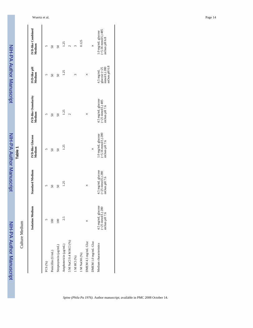
Materials and MethodsCell Isolation and Culture
For cell isolation and culture, all reagents were purchased from Invitrogen (Carlsbad, CA).Femurs from 9 skeletally mature (4–5 months) and 9 young (1 month) rats were bilaterallyexcised and the ends of the bones were removed. The bone lumen was flushed with isolationmedium (see Table 1). Isolated cells were seeded in 25 cm2 cell culture flasks and kept in anincubator at 37°C, 5% CO2. After 24 hours, culture medium was changed to removenonadherent cells (using standard medium), therefore identifying MSCs by the colony formingunit-fibroblast assay (CFU-F assay) as first described by Friedenstein et al.26 MSCs wereexpanded in monolayer for one passage as described above and then seeded in either 24-wellplates for measurement of cell viability and proliferation or in 25 cm2 cell culture flasks forgene expression analysis. Twenty-four hours later, medium was changed and either standardmedium or specific medium with an IVD-like low glucose content, an IVD-like high osmolarityor an IVD-like low pH or the combination of these characteristics was used (Table 1). IVD-like glucose medium was prepared by using a DMEM medium similar to standard DMEM,
Wuertz et al. Page 2
Spine (Phila Pa 1976). Author manuscript; available in PMC 2008 October 14.
NIH
-PA Author Manuscript
NIH
-PA Author Manuscript
NIH
-PA Author Manuscript

but with a glucose content of only 1.0 mg/mL, instead of 4.5 mg/mL.21 IVD-like osmolaritymedium was prepared by adding a sterile solution of NaCl (5 M) and KCl (0.4 M) to the culturemedium, therefore increasing the osmolarity from 280 to 485 mOsm.19 For the IVD-like pHmedium, the pH was adjusted from 7.6 to 6.8 by adding 1 M HCl and 1 M NaOH.27,28 As thepH-adjusted medium had to be incubated for 18 hours at 5% CO2 and 21% O2 to reach bufferequilibrium, each culture medium was kept in the incubator 18 hours before use. MSCs werecultured under these specific conditions for 2 weeks with medium changes twice a week. MSCscultured under standard culture conditions served as a control group for all other conditions.Analysis of gene expression, proliferation, and viability were performed on up to 9 differentMSC cultures for each age group.
Real Time RT-PCRAfter 2 weeks, cells were trypsinized and lysed with β-mercaptoethanol (Sigma, St. Louis,MO). RNA was isolated by use of the GenElute mammalian total RNA Kit (Sigma) and reversetranscribed into cDNA with MultiScribe reverse transcriptase using the TaqMan reversetranscription reagents (Applied Biosystems, Foster City, CA). For each sample, duplicateanalysis of the mRNA levels were measured using real-time RT-PCR on the GeneAmp 7700Sequence Detection System (Applied Biosystems) and the TaqMan Universal PCR MasterMix (Applied Biosystems). Gene expression of aggrecan, collagen-1, collagen-2, p53, and 18S-RNA (=housekeeping gene) was measured as previously described.29,30 Duplicate Ct valuesfor each sample were analyzed, and the relative amount of mRNA was computed according tothe comparative Ct-method.
Cell ProliferationAfter 2 weeks, the MTT assay was used to determine cell numbers relative to standardconditions, thus being an indicator for cell proliferation. To each well, 500 μL of a sterilesolution of MTT (0.25 mg MTT in DMEM, Sigma) was added. MSCs were incubated for 4hours at 37°C, and then 200 μL sterile DMSO (Sigma) was added to lyse cells. The solutionwas transferred to a 96-well plate and absorbance of samples was measured in duplicate (80μL each) at 565 nm (VersaMax Micro-Plate Reader, Molecular Devices, USA). Absorbanceof samples was calculated relative to the absorbance of cells cultured under standard conditions.
Cell ViabilityAfter 2 weeks, fluorescence double-staining with Fluorescein-diacetate (FDA, Sigma) andPropidiumiodide (PI, Sigma) was used to analyze viability of cells. MSCs were washed withsterile PBS and 0.5 mL of fresh staining solution were added to each well and incubated in thedark at 37°C for 20 minutes. Staining solution consisted of 20.8 μg FDA (dissolved in acetone)and 16.7 μg PI per 1 mL Ringer. After incubation, cells were washed extensively with sterilePBS and an inverted fluorescence microscope was used to detect red and green fluorescenceand therefore determine cell viability under different culture conditions. Green and red cellswere counted and qualitatively assessed in a randomly chosen area to estimate cell viability.
Statistical AnalysisExperimental data for proliferation (MTT) and gene expression (real time RT-PCR) of MSCscultured under IVD specific microenvironmental conditions was normalized to cells culturedunder standard conditions from the same rat. Therefore, MSCs cultured under standardconditions served as a control group. A 2-way ANOVA was used with Fisher PLSD to evaluateeffects of age (2 ages) and environmental condition (standard + 4 experimental conditions).All analyses were performed using StatView (Version 5.0.1., SAS Institute Inc., Cary, NC)with a significance level of P < 0.05.
Wuertz et al. Page 3
Spine (Phila Pa 1976). Author manuscript; available in PMC 2008 October 14.
NIH
-PA Author Manuscript
NIH
-PA Author Manuscript
NIH
-PA Author Manuscript

ResultsGene Expression
Simulating the IVD chemical microenvironment influenced aggrecan and collagen-1expression of MSCs, with a similar pattern for both age groups. On the basis of a 2-wayANOVA, we found a highly significant influence of microenvironment (P < 0.0001) as wellas a significant interaction between microenvironment and age (P = 0.002) and a trend for agealone (P = 0.09) for aggrecan (Figure 1). For collagen-1, statistical analysis indicated asignificant effect of microenvironment (P = 0.006) and age (P = 0.03) as well as a trend for aninteraction between microenvironment and age (P = 0.06) (Figure 2). Collagen-2 and p53 wereunder the detection level and therefore could not be analyzed. In general, IVD-like glucoseconditions were found to result in an increased expression of matrix proteins, with a 5.4-foldincrease for aggrecan expression (P = 0.0002) and a twofold increase for collagen-1 expression(P = 0.02) in the mature group, but with no significant changes in the young group. In contrast,IVD-like osmolarity resulted in a strong inhibition of gene expression of aggrecan (33-foldmature P < 0.0001, 10-fold young P = 0.001) and a slight inhibition (twofold) of collagen-1in both age groups (mature P = 0.01, young P = 0.02). Independent from age, IVD-like pHshowed inhibitory effects on aggrecan expression (threefold mature P = 0.01, fourfold youngP = 0.006), but no significant effects on collagen-1. Under IVD-like combined conditions,MSCs still showed an inhibition in matrix protein expression especially for aggrecan (12-foldmature, fivefold young, both P < 0.0001), demonstrating that pH and osmolarity stronglydominated the effects observed by low glucose conditions alone. Significant age differenceswere observed for low glucose conditions for both, aggrecan (P = 0.008) and collagen-1 (P =0.0003) as well as for high osmolarity for aggrecan alone (P = 0.005).
ProliferationProliferation of mature and young MSCs was similarly influenced by the discmicroenvironment in a highly significant manner (P < 0.0001), but there was neither asignificant effect of age nor an interaction between age and microenvironment (2-wayANOVA). Under low glucose conditions, cell proliferation was slightly stimulated in maturerats (P = 0.05) and not influenced in young rats. Proliferation was strongly inhibited under thelow pH (twofold mature/young) and high osmolarity (threefold mature, fourfold young)conditions, with more pronounced effects for IVD-like osmolarity (Figure 3). Under combinedIVD-like conditions, cell proliferation was strongly inhibited (eightfold mature, 11-foldyoung), showing that osmolarity and pH dominated effects of glucose. All effects of osmolarity,pH, and combined conditions were highly significant (P < 0.0001). No significant agedifferences were observed with regards to proliferation.
ViabilityWhen staining MSCs with FDA and PI in 2D culture, we were only able to detect greenfluorescence, representing viable cells, but no red fluorescence from dead/necrotic cells (Figure4). Similar to the cell proliferation assay, fluorescence staining showed a decreased number ofcells under IVD-like osmolarity and pH conditions as well as under combined IVD-likeconditions. The number of viable cells in IVD-like glucose conditions was comparable tostandard conditions.
DiscussionThis study characterized gene expression, proliferation, and viability of MSCs from young andskeletally mature rats under a variety of IVD-like chemical microenvironment conditions toevaluate the MSC responses and isolate critical factors after 2 weeks. The 2-week time pointwas chosen to assure stability of cellular responses beyond their immediate adaptation. The
Wuertz et al. Page 4
Spine (Phila Pa 1976). Author manuscript; available in PMC 2008 October 14.
NIH
-PA Author Manuscript
NIH
-PA Author Manuscript
NIH
-PA Author Manuscript

2D culture allowed high proliferation rates and high cell yields enabling evaluation of severalenvironmental conditions while keeping passage number low. Cell expansion and consequentlygene expression results are best interpreted as a measure of general biosynthesis rate ratherthan specific phenotype. Collagen-2 levels were not detectable, consistent with the findingsthat monolayer culture is known to decrease collagen-2 expression and increase collagen-1expression31; however, the primers/probe were shown to be specific and efficient in previousstudies measuring collagen-2 expression in rat IVD tissue.29,30
Results indicated that IVD-like glucose conditions increased MSC expression of aggrecan andcollagen-1 and also increased/maintained proliferation. In contrast, high osmolarity, low pH,and combined conditions strongly inhibited MSC cell proliferation and significantly decreasedanabolic gene expression, indicating that osmolarity and pH dominated the glucose effects andwere critical factors that must be overcome for MSC cell and tissue engineering therapies.Finally, chemical microenvironment conditions had similar effects on MSCs of young andskeletally mature animals with differences only for gene expression (especially for low glucoseconditions), but not for proliferation.
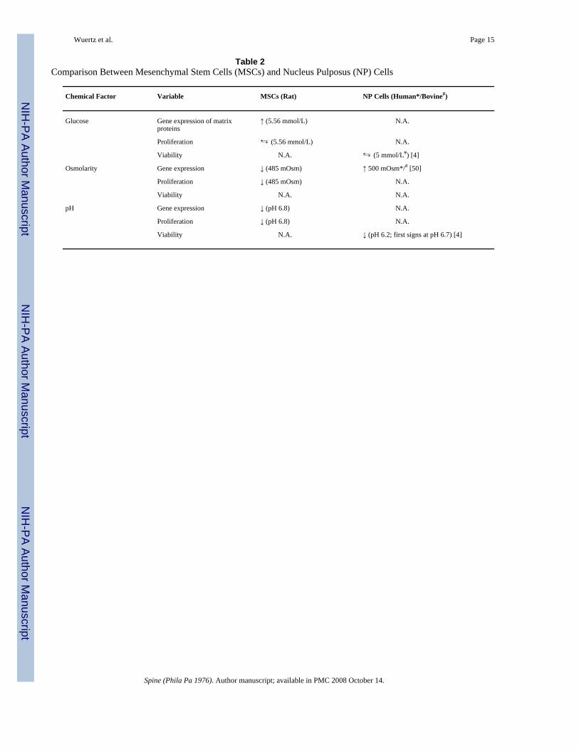
Values for media conditions were chosen as in a healthy or mildly degenerated disc, withparticular reference to the nucleus pulposus. As discs degenerate, glucose and pH levelsdecrease, creating a harsher environment; yet, osmolarity decreases during degenerationbecause of a loss of GAG, creating a less harsh environment. From a tissue engineering pointof view, it is crucial that MSCs survive, proliferate, and synthesize the appropriate matrix invivo after implantation into the target tissue whose degeneration grade can differ widely.Healthy or mild degeneration conditions were considered reasonable since early interventionis likely to offer the greatest promise for cell therapies, and this choice also allowedcomparisons with prior studies on IVD cells. In general, similar effects of chemicalmicroenvironment were found on MSCs in this study and IVD cells in the literature (Table 2),with some differences in extent. However, a lack of studies in this research area requirescomparisons of different culture systems and species. When investigating the effects of a fairlyhealthy or only mildly degenerated disc, similar effects of chemical microenvironment werefound in general on MSCs in this study and IVD cells in the literature (Table 2), with somedifferences in extent. However, a lack of studies in this research area requires comparisons ofdifferent culture systems and species and future studies on MSCs will also need to clarify theirresponses to the conditions found in a more degenerated disc. As discs degenerate glucose andpH levels decrease, creating a harsher environment, yet osmolarity decreases duringdegeneration because of a loss of GAG, creating a less harsh environment. To better understandprogressing degeneration, dose-response studies investigating decreasing pH, glucose, andosmolarity levels on the different variables will need to be performed.
Interestingly, low glucose did not negatively affect MSC proliferation and gene expression.However, this is consistent with the literature, which shows a decrease in apoptosis and anincrease in proliferation of MSCs under low glucose conditions.21,22 Similar to MSCs,glucose levels of 5 mmol/L (5.56 mmol/L = 1.0 mg/mL) are not detrimental to IVD cells;glucose levels need to drop much lower (0–0.5 mmol/L) to reduce cell viability.12
IVD-like osmolarity negatively affected MSC proliferation and gene expression, whichcontrasted the less dramatic response of IVD cells to high osmolarities. Specifically, an increasein osmolarity from 300 to 500 mOm strongly stimulated aggrecan,20 which is in contrast tothe downregulation measured in this study. Viability of IVD cells resulted in increased celldeath at high osmolarities, but effects were less pronounced than in this study and were onlyobserved at the beginning of the culture period.14 The ability of disc cells to adapt to thehyperosmotic microenvironment is potentially based on expression of acid-sensing ionchannels (ASIC3).32 We speculate that predifferentiation of MSCs towards a IVD-like
Wuertz et al. Page 5
Spine (Phila Pa 1976). Author manuscript; available in PMC 2008 October 14.
NIH
-PA Author Manuscript
NIH
-PA Author Manuscript
NIH
-PA Author Manuscript

phenotype may enhance the cells' ability to resist the chemical microenvironment of the disc,although this hypothesis will need to be tested in future studies.
To the authors' knowledge, no other studies have been performed investigating responses ofMSCs to an IVD-like pH, and only little is known about responses of IVD cells to an acidicmicroenvironment. Although results of this study suggest pH effects were less prominent thanosmolarity effects, pH may remain a larger factor limiting the use of MSCs for disc repairbecause the pH in a severely degenerated disc can drop to as low as 5.7.27 In this study, wehave chosen a pH of 6.8, which represents a healthy or only mildly degenerated disc.16,27,28 It can be anticipated that MSC responses to a lower pH as found in a moderately or severelydegenerated disc would be more substantial than measured in this study, especially consideringthe log nature of the pH scale, and we subjected MSCs to pH values as low as 6.5 in a follow-up study and found severe effects on gene expression, proliferation, and viability.33 For pH,cellular responses seem to be more substantial for MSCs than for IVD cells. Bovine nucleuspulposus cells decreased synthesis of sulfated GAG below pH 6.817,18 and Bibby et al founda decrease in cell viability at pH to 6.7 and a more obvious decrease at pH 6.2, especially whenculturing cells under nutrient deficit conditions.12 Matrix acidity, therefore, is critical for MSCgene expression and proliferation and may also accelerate disc degeneration by negativelyaffecting resident cells. However, ASIC3 expression has been reported for IVD cells, whichmay enable them to adapt more easily to an acidic environment32 than undifferentiated MSCs.
Donor age was considered an important variable to investigate in the context of MSC-baseddisc repair, because disc degeneration is an age-related process. Recent investigations showedthat the differentiation and proliferation potential of MSCs may depend on the donor's age,with a higher potential in younger donors compared with older donors.23-25 In contrast, ourexperiments show only minor biologically relevant age effects (some differences with regardsto gene expression, no differences for proliferation), suggesting that pH and osmolarityconditions dominate the effects on age, so that microenvironment must be considered morestrongly.
There is currently no commonly accepted marker for disc cells, and it is impossible to provea discogenic differentiation process, or to precisely know whether MSCs can differentiate intodisc cells.34 The 2D culture conditions are considered to be a more appropriate choice foranulus fibrosus cells,35 whereas 3D culture is more relevant for nucleus pulposus cells.35-37 Cells were not cultured under normoxic conditions, and it is known that IVD-typicalconditions are hypoxic with specific and important effects on cell metabolism38,39; therefore,an added hypoxic challenge is anticipated. It has been suggested that nucleus pulposus cellsare specifically adapted to a hypoxic environment, as indicated by normoxic stabilization ofHIF-1alpha,39 suggesting that interactions between chemical microenvironment and hypoxiaare important questions deserving further study.
Fluorescence staining of viable cells strongly supported cell proliferation results, as the numberof cells under IVD-like pH, osmolarity, and combined conditions was clearly lower than understandard and IVD-like glucose conditions. The ratio of viable and dead/necrotic cells was notpossible in this study because no red fluorescence was detectable. Conclusions about viabilityhad to be made based on relative numbers of live cells since extensive washing with PBS toreduce background staining also eliminated detection of dead/necrotic cells. Subsequentexperiments demonstrated that extensive washing did not affect live cells but did remove dead/necrotic cells. It was further determined that 3 gentle wash steps with PBS were optimal toremove background without affecting dead/necrotic cells. Expression of p53 (apoptosismarker) was also too low to be detected using the real time RT-PCR technique, but the primers/probe were shown be specific and efficient using RNA isolated from disc material.
Wuertz et al. Page 6
Spine (Phila Pa 1976). Author manuscript; available in PMC 2008 October 14.
NIH
-PA Author Manuscript
NIH
-PA Author Manuscript
NIH
-PA Author Manuscript
In conclusion, the most important findings of this study were that IVD-like osmolarity andacidity resulted in decreased proliferation and matrix protein expression whereas low glucoselevels demonstrated biosynthesis stimulation with positive effects on proliferation. Resultsdemonstrated that when chemical microenvironment factors were combined, pH andosmolarity were critical factors dominating the MSC response. Since in advanced IVDdegeneration, osmolarity will be decreased because of loss of GAGs but matrix acidity will beworsened, we believe that addressing the effects of pH alterations on MSC survival,proliferation, and biosynthesis is a crucial step that must be overcome for successful celltherapy or tissue engineering treatments. MSCs in this study responded more negatively to asimilar chemical niche than IVD cells as reported in the literature, suggesting that inducingdiscogenic differentiation before implantation (e.g., via growth factors40) may improve theMSCs resistance to the harsh microenvironment of the disc; however, this hypothesis remainsto be tested in future studies exploring the IVD niche.
Key Points• IVD-like low glucose enhanced matrix biosynthesis and maintained cell
proliferation whereas IVD-like high osmolarity and low pH conditions reducedmatrix protein expression and proliferation of young and mature MSCs.
• Osmolarity and pH dominated glucose effects in combined media conditions.• Chemical microenvironmental conditions had similar effects on MSCs of young
and mature cells.• Matrix acidity increases and osmolarity decreases in advanced IVD degeneration
so that addressing the effects of pH alterations on MSC survival, proliferation, andbiosynthesis is expected to be the most crucial step towards IVD repair.
AcknowledgmentsSupported by a grant from the National Institutes of Health (R01 AR051146).
Federal funds were received in support of this work. No benefits in any form have been or will be received from acommercial party related directly or indirectly to the subject of this manuscript.
References1. Andersson GB. Epidemiological features of chronic low-back pain. Lancet 1999;354:581–5. [PubMed:
10470716]2. Katz JN. Lumbar disc disorders and low-back pain: socioeconomic factors and consequences. J Bone
Joint Surg Am 2006;88(suppl 2):21–4. [PubMed: 16595438]3. Buckwalter JA. Aging and degeneration of the human intervertebral disc. Spine 1995;20:1307–14.
[PubMed: 7660243]4. Holm S. Pathophysiology of disc degeneration. Acta Orthop Scand Suppl 1993;251:13–5. [PubMed:
8451970]5. Osti OL, Cullum DE. Occupational low back pain and intervertebral disc degeneration: epidemiology,
imaging, and pathology. Clin J Pain 1994;10:331–4. [PubMed: 7858365]6. Yoon ST, Patel NM. Molecular therapy of the intervertebral disc. Eur Spine J 2006;15(suppl 3):S379–
88. [PubMed: 16835736]7. Masuda K, An HS. Prevention of disc degeneration with growth factors. Eur Spine J 2006;15(suppl
3):S422–32. [PubMed: 16865380]8. Javazon EH, Colter DC, Schwarz EJ, et al. Rat marrow stromal cells are more sensitive to plating
density and expand more rapidly from single-cell-derived colonies than human marrow stromal cells.Stem Cells 2001;19:219–25. [PubMed: 11359947]
Wuertz et al. Page 7
Spine (Phila Pa 1976). Author manuscript; available in PMC 2008 October 14.
NIH
-PA Author Manuscript
NIH
-PA Author Manuscript
NIH
-PA Author Manuscript
9. Pittenger MF, Mackay AM, Beck SC, et al. Multilineage potential of adult human mesenchymal stemcells. Science 1999;284:143–7. [PubMed: 10102814]
10. Moore KA, Lemischka IR. Stem cells and their niches. Science 2006;311:1880–5. [PubMed:16574858]
11. Bibby SR, Jones DA, Ripley RM, et al. Metabolism of the intervertebral disc: effects of low levelsof oxygen, glucose, and pH on rates of energy metabolism of bovine nucleus pulposus cells. Spine2005;30:487–96. [PubMed: 15738779]
12. Bibby SR, Urban JP. Effect of nutrient deprivation on the viability of intervertebral disc cells. EurSpine J 2004;13:695–701. [PubMed: 15048560]
13. Grunhagen T, Wilde G, Soukane DM, et al. Nutrient supply and intervertebral disc metabolism. JBone Joint Surg Am 2006;88(suppl 2):30–5. [PubMed: 16595440]
14. Haschtmann D, Stoyanov JV, Ferguson SJ. Influence of diurnal hyperosmotic loading on themetabolism and matrix gene expression of a wholeorgan intervertebral disc model. J Orthop Res2006;24:1957–66. [PubMed: 16917902]
15. Holm S, Maroudas A, Urban JP, et al. Nutrition of the intervertebral disc: solute transport andmetabolism. Connect Tissue Res 1981;8:101–19. [PubMed: 6453689]
16. Ichimura K, Tsuji H, Matsui H, et al. Cell culture of the intervertebral disc of rats: factors influencingculture, proteoglycan, collagen, and deoxyribonucleic acid synthesis. J Spinal Disord 1991;4:428–36. [PubMed: 1810565]
17. Ohshima H, Urban JP. The effect of lactate and pH on proteoglycan and protein synthesis rates in theintervertebral disc. Spine 1992;17:1079–82. [PubMed: 1411761]
18. Razaq S, Wilkins RJ, Urban JP. The effect of extracellular pH on matrix turnover by cells of thebovine nucleus pulposus. Eur Spine J 2003;12:341–9. [PubMed: 12883962]
19. Urban JP. The role of the physicochemical environment in determining disc cell behaviour. BiochemSoc Trans 2002;30:858–64. [PubMed: 12440933]
20. Wuertz K, Urban JP, Klasen J, et al. Influence of extracellular osmolarity and mechanical stimulationon gene expression of intervertebral disc cells. J Orthop Res 2007;25:1513–22. [PubMed: 17568421]
21. Eggum TJ, Hunter CJ. “Educated” cells for IVD therapy: differential response of MSC to oxygen,glucose, and notochord conditioned medium. Trans Orthop Res Soc. 2006
22. Stolzing A, Coleman N, Scutt A. Glucose-induced replicative senescence in mesenchymal stem cells.Rejuvenation Res 2006;9:31–5. [PubMed: 16608393]
23. Schafer R, Knauf U, Zweyer M, et al. Age dependence of the human skeletal muscle stem cell informing muscle tissue. Artif Organs 2006;30:130–40. [PubMed: 16480387]
24. Stolzing A, Scutt A. Age-related impairment of mesenchymal progenitor cell function. Aging Cell2006;5:213–24. [PubMed: 16842494]
25. Zheng, H.; Martin, JA.; Buckwalter, JA. Age related effects on chondrogenic differentiation of ratbone marrow stromal cells; Meeting of the Orthopaedic Research Society Chicago; 2006.
26. Friedenstein AJ, Chailakhjan RK, Lalykina KS. The development of fibroblast colonies in monolayercultures of guinea-pig bone marrow and spleen cells. Cell Tissue Kinet 1970;3:393–403. [PubMed:5523063]
27. Diamant B, Karlsson J, Nachemson A. Correlation between lactate levels and pH in discs of patientswith lumbar rhizopathies. Experientia 1968;24:1195–6. [PubMed: 5703005]
28. Kitano T, Zerwekh JE, Usui Y, et al. Biochemical changes associated with the symptomatic humanintervertebral disk. Clin Orthop Relat Res 1993:372–7. [PubMed: 8339506]
29. Maclean JJ, Lee CR, Alini M, et al. Anabolic and catabolic mRNA levels of the intervertebral discvary with the magnitude and frequency of in vivo dynamic compression. J Orthop Res 2004;22:1193–200. [PubMed: 15475197]
30. MacLean JJ, Lee CR, Alini M, et al. The effects of short-term load duration on anabolic and catabolicgene expression in the rat tail intervertebral disc. J Orthop Res 2005;23:1120–7. [PubMed: 16140193]
31. Marlovits S, Hombauer M, Truppe M, et al. Changes in the ratio of type-I and type-II collagenexpression during monolayer culture of human chondrocytes. J Bone Joint Surg Br 2004;86:286–95.[PubMed: 15046449]
Wuertz et al. Page 8
Spine (Phila Pa 1976). Author manuscript; available in PMC 2008 October 14.
NIH
-PA Author Manuscript
NIH
-PA Author Manuscript
NIH
-PA Author Manuscript
32. Uchiyama Y, Cheng CC, Danielson KG, et al. Expression of acid-sensing ion channel 3 (ASIC3) innucleus pulposus cells of the intervertebral disc is regulated by p75NTR and ERK signaling. J BoneMiner Res 2007;22:1996–2006. [PubMed: 17696763]
33. Wuertz, K.; Godburn, K.; Iatridis, CJ. pH dose response of mesenchymal stem cells: investigation ofa critical factor in intervertebral disc repair; Meeting of the Orthopaedic Research Society; SanFrancisco. 2008.
34. Leung VY, Chan D, Cheung KM. Regeneration of intervertebral disc by mesenchymal stem cells:potentials, limitations, and future direction. Eur Spine J 2006;15(suppl 15):406–13.
35. Wang JY, Baer AE, Kraus VB, et al. Intervertebral disc cells exhibit differences in gene expressionin alginate and monolayer culture. Spine 2001;26:1747–51. [PubMed: 11493844]discussion 52
36. Horner HA, Roberts S, Bielby RC, et al. Cells from different regions of the intervertebral disc: effectof culture system on matrix expression and cell phenotype. Spine 2002;27:1018–28. [PubMed:12004167]
37. Melrose J, Smith S, Ghosh P, et al. Differential expression of proteoglycan epitopes and growthcharacteristics of intervertebral disc cells grown in alginate bead culture. Cells Tissues Organs2001;168:137–46. [PubMed: 11173799]
38. Agrawal A, Guttapalli A, Narayan S, et al. Normoxic stabilization of HIF-1alpha drives glycolyticmetabolism and regulates aggrecan gene expression in nucleus pulposus cells of the rat intervertebraldisk. Am J Physiol Cell Physiol 2007;293:C621–31. [PubMed: 17442734]
39. Risbud MV, Guttapalli A, Stokes DG, et al. Nucleus pulposus cells express HIF-1alpha undernormoxic culture conditions: a metabolic adaptation to the intervertebral disc microenvironment. JCell Biochem 2006;98:152–9. [PubMed: 16408279]
40. Steck E, Bertram H, Abel R, et al. Induction of intervertebral disc-like cells from adult mesenchymalstem cells. Stem Cells 2005;23:403–11. [PubMed: 15749935]
Wuertz et al. Page 9
Spine (Phila Pa 1976). Author manuscript; available in PMC 2008 October 14.
NIH
-PA Author Manuscript
NIH
-PA Author Manuscript
NIH
-PA Author Manuscript

Figure 1.Relative expression of aggrecan mRNA under different medium conditions, compared withstandard conditions. Data are presented as mean ± SEM with P < 0.05 for n = 9 in both agegroups. Asterisks above bars mark a significant change in gene expression relative to standardconditions; asterisks above lines mark a significant age effect for the respectivemicroenvironment condition.
Wuertz et al. Page 10
Spine (Phila Pa 1976). Author manuscript; available in PMC 2008 October 14.
NIH
-PA Author Manuscript
NIH
-PA Author Manuscript
NIH
-PA Author Manuscript

Figure 2.Relative expression of collagen-1 mRNA under different medium conditions, compared withstandard conditions. Data are presented as mean ± SEM with P < 0.05 for n = 9 in both agegroups. Asterisks above bars mark a significant change in gene expression relative to standardconditions; asterisks above lines mark a significant age effect for the respectivemicroenvironment condition.
Wuertz et al. Page 11
Spine (Phila Pa 1976). Author manuscript; available in PMC 2008 October 14.
NIH
-PA Author Manuscript
NIH
-PA Author Manuscript
NIH
-PA Author Manuscript
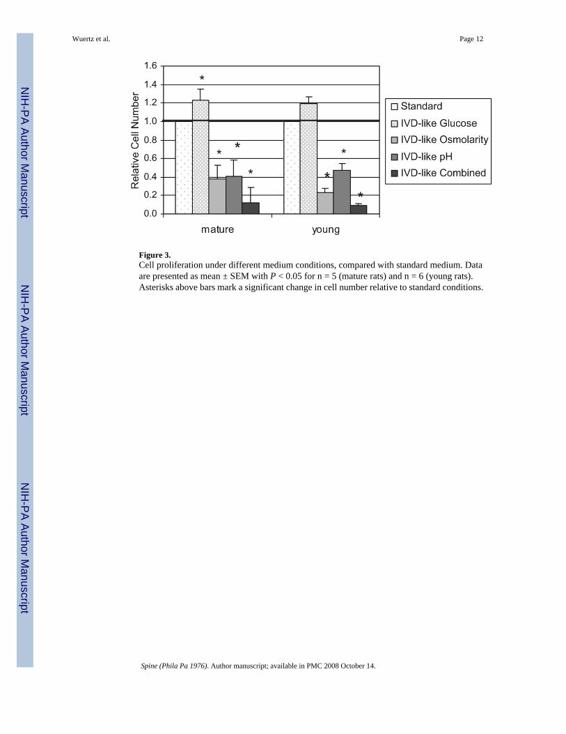
Figure 3.Cell proliferation under different medium conditions, compared with standard medium. Dataare presented as mean ± SEM with P < 0.05 for n = 5 (mature rats) and n = 6 (young rats).Asterisks above bars mark a significant change in cell number relative to standard conditions.
Wuertz et al. Page 12
Spine (Phila Pa 1976). Author manuscript; available in PMC 2008 October 14.
NIH
-PA Author Manuscript
NIH
-PA Author Manuscript
NIH
-PA Author Manuscript
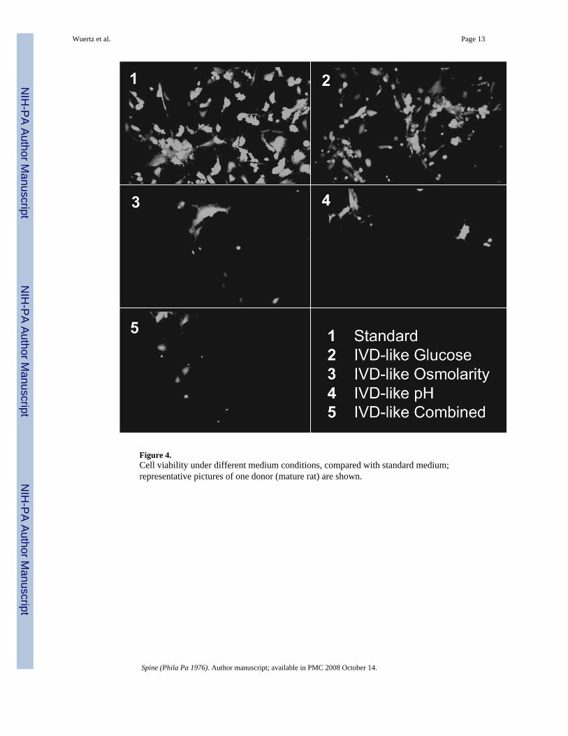
Figure 4.Cell viability under different medium conditions, compared with standard medium;representative pictures of one donor (mature rat) are shown.
Wuertz et al. Page 13
Spine (Phila Pa 1976). Author manuscript; available in PMC 2008 October 14.
NIH
-PA Author Manuscript
NIH
-PA Author Manuscript
NIH
-PA Author Manuscript
NIH
-PA Author Manuscript
NIH
-PA Author Manuscript
NIH
-PA Author Manuscript
Wuertz et al. Page 14Ta
ble
1C
ultu
re M
ediu
m
Isol
atio
n M
ediu
mSt
anda
rd M
ediu
mIV
D-li
ke G
luco
seM
ediu
mIV
D-li
ke O
smol
arity
Med
ium
IVD
-like
pH
Med
ium
IVD
-like
Com
bine
dM
ediu
m
FCS
(%)
5
5
5
5
5
5
Peni
cilli
n (U
/mL)
100
50
50
50
50
50
Stre
ptoy
mci
n (μ
g/m
L)
10
0
50
50
50
50
50
Am
phot
eric
in (μ
g/m
L)
2.5
1.2
5
1
.25
1.2
5
1
.25
1.2
5
5 M
NaC
l 0.4
M K
Cl (
%)
2
2
1 M
HC
l (%
)
3
3
1 M
NaO
H (%
)
0
.125
DM
EM 4
.5 m
g/m
L G
luc
×
×
×
×
DM
EM 1
.0 m
g/m
L G
luc
×
×
Med
ium
cha
ract
eris
tics
4.5
mg/
mL
gluc
ose
(=25
mm
ol/L
) 280
mO
sm p
H 7
.6
4.5
mg/
mL
gluc
ose
(=25
mm
ol/L
) 280
mO
sm p
H 7
.6
1.0
mg/
mL
gluc
ose
(=5.
56 m
mol
/L) 2
80m
Osm
pH
7.6
4.5
mg/
mL
gluc
ose
(=25
mm
ol/L
) 485
mO
sm p
H 7
.6
4.5
mg/
mL
gluc
ose
(=25
mm
ol/L
) 280
mO
sm p
H 6
.8
1.0
mg/
mL
gluc
ose
(=5.
56 m
mol
/L) 4
85m
Osm
pH
6.8
Spine (Phila Pa 1976). Author manuscript; available in PMC 2008 October 14.
NIH
-PA Author Manuscript
NIH
-PA Author Manuscript
NIH
-PA Author Manuscript
Wuertz et al. Page 15
Table 2Comparison Between Mesenchymal Stem Cells (MSCs) and Nucleus Pulposus (NP) Cells
Chemical Factor Variable MSCs (Rat) NP Cells (Human*/Bovine#)
Glucose Gene expression of matrixproteins
↑ (5.56 mmol/L) N.A.
Proliferation ⇆ (5.56 mmol/L) N.A.
Viability N.A. ⇆ (5 mmol/L#) [4]
Osmolarity Gene expression ↓ (485 mOsm) ↑ 500 mOsm*/# [50]
Proliferation ↓ (485 mOsm) N.A.
Viability N.A. N.A.
pH Gene expression ↓ (pH 6.8) N.A.
Proliferation ↓ (pH 6.8) N.A.
Viability N.A. ↓ (pH 6.2; first signs at pH 6.7) [4]
Spine (Phila Pa 1976). Author manuscript; available in PMC 2008 October 14.