
Gelled vegetable desserts containing pea protein, κ-carrageenan and starch

Cell, Vol. 104, 33–42, January 12, 2001, Copyright 2001 by Cell Press
Bcl10 Is a Positive Regulatorof Antigen Receptor–Induced Activationof NF-kB and Neural Tube Closure
Translocation t(1;14)(p22;q32) in MALT lymphoma leadsto overexpression of Bcl10 and is associated withframeshift mutations causing C-terminal truncationsdistal of the CARD (Willis et al., 1999; Zhang et al.,1999b). Bcl10 mutations are also found in cases of follic-
Jurgen Ruland,*† Gordon S. Duncan,*†
Andrew Elia,*† Ivan del Barco Barrantes,*†
Linh Nguyen,† Sue Plyte,*† Douglas G. Millar,†Denis Bouchard,* Andrew Wakeham,*†
Pamela S. Ohashi,† and Tak W. Mak*†‡
*Amgen Institute ular lymphoma and diffuse large B cell lymphoma (Du620 University Avenue et al., 2000).Toronto, Ontario The human and murine Bcl10 proteins are 91% identi-Canada M5G 2C1 cal. Bcl10 transcripts are expressed ubiquitously and†Ontario Cancer Institute, and Departments throughout development, with high expression levels in
of Medical Biophysics and Immunology lymphoid tissues and in the developing central nervousUniversity of Toronto system (CNS) (Costanzo et al., 1999; Koseki et al., 1999;Toronto, Ontario Srinivasula et al., 1999; Thome et al., 1999; Willis et al.,Canada M5G 2C1 1999; Yan et al., 1999; Zhang et al., 1999b). The Bcl10
CARD domain mediates self-oligomerization and theC-terminal region of Bcl10, which shows no significanthomology to any other known protein, is rich in serine
Summary and threonine residues, and can be phosphorylated.(Koseki et al., 1999; Srinivasula et al., 1999).
Bcl10, a CARD-containing protein identified from the Transient overexpression of wild-type Bcl10 in cellt(1;14)(p22;q32) breakpoint in MALT lymphomas, has lines both induces apoptosis and activates NF-kB (Ko-been shown to induce apoptosis and activate NF-kB seki et al., 1999; Thome et al., 1999; Willis et al., 1999;in vitro. We show that one-third of bcl102/2 embryos Yan et al., 1999). Whereas the truncated, tumor-deriveddeveloped exencephaly, leading to embryonic lethal- Bcl10 mutants are unable to induce cell death, the CARDity. Surprisingly, bcl102/2 cells retained susceptibility domain alone is sufficient and necessary for NF-kB acti-to various apoptotic stimuli in vivo and in vitro. How-
vation. Apoptosis induced by overexpressed Bcl10 isever, surviving bcl102/2 mice were severely immuno-
suppressed by broad-spectrum caspase inhibitors, ordeficient and bcl102/2 lymphocytes are defective in
by cotransfection of BclxL, X-IAP, cIAP1, c-IAP2, or aantigen receptor or PMA/Ionomycin-induced activa-
dominant-negative version of caspase 9 (Srinivasula ettion. Early tyrosine phosphorylation, MAPK and AP-1al., 1999; Yan et al., 1999). In addition, cotransfectionactivation, and Ca21 signaling were normal in mutantof Bcl10 and procaspase 9 results in their direct associa-lymphocytes, but antigen receptor–induced NF-kB ac-tion (Yan et al., 1999), suggesting that Bcl10 may partici-tivation was absent. Thus, Bcl10 functions as a positivepate in the Apaf1/caspase-9 mediated cell death path-regulator of lymphocyte proliferation that specificallyway. However, Bcl10 was also found to bind to TRADD,connects antigen receptor signaling in B and T cellsand Bcl10-initiated activation of NF-kB can be inhibitedto NF-kB activation.by cotransfection of dominant-negative mutants ofTRAF2, NIK, IKKa, or IkBa (Costanzo et al., 1999; KosekiIntroductionet al., 1999; Srinivasula et al., 1999).
To investigate the physiological roles of Bcl10, weThe most common type of lymphoma arising in extra-generated bcl10-deficient mice. We demonstrate thatnodal sites are B cell lymphomas of mucosa-associatedbcl10 is important for neural tube closure and lympho-lymphoid tissue (MALT lymphomas). Low grade MALTcyte activation. While dispensable for the execution oflymphomas typically develop in the context of prolongedapoptosis, bcl10 is a critical positive regulator of lym-reactive lymphoid proliferation at sites of chronic infec-phocyte proliferation and a central mediator of NF-kBtions such as Helicobacter pylori gastritis, or in autoim-activation in response to antigen receptor signaling inmune disorders (Zucca et al., 2000). The molecularB and T cells.events leading to high grade transformation and anti-
gen-independent growth are still largely unknown. How-ever, chromosomal translocation t(1;14)(p22;q32), is re-
Resultscurrent in MALT lymphoma and is associated withaggressive disease (Spencer, 1999). Molecular cloning
Generation of bcl102/2 Miceof the breakpoint identified a novel gene, Bcl10, whichThe bcl10 gene was disrupted by homologous recombi-is translocated to the immunoglobulin heavy chain locus
(Willis et al., 1999; Zhang et al., 1999b). The human Bcl10 nation in murine embryonic stem (ES) cells using stan-gene encodes a protein of 233 amino acids containing dard procedures (Figures 1A and 1B). Heterozygousan N-terminal caspase recruitment domain (CARD). (1/2) mice, were healthy up to nine months of age and
intercrossed to obtain homozygous bcl10-deficient mu-tants (2/2). The null mutation of bcl10 in bcl102/2 mice‡ To whom correspondence should be addressed (e-mail: tmak@
oci.utoronto.ca). was confirmed by Western blotting (Figure 1C).

Cell34
in the forebrain, midbrain, and hindbrain at E11.5–E15.5(Figures 2I and 2J and data not shown). These resultsindicate that bcl10 is dispensable for the execution ofapoptosis during neuronal development but may medi-ate neuronal survival.
Normal Susceptibility of bcl102/2 Cells to VariousApoptotic StimuliWe next assessed the susceptibility of bcl102/2 ES cellsand embryonic fibroblasts (EF), as well as thymocytesand peripheral lymphocytes from adult bcl102/2 mice,to various apoptotic stimuli. bcl10 1/2 and 2/2 EScells were treated with anisomycin, cisplatin, etoposide,staurosporine, or UV-irradiation and apoptosis was eval-uated at 6, 12, and 24 hr post-induction. No significantdifference in the number of apoptotic cells was observedbetween the wild-type and mutant under all conditionstested (Figure 3A and data not shown). Similar resultswere obtained when EF were tested with these stimuliand when receptor-mediated cell death was induced bytumor necrosis factor a (TNFa) plus cycloheximide orFigure 1. Targeted Disruption of the bcl10 Locusby overexpression of the death receptors 3 (DR3) or 5(A) A portion of the murine wild-type bcl10 locus (top) showing all(DR5) (Ashkenazi and Dixit, 1998) (Figure 3B and data notexons (1–3; open boxes) and a 15 kb EcoRI fragment. A targeting
vector (middle) was designed to replace exon 2 and the complete shown). Neither were any differences observed whencoding sequence of exon 3 with a neomycin resistance gene cas- thymocytes from wild-type or bcl102/2 mice weresette (neo) in antisense orientation, introducing a new EcoRI site treated with Fas-ligand, anti-CD3 plus anti-CD28 mono-(E). The mutated bcl10 locus (bottom) contains a diagnostic 6.8 kb
clonal antibodies (mAb), cisplatin, staurosporine, g- orEcoRI fragment. The position of the 39 flanking probe (FP) used forUV-irradiation, or the glucocorticoid hormone dexa-genotyping is indicated.methazone (Figure 3C and data not shown), or when(B) Southern blot analysis of bcl10 1/1, 1/2, and 2/2 ES cells.
Genomic DNA was digested with EcoRI and hybridized to the 39 splenic B or T cells were treated with cisplatin, stauro-flanking probe. sporine, etoposide, or g- or UV-irradiation (Figure 3D(C) Western blot analysis of bcl10 1/2 and 2/2 EF. Antibodies and data not shown). These results indicate that bcl10were directed against the N or C terminus of bcl10. The bcl10 and
is not required for the execution of apoptosis in severalnonspecific (ns) bands are indicated.different cell lineages in response to a wide variety ofstimuli.
bcl10 Deficiency Results in Partial EmbryonicLethality Caused by a Neural Tube T and B Cell Development in bcl102/2 Mice
Live born bcl102/2 mice were anatomically normal, butClosure DefectOf 372 offspring from heterozygous intercrosses, only 65 highly susceptible to infections. We therefore analyzed
the development and function of T and B cells in bcl10-(17.5%) homozygous mutants were identified, indicatingthat about one-third of bcl102/2 mutants died during deficient mice. The total number of thymocytes in 8–12
week old bcl102/2 mice was reduced by about 25%embryogenesis. bcl102/2 mutants that survived embry-onic development were fertile, did not show any gross compared to wild-type littermates (1/1 versus 2/2,
86.1 6 28.3 3 106 versus 63.8 6 28.6 3 106; mean 6anatomical abnormalities, and did not develop any ma-lignancies up to 6 months of age. SD, n 5 16, p , 0.05 by Student’s t test). The CD41CD81
(double positive, DP) thymocyte population was signifi-Isolation of E9.5–E18.5 embryos from heterozygousintercrosses revealed that about 30% of bcl102/2 em- cantly decreased (1/1 versus 2/2, 73.4 6 23.4 3 106
vesus 48.1 6 21.7 3 106) while the CD42CD82 (doublebryos had a neural tube closure defect (NTD) exclusivelyin the hindbrain, leading to exencephaly (Figures 2A–2D) negative, DN) thymocyte population was increased
(1/1 versus 2/2, 1.6 6 1.1 3 106 versus 2.6 6 1.6 3and embryonic lethality between E18.5 and birth. bcl102/2
embryos without NTD were morphologically indistin- 106) (Figure 4A, left). However, bcl102/2 DP thymocyteswere able to differentiate into normal total numbers ofguishable from wild-type embryos. Histological analysis
of NTD mutants at E9.5 demonstrated that the neural CD41 or CD81 T cells (1/1 versus 2/2; CD41: 7.8 64.0 3 106 versus 9.3 6 5.4 3 106; CD81: 2.3 6 1.0 3 106folds at the hindbrain failed to elevate at either side of
the midline and did not bend toward each other (Figures vs. 3.0 6 1.5 3 106), which expressed normal levels ofTCRab/CD3 complexes (data not shown).2E and 2F). Because Bcl10 has been implicated in the
Apaf-1/caspase-9 pathway critical for brain morphogen- The expression of CD25 and CD44 on developing DNthymocytes defines four stages reflecting the steps ofesis (Kuida et al., 1996, 1998; Cecconi et al., 1998; Ha-
kem et al., 1998; Yoshida et al., 1998a), we analyzed TCR gene rearrangement: CD252CD441, CD251CD441,CD251CD442, and CD252CD442 (Godfrey and Zlotnik,brain sections of bcl102/2 embryos from E9.5 to E15.5
using TUNEL staining. Surprisingly, apoptosis was in- 1993). Expression of the b chain and pre-TCR signalingat the CD251CD442 stage are required for the progres-creased in the neuronal epithelium in the hindbrain of
bcl102/2 embryos at E9.5 (Figures 2G and 2H) but normal sion to the CD252CD442 stage (b checkpoint). Absolute

Bcl10 Transduces Antigen Receptor Signals to NF-kB35
Figure 2. Hindbrain Exencephaly of bcl102/2
Embryos
(A and B) Phenotypic comparison of E14.5wild-type (A) and exencephalic bcl102/2 (B)littermate embryos.(C and D) Scanning electron microscopy im-ages of E10.5 wild-type (C) and bcl102/2 (D)embryos. The mutant embryo exhibits a neu-ral tube closure defect at the hindbrain(arrow).(E and F) H&E stained transverse sections ofthe hindbrain region at E9.5 in wild-type (E)and bcl102/2 (F) embryos. The roof of thehindbrain (asterisk) is present in the wild typebut absent in the mutant. The neural foldsfail to elevate in bcl102/2 embryos and theneuroepithelium (ne) adopts a biconvex con-figuration.(G and H) High-power view of TUNEL assayshowing increased apoptosis (bright green)in the neuroepithelium of the hindbrain in
bcl102/2 embryos (H) compared to wild type (G). The fields correspond to the areas indicated in (E) and (F).(I and J) TUNEL assay showing normal apoptosis in the forebrain of bcl102/2 embryos (J) compared to the wild type (I) at E13.5.Scale bar: 3 mm (A and B); 0.6 mm (C and D); 100 mm (E and F); 20 mm (G and H); and 150mm (I and J).
numbers of CD252CD441, CD251CD441, and CD251 virus (LCMV) or vesicular stomatitis virus (VSV) (Bach-mann and Kundig, 1994). LCMV injection into the footCD44– DN cells were normal in bcl102/2 mice (data not
shown). However, the CD252CD442 DN cells that pad of wildtype mice leads to an initial swelling that ismediated by infiltration of CD81 cytotoxic T lympho-passed the b checkpoint were increased 4- to 5-fold in
number (1/1 versus 2/2, 4.8 6 2.1 3 105 versus 19.4 6 cytes (CTLs). The swelling reaction was reduced inbcl102/2 mice compared to wild-type mice, indicating7.2 3 105) (Figure 4A, right). These cells expressed ele-
vated levels of CD3, TCRb, and TCRa (Figure 4B). About an impaired primary immune response (Figure 5B, left).When memory responses were examined 20 days post-40% of these thymocytes are in the early stages of
apoptosis, as seen by annexinV/7-AAD staining (Figure infection by in vitro restimulation of splenocytes withthe LCMV antigen, bcl102/2 mice showed reduced or4B and data not shown), whereas the frequency of
annexinV positive wild-type DN, DP, or SP thymocytes absent CTL memory responses (Figure 5B, right).Infection of wild-type mice with VSV results in synthe-was consistently less than 5% (Figure 4B and data not
shown). The increased cell death of early thymocytes sis of neutralizing IgM antibodies against VSV that isindependent of T cell help and peaks at 4–6 days afterin bcl102/2 mice is consistent with the reduced DP popu-
lation in these animals and indicates that bcl10 plays a infection. Subsequent production of IgG anti-VSV anti-bodies requires isotype switching that depends on col-role in the differentiation and/or survival of thymocytes,
but is dispensable for overall T cell lineage development. laboration between B cells and CD41 T helper cells.Compared to wild-type mice, bcl102/2 mice producedAnalysis of B cell lineage development did not reveal
any significant differences between wild-type and mu- significantly lower levels of VSV-specific IgM at 4 dayspost-infection, indicating that bcl10 is required for opti-tant mice in either bone marrow cellularity or expression
of B220, IgM, IgD, CD23, CD24, CD43, or BP-1 on pre- mal B cell function (Figure 5C, left). In wild-type mice,VSV-specific IgG was detectable by day 8 and reachedcursor B cell populations (Figure 4C and data not
shown). The cellularity of spleens and lymph nodes was a plateau by day 12 (Figure 5C, right). However, bcl102/2
mice failed to make the class switch and did not gener-also comparable in wild-type and bcl102/2 mice. Theratios of peripheral B to T cells, and of CD41 to CD81 ate detectable VSV-specific IgG.T cells, were normal, as was the expression of IgM andIgD on peripheral B cells (Figure 4C). However, a de- Impaired Antigen Receptor–Induced Proliferationcrease in activated T cells (as detected by expression and Activation of bcl10-Deficient Lymphocytesof the markers CD25, CD44, and CD69) was consistently Upon receipt of a signal through the antigen receptor,observed in these tissues (data not shown), indicating resting T cells become activated, enter the cell cycle,that bcl10 might be necessary for proper lymphocyte proliferate, and differentiate into effector cells. TCR sig-function and activation. naling can be mimicked experimentally by stimulation
with an anti-CD3e mAb. Purified wild-type T cells stimu-lated for 24 or 48 hr with soluble or plate-bound anti-Impaired Humoral and Cellular Immune
Responses in bcl102/2 Mice CD3e antibody (with or without anti-CD28 costimulation)proliferated vigorously (Figure 6A, left, and data notThe basal concentrations of all Ig isotypes tested were
found to be severely reduced in unimmunized bcl102/2 shown). However, bcl102/2 T cells neither proliferated(Figure 6A, left) nor secreted IL-2 (Figure 6B, left) inmice compared to controls (Figure 5A). To assess re-
sponses to pathogens in vivo, wild-type and bcl102/2 response to any of these stimuli. Addition of exogenousIL-2 to bcl102/2 T cells resulted in only a marginal rescuemice were infected with lymphocytic choriomeningitis

Cell36
Figure 3. Normal Apoptotic Susceptibility of bcl102/2 ES cells, EF,Thymocytes, and Peripheral Lymphocytes
(A) Bcl10 1/2 and 2/2 ES cells were treated with apoptotic stimuliand programmed cell death was evaluated as described in Experi-mental Procedures. Cell viability was normalized to spontaneouscell death in untreated controls. Percentages of surviving cells 24hr after treatment are shown for cisplatin (10 or 100 mM), anisomycin Figure 4. Flow Cytometric Analyses of bcl102/2 Thymus, Bone Mar-(10 or 50 mM), etoposide (10 or 100 mM), staurosporine (2 or 10 mM), row, Spleen, and Lymph Node Cellsand UV-irradiation (40 or 80 mJ/cm2). Triplicate samples of each (A) Bcl10 1/1 and 2/2 thymocytes were stained with anti-CD4,treatment in three independent experiments were assayed. Results anti-CD8, anti-TCRgd, anti-B220, anti-NK1.1, anti-Mac-1 (lineageshown are the mean 6 SD. marker, Lin), anti-CD44, and anti-CD25 antibodies. Left: CD4 and(B) Apoptosis in bcl10 1/2 and 2/2 EF was induced with TNFa (10 CD8 expression. Right: CD44 and CD25 expression on Lin2 DNng/ml) plus increasing concentrations of cycloheximide (102–104 ng/ thymocytes. Percentages of positive cells within each quadrant areml); anisomycin (10 or 100 mM); etoposide (10 or 100 mM); UV- indicated.irradiation (60 or 120 mJ/cm2); or by overexpression of death recep- (B) Left: CD3 expression on DN, DP, and SP Bcl10 1/1 and 2/2tor 3 (DR3) or 5 (DR5). Percentages of surviving cells 24 hr after thymocytes. Upper right: TCRVa2 and TCRb expression on Lin2
treatment are shown. The number of surviving DR3 or DR5 trans- DN thymocytes. Lower right: AnnexinV staining of Lin2 CD3lo/int DNfected cells is expressed as a percentage relative to control trans- thymocytes.fections with an empty vector (vec). (C) Expression of B220 and IgM, IgM and IgD, or CD4 and CD8 on(C) Freshly isolated bcl10 1/1 and 2/2 thymocytes were treated bone marrow, spleen, and lymph node cells. Percentages of positivewith CD8-FasL fusion protein (2 or 10 ng/ml), anti-CD3 (1 or 10 mg/ cells within each quadrant are indicated. Experiments were repeatedml) plus anti-CD28 (1 mg/ml), staurosporine (2 or 10 mM), g-irradiation at least three times with similar results.(2 or 4 Gy), or UV-irradiation (40 or 100 mJ/cm2). Percentages ofsurviving cells normalized to spontaneous cell death in untreatedcontrols 24 hr after treatment are shown. Spontaneous cell death phase, cell cycle dynamics were analyzed by BrdU andwas similar in 1/1 and 2/2 thymocytes.
7-AAD staining (Gratzner and Leif, 1981) and flow cytom-(D) Bcl10 1/1 and 2/2 peripheral B or T cells were treated withetry. Unlike wild-type cells, bcl102/2 T cells failed tostaurosporine (2 or 10 mM), etoposide (20 or 100 mM), UV-irradiationenter S phase in response to either anti-CD3 or anti-(10 or 40 mJ/cm2), or g-irradiation (0.5 or 2 Gy). Percentages of
surviving cells 24 hr after treatment are shown. CD3 plus anti-CD28 (Figure 6A, right). TCR-induced Sphase entry of resting T cells is promoted by transcrip-tional upregulation of the IL-2 and IL-2 receptor a chain(CD25) genes (Smith, 1989). Compared to wild-type Tof TCR-induced proliferation (Figure 6A, left). Further-
more, treatment with phorbol myristate acetate (PMA) cells, expression of CD25 as well as CD44 and CD69was reduced or absent in bcl102/2 T cells at 24 hr post-alone, or in combination with calcium ionophore (Iono),
induced proliferation of wild-type but not bcl102/2 T stimulation (Figure 6B). The failure to induce IL-2 andCD25 after TCR stimulation contributes to the reducedcells (Figure 6A, left and data not shown).
To investigate whether bcl102/2 T cells fail to enter proliferative responses of bcl10-deficient T cells.We next examined the role of bcl10 in proliferativethe cell cycle upon TCR stimulation or arrest at a specific

Bcl10 Transduces Antigen Receptor Signals to NF-kB37
dicating that the observed defects are intrinsic to themutant lymphocytes (Figures 6A and 6C, middle).
bcl10 Is Required for Antigen Receptor–InducedNF-kB ActivationTo elucidate the molecular basis of the impairment inantigen receptor signaling in the absence of bcl10, wesystematically analyzed the pathways activated by TCRor BCR engagement in wild-type and mutant T and Bcells. Proximal signaling events induced by TCR stim-ulation are initiated by TCR/CD3-associated proteintyrosine kinases. Antiphosphotyrosine immunoblottingshowed that phosphorylation patterns were similar inwild-type and bcl102/2 T cells (Figure 7A, top). Proximalsignaling activates the Ras/MAPK (mitogen-activatedprotein kinase) pathway and phospholipase C (PLC) g.PLCg generates second messengers that lead to anincrease in free cytoplasmic calcium (Ca21) and activa-tion of PKC. Western blotting using phospho-specificanti-ERK1/2 antibodies showed that the MAP kinasesERK1 and ERK2 were activated with similar kineticsafter TCR crosslinking in wild-type and bcl102/2 T cells(Figure 7A, bottom). In addition, TCR-induced Ca21
fluxes in wild-type and bcl102/2 T cells showed similaractivation and inactivation kinetics (Figure 7B, top), asFigure 5. Defective Immune Responses in bcl102/2 Mice
did Ca21 currents induced by IgM crosslinking in wild-(A) Reduced basal immunoglobulin levels. Serum concentrations ofIg isotypes were determined by ELISA in 6–8 week old bcl101/1 type and bcl102/2 B cells (Figure 7B, bottom).(open circles, n 5 7) and bcl102/2 (filled circles, n 5 8) mice. Signaling pathways triggered by TCR engagement ac-(B) Impaired CTL responses. Left panel: Footpad swelling in individ- tivate several key transcription factors, including NF-kBual bcl101/1 (open symbols) and bcl102/2 (closed symbols) mice
and AP-1. These proteins play important roles in IL-2after local LCMV infection. One result representative of two experi-and CD25 expression and T cell proliferation (Weiss andments is shown. Right panel: Secondary CTL responses 20 daysLittman, 1994). Gel mobility shift assays showed thatafter the initial infection. LCMV-specific cytotoxicity of in vitro re-
stimulated bcl101/1 (open symbols) and bcl102/2 (closed symbols) substantial NF-kB DNA binding activity was induced inspleen cells was determined by 51Cr-release assay using EL4 target wildtype T cells after anti-CD3 stimulation, which couldcells pulsed with LCMV or control peptide. The range of effector- be further increased if CD28 was also engaged or if theto-target cell ratios is indicated. Results from individual mice are
cells were treated with PMA1Iono. However, none ofshown.these stimuli was able to activate NF-kB in bcl102/2 cells(C) Impaired humoral responses and isotype switching in bcl102/2
(Figure 7C, left). In contrast, AP-1 DNA binding activitymice. bcl101/1 (open bars) and bcl102/2 (closed bars) mice wereintravenously immunized with VSV. Neutralizing IgM titers (left panel) in response to the same stimuli was comparable in wild-were measured 4 days after immunization, while neutralizing serum type and bcl102/2 T cells. Similarly, in B cells, stimulationIgG (right panel) was measured after 8 and 12 days. One result by IgM cross-linking or PMA1Iono failed to activate NF-representative of 3 experiments is shown.
kB in the absence of bcl10, whereas AP-1 activationwas normal. Importantly, NF-kB activation induced byLPS was equivalent in wild-type and mutant B cells,responses of purified B cells after stimulation with anti-
IgM, anti-CD40, anti-IgM plus anti-CD40, or bacterial indicating that bcl10 is required for signal-specific NF-kB activation in lymphocytes (Figure 7C, right). Consid-lipopolysaccharide (LPS). Like bcl102/2 T cells, bcl102/2
B cells showed a severe defect in antigen receptor– ering that TNFa or IL-1 stimulation also induced compa-rable levels of NF-kB activation in both wild-type andinduced proliferation (Figure 6C, left). Proliferation of
bcl102/2 B cells was also markedly reduced compared mutant T cells and primary EF cells (Figure 7D), weconclude that bcl10 is a specific regulator of antigento the wild-type after stimulation with anti-CD40, and a
combination of anti-IgM plus anti-CD40 induced only a receptor signaling to the activation of NF-kB.Prior to activation, NF-kB/Rel family members are re-moderate response. Cell cycle analysis showed that,
like T cells, B cells in bcl102/2 mice have a defect in S tained in the cytoplasm through binding to the inhibitoryIkB proteins. IkB kinase (IKK)-mediated phosphorylationphase entry after antigen receptor stimulation. Interest-
ingly, B cell cycle progression was normal in response of regulatory serines of IkB triggers its rapid ubiquitina-tion and proteolytic degradation, allowing nuclear trans-to LPS, a stimulus that activates B cells independently
of BCR signaling via Toll-like receptor 4 (Poltorak et al., location of NF-kB (Karin and Ben-Neriah, 2000). To de-termine the effect of bcl10 deficiency on signaling via1998). This result indicates that the cell cycle machinery
itself is intact in the absence of bcl10 (Figure 6C, and this pathway, we examined the activation of IKK andthe phosphorylation and degradation of IkBa in PMA-data not shown). In addition, antigen receptor–induced
proliferation was impaired in bcl102/2 T and B cells gen- stimulated lymph node T cells from wild-type andbcl102/2 mice. In wild-type cells, IKK activity was rapidlyerated from bcl102/2 ES cells using RAG-1-deficient
blastocyst complementation (Yoshida et al., 1998a), in- induced after PMA stimulation and IkBa was phosphory-

Cell38
Figure 6. Impaired Lymphocyte Activation inbcl102/2 Mice
(A) Impaired proliferative responses of T cells.Left panel: Purified lymph node bcl101/1
(open bars) and bcl102/2 (closed bars) T cellswere stimulated with medium alone, solubleanti-CD3 (1 mg/ml), with or without anti-CD28(1 mg/ml), in the presence or absence of IL-2(50 U/ml), or with PMA (10 ng/ml) 1 Iono (100ng/ml) for 24 or 48 hr. Results shown arethe mean 6 SD [3H]thymidine incorporationfor triplicate samples. Middle panel: Prolif-erative responses of T cells isolated frombcl101/2/Rag12/2 (open bar) and bcl102/2/Rag12/2 (closed bars) somatic chimeras 24hr after treatment as indicated. Right panel:Cell cycle profile of bcl10 1/1 and 2/2 T cells36 hr after stimulation with medium (control),anti-CD3 (1 mg/ml), or anti-CD3 (1 mg/ml) plusanti-CD28 (1 mg/ml). The percentage of cellsin S phase is indicated.(B) Left panel: IL-2 concentration in the super-natants of cultures of bcl101/1 (open sym-bols) and bcl102/2 (closed symbols) T cellsstimulated with increasing concentrations ofanti-CD3e in the presence of 100 ng/ml anti-CD28. Right panel: Flow cytometric analysisof expression of CD25, CD44, and CD69 onbcl10 1/1 and 2/2 T cells 24 hr after stimula-tion with medium alone (control) or solubleanti-CD3 (1 mg/ml) plus soluble anti-CD28(1 mg/ml).(C) Impaired proliferative responses of bcl102/2
B cells. Left: Purified splenic bcl101/1 (openbars) and bcl102/2 (closed bars) B cells werestimulated with medium alone, anti-CD40 (5mg/ml), anti-IgM (10 mg/ml), anti-IgM plusanti-CD40, or LPS (20 mg/ml). Proliferation at24 and 48 hr was measured as in (A). Middle:Proliferative responses of purified B cells
from bcl101/2/Rag12/2 (open bars) and bcl102/2/Rag12/2 (closed bars) somatic chimeras 24 hr after stimulation as indicated. Right: Normalcell cycle progression of bcl102/2 B cells after LPS stimulation. Cell cycle profiles were determined as in (A). The percentage of cells in Sphase is indicated.
lated and degraded accordingly (Figure 7E). However, and Harris, 2000). Interestingly, a localized hindbrainNTD with excessive apoptosis in the neural epitheliumneither IKK activation nor IkBa phosphorylation or deg-
radation occurred in bcl102/2 T cells. These data demon- at E9.5 resembling the phenotype of bcl102/2 embryoshas recently been reported in IKKa/IKKb double mutantstrate that bcl10 is required for the regulation of IKK
activity and provide a mechanistic insight into bcl10 mice (Li et al., 2000). Our finding that bcl10 is requiredfor proper IKK regulation and NF-kB signaling leads usfunction upstream of the IKK complex.to believe that a bcl10 → IKKa/IKKb → NF-kB pathwayplays a role in normal CNS development, possibly viaDiscussionpositive regulation of neuronal survival. It should benoted that the mechanism leading to exencephaly inbcl10 was originally isolated through its involvementbcl102/2 embryos is fundamentally different from thatin chromosomal translocation t(1;14)(p22;q32) in MALTcausing aberrant brain morphogenesis in mice lackinglymphomas, but the physiological function of bcl10 hasApaf-1, caspase-9, or caspase-3. In these mutants, defi-remained obscure. We have shown that, while bcl10 isciency for proapoptotic regulators leads to an extensivedispensable for the execution of apoptosis, it is impor-deficit in developmentally regulated apoptosis, resultingtant for neural tube closure and specifically required forin supernumerary neuroepithelial cells in the hindbrain,lymphocyte proliferation dependent on antigen recep-midbrain, and forebrain and gross neuronal disorderingtor-mediated activation of NF-kB.(Kuida et al., 1996, 1998; Cecconi et al., 1998; Hakemet al., 1998; Yoshida et al., 1998a).Role of bcl10 in CNS Development
Neural tube closure involves the proper orchestrationof multiple processes, including cellular migration, dif- Role of bcl10 in Apoptosis
Since overexpression studies had suggested a role forferentiation, proliferation, and apoptosis. The multifac-torial nature of NTDs is reflected in the partial pene- bcl10 as a proapoptotic signaling molecule involved in
the Apaf-1/caspase-9 cell death pathway (Costanzo ettrance of the phenotype in bcl102/2 embryos, a patternthat is typically seen in single gene NTD mutants (Juriloff al., 1999; Koseki et al., 1999; Willis et al., 1999; Yan et
Bcl10 Transduces Antigen Receptor Signals to NF-kB39
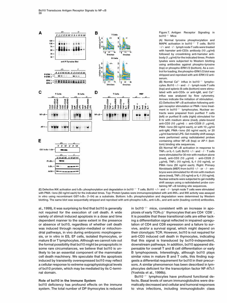
Figure 7. Antigen Receptor Signaling inbcl102/2 Mice
(A) Normal tyrosine phosphorylation andMAPK activation in bcl102/2 T cells. Bcl101/1 and 2/2 lymph node T cells were treatedwith hamster anti-CD3e antibody (10 mg/ml)followed by crosslinking anti-hamster anti-body (1 mg/ml) for the indicated times. Proteinlysates were subjected to Western blottingusing antibodies against phospho-tyrosine(top) or phospho-ERK1/2 (bottom). As a con-trol for loading, the phospho-ERK1/2 blot wasstripped and reprobed with anti-ERK1/2 anti-serum.(B) Normal Ca21 influx in bcl102/2 lympho-cytes. Bcl10 1/1 and 2/2 lymph node T cells(top) and splenic B cells (bottom) were stimu-lated with anti-CD3e or anti-IgM, and Ca21
influx was analyzed by flow cytometry.Arrows indicate the initiation of stimulation.(C) Defective NF-kB activation following anti-gen receptor stimulation or PMA1Iono treat-ment in bcl102/2 lymphocytes. Nuclear ex-tracts were prepared from purified T cells(left) or purified B cells (right) stimulated for8 hr with medium alone (med), plate-boundanti-CD3 (10 mg/ml) 6 anti-CD28 (1 mg/ml),PMA1Iono (50 ng/ml each), or with 10 mg/mlanti-IgM, PMA1Iono (50 ng/ml each), or 20mg/ml bacterial LPS. Gel mobility shift assayswere performed using radiolabeled probescontaining either NF-kB (top) or AP-1 (bot-tom) binding site sequences.(D) Normal NF-kB activation in response toTNFa or IL-1. Left: Bcl10 1/1 and 2/2 T cellswere stimulated for 30 min with medium alone(med), anti-CD3 (10 mg/ml) 1 anti-CD28 (1mg/ml), TNFa (10 ng/ml), IL-1 (10 ng/ml), orPMA1Iono (50 ng/ml each). Right: Primaryfibroblasts (MEF) from bcl10 1/2 or 2/2 em-bryos were stimulated for 45 min with mediumalone (med), TNFa (10 ng/ml) or IL-1 (10 ng/ml).Nuclear extracts were subjected to gel mobilityshift assays using a radiolabeled probe con-taining NF-kB binding site sequences.
(E) Defective IKK activation and IkBa phosphorylation and degradation in bcl102/2 T cells. Bcl10 1/1 and 2/2 lymph node T cells were stimulatedwith PMA1Iono (50 ng/ml each) for the indicated times. Top: Protein lysates were immunoprecipitated with anti-IKKa and IKK activity was assayedin vitro using recombinant GST-IkBa (1–54) as a substrate. Bottom: IkBa phosphorylation and degradation were determined by Westernblotting. The same blot was sequentially stripped and reprobed with anti-phospho-IkBa, anti-IkBa, and anti-actin (loading control) antibodies.
al., 1999), it was surprising to find that bcl10 is generally in bcl102/2 mice, consistent with an increase in apo-ptosis of early TCRab1 thymocytes that are CD42CD82.not required for the execution of cell death. A wide
variety of stimuli induced apoptosis in a dose and time It is possible that these transitional cells are either lack-ing a differentiation signal reflected in impaired upregu-dependent manner to the same extent in the presence
or absence of bcl10, regardless of whether cell death lation of CD4 and CD8 expression and a failure to sur-vive, and/or a survival signal, which might depend onwas induced through receptor-mediated or mitochon-
drial pathways, in vivo during embryonic morphogene- their clonotypic TCR. However, bcl10 is not required foranti-CD3 induced cell death in thymocytes, indicatingsis, or in vitro in ES, EF cells, isolated thymocytes, or
mature B or T lymphocytes. Although we cannot rule out that this signal is transduced by bcl10-independent,downstream pathways. In addition, bcl10 appeared dis-the formal possibility that bcl10 might be proapoptotic in
some rare circumstances, we believe that bcl10 is un- pensable for overall T and B cell differentiation or earlyB lymphopoiesis. Interestingly, although bcl10 playslikely to be an essential component of the mammalian
cell death machinery. We speculate that the apoptosis similar roles in mature B and T cells, this finding sug-gests a differential requirement for bcl10 in their precur-induced by transiently overexpressed bcl10 may reflect
a cellular response to extreme supraphysiological levels sors. A similar phenomenon has been described in lym-phocytes deficient for the transcription factor NF-ATc1of bcl10 protein, which may be mediated by its C-termi-
nal domain. (Yoshida et al., 1998b).Bcl102/2 lymphocytes have profound functional de-
fects. Basal levels of serum immunoglobulins were dra-Role of bcl10 in the Immune Systemmatically decreased and cellular and humoral responsesbcl10 deficiency has profound effects on the immune
system. The total number of DP thymocytes is reduced to virus infections, including immunoglobulin class

Cell40
switching, were impaired in vivo. Resting mature in mice. It thus seems unlikely that bcl10 has tumorsuppressor activity and that bcl10 inactivation mightbcl102/2 lymphocytes did not produce IL-2 and did not
enter the cell cycle after TCR or BCR triggering in vitro, contribute to the development of malignancies. Rather,we have identified bcl10 as a positive mediator of lym-but bcl102/2 B cells proliferated normally in response
to LPS. These data establish that bcl10 operates as a phocyte proliferation that specifically connects antigenreceptor signals to NF-kB. MALT lymphoma develop-positive regulator of lymphocyte activation and prolifer-
ation triggered specifically by antigen receptor en- ment is typically driven by chronic antigenic stimulation(Zucca et al., 2000). Translocation and upregulation ofgagement.
Early tyrosine phosphorylation, MAPK and AP-1 acti- Bcl10 (truncated or not) could conceivably mimic anti-gen receptor signaling by constitutively activating NF-vation, and mobilization of Ca21 were normal in bcl102/2
T and B cells, indicating that bcl10 is not involved in kB, thereby promoting antigen-independent growth andlymphoma progression. This hypothesis offers a molec-these signaling events. However, the fact that anti-CD3
stimulation, anti-CD3/anti-CD28 costimulation, and IgM ular explanation for the upregulation of Bcl10 in MALTtumors and the recent intriguing finding that wild-typeligation all failed to activate NF-kB in bcl102/2 lympho-
cytes demonstrates that bcl10 is a signal transducer Bcl10 is expressed in some MALT t(1;14) translocations(Du et al., 2000). It follows that rational therapies tar-between the antigen receptors and NF-kB. The activity
of NF-kB/Rel transcription factors are essential for lym- geting Bcl10 in lymphomas should be designed to inhibitrather than restore its function. Based on the assump-phocyte proliferation, cytokine production, and immu-
noglobulin isotype switching (Kontgen et al., 1995; Sha tion that Bcl10 acts as a tumor supressor, most of theclinical reports on Bcl10’s role in malignancy have fo-et al., 1995; Doi et al., 1997). Therefore, the failure to
activate NF-kB following antigen receptor engagement cused on sequence analysis rather than expression lev-els. In light of the finding that Bcl10 positively regulatesis the underlying cause of the functional defects in bcl10-
deficient lymphocytes. lymphocyte proliferation, it will be interesting to seewhether upregulation of Bcl10 expression (mutated orThe activation of IKKb and NF-kB by TCR signaling
involves a PKCu-dependent pathway directly down- not) by mechanisms other than translocation also con-tributes to human tumorigenesis.stream of vav (Dienz et al., 2000; Lin et al., 2000), but
the precise molecular mechanism linking PKCu to NF-Experimental ProcedureskB is unknown. Like bcl10, PKCu is specifically required
for TCR-induced NF-kB activation, and PKCu2/2 T cellsGeneration of bcl102/2 Mice
have the same phenotype as bcl102/2 T cells in that A genomic bcl10 clone was isolated from a 129/J library and usedthey cannot be activated, do not produce IL-2, and fail to construct a targeting vector (Figure 1) that was electroporatedto proliferate in response to TCR stimulation (Sun et al., into E14K ES cells (129/Ola). Homologous recombinants were used
to generate chimeric mice and bcl101/2 mice as described (Yoshida2000). T cells deficient in the oncogene product vavet al., 1998a). Germline transmission was confirmed by PCR andcannot proliferate in response to TCR ligation either, butSouthern blot analysis of tail DNA. Two independent ES cell linessince vav operates upstream of PKCu, these defectsresulted in mice of identical phenotypes. Bcl102/2 primary EF, ES
can be bypassed by pharmacological activation of PKC cell lines and bcl102/2/Rag12/2 somatic chimeras were generated(Fischer et al., 1998). Because bcl102/2 T cells both fail as described (Yoshida et al., 1998a).to activate NF-kB and do not proliferate in response to
EmbryologyPMA treatment, bcl10 probably acts at the level of orEmbryos were processed for histology and serial sections weredownstream from PKCu. Stimuli independent of antigenstained with hematoxylin and eosin (H&E) using standard protocols.receptor ligation, such as LPS, TNFa, and IL-1, activateDetection of apoptosis was performed by the TUNEL method usingNF-kB via discrete signal transduction systems that allthe In Situ Cell Death Detection kit (Boehringer Mannheim) according
converge on the IKK complex (Karin and Ben-Neriah, to the manufacturer’s directions. Electron microscopy was per-2000). The fact that NF-kB activation was normal in formed using standard protocols.bcl102/2 lymphocytes and fibroblasts in response to
Apoptosis in ES Cells and EFTNFa, IL-1, or LPS treatment indicates that the IKK com-To assay PCD, 1 3 105 ES cells or EF were plated in each well ofplex and its downstream elements are intact in the ab-a 24-well dish. Cell death was induced 12 hr later with anisomycinsence of bcl10. The lack of IKK activation, and IkBa(10, 50, or 100 mM), etoposide (10 or 100 mM), cisplatinum (10 or 100
phosphorylation and degradation in bcl102/2 T cells inmM), or staurosporine (2 or 10 mM) (all from Sigma); UV-irradiation
response to PMA stimulation therefore suggests that (40–120 mJ/cm2) (Stratalinker 2400, Stratagene); or 10 ng/ml TNFbcl10 acts in a unique upstream pathway specific for plus increasing concentrations of cycloheximide (30–10,000 ng/ml).
Viability was determined at the indicated time points by flow cytome-antigen receptor engagement and activation of PKC thattry after annexin V/propidium iodide (PI) costaining using the Apo-is distinct from pathways utilized by TNFa, IL-1, or LPS.ptosis Detection Kit (R&D Systems) according to the manufacturer’sdirections. Expression plasmids (2 mg) encoding cDNAs for DR3
Implications for the Role of bcl10 in Malignancy (Yeh et al., 1998) or DR5 (kind gift of V. Dixit) were transfected intoBcl10 cDNAs from t(1;14) MALT tumors have been found EF in 6-well plates in the presence of tracer amounts of pcDNA-
bGAL (0.25 mg). Cells were stained 24 hr post-transfection withto contain mutations resulting in the synthesis of trun-X-Gal, and cell viability scored and normalized to control transfec-cated proteins (Willis et al., 1999; Zhang et al., 1999b).tions with empty vector as described (Yeh et al., 1998).While it has been postulated that these truncating muta-
tions might inactivate a proapoptotic regulator, thisApoptosis in Thymocytes and Peripheral Lymphocytes
study provides evidence that complete disruption of Freshly isolated thymocytes or splenocytes from 6–8 week old miceboth bcl10 alleles neither promotes cellular survival in were plated at 1 3 106 cells/ml. Cells were stimulated with FasL-
CD8 fusion protein (2 or 10 ng/ml) (Kayagaki et al., 1997), anti-CD3a wide variety of settings nor causes tumor formation

Bcl10 Transduces Antigen Receptor Signals to NF-kB41
(1 or 10 mg/ml) plus anti-CD28 (1 mg/ml), dexamethazone (10 to 1000 double-stranded oligonucleotide probes (NF-kB: 59-ATC AGG GACTTT CCG CTG GGG ACT TTC CG-39; AP-1: 59-CGC TTG ATG ACTnM); and cisplatinum (10 or 100 mM), staurosporine (2 or 10 mM),
UV-irradiation (40 or 100 mJ/ cm2), or g-irradiation (200 or 400 rad), CAG CCG GAA-39), and fractionated on a 5% polyacrylamide gel.NF-kB binding buffer: 5 mM HEPES (pH 7.8), 50 mM KCl, 0.5 mMand viability was determined as for ES cells.dithiothreitol, 2 mg poly (dI-dC), and 10% glycerol; AP-1 bindingbuffer: 10 mM Tris-HCl (pH 7.5), 100 mM KCl, 0.5 mM MgCl2, 0.1Flow CytometrymM EDTA, 0.5 mM dithiothreitol, 2 mg poly (dI-dC), and 10% glycerol.Surface marker expression of thymocytes, splenocytes, or lymph
node or bone marrow cells was analyzed using a flow cytometerIn Vitro Kinase Assay(FACScalibur, Becton Dickinson, San Jose, CA) and CellQuest soft-Lymph node T cells were stimulated with PMA1Iono (50 ng/ml each)ware according to standard protocols.for indicated time points. Lysate proteins (500 mg) were immunopre-cipitated with anti-IKKa (Santa Cruz) and the immunoprecipitatesImmunoglobulin Isotypesassayed for kinase activity using 3 mg recombinant GST-IkBa (1–54)Ig isotypes were analyzed by ELISA performed on serially dilutedas a substrate as described (Rudolph et al., 2000).serum samples using anti-mouse IgG1, IgG2a, IgG2b, IgG3, IgA, or IgM
antibodies (Southern Biotechnology Associates, Birmingham, AL)Acknowledgmentsaccording to the manufacturer’s directions.
We thank Vuk Stambolic, James Woodgett, and Wen-Chen Yeh forLCMV Infection and Cytotoxicity Assaycritically reading the manuscript, Martin Dyer, Christopher Paige,Mice were infected with 500 PFU LCMV (WE isolate, obtained fromJosef Penninger, and Dorothea Rudolph for helpful comments andRolf Zinkernagel, University of Zurich, Switzerland) in one hind foot-discussion, Michael Bezuhly and Krista Brown for expert technicalpad and footpad thickness was assessed using a digital caliperassistance, and Mary Saunders for scientific editing. This work was(Mitutoyo, Japan). At 20 days post-infection, cytolytic activities ofsupported by the Terry Fox program of the NCIC. P. S. O. is sup-spleen cells against an immunodominant epitope of LCMV glycopro-ported by NCI, and J. R. was supported by a fellowship from thetein (LCMV-GP33) were determined using a 51Cr-release assay asDeutsche Forschungsgemeinschaft.described (Oxenius et al., 1998).
Received August 18, 2000; revised October 31, 2000.VSV Infection and Neutralization AssaysMice were intravenously immunized with 2 3 105 PFU of live VSV,
Referencesserotype Indiana (VSV-IND, obtained from Lud Prevec, McMasterUniversity, Hamilton, Ontario, Canada). Neutralizing titers of sera
Ashkenazi, A., and Dixit, V.M. (1998). Death receptors: signaling andwere determined as previously described (Roost et al., 1990).modulation. Science 281, 1305–1308.
Bachmann, M.F., and Kundig, T.M. (1994). In vivo versus in vitroProliferation Assaysassays for assessment of T- and B-cell function. Curr. Opin. Immu-T and B cells were purified using magnetic beads (Dynabeads, Dy-nol. 6, 320–326.nal). T cells were activated with PMA (10 ng/ml, Sigma) 6 Ca21
ionophore A23187 (100 ng/ml), soluble anti-CD3 (1 mg/ml), soluble Cecconi, F., Alvarez-Bolado, G., Meyer, B.I., Roth, K.A., and Gruss,anti-CD28 (1 mg/ml), in the presence or absence of IL-2 (50 U/ml). P. (1998). Apaf1 (CED-4 homolog) regulates programmed cell deathB cells were stimulated with anti-IgM (10 mg/ml), anti-CD40 (5 mg/ in mammalian development. Cell 94, 727–737.ml), or LPS (20 mg/ml). Cells were harvested at 24 or 48 hr after Costanzo, A., Guiet, C., and Vito, P. (1999). c-E10 is a caspase-an 8 hr pulse with [3H]thymidine (1 mCi/well) and incorporation of recruiting domain-containing protein that interacts with components[3H]thymidine was measured with a Matrix 96 direct b counter sys- of death receptors signaling pathway and activates nuclear factor-tem (Canberra Packard). kappaB. J. Biol. Chem. 274, 20127–20132.
Dienz, O., Hehner, S.P., Droge, W., and Schmitz, M.L. (2000). Syner-IL-2 Production
gistic activation of NF-kappaB by functional cooperation betweenThe amount of secreted IL-2 in culture supernatants was quantified
Vav and PKCtheta in T lymphocytes. J. Biol. Chem. 275, 24547–using ELISA (Opti-EIA, Pharmingen).
24551.
Doi, T.S., Takahashi, T., Taguchi, O., Azuma, T., and Obata, Y. (1997).Cell Cycle AnalysisNF-kappa B RelA-deficient lymphocytes: normal development of TCell cycle analysis of T and B cells was performed using the BrdUcells and B cells, impaired production of IgA and IgG1 and reducedFlow Kit (Pharmingen). Cells were pulsed 36 hr after stimulation withproliferative responses. J. Exp. Med. 185, 953–961.BrdU (10 mM), processed, and analyzed by flow cytometry accordingDu, M.Q., Peng, H., Liu, H., Hamoudi, R.A., Diss, T.C., Willis, T.G.,to the manufacturer’s instructions.Ye, H., Dogan, A., Wotherspoon, A.C., Dyer, M.J., and Isaacson, P.G.(2000). BCL10 gene mutation in lymphoma. Blood 95, 3885–3890.Western Blot Analysis
T cells were stimulated with anti-CD3e antibody (Pharmingen) or Fischer, K.D., Kong, Y.Y., Nishina, H., Tedford, K., Marengere, L.E.,PMA1Iono (50 ng/ml each) as previously described (Zhang et al., Kozieradzki, I., Sasaki, T., Starr, M., Chan, G., Gardener, S., et al.1999a). Protein lysates from EF, or from unstimulated or stimulated (1998). Vav is a regulator of cytoskeletal reorganization mediatedlymph node T cells, were subjected to Western blotting using anti- by the T-cell receptor. Curr. Biol. 8, 554–562.bodies against bcl10 (Santa Cruz), phospho-tyrosine (PY99, Santa Godfrey, D.I., and Zlotnik, A. (1993). Control points in early T-cellCruz), phospho-ERK1/2, ERK1/2, phospho-IkBa, IkBa (all from development. Immunol. Today 14, 547–553.NEB), or actin (Sigma), according to standard protocols.
Gratzner, H.G., and Leif, R.C. (1981). An immunofluorescencemethod for monitoring DNA synthesis by flow cytometry. Cytometry
Ca21 Flux 1, 385–393.Lymph node T cells and splenic B cells (5 3 106 cells/ml) were
Hakem, R., Hakem, A., Duncan, G.S., Henderson, J.T., Woo, M.,incubated with Indo-1 (5 mM), processed and stimulated with anti-Soengas, M.S., Elia, A., de la Pompa, J.L., Kagi, D., Khoo, W., et al.CD3e antibody (5 mg/ml) or anti-IgM antibody (5 mg/ml) as described(1998). Differential requirement for caspase 9 in apoptotic pathways(Zhang et al., 1999a). Relative Ca21 levels were detected by flowin vivo. Cell 94, 339–352.cytometric analysis of the Indo-1 violet-blue fluorescence ratio.Juriloff, D.M., and Harris, M.J. (2000). Mouse models for neural tubeclosure defects. Hum. Mol. Genet. 9, 993–1000.Gel Mobility Shift Assay
Nuclear extracts were harvested from 2 3 107 cells according to Karin, M., and Ben-Neriah, Y. (2000). Phosphorylation meets ubiqui-tination: the control of NF-B activity. Annu. Rev. Immunol. 18,previously described protocols (Sun et al., 2000). Extract proteins
(4 mg) were incubated in 20 ml binding buffer with end-labeled, 621–663.

Cell42
Kayagaki, N., Yamaguchi, N., Nagao, F., Matsuo, S., Maeda, H., L., et al. (1999). Bcl10 is involved in t(1;14)(p22;q32) of MALT B celllymphoma and mutated in multiple tumor types. Cell 96, 35–45.Okumura, K., and Yagita, H. (1997). Polymorphism of murine Fas
ligand that affects the biological activity. Proc. Natl. Acad. Sci. USA Yan, M., Lee, J., Schilbach, S., Goddard, A., and Dixit, V. (1999).94, 3914–3919. mE10, a novel caspase recruitment domain-containing proapoptotic
molecule. J. Biol. Chem. 274, 10287–10292.Kontgen, F., Grumont, R.J., Strasser, A., Metcalf, D., Li, R., Tarlinton,D., and Gerondakis, S. (1995). Mice lacking the c-rel proto-oncogene Yeh, W.C., Pompa, J.L., McCurrach, M.E., Shu, H.B., Elia, A.J., Shahi-exhibit defects in lymphocyte proliferation, humoral immunity, and nian, A., Ng, M., Wakeham, A., Khoo, W., Mitchell, K., et al. (1998).interleukin-2 expression. Genes Dev. 9, 1965–1977. FADD: essential for embryo development and signaling from some,
but not all, inducers of apoptosis. Science 279, 1954–1958.Koseki, T., Inohara, N., Chen, S., Carrio, R., Merino, J., Hottiger,M.O., Nabel, G.J., and Nunez, G. (1999). CIPER, a novel NF kappaB- Yoshida, H., Kong, Y.Y., Yoshida, R., Elia, A.J., Hakem, A., Hakem,activating protein containing a caspase recruitment domain with R., Penninger, J.M., and Mak, T.W. (1998a). Apaf1 is required forhomology to Herpesvirus-2 protein E10. J. Biol. Chem. 274, 9955– mitochondrial pathways of apoptosis and brain development. Cell9961. 94, 739–750.Kuida, K., Haydar, T.F., Kuan, C.Y., Gu, Y., Taya, C., Karasuyama, Yoshida, H., Nishina, H., Takimoto, H., Marengere, L.E., Wakeham,H., Su, M.S., Rakic, P., and Flavell, R.A. (1998). Reduced apoptosis A.C., Bouchard, D., Kong, Y.Y., Ohteki, T., Shahinian, A., Bachmann,and cytochrome c-mediated caspase activation in mice lacking cas- M., et al. (1998b). The transcription factor NF-ATc1 regulates lym-pase 9. Cell 94, 325–337. phocyte proliferation and Th2 cytokine production. Immunity 8,
115–124.Kuida, K., Zheng, T.S., Na, S., Kuan, C., Yang, D., Karasuyama, H.,Rakic, P., and Flavell, R.A. (1996). Decreased apoptosis in the brain Zhang, J., Shehabeldin, A., da Cruz, L.A., Butler, J., Somani, A.K.,and premature lethality in CPP32-deficient mice. Nature 384, McGavin, M., Kozieradzki, I., dos Santos, A.O., Nagy, A., Grinstein,368–372. S., et al. (1999a). Antigen receptor-induced activation and cytoskele-
tal rearrangement are impaired in Wiskott-Aldrich syndrome protein-Li, Q., Estepa, G., Memet, S., Israel, A., and Verma, I. (2000). Com-deficient lymphocytes. J. Exp. Med. 190, 1329–1342.plete lack of NF-kB activity in IKK1 and IKK2 double-deficient mice:
additional defect in neurolation. Genes Dev. 14, 1729–1733. Zhang, Q., Siebert, R., Yan, M., Hinzmann, B., Cui, X., Xue, L., Rakes-traw, K.M., Naeve, C.W., Beckmann, G., Weisenburger, D.D., et al.Lin, X., O’Mahony, A., Mu, Y., Geleziunas, R., and Greene, W.C.(1999b). Inactivating mutations and overexpression of BCL10, a cas-(2000). Protein kinase C-theta participates in NF-kappaB activationpase recruitment domain-containing gene, in MALT lymphoma withinduced by CD3–CD28 costimulation through selective activationt(1;14)(p22;q32). Nat. Genet. 22, 63–68.of IkappaB kinase beta. Mol. Cell. Biol. 20, 2933–2940.Zucca, E., Bertoni, F., Roggero, E., and Cavalli, F. (2000). The gastricOxenius, A., Zinkernagel, R.M., and Hengartner, H. (1998). Compari-marginal zone B-cell lymphoma of MALT type. Blood 96, 410–419.son of activation versus induction of unresponsiveness of virus-
specific CD41 and CD81 T cells upon acute versus persistent viralinfection. Immunity 9, 449–457.
Poltorak, A., He, X., Smirnova, I., Liu, M.Y., Huffel, C.V., Du, X.,Birdwell, D., Alejos, E., Silva, M., Galanos, C., et al. (1998). DefectiveLPS signaling in C3H/HeJ and C57BL/10ScCr mice: mutations inTlr4 gene. Science 282, 2085–2088.
Roost, H.P., Charan, S., and Zinkernagel, R.M. (1990). Analysis ofthe kinetics of antiviral memory T help in vivo: characterization ofshort-lived cross-reactive T help. Eur. J. Immunol. 20, 2547–2554.
Rudolph, D., Yeh, W.C., Wakeham, A., Rudolph, B., Nallainathan,D., Potter, J., Elia, A.J., and Mak, T.W. (2000). Severe liver degenera-tion and lack of NF-kappaB activation in NEMO/IKKgamma-deficientmice. Genes Dev. 14, 854–862.
Sha, W.C., Liou, H.C., Tuomanen, E.I., and Baltimore, D. (1995).Targeted disruption of the p50 subunit of NF-kappa B leads tomultifocal defects in immune responses. Cell 80, 321–330.
Smith, K.A. (1989). The interleukin 2 receptor. Annu. Rev. Cell Biol.5, 397–425.
Spencer, J. (1999). Aggressive mucosa associated lymphoid tissuelymphomas are associated with mutations in Bcl10. Gut 44, 778–779.
Srinivasula, S.M., Ahmad, M., Lin, J.H., Poyet, J.L., Fernandes-Alnemri, T., Tsichlis, P.N., and Alnemri, E.S. (1999). CLAP, a novelcaspase recruitment domain-containing protein in the tumor necro-sis factor receptor pathway, regulates NF-kappaB activation andapoptosis. J. Biol. Chem. 274, 17946–17954.
Sun, Z., Arendt, C.W., Ellmeier, W., Schaeffer, E.M., Sunshine, M.J.,Gandhi, L., Annes, J., Petrzilka, D., Kupfer, A., Schwartzberg, P.L.,and Littman, D.R. (2000). PKC-theta is required for TCR-inducedNF-kappaB activation in mature but not immature T lymphocytes.Nature 404, 402–407.
Thome, M., Martinon, F., Hofmann, K., Rubio, V., Steiner, V., Schnei-der, P., Mattmann, C., and Tschopp, J. (1999). Equine herpesvirus-2E10 gene product, but not its cellular homologue, activates NF-kappaB transcription factor and c-Jun N-terminal kinase. J. Biol.Chem. 274, 9962–9968.
Weiss, A., and Littman, D.R. (1994). Signal transduction by lympho-cyte antigen receptors. Cell 76, 263–274.
Willis, T.G., Jadayel, D.M., Du, M.Q., Peng, H., Perry, A.R., Abdul-Rauf, M., Price, H., Karran, L., Majekodunmi, O., Wlodarska, I., Pan,
Copyright © 2022 FDOKUMEN


![Bis{2-amino-2-oxo- N -[(1 E )-1-(pyridin-2-yl-κ N )ethylidene]acetohydrazidato-κ 2 N ′, O 1 }nickel(II)](https://static.fdokumen.com/doc/165x107/632cc300a7940c776c01fe7e/bis2-amino-2-oxo-n-1-e-1-pyridin-2-yl-k-n-ethylideneacetohydrazidato-k.jpg)


