Effective Cardiac Myocyte Differentiation of Human Induced Pluripotent Stem Cells Requires VEGF
Upload
independentCategory
view
1download
0
JAACT SPECIAL ISSUE
Matrine suppresses breast cancer cell proliferationand invasion via VEGF-Akt-NF-jB signaling
Pengfei Yu Æ Qian Liu Æ Kun Liu ÆKazumi Yagasaki Æ Erxi Wu Æ Guoying Zhang
Received: 23 May 2009 / Accepted: 20 August 2009 / Published online: 17 September 2009
� Springer Science+Business Media B.V. 2009
Abstract Matrine has shown therapeutic and/or
adjuvant therapeutic effects on the treatment of some
patients with breast cancer. However, its mechanisms
of action are largely unknown. To disclose the
mechanisms, we investigated in vitro and ex vivo
effects of matrine on the cancer cells. Our results
confirmed that matrine significantly suppressed the
proliferation of highly-metastatic human breast
cancer MDA-MB-231 cell line. Matrine displayed
synergistic effects with existing anticancer agents
celecoxib (the inhibitor of cyclooxygenase-2),
trichostatin A (the histone deacetylase inhibitor) and
rosiglitazone against the proliferation and VEGF
excretions in MDA-MB-231 cells. Matrine induced
the apoptosis and cell cycle arrest by reducing the
ratios of Bcl-2/Bax protein and mRNA levels in the
cancer cells. Matrine significantly reduced the inva-
sion, MMP-9/MMP-2 activation, Akt phosphoryla-
tion, nuclear factor jB p-65 expression and DNA
binding activity, and mRNA levels of MMP-9, MMP-
2, EGF and VEGFR1 in MDA-MB-231 cells.
Collectively, our results suggest that matrine inhibits
the cancer cell proliferation and invasion via EGF/
VEGF-VEGFR1-Akt-NF-jB signaling pathway.
Keywords Matrine � Anticancer agents �Human breast cancer � Proliferation �Invasion � MMP-9/MMP-2 � Akt signaling �Nuclear factor jB
Abbreviations
MA Matrine
T Trichostatin A
LY Ly294002
Bay Bay 11-7082
S Celecoxib
KA Carmofur
NA Navelbine
Ro Rosiglitazone
LO Lovastatin
VEGF Vascular endothelial growth factor
VEGFR1 VEGF receptor-1
EGF Epidermal growth factor
pro-MMP-9 Pro-matrix metalloproteinase-9
pro-MMP-2 Pro-matrix metalloproteinase-2
NF-jB Nuclear factor jB
EMSA Electrophoretic mobility shift assay
Pengfei Yu and Qian Liu contributed equally to this work.
P. Yu � Q. Liu � K. Liu � G. Zhang (&)
Laboratory of Molecular Pharmacology, School of
Pharmacy, Yantai University, No. 30, Qing Quan Lu,
Lai Shan Qu, 264005 Yantai, Shandong Province, China
e-mail: [email protected]
K. Yagasaki
Department of Applied Biological Science, Tokyo Noko
University, Saiwai-cho 3-5-8, Fuchu, Tokyo 183-8509,
Japan
E. Wu
Department of Pharmaceutical Sciences, North Dakota
State University, Fargo, ND 58105, USA
123
Cytotechnology (2009) 59:219–229
DOI 10.1007/s10616-009-9225-9
Introduction
Breast cancer is the second leading cause of cancer
related deaths among females worldwide (Jemal et al.
2008; Park et al. 2008), and its rate in China and other
Asian countries is also increasing rapidly (Park et al.
2008; Ziegler et al. 2008). To find novel natural
compounds with low toxicity and high selectivity of
killing cancer cells is an important area in cancer
research. To date, chemotherapy has been the most
frequently used treatment for breast cancer and other
cancers. However, some normal cells are destroyed as
well by this method of treatment. Due to their wide
range of biological activities and low toxicity in
animal models, some natural products have been used
as alternative treatments for cancers including breast
cancer. Matrine is a naturally occurring small-mole-
cule compound from Traditional Chinese Medicine
Sophora flavescens Ait. In China, matrine as a clinical
drug has been used to treat breast cancer as well as
other diseases such as viral hepatitis, cardiac arrhythmia
and skin inflammations. The chemical structure of
matrine is shown in Fig. 1A. Matrine induced the
apoptosis of murine hepatoma cells in vitro and in
N
N
O
H
H
H
H
0
20
40
60
80
100
120
0 1 5 10 12.5 25 50 100 250
Matrine concentration (µg/ml)
Rel
ativ
e p
rolif
erat
ion
(% o
f C
on
tro
l/FB
S) 24h
48h72h
* *
* *
* **
* *
A
0
20
40
60
80
100
120
0h 0.5h 1h 2h
Time points of samples taken (hours)
Rel
ativ
e p
rolif
erat
ion
(% o
f co
ntr
ol/r
abb
it s
era) 24h 48h 72h
** **
* *
C
D
B
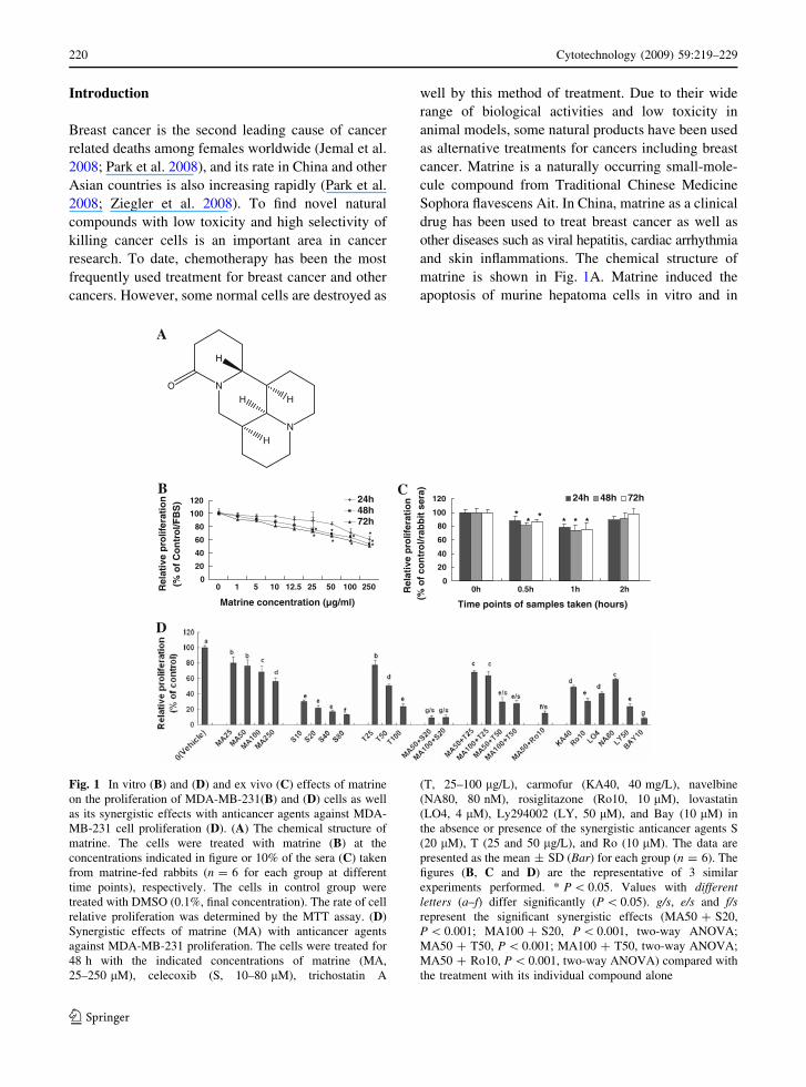
Fig. 1 In vitro (B) and (D) and ex vivo (C) effects of matrine
on the proliferation of MDA-MB-231(B) and (D) cells as well
as its synergistic effects with anticancer agents against MDA-
MB-231 cell proliferation (D). (A) The chemical structure of
matrine. The cells were treated with matrine (B) at the
concentrations indicated in figure or 10% of the sera (C) taken
from matrine-fed rabbits (n = 6 for each group at different
time points), respectively. The cells in control group were
treated with DMSO (0.1%, final concentration). The rate of cell
relative proliferation was determined by the MTT assay. (D)
Synergistic effects of matrine (MA) with anticancer agents
against MDA-MB-231 proliferation. The cells were treated for
48 h with the indicated concentrations of matrine (MA,
25–250 lM), celecoxib (S, 10–80 lM), trichostatin A
(T, 25–100 lg/L), carmofur (KA40, 40 mg/L), navelbine
(NA80, 80 nM), rosiglitazone (Ro10, 10 lM), lovastatin
(LO4, 4 lM), Ly294002 (LY, 50 lM), and Bay (10 lM) in
the absence or presence of the synergistic anticancer agents S
(20 lM), T (25 and 50 lg/L), and Ro (10 lM). The data are
presented as the mean ± SD (Bar) for each group (n = 6). The
figures (B, C and D) are the representative of 3 similar
experiments performed. * P \ 0.05. Values with differentletters (a–f) differ significantly (P \ 0.05). g/s, e/s and f/srepresent the significant synergistic effects (MA50 ? S20,
P \ 0.001; MA100 ? S20, P \ 0.001, two-way ANOVA;
MA50 ? T50, P \ 0.001; MA100 ? T50, two-way ANOVA;
MA50 ? Ro10, P \ 0.001, two-way ANOVA) compared with
the treatment with its individual compound alone
220 Cytotechnology (2009) 59:219–229
123

vivo as well as inhibited tumor growth (Ma et al.
2008). Matrine also inhibited the invasiveness and
matastasis of human malignant melanoma cell line
A375 (Liu et al. 2008). However, the mechanisms of
action of matrine against cancer such as human breast
cancer are largely unknown. In this study, we
investigated the effects of matrine on proliferation
and invasion of highly-metastatic human breast can-
cer cells and its mechanisms of action. We confirmed
that matrine inhibited the proliferation and invasion of
the human breast cancer cells as well as induced the
apoptosis and cell cycle arrest in the cancer cells. We
have demonstrated that the anticancer activities of
matrine are associated with Akt signaling and its
upstream and downstream targets by suppression of
the related proteins and mRNA levels as well as
reduction of activation of MMP-9 and MMP-2 in the
cancer cells. Matrine also displayed synergistic effects
with anticancer agents against the breast cancer cells.
Materials and methods
Chemicals and antibodies
Matrigel and Boyden chambers were purchased from
BD Bioscience (Bedford, MA) and Costar (Corning,
NY), respectively. The primary antibodies to human
Bcl-2, Bax, nuclear factor (NF-jB p-65), Akt, p-Akt,
and b-actin were purchased from Cell Signaling
Technology Inc. (Beverley, MA). Matrine, trichosta-
tin A, Ly294002 (LY), Bay 11-7082 (Bay), DMEM,
penicillin, streptomycin, propidium iodide, gelatin
3-[4,5-Dimethylthiazol-2-yl]-2,5-diphenyltetrazolium
bromide (MTT), fetal bovine serum (FBS), trypsin/
EDTA, Hoechst 33258 and all other chemicals were
purchased from Sigma Chemical Co. (St. Louis,
MO). Celecoxib, carmofur, navelbine, rosiglitazone,
and lovastatin were obtained from Yuhuangding
Hospital of Yantai, Yantai, China. LightShiftTM
chemiluminescent EMSA kit (Product # 20148) and
Biotin 30 End DNA Labeling Kit (Product # 89818)
were purchased from Pierce, Rockford.
Animal experimentation and preparation of sera
from matrine-fed rabbits
These were done according to our published methods
with slight modifications (Zhang et al. 2000). In brief,
New Zealand White female rabbits (3.5–4 kg; from
Luye Pharmaceutical Company, Yantai, China) were
treated in accordance with guidelines established by
the Animal Care and Use Committee at Yantai
University. Matrine was orally intubated into the
rabbits once daily at a dose of 10 mg/mL/kg body
weight for 3 days. On the third day, the blood was
then collected at 0, 0.5, 1, and 2 h from the rabbits
(fasted for 16 h) after oral intubation of matrine. The
prepared sera were aliquoted, and stored at -80 �C
until ex vivo assays.
Cell culture and in vitro and ex vivo proliferation
assays
The highly metastatic human breast cancer cell line
MDA-MB-231 was obtained from the American
Type Culture Collection. The cell line was cultured
in DMEM medium containing 10% FBS, glutamine
(2 mM), penicillin (100 U/mL) and streptomycin
(100 lg/ml) at 37 �C in a humidified incubator with
95% air/5% CO2 atmosphere. The in vitro and
ex vivo assays were done according to our published
methods (Zhang et al. 2000, 1999). The cells in
control group were treated with DMSO (0.1%, final
concentration). The cells were cultured in DMEM
supplemented with 10% FBS (in the case of in vitro
assay) containing different concentrations of matrine
or in combination with or without an existing
anticancer agent (celecoxib, trichostatin A, carmofur,
navelbine, rosiglitazone, lovastatin, Ly294002 and
Bay), or 10% the prepared rabbits sera (in the case of
ex vivo assay) mentioned above. The rate of relative
cell proliferation was measured 24, 48 and 72 h after
the treatments using a MTT assay kit. Each exper-
iment was repeated three times.
Apoptosis assays and cell cycle analysis
These were done according to our published methods
(Zhang et al. 2000). The treated cells were stained with
Hoechst 33258. The morphological changes in the
nuclear chromatin were observed under a fluorescent
microscope (Nikon, TE2000-U, Japan), using 409
lens. For flow cytometry for cell cycle analysis and
apoptosis, the treated cells were labeled with
propidium iodide solution containing RNase A. The
DNA content was analyzed by flow cytometry (Becton
Dickinson FACS Vantage SE, San Jose, CA).
Cytotechnology (2009) 59:219–229 221
123

In vitro invasion assay
Tumor cell invasion was measured by examining cell
invasion through matrigel-coated polycarbonate fil-
ters, using modified transwell chambers. MDA-MB-
231 cells (5 9 104) were seeded into the upper
chamber in 200 lL of serum-free medium containing
matrine at different concentrations; the lower com-
partment was filled with 0.66 ml of DMEM medium
supplemented with 10% of FBS. The cells in control
group were treated with DMSO (0.1%, final concen-
tration). After incubation for 16 h at 37 �C, the cells
that invaded to the lower surface of the filter were
fixed and stained using propidium iodide. The cells
on the upper side of the filter were removed using a
rubber scraper. The invaded cells on the underside of
the filter were counted and recorded for images under
a fluorescent microscope (Nikon, TE2000-U, Japan).
Experiments were performed in triplicate.
Gelatin zymography
This was performed according to the method of
Cheung et al. (2006) The supernatants from the upper
chamber of the transwell chamber in the aforemen-
tioned invasion assay, which was serum-free, were
analyzed for MMP-9/MMP-2 activation. Each exper-
iment was repeated three times.
Western blot analysis
The cells were treated with either matrine at the
different concentrations, or 10% of rabbit sera
prepared as mentioned above, respectively and
collected at 45 min (for detection of p-Akt) or 48 h.
The cells in control group were treated with DMSO
(0.1%, final concentration). The treated cells were
analyzed by Western Blotting according to the
method of Chen et al. (2001).
Cell fractionation and electrophoretic mobility
shift assay
Cells were grown and treated as indicated. They were
then pelleted by centrifugation at 1,000 rpm for
5 min at 4 �C and resuspended in ice-cold buffer A
(10 mM HEPES (pH 7.9), 10 mM KCl, 0.1 mM
EDTA, 1 mM DTT, 0.5 mM phenylmethysulfonyl-
fluoride (PMSF), 1 lg/mL leupeptin, 5 lg/mL
aprotinin). Following the addition of 25 lL 10%
NP40, the suspension was vortexed and centrifuged at
14,500 rpm for 1 min at 4 �C; the supernatant was
designated as the cytoplasmic fraction. Nuclei were
resuspended in 50 lL of ice-cold buffer B (20 mM
HEPES (pH 7.9), 0.4 M NaCl, 1 mM EDTA, 1 mM
DTT, 1 mM PMSF, 25% glycerol, 1 lg/mL
leupeptin, 5 lg/mL aprotinin) and centrifuged at
14,500 rpm for 5 min. The supernatant was used as
the nuclear fraction and protein concentration deter-
mined by the Bradford method. Electrophoretic
mobility shift assay (EMSA) was done with 10-lg
nuclear protein using the Biotin 30 End DNA
Labeling Kit (Pierce, Rockford, Product # 89818)
and Biotinlabeled NF-jB consensus oligonucleotide
(50-AGT TGA GGG GAC TTT CCC AGG C-30).Nuclear protein-DNA complexes were separated by
4% PAGE. Transferred DNAs were UVcross-linked
to the membrane and detected using horseradish
peroxidase-conjugated streptavidin (LightShiftTM
chemiluminescent EMSA kit, Pierce, Rockford,
Product # 20148) according to the manufacturer’s
instructions.
ELISA for detection of human VEGF protein
levels secreted by human breast cancer cells
For detection of effects of matrine and its synergistic
anticancer agents on the secretion of vascular
endothelial growth factor (VEGF) in MDA-MB-231
cells, the cells were treated for 48 h with the
indicated concentrations of matrine and the anti-
cancer agents mentioned above. Then each super-
natant of the cell culture was respectively collected
and analyzed by ELISA using a kit (VEGF) from R &
D Systems (Minneapolis, MN). ELISA was done
according to the instructions of the manufacturer.
Each experiment was repeated three times.
Semi-quantitative reverse transcription-PCR
Total cellular RNA was extracted using TRIzol
reagent (Invitrogen, USA) from the cancer cells
treated for 48 h with matrine at different concentra-
tions or the prepared rabbit sera mentioned above,
respectively according to the manufacturer’s instruc-
tions, and quantified by spectrophotometry. The cells
in control group were treated with DMSO (0.1%,
final concentration). RT reaction was done using total
222 Cytotechnology (2009) 59:219–229
123

RNA as a template and a RT-for-PCR kit (Promega,
Madison, WI). PCR amplification was carried out
with the following primers:
Bcl-2, 50-GGAGGATTGTGGCCTTCTTT-3 and
50-TCACTTGTGGCTCAGATAGGC-30;Bax, 50-TCTGACGGCAACTTCAACTG-30 and
50-CACTGTGACCTGCTCCAGAA-30;b-Actin, 50-ATCATGTTTGAGACCTTCAACAC
C-30 and 50-TAGCTCTTCTCCAGGGAGG-30;MMP-2, 50-GGATGATGCCTTTGCTCG-30 and
50- CAGTGGACATGGCGGTCT-30;MMP-9, 50-TCCCTGGAGACCTGAGAACC-30 and
50-GGCAAGTCTTCCGAGTAGTTT-30;EGF, 50-TGCCAACTGGGGGTGCACAG-30 and
50-CTGCCCGTGGCCAGCGTGGC-30;VEGFR1, 50-GAGAATTCACTATGGAAGATCT
GATTTCTTACAGT-30 and 50-GAGCATGCGG
ATAAATACACATGTGCTTCTAG-30.
PCR conditions for the expression of human genes
included an initial denaturation of 3 min at 94 �C
followed by 30 cycles of denaturation for 45 s at
94 �C, annealing for 1 min at 60 �C, and extension
for 1 min at 72 �C. Aliquots (10 lL) of the ampli-
fication products were separated by electrophoresis
through a 1.5% agarose gel and visualized by
ethidium bromide staining. The intensity of each
band was quantified using Scion Image software
(Scion, Frederick, MD). Results for each detected
band intensity were normalized to b-Actin band
intensity values. RNA only samples that gave com-
pletely negative results in PCR without reverse
transcriptase were used to rule out the presence of
genomic DNA contamination.
Statistical analysis
The data were expressed as mean ± SD and analyzed
by the SPSS 13.0 software to evaluate the statistical
difference. One-way or two-way ANOVA followed
by the appropriate post hoc test (Bonferroni) was
used to establish whether significant differences
existed among groups. For confirming the synergistic
effect between matrine and celecoxib, trichostatin A,
or rosiglitazone, comparison was made by two-way
ANOVA followed by Bonferroni post hoc test.
Values among different treatment groups at different
times were compared. Mean concentrations and
inhibition (%) are shown for each group; Asterisk
P \ 0.05. For all tests, P values less than 0.05 were
considered statistically significant. All statistical tests
were two-sided.
Results and discussion
Matrine shows in vitro and ex vivo inhibition
of proliferation in MDA-MB-231 cells
and synergistic activity with anticancer agents
against MDA-MB-231 cell proliferation
We first confirmed that matrine reduced the rates of
relative proliferation of highly-metastatic human
breast cancer cell line MDA-MB-231 in a dose- and
time-dependent manner after the cells were treated
with matrine at 1–250 lg/mL for 24, 48 and 72 h,
respectively (Fig. 1B). The ex vivo assay showed that
the rabbit sera obtained 0.5 and 1 h after oral
intubation of matrine significantly reduced the rate
of relative proliferation of MDA-MB-231 cells after
the cells were treated with these sera for 48 and 72 h,
while the 2 h rabbit sera did not show the significant
inhibitory effect (Fig. 1C). This result suggests that
matrine has a certain bioavailability by oral admin-
istration and the peak inhibition of the cancer cell
proliferation is at 1 h after oral intubation of matrine.
More importantly, matrine displayed synergistic
effects with existing anticancer agents such as
celecoxib (the inhibitor of COX-2), trichostatin A
(the histone deacetylase inhibitor) and rosiglitazone
against the proliferation of MDA-MB-231 cells,
which enhanced inhibitory activity on cancer cell
proliferation more than twice. In addition, the anti-
cancer agents celecoxib, trichostatin A, carmofur,
rosiglitazone, lovastatin, navelbine, Ly294002 and
Bay showed significant reduction of the relative
proliferation rate of MDA-MB-231 cells (Fig. 1D).
These results partially explain why matrine has its
therapeutic and/or adjuvant therapeutic effects on
treatment for some patients with breast cancer.
Matrine induced apoptosis and cell cycle arrest
by reducing the ratios of Bcl-2/Bax protein and
mRNA levels in MDA-MB-231 cells
To understand the mechanisms of action of matrine
against the proliferation in human breast cancer cells,
Cytotechnology (2009) 59:219–229 223
123

we investigated the effects of matrine on apoptosis
and cell cycle arrest in the cancer cells as well as the
expressions of related protein and mRNA. Hoechst
33258 staining showed that the typical morphological
changes, such as formation of apoptotic bodies
appeared in both MDA-MB-231 cells after the cells
were treated for 48 h with matrine at 100 and 250 lg/
mL, whereas the control cells without matrine
treatment did not show the evident apoptotic
morphological changes (Fig. 2A). Flow cytometric
analysis confirmed that matrine at concentrations of
25–250 lg/mL dose-dependently induced apoptosis
and cell cycle arrest at the S phase in MDA-MB-231
cells (Fig. 2B). Furthermore, matrine down-regulated
Bcl-2 protein and mRNA levels and up-regulated Bax
protein and mRNA levels, eventually leading to the
reduction of ratios of Bcl-2/Bax protein (Fig. 2C1)
and mRNA (Fig. 2C2) levels in the cancer cells.
There have been studies showing that Bcl-2 and its
dominant inhibitor Bax are key regulators of cell
proliferation and apoptosis. Overexpression of Bcl-2
enhances cell survival by suppressing apoptosis, but
overexpression of Bax accelerates cell death (Oltvai
et al. 1993). Induction of apoptosis and cell cycle
arrest and decrease in the ratios of Bcl-2/Bax protein
and mRNA levels by matrine may be one of the
important mechanisms of action of matrine against
the cancer cell proliferation.
Matrine suppressed the invasion and MMP-9/
MMP-2 activation and reduced the mRNA levels
of MMP-9, MMP-2, EGF, and VEGFR1 in MDA-
MB-231 cells
The presence of metastasis is the main cause of
morbidity and mortality in millions of patients with
cancer. During the complicated process of metastasis,
the invasion of cancer cells is the most important and
Fig. 2 In vitro effects of matrine on induction of apoptosis and
cell cycle arrest as well as protein and mRNA levels of Bcl-2
and Bax in MDA-MB-231 cells. (A) Induction of apoptosis in
MDA-MB-231 cells by treatment for 48 h with matrine at
concentrations of 0 (A1: vehicle, 0.1% DMSO as the control),
100 lg/mL (A2) and 250 lg/mL (A3). The cells stained with
Hoechst 33258 were observed under a fluorescent microscope.
(B) Induction of apoptosis (cells in Sub-G1 phase) and cell
cycle arrest (at S phase) in MDA-MB-231 cells by matrine was
analyzed by flow cytometry. (C) Reduction of protein and
mRNA levels in MDA-MB-231 cells by treatment for 48 h
with matrine at the concentrations indicated in figure. The
protein expressions (C1) and mRNA levels (C2) of Bcl-2 and
Bax were analyzed by Western blotting and RT-PCR,
respectively. ß-Actin was used as a sample loading control.
The ratio of Bcl-2 and Bax, (the ratio of relative density of each
band normalized to b-actin), shown as mean ± SD (Bar) is
relative to that of 0 (0.1% DMSO vehicle) as the control
(designated as 1.0). For one experiment, 3 assays were carried
out and only one set of gels is shown. * P \ 0.05
224 Cytotechnology (2009) 59:219–229
123

characteristic step. Clearly, an agent which could
efficiently inhibit the proliferation and invasion of
cancer cells would be a hopeful candidate to suppress
cancer progression and metastasis and thus could
reduce mortality. Therefore, we examined the effects
of matrine on the invasion and related factors in
0
20
40
60
80
100
120
140
0 25 50 100
Matrine concentration (µg/ml)
Rel
ativ
e in
vasi
on (
%)
* *
A
C
D
B
Fig. 3 Suppression of invasion and activation of MMP-9 and
MMP-2 as well as reduction of mRNA levels of MMP-9,
MMP-2, EGF and VEGFR1 in MDA-MB-231 cells by matrine.
(A) Relative invasion (%) ±SD (n = 6) are shown for the
indicated matrine concentrations and 0 (0.1% DMSO vehicle)
is the control. (B) The photos show the propidium iodide-
stained MDA-MB-231 cells invading through Matrigel-coated
transwell chamber. The cells were treated for 16 h with 0 (B1:
0.1% DMSO vehicle as the control) and matrine at 100 lg/mL
(B2). (C) The activation of active MMP-9 and MMP-9/MMP-2
zymogens (z-MMP9/z-MMP2) in the supernatants (serum-free)
of invading MDA-MB-231 cells mentioned in (A) was
determined by gelatin zymography analysis. Values (the
relative activation of active MMP9, z-MMP9/z-MMP2) are
shown as mean ± SD of 3 runs for each sample, only one set
of gels is shown (n = 3). (D) In vitro (D1) and ex vivo (D2)
effects of matrine on mRNA levels of MMP-9, MMP-2, EGF
and VEGFR1 in MDA-MB-231 cells. The cells were treated
for 48 h with matrine at the indicated concentrations (D1) or
the matrine-fed rabbit sera (D2) obtained at the indicated time
points. The mRNA levels were determined by semi-quantita-
tive RT-PCR. For one experiment, 3 assays were carried out
and only one set of gels is shown. The density of the band
(normalized to b-actin) shown as mean ± SD is relative to that
of the control (designated as 1.00). The figures (A, B, C and D)
are the representative of 3 similar experiments performed.
* P \ 0.05
Cytotechnology (2009) 59:219–229 225
123

cancer cells. The invasion assay indicated that
matrine at 50–100 lg/mL significantly suppressed
the invasion of MDA-MB-231 cells (Fig. 3A, B). In
addition, Ly294002 and Bay significantly reduced the
MDA-MB-231 invasion (data not shown). The gel-
atin zymography analysis further demonstrated that
the activation of active MMP-9, and MMP-9/MMP-2
zymogens in the supernatants of invading MDA-MB-
231 cells was significantly suppressed by matrine at
25–100 lg/mL (Fig. 3C). Moreover, RT-PCR detec-
tion indicated that matrine at 50–100 lg/mL signif-
icantly down-regulated the mRNA levels of MMP-9,
MMP-2, VEGFR1 and EGF in MDA-MB-231 cells
in vitro (Fig. 3D1). The rabbit sera obtained 1 h after
oral intubation of matrine for 3 days significantly
reduced the mRNA levels of MMP-9, MMP-2,
VEGFR1 and EGF in MDA-MB-231 cells ex vivo
(Fig. 3D2). These results suggest that the inhibition
of invasion and MMP-9/MMP-2 activity as well as
the reduction of mRNA levels of MMP-9, MMP-2,
VEGFR1 and EGF by matrine may play an important
role in the clinical effects of matrine against cancer
progression.
Matrine reduced the phosphorylation of Akt
and expressions and activity of NF-jB p65
in vitro and/or ex vivo as well as VEGF secretions
in MDA-MB-231 cells
Many reports indicated that in Akt signaling, pAkt,
NF-jB, EGF, VEGF, VEGFR1, MMP-9 and MMP-2
play important roles in promoting proliferation,
migration, invasion, angiogenesis, and metastasis of
cancer cells (Price et al. 1999; Helbig et al. 2003;
Rolli et al. 2003; Koivunen et al.1999; Kletsas et al.
2000; John and Tuszynski 2001; Shibata et al. 2002;
Gibson et al. 2002; Lee et al. 2007). Thus, we next
investigated if the suppression of the cancer cell
Time points of samples taken (hours)
Time points of samples taken (hours)
0
20
40
60
80
100
120
0h 0.5h 1h 2h
% o
f N
F-
B p
rote
in le
vel
* *
0
20
40
60
80
100
120
0 25 50 100 bay ly
Matrine concentration (µg/ml)
% o
f NF
-B
pro
tein
leve
l
* * * *
*
0
20
40
60
80
100
120
0h 0.5h 1h 2h
% o
f p
-Akt
(ser
473)
pro
tein
leve
l
* *
A
B
Fig. 4 Effects of matrine on in vitro (left panels) and ex vivo
(right panels) phosphorylation of Akt (A) and NF-jB p65
expressions (B) in MDA-MB-231 cells. The cells were treated
for 48 h with matrine at the indicated concentrations or sera
taken at the indicated time points from matrine-fed rabbits.
p-Akt and NF-jB p65 were analyzed by Western Blotting. The
density of the band (normalized to Akt) in (A) or (normalized
to b-actin) in (B) shown as mean ± SD is relative to that of
0 lg/mL (0.1% DMSO vehicle) or 0 h as the control
(designated as 100%). Ly (Ly294002) and Bay are the
inhibitors of PI3 K/Akt and NF-jB, respectively. For one
experiment, 3 assays were carried out and only one set of gels
is shown. * P \ 0.05
226 Cytotechnology (2009) 59:219–229
123

proliferation and invasion by matrine is associated
with Akt signaling. Western blot analysis confirmed
that matrine at 25–100 lg/mL and the positive
control Ly294002 (the inhibitor of PI3 K/Akt) and
Bay (the inhibitor of NF-jB) significantly suppressed
the phosphorylation of Akt (Fig. 4A) and NF-jB
p-65 expression (Fig. 4B) in MDA-MB-231 cells in
vitro. The rabbit sera obtained 0.5 and 1 h after oral
intubation of matrine also significantly suppressed the
phosphorylation of Akt (Fig. 4A) and NF-jB p-65
expression (Fig. 4B) in MDA-MB-231 cells ex vivo.
Furthermore, the result of EMSA indicated that
matrine at 50–250 lg/mL dose-dependently and
significantly suppressed the activity of NF-jB p65
DNA binding (Fig. 5A) in MDA-MB-231 cells in
vitro. In addition, matrine also displayed synergistic
effects with anticancer agents celecoxib, trichostatin
A, and rosiglitazone on reducing VEGF secretion in
MDA-MB-231 cells (Fig. 5B). The anticancer agents
celecoxib, trichostatin A, carmofur, rosiglitazone,
lovastatin, Ly294002 and Bay also reduced VEGF
secretion in MDA-MB-231 cells (Fig. 5B). These
results suggest that the suppression of the cancer cell
proliferation and invasion by matrine is associated
with the inhibition of Akt signaling and its upstream
targets such as EGF, VEGF and VEGFR1, and
downstream targets such as NF-jB p-65, Bcl-2/Bax,
MMP-9 and MMP-2 in the breast cancer cells. There
have been studies reporting that EGF, VEGF and
VEGFR1 protect from apoptosis in MDA-MB-231
cells (Gibson et al. 2002; Lee et al. 2007). EGF and
NF-jB promote migration of breast cancer cells
Fig. 5 Effects of matrine on NF-jB activity (A) and its
synergistic effects with anticancer agents on VEGF secretion
(B) in MDA-MB-231 cells. (A) Suppression of the activity of
NF-jB p65 DNA binding in MDA-MB-231 cells by matrine.
The cells were treated for 48 h with matrine at the concentra-
tions of 50–250 lg/mL and Bay (10 lM), respectively. NF-jB
p65 DNA binding was analyzed by EMSA as described in the
section of ‘‘Materials and methods’’. (B) Reduction of VEGF
secretion in MDA-MB-231 cells by matrine and its synergistic
anticancer agents celecoxib (S), trichostatin A (T) and
rosiglitazone (Ro). The cells were treated for 48 h with the
indicated concentrations of matrine (MA, 12.5–100 lM),
trichostatin A (T, 12.5–100 lg/L), celecoxib (S, 10–80 lM),
carmofur (KA, 40 mg/L), rosiglitazone (Ro10, 10 lM),
lovastatin (LO4, 4 lM), Ly294002 (LY, 50 lM), and Bay
(10 lM) in the absence or presence of the synergistic
anticancer agents S (20 lM), T (50 lg/L), and Ro (10 lM).
The VEGF secreted in the supernatants of MDA-MB-231 cells
was analyzed by ELISA as described in the section of
‘‘Materials and methods’’. Values are shown as mean ± SD
(bar) for the indicated concentration (n = 3). Values with
different letters (a–g) differ significantly (P \ 0.05). f/s, e/sand g/s represent the significant synergistic effects
(MA50 ? S20, P \ 0.001; MA100 ? S20, P \ 0.001, two-
way ANOVA; MA50 ? T50, P \ 0.001; MA100 ? T50,
two-way ANOVA; MA100 ? Ro10, P \ 0.001, two-way
ANOVA) compared with the treatment with its individual
compound alone
Cytotechnology (2009) 59:219–229 227
123

(Price et al. 1999; Helbig et al. 2003). MMP-9 is a
NF-jB-regulated gene. MMP-9 and MMP-2 are not
only associated with invasion and metastasis but also
MMP-9 and VEGF have been implicated in angio-
genesis, and hence they are considered to be the
therapeutic targets of high priority (Rolli et al. 2003;
Koivunen et al. 1999; Mitropoulou et al. 2003).
Inhibition of NF-jB activity decreases the VEGF
mRNA expression in MDA-MB-231 (Shibata et al.
2002). NF-jB is a nuclear transcription regulator
with a specific motif for bcl-2 transcription (Marsden
et al. 2002; Wang et al. 1996). Activation of p-Akt
and the NF-jB/bcl-2 pathway leads to inhibition of
chemotherapy-induced apoptosis, which results in
treatment resistance (Wang et al. 1996). Therefore,
the p-Akt, NF-jB, Bcl-2/Bax, EGF, MMP-9, MMP-
2, VEGF and VEGFR1 in the Akt signaling have
become the important targets of action by anticancer
agents against the proliferation, invasion, angiogen-
esis and metastasis in cancer cells (Price et al. 1999;
Helbig et al. 2003; Rolli et al. 2003; Koivunen et al.
1999; John and Tuszynski 2001; Gibson et al. 2002;
Mitropoulou et al. 2003; Wang et al. 1996; Emi et al.
2005). Our present results have demonstrated that
matrine significantly inhibits these important targets
in the Akt signaling that is associated with the
proliferation and invasion of MDA-MB-231 cells.
These results suggest that the inhibition of both
VEGF excretion and activation of pro-MMP-9/MMP-
2 (z-MMP-9/z-MMP-2) and active MMP-9 as well as
the reduction of MMP-9/MMP-2 mRNA levels by
matrine may be one of the mechanisms of action of
matrine against the proliferation and invasion of
MDA-MB-231 cells. In addition, the suppression of
the phosphorylation of p-Akt and the expression and
activity of NF-jB as well as the EGF/VEGFR1
mRNA levels by matrine may also be the important
mechanisms of action of matrine against the prolif-
eration and invasion of the human breast cancer cells.
Abnormal proliferation and metastasis of cancer
cells are regarded as the important biological char-
acteristics of cancers. Our present results have
confirmed that matrine significantly suppressed the
proliferation and invasion of highly-metastatic human
breast cancer MDA-MB-231 cells by affecting the
expressions of Bcl-2/Bax/VEGF/p-Akt/NF-jB pro-
teins and MMP-9/MMP-2/Bcl-2/Bax/EGF/VEGFR1
mRNA as well as activation of MMP-9/MMP-2 in the
Akt signaling. The sera from matrine-fed rabbits also
showed ex vivo inhibitory effects on the proliferation
of MDA-MB-231 cells by affecting these protein and
mRNA levels in the Akt signaling. Moreover, matrine
displayed synergistic effects with the anticancer
agents against the proliferation and VEGF excretion
in MDA-MB-231 cells. All these findings suggest
that matrine may have a wide therapeutic and/or
adjuvant therapeutic application in the treatment of
human breast cancer; the mechanisms of action of
matrine against cancer cell proliferation and invasion
are associated with the EGF/VEGF-VEGFR1-Akt-
NF-jB signaling.
Acknowledgments The authors would like to extend our
thanks to Ronald E. Vincent (BIOCON Scientific) for his
thoughtful reading and the help of Drs. Jianyuan Li and
Shaohua Jin for FACS analysis. This work is supported in part
by grants from the Ministry of Education of the People’s
Republic of China to G.Z, from the Ministry of Human
Resources and Social Security of the People’s Republic of
China to G.Z, Projects of Yantai University to G.Z, and
Projects from the Department of Science and Technology of
Shandong Province to G.Z. (Y2008C71; 2009GG10002089).
References
Chen Q, Gong B, Mahmoud-Ahmed AS, Zhou A, Hsi ED,
Hussein M, Almasan A (2001) Apo2L/TRAIL and Bcl-2-
related proteins regulate type I interferon-induced apop-
tosis in multiple myeloma. Blood 98:2183–2192
Cheung LW, Leung PC, Wong AS (2006) Gonadotropin-
releasing hormone promotes ovarian cancer cell inva-
siveness through c-Jun NH2-terminal kinase-mediated
activation of matrix metalloproteinase (MMP)-2 and
MMP-9. Cancer Res 66:10902–10910
Emi M, Kim R, Tanabe K, Uchida Y, Toge T (2005) Targeted
therapy against Bcl-2-related proteins in breast cancer
cells. Breast Cancer Res 7:R940–R952
Gibson EM, Henson ES, Haney N, Villanueva J, Gibson SB
(2002) Epidermal growth factor protects epithelial-
derived cells from tumor necrosis factor-related apoptosis-
inducing ligand-induced apoptosis by inhibiting cyto-
chrome c release. Cancer Res 62:488–496
Helbig G, Christopherson KW, Bhat-Nakshatri P, Kumar S,
Kishimoto H, Miller KD, Broxmeyer HE, Nakshatri H
(2003) NF-kappaB promotes breast cancer cell migration
and metastasis by inducing the expression of the chemo-
kine receptor CXCR4. J Biol Chem 278:21631–21638
Jemal A, Siegel R, Ward E, Hao Y, Xu J, Murray T, Thun MJ
(2008) Cancer statistics, 2008. CA Cancer J Clin 58:71–
96
John A, Tuszynski G (2001) The role of matrix metallopro-
teinases in tumor angiogenesis and tumor metastasis.
Pathol Oncol Res 7:14–23
Kletsas D, Pratsinis H, Zervolea I, Handris P, Sevaslidou E,
Ottaviani E, Stathakos D (2000) Fibroblast responses to
228 Cytotechnology (2009) 59:219–229
123

exogenous and autocrine growth factors relevant to tissue
repair. The effect of aging. Ann N Y Acad Sci 908:155–
166
Koivunen E, Arap W, Valtanen H, Rainisalo A, Medina OP,
Heikkila P, Kantor C, Gahmberg CG, Salo T, Konttinen
YT, Sorsa T, Ruoslahti E, Pasqualini R (1999) Tumor
targeting with a selective gelatinase inhibitor. Nat Bio-
technol 1776:8–17774
Lee TH, Seng S, Sekine M, Hinton C, Fu Y, Avraham HK,
Avraham S (2007) Vascular endothelial growth factor
mediates intracrine survival in human breast carcinoma
cells through internally expressed VEGFR1/FLT1. PLoS
Med 4:e186
Liu XY, Fang H, Yang ZG, Wang XY, Ruan LM, Fang DR,
Ding YG, Wang YN, Zhang Y, Jiang XL, Chen HC
(2008) Matrine inhibits invasiveness and metastasis of
human malignant melanoma cell line A375 in vitro. Int J
Dermatol 47:448–456
Ma L, Wen S, Zhan Y, He Y, Liu X, Jiang J (2008) Anticancer
effects of the Chinese medicine matrine on murine
hepatocellular carcinoma cells. Planta Med 74:245–251
Marsden VS, O’Connor L, O’Reilly LA, Silke J, Metcalf D,
Ekert PG, Huang DC, Cecconi F, Kuida K, Tomaselli KJ,
Roy S, Nicholson DW, Vaux DL, Bouillet P, Adams JM,
Strasser A (2002) Apoptosis initiated by Bcl-2-regulated
caspase activation independently of the cytochrome
c/Apaf-1/caspase-9 apoptosome. Nature 419(6907):634–637
Mitropoulou TN, Tzanakakis GN, Kletsas D, Kalofonos HP,
Karamanos NK (2003) Letrozole as a potent inhibitor of
cell proliferation and expression of metalloproteinases
(MMP-2 and MMP-9) by human epithelial breast cancer
cells. Int J Cancer 104:155–160
Oltvai ZN, Milliman CL, Korsmeyer SJ (1993) Bcl-2 hetero-
dimerizes in vivo with a conserved homolog, Bax, that
accelerates programmed cell death. Cell 74:609–619
Park S, Bae J, Nam BH, Yoo KY (2008) Aetiology of cancer in
Asia. Asian Pac J Cancer Prev 9:371–380
Price JT, Tiganis T, Agarwal A, Djakiew D, Thompson EW
(1999) Epidermal growth factor promotes MDA-MB-231
breast cancer cell migration through a phosphatidylinosi-
tol 3’-kinase and phospholipase C-dependent mechanism.
Cancer Res 59:5475–5478
Rolli M, Fransvea E, Pilch J, Saven A, Felding-Habermann B
(2003) Activated integrin alphavbeta3 cooperates with
metalloproteinase MMP-9 in regulating migration of
metastatic breast cancer cells. Proc Natl Acad Sci USA
100:9482–9487
Shibata A, Nagaya T, Imai T, Funahashi H, Nakao A, Seo H
(2002) Inhibition of NF-kappaB activity decreases the
VEGF mRNA expression in MDA-MB-231 breast cancer
cells. Breast Cancer Res Treat 73:237–243
Wang CY, Mayo MW, Baldwin AS Jr (1996) TNF- and cancer
therapy-induced apoptosis: potentiation by inhibition of
NF-kappaB. Science 274:784–787
Zhang G, Miura Y, Yagasaki K (1999) Effects of green, oolong
and black teas and related components on the proliferation
and invasion of hepatoma cells in culture. Cytotechnology
31:37–44
Zhang G, Miura Y, Yagasaki K (2000) Induction of apoptosis
and cell cycle arrest in cancer cells by in vivo metabolites
of teas. Nutr Cancer 38:265–273
Ziegler RG, Anderson WF, Gail MH (2008) Increasing breast
cancer incidence in China: the numbers add up. J Natl
Cancer Inst 100:1339–1341
Cytotechnology (2009) 59:219–229 229
123
Copyright © 2022 FDOKUMEN