Bao - Effectiveness of 4 tonometers after LASIK, SMILE, PRK ...
25
Downloaded from https://journals.lww.com/jcrs by BhDMf5ePHKav1zEoum1tQfN4a+kJLhEZgbsIHo4XMi0hCywCX1AWnYQp/IlQrHD3PE2KhmxLsUKQ6YVZvYQq3rC97dKVeUq0/b7CQ/8Ex/s3V54i10jidA== on 04/14/2020 Journal of Cataract and Refractive Surgery Publish Ahead of Print DOI: 10.1097/j.jcrs.0000000000000204 1 Effectiveness of four tonometers in measuring intraocular pressure following femtosecond laser–assisted LASIK, SMILE and transepithelial PRK ShiHao Chen, M.D, O.D, Bernardo T Lopes, M.D, Ph.D, Wei Huang, M.D, XiaoBo Zheng, M.Sc, JunJie Wang, Ph.D, Rong Zhu, M.D, Riccardo Vinciguerra, M.D, YiYu Li, Ph.D, QinMei Wang, M.D, HuiRong Li, Ph.D, FangJun Bao, M.D, Ph.D, Ahmed Elsheikh, Ph.D From the Eye Hospital (Chen, Zheng, J. Wang, Zhu, Y. Li, Q. Wang, H. Li, Bao), Institution of Ocular Biomechanics (Chen, Zheng, J. Wang, Q. Wang, Bao), WenZhou Medical University, Wenzhou Medical University, Wenzhou, Ningbo Hangzhou Bay Hospital (Huang), NingBo, Beijing Advanced Innovation Center for Biomedical Engineering, Beihang University (Elsheikh), Beijing, China; 3 School of Engineering, University of Liverpool (Lopes, Elsheikh), Liverpool, 6 National Institute for Health Research Biomedical Research Centre for Ophthalmology, Moorfields Eye Hospital NHS Foundation Trust, and UCL Institute of Ophthalmology (Elsheikh), London, United Kingdom; and 5 Humanitas San Pio X Hospital (Vinciguerra), Milan, Italy. S. Chen and B.T. Lopes contributed equally to this work. Supported by the National Natural Science Foundation of China (81600712, 31771020), Zhejiang Provincial Natural Science Foundation of China under grant LY18A020008, Projects of Medical and Health Technology Development Program in ZheJiang Province (2016ZHB012, 2018RC057, 2019RC056, 2018KY541), and Science and Technology Plan Project of Wenzhou Science and Technology Bureau (Y20180172, Y20170198, Y20170792). ACCEPTED Copyright © 20 Published by Wolters Kluwer on behalf of ASCRS and ESCRS. Unauthorized reproduction of this article is prohibited. 20
-
Upload
khangminh22 -
Category
Documents
-
view
0 -
download
0
Transcript of Bao - Effectiveness of 4 tonometers after LASIK, SMILE, PRK ...

Dow
nloadedfrom
https://journals.lww.com
/jcrsby
BhDMf5ePH
Kav1zEoum1tQ
fN4a+kJLhEZgbsIH
o4XMi0hC
ywCX1AW
nYQp/IlQ
rHD3PE2Khm
xLsUKQ
6YVZvYQq3rC
97dKVeUq0/b7C
Q/8Ex/s3V54i10jidA==
on04/14/2020
Downloadedfromhttps://journals.lww.com/jcrsbyBhDMf5ePHKav1zEoum1tQfN4a+kJLhEZgbsIHo4XMi0hCywCX1AWnYQp/IlQrHD3PE2KhmxLsUKQ6YVZvYQq3rC97dKVeUq0/b7CQ/8Ex/s3V54i10jidA==on04/14/2020
Journal of Cataract and Refractive Surgery Publish Ahead of PrintDOI: 10.1097/j.jcrs.0000000000000204
1
Effectiveness of four tonometers in measuring intraocular pressure following femtosecond laser–assisted LASIK, SMILE and transepithelial PRK
ShiHao Chen, M.D, O.D, Bernardo T Lopes, M.D, Ph.D, Wei Huang, M.D, XiaoBo Zheng,
M.Sc, JunJie Wang, Ph.D, Rong Zhu, M.D, Riccardo Vinciguerra, M.D, YiYu Li, Ph.D,
QinMei Wang, M.D, HuiRong Li, Ph.D, FangJun Bao, M.D, Ph.D, Ahmed Elsheikh, Ph.D
From the Eye Hospital (Chen, Zheng, J. Wang, Zhu, Y. Li, Q. Wang, H. Li, Bao), Institution
of Ocular Biomechanics (Chen, Zheng, J. Wang, Q. Wang, Bao), WenZhou Medical
University, Wenzhou Medical University, Wenzhou, Ningbo Hangzhou Bay Hospital
(Huang), NingBo, Beijing Advanced Innovation Center for Biomedical Engineering, Beihang
University (Elsheikh), Beijing, China; 3 School of Engineering, University of Liverpool
(Lopes, Elsheikh), Liverpool, 6 National Institute for Health Research Biomedical Research
Centre for Ophthalmology, Moorfields Eye Hospital NHS Foundation Trust, and UCL
Institute of Ophthalmology (Elsheikh), London, United Kingdom; and 5 Humanitas San Pio X
Hospital (Vinciguerra), Milan, Italy.
S. Chen and B.T. Lopes contributed equally to this work.
Supported by the National Natural Science Foundation of China (81600712, 31771020),
Zhejiang Provincial Natural Science Foundation of China under grant LY18A020008,
Projects of Medical and Health Technology Development Program in ZheJiang Province
(2016ZHB012, 2018RC057, 2019RC056, 2018KY541), and Science and Technology Plan
Project of Wenzhou Science and Technology Bureau (Y20180172, Y20170198, Y20170792).
ACCEPTED
Copyright © 20 Published by Wolters Kluwer on behalf of ASCRS and ESCRS. Unauthorized reproduction of this article is prohibited.20

2
Corresponding authors: HuiRong Li, PhD, FangJun Bao, MD, PhD, No. 270 Xueyuan
West Road, WenZhou City, ZheJiang Prov, 325027, China. Email:
[email protected]; [email protected].
Running Head
IOP measurement following 3 forms of refractive surgery
Abstract
Purpose: To test the performance of 4 tonometers in estimating intraocular pressure (IOP)
after 3 forms of refractive surgery.
Setting: Eye Hospital, WenZhou Medical University, China.
Design: Prospective case series.
Methods: Patients matched for preoperative age, corneal thickness and myopic correction
enrolled for femtosecond laser–assisted laser in situ keratomileusis (FS-LASIK),
small-incision lenticule extraction (SMILE), or transepithelial photorefractive keratectomy
(TransPRK) were included in the study. For each patient, 4 measurements of IOP were
obtained preoperative and 3 months postoperative, using the Goldmann applanation
tonometer (GAT-IOP), the Dynamic Contour Tonometer (DCT-IOP), corneal-compensated
IOP (IOPcc) from the Ocular Response Analyzer, and biomechanically-corrected IOP (bIOP)
from the Corvis ST. Overall corneal stiffness was also estimated based on the stiffness
parameter (SP-A1) provided by the Corvis ST.
ACCEPTED
Copyright © 20 Published by Wolters Kluwer on behalf of ASCRS and ESCRS. Unauthorized reproduction of this article is prohibited.20
3
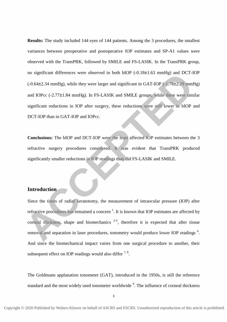
Results: The study included 144 eyes of 144 patients. Among the 3 procedures, the smallest
variances between preoperative and postoperative IOP estimates and SP-A1 values were
observed with the TransPRK, followed by SMILE and FS-LASIK. In the TransPRK group,
no significant differences were observed in both bIOP (-0.18±1.63 mmHg) and DCT-IOP
(-0.64±2.34 mmHg), while they were larger and significant in GAT-IOP (-1.78±2.29 mmHg)
and IOPcc (-2.77±1.84 mmHg). In FS-LASIK and SMILE groups, while there were similar
significant reductions in IOP after surgery, these reductions were still lower in bIOP and
DCT-IOP than in GAT-IOP and IOPcc.
Conclusions: The bIOP and DCT-IOP were the least affected IOP estimates between the 3
refractive surgery procedures considered. It was evident that TransPRK produced
significantly smaller reductions in IOP readings than did FS-LASIK and SMILE.
Introduction
Since the times of radial keratotomy, the measurement of intraocular pressure (IOP) after
refractive procedures has remained a concern 1. It is known that IOP estimates are affected by
corneal thickness, shape and biomechanics 2-5, therefore it is expected that after tissue
removal and separation in laser procedures, tonometry would produce lower IOP readings 6.
And since the biomechanical impact varies from one surgical procedure to another, their
subsequent effect on IOP readings would also differ 7, 8.
The Goldmann applanation tonometer (GAT), introduced in the 1950s, is still the reference
standard and the most widely used tonometer worldwide 9. The influence of corneal thickness
ACCEPTED
Copyright © 20 Published by Wolters Kluwer on behalf of ASCRS and ESCRS. Unauthorized reproduction of this article is prohibited.20

4
and biomechanics on GAT-IOP measurements has led to several methods to correct its values
after refractive procedures 10-12. The methods are often based on population analyses and have
resulted in corrections that ranged widely between 0.7 and 7.1 mmHg for each change in
central corneal thickness of 100 µm, and between 0.12 and 0.50 mmHg for a change in age of
10 years 2, 13, 14. These wide variations have encouraged efforts to develop alternative, and
possibly more accurate, tonometry techniques, a notable example of which is the PASCAL
Dynamic Contour Tonometer (DCT) (Ziemer Ophthalmic Systems AG). Unlike the GAT, the
DCT has a curved tip, which allows the cornea to assume its natural shape when the pressure
is the same on both sides 15. The success of the DCT in reducing the effect of corneal stiffness
on its IOP readings, compared with the GAT, has been evident in several earlier studies 16-21.
Other tonometry devices, based on the noncontact, air-puff principle, have also been
developed, including the Ocular Response Analyzer (ORA, Reichert Ophthalmic
Instruments) and the Corvis ST (CVS) (OCULUS Optikgerate GmbH). Both tonometers
produce IOP measurements that are intended to be less affected by corneal biomechanics
relative to GAT 22, 23. This study sought to evaluate these new devices in a systematic
simultaneous assessment that considered their performance and the effect the corneal
biomechanical changes caused by femtosecond laser-assisted LASIK (FS-LASIK),
small-incision lenticule extraction (SMILE) and transepithelial PRK (TransPRK) on their
IOP estimates.
Patients and Methods
One hundred forty-four eyes of 144 myopes, (26.3±5.2 years, range 17-42) including 55 men
and 89 women, who underwent refractive surgery in the Eye Hospital of WenZhou Medical
University were included in this prospective study. They included 50 eyes that underwent
ACCEPTED
Copyright © 20 Published by Wolters Kluwer on behalf of ASCRS and ESCRS. Unauthorized reproduction of this article is prohibited.20

5
FS-LASIK, 50 that underwent SMILE and 44 that underwent TransPRK. The patients were
selected to ensure that the 3 treatment groups had almost the same means and ranges in age,
central corneal thickness and refractive correction. The study followed the tenets of the
Declaration of Helsinki. It was approved by the Institutional Review Board of the Eye
Hospital of Wenzhou Medical University. Informed consent was provided by all participants
to use their data in research.
Surgical parameters, including refractive error correction (REC), optical zone diameter
(OZD), ablation depth (AD), and residual stromal bed thickness (RSB), were recorded from
surgery planning/treatment printouts. REC was converted into a corrected mean spherical
equivalent (cMSE). Mean curvature power in the central 3 mm of the anterior surface (Km)
and central corneal thickness (CCT) was measured with a Pentacam (OCULUS Optikgerate
GmbH) and RSB ratio was defined as RSB divided by presurgery CCT. One eye per patient
was selected to match the 3 treatment groups in cMSE and CCT, in addition to age, in order
to avoid these variables acting as confounding factors in the statistical analyses.
IOP and biomechanical measurements
Each participant was submitted to IOP measurements using 4 tonometers: the GAT (AT900,
Haag-Streit), the DCT, the ORA (model SW-5000), from which the corneal-compensated
IOP (IOPcc) was recorded and the CVS (software version 6.08r19), from which the
biomechanically-corrected IOP (bIOP) was recorded. The CVS exam additionally provided
the stiffness parameter at first applanation (SP-A1), which has been shown in earlier studies
to offer a good measure of overall corneal stiffness 24. The stiffness parameter (SP-A1) was
calculated as:
SP-A1 = (adjAP1 – bIOP) / (A1DeflAmp) Eq 1
ACCEPTED
Copyright © 20 Published by Wolters Kluwer on behalf of ASCRS and ESCRS. Unauthorized reproduction of this article is prohibited.20

6
where Adj AP1 is the adjusted air puff pressure at first applanation, and A1 DeflAmp the
defection amplitude at first applanation 25. Since earlier studies have demonstrated a
significant positive correlation between SP-A1 and the ORA parameters corneal hysteresis
[CH] and corneal resistance factor [CRF], for the sake of simplicity SP-A1 was chosen as the
single estimate of corneal stiffness” 26.
All exams were by a single experienced examiner (WH), with the patient in a sitting position
and in a single clinic visit, in the same half-day session (morning 08:30-11:30 or afternoon
01:30-04:30) to minimize diurnal effects 27. In compliance with the eye hospital guidelines,
IOP exams were carried out after the topography measurements. The noncontact tonometers
(ORA and CVS) were used before the contact tonometers (GAT and DCT). However, the
order of measurements taken with ORA and CVS and with GAT and DCT was random to
avoid bias toward one specific tonometer 28. Noncontact IOP measurements were repeated
with 3 minute intervals until 3 readings with less than 2 mmHg difference between the
highest and lowest values were obtained. Contact measurements were carried out with topical
anaesthesia using Alcaine 0.5% applied 20 minutes after completion of all noncontact
measurements. In case of GAT, fluorescein was applied with a fluorescein strip (JinMing
Con., Ltd.). Each contact tonometer was used twice with a pause of at least 5 minutes
between measurements. Data were collected preoperatively and 3 months after surgery.
Surgical techniques
In the FS-LASIK procedure, the lamellar flap was created using a femtosecond laser (Ziemer
Ophthalmic Systems AG). The flaps had a superior hinge, their maximum thickness ranged
between 95 and 110 µm, and diameter between 8.5 and 9.0 mm. Tissue ablation was
ACCEPTED
Copyright © 20 Published by Wolters Kluwer on behalf of ASCRS and ESCRS. Unauthorized reproduction of this article is prohibited.20

7
performed using a Amaris 750Hz excimer laser (Schwind eye-tech-solutions). In the
TransPRK procedure, the epithelium and stroma were ablated in a single step using the
aberration-free mode of the Amaris laser. The SMILE procedure was performed using the
VisuMax femtosecond laser (Carl Zeiss Meditec AG). It involved removing a stromal
lenticule, leaving a 120 µm-thick cap. The postoperative care was similar for the 3
procedures: 1 drop of tobramycin/dexamethasone (Tobradex) was instilled at the surgical site.
A bandage contact lens (Acuvue Oasy, Johnson & Johnson Vision) was placed on the cornea
and kept for 1 day after FS-LASIK, or for 5 to 7 days after TransPRK and until complete
corneal re-epithelisation. Following this period, fluorometholone 0.1% (Flumetholon) and
topical levofloxacin 0.5% (Cravit) were applied 4 times a day for 1 week. The
fluorometholone dosage was then tapered each subsequent week until it was stopped 1 month
after FS-LASIK and SMILE. In the TransPRK group, the fluorometholone dosage was
tapered each subsequent 2 to 3 weeks and stopped 2 to 3 months after surgery.
Statistical Analysis
Statistical analysis was accomplished using the R Core Team, a language and environment
for statistical computing (2016 version, R Foundation for Statistical Computing
https://www.R-project.org/). The one-sample Shapiro-Wilk test was used to check the
normality of distribution of the continuous variables. Comparisons between the 3 surgery
methods were made with one-way analysis of variance (ANOVA) or Kruskal-Wallis
according to the normality test. The differences between the preoperative and postoperative
measurements were assessed with the parametric paired t-test or with the non-parametric
Wilcoxon signed-rank test. Pairwise comparisons were adjusted with the Bonferroni
correction. Bland-Altman plots were used to evaluate the level of agreement between the
ACCEPTED
Copyright © 20 Published by Wolters Kluwer on behalf of ASCRS and ESCRS. Unauthorized reproduction of this article is prohibited.20

8
preoperative and postoperative IOP measurements 29. A P value of less than 0.05 was
considered indicative of statistical significance.
Results
The baseline patient characteristics are shown in Table 1. No significant differences were
observed in the baseline age, Km, CCT, cMSE, OZD, and SP-A1 between the 3 surgery
groups. However, statistical differences were present in each pairwise comparison (p <0.050)
in AD, flap/cap thickness and RSB.
Analysis of the IOP and SP-A1 measurements in preoperative and postoperative stages is
shown in Table 2. There was no significant difference in the preoperative IOP estimates for
corneas undergoing FS-LASIK, SMILE and TransPRK for either GAT, DCT or bIOP (p=
0.531, 0.730, 0.990, respectively). Conversely, the differences were significant in the IOPcc
estimates within the 3 surgery groups (p< 0.001).
When considering the difference of IOP between pre-surgery and post-surgery the
comparative analysis showed that the differences between pre-surgery and post-surgery bIOP
and DCT-IOP, (∆bIOP and ∆DCT-IOP), presented the smallest reductions compared with
∆GAT-IOP and ∆IOPcc. Figure 1 illustrates the differences within the groups.
When considering each surgery, all 4 IOP estimates showed a significant decrease in IOP
values in FS-LASIK and SMILE. Conversely, in TransPRK group no significant difference
between preoperative and postoperative IOP was observed in bIOP (p= 0.678) and DCT (p=
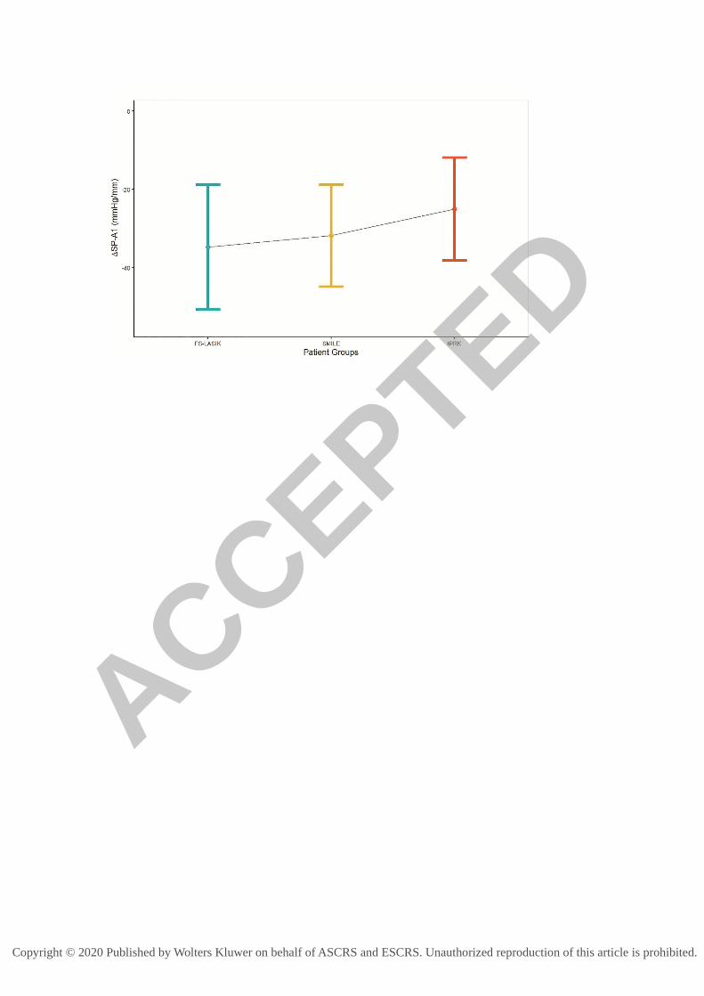
0.262). The SP-A1 reduction was statistically significant in all 3 surgery groups with this
reduction being highest in FS-LASIK, intermediate in SMILE and lowest in TransPRK.
ACCEPTED
Copyright © 20 Published by Wolters Kluwer on behalf of ASCRS and ESCRS. Unauthorized reproduction of this article is prohibited.20

9
Pairwise comparisons showed no difference in SP-A1 reductions between FS-LASIK and
SMILE (p= 0.106) but both sets of reductions were statistically different than TransPRK (p<
0.010). Even though the RSB ratio was lower (higher tissue ablation) in SMILE than in
FS-LASIK (0.57±0.04 vs 0.64±0.03), the reductions in GAT-IOP and IOPcc were higher in
the FS-LASIK group (p= 0.041 and p< 0.001, respectively). This difference between the 2
procedures was not as evident in DCT-IOP and bIOP (p= 0.069 and p= 0.051, respectively).
The distribution in SP-A1 is illustrated in Figure 2, and the relation between RSB ratio and
reduction in IOP is shown in Figure 3.
The Bland-Altman plots (Figure 4) showing the mean differences (∆ = postoperative -
preoperative IOP values) and the 95% limits of agreement (LoA) for each IOP estimate and
for each surgical technique are illustrated in Figure 3. The smallest differences are found in
bIOP, followed closely by DCT. On the other hand, GAT and IOPcc exhibited both the
largest postoperative-preoperative IOP differences and the highest LoA.
Discussion
To correct refractive error and enable the ocular optical system to focus light on the retina,
refractive surgery changes corneal shape by removing part of stromal tissue, leading to
considerable reductions in overall corneal stiffness and possible underestimations in IOP
measurement. Four commonly-used tonometers, the contact Goldmann applanation
tonometer and the Dynamic Contour Tonometer, and the noncontact Ocular Response
Analyzer and the Corvis ST, were assessed in this study to quantify and compare the effects
of refractive surgical procedures on their IOP estimates in a Chinese adult population. The
results demonstrated that the bIOP and the DCT-IOP were less affected than GAT-IOP and
ACCEPTED
Copyright © 20 Published by Wolters Kluwer on behalf of ASCRS and ESCRS. Unauthorized reproduction of this article is prohibited.20

10
IOPcc, and that TransPRK caused smaller reductions in IOP readings by all 4 tonometers
than both FS-LASIK and SMILE. The study also pointed at slightly more effect of
FS-LASIK than SMILE on IOP measurements, particularly in GAT-IOP and IOPcc, even
though the tissue loss in SMILE was larger.
Two recent meta-analyses investigated the biomechanical changes in different surgical
procedures. Guo et al. used the corneal biomechanical assessment provided by the ORA and
found that the reduction in corneal biomechanics was greater with FS-LASIK than SMILE, in
agreement with the present study, and although non-statistically significant,
PRK/laser-assisted subepithelial keratectomy (LASEK) showed less decrease in corneal
biomechanics than SMILE 8. Similarly, Rævdal et al. observed higher reductions in corneal
viscoelastic properties following LASIK compared with SMILE in nonrandomised studies 30.
The stiffness parameter was developed to facilitate the interpretation of the corneal
deformation parameters produced by the Corvis ST. The SP-A1 allows evaluating how
individual parameters respond to the decrease in corneal resistance to deformation 25. It was
observed that the reduction in SP-A1 was smaller in TransPRK than in SMILE and
FS-LASIK. One limitation to this analysis is that even though the patients had been matched
for age, CCT and the cMSE, the amount of tissue removed was not the same among the
procedures and the SMILE cap depth was bigger than the FS-LASIK flap depth. The SMILE
group presented the highest values of depth of tissue removed, which can increase the
reduction in the SP-A1. Further, the significantly less stiffness reduction in TransPRK than in
the other two procedures supports the fact that the IOP readings in this group were the least
influenced by surgery.
ACCEPTED
Copyright © 20 Published by Wolters Kluwer on behalf of ASCRS and ESCRS. Unauthorized reproduction of this article is prohibited.20

11
The absence of statistical difference in SP-A1 between FS-LASIK and SMILE is in
accordance with the similar reductions in bIOP, DCT-IOP and GAT-IOP (p> 0.051). The
IOPcc presented different behaviour. It was the estimate that was the most reduced among the
4 procedures and no statistically significant difference was found between SMILE and
TransPRK, -3.08±1.53 mmHg and -2.77±1.84 mmHg, respectively (p= 0.128). This
behaviour can be partially explained by the fact that the IOPcc was the only measurement
that was significantly lower in the preoperative of the SMILE group compared with LASIK
and TransPRK (p<0.001).
Analysing the relationship between the RSB ratio and the reduction in IOP estimates, it is
observed that in GAT-IOP and IOPcc the reduction was higher in FS-LASIK than in SMILE,
even with SMILE presenting the lowest RSB ratios. This difference was not evident in bIOP
or DCT-IOP. For all 4 estimates, the TransPRK IOP reduction was significantly less than
with the other 2 procedures. These differences suggest that the higher biomechanical impact
caused by FS-LASIK was more prominent in GAT-IOP and IOPcc estimates, while the bIOP
and DCT-IOP estimates were less affected.
Similar reductions in IOP readings in patients undergoing refractive surgery procedures are
reported in the literature. In a large cohort of 174,666 cases, Schallhorn et al. found a
significantly higher reduction in IOP estimate from preoperative to postoperative in LASIK
cases compared to PRK 7. They found that the reduction in IOP using noncontact tonometry
after LASIK was on average 0.94 mmHg higher than after PRK. This result is in accordance
with this study, in which the mean difference between preoperative and postoperative stages
was higher in FS-LASIK than PRK for all 4 tonometers. The highest mean difference was in
GAT-IOP (1.60 mmHg) and the lowest in bIOP (1.03 mmHg). Larger reductions in IOP
ACCEPTED
Copyright © 20 Published by Wolters Kluwer on behalf of ASCRS and ESCRS. Unauthorized reproduction of this article is prohibited.20

12
measurement were also found after SMILE compared with a procedure that was comparable
to PRK (without stromal flap creation, LASEK) as reported by Yu et al 31. At 3 months
postoperatively, the average IOPcc was 1.40 mmHg higher in the LASEK group. Similarities
in IOP reduction after SMILE and LASIK were also found by Li et al 22, where a mean
reduction of approximately 3 mmHg in IOPcc was observed, similar to that found in this
study.
Different attempts to correct GAT-IOP readings after laser refractive surgery have been
discussed in the literature. De Bernardo et al. studied GAT measurements obtained
preoperative and postoperative PRK,12 and observed a similar behaviour to that found in this
study with an average IOP reduction of approximately 2 mmHg and a wide LoA (from -7 to
3mmHg). They compared several correction formulas, and obtained the best result with
Rashad’s method 32, with an average reduction of approximately 1 mmHg and LoA from
-3.25 to 0.99 mmHg. Even though this correction was successful in reducing the difference in
IOP between preoperative and post-TransPRK, the differences were still higher than those
obtained in our study with the relatively newer tonometry methods, bIOP and DCT
(-0.18±1.63 mmHg, p= 0.678 and -0.64±2.34 mmHg, p= 0.262, respectively). In another
related study, Lee et al. found no statistical difference in bIOP before and after TransPRK in
cases with or without accelerated crosslinking (p> 0.101) 33, 34, supporting the expectation
that bIOP, and also DCT, were less affected by CCT and the stiffness reductions induced by
TransPRK. On the other hand, IOPcc presented reductions (-2.77±1.84 mmHg) that were
even higher than those observed with GAT in the TransPRK group, even though the LoA was
narrower with IOPcc.
ACCEPTED
Copyright © 20 Published by Wolters Kluwer on behalf of ASCRS and ESCRS. Unauthorized reproduction of this article is prohibited.20
13
The reductions in IOP readings observed both in FS-LASIK and SMILE, whereas higher than
those observed in TransPRK were still low especially for bIOP (less than 1.5 mmHg) and for
DCT (less than 2.2 mmHg), both statistically significant (p< 0.050). These results are in line
with a study by Fernandez et al. that reported a significant reduction in bIOP by an average of
1.4 mmHg (p< 0.010) after SMILE. They are compatible with the findings by Sales-Sanz et
al. including a reduction in postoperative DCT-IOP of 1.29 mmHg in LASIK cases (p=
0.036) 35, 36. Chen et al. found smaller differences with non-statistically significance
between the preoperative and postoperative reading of bIOP in FS-LASIK and SMILE. 23. A
similar result was also reported by Lee et al. before and after FS-LASIK 34.
One limitation of the study could be a possible IOP mis-estimation due to the consecutive
application of different tonometers 1, 37-39. The interval adopted in the study of at least 3
minutes between consecutive measurements and the random order followed within the
contact and noncontact tonometer groups are expected to have helped reduce any possible
bias. This expected is supported by an observation made by Tejwani et al. that there was no
influence of sequential measurements using GAT, DCT, ORA and Corvis with 5-minute
intervals 40. Another study limitation was the diurnal fluctuations of the true IOP. Despite
efforts to minimise their effects there is still the possibility that the variation observed
between the pre-surgery and post-surgery measurements could have been affected by this
physiological behaviour.
In conclusion, the bIOP and DCT-IOP estimates were the least influenced IOP measurements
by FS-LASIK, SMILE and TransPRK refractive surgeries, while GAT-IOP and IOPcc were
more considerably impacted. The effects were less pronounced in TransPRK, which caused
the smallest reduction in corneal stiffness compared with FS-LASIK and SMILE.
ACCEPTED
Copyright © 20 Published by Wolters Kluwer on behalf of ASCRS and ESCRS. Unauthorized reproduction of this article is prohibited.20

14
WHAT WAS KNOWN
� Intraocular pressure (IOP) measurement will be influenced by corneal stiffness.
� Stiffness change varies from one surgical procedure to another.
� IOP measurement decreases after corneal refractive surgery.
WHAT THIS PAPER ADDS
� Biomechanically-corrected IOP and Dynamic Contour Tonometer IOP were the least
affected by the stiffness change after refractive surgeries.
� Reductions in IOP measurements were different between 3 kinds of corneal refractive
surgeries according to their level of reduction in corneal stiffness, lowest in TransPRK,
then SMILE, and highest in FS-LASIK.
References 1. Faucher A, Gregoire J, Blondeau P. Accuracy of Goldmann tonometry after refractive surgery. J
Cataract Refract Surg 1997;23(6):832-8.
2. Kotecha A, White ET, Shewry JM, Garway-Heath DF. The relative effects of corneal thickness
and age on Goldmann applanation tonometry and dynamic contour tonometry. Br J Ophthalmol
2005;89(12):1572-5.
3. Liu J, Roberts CJ. Influence of corneal biomechanical properties on intraocular pressure
measurement: quantitative analysis. J Cataract Refract Surg 2005;31(1):146-55.
4. Vinciguerra R, Rehman S, Vallabh NA, et al. Corneal biomechanics and biomechanically
corrected intraocular pressure in primary open-angle glaucoma, ocular hypertension and controls. Br
J Ophthalmol 2020;104(1):121-6.
5. Chen KJ, Eliasy A, Vinciguerra R, et al. Development and validation of a new intraocular pressure
estimate for patients with soft corneas. J Cataract Refract Surg 2019;45(9):1316-23.
6. Khamar P, Shetty R, Vaishnav R, et al. Biomechanics of LASIK Flap and SMILE Cap: A Prospective,
Clinical Study. J Refract Surg 2019;35(5):324-32.
7. Schallhorn JM, Schallhorn SC, Ou Y. Factors that influence intraocular pressure changes after
myopic and hyperopic LASIK and photorefractive keratectomy: a large population study.
Ophthalmology 2015;122(3):471-9.
8. Guo H, Hosseini-Moghaddam SM, Hodge W. Corneal biomechanical properties after SMILE
versus FLEX, LASIK, LASEK, or PRK: a systematic review and meta-analysis. BMC Ophthalmol
2019;19(1):167.
9. Stamper RL. A history of intraocular pressure and its measurement. Optom Vis Sci
2011;88(1):E16-28.
10. Kohlhaas M, Spoerl E, Boehm AG, Pollack K. A correction formula for the real intraocular
pressure after LASIK for the correction of myopic astigmatism. J Refract Surg 2006;22(3):263-7.
ACCEPTED
Copyright © 20 Published by Wolters Kluwer on behalf of ASCRS and ESCRS. Unauthorized reproduction of this article is prohibited.20

15
11. Silva TG, Polido JG, Pinheiro MV, et al. [Application of corrective formula for intraocular
pressure changes in patients that underwent LASIK]. Arq Bras Oftalmol 2011;74(2):102-5.
12. De Bernardo M, Capasso L, Caliendo L, et al. Intraocular Pressure Evaluation after Myopic
Refractive Surgery: A Comparison of Methods in 121 Eyes. Semin Ophthalmol 2016;31(3):233-42.
13. Tonnu PA, Ho T, Newson T, et al. The influence of central corneal thickness and age on
intraocular pressure measured by pneumotonometry, non-contact tonometry, the Tono-Pen XL, and
Goldmann applanation tonometry. Br J Ophthalmol 2005;89(7):851-4.
14. Elsheikh A, Alhasso D, Gunvant P, Garway-Heath D. Multiparameter correction equation for
Goldmann applanation tonometry. Optom Vis Sci 2011;88(1):E102-12.
15. Kanngiesser HE, Kniestedt C, Robert YC. Dynamic contour tonometry: presentation of a new
tonometer. J Glaucoma 2005;14(5):344-50.
16. Kaufmann C, Bachmann LM, Thiel MA. Intraocular pressure measurements using dynamic
contour tonometry after laser in situ keratomileusis. Invest Ophthalmol Vis Sci 2003;44(9):3790-4.
17. Mollan SP, Wolffsohn JS, Nessim M, et al. Accuracy of Goldmann, ocular response analyser,
Pascal and TonoPen XL tonometry in keratoconic and normal eyes. Br J Ophthalmol
2008;92(12):1661-5.
18. Doyle A, Lachkar Y. Comparison of dynamic contour tonometry with goldman applanation
tonometry over a wide range of central corneal thickness. J Glaucoma 2005;14(4):288-92.
19. Lee SY, Kim EW, Choi W, et al. Significance of dynamic contour tonometry in evaluation of
progression of glaucoma in patients with a history of laser refractive surgery. Br J Ophthalmol
2019;104(2):276-81.
20. Lanza M, Borrelli M, De Bernardo M, et al. Corneal parameters and difference between
goldmann applanation tonometry and dynamic contour tonometry in normal eyes. J Glaucoma
2008;17(6):460-4.
21. Ozcura F, Yildirim N, Tambova E, Sahin A. Evaluation of Goldmann applanation tonometry,
rebound tonometry and dynamic contour tonometry in keratoconus. J Optom 2017;10(2):117-22.
22. Li H, Wang Y, Dou R, et al. Intraocular Pressure Changes and Relationship With Corneal
Biomechanics After SMILE and FS-LASIK. Invest Ophthalmol Vis Sci 2016;57(10):4180-6.
23. Chen KJ, Joda A, Vinciguerra R, et al. Clinical evaluation of a new correction algorithm for
dynamic Scheimpflug analyzer tonometry before and after laser in situ keratomileusis and
small-incision lenticule extraction. J Cataract Refract Surg 2018;44(5):581-8.
24. Eliasy A, Chen KJ, Vinciguerra R, et al. Determination of Corneal Biomechanical Behavior in-vivo
for Healthy Eyes Using CorVis ST Tonometry: Stress-Strain Index. Front Bioeng Biotechnol
2019;7:105.
25. Roberts CJ, Mahmoud AM, Bons JP, et al. Introduction of Two Novel Stiffness Parameters and
Interpretation of Air Puff-Induced Biomechanical Deformation Parameters With a Dynamic
Scheimpflug Analyzer. J Refract Surg 2017;33(4):266-73.
26. Zhao Y, Shen Y, Yan Z, et al. Relationship Among Corneal Stiffness, Thickness, and Biomechanical
Parameters Measured by Corvis ST, Pentacam and ORA in Keratoconus. Front Physiol 2019;10:740.
27. Theelen T, Meulendijks CF, Geurts DE, et al. Impact factors on intraocular pressure
measurements in healthy subjects. Br J Ophthalmol 2004;88(12):1510-1.
28. Bao F, Huang Z, Huang J, et al. Clinical Evaluation of Methods to Correct Intraocular Pressure
Measurements by the Goldmann Applanation Tonometer, Ocular Response Analyzer, and Corvis ST
Tonometer for the Effects of Corneal Stiffness Parameters. J Glaucoma 2016;25(6):510-9.
29. Altman DG, Bland JM. Measurement in Medicine: The Analysis of Method Comparison Studies.
The Statistician 1983;32:307-17.
30. Raevdal P, Grauslund J, Vestergaard AH. Comparison of corneal biomechanical changes after
refractive surgery by noncontact tonometry: small-incision lenticule extraction versus flap-based
refractive surgery - a systematic review. Acta Ophthalmol 2019;97(2):127-36.
ACCEPTED
Copyright © 20 Published by Wolters Kluwer on behalf of ASCRS and ESCRS. Unauthorized reproduction of this article is prohibited.20

16
31. Yu M, Chen M, Dai J. Comparison of the posterior corneal elevation and biomechanics after
SMILE and LASEK for myopia: a short- and long-term observation. Graefes Arch Clin Exp Ophthalmol
2019;257(3):601-6.
32. Rashad KM, Bahnassy AA. Changes in intraocular pressure after laser in situ keratomileusis. J
Refract Surg 2001;17(4):420-7.
33. Lee H, Roberts CJ, Ambrosio R, Jr., et al. Effect of accelerated corneal crosslinking combined
with transepithelial photorefractive keratectomy on dynamic corneal response parameters and
biomechanically corrected intraocular pressure measured with a dynamic Scheimpflug analyzer in
healthy myopic patients. J Cataract Refract Surg 2017;43(7):937-45.
34. Lee H, Roberts CJ, Kim TI, et al. Changes in biomechanically corrected intraocular pressure and
dynamic corneal response parameters before and after transepithelial photorefractive keratectomy
and femtosecond laser-assisted laser in situ keratomileusis. J Cataract Refract Surg
2017;43(12):1495-503.
35. Fernandez J, Rodriguez-Vallejo M, Martinez J, et al. New parameters for evaluating corneal
biomechanics and intraocular pressure after small-incision lenticule extraction by Scheimpflug-based
dynamic tonometry. J Cataract Refract Surg 2017;43(6):803-11.
36. Sales-Sanz M, Arranz-Marquez E, Pinero DP, et al. Effect of Laser in Situ Keratomileusis on
Schiotz, Goldmann, and Dynamic Contour Tonometric Measurements. J Glaucoma
2016;25(4):e419-23.
37. Stocker FW. On changes in intraocular pressure after application of the tonometer; in the same
eye and in the other eye. Am J Ophthalmol 1958;45(2):192-6.
38. Wilke K. Effects of repeated tonometry: genuine and sham measurements. Acta Ophthalmol
(Copenh) 1972;50(4):574-82.
39. Recep OF, Hasiripi H, Vayisoglu E, et al. Accurate time interval in repeated tonometry. Acta
Ophthalmol Scand 1998;76(5):603-5.
40. Tejwani S, Dinakaran S, Joshi A, et al. A cross-sectional study to compare intraocular pressure
measurement by sequential use of Goldman applanation tonometry, dynamic contour tonometry,
ocular response analyzer, and Corvis ST. Indian J Ophthalmol 2015;63(11):815-20.
Disclosures: None of the authors has a financial or proprietary interest in any material or method
mentioned.
Table Captions:
Table 1 –Baseline characteristics of different surgery groups.
Table 2 –Pre and Postoperative intraocular pressure and stiffness parameter estimates in
corneas undergoing FS-LASIK, SMILE and TransPRK.
ACCEPTED
Copyright © 20 Published by Wolters Kluwer on behalf of ASCRS and ESCRS. Unauthorized reproduction of this article is prohibited.20

17
Figure Captions:
Figure 1 Variation of the 4 IOP estimates (differences postoperative between and
preoperative values) in different surgical groups. *Pairwise comparisons with p> 0.05
(non-statistically significant differences)
Figure 2 Variation (postoperative-preoperative values) in stiffness parameter at first
applanation (SP-A1) in different surgical groups. Error bars represent standard deviation
values. Pairwise comparison revealed no differences between FS-LASIK and SMILE (p=
0.106) but the reduction in both groups were statistically higher than in TransPRK (p<
0.010).
Figure 3 Correlation of RSB ratio with reduction in IOP in different surgical groups. Error bars
represent standard deviation values
Figure 4 Bland-Altman plots for agreement between IOP estimates obtained pre and
postoperative in different surgical groups. The solid lines represent mean differences in IOP
and the dashed lines represent the 95% LoA
ACCEPTED
Copyright © 20 Published by Wolters Kluwer on behalf of ASCRS and ESCRS. Unauthorized reproduction of this article is prohibited.20

Table 1 – Baseline characteristics of different surgery groups.
FS-LASIK (n=50) SMILE (n=50) TransPRK (n=44)
Characteristic Mean SD Min Max Mean SD Min Max Mean SD Min Max P Value
Km (D)* 43.19 1.56 39.1 47.49 43.44 1.50 40.00 47.94 43.51 1.74 35.34 46.47 0.4839
Age (y) 25.22 5.81 17 36 27.16 5.05 18 42 26.73 4.51 17 39 0.1529
CCT (µm) 544.74 18.46 498 572 544.76 19.59 512 591 540.18 24.29 503 618 0.1554
cMSE (D) -5.94 1.78 -10.38 -1.75 -5.62 1.72 -9.38 -1.88 -5.78 2.10 -9.88 -2.25 0.6964
OZD (mm) 6.68 0.36 5.8 7.3 6.65 0.28 6 7.6 6.52 0.43 5.7 7.3 0.0956
AD (µm) 95.98 20.85 38 136 112.98 19.59 63 146 87.82 22.30 48 133 <0.0001
Flap/Cap (µm) 99.60 3.00 95 110 120.00 - 120 120 - - - - <0.0001
RSB (µm) 346.14 21.72 306 394 310.94 23.95 276 381 452.36 36.85 380.00 553.00 <0.0001
RSB/CCT 0.64 0.03 0.57 0.74 0.57 0.04 0.52 0.68 0.84 0.04 0.74 0.91 <0.0001
SP-A1 (mmHg/mm) 96.93 14.49 60.00 121.95 95.53 8.73 73.09 114.95 93.45 14.34 73.00 125.97 0.2835 AD = ablation depth; CCT = central corneal thickness; cMSE = corrected mean spherical equivalent; FS-LASIK = femtosecond laser–assisted
laser in situ keratomileusis; OZD = optical zone diameter; RSB = residual stromal bed; SMILE = small-incision lenticule extraction; SP-A1 =
stiffness parameter at the first applanation; TransPRK = transepithelial photorefractive keratectomy
*Km: mean curvature power in the central 3 mm of the anterior surface;
ACCEPTED
Copyright © 20 Published by Wolters Kluwer on behalf of ASCRS and ESCRS. Unauthorized reproduction of this article is prohibited.20

Table 2. Preoperative and postoperative IOP and stiffness parameter estimates in corneas undergoing FS-LASIK, SMILE, and TransPRK.
FS-LASIK (n=50) SMILE (n=50) TransPRK (n=44)
Tonometer Measurement Mean SD Min Max Mean SD Min max Mean SD Min Max P Value* (comparison
among the groups)
Pre 13.39 2.26 7.5 18.5 13.13 1.88 8.50 17.75 12.90 2.19 8.50 17.50 0.5305
Post 9.95 2.16 5.5 15 10.30 1.93 7.50 16.00 11.12 2.23 7.00 16.00 0.0289
∆ -3.38 2.76 -10 2.75 -2.83 2.08 -6.50 2.50 -1.78 2.29 -5.25 3.00 0.0139 GAT-IOP
P value
(pre vs post) 0.0000 0.0000 0.0003
Pre 15.58 1.94 10.33 19.90 14.28 1.74 10.83 19.03 15.83 2.01 11.50 19.73 0.0002
Post 11.64 1.65 7.93 15.87 11.19 1.74 8.37 17.33 13.11 2.29 8.70 19.87 0.0000
∆ -3.94 1.70 -8.37 0.27 -3.08 1.53 -7.60 1.07 -2.77 1.84 -6.13 2.37 0.0027 IOPcc
P value
(pre vs post) 0.0000 0.0000 0.0000
Pre 17.35 2.26 12.35 21.5 17.26 2.54 13.13 22.83 16.96 2.70 10.65 24.5 0.7301
Post 15.41 1.74 9.25 18.75 15.15 2.31 9.73 21.45 16.31 2.62 11.30 23.17 0.0436
DCT-IOP
∆ -1.87 1.95 -6.3 1.8 -2.11 2.27 -7.77 2.27 -0.64 2.34 -4.90 5.27 0.0035
ACCEPTED
Copyright © 20 Published by Wolters Kluwer on behalf of ASCRS and ESCRS. Unauthorized reproduction of this article is prohibited.20

P value
(pre vs post) 0.0001 0.0000 0.2622
Pre 13.75 1.82 9.33 19.07 13.71 1.21 10.63 16.63 13.71 1.69 10.20 17.23 0.9903
Post 12.53 1.78 8.80 17.27 12.25 1.79 8.57 16.73 13.53 2.23 8.30 19.37 0.0052
∆ -1.21 1.72 -4.67 2.63 -1.46 1.43 -4.62 2.03 -0.18 1.63 -3.30 3.57 0.0005 bIOP
P value
(pre vs post) 0.0013 0.0000 0.6784
Pre 96.93 14.49 60.00 121.95 95.53 8.73 73.09 114.95 93.45 14.34 73.00 125.97 0.2836
Post 62.20 14.29 35.41 113.75 63.71 13.96 30.84 93.73 68.41 19.41 34.51 113.08 0.1519
∆ -34.74 15.97 -63.81 -8.11 -31.82 13.06 -65.95 -5.52 -25.03 13.14 -46.71 15.56 0.0291 SP-A1
P value
(pre vs post) 0.0000 0.0000 0.0000
∆ = difference; bIOP = Corvis ST biomechanically-corrected IOP; DCT-IOP = IOP measured by Dynamic Contour Tonometry; FS-LASIK =
femtosecond laser–assisted laser in situ keratomileusis; GAT-IOP = IOP measured by Goldmann applanation tonometry; IOP = intraocular
pressure; IOPcc = ORA corneal-compensated IOP; post = post-surgery; pre = pre-surgery; SMILE = small-incision lenticule extraction; SP-A1 =
stiffness parameter at first applanation provided by the Corvis ST; TransPRK = transepithelial photorefractive keratectomy
ACCEPTED
Copyright © 20 Published by Wolters Kluwer on behalf of ASCRS and ESCRS. Unauthorized reproduction of this article is prohibited.20

ACCEPTED
Copyright © 20 Published by Wolters Kluwer on behalf of ASCRS and ESCRS. Unauthorized reproduction of this article is prohibited.20

ACCEPTED
Copyright © 20 Published by Wolters Kluwer on behalf of ASCRS and ESCRS. Unauthorized reproduction of this article is prohibited.20
ACCEPTED
Copyright © 20 Published by Wolters Kluwer on behalf of ASCRS and ESCRS. Unauthorized reproduction of this article is prohibited.20

ACCEPTED
Copyright © 20 Published by Wolters Kluwer on behalf of ASCRS and ESCRS. Unauthorized reproduction of this article is prohibited.20

ACCEPTED
Copyright © 20 Published by Wolters Kluwer on behalf of ASCRS and ESCRS. Unauthorized reproduction of this article is prohibited.20