Author's personal copy Orthodontic anomalies and malocclusions in Late Antique and Early Mediaeval...
12
Author's personal copy Orthodontic anomalies and malocclusions in Late Antique and Early Mediaeval period in Croatia Marin Vodanovic ´ a, *, Ivan Galic ´ b , Mihovil Strujic ´ c , Kristina Peros ˇ d , Mario S ˇ laus e , Hrvoje Brkic ´ a a Department of Dental Anthropology, School of Dental Medicine, University of Zagreb, Gundulic ´eva 5, HR-10000 Zagreb, Croatia b Splitsko-Dalmatinska County – Public Health Center, HR-21000 Split, Croatia c Department of Orthodontics, School of Dental Medicine, University of Zagreb, Gundulic ´eva 5, HR-10000 Zagreb, Croatia d Department of Pharmacology, School of Dental Medicine, University of Zagreb, S ˇ alata 11, HR-10000 Zagreb, Croatia e Department of Archaeology, Croatian Academy of Sciences and Arts, Ante Kovac ˇic ´a 5, HR-10000 Zagreb, Croatia 1. Introduction Diagnosis and interpretation of dental diseases and their analysis in paleodemographic framework are an indispens- able part of any attempt to reconstruct past life ways from human skeletal remains. The prevalence and distribution of dental diseases in skeletal series may yield valuable clues regarding diet, nutrition and methods of procuring the diet. 1–4 Considering this, dental caries, antemortem tooth loss, periapical pathology and tooth wear are probably the most examined dental and oral features on skeletal remains of archaeological origin. 5–13 Thanks to numerous studies, pub- lished papers and books, today, teeth and dental diseases, a r c h i v e s o f o r a l b i o l o g y 5 7 ( 2 0 1 2 ) 4 0 1 – 4 1 2 a r t i c l e i n f o Article history: Accepted 15 September 2011 Keywords: Orthodontic anomalies Malocclusions Paleodontology Late Antique Early Mediaeval Croatia a b s t r a c t Objective: Malocclusions are relative infrequently analysed in bioarchaeological investiga- tions and if investigated the samples are very small. This research provides analysis of orthodontic anomalies of even 1118 individuals from the Late Antique (LA) and Early Mediaeval (EM) period. Aims were to describe the prevalence of orthodontic anomalies in this historical period and to analyse which orthodontic anomalies are best suitable for bioarchaeological investigations. Methods: 1118 skulls were examined for anomalies of tooth number, tooth displacement (rotation, malposition, diastema and crowding) as well as for malocclusions. Results: The prevalence of hypodontia in the LA was 41.02% and 30.61% in the EM sample. Tooth displacement was noticed in 15.63% individuals from the LA and in 12.42% individuals from EM. About 26% of the LA sample and 7.19% of the EM sample were affected with tooth crowding and the difference was statistically significant. Conclusion: Orthodontic anomalies affecting only one tooth or group of teeth are more suitable for examination in bioarchaeological investigations than orthodontic features requiring presence of both jaws and all or almost all teeth. Clinical investigation protocols and methodology should be adopted for bioarchaeological researches and international standards and recommendations should be established for this kind of investigation on skeletal remains. # 2011 Elsevier Ltd. All rights reserved. * Corresponding author. Tel.: +385 1 4899 214; fax: +385 1 4802 159. E-mail address: [email protected] (M. Vodanovic ´). URL: http://www.marinvodanovic.com Available online at www.sciencedirect.com journal homepage: http://www.elsevier.com/locate/aob 0003–9969/$ – see front matter # 2011 Elsevier Ltd. All rights reserved. doi:10.1016/j.archoralbio.2011.09.006
Transcript of Author's personal copy Orthodontic anomalies and malocclusions in Late Antique and Early Mediaeval...

Author's personal copy
Orthodontic anomalies and malocclusions in Late Antiqueand Early Mediaeval period in Croatia
Marin Vodanovic a,*, Ivan Galic b, Mihovil Strujic c, Kristina Peros d, Mario Slaus e,Hrvoje Brkic a
aDepartment of Dental Anthropology, School of Dental Medicine, University of Zagreb, Gunduliceva 5, HR-10000 Zagreb, Croatiab Splitsko-Dalmatinska County – Public Health Center, HR-21000 Split, CroatiacDepartment of Orthodontics, School of Dental Medicine, University of Zagreb, Gunduliceva 5, HR-10000 Zagreb, CroatiadDepartment of Pharmacology, School of Dental Medicine, University of Zagreb, Salata 11, HR-10000 Zagreb, CroatiaeDepartment of Archaeology, Croatian Academy of Sciences and Arts, Ante Kovacica 5, HR-10000 Zagreb, Croatia
1. Introduction
Diagnosis and interpretation of dental diseases and their
analysis in paleodemographic framework are an indispens-
able part of any attempt to reconstruct past life ways from
human skeletal remains. The prevalence and distribution of
dental diseases in skeletal series may yield valuable clues
regarding diet, nutrition and methods of procuring the diet.1–4
Considering this, dental caries, antemortem tooth loss,
periapical pathology and tooth wear are probably the most
examined dental and oral features on skeletal remains of
archaeological origin.5–13 Thanks to numerous studies, pub-
lished papers and books, today, teeth and dental diseases,
a r c h i v e s o f o r a l b i o l o g y 5 7 ( 2 0 1 2 ) 4 0 1 – 4 1 2
a r t i c l e i n f o
Article history:
Accepted 15 September 2011
Keywords:
Orthodontic anomalies
Malocclusions
Paleodontology
Late Antique
Early Mediaeval
Croatia
a b s t r a c t
Objective: Malocclusions are relative infrequently analysed in bioarchaeological investiga-
tions and if investigated the samples are very small. This research provides analysis of
orthodontic anomalies of even 1118 individuals from the Late Antique (LA) and Early
Mediaeval (EM) period. Aims were to describe the prevalence of orthodontic anomalies
in this historical period and to analyse which orthodontic anomalies are best suitable for
bioarchaeological investigations.
Methods: 1118 skulls were examined for anomalies of tooth number, tooth displacement
(rotation, malposition, diastema and crowding) as well as for malocclusions.
Results: The prevalence of hypodontia in the LA was 41.02% and 30.61% in the EM sample.
Tooth displacement was noticed in 15.63% individuals from the LA and in 12.42% individuals
from EM. About 26% of the LA sample and 7.19% of the EM sample were affected with tooth
crowding and the difference was statistically significant.
Conclusion: Orthodontic anomalies affecting only one tooth or group of teeth are more
suitable for examination in bioarchaeological investigations than orthodontic features
requiring presence of both jaws and all or almost all teeth. Clinical investigation protocols
and methodology should be adopted for bioarchaeological researches and international
standards and recommendations should be established for this kind of investigation on
skeletal remains.
# 2011 Elsevier Ltd. All rights reserved.
* Corresponding author. Tel.: +385 1 4899 214; fax: +385 1 4802 159.E-mail address: [email protected] (M. Vodanovic).URL: http://www.marinvodanovic.com
Available online at www.sciencedirect.com
journal homepage: http://www.elsevier.com/locate/aob
0003–9969/$ – see front matter # 2011 Elsevier Ltd. All rights reserved.doi:10.1016/j.archoralbio.2011.09.006

Author's personal copy
especially dental caries provide us with an inappreciable
insight in the everyday life of ancient populations from
different historical periods as well as from different parts of
the world.14–18
Compared with dental markers of health (dental caries,
antemortem tooth loss, periapical pathology and tooth wear)
orthodontic anomalies of any kind and malocclusions are
relative infrequently analysed in bioarchaeological investiga-
tions. There are at least three reasons for this. First:
orthodontic anomalies and malocclusions are not recognised
by bioarchaeologists, anthropologists, and related scientists as
an important and useful source of data about the oral and
general health of ancient populations, as well as an important
source of data about other aspects of their everyday life.
Second: scientists involved in the examination of skeletal
remains derived from archaeological contexts usually do not
have enough knowledge for the identification (diagnosis) of
orthodontic anomalies, or for the interpretation of malocclu-
sions. Dentists, as experts educated in the diagnosis and
interpretations of orthodontic status are rarely involved in
bioarchaeological investigations. Third: even when the im-
portance of orthodontic anomalies is recognised, and they are
a focus of the investigation, and even if one of the researchers
is a dentist, there is the final and possibly most important
problem: the lack of usable samples.4
Modern indices of malocclusion consider few features that
differentiate individuals with normal occlusion, from those
with varying degrees of malocclusion: first molar relationship,
overjet (horizontal incisor relation), overbite and open bite
(vertical incisor relation), tooth displacement (crowding,
rotations, etc.), congenitally missing teeth, and posterior
crossbite.19,20 These features can be analysed relatively simply
in living patients, but in skeletal remains where soft tissues
are missing and there is no connection between the upper and
lower jaws the examination of these features is complicated as
the jaws cannot be placed in their correct anatomical position
as when the individual was alive.
Papers and reports about malocclusions and orthodontic
anomalies in skeletal populations from archaeological con-
texts are relatively rare and often based on small samples.
Lukacs described two prehistoric cases of maxillary canine-
first premolar transposition from the Bronze and Iron ages of
South Asia.21 Mockers et al. investigated dental crowding in 43
adult mandibles from the Copper Age.22 Corruccini and
Pacciani examined a sample of 50 Etruscan skulls for tooth
rotation, tooth displacement, first molar transverse crossbite
and horizontal buccal segment relation.23 Evensen and Ogaard
examined 85 male and 61 female skulls to describe the
prevalence and severity of malocclusions in a sample of
mediaeval Norwegians.24 Lindsten et al. measured dental arch
space and permanent tooth size in the mixed dentition of a
skeletal sample (48 skulls of Norwegian descent) from the 14th
to the 19th centuries.25,26 Rose and Roblee studied ancient
Egyptian skeletons from Amarna, Egypt and wrote about the
origins of dental crowding and malocclusions from an
anthropological perspective.27 Studies about malocclusions
in Croatian skeletal populations are extremely rare and so far
only a couple of reports have been published. Cabov et al.
described a case of canine hypodontia in the early Croatian
cemetery Strance-Gorica.28 Rajic et al. reported an impacted
canine in a prehistoric skull from the eneolithic site of Vucedol
in Croatia.29
Traditional history describes the transition from the Late
Antique (3rd–5th centuries AD) to the Early Mediaeval period
(6th–10th centuries AD) in Croatia as uniformly catastrophic
with destruction of major urban centres, depopulation,
famine, and the spread of epidemic diseases. Except historical
and archaeological evidence, this has been confirmed by
bioarchaeological investigations. Slaus et al. examined four
markers of health: cribra orbitalia, linear enamel hypoplasia,
nonspecific periostitis, and trauma in 981 skeletons from the
Late Antique and Early Mediaeval period. They found that the
transition from the Late Antique to the Early Mediaeval period
was not a uniform process, but differentially affected the past
inhabitants of Croatia because of local cultural, socio-
economical, or political factors.30 Dento-alveolar pathologies:
caries, antemortem tooth loss, abscesses, calculus, alveolar
resorption and tooth wear analysed in two composite skeletal
series from Croatia’s eastern Adriatic coast from the same
historical periods confirmed the deterioration of dental health
during the Early Mediaeval period.31
Our aims in this study are to describe the prevalence and
severity of malocclusions and orthodontic anomalies in a large
sample of individuals that inhabited Croatia during the Late
Antique and Early Mediaeval period; to investigate whether
dental health was equally adversely affected by malocclusions
and orthodontic anomalies during the Late Antique/Early
Mediaeval transition; and to analyse and determine which
orthodontic anomalies are best suited for bioarchaeological
investigations.
2. Materials and methods
This investigation was carried out on skeletal series that are
currated in the Osteological collection of the Department of
Archaeology, of the Croatian Academy of Sciences and Arts in
Zagreb. The examined sample consists of 1118 skulls. The
osteological material analysed in this study was divided into
two composite series – a Late Antique and an Early Mediaeval
skeletal series. The Late Antique series consists of a grand total
of 457 skeletons from 5 urban sites (Zmajevac, Strbinci, Osijek,
Vinkovci, and Zadar) located in the eastern part of continental
Croatia and along the eastern Adriatic coast. The sites were
inhabited from the 1st to 5th centuries AD. The Early
Mediaeval series consists of a grand total of 661 skeletons
from 6 rural sites (Privlaka, Stari Jankovci, Velim Velistak,
Glavice, Radasinovci, and Sibenik) also located in the eastern
part of continental Croatia, and along the eastern Adriatic
coast. These sites were inhabited from the 7th to 10th
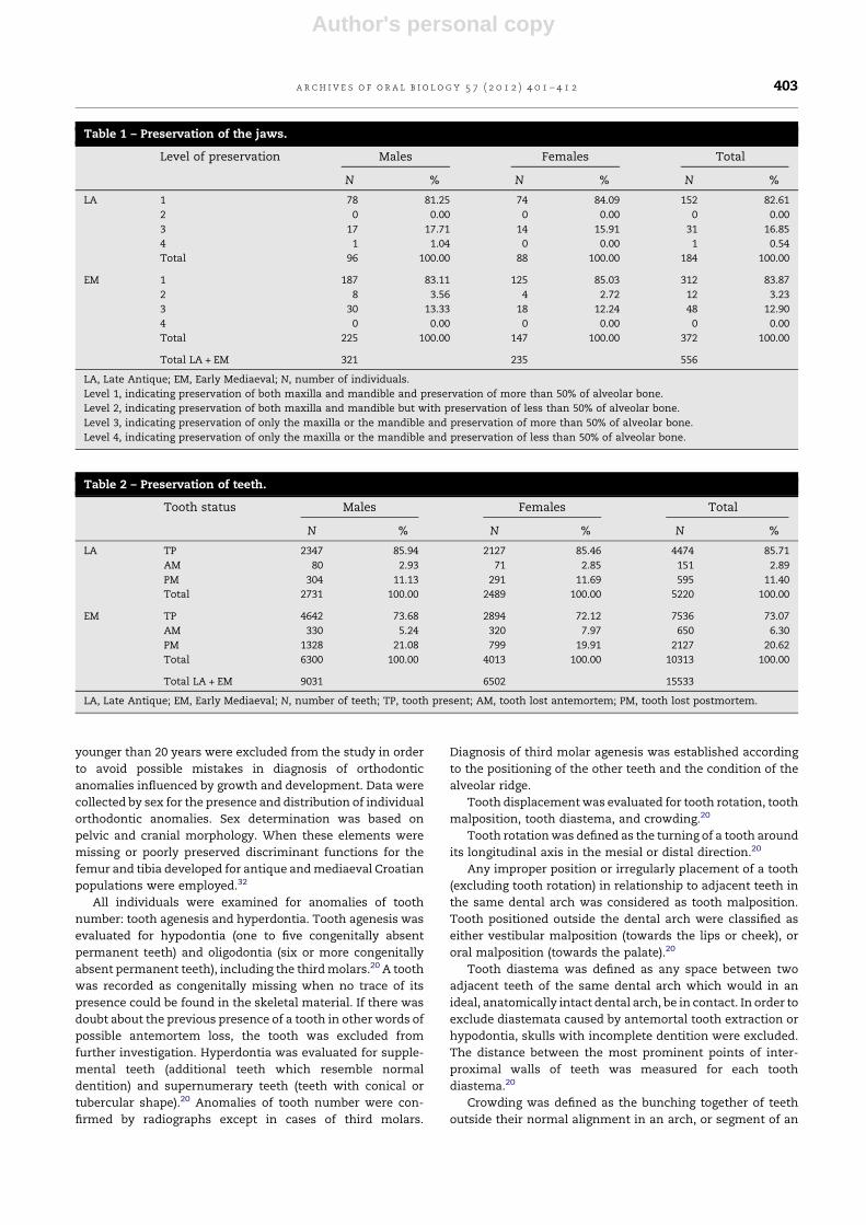
centuries AD. Because of differential preservation, the final
number of skulls included in the investigation is significantly
smaller and varies from 36 to 362 depending on the examined
feature. The state of preservation varied from completely
preserved skulls with complete mandibles, to cases where
only small fragments of the maxilla or mandible with only few
teeth were preserved (Tables 1 and 2). Adult age at death was
estimated using as many methods as possible, including pubic
symphysis morphology, auricular surface morphology, sternal
rib end changes, and ectocranial suture fusion. Individuals
a r c h i v e s o f o r a l b i o l o g y 5 7 ( 2 0 1 2 ) 4 0 1 – 4 1 2402
Author's personal copy
younger than 20 years were excluded from the study in order
to avoid possible mistakes in diagnosis of orthodontic
anomalies influenced by growth and development. Data were
collected by sex for the presence and distribution of individual
orthodontic anomalies. Sex determination was based on
pelvic and cranial morphology. When these elements were
missing or poorly preserved discriminant functions for the
femur and tibia developed for antique and mediaeval Croatian
populations were employed.32
All individuals were examined for anomalies of tooth
number: tooth agenesis and hyperdontia. Tooth agenesis was
evaluated for hypodontia (one to five congenitally absent
permanent teeth) and oligodontia (six or more congenitally
absent permanent teeth), including the third molars.20 A tooth
was recorded as congenitally missing when no trace of its
presence could be found in the skeletal material. If there was
doubt about the previous presence of a tooth in other words of
possible antemortem loss, the tooth was excluded from
further investigation. Hyperdontia was evaluated for supple-
mental teeth (additional teeth which resemble normal
dentition) and supernumerary teeth (teeth with conical or
tubercular shape).20 Anomalies of tooth number were con-
firmed by radiographs except in cases of third molars.
Diagnosis of third molar agenesis was established according
to the positioning of the other teeth and the condition of the
alveolar ridge.
Tooth displacement was evaluated for tooth rotation, tooth
malposition, tooth diastema, and crowding.20
Tooth rotation was defined as the turning of a tooth around
its longitudinal axis in the mesial or distal direction.20
Any improper position or irregularly placement of a tooth
(excluding tooth rotation) in relationship to adjacent teeth in
the same dental arch was considered as tooth malposition.
Tooth positioned outside the dental arch were classified as
either vestibular malposition (towards the lips or cheek), or
oral malposition (towards the palate).20
Tooth diastema was defined as any space between two
adjacent teeth of the same dental arch which would in an
ideal, anatomically intact dental arch, be in contact. In order to
exclude diastemata caused by antemortal tooth extraction or
hypodontia, skulls with incomplete dentition were excluded.
The distance between the most prominent points of inter-
proximal walls of teeth was measured for each tooth
diastema.20
Crowding was defined as the bunching together of teeth
outside their normal alignment in an arch, or segment of an
Table 1 – Preservation of the jaws.
Level of preservation Males Females Total
N % N % N %
LA 1 78 81.25 74 84.09 152 82.61
2 0 0.00 0 0.00 0 0.00
3 17 17.71 14 15.91 31 16.85
4 1 1.04 0 0.00 1 0.54
Total 96 100.00 88 100.00 184 100.00
EM 1 187 83.11 125 85.03 312 83.87
2 8 3.56 4 2.72 12 3.23
3 30 13.33 18 12.24 48 12.90
4 0 0.00 0 0.00 0 0.00
Total 225 100.00 147 100.00 372 100.00
Total LA + EM 321 235 556
LA, Late Antique; EM, Early Mediaeval; N, number of individuals.
Level 1, indicating preservation of both maxilla and mandible and preservation of more than 50% of alveolar bone.
Level 2, indicating preservation of both maxilla and mandible but with preservation of less than 50% of alveolar bone.
Level 3, indicating preservation of only the maxilla or the mandible and preservation of more than 50% of alveolar bone.
Level 4, indicating preservation of only the maxilla or the mandible and preservation of less than 50% of alveolar bone.
Table 2 – Preservation of teeth.
Tooth status Males Females Total
N % N % N %
LA TP 2347 85.94 2127 85.46 4474 85.71
AM 80 2.93 71 2.85 151 2.89
PM 304 11.13 291 11.69 595 11.40
Total 2731 100.00 2489 100.00 5220 100.00
EM TP 4642 73.68 2894 72.12 7536 73.07
AM 330 5.24 320 7.97 650 6.30
PM 1328 21.08 799 19.91 2127 20.62
Total 6300 100.00 4013 100.00 10313 100.00
Total LA + EM 9031 6502 15533
LA, Late Antique; EM, Early Mediaeval; N, number of teeth; TP, tooth present; AM, tooth lost antemortem; PM, tooth lost postmortem.
a r c h i v e s o f o r a l b i o l o g y 5 7 ( 2 0 1 2 ) 4 0 1 – 4 1 2 403

Author's personal copy
arch, causing them to be irregular or crooked. Crowding was
qualitatively assessed by the number of individuals with
malpositioned teeth and the severity of their displacement.
According to Iscan three grades of crowding were recognised:
slight, moderate and severe.1
Qualitative evaluation of malocclusion was performed by
Angle’s classification – one of the most widely used malocclu-
sion classification system. Considering the limitations of
working on skeletal remains all available samples were
classified in one of four groups: Class I, Class II division 1,
Class II division 2, and Class III according to Angle’s
classification.4,20
Malocclusions in the anterior region of the dental arches in
the occlusal plane were recorded as: regular sagittal relation-
ship, anterior crossbite and strong horizontal overlap (over-
jet).4,20 Overjet was evaluated to the nearest 0.5 mm and
normal range was determined at 0.5–4.0 mm. Anterior cross-
bite included mandibular overbite, and edge-to-edge position.
Malocclusions in the anterior region of the dental arches in
the vertical plane were recorded as: deep bite (overbite),
regular vertical relationship and anterior openbite.4,20
Transverse occlusal deviations of the posterior teeth were
classified as: regular transverse occlusion, edge-to-edge bite
(posterior crossbite tendency), posterior crossbite, buccal non-
occlusion and lingual non-occlusion.4,20
Chi-square test was used to test differences in the
frequencies of malocclusions and orthodontic anomalies
between Late Antique and Early Mediaeval samples and to
test the sex differences for those anomalies.
Because of the evident impact of tooth wear on teeth
position and possible influence on the correctness of results
obtained in this study for each examined individual average
tooth wear score was calculated using the Smith and Knight
tooth wear index.33,34
3. Results
A total of 362 individuals were examined for anomalies of
teeth number. In the group of individuals with anomalies of
teeth number, only individuals with hypodontia were found.
No cases of hyperdontia or oligodontia were recorded.
Although both jaws, and all teeth/tooth sockets were exam-
ined congenitally missing teeth were noted only in the upper
second incisors, upper third molars, lower second premolars,
and lower third molars. Of the 117 individuals examined from
the Late Antique sample, 48 individuals (41.02%) were found to
have at least one absent permanent tooth. As shown in Table 3
the most often missing teeth were the lower third molars,
followed by the upper third molars. There were 4 cases of
missing upper second incisor, and only one case of missing
lower second premolar. In two cases the absence of the second
incisor was unilateral, in one case it was bilateral. Of the 245
individuals examined from the Early Mediaeval sample, 75
individuals (30.61%) were found to have at least one absent
permanent tooth. As shown in Table 3 the only missing teeth
were the third molars and the lower second premolar.
Although there was no statistically significant difference
between the Late Antique and Early Mediaeval sample when
calculated by the number of individuals with anomalies of
teeth number, statistically significant differences were found
between the frequencies of hypodontia for some tooth types
(Table 3). Hypodontia of the upper second incisor is signifi-
cantly higher in the Late Antique sample than in the Early
Mediaeval sample ( p = 0.01, x2 = 6.69) and the same is true for
hypodontia of the lower third molars ( p = 0.01, x2 = 8.49). In
addition, Late Antique males have significantly more cases of
hypodontia of upper third molars than Early Mediaeval males
( p = 0.01, x2 = 12.51).
A total of 225 individuals were examined for tooth
displacement. The results according to tooth rotation (in
either mesial or distal direction), tooth malposition (vestibular
or oral position) are presented in Tables 4 and 5. There are no
significant differences between the Late Antique (15.63%) and
Early Mediaeval sample (12.42%) according to the number of
individuals with these types of tooth displacement. The most
common type of tooth displacement was tooth rotation. In
both samples rotation of teeth in the distal direction was more
common than in the mesial direction. In both samples,
premolars were most often affected by tooth rotation,
Table 3 – Frequency of hypodontia.
N UI2 UM3 LP2 LM3
NS NH % NS NH % NS NH % NS NH %
LA Females 59 35 4 11.43a 53 6 11.32 46 0 0.00 49 24 48.98
Males 58 25 0 0.00 34 21 61.76c 28 1 3.57 49 33 67.35d
Total 117 60 4 6.67b 87 27 31.03 74 1 1.35 98 57 58.16e
EM Females 101 71 0 0.00a 114 23 20.18 96 2 2.08 119 40 33.61
Males 144 84 0 0.00 166 30 18.07c 114 0 0.00 182 55 30.22d
Total 245 155 0 0.00b 280 53 18.92 210 2 0.95 301 95 31.56e
Total LA + EM 362 215 4 1.86 367 80 21.80 284 3 1.06 399 152 38.10
LA, Late Antique; EM, Early Mediaeval; UI2, upper second incisor; UM3, upper third molar; LP2, lower second premolar; LM3, lower third molar;
N, number of individuals; NS, number of tooth sockets examined; NH, number of teeth missing.a p = 0.05, x2 = 4.91.b p = 0.01, x2 = 6.69.c p = 0.01, x2 = 12.51.d p = 0.01, x2 = 8.02.e p = 0.01, x2 = 8.49.
a r c h i v e s o f o r a l b i o l o g y 5 7 ( 2 0 1 2 ) 4 0 1 – 4 1 2404

Author's personal copy
followed by canines, and incisors. Vestibular malposition of
teeth was more common than palatinal. According to tooth
type, canines were most often in the vestibular malposition.
A total of 212 individuals were examined for tooth
diastema. The frequency of individuals with tooth diastema
was almost identical in both samples, slightly less than 5%
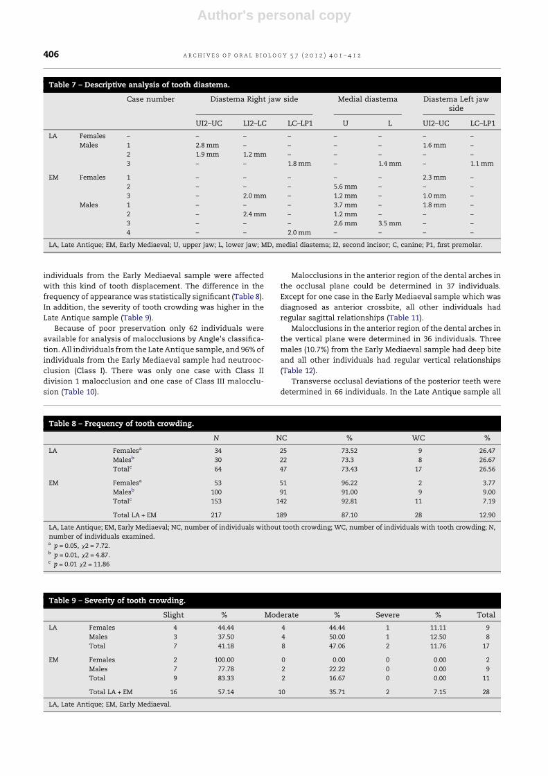
(Table 6). Descriptive analysis of tooth diastema is shown in
Table 7. Although only 10 individuals had diastemata (3 in the
Late Antique sample and 7 in the Early Mediaeval sample),
there were 19 cases of diastema. The most frequent were
diastemata between the upper second incisors and the upper
canines (31.6% of all diastemata), followed by upper medial
diastema (26.3% of all diastemata). The average distance
between upper central incisors was 3.4 mm for females, and
2.5 mm for males in the Early Mediaeval samples where all
cases of medial diastema were found.
A total of 217 individuals were examined for tooth
crowding. Tooth crowding appeared more often in the Late
Antique sample than in the Early Mediaeval sample. About
26% individuals from the Late Antique sample, and only 7.19%
Table 4 – Frequency of tooth displacements.
NTP PM VP TR PM + VM + -TR
Total
N % N % N % N % N % N
LA Females 27 79.41 1 2.94 2 5.88 4 11.76 7 20.59 34
Males 27 90.00 0 0.00 0 0.00 3 10.00 3 10.00 30
Total 54 84.38 1 1.56 2 3.13 7 10.94 10 15.63 64
EM Females 52 91.23 0 0.00 2 3.51 3 5.26 5 8.77 57
Males 89 85.58 0 0.00 3 2.88 12 11.54 15 14.42 104
Total 141 87.58 0 0.00 5 3.11 15 9.32 20 12.42 161
Total LA + EM 195 86.67 1 0.44 7 3.11 22 9.78 30 13.33 225
LA, Late Antique; EM, Early Mediaeval; N, number of individuals; NTP, normal tooth position; PM, palatinal malposition; VM, vestibular
malposition; TR, tooth rotation.
Table 6 – Frequency of tooth diastema.
N ND % WD %
LA Females 31 31 0.00 0 0.00
Males 30 27 90.00 3 10.00
Total 61 58 95.08 3 4.92
EM Females 52 49 94.23 3 5.77
Males 99 95 95.96 4 4.04
Total 151 144 95.36 7 4.64
Total LA + EM 212 202 95.28 10 4.72
LA, Late Antique; EM, Early Mediaeval; N, number of individuals examined; ND, number of individuals without tooth diastema; WD, number of
individuals with tooth diastema.
Table 5 – Distribution of tooth displacements according to the tooth type.
TRD TRM VM PM Total
LA
Females N 6 – 5 1 12
Teeth UI2 (2), UP1 (1), LC (1), LP1 (1), LP2(1) – UI2 (2), LC (3) UC (1)
Males N 1 2 – – 3
Teeth LP1 (1) UP2 (1), LC (3) – –
Total 7 2 5 1 15
EM
Females N 2 1 1 – 4
Teeth LP2 (2) UI2 (1) LC (1) –
Males N 12 3 3 1 19
Teeth UI2 (1), UP1 (3), UP2 (3), UM1 (1), LC (1), LP1 (1), LP2 (2) UP1 (2), LC (1) UC (1), UM3 (1), LM1 (1) UC (1)
Total 14 4 4 1 23
LA, Late Antique; EM, Early Mediaeval; N, number of teeth; TRD, distal tooth rotation; TRM, mesial tooth rotation; PM, palatinal malposition;
VM, vestibular malposition; U, upper jaw; L, lower jaw; I, incisor; C, canine; P, premolar; M, molar; number in parenthesis indicates the number
of affected teeth.
a r c h i v e s o f o r a l b i o l o g y 5 7 ( 2 0 1 2 ) 4 0 1 – 4 1 2 405
Author's personal copy
individuals from the Early Mediaeval sample were affected
with this kind of tooth displacement. The difference in the
frequency of appearance was statistically significant (Table 8).
In addition, the severity of tooth crowding was higher in the
Late Antique sample (Table 9).
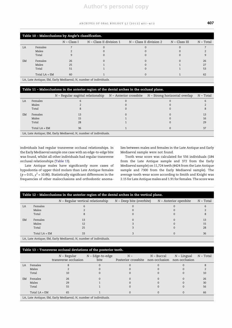
Because of poor preservation only 62 individuals were
available for analysis of malocclusions by Angle’s classifica-
tion. All individuals from the Late Antique sample, and 96% of
individuals from the Early Mediaeval sample had neutrooc-
clusion (Class I). There was only one case with Class II
division 1 malocclusion and one case of Class III malocclu-
sion (Table 10).
Malocclusions in the anterior region of the dental arches in
the occlusal plane could be determined in 37 individuals.
Except for one case in the Early Mediaeval sample which was
diagnosed as anterior crossbite, all other individuals had
regular sagittal relationships (Table 11).
Malocclusions in the anterior region of the dental arches in
the vertical plane were determined in 36 individuals. Three
males (10.7%) from the Early Mediaeval sample had deep bite
and all other individuals had regular vertical relationships
(Table 12).
Transverse occlusal deviations of the posterior teeth were
determined in 66 individuals. In the Late Antique sample all
Table 7 – Descriptive analysis of tooth diastema.
Case number Diastema Right jaw side Medial diastema Diastema Left jawside
UI2–UC LI2–LC LC–LP1 U L UI2–UC LC–LP1
LA Females – – – – – – – –
Males 1 2.8 mm – – – – 1.6 mm –
2 1.9 mm 1.2 mm – – – – –
3 – – 1.8 mm – 1.4 mm – 1.1 mm
EM Females 1 – – – – – 2.3 mm –
2 – – – 5.6 mm – – –
3 – 2.0 mm – 1.2 mm – 1.0 mm –
Males 1 – – – 3.7 mm – 1.8 mm –
2 – 2.4 mm – 1.2 mm – – –
3 – – – 2.6 mm 3.5 mm – –
4 – – 2.0 mm – – – –
LA, Late Antique; EM, Early Mediaeval; U, upper jaw; L, lower jaw; MD, medial diastema; I2, second incisor; C, canine; P1, first premolar.
Table 8 – Frequency of tooth crowding.
N NC % WC %
LA Femalesa 34 25 73.52 9 26.47
Malesb 30 22 73.3 8 26.67
Totalc 64 47 73.43 17 26.56
EM Femalesa 53 51 96.22 2 3.77
Malesb 100 91 91.00 9 9.00
Totalc 153 142 92.81 11 7.19
Total LA + EM 217 189 87.10 28 12.90
LA, Late Antique; EM, Early Mediaeval; NC, number of individuals without tooth crowding; WC, number of individuals with tooth crowding; N,
number of individuals examined.a p = 0.05, x2 = 7.72.b p = 0.01, x2 = 4.87.c p = 0.01 x2 = 11.86
Table 9 – Severity of tooth crowding.
Slight % Moderate % Severe % Total
LA Females 4 44.44 4 44.44 1 11.11 9
Males 3 37.50 4 50.00 1 12.50 8
Total 7 41.18 8 47.06 2 11.76 17
EM Females 2 100.00 0 0.00 0 0.00 2
Males 7 77.78 2 22.22 0 0.00 9
Total 9 83.33 2 16.67 0 0.00 11
Total LA + EM 16 57.14 10 35.71 2 7.15 28
LA, Late Antique; EM, Early Mediaeval.
a r c h i v e s o f o r a l b i o l o g y 5 7 ( 2 0 1 2 ) 4 0 1 – 4 1 2406
Author's personal copy
individuals had regular transverse occlusal relationships. In
the Early Mediaeval sample one case with an edge-to-edge bite
was found, whilst all other individuals had regular transverse
occlusal relationships (Table 13).
Late Antique males have significantly more cases of
hypodontia of upper third molars than Late Antique females
( p = 0.01, x2 = 10.86). Statistically significant differences in the
frequencies of other malocclusions and orthodontic anoma-
lies between males and females in the Late Antique and Early
Mediaeval sample were not found.
Tooth wear score was calculated for 556 individuals (184
from the Late Antique sample and 372 from the Early
Mediaeval sample) on 11,724 teeth (4424 from the Late Antique
sample and 7300 from the Early Mediaeval sample). The
average tooth wear score according to Smith and Knight was
2.15 for Late Antique males and 1.91 for females. The score was
Table 10 – Malocclusions by Angle’s classification.
N – Class I N – Class II division 1 N – Class II division 2 N – Class III N – Total
LA Females 7 0 0 0 7
Males 2 0 0 0 2
Total 9 0 0 0 9
EM Females 26 0 0 0 26
Males 25 1 0 1 27
Total 51 1 0 1 53
Total LA + EM 60 1 0 1 62
LA, Late Antique; EM, Early Mediaeval; N, number of individuals.
Table 11 – Malocclusions in the anterior region of the dental arches in the occlusal plane.
N – Regular sagittal relationship N – Anterior crossbite N – Strong horizontal overlap N – Total
LA Females 6 0 0 6
Males 2 0 0 2
Total 8 0 0 8
EM Females 13 0 0 13
Males 15 1 0 16
Total 28 1 0 29
Total LA + EM 36 1 0 37
LA, Late Antique; EM, Early Mediaeval; N, number of individuals.
Table 12 – Malocclusions in the anterior region of the dental arches in the vertical plane.
N – Regular vertical relationship N – Deep bite (overbite) N – Anterior openbite N – Total
LA Females 6 0 0 6
Males 2 0 0 2
Total 8 0 0 8
EM Females 13 0 0 13
Males 12 3 0 15
Total 25 3 0 28
Total LA + EM 33 3 0 36
LA, Late Antique; EM, Early Mediaeval; N, number of individuals.
Table 13 – Transverse occlusal deviations of the posterior teeth.
N – Regulartransverse occlusion
N – Edge-to-edgebite
N –Posterior crossbite
N – Buccalnon-occlusion
N – Lingualnon-occlusion
N – Total
LA Females 8 0 0 0 0 8
Males 2 0 0 0 0 2
Total 10 0 0 0 0 10
EM Females 26 0 0 0 0 26
Males 29 1 0 0 0 30
Total 55 1 0 0 0 56
Total LA + EM 65 1 0 0 0 66
LA, Late Antique; EM, Early Mediaeval; N, number of individuals.
a r c h i v e s o f o r a l b i o l o g y 5 7 ( 2 0 1 2 ) 4 0 1 – 4 1 2 407

Author's personal copy
2.08 for Early Mediaeval males and 1.98 for females. There
were no statistically significant differences between the
samples (Table 14).
4. Discussion
In this research 1118 skulls from the Late Antique and Early
Mediaeval period were examined for malocclusions and
orthodontic anomalies. According to available literature, most
anthropologic studies of malocclusion have been based on
relatively small samples so this analysis probably presents one
of the most comprehensive bioarchaeological investigations
focusing on malocclusions and orthodontic anomalies. This
sample size provided an opportunity to get reliable data about
factors that limit research of malocclusions and orthodontic
anomalies in skeletal remains derived from archaeological
contexts. As, however, previously noted – whilst the total
sample was sizable the number of individuals available for
examination was significantly decreased for some features,
resulting in a data base that contained from 36 to 362
individuals. Features requiring the presence of both jaws
and all, or almost teeth were most difficult for examination.
Heavy damage and fragmentation of the skulls and jaws,
skulls without accompanying mandibles and a high preva-
lence of postmortem lost teeth (particularly one-rooted teeth
in the frontal region) are the main reason why malocclusions
in the anterior region of the dental arches in the vertical or
occlusal plane were analysed only in 3.2% individuals of the
total sample; why the analysis of malocclusions by Angle’s
classification was possible only on 5.5% individuals, and why
the transverse occlusal deviations of the posterior teeth was
performed only on 5.9% individuals. Orthodontic anomalies
affecting single tooth like hypodontia, hyperdontia, tooth
rotation or tooth malposition or group of teeth (frontal teeth)
like diastema or tooth crowding were analysed in significantly
more individuals than features requiring the presence of both
jaws, and all or almost all teeth, varying from about 19%
individuals (diastema and crowding) up to about 32%
individuals (anomalies of teeth number). In archaeologically
derived skeletal series it has generally been easier to describe
the alignment of teeth than occlusal relationships because the
mandible frequently becomes separated from the rest of the
skull in archaeological skeletal remains.24
Malocclusion can occur as a result of: genetically deter-
mined factors, environmental factors, or more commonly a
combination of both.20 A number of primarily environmental
causes are known including habits such as digit-sucking and
nail biting, trauma, caries, periodontal disease, chronic nasal
obstruction with mouth breathing, and reduced masticatory
stress resulting from the soft consistency of foods in urbanised
societies. Other known causes of malocclusion include clefts
of the alveolus and palate that occur during foetal growth,
genetic syndromes that affect the development of craniofacial
structures, and supernumerary and congenitally absent teeth
that predominantly result from heritable factors.19 Analysis of
malocclusion and orthodontic anomalies and their aetiology
can provide a unique insight into the everyday life, diet,
nutrition and methods of procuring the diet in archaeological
populations.
Hypodontia is defined as a condition in which a person has
missing teeth as a result of their failure to develop. Congenital
hypodontia arises because of an abnormality in the induction
of oral ectoderm by the ectomesenchyme. The congenital
absence of some teeth is common with the most commonly
absent teeth being the last teeth in each series (i.e. the lateral
incisor, second premolar and third molar).35 In this study, the
frequency of individuals with at least one congenitally missing
tooth varied from 30.61% in the Early Mediaeval sample to
41.02% in the Late Antique sample. Except for third molars
(upper and lower) that were the most frequently missing teeth
in both samples (15.64% of all examined tooth sockets in the
Early Mediaeval sample, and 30.21% of all examined tooth
sockets in the Late Antique sample), four cases of missing
upper second incisors (1.43%) were recorded in females from
the Late Antique sample, and three cases of missing lower
second premolar (1 case or 0.36%. of all examined tooth
sockets in the Late Antique sample and 2 cases or 0.21% of all
examined tooth sockets in the Early Mediaeval sample).
The incidence of hypodontia in the permanent dentition in
modern samples varies from 3.5 to 6.5% (excluding wisdom
teeth, 9–37%), with a 1:1.4 male/female ratio.35 There are only a
few studies that published comparable data about hypodontia
in archaeological populations. Nelsen et al. examined a
skeletal sample from an Iron Age cemetery in Thailand and
found a very high prevalence of 79% in adults (30/38) with at
least one incisor missing in either maxilla or mandible.36
Evensen and Ogaard examined the prevalence and severity of
malocclusions in a sample of mediaeval Norwegians using the
Norwegian need for orthodontic treatment index (NOTI).24
Although this index includes the prevalence of hypodontia,
separately presented data for the mediaeval population are
not available. Cabov et al. examined 27 persons and a total
number of 412 teeth and established a lower and an upper
hypodontia incidence of 3.7% for the Croatian population in
the Vinodol region in the period from the 9th to 11th century.28
Their research included third molars. In an investigation of the
prevalence of second incisor hypodontia in contemporary
Table 14 – Tooth wear score according to the Smith and Knight tooth wear index.
Males Females Total
N NT AWTS N NT AWTS N NT AWTS
LA 96 2315 2.15 88 2109 1.91 184 4424 2.03
EM 225 4519 2.08 147 2781 1.98 372 7300 2.04
Total LA + EM 321 6834 235 4890 556 11724
LA, Late Antique; EM, Early Mediaeval; N, number of individuals; NT, number of teeth; ATWS, average tooth wear score according to the Smith
and Knight tooth wear index.
a r c h i v e s o f o r a l b i o l o g y 5 7 ( 2 0 1 2 ) 4 0 1 – 4 1 2408

Author's personal copy
Croatian inhabitants, Prskalo et al. found a prevalence of
2.46%.37 In this study the prevalence of hypodontia expressed
by teeth (all teeth including third molars) varied from 15.86%
in the Early Mediaeval sample to 32.01% in the Late Antique
sample.
Regarding malpositions of single teeth, in this study
rotation, displacement, diastema and crowding were evaluat-
ed. Although it seems that dental malpositions are mostly
caused by dysgnathia and exogenous factors, several observa-
tions give reasons to believe that, at least to some degree,
hereditary factors also play a role.4 Tooth rotation as the
movement of a tooth around its long axis in the mesial or
distal direction was found in 10.94% of individuals in the Late
Antique sample and in 9.32% of individuals of the Early
Mediaeval sample. Corruccini and Pacciani examined a
sample of 50 Etruscan skulls for tooth rotation, and found
6.8% individuals with rotated teeth.23 Vestibular and palatinal
tooth malposition was registered in 5.26% of individuals in the
Late Antique sample, and in 3.42% of individuals in the Early
Mediaeval sample. Canine displacement is the most frequent
case of tooth displacement and is generally classified into
buccal or palatal displacement. More rarely, canines can be
found lying horizontally above the apices of the teeth of the
upper arch or displaced high adjacent to the nose.20 In this
research canines were the most affected teeth by tooth
displacement: 63.64% of all displaced teeth were canines. The
absence of radiological confirmation of some examined
features, like third molar agenesis can be considered a
limitation of the reliability of the data provided by this study.
If non-invasive techniques are used in the examination of
skeletal remains, like in this study, tooth agenesis and tooth
displacement (including tooth impaction) can be only con-
firmed by radiographic analysis. Rajic et al. described a case of
an impacted canine in a prehistoric female skull from Croatian
enolithic site of Vucedol.29 Their finding was confirmed by
roentgencephalometric analysis. Although Lukacs recom-
mended epidemiological analysis of large skeletal collections
from different ecological and cultural contexts in order to yield
important clues to the aetiology of dental transposition,
expecting that a large sample size can provide more accurate
data about dental transposition prevalence,21 transposition of
teeth as a change of position between two teeth in a dental
arch was not registered in this study.
Criteria for diagnosis of tooth diastema were well defined
(as described in Section 2) and strictly followed in this study.
All cases where there was any doubt that the diastema was
caused by an environmental factor (i.e. tooth extraction)
were excluded from the study. In spite of this, 19 cases of
diastema in 10/212 individuals available for examination
were recorded. The most frequent were diastemata between
upper second incisors and upper canines (the upper
‘‘primate space’’), 31.6% of all diastemata, followed by
upper medial diastema (26.3% of all diastemata). The lower
‘‘primate space’’, the space that occurs between canine and
first premolar in lower jaw was recorded three times in two
individuals (15.8% of all diastemata) (Table 7). The preva-
lence of individuals with tooth diastema is approximately
5%. The prevalence of individuals with maxillary medial
diastema is 2.4%. Lavelle studied the prevalence of maxil-
lary medial diastema in 18–25 year old UK residents
belonging to different races. He found that 3.4% of
Caucasians, 5.5% of Negroids and 1.7% of Mongoloids were
affected by this condition.38
The crowding of permanent teeth is considered to be an
indicator for disproportion between the sizes of the jaw and
the teeth. The most common location of crowding is in the
frontal region of the dental arch.4 Crowding is extremely
common in Caucasians, affecting approximately two-thirds of
the modern population. Whilst the size of the jaws and teeth is
mainly genetically determined, environmental factors, such
as premature deciduous tooth loss, can precipitate or
exacerbate crowding. In evolutionary terms both jaw size
and tooth size appear to be reducing. However, crowding is
much more prevalent in modern populations than in
prehistoric skeletal series. It has been suggested that this is
due to the introduction of a less abrasive diet, so that less
interproximal tooth wear occurs during the lifetime of an
individual.20 In this study the frequency of tooth crowding was
about 26% of all individuals in the Late Antique sample, and
7.19% of all individuals in the Early Mediaeval sample. Mockers
et al. examined dental crowding in a prehistoric population
from the Copper Age in France. The prevalence of crowding
was 100% in this prehistoric population.22 In an epidemiologi-
cal study of malocclusions in the modern population from
island of Hvar, Croatia, Lauc found that the frequency of
crowding was 57.1%.39 Compared with our results this
presents a significant temporal increase in the frequency of
dental crowding. The increased prevalence of dental crowding
is generally believed to have been caused by changes in diet
(introducing a less abrasive diet) and lifestyle, but Rose and
Roblee go a step further to find the underlying problem. They
consider the problem of adaptation of the alveolar bone to the
changes in diet and alveolar bone discrepancies as a leading
cause of dental crowding and malocclusion.27
The importance and comparability of the results of the
analyses of: malocclusions by Angle’s classification, maloc-
clusions in the anterior region of the dental arches (in the
occlusal and vertical plane), and of transverse occlusal
deviations of the posterior teeth in this study is of limited
value because of the small sample size available for
investigation. Angle’s classification is based upon the premise
that the first permanent molars erupted into a constant
position within the facial skeleton, and can be used to assess
the anteroposterior relationship of the arches.20 Unfortunate-
ly first permanent molars are amongst the teeth that are most
frequently lost antemortem in archaeological skeletal se-
ries.6,7,9,15,17 This, together with the preservation of the
sample, explains why malocclusions by Angle’s classification
were analysed in only 62 individuals (5.54% of the whole
sample) in this investigation. The most frequent Angle Class
was Class I (100.0% in the Late Antique sample, and 96.2% in
the Early Mediaeval sample), followed by Class II division 1
(1.9%) and Class III (1.9%) in the Early Mediaeval sample. In the
epidemiological study of malocclusions in recent population
on Hvar Island, Croatia, the frequency of Angle Class I was
43.8%, followed by Class II (22.8%) and Class III (0.9%).39 Uslu
examined the malocclusions on living white subjects in
Turkey and prevalence of malocclusions by Angle’s classifica-
tion was: Class I 39.8%, Class II 36.1%, Class II division 25.7%
and Class III 18.4%.40
a r c h i v e s o f o r a l b i o l o g y 5 7 ( 2 0 1 2 ) 4 0 1 – 4 1 2 409
Author's personal copy
The term crossbite is a collective term for malocclusions in
the anterior or posterior region of the dental arches in which
the normal labio-lingual or bucco-lingual relationship be-
tween the teeth of the upper and lower dental arch is
reversed.4 In this study malocclusions in the anterior region
of the dental arches in the occlusal plane were recorded as:
regular sagittal relationship, anterior crossbite and strong
horizontal overlap (overjet) and transverse occlusal deviations
of the posterior teeth were classified as: regular transverse
occlusion, edge-to-edge bite (posterior crossbite tendency),
posterior crossbite, buccal non-occlusion and lingual non-
occlusion. Malocclusions in the anterior region of the dental
arches in the occlusal plane were analysed on 37 individuals
(3.3% of the whole sample) and only one case of anterior
crossbite was found. All other cases were considered as
regular sagittal relationships. Of the transverse occlusal
deviations on the posterior teeth only one case of edge-to-
edge bite in the Early Mediaeval sample was found. In the
investigation performed by Lauc on Island Hvar, Croatia, a
normal overjet predominated with a frequency of 69.6% whilst
anterior crossbite was observed in 1.0% of the cases.39 In a
Turkish sample Celikoglu et al. found the increased overjet in
41.7% of all examined cases, and 12.1% of individuals with
edge-to-edge bite.41 The two most important form of maloc-
clusion in the anterior region of the dental arches in the
vertical plane are an open bite and a deep bite.4 In our research
36 individuals (3.2% of the whole sample) were examined. This
feature cannot be examined without incisors and canines.
Although these teeth are infrequently lost during life, they are
easily lost postmortem. Because they have only one root, and a
relatively thin bony coverage, they are amongst the most
frequently postmortem lost teeth. In the Late Antique sample
all of the examined individuals had regular vertical relation-
ship in anterior regions (100.0%). In the Early Mediaeval
sample three males (10.7%) with deep bite (overbite) were
found, all other individuals had regular vertical relationship.
In the modern population on Hvar, Lauc found deep bite in
49.1% of the cases.39 Celikoglu et al. examined a sample of
orthodontic patients from Turkey and found overbite in 36.6%
of the cases.41
The composition and consistency of foods consumed
determine the kinds of microorganisms that flourish in the
oral cavity and the nature of biomechanical forces affecting
the teeth and jaws.1 Tooth wear is produced by tooth–tooth
contact (attrition) and tooth–food interaction (abrasion) during
mastication.4 Wear of teeth increases proportionally to the
abrasiveness of food and it is well known that diet of ancient
populations was not so soft like today. This permits dental
wear to be used for dietary reconstruction. All factors
influencing the dimensions of teeth (tooth wear decreases
the tooth height and tooth width in mesiodistal direction)
could affect the normal intra- and inter-jaw relationships
causing orthodontic anomalies and malocclusions.19 Al-
though there are different dietary patterns between Late
Antique and Early Mediaeval samples analysed in this study
which were confirmed by archaeological and bioarchaeologi-
cal studies30,31 our results of the tooth wear analysis showed
that there was no statistically significant difference between
this two samples which had almost the same average tooth
wear score (2.03 in the Late Antique sample and 2.04 in the
Early Mediaeval sample). Therefore, the influence of tooth
wear on the differences in the frequencies of orthodontic
anomalies and malocclusions analysed in this study was
negligible.
Previously published studies which analysed markers of
health in skeletal remains, demonstrated that the transition
from the Late Antique to the Early Mediaeval period was not a
uniform process, but differentially affected the past inhabi-
tants of Croatia with, however, a clear trend of worsening
living conditions during the Early Mediaeval period.30,31
Although this study analysed a large skeletal sample dating
to these periods it could not confirm that the clear worsening
of living conditions in the Early Mediaeval period had a
significant influence on the prevalence of malocclusions and
orthodontic anomalies. Probably there are numerous expla-
nations for this, but if it is considered in the light of the
influence of genotype and phenotype it seems that genotype is
more important for the development of malocclusions and
orthodontic anomalies examined in this study. Namely,
changes in diet, way of preparation of food, different diseases
affecting growth and development, dental caries and other
oral diseases which are usually considered as etiological
contributors for development of malocclusions and orthodon-
tic anomalies and which are confirmed in the Early Mediaeval
period did not have influence on the examined features as
shown in previous studies. Maybe the time span covered by
this study was too short for the expression of the phenotypic
(environmental) influence on the development of malocclu-
sions and orthodontic anomalies.
Statistically significant differences between the Late
Antique and the Early Mediaeval samples were recorded only
in the frequencies of hypodontia and tooth crowding. In both
cases individuals from the Late Antique period had higher
frequencies of these features. According to criteria used in this
study for recording hypodontia, this result suggests that the
peoples that inhabited the territory of modern Croatia during
the Late Antique and Early Mediaeval periods belonged to
different populations. This assumption is supported by
historical sources which indicate that during the Late Antique
period modern Croatia was inhabited by a fairly heteroge-
neous mixture of various Romanised populations that were
dislodged by Slavs, Avars, and finally Croats arriving from the
east during the Early Mediaeval period.30 Differences in the
prevalence of tooth crowding can be explained by environ-
mental factors like diet, and methods of procuring the diet.42
Slavs, Avars, and Croats had simpler methods of procuring the
diet and consumed more abrasive foods rich in grains and
cereals. This caused more extensive wear of occlusal and
interproximal tooth surfaces resulting in lower frequencies of
tooth crowding.
5. Conclusion
Anthropological investigations of skeletal remains should be
limited to abnormalities of the relationship between the upper
and lower jaws, and to the identification of localised eugnathic
anomalies.4 Orthodontic anomalies affecting only one tooth or
group of teeth are more suitable for examination in bioarch-
aeological investigations than orthodontic features that
a r c h i v e s o f o r a l b i o l o g y 5 7 ( 2 0 1 2 ) 4 0 1 – 4 1 2410

Author's personal copy
require the presence of both jaws and all, or almost all teeth.
Malocclusions can be examined only on excellent preserved
skulls that have almost all teeth preserved. Comparisons
between the frequencies of orthodontic anomalies and
malocclusion in archaeological remains are somewhat diffi-
cult because of different methodologies. Clinical investigation
protocols and methodologies should be adopted for bioarch-
aeological analyses, and international standards and recom-
mendations should be established for this type of
investigation on skeletal remains.
6. Contributors
Marin Vodanovic is the main researcher and contributed to
the design of the study, measurements and work on the
sample, manuscript preparation and interpretation of the
results, literature search. Ivan Galic helped in measure-
ments and work on the sample, manuscript preparation.
Mihovil Strujic helped in the interpretation of the results,
literature search and manuscript preparation. Mario Slaus
contributed in sample setup, demographic analyses of the
sample, sex and age estimation. Kristina Peros helped in
measurements and work on the sample and interpretation
of the results. Hrvoje Brkic is the principal investigator and
grant holder.
Funding
This research was supported by theMinistry of Science,
Education and Sports of the Republic of Croatia, Grant No.
065-0650445-0423 (Human dentition in forensic and arche-
ological researches); principal investigator: Hrvoje Brkic.
Competing interest
None declared.
Ethical approval
This investigation was carried out on skeletal series that are
currated in the Osteological collection of the Department of
Archaeology, of the Croatian Academy of Sciences and Arts in
Zagreb. Ethical Committee of the School of Dental Medicine
University of Zagreb, Croatia, approved usage of this collection
for the purpose of the Grant No. 065-0650445-0423 Human
dentition in forensic and archeological researches supported by
Ministry of Science, Education and Sports of the Republic of
Croatia.
r e f e r e n c e s
1. Iscan MY, Kennedy KAR. Reconstruction of life from theskeleton. New York: Alan R. Liss, Inc.; 1989.
2. Slaus M. Bioarheologija. Zagreb: Skolska knjiga; 2006.
3. Bowen WH. Food components and caries. Adv Dent Res1994;8(2):215–20.
4. Alt KW, Rosing FW, Teschler-Nicola M. Dental anthropology:fundamentals, limits, and prospects. Wien, New York: Springer;1998.
5. Wesolowski V. Caries prevalence in skeletal series – is itpossible to compare? Mem Inst Oswaldo Cruz 2006;101(Suppl.2):9–145.
6. Manzi G, Salvadei L, Vienna A, Pssarello P. Discontinuity oflife conditions at the transition from the Roman ImperialAge to the early Middle Ages: example from central Italyevaluated by pathological dento-alveolar lesions. Am J HumBiol 1999;11(3):327–41.
7. Slaus M, Pecina-Hrncevic A, Jakovljevic G. Dental disease inthe late Medieval population from Nova Raca, Croatia. CollAntropol 1997;21(2):561–72.
8. Belcastro MG, Mariotti V, Facchini F, Bonfiglioli B. Proposalof a data collection form to record dento-alveolar features –application to two Roman skeletal samples from Italy. CollAntropol 2004;28(1):161–77.
9. Nelson GC, Lukacs JR, Yule P. Dates, caries, and early toothloss during the Iron Age of Oman. Am J Phys Anthropol1999;108(3):333–43.
10. Chazel JC, Tramini P, Valcarcel J, Pelissier B. A comparativeanalysis of periapical health based on historic and currentdata. Int Endod J 2005;38(5):277–84.
11. Kaidonis JA. Tooth wear: the view of the anthropologist. ClinOral Investig 2008;12(Suppl. 1):S21–6.
12. Eshed V, Gopher A, Hershkovitz I. Tooth wear and dentalpathology at the advent of agriculture: new evidence fromthe Levant. Am J Phys Anthropol 2006;130(2):145–59.
13. Vodanovic M, Slaus M, Galic I, Marotti MHB. Stafne’s defectsin two mandibles from archaeological sites in Croatia. Int JOsteoarchaeol 2011;21(1):119–26.
14. Keene HJ. Dental caries prevalence in early Polynesiansfrom the Hawaiian Islands. J Dent Res 1986;65(6):935–8.
15. Kerr NW, Bruce MF, Cross JF. Caries experience in thepermanent dentition of late mediaeval Scots (1300–1600A.D.). Arch Oral Biol 1988;33(3):143–8.
16. Whittaker DK, Molleson T. Caries prevalence in thedentition of a late eighteenth century population. Arch OralBiol 1996;41(1):55–61.
17. Vodanovic M, Brkic H, Slaus M, Demo Z. The frequency anddistribution of caries in the mediaeval population of BijeloBrdo in Croatia (10th–11th century). Arch Oral Biol2005;50(7):669–80.
18. Gauta I, Vazdar MA, Vodanovic M. Human molar crowntraits in Croatian medieval and contemporary populations.Acta Stomatol Croat 2010;44(1):3–16.
19. Bishara SE. Textbook of orthodontics. Philadelphia: Saunders;2001.
20. Mitchell L, Carter NE, Doubleday B. An introduction toorthodontics. 2nd ed. Oxford: Oxford University Press; 2001.
21. Lukacs JR. Canine transposition in prehistoric Pakistan:Bronze Age and Iron Age case reports. Angle Orthod1998;68(5):475–80.
22. Mockers O, Aubry M, Mafart B. Dental crowding in aprehistoric population. Eur J Orthod 2004;26(2):151–6.
23. Corruccini RS, Pacciani E. Orthodontistry and dentalocclusion in Etruscans. Angle Orthod 1989;59(1):61–4.
24. Evensen JP, Ogaard B. Are malocclusions more prevalentand severe now? A comparative study of medieval skullsfrom Norway. Am J Orthod Dentofacial Orthop 2007;131(6):710–6.
25. Lindsten R, Ogaard B, Larsson E. Dental arch space andpermanent tooth size in the mixed dentition of a skeletalsample from the 14th to the 19th centuries and 3contemporary samples. Am J Orthod Dentofacial Orthop2002;122(1):48–58.
a r c h i v e s o f o r a l b i o l o g y 5 7 ( 2 0 1 2 ) 4 0 1 – 4 1 2 411

Author's personal copy
26. Lindsten R, Ogaard B, Larsson E, Bjerklin K. Transversedental and dental arch depth dimensions in the mixeddentition in a skeletal sample from the 14th to the 19thcentury and Norwegian children and Norwegian Samichildren of today. Angle Orthod 2002;72(5):439–48.
27. Rose JC, Roblee RD. Origins of dental crowding andmalocclusions: an anthropological perspective. CompendContin Educ Dent 2009;30(5):292–300.
28. Cabov T, Tomljenovic K, Legovic A, Kovac Z, Peric B, Jokic D.A case of canine hypodontia in an early Croatian cemeteryStrance-Gorica. Coll Antropol 2006;30(2):443–6.
29. Rajic S, Muretic Z, Percac S. Impacted canine in a prehistoricskull. Angle Orthod 1996;66(6):477–80.
30. Slaus M. Osteological and dental markers of health in thetransition from the Late Antique to the Early Medievalperiod in Croatia. Am J Phys Anthropol 2008;136(4):455–69.
31. Slaus M, Bedic Z., Rajic Sikanjic P, Vodanovic M, DomicKunic A. Dental health at the transition from the LateAntique to the early Medieval period on Croatia’s easternAdriatic coast. Int J Osteoarchaeol 2011;2:1. 10.1002/oa.1163.
32. Slaus M, Tomicic Z. Discriminant function sexing offragmentary and complete tibiae from medieval Croatiansites. Forensic Sci Int 2005;147(2–3):147–52.
33. Smith BG, Knight JK. An index for measuring the wear ofteeth. Br Dent J 1984;156(12):435–8.
34. Bardsley PF. The evolution of tooth wear indices. Clin OralInvestig 2008;12(Suppl. 1):S15–9.
35. Heasman PA. Restorative dentistry, paediatric dentistry andorthodontics. Edinburgh: Churchill Livingstone; 2003.
36. Nelsen K, Tayles N, Domett K. Missing lateral incisors inIron Age South-East Asians as possible indicators of dentalagenesis. Arch Oral Biol 2001;46(10):963–71.
37. Prskalo K, Zjaca K, Skaric-Juric T, Nikolic I, Anic-Milosevic S,Lauc T. The prevalence of lateral incisor hypodontia andcanine impaction in Croatian population. Coll Antropol2008;32(4):1105–9.
38. Lavelle CL. The distribution of diastemas in different humanpopulation samples. Scandinavian J Dental Res 1970;78(6):530–4.
39. Lauc T. Orofacial analysis on the Adriatic islands: anepidemiological study of malocclusions on Hvar Island. Eur JOrthod 2003;25(3):273–8.
40. Uslu O, Akcam MO, Evirgen S, Cebeci I. Prevalence of dentalanomalies in various malocclusions. Am J Orthod DentofacialOrthop 2009;135(3):328–35.
41. Celikoglu M, Akpinar S, Yavuz I. The pattern ofmalocclusion in a sample of orthodontic patients fromTurkey. Med Oral Patol Oral Cir Bucal 2010;15(5):e791–6.
42. Corruccini RS. Australian aboriginal tooth succession,interproximal attrition, and Begg’s theory. Am J OrthodDentofacial Orthop 1990;97(4):349–57.
a r c h i v e s o f o r a l b i o l o g y 5 7 ( 2 0 1 2 ) 4 0 1 – 4 1 2412