Atrial Natriuretic Peptide: Beyond Natriuresis to an Understanding of the Clinical Findings in Upper...
38
Unique Role of Atrial Natriuretic Peptide MEDICAL HYPOTHESES AND RESEARCH, VOL. 8, NO. 1/2, DECEMBER 2012 1 D. E. Wardly [2012] Med. Hypotheses Res. 8:1‐38. Atrial Natriuretic Peptide: Beyond Natriuresis to an Understanding of the Clinical Findings in Upper Airway Resistance Syndrome Deborah E. Wardly* 7901 Autumn Gate Avenue, Las Vegas, NV 89131, USA Abstract. This hypothesis paper discusses the “ANP hypothesis” and the scientific evidence which suggests the following: Atrial Natriuretic Peptide (ANP) levels are markedly elevated in upper airway resistance syndrome (UARS), more so than in obstructive sleep apnea syndrome (OSAS), and are responsible for much of the symptomatology in UARS outside of sleep deprivation. In OSAS ANP resistance develops, as a result of hypoxia as well as obesity, and this will decrease the somatic and other symptoms that are caused by ANP. The ANP resistance caused by hypoxia implicates OSAS as a cause of obesity, due to ANP effect on lipolysis. The effects of ANP on the hypothalamic‐pituitary‐adrenal axis implicate UARS as the cause of “adrenal fatigue” and chronic fatigue syndrome (CFS). These effects may be able to be used to develop a biomarker panel to aid diagnosis and follow up in UARS. ANP effect on magnesium excretion may explain magnesium deficiency mediated illnesses which are associated with sleep disordered breathing. Gender differences in ANP secretion may explain gender differences seen between UARS and OSAS, as well as within the somatic syndromes. The actions of ANP on Nitric Oxide (NO) suggest the “ANP/NO/ONOO hypothesis” via upregulation of the NO/ONOO (nitric oxide/peroxynitrite) cycle which has been implicated in the somatic syndromes. The extreme ANP elevations seen in UARS would act as a perpetuating factor to maintain the NO/ONOO vicious cycle mechanism and perpetuate these chronic illnesses once triggered; therefore it is suggested that UARS underlies all of these chronic somatic syndromes. * Correspondence: Deborah E. Wardly, M.D., 7901 Autumn Gate Avenue, Las Vegas, NV 89131, USA. PHONE: 916‐ 712‐0704. FAX: 505‐212‐1712. E‐MAIL: [email protected] Received on 02‐15‐2012; accepted on 04‐01‐2012. REVIEW & HYPOTHESIS
-
Upload
independent -
Category
Documents
-
view
1 -
download
0
Transcript of Atrial Natriuretic Peptide: Beyond Natriuresis to an Understanding of the Clinical Findings in Upper...
Unique Role of Atrial Natriuretic Peptide
MEDICAL HYPOTHESES AND RESEARCH, VOL. 8, NO. 1/2, DECEMBER 2012
1
D. E. Wardly [2012] Med. Hypotheses Res. 8: 1‐38.
Atrial Natriuretic Peptide: Beyond Natriuresis to an Understanding of the Clinical Findings in Upper Airway Resistance Syndrome Deborah E. Wardly* 7901 Autumn Gate Avenue, Las Vegas, NV 89131, USA
Abstract. This hypothesis paper discusses the “ANP hypothesis” and the scientificevidence which suggests the following: Atrial Natriuretic Peptide (ANP) levels are markedly elevated in upper airway resistance syndrome (UARS), more so than in obstructive sleep apnea syndrome (OSAS), and are responsible for much of the symptomatology in UARS outside of sleep deprivation. In OSAS ANP resistance develops,as a result of hypoxia as well as obesity, and this will decrease the somatic and othersymptoms that are caused by ANP. The ANP resistance caused by hypoxia implicates OSASas a cause of obesity, due to ANP effect on lipolysis. The effects of ANP on thehypothalamic‐pituitary‐adrenal axis implicate UARS as the cause of “adrenal fatigue” andchronic fatigue syndrome (CFS). These effects may be able to be used to develop abiomarker panel to aid diagnosis and follow up in UARS. ANP effect on magnesiumexcretion may explain magnesium deficiency mediated illnesses which are associated with sleep disordered breathing. Gender differences in ANP secretion may explain gender differences seen between UARS and OSAS, as well as within the somatic syndromes. Theactions of ANP on Nitric Oxide (NO) suggest the “ANP/NO/ONOO hypothesis” viaupregulation of the NO/ONOO (nitric oxide/peroxynitrite) cycle which has been implicated in the somatic syndromes. The extreme ANP elevations seen in UARS would act as aperpetuating factor to maintain the NO/ONOO vicious cycle mechanism and perpetuatethese chronic illnesses once triggered; therefore it is suggested that UARS underlies all of these chronic somatic syndromes. * Correspondence: Deborah E. Wardly, M.D., 7901 Autumn Gate Avenue, Las Vegas, NV 89131, USA. PHONE: 916‐712‐0704. FAX: 505‐212‐1712. E‐MAIL: [email protected]
Received on 02‐15‐2012; accepted on 04‐01‐2012.
REVIEW & HYPOTHESIS
D. E. WARDLY 2
1. Background on UARS
Upper Airway Resistance Syndrome (UARS) is a relatively recently described condition for which there remains insufficient awareness among physicians. The ability to diagnose UARS once the possibility is entertained is also limited. The findings on polysomnogram (PSG) in UARS show an apnea‐hypopnea index (AHI) of less than 5, oxygen saturation >92%, and the presence of respiratory effort‐related arousals (RERAs) as well as other nonapnea/hypopnea respiratory events. The current gold standard for detecting the respiratory abnormalities in UARS is the use of esophageal manometry, a technique that is available at very few locations. In the absence of esophageal manometry, nasal pressure transducers must be used, however they do not have comparable sensitivity for detecting respiratory events [1]. Newman, et al. describes patients with UARS to have abnormal intrathoracic pressure measurement despite normal RDIs (respiratory disturbance indices) [2]. This may relate to differences in scoring hypopneas that lead to the respiratory abnormalities in UARS being simply missed. The scoring of hypopneas varies between institutions and while the criteria used at Stanford University are more sensitive to pick up sleep disordered breathing (SDB), the 2007 AASM (American Academy of Sleep Medicine) pediatric scoring criteria was shown to miss a large proportion of children evaluated in a recently published study [3]. This is consistent with a comparison study showing that the “Chicago criteria” for scoring hypopneas will yield an AHI that is approximately three times the AHI calculated using the current “recommended” AASM hypopnea criteria. The criteria which are used is left to the discretion of the clinician; therefore in sleep medicine there is no diagnostic standardization [4]. This leaves a large percentage of patients with “mild” SDB with the possibility that their sleep study may not definitively diagnose them, leaving them still
in limbo; at risk of being categorized with a psychiatric or somatic condition and left with treatments that do not address their SDB and therefore inadequately treat its sequelae. UARS presents differently from Obstructive Sleep Apnea Syndrome (OSAS). UARS patients tend to be thin, and significantly more fatigued than patients with OSAS. UARS patients tend to have chronic insomnia, have difficulty getting up in the morning, and can have sleepwalking and sleep terrors. One third of UARS patients do not snore. They also can present with myalgias, depression, anxiety, as well as somatic syndrome symptoms like headaches and irritable bowel syndrome. Misdiagnoses seen in UARS are chronic fatigue syndrome, fibromyalgia, depression and ADD/ADHD. Also chronic nasal allergies can be seen [1], as well as asthma, migraine, diarrhea, and hypothyroidism. The percentage of women is significantly greater among UARS patients than among OSAS patients. These somatic symptoms are much less prevalent in patients with OSAS; among patients with SDB, the prevalence of somatic symptoms decreases as the AHI increases [5]. UARS patients tend to be lightheaded on positional change, can have a history of fainting, and some will have a history of cold hands and feet. One quarter of UARS patients will have hypotension, and some will have orthostatic hypotension. Structural differences seen in patients with UARS will reflect altered airway anatomy from the external nasal valves to the oropharynx; high arched palate, crowded teeth, overbite, tongue scalloping, long narrow nose, enlarged nasal turbinates, and deviated septum are some of the findings seen. There will be medical history referable to these changes, such as orthodontia and wisdom teeth removal [1].
UARS has been described as being on a continuum of sleep breathing disorders which as it worsens will progress into OSAS. Gold et al. demonstrated that from UARS to mild‐to‐moderate OSAS to moderate‐to‐severe OSAS there is a continuum of increasing upper airway collapsibility [5]. However, a study done by
MEDICAL HYPOTHESES AND RESEARCH, VOL. 8, NO. 1/2, DECEMBER 2012
Unique Role of Atrial Natriuretic Peptide 3
Guilleminault et al. showed that only about 5% of a sample of 100 UARS patients progressed to OSAS after 5 years [6]. The cases that progressed to OSAS were associated with significant weight gain. Pathophysiologically, what has been shown is that in OSAS there is a local neurogenic lesion at the level of the oropharynx and larynx, impairing the response to inspiratory effort related information, which correlates to the AHI. UARS patients do not have these local sensory deficiencies, and this leads to a faster arousal due to inspiratory flow limitation before apnea results. There is some evidence that the neuropathy which develops is related to vibrational damage from snoring. It has been demonstrated that sympathetic tone is increased in those with OSAS, and may be involved in the development of hypertension, and this is thought to be a result of hypoxia due to apnea which is secondary to the neuropathy. By contrast, it has been shown that UARS patients have an active vagal tone as compared to sympathetic tone during sleep and sometimes in wakefulness, explaining the hypotension and orthostasis, and it is suggested that this is related to the abnormal inspiratory effort associated with increased airway resistance [1]. The data regarding the pharyngolaryngeal neuropathy in OSAS imply that patients with UARS who snore may progress to OSAS if they develop a pharyngolaryngeal neurogenic lesion as a result. The implications if this is true, are quite staggering, if you look at the study at the beginning of this paragraph in a different light. If UARS with snoring must be present prior to the development of OSAS, and only 5% of UARS patients progress to OSAS in 5 years, this implies that for every one person with OSAS, there may be a significant order of magnitude more people with UARS. The ratio of UARS to OSAS patients would clearly decrease with age as people grow older and more move into OSAS. But if we consider the high percentage of people with OSAS who are estimated to go undiagnosed, it is clear that the numbers of undiagnosed UARS patients could be quite large. This would be in
keeping with the sleep breathing paradigm proposed by Dr. Steven Park in his book Sleep, Interrupted, that all humans are at significant risk for sleep breathing disorders based on the anatomy of the human upper airway which is different from all other animals [7]. Indeed, two recent studies suggest that the prevalence of SDB is much higher than previously thought: The Hypnolaus study showed that using the AASM 1999 criteria for scoring hypopneas led to SDB prevalence rates of 77.2% in middle‐aged men for an AHI threshold of 5 per hour [8], and a recent study which used the “alternative” AASM criteria for scoring hypopneas found a diagnosis of OSAS in 58.9% of all men aged 40‐49 years [9].
Treatment of UARS is similar to OSAS but can be more problematic because UARS patients tend to not tolerate CPAP as well [1]. However, it seems apparent that the most immediate problem for UARS patients is just being able to obtain the correct diagnosis. A proper diagnosis of UARS is hindered at every level, from lack of awareness among physicians to inadequacy of the diagnostic methods available. The data presented in this paper will suggest adding another dimension to the diagnostic process for UARS in order to increase accuracy.
2. Atrial Natriuretic Peptide in Sleep Disordered Breathing
In OSAS and UARS there is a condition of
significant negative intrathoracic pressure created by the struggle to breathe [1,10]. This ultimately leads to cardiac distension which triggers the release of Atrial Natriuretic Peptide or ANP. This ANP secretion during OSAS has been correlated with the degree of hypoxia and the degree of esophageal pressure changes during sleep apnea. The natriuresis which results from the increased release of ANP in sleep disordered breathing is considered to be the mechanism for the nocturia seen in these conditions [10]. This hypothesis paper will take this knowledge of abnormal ANP secretion in sleep disordered breathing (SDB) several steps
MEDICAL HYPOTHESES AND RESEARCH, VOL. 8, NO. 1/2, DECEMBER 2012
D. E. WARDLY 4
further, in an investigation of the literature regarding other known effects of ANP. We will find that the multitude of effects of ANP can explain other symptoms seen in sleep disordered breathing, and that differences in sensitivity to ANP in obesity may help to explain the differences in blood pressure and weight seen between UARS and OSAS. These conclusions based on the data presented will be collectively referred to as the “ANP hypothesis”.
Natriuretic peptides are a family of structurally related hormones, which along with their receptors have been recognized to be involved in the regulation of diuresis, natriuresis, and blood flow [11]. ANP is secreted from the cardiac atria in order to decrease blood pressure and cardiac hypertrophy. The B‐type Natriuretic Peptide (BNP) is secreted from the ventricles, and acts locally to reduce ventricular fibrosis. The C‐type Natriuretic Peptide (CNP) is primarily derived from the central nervous system and from chondrocytes and endothelial cells [12]. DNP (dendroapsis natriuretic peptide) is a recently isolated member of this family but little is known regarding its relevance to human physiology [11]. ANP and BNP stimulate the transmembrane receptor guanylyl cyclase, also called natriuretic peptide receptor‐A (NPR‐A), which leads to the production of the second messenger cyclic GMP. The cGMP then mediates most known effects of natriuretic peptides, intracellularly [12]. Elevated levels of natriuretic peptides can be seen in many pathological states, (such as advanced heart failure, advanced chronic obstructive pulmonary disease, and some malignancies) and tend to reflect the severity of the disease [11]. Pharmacokinetic studies on ANP show that its half life in plasma is only 2.5 minutes [13], much shorter than the half life of BNP which is about 12 minutes [14]. Therefore clearly, as the ANP is rapidly metabolized from the circulation, the longer term effect of ANP will be mediated intracellularly by cGMP. As a result of the short half life, measuring ANP in plasma may not reveal much information unless the stimulus for
its release is ongoing (like congestive heart failure) rather than an episodic stimulus as seen in SDB. It might be that a 24 hour or overnight urine collection to look at ANP fragments would be more clinically useful for evaluating ANP elevations in SDB. The N‐terminal fragment of pro‐ANP has a longer half‐life of 1 hour, therefore this would be most useful for plasma or urine measurements to get an indication of ANP levels over time [15].
There have been many studies looking at ANP levels in patients with OSAS, however there have been no studies looking at ANP levels specifically in patients with UARS. This association of elevated ANP levels with UARS is something that should be confirmed in future studies that will hopefully grow out of the ideas proposed in this paper. For the purpose of this discussion, we will assume that because it is known that significant intrathoracic pressure changes are present in UARS and that significant intrathoracic pressure changes lead to secretion of ANP, that therefore there will be increased secretion of ANP in UARS. 3. Effects of ANP on Blood Pressure and
Autonomic Tone
ANP has been shown to have antihypertensive effects, and an ANP analogue is currently being investigated for use as an antihypertensive drug [16]. The hypotension caused by ANP is often not accompanied by the expected increase in heart rate and sympathetic nerve activity. It has been shown that ANP alters sympathetic nerve activity by affecting cardiopulmonary baroreceptors in a mechanism mediated by vagal afferents. It also influences arterial baroreceptors and dilates the ascending aorta, as well as augments cardiac parasympathetic effects on heart rate, and inhibits sympathetic ganglionic transmission [17]. An elevation of ANP would thus explain the increased vagal tone seen in UARS. As well, elevated ANP levels would be expected to produce hypotension, as is seen in UARS
MEDICAL HYPOTHESES AND RESEARCH, VOL. 8, NO. 1/2, DECEMBER 2012
Unique Role of Atrial Natriuretic Peptide 5
patients. This does not explain the hypertension in OSAS, but here the story gets more complicated, as will be discussed later. 4. Adrenal and Hypothalamic Effects of
ANP: Implications for CFS
All natriuretic peptides and their receptors are widely present in the hypothalamus, pituitary, adrenal cortex, and medulla. In the hypothalamus, they reduce norepinephrine release, inhibit oxytocin, vasopressin, corticotropin releasing hormone (CRH), and luteinizing hormone releasing hormone release. In the hypophysis, natriuretic peptides inhibit basal and induced ACTH release. They are also well known to inhibit basal and stimulated aldosterone secretion. The effect on growth hormone is not yet clear. Natriuretic peptides inhibit catecholamine release in the adrenal medulla [18]. Multiple publications refer to the well documented inhibitory effect of ANP on aldosterone [18‐20]. ANP has been shown to inhibit basal aldosterone production and also antagonizes aldosterone stimulation by the agonists angiotensin II, ACTH, dibutryl cyclic AMP, and potassium [20]. What this suggests, is that it may be possible to use an aldosterone level as a biomarker in UARS, not just for an aid in diagnosis, but also to follow response to treatment, given that UARS is so difficult to detect on PSG. Measuring some of the other hormones mentioned above may also be helpful, and perhaps a “UARS panel” can be developed such that a statistical likelihood of having UARS can be generated based on the number of abnormal results within the panel, so long as the clinical picture is also consistent with UARS. With so many adrenal related hormones being suppressed by ANP, it suggests that in UARS there may be an adrenal dysfunction that because of the episodic nature of the ANP secretion would look like a partial adrenal insufficiency, something that endocrinologists do not acknowledge as a possibility. In endocrinology it seems that the adrenals must be
either “on” or “off”, with no acceptance of the concept of “adrenal fatigue”. Over 50 years ago, Tintera proposed adrenal exhaustion as a cause of chronic fatigue syndrome (CFS). There are several studies which point to a disruption of the hypothalamic‐pituitary‐adrenal (HPA) axis in CFS. Symptoms seen in CFS and in adrenal insufficiency are myalgias, postexertional fatigue, as well as disturbances of mood and sleep [21]. Demitrack et al. demonstrated a pattern of HPA suppression in CFS which suggested a CRH deficiency. They showed blunted ACTH responses to CRH but enhanced adrenocortical sensitivity to exogenous ACTH. They also noted that evening basal plasma ACTH levels were elevated in the CFS patients, something they could not fully explain [22], however a rebound by the evening after suppression the prior night by ANP could certainly explain this finding. Here it is also of interest to note that the evening basal plasma ACTH levels were positively correlated with levels of fatigue and depression [22]. Demiralay et al. showed that exogenous intravenous ANP produced a pattern of HPA suppression which is consistent with it behaving as a CRH inhibitor. The researchers noted that ACTH was suppressed during the ANP infusion and then rebounded after the infusion stopped [23]. Another study, using an intranasal form of ANP, showed that it has a direct action on the central nervous system to inhibit stimulated HPA activity at the hypothalamic level (via suppressing CRH) [24]. Taken together, this information suggests that: 1) ANP clearly suppresses HPA function at the level of CRH; 2) this is the same pattern seen in CFS; 3) therefore, elevated ANP as we assume is present in UARS probably causes an adrenal suppression which may explain some of the hypotension and the orthostasis seen in these patients, as well as the excessive fatigue as compared to OSAS patients. Because the adrenals are presumably not damaged and the ANP secretion in UARS by definition must be episodic, this would appear as a partial adrenal suppression and mixed
MEDICAL HYPOTHESES AND RESEARCH, VOL. 8, NO. 1/2, DECEMBER 2012
D. E. WARDLY 6
results might be expected with an ACTH stimulation test (done with the patient awake), thereby confounding our endocrinology colleagues. Therefore two important conclusions result: the data here suggest that “adrenal fatigue” is simply a manifestation of undiagnosed UARS, and by extension that CFS in many if not most cases may also be, quite simply, undiagnosed UARS. Indeed, Gold et al. also intimated this connection in their 2003 paper, suggesting that patients with somatic syndromes should be investigated for inspiratory flow limitations on PSG [5]. Finally, based on the above discussion regarding ANP suppression of CRH, it may be possible to use CRH as another biomarker of UARS, in the panel previously mentioned.
There is more suggestive evidence for the CFS/UARS connection as it relates to the adrenal and hypothalamic effect of ANP: Boneva et al. found that aldosterone levels are low in CFS patients. The authors commented that the mechanism for this remains unexplained [25], however we can see that this is consistent with an elevation of ANP as would be seen in UARS. Anderberg et al. found that a subgroup of Fibromyalgia patients with perception of severe pain, depression and stress had low levels of oxytocin, with oxytocin levels being higher in patients with less pain, depression and stress [26]. A recent study found that adolescents with CFS had significantly lower antidiuretic hormone (vasopressin) levels than controls. The authors concluded this was likely due to a primary decrease in pituitary secretion, because it was associated with an increase in serum osmolality [27]. These preliminary results suggest that both oxytocin and vasopressin might also be used for the UARS biomarker panel.
A brief discussion is warranted here to review a few other connections between sleep disordered breathing and somatic syndromes, outside of our examination of ANP. Dr. Barry Krakow has looked at SDB in PTSD (post‐traumatic stress disorder), and found that
insomnia and SDB are quite prevalent in those with PTSD, and when the SDB is treated, the symptoms of PTSD, insomnia and nightmares will improve. He reviews data which show that patients with PTSD tend to have low cortisol levels and inhibition of the HPA axis [28]. Clearly this parallels what we might expect to find in UARS. Dr. Gold discusses in his recent paper how there has been a high prevalence of SDB seen in patients with fibromyalgia, IBS, and cluster headaches, and that stabilization of the upper airway in sleep using either CPAP or oral appliances can improve the symptoms of fatigue, pain, insomnia and gastrointestinal complaints, as well as cluster headache and restless legs syndrome [29]. A small study comparing those with Gulf War Illness (GWI) and asymptomatic veterans of the first Gulf war showed that veterans with GWI had significantly more SDB than the asymptomatic veterans [30]. These results all suggest that sleep disordered breathing may play a significant role in the pathogenesis of these somatic syndromes.
5. ANP and Exercise
Another interesting fact about ANP: it is increased during exercise. Atrial distension is the stimulus for the ANP release during exercise. It is known that higher basal levels of ANP are present in several disease states such as congestive heart failure, valvular heart disease, acute myocardial infarction, and supraventricular tachycardia, and evidence has suggested that the ANP response to exercise in these pathologic states is exaggerated when compared to healthy controls [31]. Atlas et al. discusses the finding that pharmacological doses of ANP may impair cardiac output [20]. This is relevant to our discussion of CFS, because a significant characteristic of CFS is exercise intolerance, with exercise causing significant fatigue. Based on this theory that CFS is caused by the consequences of UARS as mediated at least in part by increased secretion of ANP, we can see that any further increases in ANP as
MEDICAL HYPOTHESES AND RESEARCH, VOL. 8, NO. 1/2, DECEMBER 2012
Unique Role of Atrial Natriuretic Peptide 7
caused by exercise would worsen the underlying problem. Blood pressure and cardiac output are supposed to increase with exercise; if they decrease then exercise cannot be supported. Clearly in a normal person this ANP effect must be adequately balanced with hypertensive effects, but if there is an excessive amount of ANP in those with UARS and based on this theory those with CFS, it may be that in these individuals (as is seen in the cardiac conditions described above) the ANP response to exercise is exaggerated as are its physiologic effects. In this group there may be abnormal effects on cardiac output and blood pressure response as well as on the usual hormonal response to exercise.
6. ANP Levels in OSAS
There have been many studies looking at the levels of ANP in OSAS and the response to treatment with CPAP. Kita et al. showed that while baseline ANP and BNP levels were normal, the levels increased during sleep and correlated with increases in blood pressure and apnea duration, and were reduced by CPAP [32]. Schafer et al. showed that asleep levels of ANP were slightly, but not significantly higher than awake levels [33]. Svatikova et al. showed that ANP levels overnight were elevated in untreated OSAS, while they were normal in these subjects during the day. The ANP levels decreased with treatment of OSAS [34]. Lin et al. also found abnormal ANP secretion in OSAS patients, which normalized on CPAP treatment [35]. Ichioka et al. found that both plasma ANP and urinary excretion of ANP increased during apneic periods compared to wakefulness, while normal controls had no differences in ANP levels between sleep and wakefulness [36]. Other studies found either no relationship between OSAS and ANP levels or an inverse relationship between ANP levels and the AHI [37]. For the most part these were small studies, from between 6 to 14 patients in the study groups. A large community based sample of 623 patients was analyzed and showed no association
between morning (between 8‐9 am) natriuretic peptide levels and OSAS [37]. Taking these studies together, the results seem haphazard; sometimes ANP is elevated and sometimes not. The large study may have missed the ANP elevations because they were not measured during sleep and apneic periods; this is due to what we know about the half life of ANP. But here we have the question of, if ANP causes hypotension, why are we seeing it elevated in OSAS patients with known hypertension and related to increases in blood pressure? We need to search deeper for the answers.
7. Hypertension in OSAS
Animal experiments have strongly suggested that intermittent hypoxia causes hypertension via activation of the renin‐angiotensin system as mediated by stimulation of the sympathetic nervous system. Foster et al. demonstrated that intermittent hypoxia increases arterial blood pressure through activation of the type 1 angiotensin II receptor [38]. Moller et al. showed elevated angiotensin II and aldosterone levels in OSAS (a condition of intermittent hypoxia), with angiotensin II levels correlated with blood pressure. They found that long term CPAP led to decreases in blood pressure that were correlated with decreases in renin and angiotensin II levels, and concluded that hypertension in OSAS is mediated at least in part by stimulation of angiotensin II production [39]. So why are we seeing elevations in aldosterone now, when we thought that sleep disordered breathing caused elevated ANP and we know ANP suppresses aldosterone?
We can understand this better if we look at how angiotensin II, ANP and AVP (arginine vasopressin) interact in their control of blood volume and blood pressure. ANP decreases basal AVP secretion and angiotensin II induced AVP secretion. ANP also blocks angiotensin II‐evoked muscle sympathetic nerve activity in humans [40]. ANP has been described as a naturally occurring antagonist of the renin‐
MEDICAL HYPOTHESES AND RESEARCH, VOL. 8, NO. 1/2, DECEMBER 2012
D. E. WARDLY 8
angiotensin system. ANP inhibits the vasoconstricting properties of angiotensin II, and also inhibits renin secretion (which will indirectly lower angiotensin II levels). When renal perfusion is impaired ANP can actually slightly increase renin secretion [20]. By contrast, angiotensin II increases AVP secretion and inhibits stretch induced ANP release [41]. Therefore we can imagine that in states of decreased renal perfusion, ANP might actually have some hypertensive effects. More importantly, once OSAS (most likely via intermittent hypoxia) is a greater stimulus for angiotensin II production, the higher angiotensin II levels should have a greater suppressing effect on stretch induced ANP release (our mechanism in SDB), reducing the ANP effects discussed here and leading to more aldosterone production and higher blood pressures. There has been controversy over a possible stimulatory effect of angiotensin II on basal ANP release, however there have been conflicting results and it does not seem that there is a consensus [42]. In the most recent study (2011) in humans, angiotensin II infusion did not increase ANP levels [40]. There is some evidence for decreased ANP levels in hypertension and obesity as will be discussed below, and even more interesting changes seen with the altered hormonal state of obesity. Before we move on to this next topic, we should consider that given the above information, it is possible that ANP levels are markedly elevated in UARS, much greater than the levels demonstrated in OSAS. This may explain the inverse relationship found between ANP levels and the AHI in some studies, as mentioned above. 8. ANP Resistance and Lower ANP Levels
in Obesity
There are multiple studies showing that there is a state of ANP resistance among obese subjects. De Pergola et al. performed a study on obese and normal weight women, and showed normal basal ANP levels in both groups. After
an ANP IV bolus, the biological response to ANP as evaluated by changes in blood pressure and percentage increase in diuresis was significantly reduced in the obese group [43]. Beltowski et al. showed that obese rats exhibited a lower cGMP excretion to ANP than control animals, demonstrating ANP resistance in these obese animals [44]. Schiffrin et al. demonstrated that ANP resistance may occur via downregulation of the ANP receptor [45]. In addition to ANP resistance via decreases in ANP receptors, it appears that levels of natriuretic peptides are also lower in obesity. Licata et al. looked at ANP levels in response to a saline load. In this study, a saline load increased ANP levels significantly in lean subjects, while in obese subjects the ANP levels decreased. The suppression of plasma renin activity and of aldosterone activity by the saline load were more marked in lean subjects than in obese subjects. The authors conclude that the lack of ANP response and reduced suppression of renin and aldosterone activity to saline load indicate that there is a dysfunction of these systems in obese subjects which may be involved in their risk for hypertension [46]. Also, Rubattu et al. demonstrated that ANP levels are significantly reduced in hypertensive patients affected by metabolic syndrome [47]. This is consistent with the effects of angiotensin II as discussed above. In obese hypertensive subjects the ratio of NPRA/NPRC in subcutaneous adipose tissue is reduced, which implies reduced natriuretic peptide efficiency due to increased clearance. (NPRC is the receptor involved in clearing natriuretic peptides from the circulation.) An inverse association found between ANP levels and both glucose and insulin levels has suggested that lower ANP levels could be an additional manifestation of the metabolic syndrome [48]. A recent study showed that insulin downregulates NPR‐1 (ANP receptor, same as NPRA) and upregulates NPR‐3 (same as NPRC) and therefore suggested that insulin may promote lipogenesis in part by reducing the lipolytic effect of ANP via its regulation of the ANP receptors [49]. This
MEDICAL HYPOTHESES AND RESEARCH, VOL. 8, NO. 1/2, DECEMBER 2012
Unique Role of Atrial Natriuretic Peptide 9
suggests that elevated insulin levels as seen in insulin resistance play a role in ANP resistance. Moro et al. showed that ANP inhibits the secretion of TNFα, IL‐6, and RBP‐4 (retinol binding protein‐4) from adipocytes or macrophages, all substances which have been shown to promote insulin resistance [50]. Therefore, elevated ANP may be involved in protection from insulin resistance, while lower ANP levels and ANP resistance again may be associated with insulin resistance (and obesity) via a loss of suppression of these factors. Neutral endopeptidase (NEP=Neprilysin) is involved in clearing natriuretic peptides from the circulation; Standeven et al. recently demonstrated increased NEP activity associated with obesity, metabolic syndrome and insulin resistance as well as cardiovascular risk factors. NEP is produced by adipocytes, and this study showed that NEP levels increased as obesity and insulin resistance developed in mice [51]. This may also help to explain the lower ANP levels seen in this group. 9. ANP Resistance in Obesity Caused by
Leptin
It is now well known that in obesity there are high levels of the hormone leptin, which normally signals satiety [52]. Beltowski et al. looked at the effect of ANP on three groups: lean rats, obese overfed rats, and rats treated with leptin to reproduce the hyperleptinemia seen in obesity. In the obese and leptin treated rats, the effect of ANP on sodium and cGMP excretion was 3 fold lower than in the lean rats. Using a phosphodiesterase inhibitor, the researchers were able to restore normal ANP sensitivity; this would prolong the effect of cGMP after receptor stimulation. They concluded that dietary obesity is associated with an impaired natriuretic effect of ANP, and that this ANP resistance can be accounted for by an increased leptin level. The ANP resistance caused by leptin appeared to be mediated by an increase in the activity of phosphodiesterase 5 [44]. Yuan et al., in a study also done in rats, demonstrated that leptin
increased blood pressure and decreased ANP levels, but also showed that leptin inhibits ANP secretion indirectly via a nitric oxide mechanism; leptin stimulates nitric oxide to lead to reduced ANP secretion, probably by way of downregulating expression of ANP mRNA [53].
To summarize what we have learned from the studies described above: in obesity there is hyperleptinemia, which causes resistance to ANP at the receptor level, in addition to an inhibition of ANP secretion. There may be other mechanisms for this in obesity involving insulin affecting the NP receptors as well as increased NEP activity. The ANP hypothesis states that extremely high levels of ANP are present in UARS and mediate the symptomatology seen in this condition. Once we have ANP resistance and inhibition of ANP secretion in obesity as outlined above, all the physiologic effects of ANP are minimized, and in the setting of SDB the UARS symptoms should decrease, to be replaced by other mechanisms which lead to increased blood pressure via angiotensin II and perhaps also via leptin. Hence, the clinical findings seen in OSAS. Indeed, if we recall the study done by Guilleminault et al. described in the first section above, the 5% of people with UARS who progressed to OSAS had all gained a significant amount of weight. Therefore this change in ANP sensitivity may explain why somatic symptoms, while still present, are less prominent in those with OSAS. The data discussed above, that AHI decreases as somatic symptoms increase, and that there is an inverse relationship between the AHI and ANP levels, are both consistent with this hypothesis.
10. Lipolytic Nature of ANP
There is more to discuss regarding ANP and the link to obesity. Over the last decade, it has been demonstrated that natriuretic peptides are powerful lipolytic agents. ANP, via increasing cGMP, activates cGMP dependent protein kinase which leads to perilipin and hormone‐sensitive lipase phosphorylation and
MEDICAL HYPOTHESES AND RESEARCH, VOL. 8, NO. 1/2, DECEMBER 2012
D. E. WARDLY 10
lipolysis [54]. Miyashita, et al. performed a study published in 2009 using mice genetically engineered to have their ANP receptor cascades chronically turned on (a model of chronic ANP elevation) and showed that these mice when fed a high fat diet were protected against diet induced obesity and insulin resistance. The mice with genetically engineered decreased guanylyl cyclase activity (functional down regulation of the ANP receptor) were susceptible to increase in body weight and glucose intolerance when fed a high fat diet [55]. Sengenes et al. conducted a study on obese women and showed this lipolytic effect of NPs and also that a low calorie diet increased the lipolysis seen with IV ANP infusions [56]. The upregulation of the ANP receptor found in response to fasting by Miyashita et al. would explain this finding [55]. Exercise causes an increase in ANP; this is a mechanism for how exercise stimulates lipid mobilization [57]. It has even been suggested that this lipid mobilization as caused by natriuretic peptides might be involved in the development of cardiac cachexia [58]. A review published in 2009 looked at 75 different studies and concluded “Obese patients, especially those with hypertension and metabolic risk factors, have reduced plasma levels of natriuretic peptides. Whether this precedes or follows obesity and its complications remains unidentified. The lipolytic effect of natriuretic peptides indicates that they may be involved in the pathophysiology of obesity” [54]. It is evident that without the lipolytic effect of ANP, obesity may result. The opposite conclusion brings us back to UARS; recall that UARS patients tend to be thin. The ANP hypothesis states that the reason UARS patients are thin is because of elevated levels of ANP and its lipolytic effect. This implies that if the sleep disordered breathing in UARS is treated, by CPAP or other measures to decrease the intrathoracic pressure changes, that ANP levels will drop and these patients will gain some weight. This knowledge about the lipolytic nature of ANP also implies that UARS should be
considered in the differential diagnosis for a patient with unexplained weight loss or difficulty gaining weight. 11. ANP Receptor Upregulation Effects
The study mentioned above, by Miyashita,
et al., deserves some further comment. Again, they found that not only were the functional natriuretic peptide (NP) receptors (those for ANP and BNP) downregulated by high fat feeding (which led to obesity), but that fasting upregulated the functional NP receptors [55]. If ANP truly mediates the symptomatology in UARS, then this implies that a low fat diet or any fasting or dieting may worsen UARS symptoms in these patients by increasing the physiologic response to ANP. Conversely, a high fat diet might improve their symptomatology. This fascinating and complex study also showed that the cGK‐Tg mice (mice which over‐express cGMP protein kinase, acting as a functional upregulation of the ANP receptor) had giant mitochondria in their skeletal muscle associated with higher oxygen consumption and fat oxidation. They had increased mitochondrial DNA copy number in brown adipose tissue and skeletal muscle [55]. It is known that the ratio of oxygen delivery to oxygen consumption (DO2/VO2) must be greater than 2:1 to avoid tissue hypoxia. Organisms with a hypermetabolic state (such as might be seen with this upregulated ANP/giant mitochondria situation) will less easily tolerate a drop in oxygen delivery: in other words they will go to the 2:1 ratio at higher oxygen delivery states than those with hypometabolic or normometabolic states [59]. This would require confirmation in clinical studies, but it implies that those with UARS who we assume have elevated ANP levels to the point they are in a lipolytic state and have these mitochondrial changes and increased oxygen consumption, may develop cellular hypoxia at higher oxygen delivery states than their couterparts with OSAS. Therefore, it may be that an oxygen desaturation in the low 90s
MEDICAL HYPOTHESES AND RESEARCH, VOL. 8, NO. 1/2, DECEMBER 2012
Unique Role of Atrial Natriuretic Peptide 11
which we would otherwise consider “within normal limits” on a polysomnogram, might actually be associated with pathology for these individuals. This study also suggests that subjects with UARS would be likely to have increased lipid peroxides, due to the increased fat oxidation which would be related to ANP elevations. Therefore lipid peroxides is another candidate for the “UARS panel”. 12. Implications for Low Sodium Diet
It has been recommended that those with
hypertension have a low sodium intake, and certainly if these individuals have disordered sodium excretion as we might propose occurs in OSAS and ANP resistance, with elevated angiotensin II and aldosterone levels, a low sodium diet would be appropriate. However, if there is a larger unrecognized population of people with UARS and elevated ANP, these individuals would actually need a higher sodium intake, or at least tolerate a higher sodium intake. One of the recommended treatments for orthostatic hypotension is increasing sodium intake [60]. So we can see that recommending lower sodium intake across the board may be shortsighted and does not take into account the underlying biochemical processes at work in the individual. Just because a low sodium diet helps those with hypertension may not mean it is a good thing for all humans. 13. OSAS Causes Obesity: Consequences
of Hypoxia
Returning to the lipolytic effect of ANP, this leaves us with the question of; which comes first, obesity or OSAS? It has been described that sleep deprivation will lead to lower leptin and higher cortisol levels which will stimulate the appetite [7]. But in OSAS there is more than just sleep deprivation. It has been shown that circulating leptin increases as BMI increases, and under experimental conditions intermittent hypoxia leads to leptin production [61]. Polotsky et al.
demonstrated that lean mice exposed to intermittent hypoxia (IH) had an increase in leptin gene expression as well as serum leptin levels compared to control mice [62]. In male humans with and without OSAS, Tatsumi et al. showed that leptin levels correlated with BMI, AHI, and sleep mean arterial oxygen saturation as well as sleep lowest oxygen saturation. Statistical analysis showed that only the mean oxygen saturation and lowest oxygen saturation were explanatory variables for serum leptin levels, suggesting that sleep hypoxemia may be the main determinant of circulating leptin values. This study also found that CPAP treatment led to a decrease in leptin levels in the OSAS patients [61]. Qin et al. recently showed again that IH can increase serum leptin levels, as well as those of insulin and erythropoetin (EPO), but also that treatment with EPO alone can increase serum leptin levels [63]. We know from the discussion above that this increase in leptin will lead to ANP resistance. In addition to this effect of intermittent hypoxia on leptin and presumably on ANP resistance, it is known that hypoxia will induce increased levels of adrenomedullin, an endothelial‐derived vasodilator. Schulz et al. showed that patients with OSAS had markedly elevated adrenomedullin levels and that these levels decreased with CPAP therapy [64]. Adrenomedullin inhibits ANP gene expression [42] and will therefore lead to lower ANP levels. Competing with these effects, hypoxia (acute and chronic) has been shown to stimulate ANP gene expression and ANP release in cardiac myocytes in vitro, although there is one study which could not demonstrate increased synthesis in response to hypoxia [65]. Hypoxia has also been shown to induce ANP release from the isolated rat heart and this is thought to be related to the release of endothelins [35]. However, when examining the effects of long term intermittent hypoxia (LTIH), Kraiczi et al. demonstrated that there was no effect on ANP mRNA expression in the Wistar‐Kyoto (normotensive) rat, while this increased in the
MEDICAL HYPOTHESES AND RESEARCH, VOL. 8, NO. 1/2, DECEMBER 2012
D. E. WARDLY 12
spontaneously hypertensive rat in the right ventricle but not the left ventricle. When looking at ANP concentration in the ventricles by radioimmunoassay, there was no difference seen after LTIH [66]. It seems apparent that in vivo the situation is much more complex with many interacting factors that may be dependent on underlying genetic differences. There is additional evidence that sympathetic stimulation can also increase ANP secretion [67]. We know that many studies show ANP levels to be elevated in OSAS, and hypoxia as well as sympathetic activation may be additional contributing factors for this elevation, along with changes in intrathoracic pressure. However, ANP resistance as is likely to be present in obese patients with OSAS will clearly modulate any physiologic response that results from this increase in ANP levels.
Analyzing the data as presented allows the conclusion that the development of OSAS and obesity probably proceeds in the following fashion: 1) structural abnormalities cause UARS, 2) UARS with snoring leads to the neurogenic lesion in the larynx which leads to, 3) apneic episodes causing intermittent hypoxia (OSAS) which leads to 4) elevated EPO which leads to 5) elevated leptin which leads to 6) ANP resistance/ANP inhibition which leads to 7) loss of lipolysis mediated by ANP which will predispose to obesity. Hypoxia increasing adrenomedullin leading to ANP inhibition adds to this effect. Therefore, this suggests that OSAS causes obesity. Obesity clearly makes OSAS worse, but in this chicken and egg story, OSAS comes first. See (FIG 1) for a diagrammatic representation of these conclusions, combined with what we have also determined for the control of blood pressure in UARS and OSAS as described in previous sections. In this diagram for the sake of simplicity ANP is pictured to have a suppressing effect on angiotensin II; to clarify, this is a suppression of the effect of angiotensin II, not of the serum level of angiotensin II.
If we think about these relationships, we
can see that once intermittent hypoxia develops, the relationship of OSAS to obesity becomes a vicious cycle, wherein the OSAS becomes worse and worse as the person gains more weight because their OSAS is getting worse. This situation leads to speculation that perhaps we should work to become better at early diagnosis in sleep disordered breathing, the same way we strive to do this regarding early diagnosis of cancer. If we can diagnose and correct SDB in the early stages, before intermittent hypoxia develops, or even before severe obesity develops, we can probably prevent a significant amount of morbidity and mortality. This might include being more aggressive in terms of surgical treatment for milder forms of SDB, in order to correct it completely before it progresses. It should also include screening of at risk children, because there may be orthodontic procedures which would allow enlargement of the jaws more easily while they are still growing, to attempt to correct the narrowed airway anatomy early. Given what we know of the downstream complications of longstanding OSAS, focusing on correcting abnormal airway anatomy in childhood could have significant effects on overall disease burden and expenditures for society.
We can also see that it is intermittent hypoxia which starts the cascade of hormonal changes which leads to the differences seen between UARS and OSAS in terms of blood pressure, weight, and somatic symptoms. According to this model, if an individual is in a category defined as mild sleep apnea but they do not have hypoxia, they will clinically present with a symptom complex more characteristic of UARS. Therefore it may be that our definitions of sleep disordered breathing need to be adjusted to take this distinction into account, if it is not apnea itself but hypoxia which changes the symptomatology seen in these disorders. Indeed, Sariman et al. who found elevated homocysteine (Hcys) levels in all OSAS patients in their study, found that elevated Hcys was correlated with NOD (nocturnal oxygen desaturation) duration
MEDICAL HYPOTHESES AND RESEARCH, VOL. 8, NO. 1/2, DECEMBER 2012

Unique Role of Atrial Natriuretic Peptide
MEDICAL HYPOTHESES AND RESEARCH, VOL. 8, NO. 1/2, DECEMBER 2012
13
Figure 1. Pathogenesis in Sleep Disordered Breathing. This figure diagrammatically represents the relationships that have been discussed in the text, to explain the differences in weight and blood pressure seen between the sleep disordered breathing conditions UARS and OSAS. Each arrow represents a stimulating effect, except as noted between ANP and Angiotensin II. ANP suppresses the physiologic effect of Angiotensin II, not the plasma level of Angiotensin II.Abbreviations: UARS = Upper Airway Resistance Syndrome. OSAS = Obstructive Sleep Apnea Syndrome. ANP = AtrialNatriuretic Peptide. EPO = Erythropoetin. BP=Blood Pressure.
but not with AHI, leading them to conclude that it is hypoxia rather than apnea which is more of a determinant of disease severity in OSAS [68]. According to the ANP hypothesis as presented here, prior to the development of hypoxia the primary determinant of disease symptomatology in SDB in addition to the effect of sleep deprivation may in fact be the physiologic effects of extreme elevations of ANP.
14. Biomarkers in Sleep Disordered Breathing
In preceeding sections several tests were suggested for use as biomarkers for UARS; Aldosterone, CRH, vasopressin, oxytocin, and lipid peroxides. Alone, each test may not be specific for SDB but taken as a panel it may be that several of these abnormal in one individual may show a pattern that is more specific for UARS. Clearly, ANP ought to be on this list, however it will be difficult to use as a biomarker from a practical standpoint as it may only be helpful if it were obtained as a 24 hour or overnight urine measurement, and currently
D. E. WARDLY 14
ANP testing appears to be available only in research situations. There have been scores of studies looking at abnormal biochemistry and hormones in OSAS, but unfortunately this type of research into UARS has been minimal. This paper is written with the hope that these ideas will spur clinical research projects designed to test this hypothesis that: 1) abnormal levels of these 6 substances are seen in UARS; 2) the combination of at least 3 or 4 of these being abnormal may be predictive for a diagnosis of UARS in conjunction with the clinical syndrome and a sleep study showing at minimum fragmented sleep, such that the panel may be able to be used in place of esophageal manometry; and 3) the levels will normalize with treatment, allowing for assessment of clinical response to treatment. The ANP, Aldosterone, CRH, vasopressin and oxytocin levels would be altered ultimately as a direct result of the abnormal intrathoracic pressure changes in UARS, and so would be simply another way of measuring this abnormality that would be much easier and more comfortable than esophageal manometry. 15. ANP and Renal Excretion: Implications
for Magnesium Deficiency
We know that ANP causes as its primary effect, sodium and water excretion from the kidney as an attempt to lower blood pressure by lowering plasma volume. When that sodium and water are excreted, other substances may follow. Gaillard et al. showed in humans that with intravenous ANP injection, in addition to the increase in sodium excretion, there was also a rise in GFR, free water clearance, phosphate, lithium, uric acid and magnesium excretion [69]. Therefore under conditions in which ANP levels are overall higher than normal, one might expect to see pathology resulting from total body magnesium depletion, as well as urolithiasis from the consequences of increased uric acid and phosphate excretion in the urine. Ha et al. showed that phosphaturia is a significant risk
factor for kidney stone recurrence in first time calcium urolithiasis [70]. A study done looking at urinary uric acid excretion in OSAS before and after treatment with CPAP showed that uric acid excretion is increased in OSAS patients and normalizes after CPAP treatment. Several studies have looked at changes in urinary uric acid:creatinine ratio as a marker of tissue hypoxia [71,72]. Uric acid may be increased in hypoxic conditions due to inadequate formation of ATP [71]. However it must be considered that there may also be increased renal excretion of uric acid as mediated by ANP, if the subjects had not yet become significantly resistant to ANP, and which would be expected to decrease with CPAP treating the intrathoracic pressure changes. This ANP effect may confound any studies using uric acid excretion as a measure of tissue hypoxia.
In Gaillard et al. mentioned above, the magnesium excretion with ANP infusion was increased almost two‐fold compared to the baseline rate [69]. The implications of elevated ANP contributing to magnesium deficiency are quite significant, given the many conditions that have been linked to low magnesium, and the fact that marginal magnesium deficiency has been reported to be very common [73,74]. About 60% of adults in the US do not consume the estimated average requirement for magnesium [75]. A study in an African American population showed the incidence of hypomagnesemia to be 20% [76]. If this group were assessed looking for total body magnesium levels (such as red blood cell magnesium levels) the incidence of magnesium deficiency would likely be much higher. Magnesium deficiency has been linked to an increased prevalence of hypertension, insulin resistance, and diabetes, as well as hyperlipidemia and atherosclerosis [77]. This association would ultimately link magnesium deficiency to an increased risk of cardiac morbidity and mortality. Magnesium deficiency may influence oxidative and inflammatory stress, is associated with obesity, and may also play a role in osteoporosis and cancer [75].
MEDICAL HYPOTHESES AND RESEARCH, VOL. 8, NO. 1/2, DECEMBER 2012
Unique Role of Atrial Natriuretic Peptide 15
Experimental magnesium deficiency in rats causes a clinical inflammatory syndrome with leukocyte and macrophage activation, synthesis of inflammatory cytokines and acute phase proteins and extensive production of free radicals. In the rat model, magnesium deficiency is a clear cause of insulin resistance. The inflammation that results from experimental magnesium deficiency is the cause of the hypertriglyceridemia and pro‐atherogenic changes in lipoproteins that is seen in this model [78]. Magnesium supplementation has been shown to reduce insulin requirements in diabetics [73]. The majority of the above mentioned conditions associated with magnesium deficiency are conditions that are seen in sleep disordered breathing [7,29]. If our proposed mechanism is that UARS initially causes magnesium deficiency via ANP elevations, it would make sense that over time this will lead to increased risk of inflammation, insulin resistance, lipoprotein changes, and as ANP resistance allows, the development of obesity and hypertension, as is seen in OSAS. Even though ANP resistance ultimately develops, in the presence of a poor diet and the absence of supplementation the magnesium deficiency will likely be perpetuated.
Magnesium deficiency causes multiple EKG abnormalities and a relationship has been shown between low magnesium and various types of supraventricular tachyarrhythmias including atrial fibrillation [79], a common arrhythmia seen in OSAS. 50% of patients with atrial fibrillation have OSAS [80]. Low serum magnesium has been associated with an increased rate of premature ventricular contractions, ventricular tachycardia, torsade de pointes, and ventricular fibrillation [79]. Among patients with cardiac arrhythmias, OSAS is quite prevalent; 59% of people who have pacemakers also have OSAS [80]. Interestingly, an ANP mutant gene has been linked to familial atrial fibrillation, and this mutant ANP has been shown to have significantly increased diuretic properties compared to wild type ANP [81]. If this mutant
ANP causes greater magnesium wasting, this may be the link to at least partially explain the familial atrial fibrillation.
Serum and ionized magnesium abnormalities have been seen in women with preeclampsia, a disorder characterized by vasospasm, hypertension and increased neuromuscular irritability, all features of magnesium deficiency. Resnick et al. utilized phosphorus 31 nuclear magnetic resonance to evaluate intracellular magnesium levels in skeletal muscle and brain of nonpregnant women, pregnant women, and pre‐eclamptic women, and showed that brain magnesium levels were lowest in preeclamptics, highest in nonpregnant women. Blood pressure was significantly and inversely related to the brain magnesium levels in all pregnant women. The authors concluded that cellular magnesium depletion is characteristic of normal pregnancy and may be a factor that contributes to the development of preeclampsia [82]. A recent review article outlines how snoring and OSAS increase during pregnancy, and that the incidence of OSAS in preeclamptic pregnancies is quite high: 82% in one study quoted [83]. Guilleminault et al. studied the use of CPAP in pregnant women with sleep disordered breathing and preeclamptic risk factors, but they did not show significant reduction of preeclamptic outcome with the use of CPAP [84]. Poyares et al. in a small study initiating CPAP on hypertensive women in the 8th week of pregnancy showed an improvement in outcome in the study group (n = 7, no preeclampsia) compared to the control group (n = 9, one patient developed preeclampsia) [85]. In one study looking at preeclampsia and ANP, there was an early pregnancy rise in the level of ANP in women who had had preeclampsia in a prior pregnancy, which differed from the controls [86]. To summarize, sleep disordered breathing is associated with pregnancy and more so with preeclampsia, and we see elevations in ANP in women at risk for preeclampsia, providing a possible biochemical link between these
MEDICAL HYPOTHESES AND RESEARCH, VOL. 8, NO. 1/2, DECEMBER 2012
D. E. WARDLY 16
conditions. There is also a strong link to magnesium deficiency in preeclampsia, and we suspect there is increased risk for magnesium deficiency in sleep disordered breathing as a result of the ANP effect on magnesium excretion. This implies that magnesium deficiency may be the mechanism via which sleep disordered breathing may cause preeclampsia. This magnesium deficiency connection may explain why CPAP did not help prevent preeclampsia in Guilleminault’s study above. Patients with migraine have been shown to have lower than normal circulating magnesium levels, and low magnesium is definitely a factor in neuronal excitability [87], therefore magnesium deficiency has been identified as having a possible role in the pathophysiology of migraine [88]. Low magnesium has been shown in multiple studies to induce cerebral arterial vasospasm, potentiate the contractile responses of blood vessels to vasoactive substances, increase NMDA receptor sensitivity to glutamate, and therefore to induce cortical spreading depression [89]. Magnesium has been used successfully both as oral prophylaxis of migraine and as intravenous acute treatment of migraine [88,89]. We know that people with UARS tend to have migraine, and people with sleep disordered breathing tend to wake up with headaches, therefore a mechanism by which ANP secretion increases at night to reduce magnesium levels and cause migraine is consistent with this theory. There has been a relationship detected between magnesium deficiency and asthma; the ionized magnesium concentration was found to be 20% lower in asthmatics in one study. In an epidemiological study a reduced magnesium intake was shown to be associated with hyperreactivity of the airways to methacholine. Also, magnesium sulphate has been used in the treatment of severe asthma [90]. Remember that asthma is seen to be associated with UARS, therefore low magnesium secondary to ANP elevations might be one mechanism for this link.
16. ANP and the Intestine
The intestinal tract has specific binding sites for ANP and as such is a target organ for this hormone. In vitro studies on rat and eel showed that ANP led to decreased water absorption in the small intestine, while in vivo studies on rat and dog also showed that ANP led to decreased water absorption in the small intestine. In vitro and in vivo studies on humans showed no effect of ANP on the intestine. However, ANP can mediate its effects via nitric oxide, and nitric oxide (NO) has been shown to be a secretagogue in the human jejunum, as well as in the rat jejunum, and in the ileum and colon in in vitro studies. Gonzalez Bosc et al. concluded in their review of the topic that both ANP and NO may have a pathophysiological role in disorders characterized by diarrhea [91]. Clearly more research in humans is warranted, but this information suggests that high levels of ANP might produce the intestinal symptom of diarrhea. This would mimic IBS, as is seen in some cases of UARS.
17. ANP and Thyroid Hormones
As previously mentioned, UARS is associated with hypothyroidism. Vesely et al. looked at changes in thyroid hormones in response to IV ANP in humans. They found that ANP decreased total T4 by 55%, free T4 by 79%, and free T3 by 67%, while the ANP infusion led to increases in TSH concentration. This suggests that ANP modulates T4 and T3 concentrations in the thyroid rather than at the pituitary or hypothalamic level, and may be involved in negative feedback regulation of thyroid hormones [92]. Studies in sleep deprived rats, however, show that sleep deprivation appears to affect thyroid function by inhibition of TRH release at the hypothalamic level [93,94]. There is one report of an association between Hashimoto’s thyroiditis and OSAS; seven female patients with Hashimoto’s and no sleep complaints were given PSG and 5 of these
MEDICAL HYPOTHESES AND RESEARCH, VOL. 8, NO. 1/2, DECEMBER 2012
Unique Role of Atrial Natriuretic Peptide 17
patients had OSAS on their sleep studies while the control group’s sleep studies were normal. The assumption by the authors seems to be that the thyroid disorder predisposed to OSAS [95], however the ANP hypothesis would suggest that perhaps the SDB predisposed to the thyroid problem. To clarify these issues, more research should be done to look at the effects of ANP and sleep disordered breathing on thyroid function in humans. 18. Gender Differences in ANP Expression
There are a few studies which suggest there may be gender differences in natriuretic peptide secretion. BNP has been shown to increase significantly with age and to be significantly higher in women than in men [96]. A study looking at both ANP and BNP showed BNP to be significantly higher in women, while ANP was overall slightly higher in women but this was not statistically significant. However, before menopause, ANP was significantly higher in women, suggesting that estrogen plays a role in the difference seen [97]. Data from the Dallas Heart Study published in 2007 showed that measures of free testosterone in young women were independently and inversely related to BNP levels, and they concluded that the level of testosterone mediates the gender differences in natriuretic peptides [98]. Battista et al. examined a rat model of IUGR (intra‐uterine growth retardation) which is associated with adulthood cardiac disease and hypertension, and found that there was an increase in ANP expression in the female rats only [99]. Bridgeman et al. showed that after myocardial infarction both male and female mice had higher ANP levels, but the levels in females were significantly higher than in the males [100]. Two relatively recent studies done in mice each showed the same finding; that E2 (17β‐estradiol) when administered to the mice induced an increase in ANP expression [101,102]. Deng et al. demonstrated that estradiol administered subcutaneously in rats increased basal secretion
of ANP and did not influence stretch induced stimulation of ANP secretion, while administered testosterone did not affect basal secretion yet it completely eliminated the stretch induced increase in ANP secretion [103]. The rodent studies definitely suggest that in females ANP expression will be higher, and so we might expect to see greater consequences in females of ANP effect in SDB. There is also the suggestion that testosterone (and therefore male gender) might have a protective effect in limiting ANP secretion in response to changes in intrathoracic pressure. The information we have for this possibility in humans is incomplete, although there is the strong suggestion that sex hormones play a role in ANP levels and that further studies will likely bear out the same pattern we see in rodents. If this is the case, and if this ANP hypothesis is proven correct, it may explain the female predominance in the UARS syndrome as compared to OSAS. If UARS is the underlying cause of CFS and other somatic syndromes, it may explain the female predominance in these disorders as well.
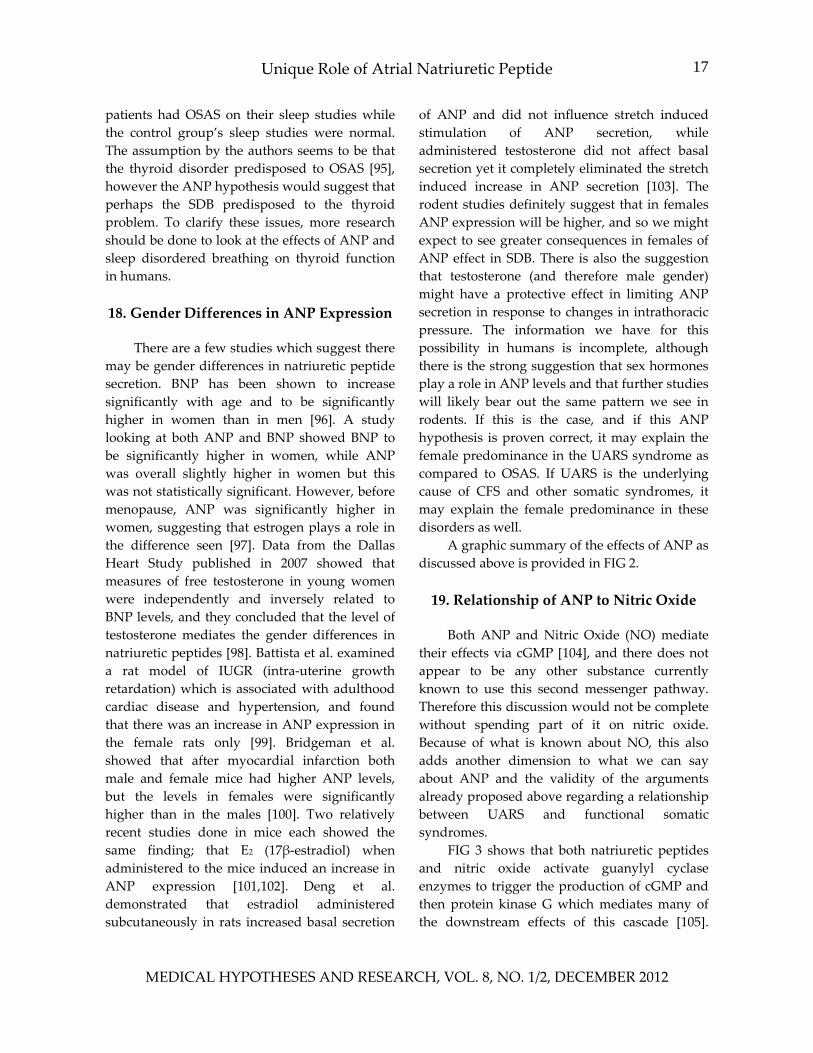
A graphic summary of the effects of ANP as discussed above is provided in FIG 2. 19. Relationship of ANP to Nitric Oxide
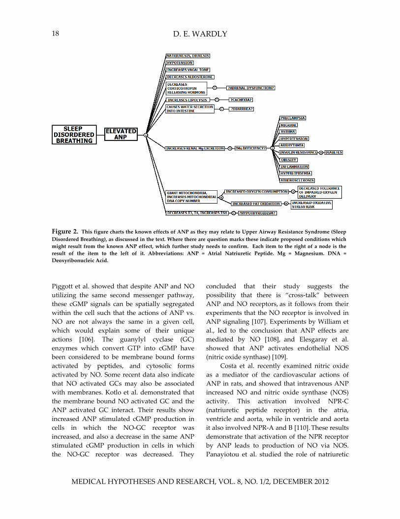
Both ANP and Nitric Oxide (NO) mediate
their effects via cGMP [104], and there does not appear to be any other substance currently known to use this second messenger pathway. Therefore this discussion would not be complete without spending part of it on nitric oxide. Because of what is known about NO, this also adds another dimension to what we can say about ANP and the validity of the arguments already proposed above regarding a relationship between UARS and functional somatic syndromes.
FIG 3 shows that both natriuretic peptides and nitric oxide activate guanylyl cyclase enzymes to trigger the production of cGMP and then protein kinase G which mediates many of the downstream effects of this cascade [105].
MEDICAL HYPOTHESES AND RESEARCH, VOL. 8, NO. 1/2, DECEMBER 2012
D. E. WARDLY 18
Figure 2. This figure charts the known effects of ANP as they may relate to Upper Airway Resistance Syndrome (Sleep Disordered Breathing), as discussed in the text. Where there are question marks these indicate proposed conditions whichmight result from the known ANP effect, which further study needs to confirm. Each item to the right of a node is the result of the item to the left of it. Abbreviations: ANP = Atrial Natriuretic Peptide. Mg = Magnesium. DNA = Deoxyribonucleic Acid.
Piggott et al. showed that despite ANP and NO utilizing the same second messenger pathway, these cGMP signals can be spatially segregated within the cell such that the actions of ANP vs. NO are not always the same in a given cell, which would explain some of their unique actions [106]. The guanylyl cyclase (GC) enzymes which convert GTP into cGMP have been considered to be membrane bound forms activated by peptides, and cytosolic forms activated by NO. Some recent data also indicate that NO activated GCs may also be associated with membranes. Kotlo et al. demonstrated that the membrane bound NO activated GC and the ANP activated GC interact. Their results show increased ANP stimulated cGMP production in cells in which the NO‐GC receptor was increased, and also a decrease in the same ANP stimulated cGMP production in cells in which the NO‐GC receptor was decreased. They
concluded that their study suggests the possibility that there is “cross‐talk” between ANP and NO receptors, as it follows from their experiments that the NO receptor is involved in ANP signaling [107]. Experiments by William et al., led to the conclusion that ANP effects are mediated by NO [108], and Elesgaray et al. showed that ANP activates endothelial NOS (nitric oxide synthase) [109].
Costa et al. recently examined nitric oxide as a mediator of the cardiovascular actions of ANP in rats, and showed that intravenous ANP increased NO and nitric oxide synthase (NOS) activity. This activation involved NPR‐C (natriuretic peptide receptor) in the atria, ventricle and aorta, while in ventricle and aorta it also involved NPR‐A and B [110]. These results demonstrate that activation of the NPR receptor by ANP leads to production of NO via NOS. Panayiotou et al. studied the role of natriuretic
MEDICAL HYPOTHESES AND RESEARCH, VOL. 8, NO. 1/2, DECEMBER 2012
Unique Role of Atrial Natriuretic Peptide 19
Figure 3. Nitric Oxide and Natriuretic Peptides Intracellular Messenger Pathway. This cartoon of a cell shows the particulate guanylyl cyclase (pGC) in the cell membrane and the soluble guanylyl cyclase (sGC) in the cytoplasm. ThepGC is activated by natriuretic peptides (NPs) and the sGC is activated by nitric oxide (NO), and the result of activation of guanylyl cyclase regardless of the activator is the production of cGMP (cyclic guanosine monophosphate) from GTP(guanosine triphosphate). cGMP is degraded by phosphodiesterases (PDE). The production of cGMP leads to thegeneration of protein kinase G (PKG), which mediates many of the effects of this cascade.(http://cgmp.blauplanet.com/path.html)
peptides in the cardiovascular dysfunction seen in endotoxic shock, for which the vascular dysfunction is thought to be mediated by excessive production of NO by induction of inducible NOS (iNOS). In endotoxic shock, levels of natriuretic peptides are elevated and correlate with the cardiovascular dysfunction. The authors took advantage of the fact that both NO and natriuretic peptides utilize the guanylate cyclase receptors and used mice who lacked the ANP receptor, NPR‐A. After inducing endotoxic shock using LPS (lipopolysaccharide), the amount of NO produced by the NPR‐A KO (knockout, or animals lacking this receptor) was much lower than in the wild type mice. Similarly, in response to induced shock the NPR‐A KO mice had lower levels of IL‐1α, TNFα, and interferonγ, and iNOS expression compared to the wild type mice. The authors concluded that
NPR‐A activation by ANP facilitates iNOS expression and contributes to the vascular dysfunction seen in endotoxic shock [104]. Again, we have more evidence that confirms that ANP will lead to NOS activity and NO production via its receptor, acting as a hormone which mediates NO action in the body. Indeed, Gonzalez Bosc et al. describes NO as another second messenger for ANP [91].
If ANP has the potential to do everything that NO can do, either by directly stimulating the NO receptor, or by its own receptor mediating an increase in NO production, this expands the significance of ANP remarkably. The half life of NO is about one second; at 2.5 minutes the half life of ANP is 150 times that [74]. Clearly the effect of NO will be local, while the effect of ANP can be body‐wide with the function of a hormone, yet with all the potential
MEDICAL HYPOTHESES AND RESEARCH, VOL. 8, NO. 1/2, DECEMBER 2012
D. E. WARDLY 20
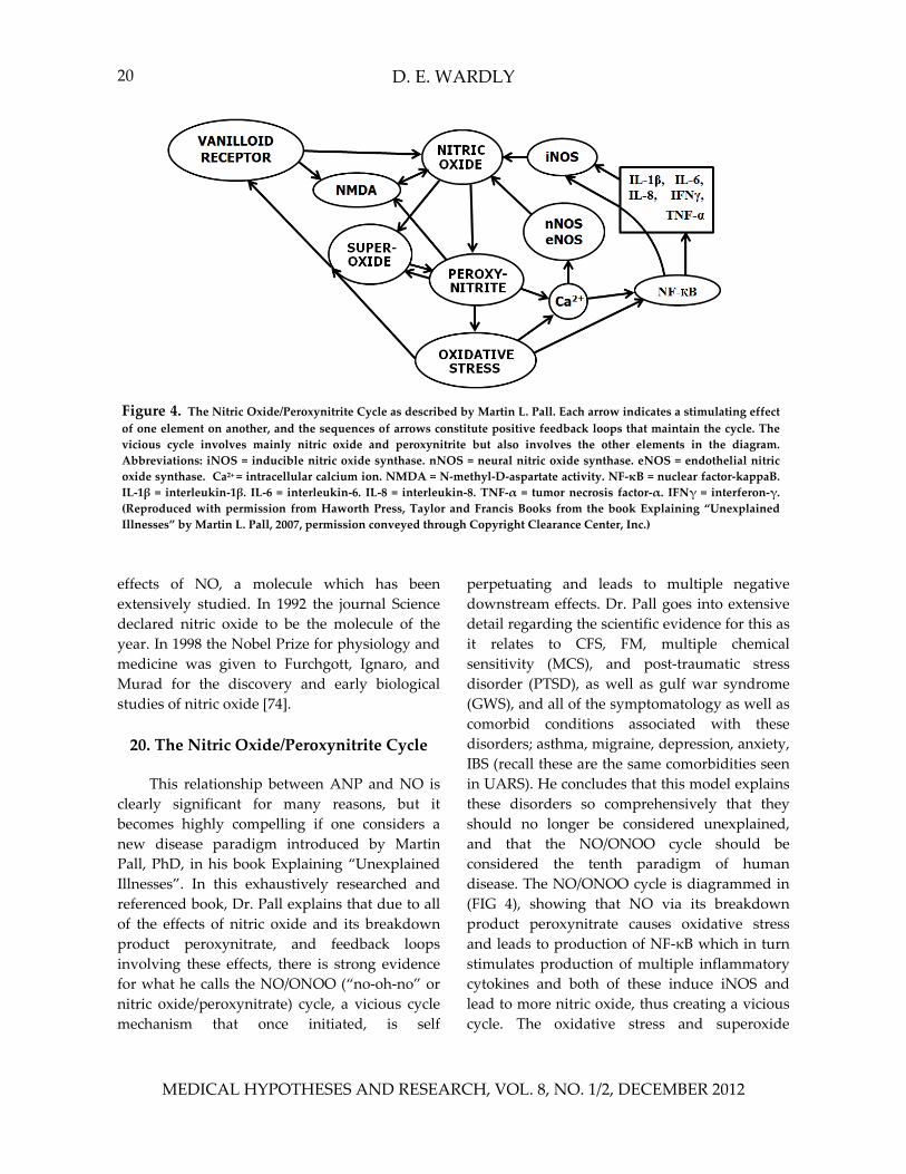
Figure 4. The Nitric Oxide/Peroxynitrite Cycle as described by Martin L. Pall. Each arrow indicates a stimulating effect of one element on another, and the sequences of arrows constitute positive feedback loops that maintain the cycle. Thevicious cycle involves mainly nitric oxide and peroxynitrite but also involves the other elements in the diagram. Abbreviations: iNOS = inducible nitric oxide synthase. nNOS = neural nitric oxide synthase. eNOS = endothelial nitricoxide synthase. Ca2+ = intracellular calcium ion. NMDA = N‐methyl‐D‐aspartate activity. NF‐κB = nuclear factor‐kappaB. IL‐1β = interleukin‐1β. IL‐6 = interleukin‐6. IL‐8 = interleukin‐8. TNF‐α = tumor necrosis factor‐α. IFNγ = interferon‐γ. (Reproduced with permission from Haworth Press, Taylor and Francis Books from the book Explaining “UnexplainedIllnesses” by Martin L. Pall, 2007, permission conveyed through Copyright Clearance Center, Inc.)
effects of NO, a molecule which has been extensively studied. In 1992 the journal Science declared nitric oxide to be the molecule of the year. In 1998 the Nobel Prize for physiology and medicine was given to Furchgott, Ignaro, and Murad for the discovery and early biological studies of nitric oxide [74]. 20. The Nitric Oxide/Peroxynitrite Cycle
This relationship between ANP and NO is
clearly significant for many reasons, but it becomes highly compelling if one considers a new disease paradigm introduced by Martin Pall, PhD, in his book Explaining “Unexplained Illnesses”. In this exhaustively researched and referenced book, Dr. Pall explains that due to all of the effects of nitric oxide and its breakdown product peroxynitrate, and feedback loops involving these effects, there is strong evidence for what he calls the NO/ONOO (“no‐oh‐no” or nitric oxide/peroxynitrate) cycle, a vicious cycle mechanism that once initiated, is self
perpetuating and leads to multiple negative downstream effects. Dr. Pall goes into extensive detail regarding the scientific evidence for this as it relates to CFS, FM, multiple chemical sensitivity (MCS), and post‐traumatic stress disorder (PTSD), as well as gulf war syndrome (GWS), and all of the symptomatology as well as comorbid conditions associated with these disorders; asthma, migraine, depression, anxiety, IBS (recall these are the same comorbidities seen in UARS). He concludes that this model explains these disorders so comprehensively that they should no longer be considered unexplained, and that the NO/ONOO cycle should be considered the tenth paradigm of human disease. The NO/ONOO cycle is diagrammed in (FIG 4), showing that NO via its breakdown product peroxynitrate causes oxidative stress and leads to production of NF‐κB which in turn stimulates production of multiple inflammatory cytokines and both of these induce iNOS and lead to more nitric oxide, thus creating a vicious cycle. The oxidative stress and superoxide
MEDICAL HYPOTHESES AND RESEARCH, VOL. 8, NO. 1/2, DECEMBER 2012
Unique Role of Atrial Natriuretic Peptide 21
increase activity of the vanilloid receptor, which also increases NO and NMDA (N‐methyl‐D‐aspartate) activity, and the NMDA activity (a mediator of neuroexcitotoxicity) is also increased by NO and peroxynitrate. Peroxynitrite is preferentially produced instead of NO when NOS is uncoupled by a depletion of its cofactors. The intent here is not to recreate Dr. Pall’s eloquently explained argument for the existence of this NO/ONOO cycle; his book is over 400 pages long and fully ¼ of this book is scientific references which support his argument for this paradigm to explain the mechanism for these chronic illnesses. Suffice it to say, there is evidence for elevated nitric oxide and the elements of the NO/ONOO cycle in all these illnesses and their comorbidities. (For example, peroxynitrite leads to lowered mitochondrial function and decreased ATP production; this will account for some of the fatigue seen in these illnesses). Because all of these illnesses have triggers that initiate them, he shows evidence for how the known triggers of these illnesses have been shown to lead to elevated NO or elevated NMDA activity or elevated cytokines, etc. Dr. Pall also explains that because the effect of NO is local, this explains the different patterns seen in each individual with a multisystem illness, depending on in which tissues the vicious cycle is activated. It should be noted that there are several different forms of NOS, the most important for the NO/ONOO cycle being the iNOS, or inducible nitric oxide synthase [74].
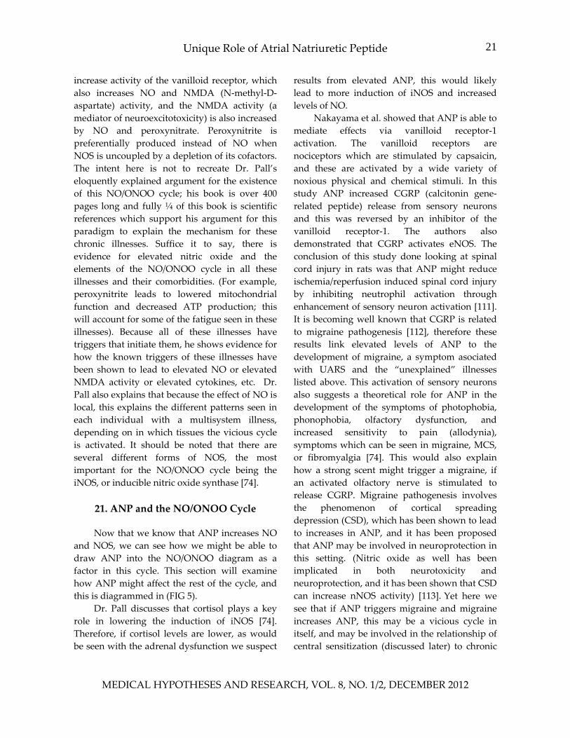
21. ANP and the NO/ONOO Cycle
Now that we know that ANP increases NO and NOS, we can see how we might be able to draw ANP into the NO/ONOO diagram as a factor in this cycle. This section will examine how ANP might affect the rest of the cycle, and this is diagrammed in (FIG 5).
Dr. Pall discusses that cortisol plays a key role in lowering the induction of iNOS [74]. Therefore, if cortisol levels are lower, as would be seen with the adrenal dysfunction we suspect
results from elevated ANP, this would likely lead to more induction of iNOS and increased levels of NO.
Nakayama et al. showed that ANP is able to mediate effects via vanilloid receptor‐1 activation. The vanilloid receptors are nociceptors which are stimulated by capsaicin, and these are activated by a wide variety of noxious physical and chemical stimuli. In this study ANP increased CGRP (calcitonin gene‐related peptide) release from sensory neurons and this was reversed by an inhibitor of the vanilloid receptor‐1. The authors also demonstrated that CGRP activates eNOS. The conclusion of this study done looking at spinal cord injury in rats was that ANP might reduce ischemia/reperfusion induced spinal cord injury by inhibiting neutrophil activation through enhancement of sensory neuron activation [111]. It is becoming well known that CGRP is related to migraine pathogenesis [112], therefore these results link elevated levels of ANP to the development of migraine, a symptom asociated with UARS and the “unexplained” illnesses listed above. This activation of sensory neurons also suggests a theoretical role for ANP in the development of the symptoms of photophobia, phonophobia, olfactory dysfunction, and increased sensitivity to pain (allodynia), symptoms which can be seen in migraine, MCS, or fibromyalgia [74]. This would also explain how a strong scent might trigger a migraine, if an activated olfactory nerve is stimulated to release CGRP. Migraine pathogenesis involves the phenomenon of cortical spreading depression (CSD), which has been shown to lead to increases in ANP, and it has been proposed that ANP may be involved in neuroprotection in this setting. (Nitric oxide as well has been implicated in both neurotoxicity and neuroprotection, and it has been shown that CSD can increase nNOS activity) [113]. Yet here we see that if ANP triggers migraine and migraine increases ANP, this may be a vicious cycle in itself, and may be involved in the relationship of central sensitization (discussed later) to chronic
MEDICAL HYPOTHESES AND RESEARCH, VOL. 8, NO. 1/2, DECEMBER 2012
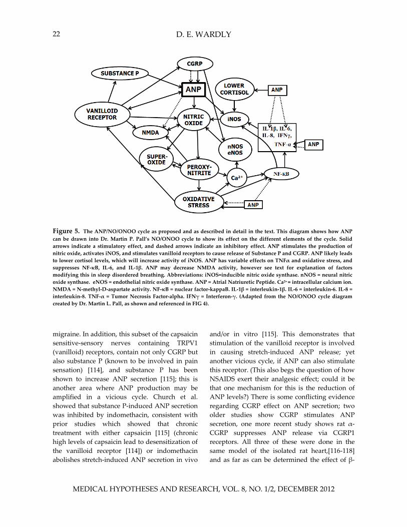
D. E. WARDLY 22
Figure 5. The ANP/NO/ONOO cycle as proposed and as described in detail in the text. This diagram shows how ANP can be drawn into Dr. Martin P. Pall’s NO/ONOO cycle to show its effect on the different elements of the cycle. Solidarrows indicate a stimulatory effect, and dashed arrows indicate an inhibitory effect. ANP stimulates the production of nitric oxide, activates iNOS, and stimulates vanilloid receptors to cause release of Substance P and CGRP. ANP likely leadsto lower cortisol levels, which will increase activity of iNOS. ANP has variable effects on TNFα and oxidative stress, and suppresses NF‐κB, IL‐6, and IL‐1β. ANP may decrease NMDA activity, however see text for explanation of factorsmodifying this in sleep disordered breathing. Abbreviations: iNOS=inducible nitric oxide synthase. nNOS = neural nitric oxide synthase. eNOS = endothelial nitric oxide synthase. ANP = Atrial Natriuretic Peptide. Ca2+ = intracellular calcium ion. NMDA = N‐methyl‐D‐aspartate activity. NF‐κB = nuclear factor‐kappaB. IL‐1β = interleukin‐1β. IL‐6 = interleukin‐6. IL‐8 = interleukin‐8. TNF‐α = Tumor Necrosis Factor‐alpha. IFNγ = Interferon‐γ. (Adapted from the NO/ONOO cycle diagram created by Dr. Martin L. Pall, as shown and referenced in FIG 4).
migraine. In addition, this subset of the capsaicin sensitive‐sensory nerves containing TRPV1 (vanilloid) receptors, contain not only CGRP but also substance P (known to be involved in pain sensation) [114], and substance P has been shown to increase ANP secretion [115]; this is another area where ANP production may be amplified in a vicious cycle. Church et al. showed that substance P‐induced ANP secretion was inhibited by indomethacin, consistent with prior studies which showed that chronic treatment with either capsaicin [115] (chronic high levels of capsaicin lead to desensitization of the vanilloid receptor [114]) or indomethacin abolishes stretch‐induced ANP secretion in vivo
and/or in vitro [115]. This demonstrates that stimulation of the vanilloid receptor is involved in causing stretch‐induced ANP release; yet another vicious cycle, if ANP can also stimulate this receptor. (This also begs the question of how NSAIDS exert their analgesic effect; could it be that one mechanism for this is the reduction of ANP levels?) There is some conflicting evidence regarding CGRP effect on ANP secretion; two older studies show CGRP stimulates ANP secretion, one more recent study shows rat α‐CGRP suppresses ANP release via CGRP1 receptors. All three of these were done in the same model of the isolated rat heart,[116‐118] and as far as can be determined the effect of β‐
MEDICAL HYPOTHESES AND RESEARCH, VOL. 8, NO. 1/2, DECEMBER 2012
Unique Role of Atrial Natriuretic Peptide 23
CGRP on ANP has not been assessed so perhaps further work here may elucidate the conflict. It may be that CGRP feeds into the vicious cycle or it may act as negative feedback to limit overstimulation of ANP; this is yet unclear. The information detailed above leads to speculation whether ANP elevations might be involved in the development of chronic pain conditions such as fibromyalgia or neuropathic pain. According to Dr. Pall, all of the parameters of the NO/ONOO cycle have been implicated in hyperalgesia using animal models [74], so ANP is likely involved in chronic pain if only by its effect on NO. A review article on the pharmacology of the vanilloid receptor discusses evidence that it may be involved in the generation of neuropathic pain, as well as that this receptor is upregulated in irritable bowel syndrome (IBS) [119], which we know to be associated with both UARS and the chronic illnesses we are discussing. Lin et al. showed that local injection of CGRP, Substance P, or capsaicin results in cutaneous vasodilation and edema, demonstrating a mechanism for neurogenic inflammation mediated by stimulation of the TRPV1 receptor [120]. There is clearly more to be learned about the role of CGRP, Substance P, and the vanilloid receptor in these chronic illnesses.
There are two studies which suggested that ANP can decrease NMDA activity. Kuribayashi et al. showed that ANP reduced NMDA induced neurotoxicity in the rat retina, via a mechanism of increasing dopamine synthesis [121]. The other showed that ANP inhibited the seizure inducing effect of NMDA in ethanol dependent mice. However it did not inhibit NMDA induced seizures in mice that were not ethanol dependent. (This paper also pointed out that high dose ethanol intake can significantly reduce ANP levels) [122]. These studies support a possible role for ANP in neuroprotection under certain conditions. It is thought that the neuroprotection from ANP may result from the fact that increases in cGMP may lead to a reduction in intracellular calcium concentrations,
which may reduce NMDA activation in the presence of ischemia [113]. However, recall that if NOS is uncoupled then the increase in peroxynitrite will favor increased intracellular calcium concentrations (see FIG 4), such that an activated NO/ONOO cycle will result in neurotoxicity with ANP stimulation. Again looking at (FIG 5), one might propose that despite a possible negative effect by ANP on NMDA activity, that the arrows indicating increased NO, iNOS, and vanilloid receptor activity from ANP in addition to NOS uncoupling might override any inhibitory effect and lead to an overall stimulatory effect on NMDA activity in an individual in whom the NO/ONOO cycle is activated. However, the knowledge that those with SDB probably have low magnesium levels (as discussed above) adds another layer here; magnesium deficiency causes NMDA receptors to be hypersensitive to stimulation [74]. Also, part of a stress response (as present in the struggle to breathe) involves the release of large amounts of glutamate and aspartate, which will stimulate the NMDA receptor [78]. Fourthly, the amino acid homocysteine has been shown to be elevated in OSAS[123] and it has a direct excitotoxic effect on the NMDA receptor [68]. Therefore it seems quite likely that the overall result in SDB might be increased NMDA activity. This would leave those with SDB vulnerable to the effects of excitotoxins like MSG (monosodium glutamate) and Aspartame, with increased risk of migraine and other consequences of neuroexcitotoxicity [74].
ANP has been seen to have either antioxidant or prooxidant effects, depending on different experimental conditions. ANP does seem to lead to the generation of reactive oxygen species (ROS), however it also appears to act as an antioxidant in some studies [124]. Scarpino et al. looked at the differences in ROS production by human umbilical vein endothelial cells depending on stimulation with the wild type ANP (TT2238) or with gene variant ANP (CC2238); the variant gene ANP led to more ROS
MEDICAL HYPOTHESES AND RESEARCH, VOL. 8, NO. 1/2, DECEMBER 2012
D. E. WARDLY 24
production and an increase in gene expression of substances involved in atherogenesis and vascular remodeling. This implied that this ANP variant would lead to more oxidative stress and increased endothelial dysfunction in vivo [125]. The presence of polymorphisms of the ANP gene of which there appear to be several [81,126], would explain the varying results in studies looking at the ANP effect on oxidative stress but may also explain, once more is known about these polymorphisms, the differences in clinical presentation of those individuals with UARS, or perhaps even why some people with nasal flow limitations are asymptomatic and some have the clinical syndrome of UARS. Polymorphisms in the ANP gene might be expected to account for the possibility of a variable effect of ANP on perpetuating or quenching the NO/ONOO cycle. It also begs the question; what results would be obtained in all of the above mentioned studies on ANP if a gene variant ANP were used instead of the wild type human ANP? Do those individuals with UARS, CFS/FM, MCS, etc, carry a mutant ANP gene? Hopefully these questions may be answered by future research.
Human ANP has been shown in many studies to have anti‐inflammatory effects. ANP was shown to reduce the lipopolysaccharide induced elevations in IL‐1β and IL‐6 in rats [127]. Three separate studies, two in mice and one in cell culture, showed that ANP is involved in reducing levels of TNF‐α and NF‐κB [128‐130]. However, these studies contradict the findings of the recent study above which utilized an endotoxic shock model and found that ANP was associated with elevated levels of TNF‐α via increases in iNOS expression, also in mice. There may be other variables involved which are not yet apparent, such as unknown genetic differences or perhaps the presence or absence of an activated NO/ONOO cycle.
Finally, it looks as though there is a mechanism for negative feedback in that NO can inhibit ANP secretion [42]. Yuan et al. as discussed above showed that leptin decreases ANP secretion via increasing NO to produce that
decrease in ANP [53]. In SDB we are interested mainly in ANP production by the atria, in response to intrathoracic pressure changes, and therefore if NO is to feedback to decrease this production it must be produced locally in the atria in order to have an effect; as we recall the effect of NO is local only, because its half life is so short. This short half life will limit its ability to change the total amount of ANP secretion over time, because in SDB the stimulus for ANP secretion will be episodic; therefore by the time another respiratory event occurs, the previously produced NO will already have dissipated and the intrathoracic pressure effect on ANP secretion at onset will be unchanged from the conditions of the prior respiratory event. If leptin is elevated, however, this is a hormone which will have a sustained effect on NO production which then may have a more sustained effect on decreasing ANP production, generating the reduction in lipolysis as previously discussed. If NO production is increased via a NO/ONOO cycle mechanism triggered locally in the atria, then ANP as produced episodically in SDB might be able to feedback to decrease its own production via NO. Based on the fact that we know ANP is elevated in OSAS, and on the striking correlation of ANP effects with the UARS syndrome, this NO feedback mechanism may ultimately be not very significant. The testing of this piece of the ANP hypothesis might be a difficult experiment to design.
To summarize the possible effect of ANP on the NO/ONOO cycle (see FIG 5); ANP has multiple stimulatory effects which could increase levels of NO and iNOS induction. ANP also has some effects which might turn down some elements of the cycle. Ultimately in each individual it would be the totality of stimulatory versus inhibitory effects which would decide if the ANP elevation predisposed to NO/ONOO cycle activation, with a potentially large factor being the ANP polymorphism that individual carries. According to this part of the theory which is termed the “ANP/NO/ONOO hypothesis”, in those with UARS the presumed
MEDICAL HYPOTHESES AND RESEARCH, VOL. 8, NO. 1/2, DECEMBER 2012
Unique Role of Atrial Natriuretic Peptide 25
higher ANP levels will act systemically to upregulate all areas in the body where the NO/ONOO cycle has been set in motion, creating a systemic illness. In OSAS, ANP resistance is presumed to be present, therefore any effect from elevation of NO and iNOS will be localized and not systemically amplified.
22. Causes of Fatigue in UARS
Most sources which discuss UARS and OSAS in terms of fatigue will discuss daytime sleepiness as measured by the Epworth Sleepiness Scale, and more objectively via the MSLT (Multiple Sleep Latency Test) [2]. However, because patients with UARS tend to have chronic insomnia, the Epworth Sleepiness Scale may not accurately measure their fatigue because with insomnia by definition one has difficulty falling asleep. Indeed, in the Sleep Heart Health Study it was found that for women (remember the greater proportion of women in UARS) the Epworth Sleepiness Scale did not correlate with their reports of feeling unrested, while in men there was a correlation between the two [131]. The ANP/NO/ONOO hypothesis provides us with many more explanations for the fatigue seen in UARS other than simply sleep deprivation. 1) Hypotension as well as decreased cardiac output as caused by ANP will cause fatigue; 2) adrenal insufficiency mediated by ANP causes fatigue and orthostatic hypotension; 3) mitochondrial dysfunction as caused by peroxynitrite elevations via ANP activation of iNOS will cause fatigue; and 4) multiple factors in migraine as triggered by ANP increasing CGRP may cause fatigue, outside of migraine pain: migraine aura, migraine premonitory symptoms, and the phenomenon of silent migraine. Therefore the ANP/NO/ONOO hypothesis suggests that UARS patients should be evaluated differently than those with OSAS, because their fatigue may reflect more than just sleepiness and their insomnia may render the Epworth scale to be an insufficient measure of these symptoms in UARS. An alternative scale
for measuring the symptoms of sleep disordered breathing has been published by Dr. Barry Krakow [132], and this may prove to be a more effective tool for assessing UARS severity and response to treatment.
23. Other Genetic Considerations
In his book on the NO/ONOO cycle paradigm, Dr. Pall suggests that genetics will play a role in why some individuals become triggered into the vicious cycle mechanism and others do not [74]. This likely does play a role within the spectrum of UARS proposed here, and as discussed above, the type of ANP polymorphism a person carries will probably contribute to the clinical presentation. Other genes coding for enzymes that are affected by the process we have discussed may also be involved in differences in illness expression. Yuan et al., using mice, studied ANP and its receptors in wild types and those with the eNOS deficient polymorphism (homozygous), a mutation that also occurs in humans. The results showed that the eNOS‐/‐ mice had increased expression of ANP as well as of the NPR‐A and NPR‐C receptors, compared to the wild type mice [133]. This suggests that those humans with the same polymorphism may have an increased susceptibility to the UARS syndrome as mediated by ANP, because their ANP levels will be higher and their ANP response may be upregulated. Dickey et al. examined a mutation in ANP that is now genetically linked to familial atrial fibrillation; they showed that the mutant ANP had a significantly greater half life (4.5 minutes) that was secondary to decreased degradation by neutral endopeptidase. It has been shown that affected individuals have ANP serum levels that are 5‐10 fold higher than unaffected individuals [134]. McKie et al. demonstrated that this mutant ANP product showed greater plasma cGMP activation, diuresis, natriuresis, renin‐angiotensin‐aldosterone inhibition, and hypotensive properties compared to the native ANP, and that
MEDICAL HYPOTHESES AND RESEARCH, VOL. 8, NO. 1/2, DECEMBER 2012

D. E. WARDLY 26
Figure 6. The Yasko Methylation Cycle: This figure shows the Methylation pathway including the 5 cycles that compriseit: 1) urea cycle; 2) neurotransmitter cycle; 3) folate cycle; 4) methionine cycle; and 5) transsulfuration cycle. This complex pathway is the means for moving methyl groups in the body; to make neurotransmitters, to make thymidine, to recyclevitamin B12, and to methylate DNA and other molecules. Cycle 5 involves the detoxification of sulfur from sulfurcontaining amino acids. The ammonia produced by Cycle 5 is detoxified by Cycle 1 (the urea cycle), however if NOS isuncoupled by BH4 depletion, peroxynitrite is produced. Abbreviations: NOS = nitric oxide synthase. NO = nitric oxide. BH4 = tetrahydrobiopterin. BH2 = dihydrobiopterin. DHPR = dihydropteridine reductase. MAO A = monoamine oxidase A.MAO B = monoamine oxidase B. HIAA = hydroxyindoleacetic acid. COMT = catechol‐O‐methyltransferase. HVA = homovanillic acid. NorEp = norepinephrine. VMA = vanillylmandelic acid. dUMP = 2’‐deoxyuridine 5’‐monophosphate. THF = tetrahydrofolate. 5methyl THF = 5methyltetrahydrofolate. MTHFR = methyltetrahydrofolate reductase.MTR=methionine synthase. MTRR = methionine synthase reductase. B12 = vitamin B12. BHMT = betaine‐homocysteine methyltransferase. TMG = trimethylglycine. DMG=dimethylglycine. SAMe = S‐adenosylmethionine. SAH = S‐adenosylhomocysteine. SAHH = S‐adenosylhomocysteine hydrolase. CBS = cystathionine‐β‐synthase. P5P = pyridoxal 5 phosphate. B6 = vitamin B6. Moly = molybdenum. SUOX = sulfite oxidase. Guanido Ac = Guanidinoacetate. (Reproduced with permission of Amy Yasko.)
this was secondary to decreased degradation [81]. It seems plausible that people with severe UARS might be carriers of this mutant ANP gene. 24. Homocysteine and the Methylation Cycle in Sleep Disordered Breathing
The ANP/NO/ONOO hypothesis suggests
that ANP may be linked to the methylation cycle via the link to NO. NOS and NO play a role in the process of methylation, a complex cycle
involving multiple enzymes, cofactors and intermediates. See (FIG 6) for a diagrammatic representation of these reactions. A detailed explanation of this complex cycle is outside the scope of this paper, therefore the reader is referred to other sources [135]. However, it should be apparent from reviewing the diagram that any stress to this cycle may cause problems which can lead to accumulation of homocysteine (Hcys), a marker which has been implicated in cardiovascular disease. Blaise et al. examined the effect of hypoxia on methylation enzymes and
MEDICAL HYPOTHESES AND RESEARCH, VOL. 8, NO. 1/2, DECEMBER 2012
Unique Role of Atrial Natriuretic Peptide 27
showed that hypoxia decreases the activity of MAT (methionine adenosyltransferase) which is needed for the production of SAMe (S‐adenosylmethionine), a molecule critical for DNA methylation [136]. So here we can see that OSAS with hypoxia will increase the risk of epigenetic changes. This study also showed that hypoxia will decrease the activity of CBS (cystathionine β synthase), which we know will lead to elevations in Hcys. The authors concluded that the remethylation pathway for Hcys may be a target for hypoxia [136]. Elevated homocysteine, which is a neurotoxic amino acid [137] and may have a direct excitotoxic effect on the NMDA receptor [123] is known to be an independent risk factor for cardiovascular disease. Sariman et al. measured Hcys levels in OSAS patients and showed that Hcys was elevated in all OSAS patients regardless of the presence of cardiac dysfunction. There was a positive correlation between the level of Hcys and the level of nocturnal oxygen desaturation [68]. Wang et al. showed that Hcys levels were higher in elderly patients than in younger patients, yet they were highest in the elderly patients with OSAS [138]. Returning to what we have learned about the relationship of ANP to NO, we can imagine that with ANP induction of NOS and depletion of methylation cofactors as a result, UARS might also lead to elevated Hcys, although this needs further investigation. What we can conclude however is that the studies above suggest that Hcys may be a marker of sleep disordered breathing, and that the cardiac risk associated with Hcys may be in part secondary to unrecognized sleep disordered breathing.
If Hcys is a marker of sleep disordered breathing, then we need to reconsider the role of sleep disordered breathing in many conditions which have been shown to be associated with hyperhomocysteinemia, among them: PTSD, Alzheimer’s disease, depression, schizophrenia, obsessive‐compulsive disorder, pregnancy induced hypertension, preeclampsia, and placental abruption [123,137,139‐141]. Hcys has
not been demonstrated to be specifically elevated in autistic children, but a recent study by James et al. indicated that Hcys levels are elevated in mothers of autistic children, suggesting that poor methylation in these women may have led to epigenetic changes that contributed to their offspring’s autism [142]. This may explain why Mann et al. recently found that maternal pre‐eclampsia (again, shown to be associated with hyperhomocysteinemia and with OSAS) is a significant risk factor for the development of an autism spectrum disorder in the child [143]. There are several studies which implicate poor methylation capacity and defects in genes necessary for methylation in autism [144,145]. Whether or not SDB in the child influences the severity of the expression of the methylation defect in autism is something that deserves further investigation.
25. Why Some and Not Others: Perpetuating Factors and Neural
Sensitization
There has been the question of why certain individuals develop an illness with CFS after EBV infection (or other triggers) and others do not, or why some develop MCS after a chemical exposure but others do not after the same exposure. Dr. Pall discusses the possibility of genetics, hormones, nutrition, alterations in the immune system as well as the strength of the triggering stressors [74], which probably all do play a role. The ANP/NO/ONOO hypothesis would state that in certain individuals who have UARS and elevated ANP, the NO/ONOO cycle reactions will already be upregulated. Therefore it will not take much of an additional trigger to increase the activity of their NO/ONOO cycles and lead to the consequences of the vicious cycle on their biochemistry; with the increase in oxidative stress and mitochondrial dysfunction and NMDA activation that results from this and leads to symptoms. It will be much more difficult to trigger the NO/ONOO cycle in a person who does not have ANP elevations as
MEDICAL HYPOTHESES AND RESEARCH, VOL. 8, NO. 1/2, DECEMBER 2012
D. E. WARDLY 28
seen in UARS; therefore as per the discussion above, it is proposed that most individuals with these multisystem illnesses will have UARS underlying their disorder. An individual may start out low on a spectrum with mild UARS or simply nasal flow limitations and not seem to have many symptoms, or they can have more severe UARS with more symptoms related to ANP elevations, or they can be triggered to develop full blown CFS, FM, MCS, PTSD or GWS more easily because their biochemistry is more vulnerable leading to initiation of the NO/ONOO vicious cycle. This model may also explain some of the variation in clinical expression of all of the above syndromes, and it might also be the case that the severity of the clinical syndrome may be proportional to the duration and height of the ANP elevation caused by sleep disordered breathing. Dr. Pall uses the self perpetuating NO/ONOO cycle to explain why, for instance, a one time infection or other stressor may lead to a chronic illness. The ANP/NO/ONOO hypothesis explanation would be that in addition to the NO/ONOO vicious cycle mechanism itself, it is the underlying sleep breathing disorder with ongoing elevations of ANP which, in addition to its multitude of effects as previously discussed, perpetuates the NO/ONOO cycle and therefore the chronic illness.
Dr. Avram Gold, a prominent researcher in the UARS field, has recently put forth a hypothesis paper looking at a new paradigm to explain an association between SDB and the functional somatic syndromes (FSS). He explains the phenomenon of neural sensitization, in which the limbic system becomes sensitized to a stimulus such that there is an amplification of the response to the stimulus after repetitive exposure. This is a phenomenon which has been suggested to explain the extreme sensitivity to odors seen in MCS. He also reviews what we know of the general adaptation syndrome, first described by Dr. Hans Selye; it is a physiological response to chronic physical stress (or allostatic challenge) which has been shown to involve
HPA (hypothalamic‐pituitary‐adrenal) axis activation. Dr. Gold suggests that pharyngeal collapse may serve as a repetitive stress (or allostatic challenge) which then perpetuates an elevation in the HPA axis that begins with a larger allostatic challenge or trigger factor, leading to a more chronic condition manifested as a FSS. According to this model, in the absence of SDB, there is no perpetuation of the allostatic challenge trigger factor, and the person recovers. In the presence of SDB, pharyngeal collapse leads to perpetuation of illness via HPA axis activation and neural sensitization of the limbic system to the pharyngeal collapse [29].
Dr. Pall also describes neural sensitization in his chapter on MCS, and he shows how the NO/ONOO cycle may lead to this phenomenon via increased NMDA activity, based on the presumed mechanism of neural sensitization, long term potentiation (LTP), which involves stimulation of NMDA receptors in the brain [74]. In fact, it has been shown that ANP can delay the extinction of active avoidance behavior in rats [146]. This suggests that without elevated ANP levels, one might be able to become habituated to pharyngeal collapse. If this is true, then it may be that the neural sensitization which leads to pharyngeal collapse becoming a perpetuating factor in Dr. Gold’s theory, may be in fact mediated by ANP as the perpetuating factor in the ANP/NO/ONOO hypothesis. Therefore we have a convergence of our hypotheses here where the ANP/NO/ONOO hypothesis simply outlines the biochemical factors underlying the same perpetuation Dr. Gold describes. This convergence is striking, and it must be pointed out that the fact that such similar theories were independently originated is an argument for the validity of the assertion that there IS such a relationship between SDB and FSS or “unexplained illness”.
Because this neural sensitization hypothesis for UARS involves sensitization of the limbic system via the olfactory nerve, which senses the pressure changes in the nose as well as odors, this is a link between the pathogenesis of UARS
MEDICAL HYPOTHESES AND RESEARCH, VOL. 8, NO. 1/2, DECEMBER 2012
Unique Role of Atrial Natriuretic Peptide 29
and that of MCS. If this is in fact the mechanism via which these disorders originate, this suggests that odor sensitivity or dysosmia in any patient ought to be a symptom that initiates a search for a sleep breathing disorder. We know that ANP can stimulate the vanilloid receptor, and according to the NO/ONOO cycle we know this can increase NMDA activity which has been implicated in neural sensitization. Dr. Pall also discusses peripheral sensitivity mechanisms which involve the vanilloid receptor. In his book he suggests that guiafenesin may be a vanilloid receptor antagonist. He discusses guiafenisen because it has been found to be helpful for some patients with CFS, and some with FM [74]. This information leads to further speculation: is it possible that guaifenesin, while suppressing hypersensitive cough reflexes, may also suppress the hypersensitive olfactory nerve/amygdala connection? And will this help patients with UARS if they take guaifenesin at bedtime, or will it lead them to develop complete pharyngeal collapse and apnea? Is the hypersensitivity to pharyngeal collapse a dysfunctional mechanism that leads to chronic illness secondary to stress responses, or is it a protective mechanism that prevents hypoxia? These questions should be fairly easy to answer in future clinical research. However, neural sensitization has been described as a mechanism which allows the individual to respond more forcefully to a stimulus that threatens tissue damage [147]. Therefore it seems apparent that neural sensitization in general is a protective mechanism, and when we see people who have evidence of this via heightened senses that we as physicians should be alert to the presence of a condition that their brains consider threatening, such as pharyngeal collapse. Dr. Gold seems to suggest that prior to a triggering event, an individual might tolerate pharyngeal collapse in a state of benign neglect (habituation, as mentioned above) [29]. According to the ANP/NO/ONOO hypothesis, this individual probably had some mild symptoms related to ANP elevations prior to the trigger that were
subclinical, and then as a result of the trigger the NO/ONOO cycle is activated to cause more serious symptoms which are then maintained by the continued ANP elevation. As we learned above, activation of the sympathetic nervous system can increase ANP levels [67]. If ANP is able to turn off habituation as well as trigger the NO/ONOO cycle, we can see how any severe physiological or psychological stress may trigger this biochemical pathway to turn off the “benign neglect” of pharyngeal collapse and start a cascade of events that leads to illness.
The only area where there is conflict between Dr. Gold’s ideas and the data presented here is in the area of HPA axis activation. This would certainly be expected to be a factor in UARS if we did not know about the physiologic effects of ANP. But with what we know about the effect of ANP on suppressing the HPA axis, supported by the observation that UARS patients tend to be hypotensive and orthostatic, and also the fact that the HPA axis has been shown to be suppressed in CFS, there is clearly something else happening in UARS (and CFS). Again, once ANP resistance develops, all the usual responses to chronic stress will lead to HPA axis activation and all of the sequelae related to this will then become manifest. Most of these adverse effects of chronic stress (for example: hypertension, hyperglycemia, myocardial infarction, polycystic ovaries) have been associated with OSAS [29], but it is not clear that they have been specifically associated with UARS, and it seems apparent from the data presented here that these conditions are different enough hormonally such that separate studies need to be done on UARS rather than assuming it has the same consequences. Dr. Gold discusses the fact that the responses to the HPA axis elevation may be not only from increasing HPA hormones, but also from increased sensitivity of the end organ to the hormones [29]. However, it must be argued that the orthostasis seen in UARS could only be related to a decrease in HPA hormones or a decrease in sensitivity to those hormones; an increase in either would
MEDICAL HYPOTHESES AND RESEARCH, VOL. 8, NO. 1/2, DECEMBER 2012
D. E. WARDLY 30
result in an increase in blood pressure. Based on the above arguments we must conclude that further research needs to formally evaluate the differences in HPA status as seen in OSAS vs. UARS. It seems apparent that ANP elevations in UARS serve to “short circuit” the normal stress responses, which actually ends up causing a more disabling illness than if they were intact. 26. Neural Sensitization and Gender?
If neural sensitization is the cause of
hypersensitivity to pharyngeal collapse, if it is mediated by ANP and the NO/ONOO cycle, and as discussed above females may be more likely to have higher ANP levels than males, then might this explain why more females have UARS and more males have OSAS? Is it the absence of neural sensitization which leads to apnea? And if neural sensitization is responsible for many of the symptoms seen in UARS and the somatic syndromes, could this also explain the gender differences seen in these symptom complexes? Recall the study discussed previously which showed in rats that testosterone may eliminate stretch induced ANP secretion [103]. There are many studies which indicate that testosterone may be involved in the development of OSAS, including a study in women with PCOS (polycystic ovarian syndrome) which showed that they have higher AHIs than weight matched controls, and that the AHI was significantly correlated to the total serum testosterone level [148,149]. In fact, the incidence of OSAS in PCOS has been shown to be 70% [83]. Could this testosterone/OSAS/male gender link be mediated by testosterone’s effect on limiting ANP secretion and as a result reducing the tendency to become neurally sensitized to pharyngeal collapse? By contrast, this suggests the possibility that men with UARS might have lower testosterone levels than men with OSAS. If all this is true, then testosterone supplementation might help those with UARS to reduce their symptomatology, however it may be at the expense of progressing to OSAS.
27. For Future Study ● Examine both UARS and OSAS patients,
correctly identifying the UARS patients with esophageal manometry and proper hypopnea criteria. Mild OSAS without hypoxia should be a separate category for statistical evaluation, for which we expect the results to be similar to UARS.
● Measure ANP levels during sleep to compare average levels between these groups, as well as assessment of the fractional excretion of sodium to ascertain the biological effect of those ANP levels.
● Measure ANP resistance in terms of natriuretic response to a saline load, to identify this condition in those with OSAS and compare to those with UARS.
● In statistical analysis compare ANP levels with the AHI, lowest oxygen desaturation, blood pressure and body weight, as well as with somatic symptoms.
● Compare degree of hypoxia to somatic symptoms.
● Treat SDB, with CPAP or other measures as tolerated, and then assess blood pressure and body weight after a period of time.
● Measure morning serum aldosterone, corticotropin releasing hormone, vasopressin, oxytocin, lipid peroxides, red blood cell magnesium level, TSH, and urinary uric acid in both UARS and OSAS, before and after treatment of SDB.
● Correlate the Pes (esophageal pressure) with the ANP, aldosterone, CRH, vasopressin and oxytocin.
● Compare ANP levels between males and females in each group (OSAS and UARS).
● Measure ANP levels and cortisol levels before and after exercise in UARS patients vs. normal controls.
● Measure basal oxygen consumption in patients with UARS, OSAS and in controls.
The results of all of the above proposed
experiments might be very useful for developing
MEDICAL HYPOTHESES AND RESEARCH, VOL. 8, NO. 1/2, DECEMBER 2012
Unique Role of Atrial Natriuretic Peptide 31
tests that may assist in the diagnosis of UARS when esophageal manometry or effective hypopnea scoring are not available. ● Assess a group of patients with “adrenal
fatigue” and determine if they indeed all have UARS, and then further investigate whether their adrenal fatigue can be reversed by treating the UARS and (importantly) lowering their ANP levels.
● Measure airway NO levels to see if they correlate with the degree of ANP elevation in UARS.
● Measure plasma nitrite to determine systemic NO levels in UARS, compare to OSAS.
● Measure UARS symptomatology with and without the use of CGRP antagonists, inhibitors of NMDA and vanilloid receptors, indomethacin and testosterone supplementation (not all of these medications are currently available).
● Measure changes in ANP levels and in the AHI with the use of these substances, and compare different responses between men and women.
● Compare testosterone levels between men with UARS and men with OSAS.
● Assess for the presence of ANP polymorphisms in UARS, OSAS and controls to determine if there is an association of the mutant ANP gene with UARS.
● Determine the incidence of homocysteine elevation in UARS.
28. Summary and Conclusion
This ANP hypothesis is truly a new
paradigm for understanding pathogenesis in sleep disordered breathing. It has been reviewed here how sleep disordered breathing will cause elevations of ANP, and how OSAS with intermittent hypoxia should ultimately lead to ANP resistance via elevations in EPO and leptin and how this may lead to the development of
hypertension, as well as contribute to the obesity which is seen in OSAS via loss of the lipolytic effect of ANP. This implies that while we know that obesity makes OSAS worse, it may be OSAS which initially causes obesity. There is clearly a relationship between ANP resistance and insulin resistance and the metabolic syndrome. The known effects of ANP on blood pressure, vagal tone and lipolysis correlate well with what is seen in UARS, as these patients tend to be thin and hypotensive. This suggests that ANP levels may be higher in UARS than they are in OSAS, and this requires further investigation. There are many other known effects of ANP which explain the clinical syndrome of UARS and which also suggest its relationship to several of the somatic syndromes. ANP is known to suppress multiple areas in the hypothalamic‐pituitary‐adrenal axis, including aldosterone and corticotropin releasing hormone. The research described links UARS via elevated ANP to chronic fatigue syndrome (CFS) and to the phenomenon of “adrenal fatigue”. ANP actions may also explain the diarrhea and hypothyroidism which are associated with UARS. ANP increases renal excretion of magnesium, therefore it may mediate magnesium deficiency in those with SDB. Magnesium deficiency is seen in preeclampsia, migraine, asthma, hypertension, cardiac arrhythmia, insulin resistance, obesity, inflammation, hyperlipidemia and atherosclerosis; all conditions which are also associated with SDB. There is evidence for gender differences in ANP levels and secretion, which may explain the gender differences seen in UARS, and based on this theory, those seen in the somatic syndromes. Again, once intermittent hypoxia occurs in OSAS, ANP resistance should develop and it is proposed that this is the reason why many of the somatic symptoms seen in SDB, including the severe fatigue, will lessen as the AHI increases. Based on the known actions of ANP, the development of a biomarker panel is proposed to aid in the diagnosis of UARS, to be used in place of esophageal manometry because of the hormone levels which are altered directly
MEDICAL HYPOTHESES AND RESEARCH, VOL. 8, NO. 1/2, DECEMBER 2012
D. E. WARDLY 32
as a result of the ANP elevations that are caused by changes in intrathoracic pressure. This may help to improve diagnostic accuracy in UARS, as detection on PSG can be difficult and dependent upon sleep lab technique and proficiency. Finally, it has been shown here how ANP and nitric oxide are closely linked in their physiologic effects. This connection allows further supposition regarding the role of ANP in the development of CFS, FM, MCS, PTSD and GWS via Dr. Martin Pall’s paradigm of the NO/ONOO vicious cycle mechanism to explain these multisystem illnesses. There are multiple known effects of ANP which allow us to further develop this theory, and to propose that sleep disordered breathing, via ANP, acts as a perpetuating factor in the maintenance of these functional somatic syndromes. ANP may play a role in neural sensitization which has been proposed as a factor in SDB causing perpetuation of these chronic illnesses. Known polymorphisms in the ANP gene as well as in NOS genes may help explain some of the variability in expression of illness in SDB. Lastly, the link between ANP and NO may link ANP elevations to depletion of the methylation cycle. There is evidence via homocysteine elevations for methylation problems in SDB, and this therefore suggests a possible link to be explored between SDB and other known illnesses characterized by homocysteine elevations and methylation issues.
A great deal of science is presented here in these pages, and the facts are put together to construct a cohesive albeit complex picture from the various puzzle pieces. There is still much of what is written here which is simply theoretical until it can be proven clinically. Therefore this paper is a challenge to others to take these ideas and develop clinical trials to show which ones hold merit. The development of a UARS biomarker panel would be a huge step towards improving diagnosis of this poorly understood condition. If those with UARS carry a mutant ANP gene, this would also aid diagnosis as well as provide more clinical definition for UARS. If
the assessment here of the prevalence of UARS and of its presence underlying the functional somatic syndromes is correct, then there is great potential to relieve the suffering of millions of people through improving the recognition and treatment of sleep disordered breathing.
Acknowledgements
I would like to thank Steven Park, M.D., and Elita Pastra‐Landis, Ph.D., for reviewing the manuscript. In addition, I would also like to thank George Wardly, Ph.D., for technical assistance with preparing the figures.
TABLE 1. Abbreviations used.
AASM American Academy of Sleep Medicine ACTH Adrenocorticotropic Hormone ADD Attention Deficit Disorder ADHD Attention Deficit Hyperactivity Disorder AHI Apnea Hypopnea Index AMP Adenosine Monophosphate ANP Atrial Natriuretic Peptide ATP Adenosine Triphosphate AVP Arginine Vasopressin BMI Body Mass Index BNP B‐type Natriuretic Peptide CBS Cystathionine‐β‐Synthase CFS Chronic Fatigue Syndrome cGMP Cyclic Guanosine Monophosphate CGRP Calcitonin Gene‐Related Peptide CNP C‐type Natriuretic Peptide CPAP Continuous Positive Airway Pressure CRH Corticotropin Releasing Hormone CRP C‐reactive Protein CSD Cortical Spreading Depression DNP Dendroapsis Natriuretic Peptide EBV Epstein‐Barr Virus EEG Electroencephalogram eNOS Endothelial Nitric Oxide Synthase EPO Erythropoetin FM Fibromyalgia GC Guanylyl Cyclase GC‐A Guanylyl Cyclase‐A (ANP receptor) GFR Glomerular filtration rate GTP Guanosine Triphosphate GWI Gulf War Illness
MEDICAL HYPOTHESES AND RESEARCH, VOL. 8, NO. 1/2, DECEMBER 2012
Unique Role of Atrial Natriuretic Peptide 33
GWS Gulf War Syndrome Hcys Homocysteine HPA Hypothalamic‐Pituitary‐Adrenal IBS Irritable Bowel Syndrome IGF‐1 Insulin‐like Growth Factor‐1 IH Intermittent Hypoxia IL Interleukin iNOS Inducible Nitric Oxide Synthase IUGR Intrauterine Growth Retardation KO Knock‐Out (implies null genotype) LPS Lipopolysaccharide LTIH Long‐Term Intermittent Hypoxia MAT Methionine Adenosyltransferase MCS Multiple Chemical Sensitivity mRNA Messenger Ribonucleic Acid MSLT Multiple Sleep Latency Test NEP Neutral Endopeptidase NF‐κB Nuclear Factor‐kappaB NMDA N‐Methyl‐D‐aspartate nNOS Neural Nitric Oxide Synthase NO Nitric Oxide NOD Nocturnal Oxygen Desaturation NO Nitric Oxide ONOO Peroxynitrite NOS Nitric Oxide Synthase NP Natriuretic Peptide NPR‐A Natriuretic Peptide Receptor‐A NPR‐B Natriuretic Peptide Receptor‐B NPR‐C Natriuretic Peptide Receptor‐C NSAIDS Non‐Steroidal Anti‐Inflammatory Drugs OSAS Obstructive Sleep Apnea Syndrome PSG Polysomnogram PTSD Post Traumatic Stress Disorder RBP‐4 Retinol Binding Protein‐4 REM Rapid Eye Movement RERAs Respiratory Effort‐Related Arousals RDI Respiratory Disturbance Index ROS Reactive Oxygen Species SAMe S‐Adenosylmethionine SDB Sleep Disordered Breathing T3 Triiodothyronine T4 Thyroxine TMJD Temporomandibular Joint Dysfunction TNFα Tumor Necrosis Factor alpha TRH Thyrotropin Releasing Hormone TRPV1 Transient Receptor Potential Vanilloid
Type 1 TSH Thyroid Stimulating Hormone UARS Upper Airway Resistance Syndrome VR1 Vanilloid Receptor‐1
References 1. Guilleminault C and De Los Reyes V [2011] Upper‐
airway resistance syndrome. Handbook of Clinical Neurology 98: 401‐409.
2. Newman JP, Clerk AA, Moore M, Utley DS and Terris DJ [1996] Recognition and surgical management of the upper airway resistance syndrome. Laryngoscope 106 (9 Pt 1): 1089‐1093.
3. Lin CH and Guilleminault C [2011] Current hypopnea scoring criteria underscore pediatric sleep disordered breathing. Sleep Medicine 12: 720‐729.
4. Ruehland WR, Rochford PD, O’Donoghue FJ, Pierce RJ, Singh P and Thornton AT [2009] The new AASM criteria for scoring hypopneas: impact on the apnea hypopnea index. Sleep 32: 150‐157.
5. Gold AR, Dipalo F, Gold MS and O’Hearn D [2003] The symptoms and signs of upper airway resistance syndrome: a link to the functional somatic syndromes. Chest 123: 87‐95.
6. Guilleminault C, Kirisoglu C, Poyares D, Palombini L, Leger D, Farid‐Moayer M and Ohayon MM [2006] Upper Airway Resistance Syndrome: a long‐term outcome study. J Psychiatr Res 40: 273‐279.
7. Park SY [2008] Sleep Interrupted. New York: Jodev Press, LLC.
8. Haba‐Rubio J, Andries D, Bastardot F, Tobback N, Vollenweider P, Tafti M and Heinzer R [2011] Prevalence of sleep disordered breathing in middle‐aged general population: the Hypnolaus study. Sleep 34: A123
9. Tufik S, Santos‐Silva R, Taddei JA and Azeredo Bittencourt LR [2010] Obstructive sleep apnea syndrome in the Sao Paolo epidemiologic sleep study. Sleep Medicine 11: 441‐446.
10. Umlauf MG and Chasens ER [2003] Sleep disordered breathing and nocturnal polyuria: nocturia and enuresis. Sleep Med Rev 7: 403‐411.
11. Kalra PR and Tigas S [2002] Regulation of lipolysis: natriuretic peptides and the development of cachexia. Int J Cardiol 85: 125‐132.
12. Potter LF, Yoder AR, Flora DR, Antos LK and Dickey DM [2009] Natriuretic peptides: their structures, receptors, physiologic functions and therapeutic applications. Handb Exp Pharmacol 191: 341‐366.
13. Yandle TG, Richards AM, Nicholls MG, Cuneo R, Espiner EA and Livesey JH [1986] Metabolic clearance rate and plasma half life of alpha‐human atrial natriuretic peptide in man. Life Sci 38: 1827‐1833.
14. Kimura K, Yamaguchi Y, Horii M, Kawata H, Yamamoto H, Uemura S and Saito Y [2007] ANP is cleared much faster than BNP in patients with congestive heart failure. Eur J Clin Pharmacol 63: 699‐702.
15. Ikeda N, Yasu T, Nishikimi T, Nakamura T, Kubo N, Kawakami M, Momomura S and Saito M [2007] N‐terminal pro‐atrial natriuretic peptide and exercise
MEDICAL HYPOTHESES AND RESEARCH, VOL. 8, NO. 1/2, DECEMBER 2012
D. E. WARDLY 34
prescription in patients with myocardial infarction. Regul Pept 141(1‐3): 154‐158.
16. McKie PM, Cataliotti A, Boerrigter G, Chen HH, Sangaralingham SJ, Martin FL, Ichiki T and Burnett JC Jr [2010] A novel atrial natriuretic peptide based therapeutic in experimental angiotensin II mediated acute hypertension. Hypertension 56: 1152‐1159.
17. Imaizumi T and Takeshita A [1993] Influence of ANP on sympathetic nerve activity and chronotropic regulation of the heart. J Cardiovasc Electrophysiol 4: 719‐729.
18. Porzionato A, Macchi V, Rucinski M, Malendowicz LK and De Caro R [2010] Natriuretic peptides in the regulation of the hypothalamic‐pituitary‐adrenal axis. Int Rev Cell Mol Biol 280: 1‐39.
19. Chartier L, Schiffrin E and Thibault G [1984] Effect of atrial natriuretic factor (ANF)‐related peptides on aldosterone secretion by adrenal glomerulosa cells: critical role of the intramolecular disulphide bond. Biochem Biophys Res Commun 122: 171‐174.
20. Atlas SA, Volpe M, Sosa RE, Laragh JH, Camargo MJ and Maack T [1986] Effects of atrial natriuretic factor on blood pressure and the renin‐angiotensin‐aldosterone system. Fed Proc 45: 2115‐2121.
21. Pizzorno JE and Murray MT [1999] Textbook of Natural Medicine, second edition. Edinburgh: Harcourt Publishers Limited.
22. Demitrack MA, Dale JK and Straus SE [1991] Evidence for impaired activation of the hypothalamic‐pituitary‐adrenal axis in patients with chronic fatigue syndrome. J Clin Endocrinol Metab 73: 1224‐1234.
23. Demiralay C, Jahn H, Kellner M, Yassouridis A and Weidemann K [2010] Influence of exogenous atrial natriuretic peptide on the nocturnal hypothalamic‐pituitary‐adrenal axis and sleep in healthy men. Psychoneuroendocrinology 35: 1438‐1445.
24. Perras B, Schultes B, Behn B, Dodt C, Born J and Fehm HL [2004] Intranasal atrial natriuretic peptide acts as central nervous inhibitor of the hypothalamic‐pituitary‐adrenal stress system in humans. J Clin Endocrinol Metab 89: 4642‐4648.
25. Boneva RS, Decker MJ, Maloney EM, Lin JM, Jones JF, Helgason HG, Heim CM, Rye DB and Reeves WC [2007] Higher heart rate and reduced heart rate variability persist during sleep in chronic fatigue syndrome: a population based study. Auton Neurosci 137: 94‐101.
26. Anderberg UM and Uvnas‐Moberg K [2000] Plasma oxytocin levels in female fibromyalgia syndrome patients. Z Rheumatol 59: 373‐379.
27. Wyller VB, Evang JA, Godang K, Solhjell KK and Bollerslev J [2010] Hormonal alterations in adolescent chronic fatigue syndrome. Acta Paediatr 99: 770‐773.
28. Krakow B, Melendrez D, Warner TD, Dorin R, Harper R and Hollifield M [2002] To breathe, perchance to sleep: sleep‐disordered breathing and chronic insomnia among trauma survivors. Sleep Breath 6: 189‐202.
29. Gold AR [2011] Functional somatic syndromes, anxiety disorders and the upper airway: A matter of paradigms. Sleep Med Rev 15: 389‐401.
30. Amin MM, Belisova Z, Hossain S, Gold MS, Broderick JE and Gold AR [2011] Inspiratory airflow dynamics during sleep in veterans with Gulf War illness: a controlled study. Sleep Breath 15: 333‐339.
31. Freund BJ, Wade CE and Claybaugh J [1988] Effects of exercise on atrial natriuretic factor. Release mechanisms and implications for fluid homeostasis. Sports Med 6: 364‐377.
32. Kita H, Ohi M, Chin K, Noguchi T, Otsuka N, Tsuboi T, Itoh H, Nakao K and Kuno K [1998] The nocturnal secretion of cardiac natriuretic peptides during obstructive sleep apnoea and its response to therapy with nasal continuous positive airway pressure. J Sleep Res 7: 199‐207.
33. Schafer H, Ehlenz K, Ewig S, Hasper E, Koehler U, Latzelsberger J, Tasci S and Luderitz B [1999] Atrial natriuretic peptide levels and pulmonary artery pressure awake, at exercise and asleep in obstructive sleep apnoea syndrome. J Sleep Res 8: 205‐210.
34. Svatikova A, Shamsuzzaman, AS, Wolk R, Phillips BG, Olson LJ and Somers VK [2004] Plasma brain natriuretic peptide in obstructive sleep apnea. Am J Cardiol 94: 529‐532.
35. Lin CC, Tsan KW and Lin CY [1993] Plasma levels of atrial natriuretic factor in moderate to severe obstructive sleep apnea syndrome. Sleep 16: 37‐39.
36. Ichioka M, Hirata Y, Inase N, Tojo N, Yoshizawa M, Chida M, Miyazato I, Taniai S and Marumo F [1992] Changes of circulating atrial natriuretic peptide and antidiuretic hormone in obstructive sleep apnea syndrome. Respiration 59: 164‐168.
37. Patwardhan AA, Larson MG, Levy D, Benjamin EF, Leip EP, Keyes MJ, Wang TJ, Gottlieb DJ and Vasan RS [2006] Obstructive sleep apnea and plasma natriuretic peptide levels in a community‐based sample. Sleep 29: 1301‐1306.
38. Foster GE, Hanly PJ, Ahmed SB, Beaudin AE, Pialoux V and Poulin MJ [2010] Intermittent hypoxia increases arterial blood pressure in humans through a Renin‐Angiotensin system‐dependent mechanism. Hypertension 56: 369‐377.
39. Møller DS, Lind P, Strunge B and Pedersen EB [2003] Abnormal vasoactive hormones and 24‐hour blood pressure in obstructive sleep apnea. Am J Hypertens 16: 274‐280.
40. Matsukawa T and Miyamoto T [2011] Angiotensin II‐stimulated secretion of arginine vasopressin is inhibited by atrial natriuretic peptide in humans. Am J Physiol Regul Integr Comp Physiol 300: R624‐R629.
41. Oh YB, Gao S, Shah A, Kim JH, Park WH and Kim SH [2011] Endogenous angiotensin II suppresses stretch‐induced ANP secretion via AT1 receptor pathway. Peptides 32: 374‐381.
42. Dietz JR [2005] Mechanisms of atrial natriuretic peptide secretion from the atrium. Cardiovasc Res 66: 8‐17.
MEDICAL HYPOTHESES AND RESEARCH, VOL. 8, NO. 1/2, DECEMBER 2012
Unique Role of Atrial Natriuretic Peptide 35
43. De Pergola G, Garruti G, Giorgino F, Cospite MR, Corso M, Cignarelli M and Giorgino R [1994] Reduced effectiveness of atrial natriuretic factor in pre‐menopausal obese women. Int J Obes Relat Metab Disord 18: 93‐97.
44. Beltowski J, Jamroz‐Wisniewka A, Borkowska E and Marciniak A [2006] Phosphodiesterase 5 inhibitor ameliorates renal resistance to atrial natriuretic peptide associated with obesity and hyperleptinemia 37: 307‐315.
45. Schiffrin EL and St‐Louis J [1987] Decreased density of vascular receptors for atrial natriuretic peptide in DOCA‐salt hypertensive rats. Hypertension 9: 504‐512.
46. Licata G, Volpe M, Scaglione R and Rubattu S [1994] Salt‐regulating hormones in young normotensive obese subjects. Effects of saline load. Hypertension 23(1 Suppl): 120‐124.
47. Rubattu, S, Sciarretta S, Ciavarella GM, Venturelli V, De Paolis P, Tocci G, De Biase L, Ferrucci A and Volpe M [2007] Reduced levels of N‐terminal‐proatrial natriuretic peptide in hypertensive patients with metabolic syndrome and their relationship with left ventricular mass. J Hypertens 25: 833‐839.
48. Martel G, Hamet P and Tremblay J [2010] Central role of guanylyl cyclase in natriuretic peptide signaling in hypertension and metabolic syndrome. Mol Cell Biochem 334: 53‐65.
49. Nakatsuji H, Maeda N, Hibuse T, Hiuge A, Hirata A, Kuroda Y, Kishida K, Kihara S, Funahashi T and Shimomura I [2010] Reciprocal regulation of natriuretic peptide receptors by insulin in adipose cells. Biochem Biophys Res Commun 392: 100‐105.
50. Moro C, Klimcakova E, Lolmede K, Berlan M, Lafontan M, Stich V, Bouloumie A, Galitzky J, Arner P and Langin D [2007] Atrial natriuretic peptide inhibits the production of adipokines and cytokines linked to inflammation and insulin resistance in human subcutaneous adipose tissue. Diabetologia 50: 1038‐1047.
51. Standeven KF, Hess K, Carter AM, Rice GI, Cordell PA, Balmforth AJ, Lu B, Scott DJ, Turner AJ, Hooper NM and Grant PJ [2011] Neprilysin, obesity and the metabolic syndrome. Int J Obes 35: 1031‐1040.
52. http://www.ncbi.nlm.nih.gov/books/NBK22251/ [1998] Obesity. Bethesda, MD: National Center for Biotechnology Information. Accessed January 19, 2011.
53. Yuan K, Yu J, Shah A, Gao S, Kim SY, Kim SZ, Park BH and Kim SH [2010] Leptin reduces plasma ANP level via nitric oxide‐dependent mechanism. Am J Physiol Regul Integr Comp Physiol 298: R1007‐R1016.
54. Beleigoli AM, Diniz MF and Ribiero AL [2009] Natriuretic peptides: linking heart and adipose tissue in obesity and related conditions — A systematic review. Obes Rev 10: 617‐626.
55. Miyashita K, Itoh H, Tsujimoto H, Tamura N, Fukunaga Y, Sone M, Yamahara K, Taura D, Inuzuka M, Sonoyama T and Nakao K [2009] Natriuretic peptides/cGMP/cGMP‐dependent protein kinase
cascades promote muscle mitochondrial biogenesis and prevent obesity. Diabetes 58: 2880‐2892.
56. Sengenes C, Stich V, Berlan M, Hejnova J, Lafontan M, Pariskova Z and Galitzky J [2002] Increased lipolysis in adipose tissue and lipid mobilization to natriuretic peptides during low‐calorie diet in obese women. Int J Obes Relat Metab Disord 26: 24‐32.
57. Lafontan M, Moro C, Berlan M, Crampes F, Sengenes C and Galitzky J [2008] Control of lipolysis by natriuretic peptides and cyclic GMP. Trends Endocrinol Metab 19: 130‐137.
58. Birkenfeld AL, Boschmann M, Moro C, Adams F, Heusser K, Franke G, Berlan M, Luft FC, Lafontan M, and Jordan J [2005] Lipid mobilization with physiological atrial natriuretic peptide concentrations in humans. J Clin Endocrinol Metab 90: 3622‐3628.
59. Fischer JE and Bland KI [2007] Master of Surgery, volume 1. Fifth edition. Philadelphia: Lippincott Williams & Wilkins, p. 116.
60. Mtinangi BL and Hainsworth R [1998] Early effects of oral salt on plasma volume, orthostatic tolerance, and baroreceptor sensitivity in patients with syncope. Clin Auton Res 8: 231‐235.
61. Tatsumi K, Kasahara Y, Kurosu K, Tanabe N, Takiguchi Y and Kuriyama T [2005] Sleep oxygen desaturation and circulating leptin in obstructive sleep apnea‐hypopnea syndrome. Chest 127: 716‐721.
62. Polotsky VY, Li J, Punjabi NM, Rubin AE, Smith PL, Schwartz AR and OʹDonnell CP [2003] Intermittent hypoxia increases insulin resistance in genetically obese mice. J Physiol 552(Part 1): 253‐264.
63. Qin L, Xiang Y, Song Z, Jing R, Hu C and Howard ST [2010] Erythropoetin as a possible mechanism for the effects of intermittent hypoxia on bodyweight, serum glucose and leptin in mice. Regul Pept 165: 168‐173.
64. Schulz R, Flötotto C, Jahn A, Eisele HJ, Weissmann N, Seeger W and Rose F [2006] Circulating adrenomedullin in obstructive sleep apnoea. J Sleep Res 15: 89‐95.
65. Chen YF [2005] Atrial natriuretic peptide in hypoxia. Peptides 26(6):1068‐77.
66. Kraiczi H, Magga J, Sun XY, Ruskoaho H, Zhao X and Hedner J [1999] Hypoxic pressor response, cardiac size, and natriuretic peptides are modified by long‐term intermittent hypoxia. J Appl Physiol 87: 2025‐2031.
67. Jiao J and Baertschi A [1993] Neural control of the endocrine rat heart. Proc Natl Acad Sci USA 90: 7799‐7803.
68. Sariman N, Levent E, Aksungar FB, Soylu AC and Bektas O [2010] Homocysteine levels and echocardiographic findings in obstructive sleep apnea syndrome. Respiration 79: 38‐45.
69. Gaillard CA, Koomans, HA and Mees EJ [1988] Enalapril attenuates natriuresis of atrial natriuretic factor in humans. Hypertension 11: 160‐165.
70. Ha YS, Tchey DU, Kang HW, Kim YJ, Yun SJ, Lee SC and Kim WJ [2010] Phosphaturia as a promising
MEDICAL HYPOTHESES AND RESEARCH, VOL. 8, NO. 1/2, DECEMBER 2012
D. E. WARDLY 36
predictor of recurrent stone formation in patients with urolithiasis. Korean J Urol 51: 54‐59.
71. Sahebjani H [1998] Changes in urinary uric acid excretion in obstructive sleep apnea before and after therapy with nasal continuous positive airway pressure. Chest 113: 1604‐1608.
72. Saito H, Nishimura M, Shibuya E, Makita H, Tsujino I, Miyamoto K and Kawakami Y [2002] Tissue hypoxia in sleep apnea syndrome assessed by uric acid and adenosine. Chest 122: 1686‐1694.
73. Nadler JL and Rude RK [1995] Disorders of magnesium metabolism. Endocrinol Metab Clin North Am 24: 623‐641.
74. Pall ML [2007] Explaining “Unexplained Illnesses” New York: The Haworth Press, Inc.
75. Nielsen FH [2010] Magnesium, inflammation, and obesity in chronic disease. Nutr Rev 68(6):333‐40.
76. Fox CH, Ramsoomair D, Mahoney MC, Carter C, Young B and Graham R [1999] An investigation of hypomagnesemia among ambulatory urban African Americans. J Fam Pract 48: 636‐639.
77. Fox CH, Mahoney MC, Ramsoomair D and Carter CA [2003] Magnesium deficiency in African‐Americans: Does it contribute to increased cardiovascular risk factors? J Nat Med Assoc 95: 257‐262.
78. Rayssiguier Y, Libako P, Nowacki W and Rock E [2010] Magnesium deficiency and metabolic syndrome: stress and inflammation may reflect calcium activation. Magnes Res 23: 73‐80.
79. Douban S, Brodsky MA, Whang DD and Whang R [1996] Significance of magnesium in congestive heart failure. Am Heart Journal 132: 664‐671.
80. Jones MD [2009] Deadly Sleep. New York: iUniverse Books.
81. McKie PM, Catalotti A, Huntley BK, Martin FL, Olson TM and Burnett JC Jr [2009] A human atrial natriuretic peptide gene mutation reveals a novel peptide with enhanced blood pressure‐lowering, renal‐enhancing, and aldosterone‐suppressing actions. J Am Coll Cardiol 54: 1024‐1032.
82. Resnick, LM, Barbagallo M, Bardicef M, Bardicef O, Sorokin Y, Evelhoch J, Dominguez LJ, Mason BA and Cotton DB [2004] Cellular‐free magnesium depletion in brain and muscle of normal and preeclamptic pregnancy: a nuclear magnetic resonance spectroscopic study. Hypertension 44: 322‐326.
83. Champagne KA, Kimoff RJ, Barriga PC and Schwartzman K [2010] Sleep disordered breathing in women of childbearing age & during pregnancy. Indian J Med Res 131: 285‐301.
84. Guilleminault C, Palombini L, Poyares D, Takaoka S, Huynh NT and El‐Sayed Y [2007] Pre‐eclampsia and nasal CPAP: part 1. Early intervention with nasal CPAP in pregnant women with risk factors for pre‐eclampsia: preliminary findings. Sleep Med 9: 9‐14.
85. Poyares D, Guilleminault C, Hachul H, Fujita L, Takaoka S, Tufik S and Sass N [2007] Pre‐eclampsia and nasal CPAP: Part 2. Hypertension during
pregnancy, chronic snoring, and early nasal CPAP intervention. Sleep Med 9: 15‐21.
86. Spaanderman M, Ekhart, van Eyck J, de Leeuw P and Peeters L [2001] Preeclampsia and maladaptation to pregnancy: a role for atrial natriuretic peptide? Kidney Int 60: 1397‐1406.
87. Welch KMA [2003] Contemporary concepts of migraine pathogenesis. Neurology 61(8 Suppl. 4): S2‐S8.
88. Demirkaya S, Vural O, Dora B and Topcuoglu MA [2001] Efficacy of intravenous magnesium sulfate in the treatment of acute migraine attacks. Headache 41: 171‐177.
89. Peikert A, Wilimzig C and Kohne‐Volland R [1996] Prophylaxis of migraine with oral magnesium: results from a prospective, multicenter, placebo‐controlled and double‐blind randomized study. Cephalalgia 16: 257‐263.
90. Swaminathan R [2003] Magnesium metabolism and its disorders. Clin Biochem Rev 24: 47‐66.
91. González Bosc LV, Majowicz MP and Vidal NA [2000] Effects of atrial natriuretic peptide in the gut. Peptides 21: 875‐887.
92. Vesely DL, San Miguel GI, Hassan I, Gower WR Jr and Schocken DD [2001] Atrial natriuretic hormone, vessel dilator, long‐acting natriuretic hormone, and kaliuretic hormone decrease the circulating concentrations of total and free T4 and free T3 with reciprocal increase in TSH. J Clin Endocrinol Metab 86: 5438‐5442.
93. Everson CA and Reed HL [1995] Pituitary and peripheral thyroid hormone responses to thyrotropin‐releasing hormone during sustained sleep deprivation in freely moving rats. Endocrinology 136: 1426‐1434.
94. Everson C and Nowak TS Jr [2002] Hypothalamic thyrotropin‐releasing hormone mRNA responses to hypothyroxinemia induced by sleep deprivation. Am J Physiol Endocrinol Metab 283: E85‐E93.
95. Erden S, Cagatay T, Buyukozturk S, Kiyan E and Cuhadaroglu C [2004] Hashimoto thyroiditis and obstructive sleep apnea syndrome: is there any relation between them? Eur J Med Res 9: 570‐572.
96. Redfield MM, Rodeheffer RJ, Jacobsen SJ, Mahoney DW, Bailey KR and Burnett JC Jr [2002] Plasma brain natriuretic peptide concentration: impact of age and gender. J Am Coll Cardiol 40: 976‐982.
97. Clerico A, Del Ry S, Maffei S, Prontera C, Emdin M and Giannessi D [2002] The circulating levels of cardiac natriuretic hormones in healthy adults: effects of age and sex. Clin Chem Lab Med 40: 371‐377.
98. Chang AY, Abdullah SM, Jain T, Stanek HG, Das SR, McGuire DK, Auchus RJ and de Lemos JA [2007] Associations among androgens, estrogens, and natriuretic peptides in young women: observations from the Dallas Heart Study. J Am Coll Cardiol 49: 109‐116.
99. Battista MC, Oligny LL, St‐Louis J and Brochu M [2002] Intrauterine growth restriction in rats is associated with hypertension and renal dysfunction in
MEDICAL HYPOTHESES AND RESEARCH, VOL. 8, NO. 1/2, DECEMBER 2012
Unique Role of Atrial Natriuretic Peptide 37
adulthood. Am J Physiol Endocrinol Metab 283: E124‐E131.
100. Bridgeman P, Aronovitz MA, Kakkar R, Oliverio MI, Coffman TM, Rand WM, Konstam MA, Mendelsohn ME and Patten RD [2005] Gender‐specific patterns of left ventricular and myocyte remodeling following myocardial infarction in mice deficient in the angiotensin II type 1a receptor. Am J Physiol Heart Circ Physiol 289: H586‐H592.
101. Babiker FA, Lips D, Meyer R, Delvaux E, Zandberg P, Janssen B, van Eys G, Grohé C and Doevendans PA [2006] Estrogen receptor beta protects the murine heart against left ventricular hypertrophy. Arterioscler Thromb Vasc Biol 26: 1524‐1530.
102. Babiker FA, De Windt LJ, van Eickels M, Thijssen V, Bronsaer RJ, Grohé C, van Bilsen M and Doevendans PA [2004] 17β‐Estradiol antagonizes cardiomyocyte hypertrophy by autocrine/paracrine stimulation of a guanylyl cyclase A receptor‐cyclic guanosine monophosphate‐dependent protein kinase pathway. Circulation 109: 269‐276.
103. Deng Y and Kaufman S [1993] The influence of reproductive hormones on ANF release by rat atria. Life Sci 53: 689‐696.
104. Panayiotou CM, Baliga R, Stidwell R, Taylor V, Singer M and Hobbs AJ [2010] Resistance to endotoxic shock in mice lacking natriuretic peptide receptor‐A. Br J Pharmacol 160: 2045‐2054.
105. http://cgmp.blauplanet.com/path.html [2007] Dr. Luis Agullo December 28, 2007 Cyclic GMP Site, accessed June 13, 2011
106. Piggott LA, Hassell KA, Berkova Z, Morris AP, Silberbach M and Rich TC [2006] Natriuretic peptides and nitric oxide stimulate cGMP synthesis in different cellular compartments. J Gen Physiol 128: 3‐14.
107. Kotlo KU, Rasenick MM and Danziger RS [2010] Evidence for cross‐talk between atrial natriuretic peptide and nitric oxide receptors. Mol Cell Biochem 338: 183‐189.
108. William M, Hamilton EJ, Garcia A, Bundgaard H, Chia KK, Figtree GA and Rasmussen HH [2008] Natriuretic peptides stimulate the cardiac sodium pump via NPR‐C‐coupled NOS activation. Am J Physiol Cell Physiol 294: C1067‐C1073.
109. Elesgaray R, Caniffi C, Ierace DR, Jaime MF, Fellet A, Arranz C and Costa MA [2008] Signaling cascade that mediates endothelial nitric oxide synthase activation induced by atrial natriuretic peptide. Regul Pept 151: 130‐134.
110. Costa MA, Elesgaray R, Caniffi C, Fellet A, MacLaughlin M and Arranz C [2010] Role of nitric oxide as a key mediator on cardiovascular actions of atrial natriuretic peptide in spontaneously hypertensive rats. Am J Physiol Heart Circ Physiol 298: H778‐H786
111. Nakayama R, Harada N, Asano M, Nomura N, Saito T, Mishima A and Okajima K [2007] Atrial natriuretic peptide reduces ischemia/reperfusion‐induced spinal
cord injury in rats by enhancing sensory neuron activation. J Pharmacol Exp Ther 322: 582‐590.
112. Eftekhari S and Edvinsson L [2010] Possible sites of action of the new calcitonin gene‐related peptide receptor antagonists. Ther Adv Neurol Disord 3: 369‐378.
113. Wiggins AK, Shen PJ and Gundlach AL [2003] Atrial natriuretic peptide expression is increased in rat cerebral cortex following spreading depression: possible contribution to sd‐induced neuroprotection. Neuroscience 118: 715‐726.
114. Palazzo E, Luongo L, de Novellis V, Berrino L, Rossi F and Maione S [2010] Moving towards supraspinal TRPV1 receptors for chronic pain relief. Mol Pain 6: 66.
115. Church DJ, Arkinstall SJ, Vallotton MB, Chollet A, Kawashima E and Lang U [1996] Stimulation of atrial natriuretic peptide release by neurokinins in neonatal rat ventricular cardiomyocytes. Am J Physiol 270(3 Part 2): H935‐H944.
116. Piao FL, Cao C, Han JH, Kim SZ and Kim SH [2004] Calcitonin gene‐related peptide‐induced suppression of atrial natriuretic peptide release through receptors for CGRP1 but not for calcitonin and amylin. Eur J Pharmacol 483: 295‐300.
117. Schiebinger RJ and Santora AC [1989] Stimulation by calcitonin gene‐related peptide of atrial natriuretic peptide secretion in vitro and its mechanism of action. Endocrinology 124: 2473‐2479.
118. Yamamoto A, Kimura S, Hasui K, Fujisawa Y, Tamaki T, Fukui K, Iwao H and Abe Y [1988] Calcitonin gene‐related peptide (CGRP) stimulates the release of atrial natriuretic peptide (ANP) from isolated rat atria. Biochem Biophys Res Commun 155: 1452‐1458.
119. Cortright DN and Szallasi A [2004] Biochemical pharmacology of the vanilloid receptor TRPV1. An update. Eur J Biochem 271: 1814‐1819.
120. Lin Q, Li D, Xu X, Zou X and Fang L [2007] Roles of TRPV1 and neuropeptidergic receptors in dorsal root reflex‐mediated neurogenic inflammation induced by intradermal injection of capsaicin. Mol Pain 3: 30.
121. Kuribayashi K, Kitaoka Y, Kumai T, Munemasa Y, Kitaoka Y, Isenoumi K, Motoki M, Kogo J, Hayashi Y, Kobayashi S and Ueno S [2006] Neuroprotective effect of atrial natriuretic peptide against NMDA‐induced neurotoxicity in the rat retina. Brain Res 1071: 34‐41.
122. Kovács GL [2000] Atrial natriuretic peptide modulates N‐methyl‐D‐aspartate‐induced hyperexcitability in ethanol‐dependent mice. Eur J Pharmacol 401: 343‐347.
123. Bjelland I, Tell GS, Vollset SE, Refsum H and Ueland PM [2003] Folate, vitamin B12, homocysteine, and the MTHFR 677C‐>T polymorphism in anxiety and depression: the Hordaland Homocysteine Study. Arch Gen Psychiatry 60: 618‐626.
124. De Vito P, Incerpi S, Pedersen JZ and Luly P [2010] Atrial natriuretic peptide and oxidative stress. Peptides 31: 1412‐1419.
125. Scarpino S, Marchitti S, Stanzione R, Evangelista A, Di Castro S, Savoia C, Quarta G, Sciarretta S, Ruco L,
MEDICAL HYPOTHESES AND RESEARCH, VOL. 8, NO. 1/2, DECEMBER 2012
D. E. WARDLY 38
Volpe M, and Rubattu S [2009] Reactive oxygen species‐mediated effects on vascular remodeling induced by human atrial natriuretic peptide T2238C molecular variant in endothelial cells in vitro. J Hypertens 27: 1804‐1813.
126. Nannipieri M, Manganiello M, Pezzatini A, De Bellis A, Seghieri G and Ferrannini E [2001] Polymorphisms in the hANP (human atrial natriuretic peptide) gene, albuminuria, and hypertension. Hypertension 37: 1416‐1422.
127. Koga H, Hagiwara S, Shingu C, Matsumoto S, Yokoi I and Noguchi T [2010] Human atrial natriuretic peptide ameliorates LPS‐induced acute lung injury in rats. Lung 188: 241‐246.
128. Vellaichamy E, Kaur K and Pandey KN [2007] Enhanced activation of pro‐inflammatory cytokines in mice lacking natriuretic peptide receptor‐A. Peptides 28: 893‐899.
129. Ladetzki‐Baehs K, Keller M, Kiemer AK, Koch E, Zahler S, Wendel A and Vollmar AM [2007] Atrial natriuretic peptide, a regulator of nuclear factor‐kappaB activation in vivo. Endocrinology 148: 332‐336.
130. Kiemer AK, Weber NC and Vollmar AM [2002] Induction of IkappaB: atrial natriuretic peptide as a regulator of the NF‐kappaB pathway. Biochem Biophys Res Commun 295: 1068‐1076.
131. Baldwin CM, Kapur VK, Holberg CJ, Rosen C, Nieto FJ and Sleep Heart Health Study Group [2004] Associations between gender and measures of daytime somnolence in the Sleep Heart Health Study. Sleep 27: 305‐311.
132. Krakow B [2007] Sound Sleep, Sound Mind. Hoboken, NJ: John Wiley and Sons, Inc.
133. Yuan K, Kim SY, Oh YB, Yu J, Shah A, Park BH and Kim SH [2010] Upregulation of ANP and NPR‐C mRNA in the kidney and heart of eNOS knockout mice. Peptides 31: 1319‐1325.
134. Dickey DM, Yoder AR and Potter LR [2009] A familial mutation renders atrial natriuretic peptide resistant to proteolytic degradation. J Biol Chem 284(29):19196‐202.
135. Yasko A [2005] Genetic ByPass. Matrix Development Publishing.
136. Blaise S, Alberto JM, Nédélec E, Ayav A, Pourié G, Bronowicki JP, Guéant JL and Daval JL [2005] Mild neonatal hypoxia exacerbates the effects of vitamin‐deficient diet on homocysteine metabolism in rats. Pediatr Res 57: 777‐782.
137. Levine J, Timinsky I, Vishne T, Dwolatzky T, Roitman S, Kaplan Z, Kotler M, Sela BA and Spivak B [2008] Elevated serum homocysteine levels in male patients with PTSD. Depress Anxiety 25: E154‐E157.
138. Wang L, Li J, Xie Y and Zhang XG [2010] Association between serum homocysteine and oxidative stress in elderly patients with obstructive sleep apnea/hypopnea syndrome. Biomed Environ Sci 23: 42‐47.
139. Jendricko T, Vidović A, Grubisić‐Ilić M, Romić Z, Kovacić Z and Kozarić‐Kovacić D [2009] Homocysteine and seum lipids concentration in male war veterans
with posttraumatic stress disorder. Prog Neuropsychopharmacol Biol Psychiatry 33: 134‐140.
140. Seshadri S, Beiser A, Selhub J, Jacques PF, Rosenberg IH, DʹAgostino RB, Wilson PW and Wolf PA [2002] Plasma homocysteine as a risk factor for dementia and Alzheimer’s disease. N Engl J Med 346(7):476‐83.
141. Scholl TO and Johnson WG [2000] Folic acid: influence on the outcome of pregnancy. Am J Clin Nutr 71(5 Suppl): 1295S‐1303S.
142. James SJ, Melnyk S, Jernigan S, Pavliv O, Trusty T, Lehman S, Seidel L, Gaylor DW and Cleves MA [2010] A functional polymorphism in the reduced folate carrier gene and DNA hypomethylation in mothers of children with autism. Am J Med Genet B Neuropsychiatr Genet 153B: 1209‐1220.
143. Mann JR, McDermott S, Bao H, Hardin J and Gregg A [2010] Pre‐eclampsia, birth weight, and autism spectrum disorders. J Autism Dev Disord 40: 548‐554.
144. James SJ, Cutler P, Melnyk S, Jernigan S, Janak L, Gaylor DW and Neubrander JA [2004] Metabolic biomarkers of increased oxidative stress and impaired methylation capacity in children with autism. Am J Clin Nutr 80: 1611‐1617.
145. Adams M, Lucock M, Stuart J, Fardell S, Baker K and Ng X [2007] Preliminary evidence for involvement of the folate gene polymorphism 19bp deletion‐DHFR in occurence of autism. Neurosci Lett 422: 24‐29.
146. Bidzseranova A, Gueron J, Penk B and Telegdy G [1991] The effects of atrial natriuretic peptide on active avoidance behavior in rats. The role of neurotransmitters. Pharmacol Biochem Behav 40: 61‐64.
147. Chila A, Fitzgerald M [2011] Foundations of Osteopathic Medicine, American Osteopathic Association. Philadelphia: Lippincott, Williams and Wilkins.
148. Liu PY, Yee B, Wishart SM, Jimenez M, Jung DG, Grunstein RR and Handelsman DJ [2003] The short‐term effects of high‐dose testosterone on sleep, breathing, and function in older men. J Clin Endocrinol Metab 88: 3605‐3613.
149. Fogel RB, Malhotra A, Pillar G, Pittman SD, Dunaif A and White DP [2001] Increased prevalence of obstructive sleep apnea syndrome in obese women with polycystic ovary syndrome. J Clin Endocrinol Metab 86: 1175‐1180.
Conflict of interest statement: The author has no conflict of
interest to declare.
MEDICAL HYPOTHESES AND RESEARCH, VOL. 8, NO. 1/2, DECEMBER 2012