ATPase family AAA domain containing 3A is an anti-apoptotic factor and a secretion regulator of PSA...
7
INTERNATIONAL JOURNAL OF MOLECULAR MEDICINE 00: 0-00, 0000 Abstract. In order to investigate the clinical value of ATPase family AAA domain containing 3A (ATAD3A), a potential anti-apoptotic factor in prostate cancer (PCa), immu- nohistochemistry was used to measure ATAD3A expression in pathological specimens from 86 Chinese patients and in 183 tissue-array samples from American patients. The effect of ATAD3A on the expression of prostate specific antigen (PSA) and drug resistance in PCa cell lines was determined by in vitro experiments. ATAD3A was detected in 74 of 86 (86.0%) Chinese specimens and in 145 of 183 (79.2%) American patient samples. No difference was found in ATAD3A expression between these two patient groups. In vitro, silencing of ATAD3A expression reduced PSA secre- tion and cisplatin resistance, suggesting that ATAD3A was associated with PSA secretion and drug resistance in PCa. Introduction Prostate cancer (PCa) is the second leading cause of cancer- related deaths in American men, of which the annual mortality rate is 24.7 per 100,000 men (1). In Taiwan, the incidence of PCa has increased noticeably in the past three decades, and accounted for 2.3% of cancer-related deaths in 2009 (2). Although prostatectomy, radiation therapy and androgen depri- vation treatment are adequate for most of the patients at the early-stages, for those with advanced stages of the disease, the tumor cells are resistant to radiotherapies and chemotherapies (3). The basic mechanism for the spontaneous resistance to radiation and anticancer drugs, however, is not yet clear. Recently, using immunohistochemistry and in vitro studies, we showed that similarly to drug-resistant human colon cancer and lung adenocarcinoma cells (4,5), PCa cells highly express dihydrodiol dehydrogenase (DDH) (6). DDH was shown to up-regulate the expression of the hepatocyte growth factor (HGF) and interleukin 8 (IL-8) when cells were exposed to hypoxic conditions (7). Increased HGF, down-regulates the apoptosis-inducing factor (AIF) (8), which correlates with expression of the AAA domain containing 3A (ATAD3A), a prospective, protein transport-related ATPase and possibly an anti-apoptotic factor (9). Interestingly, applying phage display to probe tumor-associated antigens, Geuijen et al identified ATAD3A in acute myeloid leukemic (AML) blasts (11). Using autoantibody-mediated identification of antigens (AMIDA), Gires et al detected high levels of ATAD3B, a variant of ATAD3A, in the patients with head and neck cancer (10), indicating that overexpression of ATAD3A could be common among cancers. Fang et al further showed that silencing of ATAD3A expression by siRNA increased apoptosis (9), suggesting that ATAD3A could be an anti-apoptotic factor in cancer cells. The role of ATAD3A in PCa, however, has not been examined. In this study, we proposed that ATAD3A could act as an anti-apoptotic factor in PCa, and we investi- gated ATAD3A expression in the prostate cancer pathological specimens and in cancer cell lines. The correlation between ATAD3A expression and patient survival was statistically evaluated. Materials and methods Prostate cancer cell lines. ATAD3A expression was evaluated in three prostate cancer cell lines (PC3, DU145 and LNCaP). Cells were grown at 37˚C in a monolayer in RPMI-1640 supplemented with 10% fetal calf serum, 100 IU/ml penicillin and 100 µg/ml streptomycin. Patients and tissue samples. The patients in this study were from the same cohort used in the previous study (6). The protocols of both studies were approved by the Medical ATPase family AAA domain containing 3A is an anti-apoptotic factor and a secretion regulator of PSA in prostate cancer KUO-HSUAN HUANG 1,3 , KUAN-CHIH CHOW 4 , HUI-WEN CHANG 5 , TZE-YI LIN 5 , and MENG-CHIH LEE 1,2 1 Institute of Medicine, 2 Department of Family and Community Medicine, Chung-Shan Medical University, Taichung; 3 Section of Urology, Department of Surgery, Erlin Branch of Changhua Christian Hospital, Changhua; 4 Graduate Institute of Biomedical Sciences, College of Life Science, National Chung Hsing University, Taichung; 5 Department of Pathology, China Medical University Hospital, China Medical University, Taichung, Taiwan, R.O.C. Received XXXXX; Accepted XXXXX DOI: 10.3892/ijmm_xxxxxxxx 27 28 29 30 31 32 33 34 35 36 37 38 39 40 41 42 43 44 45 46 47 48 49 50 51 52 53 54 55 56 57 58 59 60 61 62 63 64 65 66 67 68 1 2 3 4 5 6 7 8 9 10 11 12 13 14 15 16 17 18 19 20 21 22 23 24 25 26 Correspondence to: Dr Kuan-Chih Chow, Graduate Institute of Biomedical Sciences, National Chung Hsing University, Taichung, Taiwan, R.O.C. E-mail: [email protected] Dr Meng-Chih Lee, Institute of Medicine, Chung-Shan Medical University Hospital, Taichung, Taiwan, R.O.C. E-mail: [email protected] Key words: prostate cancer, disease progression, ATPase family AAA domain containing 3A, drug resistance, prostate specific antigen
-
Upload
independent -
Category
Documents
-
view
0 -
download
0
Transcript of ATPase family AAA domain containing 3A is an anti-apoptotic factor and a secretion regulator of PSA...
INTERNATIONAL JOURNAL OF MOLEcULAR MEdIcINE 00: 0-00, 0000
Abstract. In order to investigate the clinical value of ATPase family AAA domain containing 3A (ATAd3A), a potential anti-apoptotic factor in prostate cancer (Pca), immu-nohistochemistry was used to measure ATAd3A expression in pathological specimens from 86 chinese patients and in 183 tissue-array samples from American patients. The effect of ATAd3A on the expression of prostate specific antigen (PSA) and drug resistance in Pca cell lines was determined by in vitro experiments. ATAd3A was detected in 74 of 86 (86.0%) chinese specimens and in 145 of 183 (79.2%) American patient samples. No difference was found in ATAd3A expression between these two patient groups. In vitro, silencing of ATAd3A expression reduced PSA secre-tion and cisplatin resistance, suggesting that ATAd3A was associated with PSA secretion and drug resistance in Pca.
Introduction
Prostate cancer (Pca) is the second leading cause of cancer-related deaths in American men, of which the annual mortality rate is 24.7 per 100,000 men (1). In Taiwan, the incidence of Pca has increased noticeably in the past three decades, and accounted for 2.3% of cancer-related deaths in 2009 (2). Although prostatectomy, radiation therapy and androgen depri-vation treatment are adequate for most of the patients at the early-stages, for those with advanced stages of the disease, the
tumor cells are resistant to radiotherapies and chemotherapies (3). The basic mechanism for the spontaneous resistance to radiation and anticancer drugs, however, is not yet clear.
Recently, using immunohistochemistry and in vitro studies, we showed that similarly to drug-resistant human colon cancer and lung adenocarcinoma cells (4,5), Pca cells highly express dihydrodiol dehydrogenase (ddH) (6). ddH was shown to up-regulate the expression of the hepatocyte growth factor (HGF) and interleukin 8 (IL-8) when cells were exposed to hypoxic conditions (7). Increased HGF, down-regulates the apoptosis-inducing factor (AIF) (8), which correlates with expression of the AAA domain containing 3A (ATAd3A), a prospective, protein transport-related ATPase and possibly an anti-apoptotic factor (9). Interestingly, applying phage display to probe tumor-associated antigens, Geuijen et al identified ATAd3A in acute myeloid leukemic (AML) blasts (11). Using autoantibody-mediated identification of antigens (AMIDA), Gires et al detected high levels of ATAd3B, a variant of ATAd3A, in the patients with head and neck cancer (10), indicating that overexpression of ATAd3A could be common among cancers. Fang et al further showed that silencing of ATAd3A expression by siRNA increased apoptosis (9), suggesting that ATAd3A could be an anti-apoptotic factor in cancer cells. The role of ATAd3A in Pca, however, has not been examined. In this study, we proposed that ATAd3A could act as an anti-apoptotic factor in Pca, and we investi-gated ATAd3A expression in the prostate cancer pathological specimens and in cancer cell lines. The correlation between ATAd3A expression and patient survival was statistically evaluated.
Materials and methods
Prostate cancer cell lines. ATAd3A expression was evaluated in three prostate cancer cell lines (Pc3, dU145 and LNcaP). Cells were grown at 37˚C in a monolayer in RPMI-1640 supplemented with 10% fetal calf serum, 100 IU/ml penicillin and 100 µg/ml streptomycin.
Patients and tissue samples. The patients in this study were from the same cohort used in the previous study (6). The protocols of both studies were approved by the Medical
ATPase family AAA domain containing 3A is an anti-apoptotic factor and a secretion regulator of PSA in prostate cancer
KUO-HSUAN HUANG1,3, KUAN-cHIH cHOw4, HUI-wEN cHANG5, TzE-YI LIN5, and MENG-cHIH LEE1,2
1Institute of Medicine, 2department of Family and community Medicine, chung-Shan Medical University, Taichung; 3Section of Urology, department of Surgery, Erlin Branch of changhua christian Hospital, changhua; 4Graduate Institute of Biomedical Sciences, college of Life Science, National chung Hsing University, Taichung;
5department of Pathology, china Medical University Hospital, china Medical University, Taichung, Taiwan, R.O.c.
Received XXXXX; Accepted XXXXX
dOI: 10.3892/ijmm_xxxxxxxx
272829303132333435363738394041424344454647484950515253545556575859606162636465666768
123456789
1011121314151617181920212223242526
Correspondence to: dr Kuan-chih chow, Graduate Institute of Biomedical Sciences, National chung Hsing University, Taichung, Taiwan, R.O.c.E-mail: [email protected] Meng-chih Lee, Institute of Medicine, chung-Shan Medical University Hospital, Taichung, Taiwan, R.O.c.E-mail: [email protected]
Key words: prostate cancer, disease progression, ATPase family AAA domain containing 3A, drug resistance, prostate specific antigen
HUANG et al: ATAd3A IN PROSTATE cANcER2
123456789
101112131415161718192021222324252627282930313233343536373839404142434445464748495051525354555657585960
616263646566676869707172737475767778798081828384858687888990919293949596979899
100101102103104105106107108109110111112113114115116117118119120
Ethics committee of the china Medical University Hospital. All clinicopathological data were identical to those of our previous study (6). Tissue microarrays of tumor samples from 183 American patients with prostate cancer (US Biomax, Inc., Rockville, Md, USA) were used to compare ATAd3A expression between chinese and American patients.
Immunoblotting, immunological and immunofluorescent staining. For immunoblotting, proteins were separated on a polyacrylamide gel and electrotransferred to a nitrocellulose membrane. The membrane was probed with antibodies to ATAd3A (9). The signal was visualized by exposing the membrane to X-Omat film (Eastman Kodak, Rochester, NY) with enhanced chemiluminescence reagent (NEN, Boston, MA). Immunohistochemical staining was performed on paraffin sections by an immunoperoxidase method (4,6-10). crimson precipitate was identified as positive staining. Non-tumor prostate tissue served as the negative control. For immunofluorescence staining, rhodamine-conjugated secondary antibodies (Molecular Probes, Inc., Eugene, OR) were used, and nuclei were stained with 4',6-diamidino-2-phenylindole (dAPI). Slides were examined under a confocal microscope (LSM510, zeiss, chicago, IL).
Cytotoxicity assay. The cytotoxicity assay was previously described (6). Briefly, cells were seeded 18 h prior to drug challenge. The cells were treated continuously with various concentrations of drug for 72 h. The control group was only treated with drug diluent. The culture medium was replaced with PBS before addition of 10 µl of wST-1 (BioVision, Mountain View, cA, USA) and incubation continued for 4 h. The percent cell survival was quantified by comparing the experimental to the control group. All procedures were performed in triplicate (12).
Slide evaluation. Slides were evaluated by three independent pathologists blinded to the clinicopathological variables. An immunoreactive scoring system was adapted for this study (13). A specimen was considered positive if >25% of cancer cells were positively stained (ATAd3A+); negative if <25% were positively stained (ATAd3A-).
Statistical analysis. Associations between ATAd3A and clinicopathological variables were analyzed by the χ2 test. The χ2 test for trend was used when corresponding variables exceeded two categories. Statistical significance was set at p<0.05. All statistical analyses were performed by GraphPad Prism5 (GraphPad Software, Inc., La Jolla, cA).
Results
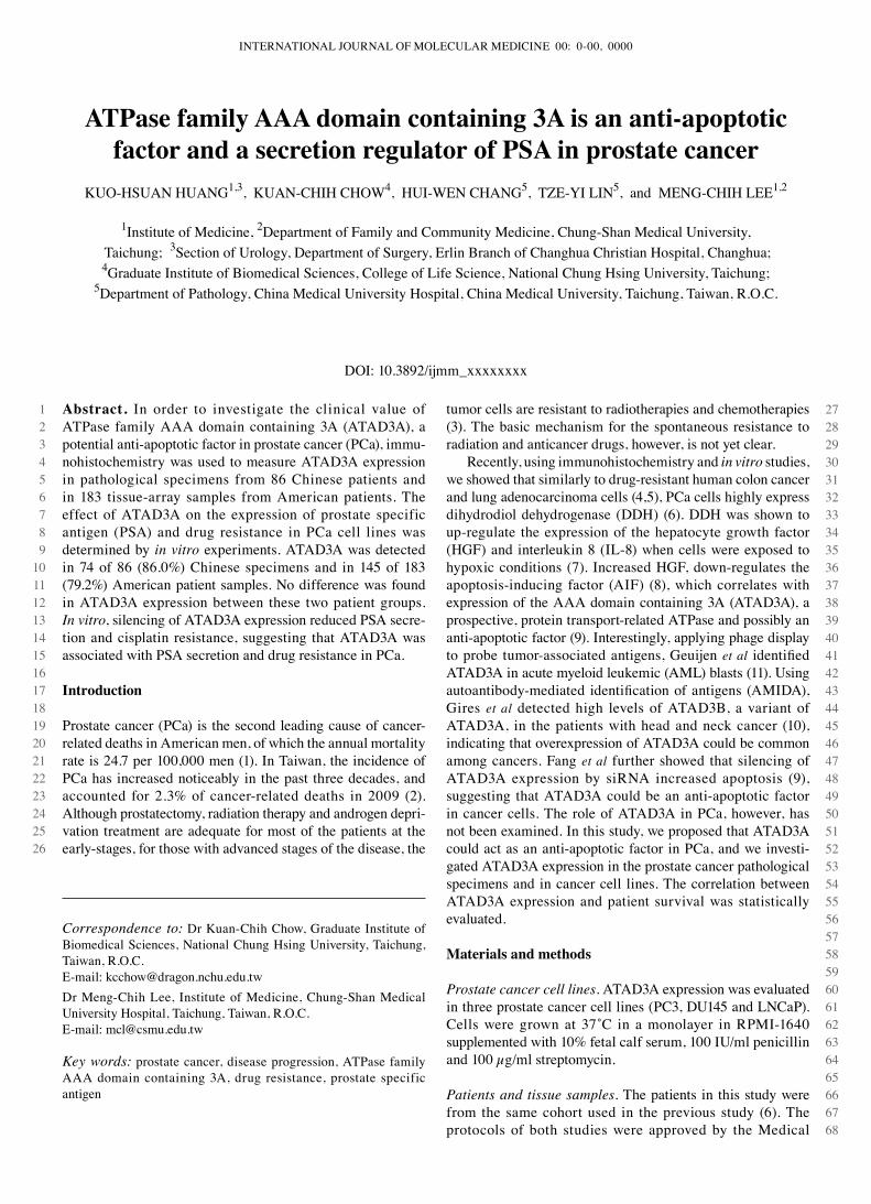
Expression of ATAD3A in prostate cancer. Immuno-histochemical analysis revealed that compared to non-tumor prostate epithelium (NTPE) (Fig. 1A) or benign hypertrophic prostate epithelia (BHPE), ATAd3A was highly expressed in the Pca tissue (Fig. 1B). As the tumor grade advanced, expression of ATAd3A became more prominent (Fig. 1c). Overexpression of ATAd3A was detected in 74 of 86 (86.0%) chinese patient specimens and in 145 of 183 (79.2%) samples from American (Fig. 1d) patients. No difference was found in ATAd3A expression between the American and chinese Pca patients (p=0.239, ratio=1.09). Results of the statistical analysis demonstrated that ATAd3A expression in Pca was associated with disease status, tumor grade, cigarette smoking, serum PSA level, lymphovascular infiltration as well as expression of the androgen receptor (AR) and of aldo-keto reductase 1c2 (AKR1c2) (Table I) (6), suggesting that ATAd3A expression was associated with the growth and invasive potential of Pca cells. As shown in Table II, ATAd3A expression was also
Figure 1. Representative examples of ATAd3A expression in prostate cancer. ATAd3A was detected by immunohistochemistry. The slide was counterstained with hematoxylin. Compared to (A) non-tumor prostate epithelial cells (original magnification x100), ATAD3A was highly expressed in (B) the prostate cancer tissue (original magnification x100); and became more prominent with (C) advanced tumor grade (original magnification x200). (D) Overexpression of ATAD3A in PCa cells of a tissue microarray of an American PCa specimen (original magnification x100).
INTERNATIONAL JOURNAL OF MOLEcULAR MEdIcINE 00: 0-00, 0000 3
123456789
101112131415161718192021222324252627282930313233343536373839404142434445464748495051525354555657585960
616263646566676869707172737475767778798081828384858687888990919293949596979899
100101102103104105106107108109110111112113114115116117118119120
Table I. Association of ATAd3A expression with clinicopathological parameters in patients with prostate cancer in Taiwan.
Expression of ATAd3A -----------------------------------------------------------------------clinicopathological parameters Low (n=12) High (n=74) p-value
Age (48-83 years) 68.9±4.1 69.4±4.7 0.872a
disease status Localized (stage A or B) (n=54) 4 50 <0.001c
Advanced (Stage c or d) (n=21) 2 19 Undetermined (n=11) 6 5Tumor grade Gleason <7 or well or moderately differentiated (n=59) 8 51 0.005c
Gleason ≥7 or poorly differentiated (n=22) 1 21 Undetermined (n=5) 3 2cigarette smoking Smoker (n=65) 4 61 0.001c
Non-smoker (n=21) 8 13Lymphovascular infiltration Positive (n=26) 0 26 0.015c
Negative (n=60) 12 48Serum PSA (ng/ml) ≤10 (n=17) 9 8 <0.001c
>10 (n=69) 3 66Expression of androgen receptor High (n=75) 7 68 0.007b
Low (n=11) 5 6Expression of aldo-keto reductase 1c2 High (n=77) 5 72 <0.001c
Low (n=9) 7 2Expression of cyclooxygenase-2 High (n=21) 4 17 0.476c
Low (n=65) 8 57
A smoker was defined as a person who smoked more than 20 packs per year. aTwo-sided p-value determined by the Mann-whitney test; btwo-sided p-value determined by the χ2 test for trend; ctwo-sided p-value determined by the χ2 test (the Fisher's exact test was used when the number in one of the cells was ≤5).
Table II. Association of ATAd3A expression with clinicopathological parameters in patients with prostate cancer in the USA.
Expression of ATAd3A ------------------------------------------------------------------------clinicopathological parameters Low (n=38) High (n=145) p-value
disease status Localized (stage A or B) (n=82) 23 59 <0.001b
Advanced (stage c or d) (n=74) 4 70 Undetermined (n=27) 11 16Tumor grade Gleason <7 or well or moderately differentiated (n=69) 20 49 0.039a
Gleason ≥7 or poorly differentiated (n=114) 18 96
aTwo-sided p-value determined by the χ2 test for trend; btwo-sided p-value determined by the χ2 test (the Fisher's exact test was used when the number in one of the cells was ≤5).
HUANG et al: ATAd3A IN PROSTATE cANcER4
123456789
101112131415161718192021222324252627282930313233343536373839404142434445464748495051525354555657585960
616263646566676869707172737475767778798081828384858687888990919293949596979899
100101102103104105106107108109110111112113114115116117118119120
correlated with the disease status and the tumor grade of American Pca patients.
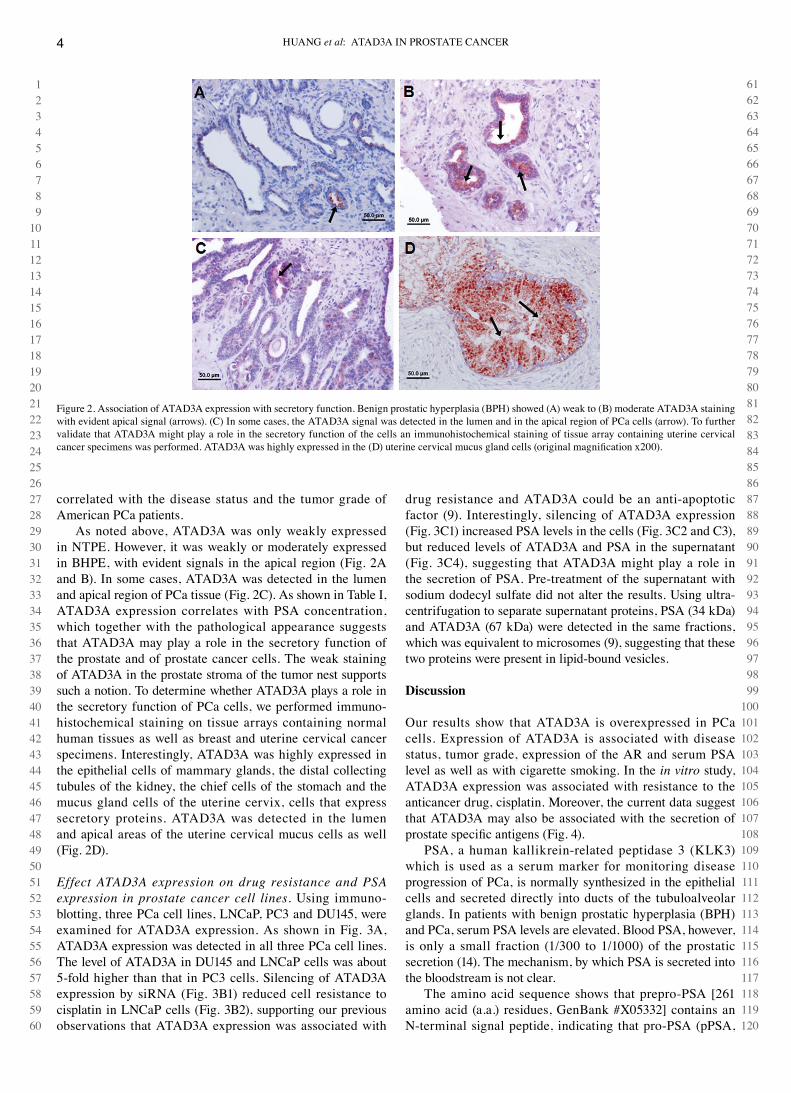
As noted above, ATAd3A was only weakly expressed in NTPE. However, it was weakly or moderately expressed in BHPE, with evident signals in the apical region (Fig. 2A and B). In some cases, ATAd3A was detected in the lumen and apical region of Pca tissue (Fig. 2c). As shown in Table I, ATAd3A expression correlates with PSA concentration, which together with the pathological appearance suggests that ATAd3A may play a role in the secretory function of the prostate and of prostate cancer cells. The weak staining of ATAd3A in the prostate stroma of the tumor nest supports such a notion. To determine whether ATAd3A plays a role in the secretory function of Pca cells, we performed immuno-histochemical staining on tissue arrays containing normal human tissues as well as breast and uterine cervical cancer specimens. Interestingly, ATAd3A was highly expressed in the epithelial cells of mammary glands, the distal collecting tubules of the kidney, the chief cells of the stomach and the mucus gland cells of the uterine cervix, cells that express secretory proteins. ATAd3A was detected in the lumen and apical areas of the uterine cervical mucus cells as well (Fig. 2d).
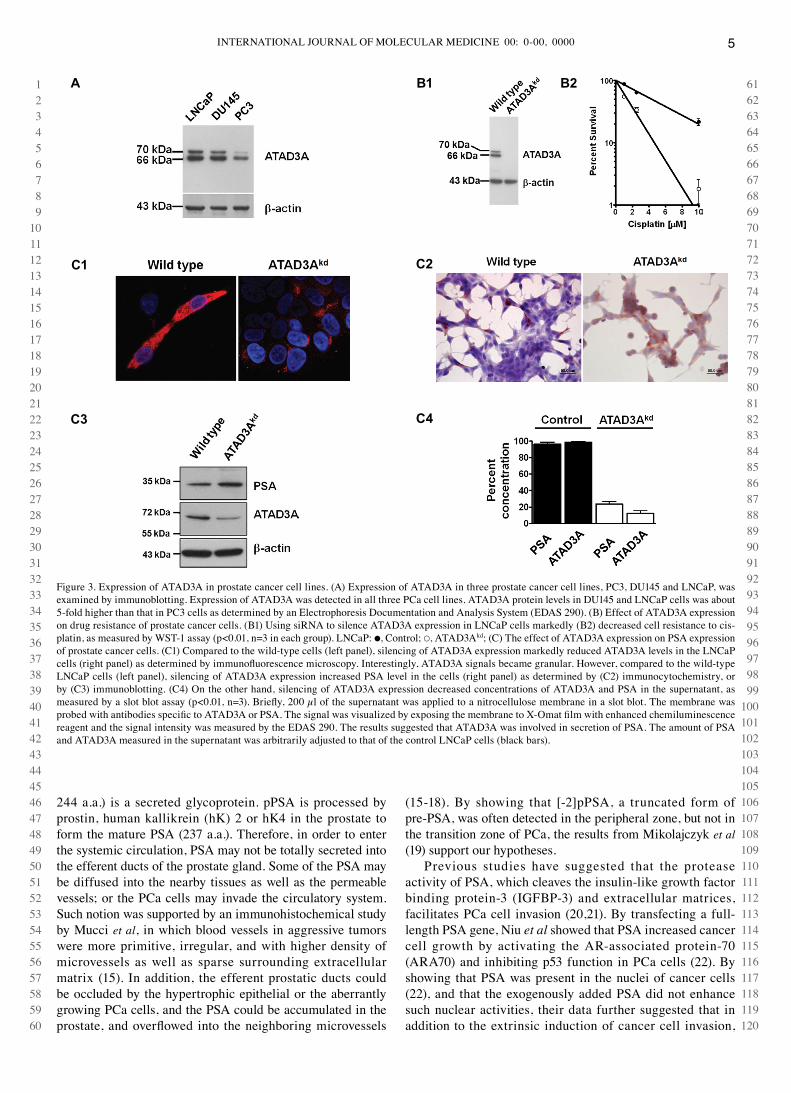
Effect ATAD3A expression on drug resistance and PSA expression in prostate cancer cell lines. Using immuno-blotting, three Pca cell lines, LNcaP, Pc3 and dU145, were examined for ATAd3A expression. As shown in Fig. 3A, ATAd3A expression was detected in all three Pca cell lines. The level of ATAd3A in dU145 and LNcaP cells was about 5-fold higher than that in Pc3 cells. Silencing of ATAd3A expression by siRNA (Fig. 3B1) reduced cell resistance to cisplatin in LNcaP cells (Fig. 3B2), supporting our previous observations that ATAd3A expression was associated with
drug resistance and ATAd3A could be an anti-apoptotic factor (9). Interestingly, silencing of ATAd3A expression (Fig. 3c1) increased PSA levels in the cells (Fig. 3c2 and c3), but reduced levels of ATAd3A and PSA in the supernatant (Fig. 3c4), suggesting that ATAd3A might play a role in the secretion of PSA. Pre-treatment of the supernatant with sodium dodecyl sulfate did not alter the results. Using ultra-centrifugation to separate supernatant proteins, PSA (34 kda) and ATAd3A (67 kda) were detected in the same fractions, which was equivalent to microsomes (9), suggesting that these two proteins were present in lipid-bound vesicles.
Discussion
Our results show that ATAd3A is overexpressed in Pca cells. Expression of ATAd3A is associated with disease status, tumor grade, expression of the AR and serum PSA level as well as with cigarette smoking. In the in vitro study, ATAd3A expression was associated with resistance to the anticancer drug, cisplatin. Moreover, the current data suggest that ATAd3A may also be associated with the secretion of prostate specific antigens (Fig. 4).
PSA, a human kallikrein-related peptidase 3 (KLK3) which is used as a serum marker for monitoring disease progression of Pca, is normally synthesized in the epithelial cells and secreted directly into ducts of the tubuloalveolar glands. In patients with benign prostatic hyperplasia (BPH) and Pca, serum PSA levels are elevated. Blood PSA, however, is only a small fraction (1/300 to 1/1000) of the prostatic secretion (14). The mechanism, by which PSA is secreted into the bloodstream is not clear.
The amino acid sequence shows that prepro-PSA [261 amino acid (a.a.) residues, GenBank #X05332] contains an N-terminal signal peptide, indicating that pro-PSA (pPSA,
Figure 2. Association of ATAd3A expression with secretory function. Benign prostatic hyperplasia (BPH) showed (A) weak to (B) moderate ATAd3A staining with evident apical signal (arrows). (c) In some cases, the ATAd3A signal was detected in the lumen and in the apical region of Pca cells (arrow). To further validate that ATAd3A might play a role in the secretory function of the cells an immunohistochemical staining of tissue array containing uterine cervical cancer specimens was performed. ATAD3A was highly expressed in the (D) uterine cervical mucus gland cells (original magnification x200).
INTERNATIONAL JOURNAL OF MOLEcULAR MEdIcINE 00: 0-00, 0000 5
123456789
101112131415161718192021222324252627282930313233343536373839404142434445464748495051525354555657585960
616263646566676869707172737475767778798081828384858687888990919293949596979899
100101102103104105106107108109110111112113114115116117118119120
244 a.a.) is a secreted glycoprotein. pPSA is processed by prostin, human kallikrein (hK) 2 or hK4 in the prostate to form the mature PSA (237 a.a.). Therefore, in order to enter the systemic circulation, PSA may not be totally secreted into the efferent ducts of the prostate gland. Some of the PSA may be diffused into the nearby tissues as well as the permeable vessels; or the Pca cells may invade the circulatory system. Such notion was supported by an immunohistochemical study by Mucci et al, in which blood vessels in aggressive tumors were more primitive, irregular, and with higher density of microvessels as well as sparse surrounding extracellular matrix (15). In addition, the efferent prostatic ducts could be occluded by the hypertrophic epithelial or the aberrantly growing Pca cells, and the PSA could be accumulated in the prostate, and overflowed into the neighboring microvessels
(15-18). By showing that [-2]pPSA, a truncated form of pre-PSA, was often detected in the peripheral zone, but not in the transition zone of Pca, the results from Mikolajczyk et al (19) support our hypotheses.
Previous studies have suggested that the protease activity of PSA, which cleaves the insulin-like growth factor binding protein-3 (IGFBP-3) and extracellular matrices, facilitates Pca cell invasion (20,21). By transfecting a full-length PSA gene, Niu et al showed that PSA increased cancer cell growth by activating the AR-associated protein-70 (ARA70) and inhibiting p53 function in Pca cells (22). By showing that PSA was present in the nuclei of cancer cells (22), and that the exogenously added PSA did not enhance such nuclear activities, their data further suggested that in addition to the extrinsic induction of cancer cell invasion,
Figure 3. Expression of ATAd3A in prostate cancer cell lines. (A) Expression of ATAd3A in three prostate cancer cell lines, Pc3, dU145 and LNcaP, was examined by immunoblotting. Expression of ATAd3A was detected in all three Pca cell lines. ATAd3A protein levels in dU145 and LNcaP cells was about 5-fold higher than that in Pc3 cells as determined by an Electrophoresis documentation and Analysis System (EdAS 290). (B) Effect of ATAd3A expression on drug resistance of prostate cancer cells. (B1) Using siRNA to silence ATAd3A expression in LNcaP cells markedly (B2) decreased cell resistance to cis-platin, as measured by wST-1 assay (p<0.01, n=3 in each group). LNcaP: ●, control; ○, ATAd3Akd; (c) The effect of ATAd3A expression on PSA expression of prostate cancer cells. (c1) compared to the wild-type cells (left panel), silencing of ATAd3A expression markedly reduced ATAd3A levels in the LNcaP cells (right panel) as determined by immunofluorescence microscopy. Interestingly, ATAD3A signals became granular. However, compared to the wild-type LNcaP cells (left panel), silencing of ATAd3A expression increased PSA level in the cells (right panel) as determined by (c2) immunocytochemistry, or by (c3) immunoblotting. (c4) On the other hand, silencing of ATAd3A expression decreased concentrations of ATAd3A and PSA in the supernatant, as measured by a slot blot assay (p<0.01, n=3). Briefly, 200 µl of the supernatant was applied to a nitrocellulose membrane in a slot blot. The membrane was probed with antibodies specific to ATAD3A or PSA. The signal was visualized by exposing the membrane to X-Omat film with enhanced chemiluminescence reagent and the signal intensity was measured by the EdAS 290. The results suggested that ATAd3A was involved in secretion of PSA. The amount of PSA and ATAd3A measured in the supernatant was arbitrarily adjusted to that of the control LNcaP cells (black bars).
A B1 B2
C1
C3 C4
C2
HUANG et al: ATAd3A IN PROSTATE cANcER6
123456789
101112131415161718192021222324252627282930313233343536373839404142434445464748495051525354555657585960
61626364656667686970717273747576777879808182838485868788899091
which was mediated by an AR-independent route (20,21), PSA might activate cancer cell growth via an intrinsic AR-dependent pathway.
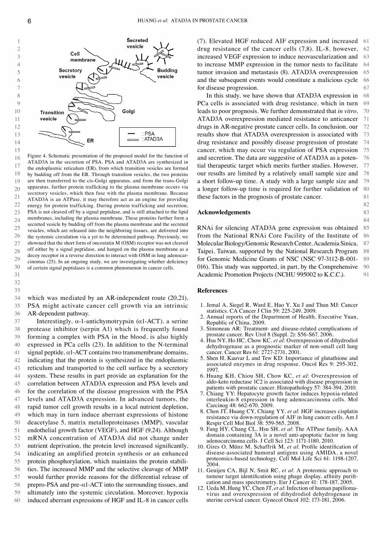
Interestingly, α-1-antichymotrypsin (α1-AcT), a serine protease inhibitor (serpin A1) which is frequently found forming a complex with PSA in the blood, is also highly expressed in Pca cells (23). In addition to the N-terminal signal peptide, α1-AcT contains two transmembrane domains, indicating that the protein is synthesized in the endoplasmic reticulum and transported to the cell surface by a secretory system. These results in part provide an explanation for the correlation between ATAd3A expression and PSA levels and for the correlation of the disease progression with the PSA levels and ATAd3A expression. In advanced tumors, the rapid tumor cell growth results in a local nutrient depletion, which may in turn induce aberrant expressions of histone deacetylase 5, matrix metalloproteinases (MMP), vascular endothelial growth factor (VEGF), and HGF (9,24). Although mRNA concentration of ATAd3A did not change under nutrient deprivation, the protein level increased significantly, indicating an amplified protein synthesis or an enhanced protein phosphorylation, which maintains the protein stabili-ties. The increased MMP and the selective cleavage of MMP would further provide reasons for the differential release of prepro-PSA and pre-α1-AcT into the surrounding tissues, and ultimately into the systemic circulation. Moreover, hypoxia induced aberrant expressions of HGF and IL-8 in cancer cells
(7). Elevated HGF reduced AIF expression and increased drug resistance of the cancer cells (7,8). IL-8, however, increased VEGF expression to induce neovascularization and to increase MMP expression in the tumor nests to facilitate tumor invasion and metastasis (8). ATAd3A overexpression and the subsequent events would constitute a malicious cycle for disease progression.
In this study, we have shown that ATAd3A expression in Pca cells is associated with drug resistance, which in turn leads to poor prognosis. we further demonstrated that in vitro, ATAd3A overexpression mediated resistance to anticancer drugs in AR-negative prostate cancer cells. In conclusion, our results show that ATAd3A overexpression is associated with drug resistance and possibly disease progression of prostate cancer, which may occur via regulation of PSA expression and secretion. The data are suggestive of ATAd3A as a poten-tial therapeutic target which merits further studies. However, our results are limited by a relatively small sample size and a short follow-up time. A study with a large sample size and a longer follow-up time is required for further validation of these factors in the prognosis of prostate cancer.
Acknowledgements
RNAi for silencing ATAd3A gene expression was obtained from the National RNAi core Facility of the Institute of Molecular Biology/Genomic Research center, Academia Sinica, Taipei, Taiwan, supported by the National Research Program for Genomic Medicine Grants of NSc (NSc 97-3112-B-001-016). This study was supported, in part, by the comprehensive Academic Promotion Projects (NcHU 995002 to K.c.c.).
References
Jemal A, Siegel R, ward E, Hao Y, Xu J and Thun MJ: cancer 1. statistics. cA cancer J clin 59: 225-249, 2009.Annual reports of the department of Health, Executive Yuan, 2. Republic of china, 2009.Simoneau AR: Treatment- and disease-related complications of 3. prostate cancer. Rev Urol 8 (Suppl. 2): S56-S67, 2006.Hsu NY, Ho Hc, chow Kc, 4. et al: Overexpression of dihydrodiol dehydrogenase as a prognostic marker of non-small cell lung cancer. cancer Res 61: 2727-2731, 2001.Shen H, Kauvar L and Tew Kd: Importance of glutathione and 5. associated enzymes in drug response. Oncol Res 9: 295-302, 1997.Huang KH, chiou SH, chow Kc, 6. et al: Overexpression of aldo-keto reductase 1c2 is associated with disease progression in patients with prostatic cancer. Histopathology 57: 384-394, 2010.chiang YY: Hepatocyte growth factor induces hypoxia-related 7. interleukin-8 expression in lung adenocarcinoma cells. Mol carcinog 48: 662-670, 2009.chen JT, Huang cY, chiang YY, 8. et al: HGF increases cisplatin resistance via down-regulation of AIF in lung cancer cells. Am J Respir cell Mol Biol 38: 559-565, 2008. Fang HY, chang cL, Hsu SH, 9. et al: The ATPase family, AAA domain containing 3A is a novel anti-apoptotic factor in lung adenocarcinoma cells. J cell Sci 123: 1171-1180, 2010.Gires O, Münz M, Schaffrik M, 10. et al: Profile identification of disease-associated humoral antigens using AMIdA, a novel proteomics-based technology. cell Mol Life Sci 61: 1198-1207, 2004.Geuijen cA, Bijl N, Smit Rc, 11. et al: A proteomic approach to tumour target identification using phage display, affinity purifi-cation and mass spectrometry. Eur J cancer 41: 178-187, 2005.Ueda M, Hung Yc, chen JT, 12. et al: Infection of human papilloma-virus and overexpression of dihydrodiol dehydrogenase in uterine cervical cancer. Gynecol Oncol 102: 173-181, 2006.
Figure 4. Schematic presentation of the proposed model for the function of ATAd3A in the secretion of PSA. PSA and ATAd3A are synthesized in the endoplasmic reticulum (ER), from which transition vesicles are formed by budding off from the ER. Through transition vesicles, the two proteins are then transferred to the cis-Golgi apparatus, and from the trans-Golgi apparatus, further protein trafficking to the plasma membrane occurs via secretory vesicles, which then fuse with the plasma membrane. Because ATAd3A is an ATPase, it may therefore act as an engine for providing energy for protein trafficking. during protein trafficking and secretion, PSA is not cleaved off by a signal peptidase, and is still attached to the lipid membranes, including the plasma membrane. These proteins further form a secreted vesicle by budding off from the plasma membrane and the secreted vesicles, which are released into the neighboring tissues, are delivered into the systemic circulation via a yet to be determined pathway. Previously, we showned that the short form of oncostatin M (OSM) receptor was not cleaved off either by a signal peptidase, and hanged on the plasma membrane as a decoy receptor in a reverse direction to interact with OSM in lung adenocar-cinomas (25). In an ongoing study, we are investigating whether deficiency of certain signal peptidases is a common phenomenon in cancer cells.
INTERNATIONAL JOURNAL OF MOLEcULAR MEdIcINE 00: 0-00, 0000 7
Remmele w and Schicketanz KH: Immunohistochemical 13. determination of estrogen and progesterone receptor content in human breast cancer. computer-assisted image analysis (QIc score) vs. subjective grading (IRS). Pathol Res Pract 189: 862-866, 1993.Sävblom c, Malm J, Giwercman A, Nilsson JA, Berglund G and 14. Lilja H: Blood levels of free-PSA but not complex-PSA signifi-cantly correlates to prostate release of PSA in semen in young men, while blood levels of complex-PSA, but not free-PSA increase with age. Prostate 65: 66-72, 2005. Mucci LA, Powolny A, Giovannucci E, 15. et al: Prospective study of prostate tumor angiogenesis and cancer-specific mortality in the health professionals follow-up study. J clin Oncol 27: 5627-5633, 2009. Spannuth wA, Sood AK and coleman RL: Angiogenesis as a 16. strategic target for ovarian cancer therapy. Nat clin Pract Oncol 5: 194-204, 2008. Igawa M, Urakami S, Shiina H, 17. et al: Immunohistochemical evaluation of proliferating cell nuclear antigen, prostate-specific antigen and alpha 1-antichymotrypsin in human prostate cancer. Br J Urol 77: 107-112, 1996.Stege RH, Tribukait B, carlström KA, Grande M, Pousette AH, 18. et al: Tissue PSA from fine-needle biopsies of prostatic carcinoma as related to serum PSA, clinical stage, cytological grade, and dNA ploidy. Prostate 38: 183-188, 1999.Mikolajczyk Sd, Millar LS, wang TJ, 19. et al: A precursor form of prostate-specific antigen is more highly elevated in prostate cancer compared with benign transition zone prostate tissue. cancer Res 60: 756-759, 2000.
cohen P, Graves Hc, Peehl dM, Kamarei M, Giudice Lc and 20. Rosenfeld RG: Prostate-specific antigen (PSA) is an insulin-like growth factor binding protein-3 protease found in seminal plasma. J clin Endocrinol Metab 75: 1046-1053, 1992.denmeade SR, Sokoll LJ, dalrymple S, 21. et al: dissociation between androgen responsiveness for malignant growth vs. expression of prostate specific differentiation markers PSA, hK2, and PSMA in human prostate cancer models. Prostate 54: 249-257, 2003.Niu Y, Yeh S, Miyamoto H, 22. et al: Tissue prostate-specific antigen facilitates refractory prostate tumor progression via enhancing ARA70-regulated androgen receptor transactivation. cancer Res 68: 7110-7119, 2008.Lilja H, cockett AT and Abrahamsson PA: Prostate specific 23. antigen predominantly forms a complex with alpha 1-antichymo-trypsin in blood. Implications for procedures to measure prostate specific antigen in serum. Cancer 70: 230-234, 1992.Lin TS, chiou SH, wang LS, 24. et al: Expression spectra of matrix metalloproteinases in metastatic non-small cell lung cancer. Oncol Rep 12: 717-723, 2004. chen dR, chu cY, chen cY, 25. et al: Expression of short form oncostatin M receptor as a decoy receptor in lung adeno-carcinomas. J Pathol 215: 290-299, 2008.