Atomic Structure of the KEOPS Complex: An Ancient Protein Kinase-Containing Molecular Machine
25
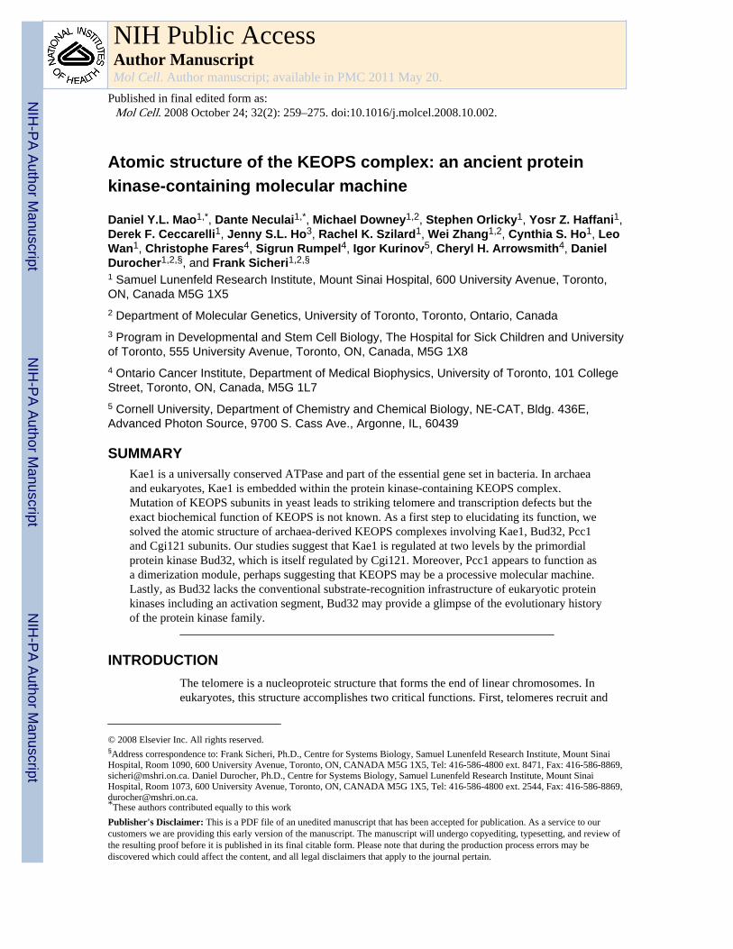
Atomic structure of the KEOPS complex: an ancient protein kinase-containing molecular machine Daniel Y.L. Mao 1,* , Dante Neculai 1,* , Michael Downey 1,2 , Stephen Orlicky 1 , Yosr Z. Haffani 1 , Derek F. Ceccarelli 1 , Jenny S.L. Ho 3 , Rachel K. Szilard 1 , Wei Zhang 1,2 , Cynthia S. Ho 1 , Leo Wan 1 , Christophe Fares 4 , Sigrun Rumpel 4 , Igor Kurinov 5 , Cheryl H. Arrowsmith 4 , Daniel Durocher 1,2,§ , and Frank Sicheri 1,2,§ 1 Samuel Lunenfeld Research Institute, Mount Sinai Hospital, 600 University Avenue, Toronto, ON, Canada M5G 1X5 2 Department of Molecular Genetics, University of Toronto, Toronto, Ontario, Canada 3 Program in Developmental and Stem Cell Biology, The Hospital for Sick Children and University of Toronto, 555 University Avenue, Toronto, ON, Canada, M5G 1X8 4 Ontario Cancer Institute, Department of Medical Biophysics, University of Toronto, 101 College Street, Toronto, ON, Canada, M5G 1L7 5 Cornell University, Department of Chemistry and Chemical Biology, NE-CAT, Bldg. 436E, Advanced Photon Source, 9700 S. Cass Ave., Argonne, IL, 60439 SUMMARY Kae1 is a universally conserved ATPase and part of the essential gene set in bacteria. In archaea and eukaryotes, Kae1 is embedded within the protein kinase-containing KEOPS complex. Mutation of KEOPS subunits in yeast leads to striking telomere and transcription defects but the exact biochemical function of KEOPS is not known. As a first step to elucidating its function, we solved the atomic structure of archaea-derived KEOPS complexes involving Kae1, Bud32, Pcc1 and Cgi121 subunits. Our studies suggest that Kae1 is regulated at two levels by the primordial protein kinase Bud32, which is itself regulated by Cgi121. Moreover, Pcc1 appears to function as a dimerization module, perhaps suggesting that KEOPS may be a processive molecular machine. Lastly, as Bud32 lacks the conventional substrate-recognition infrastructure of eukaryotic protein kinases including an activation segment, Bud32 may provide a glimpse of the evolutionary history of the protein kinase family. INTRODUCTION The telomere is a nucleoproteic structure that forms the end of linear chromosomes. In eukaryotes, this structure accomplishes two critical functions. First, telomeres recruit and © 2008 Elsevier Inc. All rights reserved. § Address correspondence to: Frank Sicheri, Ph.D., Centre for Systems Biology, Samuel Lunenfeld Research Institute, Mount Sinai Hospital, Room 1090, 600 University Avenue, Toronto, ON, CANADA M5G 1X5, Tel: 416-586-4800 ext. 8471, Fax: 416-586-8869, [email protected]. Daniel Durocher, Ph.D., Centre for Systems Biology, Samuel Lunenfeld Research Institute, Mount Sinai Hospital, Room 1073, 600 University Avenue, Toronto, ON, CANADA M5G 1X5, Tel: 416-586-4800 ext. 2544, Fax: 416-586-8869, [email protected]. * These authors contributed equally to this work Publisher's Disclaimer: This is a PDF file of an unedited manuscript that has been accepted for publication. As a service to our customers we are providing this early version of the manuscript. The manuscript will undergo copyediting, typesetting, and review of the resulting proof before it is published in its final citable form. Please note that during the production process errors may be discovered which could affect the content, and all legal disclaimers that apply to the journal pertain. NIH Public Access Author Manuscript Mol Cell. Author manuscript; available in PMC 2011 May 20. Published in final edited form as: Mol Cell. 2008 October 24; 32(2): 259–275. doi:10.1016/j.molcel.2008.10.002. NIH-PA Author Manuscript NIH-PA Author Manuscript NIH-PA Author Manuscript
Transcript of Atomic Structure of the KEOPS Complex: An Ancient Protein Kinase-Containing Molecular Machine
Atomic structure of the KEOPS complex: an ancient proteinkinase-containing molecular machine
Daniel Y.L. Mao1,*, Dante Neculai1,*, Michael Downey1,2, Stephen Orlicky1, Yosr Z. Haffani1,Derek F. Ceccarelli1, Jenny S.L. Ho3, Rachel K. Szilard1, Wei Zhang1,2, Cynthia S. Ho1, LeoWan1, Christophe Fares4, Sigrun Rumpel4, Igor Kurinov5, Cheryl H. Arrowsmith4, DanielDurocher1,2,§, and Frank Sicheri1,2,§
1 Samuel Lunenfeld Research Institute, Mount Sinai Hospital, 600 University Avenue, Toronto,ON, Canada M5G 1X52 Department of Molecular Genetics, University of Toronto, Toronto, Ontario, Canada3 Program in Developmental and Stem Cell Biology, The Hospital for Sick Children and Universityof Toronto, 555 University Avenue, Toronto, ON, Canada, M5G 1X84 Ontario Cancer Institute, Department of Medical Biophysics, University of Toronto, 101 CollegeStreet, Toronto, ON, Canada, M5G 1L75 Cornell University, Department of Chemistry and Chemical Biology, NE-CAT, Bldg. 436E,Advanced Photon Source, 9700 S. Cass Ave., Argonne, IL, 60439
SUMMARYKae1 is a universally conserved ATPase and part of the essential gene set in bacteria. In archaeaand eukaryotes, Kae1 is embedded within the protein kinase-containing KEOPS complex.Mutation of KEOPS subunits in yeast leads to striking telomere and transcription defects but theexact biochemical function of KEOPS is not known. As a first step to elucidating its function, wesolved the atomic structure of archaea-derived KEOPS complexes involving Kae1, Bud32, Pcc1and Cgi121 subunits. Our studies suggest that Kae1 is regulated at two levels by the primordialprotein kinase Bud32, which is itself regulated by Cgi121. Moreover, Pcc1 appears to function asa dimerization module, perhaps suggesting that KEOPS may be a processive molecular machine.Lastly, as Bud32 lacks the conventional substrate-recognition infrastructure of eukaryotic proteinkinases including an activation segment, Bud32 may provide a glimpse of the evolutionary historyof the protein kinase family.
INTRODUCTIONThe telomere is a nucleoproteic structure that forms the end of linear chromosomes. Ineukaryotes, this structure accomplishes two critical functions. First, telomeres recruit and
© 2008 Elsevier Inc. All rights reserved.§Address correspondence to: Frank Sicheri, Ph.D., Centre for Systems Biology, Samuel Lunenfeld Research Institute, Mount SinaiHospital, Room 1090, 600 University Avenue, Toronto, ON, CANADA M5G 1X5, Tel: 416-586-4800 ext. 8471, Fax: 416-586-8869,[email protected]. Daniel Durocher, Ph.D., Centre for Systems Biology, Samuel Lunenfeld Research Institute, Mount SinaiHospital, Room 1073, 600 University Avenue, Toronto, ON, CANADA M5G 1X5, Tel: 416-586-4800 ext. 2544, Fax: 416-586-8869,[email protected].*These authors contributed equally to this workPublisher's Disclaimer: This is a PDF file of an unedited manuscript that has been accepted for publication. As a service to ourcustomers we are providing this early version of the manuscript. The manuscript will undergo copyediting, typesetting, and review ofthe resulting proof before it is published in its final citable form. Please note that during the production process errors may bediscovered which could affect the content, and all legal disclaimers that apply to the journal pertain.
NIH Public AccessAuthor ManuscriptMol Cell. Author manuscript; available in PMC 2011 May 20.
Published in final edited form as:Mol Cell. 2008 October 24; 32(2): 259–275. doi:10.1016/j.molcel.2008.10.002.
NIH
-PA Author Manuscript
NIH
-PA Author Manuscript
NIH
-PA Author Manuscript

activate telomerase, a reverse transcriptase that polymerizes short TG-rich repeats on the 3′end of chromosomes (Gilson and Geli, 2007; Smogorzewska and de Lange, 2004; Verdunand Karlseder, 2007). Second, telomeres protect chromosome ends from nucleolyticdegradation, unscheduled DNA repair and activation of the DNA damage checkpoints. Thisactivity, termed telomere capping, is essential for cell viability and genomic integrity(Feldser et al., 2003; Gilson and Geli, 2007; Verdun and Karlseder, 2007).
To identify genes that modulate telomere capping, we screened the viable gene deletioncollection of Saccharomyces cerevisiae for suppressors of the temperature sensitivity ofcdc13-1 (Downey et al., 2006), an allele of the telomere capping protein Cdc13. This screenidentified Cgi121 as a protein that promotes telomere uncapping triggered by cdc13-1.Cgi121 is part of a five member complex in yeast termed the KEOPS (or EKC) complex(Downey et al., 2006; Kisseleva-Romanova et al., 2006). In addition to Cgi121, which hasno recognizable domains or motifs, the KEOPS complex is composed of a protein kinase,Bud32; a putative ATPase, Kae1 (see below); and two other proteins of small size with nodetectable enzymatic function or recognizable domains, Pcc1 and Gon7. The KEOPScomplex is conserved in humans, with the exception of Gon7 which could not be detected.Interestingly, human orthologues of Pcc1 have been characterized as cancer antigens,indicating that the upregulation of KEOPS subunits might be associated with cancer.
In addition to promoting telomere uncapping, the KEOPS complex profoundly affectstelomere length homeostasis and is required for cell growth in budding yeast. Indeed,deletion of BUD32 and GON7 results in dramatic telomere shortening, a drastic reduction inthe ability of telomerase to add new telomere sequences to DNA double-strand breaks andsevere growth defects. In fact, every component of KEOPS promotes telomere uncappingand elongation (Downey et al., 2006; Gatbonton et al., 2006; Shachar et al., 2008) indicatingthat the proteins function collectively as a unit.
In parallel, the KEOPS/EKC complex was identified as a novel transcription factor(Kisseleva-Romanova et al., 2006). Components of KEOPS are nuclear proteins thatassociate with chromatin in vivo and are important for inducible gene transcription(Kisseleva-Romanova et al., 2006). It is not clear how KEOPS promotes transcription butKEOPS mutant yeast strains are defective for the recruitment of coactivators, such as SAGAand Mediator, to target genes. Collectively, these studies indicate that KEOPS promotes theaccess of nucleases and telomerase to the chromosome end as well as the access oftranscriptional complexes to chromatin. A model that explains these seemingly disparateobservations is that KEOPS regulates accessibility to chromatin, perhaps by modulating itshigher order structure.
Remarkably, the KEOPS component Kae1 is universally conserved in all three domains oflife. A Kae1 homolog is present in the smallest bacterial genome, Mycoplasma genitalium,in the smallest archaeal genome, Nanoarchaeum equitans, and is an essential gene in allbacterial species tested (Arigoni et al., 1998). Kae1 is an ATPase of the ASKHA (acetate &sugar kinase/Hsp70/actin) superfamily. As proteins of this superfamily carry out diversefunctions, it is not clear what specific biochemical function Kae1 possesses althoughPasteurella haemolytica Kae1 can act as an O-sialoglycoprotein protease (Abdullah et al.,1991) and Pyrococcus abyssi Kae1 is proposed to be an apurinic (AP) endonuclease (Heckeret al., 2007). Since these two functions are dramatically different, the conserved function ofKae1 remains an open question. No other subunits of KEOPS have been identified inbacteria. However, in archaea, a nearly complete KEOPS complex can be identified (seebelow). This observation suggests that Kae1-interacting proteins may have evolved to bepart of a regulatory infrastructure that modulates the activity of Kae1. Given the importanceof KEOPS in telomere biology and gene transcription, and evidence that KEOPS is an
Mao et al. Page 2
Mol Cell. Author manuscript; available in PMC 2011 May 20.
NIH
-PA Author Manuscript
NIH
-PA Author Manuscript
NIH
-PA Author Manuscript
evolutionarily conserved molecular machine, we determined the atomic structure of KEOPScomplexes reconstituted from archaeal proteins and probed them for functional significanceboth in vitro and in vivo. Our findings validate the importance of the KEOPS architecturethat we discovered and provide a framework for deciphering the functional relationshipsbetween KEOPS subunits.
RESULTSArchitecture of the KEOPS complex
We identified homologs of Pcc1 and Cgi121 in archaea by iterative BLAST searches(Figures S3 & S4). We did not identify a Gon7 homolog in either archaea or the humangenome (data not shown). We then determined the underlying architecture of the KEOPScomplex using pull down experiments with recombinant forms of archaeal Kae1 (residues1-328 of Methanococcus jannaschii MJ1130); Pcc1 (Pyrococcus furiosus PF2011); Cgi121(M. jannaschii MJ0187); or Bud32 (residues 328-535 of MJ1130). Each protein wasexpressed as a GST fusion in Escherichia coli and purified to near homogeneity. The GSTtags were used to immobilize proteins on glutathione-Sepharose resin or cleaved with TEVprotease, followed by purification, to isolate the untagged protein. GST-Pcc1 immobilized toglutathione resin selectively retained Kae1, but not Bud32 (Figure 1A) or Cgi121 (Figure1B). In contrast, GST-Cgi121 retained Bud32 but not Kae1 (Figure 1C) while GST-Kae1retained Bud32 (Figure 1D). Lastly, Bud32 and Cgi121 bound to immobilized GST-Pcc1only in the presence of Kae1 (Figure 1B) and Kae1 bound to immobilized GST-Cgi121 onlyin the presence of Bud32 (Figure 1C). These data indicate that KEOPS is organized in alinear fashion where Cgi121 binds to Bud32, which binds to Kae1, which in turn binds toPcc1. A model depicting the architecture of the KEOPS complex is shown in Figure 1E.
X-ray and NMR structure determinationThe linear topology of KEOPS enabled us to crystallize and solve binary and ternarysubcomplexes by X-ray crystallography as a means to develop an atomic model of the wholeKEOPS complex. We solved a total of six structures using X-ray and NMR methods (seeTable 1 & S1 for data collection and refinement statistics). Structure based sequencealignments for Bud32, Kae1, Pcc1, and Cgi121 are shown in Figures S1–5. A compositemodel for a monomer of the KEOPS complex generated from the subcomplex structures isshown in Figure 1F.
The following sections describe the structure and functional characterizations of the fourindividual KEOPS subunits followed by characterizations of the three binary interactingpairs of KEOPS subunits. For clarity, the residue numbering corresponds to the M.jannaschii proteins (except Pcc1, for which numbers are P. furiosus), and, when appropriate,residue numbers corresponding to the S. cerevisiae ortholog are shown in parentheses. SeeTable S3 and Figures S1–5 for residue equivalence.
Individual subunit analysesi. Bud32 structure and function—The crystal structure of Bud32 reveals a bilobalarchitecture, characteristic of eukaryotic protein kinases, but with major atypical features(Figure 2A). The N-lobe of canonical protein kinases consists minimally of a five strandedβ-sheet and a flanking helix αC. These features are conserved in the primary (Figure S1) andatomic structures of Bud32 (Figure 2A) with the exception of strand β1, which is present butdisordered in two different crystal complexes analyzed. The C-lobe of Bud32 is unusuallysmall, consisting of two β-strands (β7 & β8) and five α-helices (αD, αE, αF, αJ & αR) ofwhich only three (αD, αE & αF) have direct equivalents in canonical protein kinasestructures (Figure 2A). Strikingly, the C-lobe of Bud32 lacks the infrastructure normally
Mao et al. Page 3
Mol Cell. Author manuscript; available in PMC 2011 May 20.
NIH
-PA Author Manuscript
NIH
-PA Author Manuscript
NIH
-PA Author Manuscript
responsible for phospho-acceptor site recognition, including the activation segment (A-loop), which is substituted by a direct link between strand β8 and helix αF (Figure 2A).Despite its atypical structure, Bud32 retains conserved elements required for ATP bindingand catalysis (Figure 2A, Table S2) and indeed can auto-phosphorylate in vitro (Figure 2B)(Facchin et al., 2002) in a manner dependent on the integrity of conserved catalyticelements.
The closest structural homologues of Bud32 are the RIO kinases (Rio1 and Rio2) (Figure2C, top panel). The Rio kinases also exhibit an abbreviated C-lobe and the absence of an A-loop (for comparison with PKA, which has an A-loop, see Figure 2C, bottom panel). Thequestion of how a protein kinase recognizes and phosphorylates its substrate in the absenceof an A-loop remains an open question.
To test the biological importance of unique features identified in archaeal KEOPS structures,we mutated residues in the budding yeast counterpart identified by structure based sequencealignments. These mutants were then introduced into S. cerevisiae strains on low copyplasmids in a context where the chromosomal copy of the KEOPS component was deleted.We assessed two biological readouts for KEOPS function: cell growth and telomere length.Consistent with Bud32 being competent for phospho-transfer in vitro, its ATP binding andcatalytic residues are required for full biological function in yeast [Figure 2DE, Table S3 &(Downey et al., 2006)]. Indeed, the catalytic cleft mutants K360A (K52A), D467A(D182A), N456A (N166A), D451A (D161A), D451R (D161R), N456A/D467A (N166A/D182) all result in cell growth defects and telomere shortening. The severity of the twophenotypes imparted by the BUD32 mutations parallel each other, and also parallel the lossof protein kinase activity in vitro caused by the equivalent mutation in archaeal Bud32(Figure 2B). Interestingly, none of the kinase inactivating mutants displayed phenotypes asstrong as bud32Δ, indicating that the phosphotransfer activity of Bud32 is not its solefunction. Finally, the catalytic cleft mutations tested do not destroy protein structure as eachmutant binds archaeal Cgi121 in vitro (Figure S6A) and mutant Bud32 expression levels inyeast are similar to wild type levels (Figure S6B).
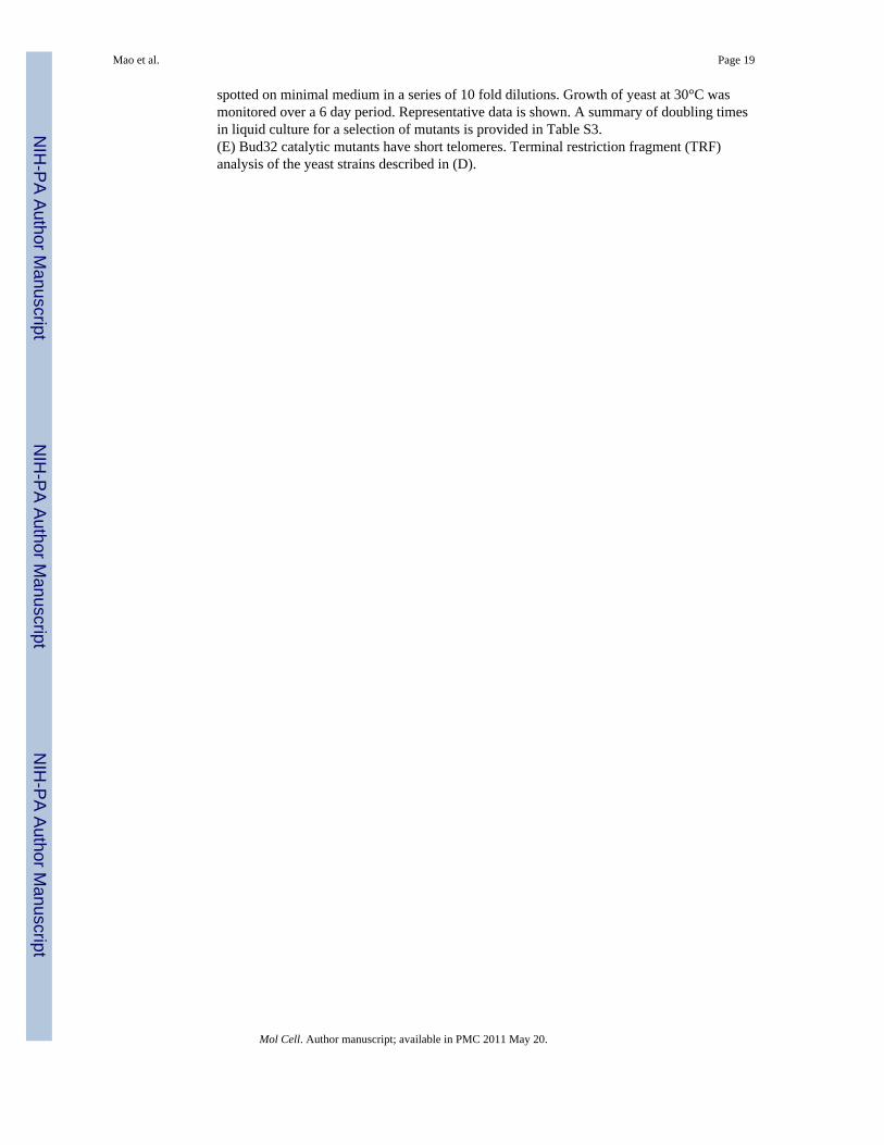
ii. Kae1 structure and function—We solved Kae1 structures corresponding to twospecies (M. jannaschi, T. acidophilum) in three different protein complexes (Kae1-Bud32,Kae1-Bud32-Cgi121 & Kae1-Pcc1) without nucleotide. The structures of Kae1 are verysimilar to each other and to the published structure of P. furiosus Kae1 (PDB 2IVO, 2IVPand 2IVN) (1.20 Å, 1.17 Å & 1.20 Å Cα rmsd respectively). As the structure of the isolatedKae1 subunit was described previously (Hecker et al., 2007), we only describe features mostrelevant to this study. Kae1 belongs to the actin-like ATPase domain superfamily ofproteins, also known as the ASKHA superfamily. The ASKHA fold serves two broadfunctions: 1) as conformational hydrolases (e.g. Hsp70, actin) and 2) as metabolite kinases(e.g. glycerol kinase). The ASKHA fold consists of two subdomains (denoted I & II) withhighly similar RNase H-like folds (Hurley, 1996). The fold of each subdomain ischaracterized by a central five strand β-sheet and flanking α-helices. The active site residesin the cleft region between the two subdomains, sandwiched between the two β-sheets. ATPbinding is dependent on extensive protein contacts with the adenine ring and sugar. ATPasecatalytic function is dependent on bound metal ions. Ribbons and schematic representationsin Figures 3AB summarize the conserved nucleotide and metal ion binding infrastructure ofKae1.
The specific function of ASKHA family members is attributable in part to the presence ofunique inserts in the core fold (Hurley, 1996). Kae1 possess two unique inserts (denotedspecific inserts 1 & 2) that comprise three surface exposed α-helices (α4, α5 & α6) and along intervening linker between α5 and α6 for the first insert, as well as a C-terminal tail
Mao et al. Page 4
Mol Cell. Author manuscript; available in PMC 2011 May 20.
NIH
-PA Author Manuscript
NIH
-PA Author Manuscript
NIH
-PA Author Manuscript

adopting an extended conformation interspersed by two short 310 helices for the secondinsert (Figure 3C). The C-terminal tail threads across the catalytic cleft of Kae1. Comparisonof Kae1 with other ASKHA family members, with kinase or hydrolase function, did not giveinsight into its specific function.
To begin our characterization of Kae1, we purified wild type and site-directed mutants of M.jannaschii Kae1 and assessed ATP-binding, ATPase activity, DNA binding and APendonuclease activity. We found that wild type Kae1 binds ATP with a Kd of 27 μM (TableS3), which is well below intracellular ATP levels. We also detected low but reproducibleATPase activity that was not affected by other KEOPS subunits (data not shown) butdepended on the presence of metal ion binding residues (Figure S7A). We did not detectapurinic (AP) endonuclease or DNA binding activity (Figure S7B–D). Mutation of residuesthat coordinate ATP, but not the metal ion, perturbed the ATP-binding function of Kae1(Table S3). None of the Kae1 mutations destroyed protein fold since all mutant proteinscould bind Pcc1 in vitro (Figure S7E). To assess the contribution of ATP- and metal ion-binding to Kae1 biological function, we made kae1 mutant yeast strains that perturbed onefunction or the other. kae1 mutants that perturbed ATP-binding in vitro or that disrupted theputative metal ion binding site, resulted in a phenocopy of the kae1Δ mutation in yeast [nearabsence of cell growth (Figure S7F)] despite wild type protein expression levels (FigureS6C). This effect on yeast growth precluded a full analysis of the telomere lengthphenotype. However, we were able to culture the ATP-binding mutant (E176R) and, aspredicted, this mutant had short telomeres (Figure 3D).
iii. Pcc1 structure and function—The Pcc1 structure was solved alone and in complexwith Kae1. The Pcc1 structure consists of a three-stranded anti-parallel β-sheet packed withtwo α helices on one face, and solvent exposed on the other (Figure 4A top panel). This foldis shared with over 700 functionally diverse proteins (DALI Z score >2.0). A close structuralhomologue of Pcc1 is the KH domain (Z score=6.7), which functions as a versatile RNA,ssDNA or RNA hairpin binder (Figure S8A). The ability of the KH domain to bind nucleicacids is dependent on a signature motif (GXXG loop), located in a kink in helix α1(Valverde et al., 2008), which is notably absent in Pcc1 (Figure S8A).
Pcc1 forms a tightly interdigitated dimer with a contact area of 555 Å2 (1110 Å2 buriedsurface area) (Figure 4AB). Dimerization involves an anti-parallel arrangement of twomonomers giving rise to a continuous six-stranded anti-parallel β-sheet packed by four αhelices on the same face. Interestingly, this mode of dimerization is widely used by KHdomains (Figure S8A). Indeed, Pcc1 dimerizes in solution, under all concentrations tested(Figure S8B). Equilibrium centrifugation analysis of a purified Kae1-Pcc1 complex revealeda 2:2 binding stochiometry (Figure S8C), suggesting that Pcc1 functions as a dimerizationmodule in the presence of other KEOPS subunits. Our attempts to disrupt Pcc1 homo-dimerization by mutation yielded insoluble protein (data not shown), suggesting that Pcc1 isan obligate dimer, a notion supported by the conserved hydrophobic nature of the dimerinterface (Figure 4B & S9A).
iv. Cgi121 structure—We solved the structures of human Cgi121 alone as well asarchaeal Cgi121 alone and in complex with Bud32 and Kae1 (Figures 4C & 6A). Thehuman and archaeal Cgi121 structures are very similar (2.0Å Cα rmsd), consisting of acentral anti-parallel β-sheet abutted by two and five α helices on either face. In surveying thePDB, we identified a close structural ortholog from P. furiosus (PDB 1ZD0). Aside fromthis protein, we did not identify other proteins with a similar fold.
Mao et al. Page 5
Mol Cell. Author manuscript; available in PMC 2011 May 20.
NIH
-PA Author Manuscript
NIH
-PA Author Manuscript
NIH
-PA Author Manuscript

The Kae1-Bud32 interactionBinding of Bud32 to Kae1 involves a contact surface of 1583 Å2 (3167 Å2 buried surface)(Figure 5A) that is highly conserved across Kae1 and Bud32 orthologs (Figure S9BC &S10A). Bud32 engages Kae1 using the forward projecting surfaces of both its N- and C-terminal lobes, which clamps onto subdomain II of Kae1. Overall, the binding mode ofBud32 to Kae1 observed in this study is very similar to the Kae1-Bud32 structure recentlyreported by Hecker et al. (2008) (PDB 2VWB, 1.75 Å Cα rmsd). Binding to Bud32 does notgreatly alter the conformation of Kae1 (1.2 Å Cα rmsd) with the exception of the β3-α1linker. The reverse cannot be assessed, as we do not have a Bud32 structure in the absenceof Kae1.
The N-lobe structural elements of Bud32 involved in Kae1 binding include the C-terminalend of helix αC and its linker to strand β3 (Figure 5B). C-lobe structural elements involvedin Kae1 binding include the αE-β7 linker containing the catalytic loop, helix αF, helix αRand the extended C-terminal tail. Kae1 structural elements involved in Bud32 bindinginclude the β3′ strand, the extended linker sequence between helices α5 and α6 (withinKae1-specific insert 1), helix α1′, and helix α2′. The only contacts to subdomain I of Kae1are mediated by the C-terminal tail of Bud32 which interacts with the C-terminal region ofhelix α1 and the β4-α2 linker.
The Bud32-Kae1 interface is composed by a mixture of hydrophobic, electrostatic andhydrogen bonding interactions. Interesting interaction clusters include a salt networkbetween Bud32 residues R380 and K365 and Kae1 residues E236 and E233, between Bud32K489 and Kae1 E148, and between the catalytic base of Bud32, D451, and Kae1 R287.Hydrophobic interactions include a network involving V486 and V482 in Bud32 and L150and K193 in Kae1 (Figure 5B top panel). Conspicuously, R534 in the C-terminal tail ofBud32 forms a hydrogen bond with N132 in Kae1 immediately adjacent to the metal ionbinding site (Figure 5B bottom panel). The R534 interaction correlates with a largedisplacement of the β3-α1 linker in subdomain I of Kae1, giving rise to a partial opening ofthe ATP binding cleft and a reduction of inter-subdomain contacts (Figure 5B bottompanel).
To assess the functional significance of the Bud32-Kae1 interaction, we probed theinteraction surfaces by site-directed mutagenesis. The K365E, R380E, V486Y, and K489Esingle site mutations in Bud32 and E233R and E236K single site mutations in Kae1markedly reduced the Bud32-Kae1 interaction in vitro (Figure 5C) without perturbingbinding to Cgi121 and Pcc1 (Figures 5D). In contrast, deletion of the C-terminal tail ofBud32 had no impact on the Bud32-Kae1 interaction (Figure S11A). We then made theequivalent mutations in yeast Kae1 and Bud32 to test for functional relevance in vivo. Asshown in Figure S10B and S6C, the Kae1 E233R (E292R) and E236K (E295K) mutationsseverely impaired growth in budding yeast without adversely affecting protein expression.Similarly, the Bud32 single site mutations K489E (R204E), V486Y (V201Y) and doublesite mutations K365E/R380E (K57E/R72E) impaired Bud32 function in vivo (Figure S10C)without adversely affecting protein expression (Figure S6B). These results indicate that thebinding of Bud32 to Kae1 is critical for KEOPS function, a conclusion also reached byHecker et al. (2008). As the catalytic base D451 (D161) of Bud32 also lies at the Kae1contact surface, a perturbation of binding to Kae1 might also explain the more potent in vivoeffect caused by the D451R (D161R) mutant relative to other Bud32 active site mutants(Figure 2D). Consistent with this notion, combination mutants targeting both Bud32catalytic residues and contact residues for Kae1 had more pronounced phenotypic effectsapproaching the severity of the BUD32 deletion (Figure S10E, Table S3) without adverselyaffecting protein expression (Figure S6B). Finally, we tested the importance of the Bud32 C-terminal tail for biological function. Despite the fact that deletion of the tail did not affect
Mao et al. Page 6
Mol Cell. Author manuscript; available in PMC 2011 May 20.
NIH
-PA Author Manuscript
NIH
-PA Author Manuscript
NIH
-PA Author Manuscript

Bud32 binding to Kae1 in vitro, its deletion in yeast Bud32 had a severe growth defect(Figure S6B, Figure S11B). This result suggests a possible role for the Bud32 tail inregulating Kae1 function.
Kae1 may be a substrate of Bud32Since Kae1 binds close to the catalytic cleft of Bud32, we explored the possibility that Kae1is a substrate of Bud32. Indeed, Bud32 can phosphorylate Kae1 in vitro (Figure S11C).Secondly, if phosphorylation of Kae1 by Bud32 is functionally relevant, the acceptor sites inKae1 should be conserved in Kae1 proteins from species containing a Bud32 ortholog. Of11 conserved S/T residues, T308 and T316 in M. jannaschii Kae1 localize to the Kae1-specific insert 2 (Figure S11D). This insert is notable for its meandering path across thecatalytic cleft of Kae1 as well as for its conservation across archaeal and eukaryotic Kae1orthologs but not bacterial counterparts that lack a Bud32 orthologue (Figure S5). Theextended and surface exposed nature of the Kae1 insert 2 also leaves it more accessible tothe active site of Bud32. In contrast, the remaining 9 conserved S/T sites are deeply buriedin the fold of Kae1 or integrated into secondary structure elements.
If Bud32 regulates Kae1 function by phosphorylation of sites within the Kae1-specific insert2, then deleting the insert might have a pronounced effect in vivo. Supporting this idea,partial (1-372) or full deletion (1-364) of the C-terminal tail of yeast Kae1 caused a severegrowth defect (Figure S11E). Moreover, a T308A (T369A) but not T316A (T377A) mutantshowed a similar phenotype, without adversely affecting protein expression (Figure S6C).This result is consistent with the notion that T308 (T369) in Kae1 is a Bud32 phospho-regulatory site. Together, these data support the possibility that Kae1 is a biologicallyrelevant substrate of Bud32 and that one phospho-acceptor site lies within the Kae1-specificinsert 2. However, deletion of the Kae1-specific insert 2 did not abolish Bud32phosphorylation of Kae1 in vitro (data not shown), implying that other residues might alsobe targeted by Bud32 in vitro.
The Bud32-Cgi121 interactionThe binding mode of Cgi121 to Bud32 was revealed in a Bud32-Cgi121-Kae1 ternarystructure (Figures 6A & S12A). Cgi121 binds exclusively to the N-lobe of Bud32, to asurface composed by helix αC, the adjacent strand β4 and the β2-β3 linker (Figure 6B). Thereciprocal binding surface on Cgi121 is composed by helices α2 and α8. The total contactsurface is 700 Å2 (1400 Å2) and relative to the two other inter-subunit contact surfaces ofthe KEOPS complex, it is the least conserved (Figure S9CD).
The Cgi121-Bud32 interface is composed of a combination of hydrophobic, hydrogenbonding, and electrostatic interactions (Figure 6B). Hydrophobic interactions consist of twoclusters. The first is formed by L353 and Y352 at the tip of the Bud32 β2-β3 loop, and L134in Cgi121 helix α8. The second is formed by A389 and V404 in Bud32 and A138 and L139in Cgi121. Salt interactions link K392 in Bud32 with D135 in Cgi121, and D403 in Bud32with K46 in Cgi121. Lastly, Y352 in Bud32 hydrogen bonds with H46 in Cgi121, and K46in Cgi121 hydrogen bonds with the backbone carbonyl of D403 in Bud32.
Of all inter-subunit contacts characterized, Bud32 displays the largest conformationalchange upon binding to Cgi121 (1.38 Å Cα rmsd by comparison to 0.9 Å Cα rmsd for Kae1).The most apparent conformational change consists of a 10 Å shift of the β2-β3 loop (FigureS12B). This shift appears stabilized by the involvement of Y352 in Bud32 in bothhydrophobic and hydrogen bond interactions with Cgi121. All other N-lobe elements ofBud32 including helix αC and the entire C-lobe appear minimally affected by Cgi121binding.
Mao et al. Page 7
Mol Cell. Author manuscript; available in PMC 2011 May 20.
NIH
-PA Author Manuscript
NIH
-PA Author Manuscript
NIH
-PA Author Manuscript

Cgi121 stimulates Bud32 activityWe first examined the impact of Cgi121 mutations on the Cgi121-Bud32 interaction. Whilethe L134E, L139R and A138R single site mutations had no effect on Bud32 binding in vitro(Figure 6C) the L134E/A138R double mutation perturbed binding severely. We confirmedby NMR analysis that the overall fold of Cgi121 was not affected by this double mutation(Figure S12C). The dispersed nature of the HSQC spectra produced by the double mutantwas consistent with a folded state and the close correspondence of the HSQC spectraproduced by wild type and mutant Cgi121 proteins was consistent with the absence of grossstructural changes.
The binding of Cgi121 to the Bud32 N-lobe on a site centered on helix αC is reminiscent ofthe intermolecular mode of Cdk (cyclin-dependent protein kinase) regulation by cyclins(Jeffrey et al., 1995). In the case of Cdks, binding to cyclins reorients helix αC to aproductive position that supports catalysis. While the position of αC in Bud32 is notoutwardly affected by Cgi121, large conformational changes to the β2-β3 loop are apparent.This led us to test if Cgi121 influences the catalytic activity of Bud32. Cgi121 was a potentactivator (>20 fold) of Bud32 auto-phosphorylation in vitro (Figures 2B and 6D). Inaddition, this activator function was dependent on the ability of Cgi121 to bind Bud32, asthe Cgi121 L134E/A138R double mutant did not stimulate Bud32 kinase activity (Figure6D). To investigate the impact of a loss of binding between Cgi121 and Bud32 in vivo, wemade the analogous double mutation (L172E/I176R) in yeast Cgi121. As shown in Figure6E, the Cgi121 (L172E/I176R) strain displayed a slow growth phenotype in the range of thecgi121Δ mutation, strongly suggesting that binding to Bud32 is central to Cgi121’sbiological function.
A role for Cgi121 in regulating Bud32 protein kinase activity is consistent with theobservation that the phenotypes imparted by the cgi121Δ mutation are milder than thosecaused by bud32Δ or kae1Δ mutations (Downey et al., 2006), and more closely resemble thephenotype of Bud32 catalytic site mutants. Moreover, Cgi121 is expressed at much lowerlevels than the other members of KEOPS (Ghaemmaghami et al., 2003). This observation isalso reminiscent of the cyclin-Cdk paradigm in which the activity of Cdks is regulated byproteosome-mediated destruction of cyclins, while Cdk levels remain fixed. This similarityled us to test if yeast Cgi121 and Bud32 are unstable proteins in GAL1 promoter shut offexperiments. We observed that Cgi121 is highly unstable with a half life of ~30 min whereasBud32 remained stable during the experiment (Figure 6F). Consistent with the possibilitythat the binding of Cgi121 to Bud32 enhances its stability in vivo, the expression level of theCgi121 (L172E/I176R) mutant was unexpectedly low relative to the wild type protein(Figure S6D). Together, these data suggest that Cgi121 may indeed function as a positiveregulator of Bud32 function.
The Kae1-Pcc1 interactionWe were unable to crystallize a 2:2 stochiometric complex of Pcc1 with Kae1 butserendipitously crystallized and solved the structure of a 2:1 complex resulting frompurification of Kae1 in the presence of an excess of Pcc1 (Figure 7A). Pcc1 and Kae1 do notundergo significant conformational changes in response to complex formation (0.73Å &1.20Å Cα rmsd, respectively). The Kae1-Pcc1 binding interface is less extensive (contactarea: 922 Å2; buried area: 1844 Å2) and less conserved (Figure S9AB) than the Kae1-Bud32binding interface.
Kae1 binds exclusively to one monomer of the Pcc1 dimer. Kae1 helices α1 and α2 packagainst helices α1 and α2 in Pcc1, giving rise to a tightly packed four-helix bundle (FigureS13A). Additional contacts are made between the Kae1 310-1 helix and the Pcc1 helix α2.
Mao et al. Page 8
Mol Cell. Author manuscript; available in PMC 2011 May 20.
NIH
-PA Author Manuscript
NIH
-PA Author Manuscript
NIH
-PA Author Manuscript
The binding interactions, primarily hydrophobic in nature, are augmented by a small numberof salt and hydrogen bonds (Figure S13A). The majority of hydrophobic interactions involvesmall side chain residues, including T89, A90, T93 on Kae1 and A75, V79, A26 in Pcc1allowing a close approach of the four participating α helices. Non-hydrophobic contactsinclude a salt interaction between E30 in Pcc1 and R92 in Kae1.
Consistent with Pcc1 functioning as a dimerization module (Figure S8C), two fullyaccessible surfaces for Kae1 binding are presented on the Pcc1 dimer structure.Superposition of a second Kae1 molecule onto the free binding surface of Pcc1 revealedcontacts involving subdomains II of two juxtaposed Kae1 molecules (Figure S13B). Wereason that these contacts would be accommodated by small changes to the inter-subunitclosure angle of the Kae1 ASKHA fold to yield the 2:2 stochiometric complex we observedin solution (Figure S8C). Extension of our modeling studies to include Bud32 and Cgi121 ina fully dimeric KEOPS complex revealed no prohibitory contacts involving Cgi121 andBud32 subunits (Figure S13C).
Functional characterizations of the Kae1-Pcc1 interactionIntroduction of the Kae1 T88R mutation, alone or in combination with T92R or R91E,perturbed binding of Kae1 to Pcc1, but not to Bud32, in vitro (Figures 7B). Mutation ofsmall hydrophobic contact residues in Pcc1 to large polar residues (Pcc1 A75Y and V79R),impaired binding to Kae1, while the double mutant A75Y/V79R had a more pronouncedeffect (Figure 7C). A charge reversal mutation E30R in Pcc1 also disrupted binding to Kae1.The Pcc1 mutations did not destroy protein fold as each mutant was easily purified (Figure7D) and eluted as a well behaved dimer during purification (data not shown). Analogousmutations were then made in the yeast PCC1 and KAE1 counterparts and introduced into atemperature sensitive pcc1-4 yeast strain (Kisseleva-Romanova et al., 2006). The Pcc1V79R (A83R) and A75Y (T79R) mutations that disrupt binding to Kae1 in vitro did notrescue the pcc1-4 temperature sensitivity phenotype whereas wild type Pcc1 did (Figure 7E,Table S3). Similarly, the T88R (I123R) and R91E (R126A) mutations in Kae1 resulted insevere loss of function in vivo (Figure 7F). Note that the R91E (R126A) Kae1 mutant wasexpressed well in yeast (Figure S6C). These data indicate that the Pcc1-Kae1 interaction isrequired for full biological function.
To further test the functional requirement for Pcc1 dimerization in vivo, we made a Pcc1-Pcc1 fusion. The close proximity of the N- and C-termini of adjacent protomers in the Pcc1homodimer suggested that a Pcc1-Pcc1 fusion would readily form intramolecular dimers. Asobserved for wild type Pcc1, the Pcc1-Pcc1 wild type fusion rescued the pcc1-4 temperaturesensitive growth phenotype (Figure S13D). Consistent with a need for two functional copiesof the Kae1 binding surface on the Pcc1 dimer, disruption of one or both binding sites on thePcc1-Pcc1 fusion by introduction of A75R (T79R), V79R (A83R) or A75R (T79R)/A75R(T79R) mutations abolished the ability of the Pcc1-Pcc1 fusion to rescue the pcc1-4 growthdefect at the restrictive temperature (Figure S13D). These data are consistent with Pcc1functioning as a dimer and further suggests that the KEOPS holocomplex acts a dimer invivo. Indeed, the five yeast KEOPS subunits Kae1, Gon7, Pcc1, Cgi121 and Bud32 co-migrate in a complex of apparent MW of ~300,000 Da (Downey et al., 2006); (Downey etal., 2006; Kisseleva-Romanova et al., 2006), which is much larger than a predictedmonomer mass of 116,872 Da and more closely matches a predicted dimer mass of 233,744Da.
DISCUSSIONThe KEOPS complex is an ancient, protein kinase-containing molecular machine critical fortelomere function and gene transcription in budding yeast. This work provides a framework
Mao et al. Page 9
Mol Cell. Author manuscript; available in PMC 2011 May 20.
NIH
-PA Author Manuscript
NIH
-PA Author Manuscript
NIH
-PA Author Manuscript

for its in-depth functional and biochemical characterization by providing an atomic model ofthe complex. We also report that the catalytic activity of two KEOPS subunits, Bud32(protein kinase) and Kae1 (ATPase), along with the four inter-subunit interaction surfaces,are important for the overall function of the KEOPS complex in vivo. A challenge for thefuture will be to identify the substrate(s) of KEOPS in order to unravel how this molecularmachine exerts a profound impact on diverse aspects of cell biology.
Kae1: a universal ASKHA-type ATPaseKae1 is part of the minimal gene set of all organisms. In eukaryotes, a nuclear encoded Kae1paralog (Qri7 in S. cerevisiae) is localized to mitochondria, itself a bacterial symbiont(Ghaemmaghami et al., 2003; Reinders et al., 2006), underscoring the ancient and criticalrole that Kae1 must play. ASKHA type ATPases function primarily as either conformationalhydrolases or metabolite kinases. Moreover, Kae1 has been proposed to be a glycoproteaseand an AP endonuclease, two strikingly different biochemical functions. Our studies with M.jannaschii Kae1 revealed ATPase activity but no DNA binding or AP endonucleaseactivities. Hence, the exact function of Kae1 remains mysterious.
Bud32 as a regulator of Kae1It is striking that archaeal species produce a single protein encoding both Bud32 and Kae1,indicating an intimate relationship between proteins. Since Kae1 is conserved in all species,it is tempting to speculate that Bud32 (and other members of KEOPS) has evolved tomodulate Kae1 function. However, we cannot rule out that Kae1 modulates Bud32 or thatKae1 and Bud32 collaborate to modify other substrates. In fact, Kae1 was recently proposedto be an inhibitor of Bud32 kinase activity (Hecker et al. 2008). Nevertheless, our worksuggests two means by which Bud32 may regulate Kae1, first by phosphorylating Kae1, andsecond by binding Kae1. Dual regulation of Kae1 by Bud32 is experimentally supported byour observation that the bud32Δ mutation is phenotypically more severe than disruption ofeither Bud32 catalytic activity or its binding to Kae1. In fact, a close phenocopy of thebud32Δ mutation with bud32 mutants occurs only when both catalysis and binding to Kae1are impaired (Figure S10E, Table S3).
ATPases of the ASKHA fold superfamily are often regulated by intermolecular bindingpartners (Hurley, 1996; Mayer and Bukau, 2005). For example, the low ATPase activity ofthe E. coli chaperone DnaK is enhanced >1000 fold by the binding of peptide substrate andthe co-chaperone regulator DnaJ (Liberek et al., 1991). Additionally, the catalytic cycle ofDnaK and other ASKHA family ATPases is limited by ADP exchange. This necessitates theaction of exchange factors such as GrpE, in the case of DnaK, that opens the nucleotidebinding cleft (Harrison et al., 1997).
How does binding of Bud32 influence Kae1 catalytic function? The C-terminal tail ofBud32 binds in the catalytic cleft of Kae1 directly adjacent to the metal ion coordinatingcenter. This leaves the Bud32 tail well placed to influence ATPase activity or binding of abiological substrate. Alternatively the Bud32 induced movement of the β3-α1 linker in Kae1creates a more open catalytic cleft that might potentiate nucleotide exchange. Investigatingthese possibilities awaits a better understanding of the biochemical function of Kae1 and thekey players in Kae1’s catalytic cycle. Indeed, while we detect Kae1 ATPase activity in vitro,the low levels suggest a key factor is missing in our reconstitution assays.
Bud32 and Rio kinase: living protein kinase fossils?The protein kinase superfamily is a diverse group of enzymes with phospho-transfer activitycarried out by an evolutionary conserved bilobal structure. Bud32, along with two Rioparalogs, form the minimal set of protein kinases conserved in all archaeal and eukaryotic
Mao et al. Page 10
Mol Cell. Author manuscript; available in PMC 2011 May 20.
NIH
-PA Author Manuscript
NIH
-PA Author Manuscript
NIH
-PA Author Manuscript

species (Kannan et al., 2007). A general feature of the Rio and Bud32 protein kinases is aminimal C-lobe and absence of an A-loop. The role of the C-lobe and A-loop in substraterecognition raises the question of how Rio and Bud32 kinases recognize their substrates.
We suggest that the higher order binding mode revealed by the Bud32-Kae1 structure mayreflect an unconventional kinase substrate targeting mechanism, one involving therecognition of the tertiary fold of the substrate rather than a simple linear peptide motif. Thisapproach to substrate recognition is employed infrequently by canonical eukaryotic proteinkinases and has only been visualized at atomic resolution for the eIF2α protein kinase PKR(Dar et al., 2005) and the splicing related kinase SRPK1 (Ngo et al., 2008). The latterkinase-substrate complex is notably similar to the Bud32-Kae1 complex in that the substrateof SRPK1 binds across the front face of the kinase domain in manner that bridges the twokinase lobes.
Our work also identified Cgi121 as a positive regulator of Bud32 kinase activity. Thisconclusion is based on the stimulatory effect that Cgi121 binding to Bud32 has on Bud32autocatalytic activity. Moreover, yeast Cgi121 is a substochiometric component of KEOPSand its abundance is regulated by protein degradation. Genetic analysis of CGI121 inbudding yeast is also consistent with it being a positive regulator of KEOPS. We posit thatthe identification of cellular conditions where Cgi121 is stabilized will provide an importantclue as to the core function of the KEOPS complex.
Pcc1: a dimerization subunitOur study identifies Pcc1 as a dimeric KH domain-like protein. Highlighting the functionalimportance of Pcc1 in the KEOPS complex, deletion of PCC1 in yeast results in phenotypesthat are as severe as the BUD32 and KAE1 deletions (Kisseleva-Romanova et al., 2006).Also, we show that the ability of the Pcc1 dimer to bind two Kae1 molecules is needed forits function in vivo. These data are consistent with our studies that place Pcc1 at the centerof a dimeric KEOPS holocomplex. Why KEOPS must function as a dimer remains an openquestion. One answer to this question might be that KEOPS is a processive molecularmachine. Indeed, dimerization and multimerization is a common theme of processiveenzymes [e.g. helicases, kinesins, ubiquitin ligases and polymerases (Lohman et al., 1998;Tang et al., 2007)] that perform multiple catalytic cycles on a single substrate. Theidentification of KEOPS substrate(s) will provide an invaluable tool to decipher the innerworkings of this complex, its regulation and its biological function.
EXPERIMENTAL PROCEDURESBud32 in vitro kinase assays
Bud32 was purified in vitro as a hexahistidine tag fusion protein (pProEx-HTa) andconcentrated in buffer containing 10 mM HEPES pH 7.5, 300 mM NaCl and 2 mMdithiothreitol (DTT). Kinase assays were performed using [γ-32P]-ATP, 30 μM cold ATP,20 μM MnCl2 and 0.2–1 μM Bud32 at 60°C. Reactions were resolved by sodiumdodecylsulfate polyacrylamide gel electrophoresis (SDS-PAGE) and visualized with aPhosphorImager (Molecular Dynamics).
GST pull down assaysGST-tagged Cgi121, Kae1, or Pcc1 proteins were expressed in Escherichia coli BL21 (DE3)Codon Plus cells and lysed in a buffer containing 20 mM HEPES pH 7.5, 300 mM NaCl and2 mM DTT. Clarified lysate was mixed with glutathione-Sepharose resin at 20°C andallowed to bind for 30 min. 15 μl aliquots of resin bound fusion proteins was incubated with
Mao et al. Page 11
Mol Cell. Author manuscript; available in PMC 2011 May 20.
NIH
-PA Author Manuscript
NIH
-PA Author Manuscript
NIH
-PA Author Manuscript

molar equivalents of purified prey proteins for 30 min, washed 6 times using 100X bedvolumes of buffer and resolved by SDS-PAGE.
PlasmidsA description of the plasmids used can be found in the Supplementary ExperimentalProcedures.
Telomere Length DeterminationTelomere length assays were performed as previously described (Downey et al., 2006).
Yeast StrainsSee Table S4 for a list of yeast strains. Generation of yeast strains in this study wereperformed using standard genetic techniques.
Protein Expression, Crystallization and Data CollectionAll proteins described in this study were purified using conventional affinity tag purificationtechniques (either glutathione-Sepharose or nickel chelate chromatography). Affinity weretags removed by TEV protease cleavage where indicated. Seleno-methionine substitutedproteins were prepared as described previously (Tang et al., 2007). Heavy atomderivatizations were carried out by soaking crystals in mother liquor solutions containingsaturated [Ta6Br12]Br2. For x-ray diffraction experiments, crystals were flash frozen inmother liquors supplemented with 20–35% (v/v) glycerol. All x-ray data sets were processedusing the software XDS (Kabsch, 1993).
Cgi121 isolated structures—The full length Homo sapiens and Methanococcusjannaschii Cgi121 proteins were expressed in E. coli BL21 (DE3) CodonPlus cells as aglutathione S-transferase (GST) fusion protein using a pGEX2T plasmid modified foraffinity tag cleavage with the Tobacco Etch Virus (TEV) protease. H. sapiens CGi121crystals of the space group P62 (a=b=93.56Å, c=87.16Å, α=β=90°, γ=120°) with twomolecules in the asymmetric unit were obtained using the hanging drop method by mixingequal volumes of 15 mg/mL protein solution with 20% (w/v) PEG1500, 0.2 M calciumacetate, and 0.1 M imidazole pH 8 at 4°C. A selenium SAD data set was collected at NE-CAT beamline 24-ID-C. NMR analyses on M. jannaschii Cgi121 were performed using aBruker AVANCE 600 or 800 spectrometer at 298K. NMR samples contained 1.6mM 15N, 13C-labeled M. jannaschii CGI121 in 20 mM sodium phosphate pH 6.3 and 100mM NaCl with 10% D2O.
Kae1-Bud32 complex—The Methanococcus jannaschii protein MJ1130 (Kae1-Bud32fusion) with an internal deletion of two amino acids (MJ1130 ΔR332-K333) was expressedand purified as an N-terminal GST-fusion protein as per human Cgi121. Crystals of thespace group P4122 (a=b=148.73Å, c=136.42Å, α=β=γ=90°) with two molecules in theasymmetric unit were obtained using the hanging drop method by mixing of equal volumesof 6 mg/ml protein solution with 10 mM HEPES pH 7.5, 10% PEG3000 and 11% sperminetetrahydrochloride at 20°C. A tantalum SAD data set was collected at NE-CAT beamline24-ID-C.
Kae1-Bud32-Cgi121 complex—A 1:1 molar complex of M. jannaschii Cgi121 andMJ1130 was obtained by mixing excess molar Cgi121 with MJ1130 followed by sizeexclusion chromatography to remove excess Cgi121. Crystals of the space group P212121(a=76.99Å, b=106.91Å, c=209.99Å, α=β=γ=90°) with two 1:1 complexes of Cgi121 toMJ1130 in the asymmetric unit were obtained using the hanging drop method by mixing
Mao et al. Page 12
Mol Cell. Author manuscript; available in PMC 2011 May 20.
NIH
-PA Author Manuscript
NIH
-PA Author Manuscript
NIH
-PA Author Manuscript

equal volumes of 8 mg/mL of protein complex with 0.1 M MES pH 6.5 and 15% PEG3000.A tantalum SAD data set was collected at NE-CAT beamline 24-ID-C.
Pcc1 structure—Full length Pyrococcus furiosus Pcc1 protein (pfuPcc1) was expressedand purified as a N-terminal GST-fusion protein as per human Cgi121. Crystals of the spacegroup P63 (a=b=78.81Å, c=60.02Å, α=β=90°, γ=120°) with two molecule in the asymmetricunit were obtained using the hanging drop method by mixing of equal volumes of 5 mg/mLprotein solution with 45% PEG300, 0.1 M Na/K phosphate pH 6.1 at 20°C. A seleniumSAD data set was collected at CHESS East - F2 beamline and processed using the softwareXDS (Kabsch, 1993).
Kae1-Pcc1 complex—The Thermoplasma acidophilum Kae1 protein (taKae1) wasexpressed and purified as a N-terminal GST-fusion protein as per human Cgi121. A 2:1molar complex of taKae1-pfuPcc1 was obtained by mixing 20 fold molar excess pfuPcc1with taKae1 followed by size exclusion chromatography to remove excess Pcc1. Crystals ofthe space group P65 (a=b=66.56Å, c=435.56Å, α=β=γ=90°) with two 2:1 complexes ofPcc1:Kae1, respectively, in the asymmetric unit were obtained using the hanging dropmethod by mixing equal volumes of 5 mg/mL of protein complex with 0.2 M NaCl, 40%PEG300, 0.1 M HEPES pH 7.5 at 20°C. A native data set was collected at NE-CATbeamline 24-ID-E.
X-ray Structure DeterminationsFor experimental phasing, heavy atoms positions were located using SHELXD (Schneiderand Sheldrick, 2002) and refined using SHARP/AUTOSHARP (Vonrhein et al., 2007).RESOLVE (Terwilliger, 2004) was used for density modification and model autobuilding.Coot (Emsley and Cowtan, 2004) was used for all manual building and electron density mapinspection. Refinement was carried out using Refmac (Winn et al., 2003) or Phenix (Zwartet al., 2008). The HsCgi121 and PfuPcc1 crystal structures were solved by the seleniumSAD method. The MJ1130 crystal structure was solved by the tantalum SAD method. Thestructure of MjCgi121/MJ1130 complex was solved by a combination of the tantalum SADmethod and molecular replacement using Phaser (Storoni et al., 2004). Although theresolution of MjCgi121/MJ1130 complex analysis was low (3.6 Å) in comparison to theother structure analyses in this study, the high quality of our experimentally phased electrondensity maps (Figure S6A) in conjunction with our knowledge of the M. jannaschii Cgi121NMR structure allowed us to unambiguously model the position of Cgi121 with respect toBud32.
The crystal structure of the TaKae1-PfuPcc1 complex was solved by molecular replacementusing the aforementioned Kae1 and Pcc1 structures as search models in Phaser (Storoni etal., 2004).
MjCGI121 NMR resonance assignment and structure calculationThe 3D spectra were measured with non-uniform sampling (NUS) of the indirectdimensions (down to 30% sampling) and reconstructed using multidimensionaldecomposition (MDD) using MDDNMR (Jaravine and Orekhov, 2006). All spectra wereprocessed using NMRPipe/NMRDraw (Delaglio et al., 1995) and analyzed using Sparky(Goddard T. D.). Backbone assignments were obtained using standard triple-resonanceexperiments (Bax and Grzesiek, 1993). Aliphatic side chain assignments were obtained from3D HCCH-TOCSY and aromatic side chain assignments from 2D (Hβ)Cβ(CγCδ)Hδ and(Hβ)Cβ(CγCδCε)Hε experiments (Yamazaki et al., 1993). Distance restraints, derived froma 15N- and 13C-edited NOESY-HSQC (using cross-relaxation mixing time of 100 ms) anddihedral angle restraints, predicted from chemical shifts with the software TALOS
Mao et al. Page 13
Mol Cell. Author manuscript; available in PMC 2011 May 20.
NIH
-PA Author Manuscript
NIH
-PA Author Manuscript
NIH
-PA Author Manuscript

(Cornilescu et al., 1999; Jaravine and Orekhov, 2006), were used as input for combinedautomated NOE assignment and structure calculation with the program CYANA (Guntert,2004). Residual dipolar couplings (RDCs) were gained from the difference betweenthe 1HN-15N splittings in 2D IPAP-1H-15N HSQC experiments measured under isotropicand anisotropic conditions (Ottiger et al., 1998). Anisotropic media were prepared byaddition of filamentous phages to a concentration of 10 mg/mL (Hansen et al., 1998;Zweckstetter and Bax, 2001). The final 20 structures with the lowest target function after thefinal CYANA cycle were used for further refinement in the presence of 122 HN-RDCs inexplicit solvent (Linge et al., 2003). Structure statistics for the 10 lowest energy structuresafter refinement are shown in Table S1.
Supplementary MaterialRefer to Web version on PubMed Central for supplementary material.
AcknowledgmentsWe thank Steve Bell for help in identifying archaeal KEOPS orthologs and Domenico Libri for strains/plasmids.Research conducted at the NE-CAT beamlines (Advanced Photon Source) was supported by the National Centerfor Research Resources (NIH, award RR 15301) and the U.S. Department of Energy (Office of Basic EnergySciences, contract DE-AC02-06CH11357). DD is a Canada Research Chair (Tier II). This work was funded bygrants from the CIHR (MOP 79441) to DD and the Canadian Cancer Society to FS (Grant 017220). DYLM and DNwere supported by NCIC and CIHR post-doctoral fellowships, respectively. SR was supported by a postdoctoralfellowship of the German Academic Exchange Service (DAAD).
Coordinates have been deposited to the PDB (accession codes 3EN9, 3ENH, 3ENP, 3ENC, 3ENO, 2K8Y).
ReferencesAbdullah KM, Lo RY, Mellors A. Cloning, nucleotide sequence, and expression of the Pasteurella
haemolytica A1 glycoprotease gene. J Bacteriol. 1991; 173:5597–5603. [PubMed: 1885539]Arigoni F, Talabot F, Peitsch M, Edgerton MD, Meldrum E, Allet E, Fish R, Jamotte T, Curchod ML,
Loferer H. A genome-based approach for the identification of essential bacterial genes. NatBiotechnol. 1998; 16:851–856. [PubMed: 9743119]
Bax A, Grzesiek S. Methodological Advances in Protein Nmr. Accounts of Chemical Research. 1993;26:131–138.
Cornilescu G, Delaglio F, Bax A. Protein backbone angle restraints from searching a database forchemical shift and sequence homology. J Biomol NMR. 1999; 13:289–302. [PubMed: 10212987]
Dar AC, Dever TE, Sicheri F. Higher-order substrate recognition of eIF2alpha by the RNA-dependentprotein kinase PKR. Cell. 2005; 122:887–900. [PubMed: 16179258]
Delaglio F, Grzesiek S, Vuister GW, Zhu G, Pfeifer J, Bax A. NMRPipe: a multidimensional spectralprocessing system based on UNIX pipes. J Biomol NMR. 1995; 6:277–293. [PubMed: 8520220]
Downey M, Houlsworth R, Maringele L, Rollie A, Brehme M, Galicia S, Guillard S, Partington M,Zubko MK, Krogan NJ, et al. A genome-wide screen identifies the evolutionarily conservedKEOPS complex as a telomere regulator. Cell. 2006; 124:1155–1168. [PubMed: 16564010]
Emsley P, Cowtan K. Coot: model-building tools for molecular graphics. Acta crystallographica. 2004;60:2126–2132.
Facchin S, Lopreiato R, Stocchetto S, Arrigoni G, Cesaro L, Marin O, Carignani G, Pinna LA.Structure-function analysis of yeast piD261/Bud32, an atypical protein kinase essential for normalcell life. Biochem J. 2002; 364:457–463. [PubMed: 12023889]
Feldser DM, Hackett JA, Greider CW. Telomere dysfunction and the initiation of genome instability.Nat Rev Cancer. 2003; 3:623–627. [PubMed: 12894250]
Gatbonton T, Imbesi M, Nelson M, Akey JM, Ruderfer DM, Kruglyak L, Simon JA, Bedalov A.Telomere length as a quantitative trait: genome-wide survey and genetic mapping of telomerelength-control genes in yeast. PLoS Genet. 2006; 2:e35. [PubMed: 16552446]
Mao et al. Page 14
Mol Cell. Author manuscript; available in PMC 2011 May 20.
NIH
-PA Author Manuscript
NIH
-PA Author Manuscript
NIH
-PA Author Manuscript

Ghaemmaghami S, Huh WK, Bower K, Howson RW, Belle A, Dephoure N, O’Shea EK, WeissmanJS. Global analysis of protein expression in yeast. Nature. 2003; 425:737–741. [PubMed:14562106]
Gilson E, Geli V. How telomeres are replicated. Nat Rev Mol Cell Biol. 2007; 8:825–838. [PubMed:17885666]
Goddard, TD.; KDG. SPARKY. Vol. 3. University Of California; San Francisco:Guntert P. Automated NMR structure calculation with CYANA. Methods Mol Biol. 2004; 278:353–
378. [PubMed: 15318003]Hansen MR, Mueller L, Pardi A. Tunable alignment of macromolecules by filamentous phage yields
dipolar coupling interactions. Nature Structural Biology. 1998; 5:1065–1074.Harrison CJ, Hayer-Hartl M, Di Liberto M, Hartl F, Kuriyan J. Crystal structure of the nucleotide
exchange factor GrpE bound to the ATPase domain of the molecular chaperone DnaK. Science.1997; 276:431–435. [PubMed: 9103205]
Hecker A, Leulliot N, Gadelle D, Graille M, Justome A, Dorlet P, Brochier C, Quevillon-Cheruel S,Le Cam E, van Tilbeurgh H, et al. An archaeal orthologue of the universal protein Kae1 is an ironmetalloprotein which exhibits atypical DNA-binding properties and apurinic-endonuclease activityin vitro. Nucleic Acids Res. 2007; 35:6042–6051. [PubMed: 17766251]
Hurley JH. The sugar kinase/heat shock protein 70/actin superfamily: implications of conservedstructure for mechanism. Annu Rev Biophys Biomol Struct. 1996; 25:137–162. [PubMed:8800467]
Jaravine VA, Orekhov VY. Targeted acquisition for real-time NMR spectroscopy. Journal of theAmerican Chemical Society. 2006; 128:13421–13426. [PubMed: 17031954]
Jeffrey PD, Russo AA, Polyak K, Gibbs E, Hurwitz J, Massague J, Pavletich NP. Mechanism of CDKactivation revealed by the structure of a cyclinA-CDK2 complex. Nature. 1995; 376:313–320.[PubMed: 7630397]
Kabsch W. Automatic processing of rotation diffraction data from crystals of initially unknownsymmetry and cell constants. Journal of Applied Crystallography. 1993; 26:795–800.
Kannan N, Taylor SS, Zhai Y, Venter JC, Manning G. Structural and functional diversity of themicrobial kinome. PLoS Biol. 2007; 5:e17. [PubMed: 17355172]
Kisseleva-Romanova E, Lopreiato R, Baudin-Baillieu A, Rousselle JC, Ilan L, Hofmann K, NamaneA, Mann C, Libri D. Yeast homolog of a cancer-testis antigen defines a new transcriptioncomplex. Embo J. 2006; 25:3576–3585. [PubMed: 16874308]
Liberek K, Marszalek J, Ang D, Georgopoulos C, Zylicz M. Escherichia coli DnaJ and GrpE heatshock proteins jointly stimulate ATPase activity of DnaK. Proc Natl Acad Sci U S A. 1991;88:2874–2878. [PubMed: 1826368]
Linge JP, Williams MA, Spronk CA, Bonvin AM, Nilges M. Refinement of protein structures inexplicit solvent. Proteins. 2003; 50:496–506. [PubMed: 12557191]
Lohman TM, Thorn K, Vale RD. Staying on track: common features of DNA helicases andmicrotubule motors. Cell. 1998; 93:9–12. [PubMed: 9546385]
Mayer MP, Bukau B. Hsp70 chaperones: cellular functions and molecular mechanism. Cell Mol LifeSci. 2005; 62:670–684. [PubMed: 15770419]
Ngo JC, Giang K, Chakrabarti S, Ma CT, Huynh N, Hagopian JC, Dorrestein PC, Fu XD, Adams JA,Ghosh G. A sliding docking interaction is essential for sequential and processive phosphorylationof an SR protein by SRPK1. Mol Cell. 2008; 29:563–576. [PubMed: 18342604]
Ottiger M, Delaglio F, Bax A. Measurement of J and dipolar couplings from simplified two-dimensional NMR spectra. Journal of Magnetic Resonance. 1998; 131:373–378. [PubMed:9571116]
Reinders J, Zahedi RP, Pfanner N, Meisinger C, Sickmann A. Toward the complete yeastmitochondrial proteome: multidimensional separation techniques for mitochondrial proteomics. JProteome Res. 2006; 5:1543–1554. [PubMed: 16823961]
Schneider TR, Sheldrick GM. Substructure solution with SHELXD. Acta crystallographica. 2002;58:1772–1779.
Shachar R, Ungar L, Kupiec M, Ruppin E, Sharan R. A systems-level approach to mapping thetelomere length maintenance gene circuitry. Mol Syst Biol. 2008; 4:172. [PubMed: 18319724]
Mao et al. Page 15
Mol Cell. Author manuscript; available in PMC 2011 May 20.
NIH
-PA Author Manuscript
NIH
-PA Author Manuscript
NIH
-PA Author Manuscript

Smogorzewska A, de Lange T. Regulation of telomerase by telomeric proteins. Annu Rev Biochem.2004; 73:177–208. [PubMed: 15189140]
Storoni LC, McCoy AJ, Read RJ. Likelihood-enhanced fast rotation functions. Acta crystallographica.2004; 60:432–438.
Tang X, Orlicky S, Lin Z, Willems A, Neculai D, Ceccarelli D, Mercurio F, Shilton BH, Sicheri F,Tyers M. Suprafacial orientation of the SCFCdc4 dimer accommodates multiple geometries forsubstrate ubiquitination. Cell. 2007; 129:1165–1176. [PubMed: 17574027]
Terwilliger T. SOLVE and RESOLVE: automated structure solution, density modification and modelbuilding. Journal of synchrotron radiation. 2004; 11:49–52. [PubMed: 14646132]
Valverde R, Edwards L, Regan L. Structure and function of KH domains. Febs J. 2008; 275:2712–2726. [PubMed: 18422648]
Verdun RE, Karlseder J. Replication and protection of telomeres. Nature. 2007; 447:924–931.[PubMed: 17581575]
Vonrhein C, Blanc E, Roversi P, Bricogne G. Automated structure solution with autoSHARP. Methodsin molecular biology (Clifton, NJ). 2007; 364:215–230.
Winn MD, Murshudov GN, Papiz MZ. Macromolecular TLS refinement in REFMAC at moderateresolutions. Methods in enzymology. 2003; 374:300–321. [PubMed: 14696379]
Yamazaki T, Forman-Kay JD, Kay LE. Two-dimensional NMR experiments for correlating 13C-betaand 1H-delta/epsilon chemical shifts of aromatic residues in 13C-labeled proteins via scalarcouplings. J Am Chem Soc. 1993; 115:11054–11055.
Zwart PH, Afonine PV, Grosse-Kunstleve RW, Hung LW, Ioerger TR, McCoy AJ, McKee E,Moriarty NW, Read RJ, Sacchettini JC, et al. Automated structure solution with the PHENIXsuite. Methods in molecular biology (Clifton, NJ). 2008; 426:419–435.
Zweckstetter M, Bax A. Characterization of molecular alignment in aqueous suspensions of Pf1bacteriophage. Journal of Biomolecular Nmr. 2001; 20:365–377. [PubMed: 11563559]
Mao et al. Page 16
Mol Cell. Author manuscript; available in PMC 2011 May 20.
NIH
-PA Author Manuscript
NIH
-PA Author Manuscript
NIH
-PA Author Manuscript

Figure 1. Architecture of the KEOPS complex(A–D) GST pull down experiments with purified KEOPS subunits. Proteins were separatedby SDS-PAGE and visualized by Coomassie blue staining.(E) Architectural model of the KEOPS complex derived from (A–D).(F) Composite model of the KEOPS complex based on the X-ray crystal structures of singlesubunits, binary complexes and a ternary complex summarized in Table 1.
Mao et al. Page 17
Mol Cell. Author manuscript; available in PMC 2011 May 20.
NIH
-PA Author Manuscript
NIH
-PA Author Manuscript
NIH
-PA Author Manuscript
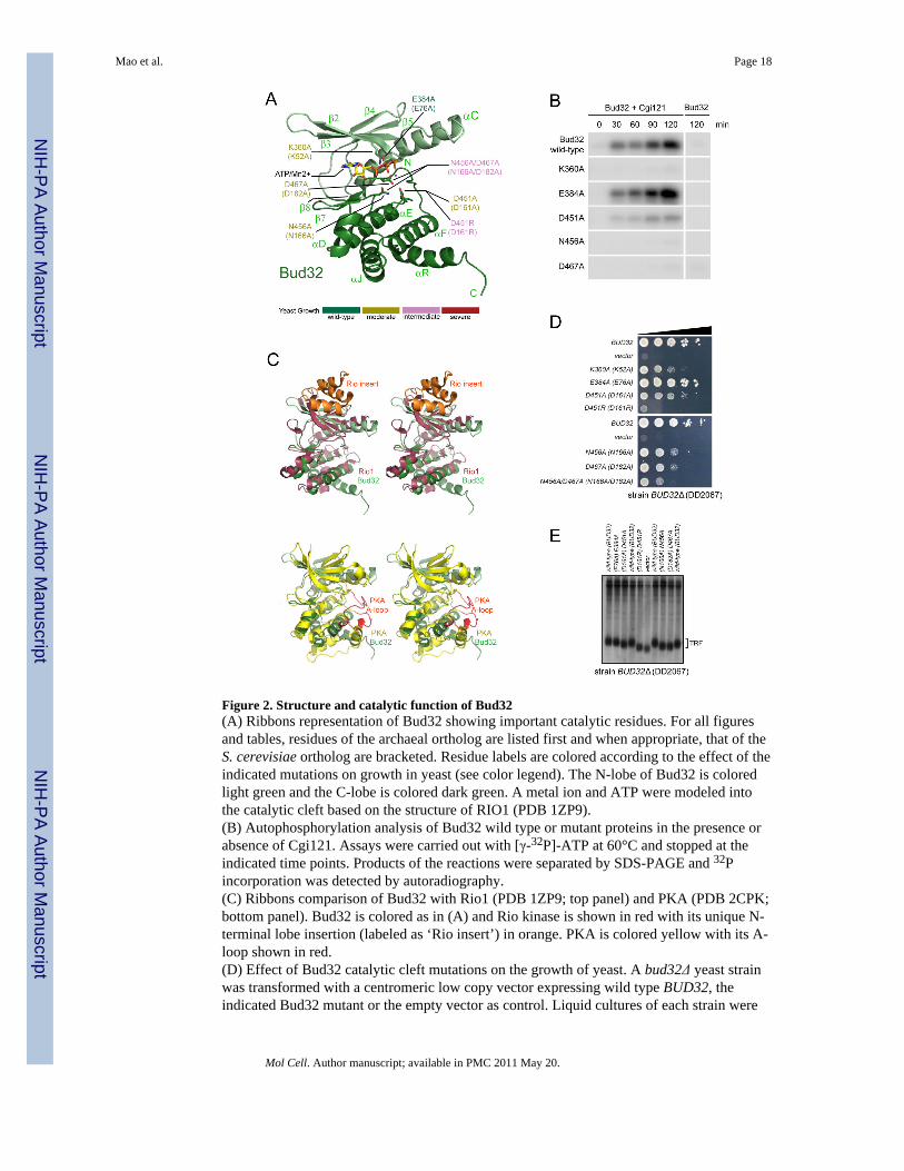
Figure 2. Structure and catalytic function of Bud32(A) Ribbons representation of Bud32 showing important catalytic residues. For all figuresand tables, residues of the archaeal ortholog are listed first and when appropriate, that of theS. cerevisiae ortholog are bracketed. Residue labels are colored according to the effect of theindicated mutations on growth in yeast (see color legend). The N-lobe of Bud32 is coloredlight green and the C-lobe is colored dark green. A metal ion and ATP were modeled intothe catalytic cleft based on the structure of RIO1 (PDB 1ZP9).(B) Autophosphorylation analysis of Bud32 wild type or mutant proteins in the presence orabsence of Cgi121. Assays were carried out with [γ-32P]-ATP at 60°C and stopped at theindicated time points. Products of the reactions were separated by SDS-PAGE and 32Pincorporation was detected by autoradiography.(C) Ribbons comparison of Bud32 with Rio1 (PDB 1ZP9; top panel) and PKA (PDB 2CPK;bottom panel). Bud32 is colored as in (A) and Rio kinase is shown in red with its unique N-terminal lobe insertion (labeled as ‘Rio insert’) in orange. PKA is colored yellow with its A-loop shown in red.(D) Effect of Bud32 catalytic cleft mutations on the growth of yeast. A bud32Δ yeast strainwas transformed with a centromeric low copy vector expressing wild type BUD32, theindicated Bud32 mutant or the empty vector as control. Liquid cultures of each strain were
Mao et al. Page 18
Mol Cell. Author manuscript; available in PMC 2011 May 20.
NIH
-PA Author Manuscript
NIH
-PA Author Manuscript
NIH
-PA Author Manuscript
spotted on minimal medium in a series of 10 fold dilutions. Growth of yeast at 30°C wasmonitored over a 6 day period. Representative data is shown. A summary of doubling timesin liquid culture for a selection of mutants is provided in Table S3.(E) Bud32 catalytic mutants have short telomeres. Terminal restriction fragment (TRF)analysis of the yeast strains described in (D).
Mao et al. Page 19
Mol Cell. Author manuscript; available in PMC 2011 May 20.
NIH
-PA Author Manuscript
NIH
-PA Author Manuscript
NIH
-PA Author Manuscript
Figure 3. Structure and catalytic function of Kae1(A) Ribbons representation of Kae1 highlighting catalytic cleft residues. Subdomains I andII are colored dark and light blue respectively. The metal ion and ADP were modeled intothe cleft based on the structure of P. abyssi Kae1 (PDB 2IVP).(B) Schematic of catalytic cleft residues of Kae1 that coordinate ATP and metal ion (basedon PDB 2IVP).(C) Kae1 has two characteristic inserts (colored pink and red) that distinguish it from otherASKHA fold proteins. Secondary structure elements within the unique inserts are labeled.(D) The kae1-E176R (E213R) mutant has short telomeres. TRF analysis was performed onthe kae1Δ strain transformed with a low copy vector encoding wild type Kae1 or the E176(E213R) mutant. Telomeres from wild type, bud32Δ and cgi121Δ strains were used asreference.
Mao et al. Page 20
Mol Cell. Author manuscript; available in PMC 2011 May 20.
NIH
-PA Author Manuscript
NIH
-PA Author Manuscript
NIH
-PA Author Manuscript

Figure 4. Structures of Pcc1 and Cgi121(A) Ribbons representation of the Pcc1 monomer (top) and dimer (bottom). Pcc1 protomers1 and 2 are colored light and dark orange respectively.(B) Stereo view of conserved hydrophobic contacts within the Pcc1 dimer interface.(C) Ribbons representation of human Cgi121 (left) and M.jannaschii Cgi121 (right)(rmsd=1.92 Å).
Mao et al. Page 21
Mol Cell. Author manuscript; available in PMC 2011 May 20.
NIH
-PA Author Manuscript
NIH
-PA Author Manuscript
NIH
-PA Author Manuscript

Figure 5. Binding mode of Bud32 to Kae1(A) Ribbons representation of the Bud32-Kae1 complex. Kae1 and Bud32 are colored as inFigures 2A and 3A. Interface residues whose mutation gives rise to growth phenotypes inyeast are shown in stick representation.(B) Stereo zoom-in view of the Kae1-Bud32 binding interface centered on (top) Kae1-specific insert 1 and (bottom) C-terminal tail region of Bud32. Figures are colored as in (A).(C,D) Mutation of the Kae1-Bud32 binding interface. Binding experiments with theindicated GST fusion baits and prey proteins were performed as in Figure 1. (C)Examination of the Bud32-Kae1 interaction. Lane 1 (lower panel) is a loading control forBud32. (D) Examination of the Cgi121-Bud32 interaction using Bud32 mutants (top) andthe Pcc1-Kae1 interaction using Kae1 mutants (bottom).
Mao et al. Page 22
Mol Cell. Author manuscript; available in PMC 2011 May 20.
NIH
-PA Author Manuscript
NIH
-PA Author Manuscript
NIH
-PA Author Manuscript

Figure 6. Binding mode of Cgi121 to Bud32(A) Ribbons representation of the Cgi121-Bud32-Kae1 complex. Bud32, Kae1 and Cgi121are colored as in Figures 2A, 3A and 4B, respectively. Residues whose mutation gives riseto strong phenotypes in yeast are highlighted.(B) Stereo zoom-in view of the Bud32-Cgi121 binding interface showing side chains thatparticipate in the interaction.(C) In vitro binding experiments with the indicated GST fusion baits and purified preyproteins.(D) Disruption of Cgi121 binding to Bud32 impairs the ability of Cgi121 to potentiateBud32 autophosphorylation activity in vitro. Bud32 kinase reactions were performed as inFigure 2B.(E) Functional analysis in yeast of Bud32-Cgi121 binding interface mutations on cellgrowth. cgi121Δ yeast was transformed with empty vector, a vector encoding wild typeCgi121 or the L134E/A138R (L172E/I176R) double mutant. All strains were plated byserial dilution on minimal medium as described in Figure 2D.(F) Analysis of Cgi121 and Bud32 stability in vivo. Yeast encoding galactose-inducible3xHA-Cgi121 and Bud32-FLAG proteins were grown for 4 hrs in media containing 2%glucose or 2% galactose. Cells grown in galactose were washed and resuspended in 2%glucose-containing media to repress the GAL1 promoter. Cells were harvested at timesfollowing glucose addition. α-HA and α-FLAG immunoblots are shown.
Mao et al. Page 23
Mol Cell. Author manuscript; available in PMC 2011 May 20.
NIH
-PA Author Manuscript
NIH
-PA Author Manuscript
NIH
-PA Author Manuscript
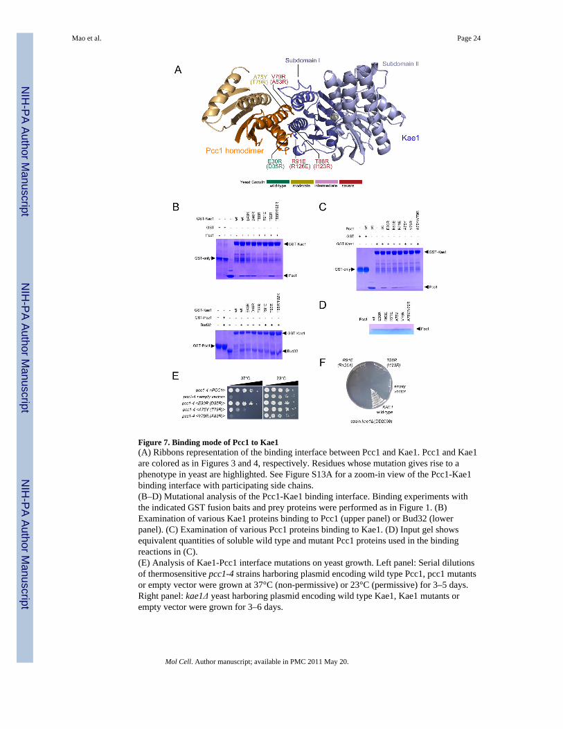
Figure 7. Binding mode of Pcc1 to Kae1(A) Ribbons representation of the binding interface between Pcc1 and Kae1. Pcc1 and Kae1are colored as in Figures 3 and 4, respectively. Residues whose mutation gives rise to aphenotype in yeast are highlighted. See Figure S13A for a zoom-in view of the Pcc1-Kae1binding interface with participating side chains.(B–D) Mutational analysis of the Pcc1-Kae1 binding interface. Binding experiments withthe indicated GST fusion baits and prey proteins were performed as in Figure 1. (B)Examination of various Kae1 proteins binding to Pcc1 (upper panel) or Bud32 (lowerpanel). (C) Examination of various Pcc1 proteins binding to Kae1. (D) Input gel showsequivalent quantities of soluble wild type and mutant Pcc1 proteins used in the bindingreactions in (C).(E) Analysis of Kae1-Pcc1 interface mutations on yeast growth. Left panel: Serial dilutionsof thermosensitive pcc1-4 strains harboring plasmid encoding wild type Pcc1, pcc1 mutantsor empty vector were grown at 37°C (non-permissive) or 23°C (permissive) for 3–5 days.Right panel: kae1Δ yeast harboring plasmid encoding wild type Kae1, Kae1 mutants orempty vector were grown for 3–6 days.
Mao et al. Page 24
Mol Cell. Author manuscript; available in PMC 2011 May 20.
NIH
-PA Author Manuscript
NIH
-PA Author Manuscript
NIH
-PA Author Manuscript

NIH
-PA Author Manuscript
NIH
-PA Author Manuscript
NIH
-PA Author Manuscript
Mao et al. Page 25
Tabl
e 1
Dat
a co
llect
ion
and
refin
emen
t sta
tistic
s.
mjB
ud32
/Kae
1m
jBud
32/K
ae1/
mjC
gi12
1hs
Cgi
121
pfuP
cc1
taK
ae1/
pfuP
cc1
Wav
elen
gth
(Å)
0.97
888
0.97
888
0.97
930.
979
0.97
935
Res
olut
ion
(Å)
2.67
3.6
2.48
2.63
3.02
Rsy
m (%
)5.
78 (2
2.86
)7.
59(1
8.38
)5.
6(31
.17)
3.31
(26.
77)
9.50
(44.
59)
Tota
l Ref
lect
ions
8297
120
745
2958
412
370
7661
7
Uni
que
Ref
lect
ions
4397
120
745
1545
564
1321
362
Com
plet
enes
s (%
)99
.8 (1
00)
99.7
(99.
8)99
.8(9
9.1)
97.3
(66.
1)99
.9(9
9.7)
I/σ20
.06
(3.4
)14
.21(
3.67
)14
.84(
2.24
)24
.16(
2.86
)16
.43(
2.43
)
Twin
Law
, Tw
in fr
actio
nh,
-h-k
,-l, 0
.49
h,-h
-k,-l
0.4
89
Ref
inem
ent
Res
olut
ion
rang
e (Å
)15
0–2.
995
–3.6
31.8
–2.4
868
.25–
2.63
39.6
–3.0
2
Ref
lect
ions
4396
619
689
1539
863
9021
252
Rfa
ctor
/Rfr
ee (%
)20
.96/
26.7
927
.64(
32.5
5)22
.40(
28.7
8)19
.2(2
2.2)
20.1
4(24
.71)
Rm
s dev
iatio
ns
Bon
ds (Å
)0.
026
0.04
70.
003
0.08
20.
003
Ang
les (
°)0.
825
1.37
50.
644
1.23
20.
706
Spac
e gr
oup
P412
2a=
b=14
8.73
Å, c
=136
.42
ÅP2
1212
1a=
76.9
9Å b
=106
.91Å
, c=2
09.9
9ÅP6
2a=
b=93
.521
Å, c
=87.
155Å
P63
a=b=
78.8
1Å, c
=60.
02 Å
P65
a=b=
66.5
6Å, c
=435
.56
Å
Mol
elcu
les p
er a
.u.(r
msd
)2
(1.2
0 Å
)2
(0.5
0 Å
)2(
0.04
Å)
2(0.
78 Å
)2
(0.0
2 Å
)
Ram
acha
ndra
n D
ata
Mos
t fav
ored
89.8
%82
.6%
92.0
%84
.9%
84.6
%
Add
ition
ally
allo
wed
9.6%
17.2
%8.
0%14
.4%
14.8
%
Gen
erou
sly
allo
wed
0.5
%0.
2%0%
0.7%
0.6%
Dis
allo
wed
0%0%
0%0%
0%
Rsy
m =
ΣhΣ
i | I h
,i −
I h |/Σ hΣ i
I h,i,
whe
re I h
is th
e m
ean
inte
nsity
of t
he i
obse
rvat
ions
of s
ymm
etry
rela
ted
refle
ctio
ns o
f h. R
= Σ
| F o
bs −
Fca
lc |/ΣF
obs,
whe
re F
obs =
FP
and
F cal
c is
the
calc
ulat
edpr
otei
n st
ruct
ure
fact
or fr
om th
e at
omic
mod
el.
Mol Cell. Author manuscript; available in PMC 2011 May 20.