aspects of dose, dose rate and radioisotopes in brachytherapy ...
67
From THE DEPARTMENT OF CLINICAL NEUROSCIENCE ST ERIK EYE HOSPITAL Karolinska Institutet, Stockholm, Sweden ASPECTS OF DOSE, DOSE RATE AND RADIOISOTOPES IN BRACHYTHERAPY OF UVEAL MELANOMA Maria Fili Stockholm 2021
-
Upload
khangminh22 -
Category
Documents
-
view
0 -
download
0
Transcript of aspects of dose, dose rate and radioisotopes in brachytherapy ...

From THE DEPARTMENT OF CLINICAL NEUROSCIENCE
ST ERIK EYE HOSPITAL
Karolinska Institutet, Stockholm, Sweden
ASPECTS OF DOSE, DOSE RATE AND RADIOISOTOPES IN BRACHYTHERAPY
OF UVEAL MELANOMA
Maria Fili
Stockholm 2021

All previously published papers were reproduced with permission from the publisher.
Published by Karolinska Institutet.
Printed by Universitetsservice US-AB, 2021
© Maria Fili, 2021
ISBN 978-91-7831-885-8

ASPECTS OF DOSE, DOSE RATE AND RADIOISOTOPES IN BRACHYTHERAPY OF UVEAL MELANOMA
THESIS FOR DOCTORAL DEGREE (Ph.D.)
By
Maria Fili
The thesis will be defended in public at Aula, St. Erik Eye Hospital, Eye Center of Excellence,
Eugeniavägen 12, Solna, Friday 15th of January 2021, 9:00 AM.
Principal Supervisor:
Associate Professor Gustav Stålhammar
Karolinska Institutet
Department of Clinical Neuroscience
Division of Eye and Vision
Co-supervisor(s):
Professor Stefan Seregard
Karolinska Institutet
Department of Clinical Neuroscience
Division of Eye and Vision
Professor Olle Larsson
Karolinska Institutet
Department of Oncology and Pathology
Opponent:
Professor Sören Mattsson
Lund University
Department of Radiation Physics, Malmö
Examination Board:
Professor Malin Malmsjö
Lund University
Department of Ophthalmology, Clinical Sciences
Professor Anders Ahnesjö
Uppsala University
Department of Immunology, Genetics and
Pathology
Division of Medical Radiation Science
Associate Professor Eva Dafgård-Kopp
Karolinska Institutet
Department of Clinical Neuroscience
Division of Eye and Vision


To all patients with uveal melanoma.


POPULAR SCIENCE SUMMARY OF THE THESIS
Uveal melanoma is a rare malignant tumor in the eye which affects 5-10
persons per million inhabitants per year. People with northern appearance
have a higher risk of the disease, but the correlation to ultraviolet exposure is
not as certain as it is to malignant melanoma in the skin. The age of
diagnosis is generally around 60 years old and it is equally common in males
and females. Uveal melanoma rarely affects children. Unfortunately, the
cancer has spread to other organs in up to one half of patients within 15
years from diagnosis. After the tumor has spread, most patients die within a
year.
In the late 60’s local radiation treatment (brachytherapy)
became more and more available as an alternative treatment to removal of
the entire eye. It meant a possibility for the patient to keep the eye and
preserve vision. Brachytherapy gained even more popularity as a beneficial
treatment option for medium sized tumors when it was shown that these
patients have the same survival rate as patients who have the eye removed.
In Sweden we use the radionuclide ruthenium-106 for tumors thinner than 6-
7 mm, and iodine-125 for tumors 7-10 mm thick. Tumors thicker than 10
mm are treated with eye removal. Between 1979 and 1999 only ruthenium-
106 was available, and thus there are some tumors thicker than 7 mm that
have been treated with ruthenium-106 in the past.
In paper I we evaluated the radiation dose rate, i.e. radiation
dose per time unit in ruthenium-106 brachytherapy. We investigated if there
was an association between dose rate and the risk of having to remove the
eye after brachytherapy, due to insufficient effect of the treatment/tumor
relapse or due to a blind and/or painful eye because of radiation damage to
normal ocular tissue surrounding the tumor. We found no such correlation.
In paper II we compared ruthenium-106 and iodine-125 for the
treatment of tumors 5.5 mm or thicker. We compared matched subgroups
and found no difference in risk of dying from the melanoma, but the patients
treated with ruthenium-106 had a higher risk of needing another round of
brachytherapy due to insufficient effect of the treatment or tumor recurrence.

We thereby confirmed that iodine-125 is preferable for thicker tumors, in
accordance with today’s treatment regime.
In paper III we investigated whether there are any gender
differences regarding tumor characteristics, effect of treatment or patient
survival. We found no differences between men and women that would
indicate that the patient’s gender should be considered when handling
patients with uveal melanoma.
In paper IV we analyzed whether radiation dose or dose rate in
brachytherapy with ruthenium-106 and iodine-125 correlated to the risk of
developing subsequent tumors and death. We found no such correlation.

ABSTRACT
Uveal melanomas constitute melanomas arising from the choroid, the ciliary
body or the iris, and in adults stands for the most common primary
malignant intraocular tumor. It has a high risk of metastatic spread followed
by inevitable death, and unfortunately the prognosis has not improved over
the last decades.
In the beginning the only considerable treatment was
enucleation, i.e. removal of the entire eye. However, brachytherapy has since
the 70’s become the primary treatment for medium sized tumors, and the
survival rate has been shown to be equal compared to primary enucleation.
The aim of our studies included in this thesis was to
investigate the efficacy of today’s brachytherapy treatment regime and if any
differences in ocular or patient survival could be found considering different
brachytherapy aspects. When investigating only ruthenium-106
brachytherapy, no association was found between high or low dose rate and
the risk of secondary enucleation, i.e. enucleation due to extensive unwanted
side effects, insufficient treatment effect on the tumor or tumor relapse. In
further analysis with both ruthenium-106 and iodine-125 brachytherapy
included in the study and the risk of tumor related mortality considered,
there was no statistical difference in outcome related to either dose or dose
rate applied and we found no negative consequences for patients that
received lower dose and dose rates than intended.
As there is a gender difference in survival outcome for some
cancers, we investigated this issue regarding uveal melanoma, but no
differences could be found indicating this should be a factor to consider
when planning treatment.
Ruthenium-106 brachytherapy is generally used throughout
Europe. In Sweden, iodine-125 for larger medium sized tumors became
available 20 years after ruthenium-106 brachytherapy had begun. We
therefore had a great opportunity to analyze the larger tumors treated with
ruthenium-106 in the earlier years and compare them to same size tumors
treated with iodine-125. There was no difference in survival between

matched groups, but ruthenium-106-treated patients had a significantly
higher risk of needing retreatment, thus making iodine-125 the preferred
nuclide for thicker tumors.
In conclusion gender is not a factor that needs considering
when planning treatment for uveal melanoma. Treatment with brachytherapy
is both safe and effective within the current recommended doses and dose
rates. Our findings suggest that lower doses and dose rates still would be
adequate, and this should be investigated further. The current protocol for
radioisotope selection based on tumor thickness also seems adequate for the
best possible ocular and survival outcome.
Key words: uveal melanoma, brachytherapy, iodine-125, ruthenium-106,
dose, dose rate

LIST OF SCIENTIFIC PAPERS
I. Fili, M., Lundell, G., Lundell, M., & Seregard, S. (2014). High dose rate and
low dose rate ruthenium brachytherapy for uveal melanoma. No
association with ocular outcome. Br J Ophthalmol, 98(10), 1349-
1354. doi:10.1136/bjophthalmol-2014-305055
II. Fili, M., Trocme, E., Bergman, L., See, T. R. O., Andre, H., Bartuma, K., . . .
Stalhammar, G. (2020). Ruthenium-106 versus iodine-125 plaque
brachytherapy of 571 choroidal melanomas with a thickness of >/=5.5
mm. Br J Ophthalmol, 104(1), 26-32. doi:10.1136/bjophthalmol-
2018-313419
III. Stalhammar, G., See, T. R., Fili, M., & Seregard, S. (2019). No Gender
Differences in Long-Term Survival after Brachytherapy of 1,541
Patients with Uveal Melanoma. Ocul Oncol Pathol, 5(6), 432-439.
doi:10.1159/000497186
IV. Fili, M., Trocme, E., Herrspiegel, C., Seregard, S., & Stalhammar, G. (2020).
Effect of plaque brachytherapy dose and dose rate on risk for disease-
related mortality in 1238 patients with choroidal melanoma. Br J
Ophthalmol. doi:10.1136/bjophthalmol-2019-315722

LIST OF RELATED PAPERS/MANUSCRIPTS
V. Force, A. O. O. T. (2015). International Validation of the American Joint
Committee on Cancer's 7th Edition Classification of Uveal
Melanoma. JAMA Ophthalmol, 133(4), 376-383.
doi:10.1001/jamaophthalmol.2014.5395
VI. Ophthalmic Oncology Task, F. (2016). Local Recurrence Significantly
Increases the Risk of Metastatic Uveal Melanoma. Ophthalmology,
123(1), 86-91. doi:10.1016/j.ophtha.2015.09.014
VII. Al-Jamal, R. T., Cassoux, N., Desjardins, L., Damato, B., Konstantinidis, L.,
Coupland, S. E., . . . Kivela, T. T. (2016). The Pediatric Choroidal and
Ciliary Body Melanoma Study: A Survey by the European
Ophthalmic Oncology Group. Ophthalmology, 123(4), 898-907.
doi:10.1016/j.ophtha.2015.12.024
VIII.
Fili, M., Seregard, S. & Stalhammar, G. (2020). Presenting symptoms are
associated with uveal melanoma-related death. Accepted for
publication in Ophthalmology November 2020

TABLE OF CONTENTS
1 INTRODUCTION..................................................................... 1
2 LITERATURE REVIEW ......................................................... 2
2.1 Uveal melanoma ............................................................... 2
2.1.1 Anatomy ............................................................... 2
2.1.2 Epidemiology ....................................................... 4
2.1.3 Pathogenesis ......................................................... 5
2.1.4 Genetics ................................................................ 6
2.1.5 Diagnosis .............................................................. 7
2.1.6 Prognosis .............................................................. 8
2.2 Treatment ........................................................................ 11
2.2.1 Brachytherapy .................................................... 11
2.2.2 Ophthalmic applicators ...................................... 14
2.2.3 Brachytherapy treatment procedure ................... 15
2.2.4 Ophthalmic brachytherapy in Sweden ............... 16
2.2.5 Radiation biology ............................................... 16
2.2.6 Radiation effects on ocular tissue ...................... 16
2.2.7 Radiation effects on uveal melanoma ................ 17
2.2.8 Other treatments ................................................. 19
2.2.9 Follow up and screening for metastases ............ 19
2.2.10 Adjuvant treatment and treatment of metastatic
disease ................................................................. 20
3 RESEARCH AIMS ................................................................. 22
4 MATERIALS AND METHODS ........................................... 23
4.1 Patients and Methods ...................................................... 23
4.2 Statistical analysis........................................................... 25
4.3 Ethical considerations ..................................................... 26
5 RESULTS ................................................................................ 27
5.1 Paper I ............................................................................. 27
5.2 Paper II ............................................................................ 27
5.3 Paper III .......................................................................... 28
5.4 Paper IV .......................................................................... 29
6 DISCUSSION ......................................................................... 31
7 CONCLUSIONS ..................................................................... 34
8 POINTS OF PERSPECTIVE ................................................. 35

9 ACKNOWLEDGEMENTS .................................................... 36
10 REFERENCES ........................................................................ 37

LIST OF ABBREVIATIONS
AJCC American Joint Committee on Cancer
BAP1 BRCA associated protein 1
BRUM Brachytherapy Registry of Uveal Melanoma
COMS Collaborative Ocular Melanoma Study
EBRT external beam radiation therapy
EIF1AX encoding eukaryotic translation initiation factor 1 A X-linked
eV electron Volt
GEP gene expression profile
GNA11 guanine nucleotide-binding protein subunit alpha 11
GNAQ guanine nucleotide-binding protein subunit alpha q
Gy Gray
IDO indoleamine 2,3-dioxygenase
MAPK mitogen-activated protein kinase
OCT optical coherence tomography
OOTF Ophthalmic Oncology Task Force
PAS periodic acid-schiff
SF3B1 encoding splicing factor 3B subunit 1A
TIGIT T cell Ig and ITIM domain
TTT transpupillary thermotherapy
UBM ultrasound biomicroscopy
UV ultraviolet
VEGF vascular endothelial growth factor


1
1 INTRODUCTION
Uveal melanoma is the most common primary malignant intraocular tumor, with a high risk
for metastasis and tumor-related death.(A. D. Singh, Turell, & Topham, 2011) The tumor’s
tissue of origin - the uvea - consists of three anatomical structures: the choroid, the ciliary
body and the iris. The melanoma originates from melanocytes that normally occur in these
structures, and spreads hematogenously predominantly to the liver. Historically, the primary
treatment was surgical removal of the entire eye - enucleation . Enucleation is still the
primary treatment for large tumors, but for small to medium-sized tumors, eye-preserving
local radiation treatment - plaque brachytherapy – has been the standard treatment for four
decades (American Brachytherapy Society - Ophthalmic Oncology Task Force. Electronic
address & Committee, 2014) without negative survival consequences.(Diener-West et al.,
2001b) Brachytherapy treatment recommendations generally have been based on consensus,
as studies on dosimetric issues have been low in number. Patient and ocular factors that might
influence the effect of brachytherapy is also a field highly unexplored.

2
2 LITERATURE REVIEW
2.1 UVEAL MELANOMA
2.1.1 Anatomy
Uveal melanoma is located in the iris and ciliary body in 4 % and 6 % of cases respectively.
The absolute majority, 90 %, originate from the choroid.(Kaliki & Shields, 2017) The
choroidal melanomas are mostly dome shaped or mushroom-shaped and greyish in tone. Iris
melanomas are mostly located inferiorly.(Shields, Kaliki, Shah, et al., 2012)
Figure 1. Locations of uveal melanoma. Used with permission of Mayo Foundation for
medical education and research. All rights reserved.
The tumors are categorized as small, medium or large in size by a widely
accepted set of criteria established by the Collaborative Ocular Melanoma Study group
(COMS) in the late 1980’s. Small tumors are defined as < 2.5 mm in height and >5 mm in
diameter. Medium sized tumors are 2.5-10 mm thick and a maximum of 16 mm in diameter.
Tumors exceeding 10 mm in thickness, or 16 mm in diameter at the base are defined as
large.("Design and methods of a clinical trial for a rare condition: the Collaborative Ocular
Melanoma Study. COMS Report No. 3," 1993) If the melanoma covers more than one

3
quarter of the choroid but still is not thicker than 5 mm it is referred to as a diffuse
melanoma.(Reese & Howard, 1967) Men have been reported to have a larger and more
posterior located tumors than women.(B. E. Damato & Coupland, 2012)
Figure 2. Typical appearance of a choroidal melanoma. Picture from patient at St. Erik Eye
Hospital.
Histologically, uveal melanoma consists of tumor cells, fibroblasts,
macrophages, lymphocytes and blood vessels. There are two types of tumor cells; spindle
cells and epithelioid cells. The most common tumor cell type is type B spindle cells, which
are fusiform with a spindle shaped nucleus and a clearly defined nucleolus (Figure 3). Type A
spindle cells have a less clearly defined or absent nucleolus, but these cells are now
considered nevus cells. The epithelioid cells are round or polygonal and the large nucleus
contains one or two prominent nucleoli (Figure 4).(McLean, Foster, Zimmerman, & Gamel,
2018) The most common tumor type is the mixed cell type, as the tumor need to be
composed of at least 90 % of spindle or epithelioid cell type to be classified as one or the
other.("Histopathologic characteristics of uveal melanomas in eyes enucleated from the
Collaborative Ocular Melanoma Study. COMS report no. 6," 1998; McLean et al., 2018)
Spindle cell tumors have been associated with higher age at diagnosis.(Andreoli, Mieler, &
Leiderman, 2015)

4
Figure 3. Spindle cell-type tumor. Laminin staining (left, ×20) and PAS staining (right, ×40).
Picture from patient at St. Erik Eye Hospital.
Figure 4. Epithelioid cell-type tumor. Laminin staining (left, ×40) and PAS staining (right,
×40). Picture from patient at St. Erik Eye Hospital.
2.1.2 Epidemiology
Uveal melanoma comprise 85 % of all ocular melanomas, which in turn represents 5 % of all
melanomas. (A. D. Singh, Bergman, & Seregard, 2005) The natural course including
recurrences and metastases was first described in parallel by Scottish surgeon James Wardrop
(1782-1869) and Scottish ophthalmologist-anatomist Allan Burns (1781-1813). (Kivela,
2018)
The age of diagnosis is in general just over 60 years old (Andreoli et al., 2015;
Aronow, Topham, & Singh, 2018; Bergman et al., 2002) but around a decade younger for
Hispanics and Asians.(Shields, Kaliki, Cohen, et al., 2015) Some studies show a slight
overrepresentation in men,(Bergman et al., 2002; Mahendraraj, Lau, Lee, & Chamberlain,

5
2016) while others don’t.(Aronow et al., 2018; Shields, Kaliki, Cohen, et al., 2015) Uveal
melanoma in children and adolescents is very uncommon.(Al-Jamal et al., 2016) .
The incidence in Europe is increasing from south to north with an incidence of
less than 2 per million in Italy and Spain, increasing to more than 8 per million in the Nordic
countries.(Bergman et al., 2002; Virgili et al., 2007) (Krohn, Monge, Skorpen, Mork, & Dahl,
2008) In the US, the general incidence has been estimated to 5.1-5.2(Aronow et al., 2018;
Mahendraraj et al., 2016) with a small but significant increase of 0.5 % in whites.(Aronow et
al., 2018) The incidence in Canada is 3.8 per million with a steady slow increase.(Ghazawi et
al., 2019) Uveal melanoma is very uncommon in Hispanic, Asian and Black populations,
representing only 2-5 percent of all affected individuals while Caucasians stand for the
remaining 95-98 %.(Aronow et al., 2018; Mahendraraj et al., 2016; Phillpotts et al., 1995;
Shields, Kaliki, Cohen, et al., 2015) High incidences of 9.5 and 8.0 per million have been
reported in Ireland and Australia, respectively. (Baily et al., 2019) (Vajdic et al., 2003) A
very low incidence of 1.9 per million has been reported from Japan.(Tomizuka, Namikawa,
& Higashi, 2017) Generally, incidences increase with the latitude.(Yu, Hu, & McCormick,
2006)
2.1.3 Pathogenesis
Risk factors for developing choroidal or ciliary body melanoma is having fair skin and light-
colored eyes. Having congenital ocular melanocytosis or melanocytoma has also been
identified. Further, the BAP1-tumor disposition syndrome is a risk factor, (Walpole et al.,
2018) which is a cancer syndrome including mesothelioma, cutaneous melanoma, uveal
melanoma and renal cell carcinoma. (Masoomian, Shields, & Shields, 2018) Whether there is
a correlation between exposure to ultraviolet (UV) radiation and uveal melanoma is still not
clearly understood, some studies show weak association and some studies show no
association.(Mallet, Gendron, Drigeard Desgarnier, & Rochette, 2014; A. D. Singh, Rennie,
Seregard, Giblin, & McKenzie, 2004) Very little UV radiation reaches the retina and
underlying choroid after passing the cornea, lens and vitreous body in adults(Sliney, 2002)
and uveal melanomas show low presence of mutations associated with UV damage.(Mallet et
al., 2014) As the incidence of uveal melanoma and cancers in the periocular region remain
stable, while cutaneous melanoma is steadily increasing in incidence, it has been suggested
that UV exposure differs in the periocular region from the rest of the body.(Weis et al., 2019)
However, as fair skin color with easy sunburn, as well as freckles and light eye color have
been identified as risk factors for uveal melanoma in a large meta-analyses, the correlation to
UV light is still under investigation. Cutaneous and iris nevi were also risk factors as well as
welding and occupational cooking. In contrast, free time activities outside, work-related
sunlight exposure, latitude of birth, and hair color had no significant correlation.(Nayman,
Bostan, Logan, & Burnier, 2017)

6
Choroidal melanoma can arise de novo or from malignant transformation of
choroidal nevi. The rate of choroidal nevi transforming in to melanomas has been estimated
to less than 1 % in white Americans.(A. D. Singh, Kalyani, & Topham, 2005) Choroidal nevi
are round to oval, flat and usually pigmented lesions and present in approximately 5 % of the
adult population (>40 years, Americans).(Chien, Sioufi, Surakiatchanukul, Shields, &
Shields, 2017) A commonly used mnemonic to remember risk factors for transformation of
nevi into melanomas was created by professor Carol Shields (Wills Eye Hospital,
Philadelphia, USA); “To find small ocular melanoma using helpful hints daily“, where T
represents thickness exceeding 2 mm, F represents subretinal fluid, S represents symptoms of
the vision being disturbed, O represents orange lipofuscin pigment, M represents margin with
a shorter distance than 3 mm from the optic disc, U represents ultrasonographic hollowness,
H represents absence of halo on ultrasound, and D represents absence of drusen.(Shields,
Dalvin, et al., 2018; Shields, Furuta, Berman, et al., 2009) Melanomas less than 3 mm thick
rarely spread, but share the same risk factors as larger melanomas.(Jouhi et al., 2019)
Risk factors for developing iris melanoma from a nevus include being younger
than 40 years old, episodes of hyphema (blood in the anterior chamber of the eye), location
clockwise between 4 and 9, diffuse tumor involving the entire iris surface and feathery
margins.(Shields, Kaliki, Hutchinson, et al., 2013)
Metastases spread hematogenously, with 90% spreading to the liver but also to
the lungs (28-31%) and bone (18-23%). Brain metastases and spread to lymph nodes,
subcutaneous tissue or the gastrointestinal tract is less common. More than half of the patients
have only 1 organ involved, around 25 % 2 organs and very few 3 organs or more. (Diener-
West et al., 2004; Lorigan, Wallace, & Mavligit, 1991)
2.1.4 Genetics
In 2004 uveal melanoma was categorized in two different gene expression profiles (GEP) and
a classifier for clinical use is available.(Onken, Worley, Ehlers, & Harbour, 2004; Onken,
Worley, Tuscan, & Harbour, 2010) GEP1 is associated with a more favorable prognosis than
GEP2.(Onken et al., 2004) Lately however four categories of uveal melanoma has been
defined (A-D) where GEP1 are A and B tumors and GEP2 are C and D tumors, divided
further after their chromosome 6 and 8 status and inflammatory phenotype.(Robertson et al.,
2017) (Jager, Brouwer, & Esmaeli, 2018)
There are five different genes identified to contain mutations important for the
prognosis of uveal melanoma; BAP1, EIF1AX, SF3B1, GNAQ and GNA11. The notion that
there are consistent alterations in a few chromosomes is known from cytogenetic studies from
over 30 years back.(Sisley et al., 1990; Sisley et al., 1997) Lack of the whole chromosome 3;
monosomy 3, occurs in around half of patients with uveal melanoma and is the most
significant chromosome abnormality with a connection to worse prognosis,(B. Damato et al.,
2007) and has also been associated with epithelioid cell type tumors.(Scholes et al., 2003)

7
Amplification of the long arm of chromosome 8q (around 40 % of patients), or loss of 8p
(around 25 %) is also associated with shorter survival,(van den Bosch et al., 2012) as well as
loss of chromosome 6q which is seen in one quarter of patients. Gain of chromosome 6p is
present in around 25 % and is on the contrary a positive alteration. Whether it depends on the
aberration itself or the fact that gain of chromosome 6p is associated with a lack of
monosomy 3, is yet not clear.(Nichols, Richmond, & Daniels, 2016) Mutation in the BAP1
tumor suppressor gene is strongly associated with high risk of metastatic disease and tumor-
related death.(Decatur et al., 2016; Harbour et al., 2010; Jager et al., 2018) BAP1 is a tumor
suppressor located at chromosome 3p21 and has been suggested as explanation to why
monosomy 3 is associated with poorer prognosis.(Harbour & Chao, 2014; Robertson et al.,
2017) Mitogen-activated protein kinase (MAPK) is a pathway which in the end affects cell
proliferation, differentiation and apoptosis. The vast majority of uveal melanoma have an
activated MAPK pathway, which is seldom caused by a BRAF mutation but rather from
mutations in the mutually exclusive driver mutations in the guanine nucleotide-binding
protein GNAQ and GNA11.(Van Raamsdonk et al., 2009; Van Raamsdonk et al., 2010)
GNAQ or GNA11 is present in 85 % of uveal melanomas, whereas BRAF mutations, which
are quite common in cutaneous melanomas, are rare (Davies et al., 2002; Patel et al., 2011). It
has also been shown that a majority of choroidal nevi carry a mutation in GNAQ or
GNA11.(Vader et al., 2017) Mutations in EIF1AX and SF3B1 has been identified to be
frequently present in uveal melanomas with disomy 3, which carry a positive
prognosis.(Martin et al., 2013)
In contrary to most tumors in which the metastases carry similar mutations to
the primary tumor,(Reiter et al., 2018; Vogelstein et al., 2013) uveal melanoma metastases
have quite many mutational changes compared to the primary tumor.(Shain et al., 2019)
2.1.5 Diagnosis
The diagnosis of choroidal melanoma is established with fundus examination through slit
lamp biomicroscopy and/or indirect ophthalmoscopy in conjunction with B-scan
ultrasonography, which provides a better possibility than optical coherence tomography
(OCT) to determine the shape of the tumor and also see acoustic hollowness and choroidal
excavation.(Coleman et al., 2004) A-scan ultrasonography is also performed to evaluate
internal reflectivity, being low in choroidal melanoma as opposed to choroidal hemangioma,
which can have a similar shape but have high internal reflectivity. In case of intermediate
internal reflectivity, choroidal metastasis should be suspected. These examinations are
sufficient for diagnosis of choroidal melanoma and initiation of treatment, as the accuracy has
been shown to be almost 100 %.("Accuracy of diagnosis of choroidal melanomas in the
Collaborative Ocular Melanoma Study. COMS report no. 1," 1990; "Histopathologic
characteristics of uveal melanomas in eyes enucleated from the Collaborative Ocular
Melanoma Study. COMS report no. 6," 1998)

8
Transscleral or transvitreal biopsy is indicated when diagnostic uncertainty
occurs, usually between amelanotic melanoma and metastatic adenocarcinoma, or other even
less frequent tumors. To visualize iris and ciliary body tumors ultrasound biomicroscopy
(UBM) provides a superior image of the tumor than anterior segment OCT.(Bianciotto et al.,
2011)
Figure 5. Iridociliary melanoma (middle) visualized by AS-OCT (left) versus UBM (right).
Note how the margins are much more clearly visible in UBM. Reprinted with permission by
Elsevier.(Bianciotto et al., 2011)
Fluorescein and/or indocyanine green angiography are not needed for diagnosis but can be of
value when there are differential diagnostic considerations from other choroidal
tumors.(Shields, Shields, & De Potter, 1995) MRI can be valuable when extraocular tumor
growth needs to be excluded.(Beenakker et al., 2016) Autofluorescence images show
hyperfluorescence if there is presence of orange pigment in the melanoma, while benign nevi
generally show hypo- or isofluorescence.(Shields, Bianciotto, Pirondini, et al., 2008; Shields,
Pirondini, et al., 2008)
Specific biomarkers for early detection of uveal melanoma and also detection of
metastatic disease is a hot research field, but no markers are yet clinically in use.(Bande
Rodriguez et al., 2020; Triozzi & Singh, 2012)
2.1.6 Prognosis
While only 2 % of patients have metastatic disease at presentation, unfortunately 15 % and 25
% of the patients suffer from metastatic spread after 5 and 10 years respectively, when
considering all locations.(Shields, Furuta, Thangappan, et al., 2009)
Ciliary body melanomas and diffuse melanomas are associated with a higher
risk of metastatic disease compared to choroidal melanomas, while iris melanomas have a
lower risk, which can be attributed to the hidden or clearly visible location of these respective
lesions which affects the time to discovery.(Kaliki, Shields, & Shields, 2015; Nichols et al.,
2016; A. D. Singh, Shields, & Shields, 2001) More specifically at 5 and 10 years, iris

9
melanomas metastasize in 4 % and 7 %, choroidal melanomas in 15 % and 25 % and ciliary
body melanomas in 19 % and 33 % respectively.(Shields, Furuta, Thangappan, et al., 2009)
Every mm increase in thickness and diameter is associated with higher risk of
tumor spread (Diener-West et al., 2004; Shields, Furuta, Thangappan, et al., 2009; Shields,
Sioufi, et al., 2018). Small melanomas up to 3 mm thick have a 5-, 10- and 20-year risk of 6
%, 10-12 % and 20 % for metastatic disease, medium sized tumors 3 to 8 mm have a 14 %,
26 % and 37 % risk respectively, and large tumors more than 8 mm thick a considerably
higher risk of 35-38 %, 49 % and 67 %.(Shields, Furuta, Thangappan, et al., 2009)
Higher age at diagnosis is also a risk factor for metastatic disease.(Shields,
Kaliki, Furuta, Mashayekhi, & Shields, 2012) Tumors in non-Caucasian patients are
generally thicker, more hemorrhagic and more peripheral at diagnosis, but the risk of
metastatic spread and overall survival does not differ from Caucasians.(Andreoli et al., 2015;
Shields, Kaliki, Cohen, et al., 2015) Patients with ocular or oculodermal melanocytosis,
which have an increased risk of uveal melanoma, have a doubled risk for metastatic spread
compared to uveal melanoma patients without melanocytosis.(Shields, Kaliki, Livesey, et al.,
2013)
In a study of pediatric uveal melanoma, that our group contributed to as part of
the European Ophthalmic Oncology Group, uveal melanoma has been described in patients
as young as less than 3 years old. In young patients with uveal melanoma, the prognosis
seems to be more favorable for children younger than 10 years old, with no metastases found
after 10 years follow up. In children 10 to 17 years, the proportion of patients suffering from
metastatic disease was 9 %, and in the group of young adults 18 to 25 years old it was 17 %.
In patients older than 10 years old there was also a worse prognosis for female patients,
which was not observed in the group younger than 10 years old.(Al-Jamal et al., 2016)
If the melanoma consists almost exclusively of spindle cells the risk of
metastatic disease is lower than if the melanoma is mixed with epithelioid cells. The higher
the percentage of epithelioid cells and the bigger they are, the higher the risk of tumor
spread.(B. Damato et al., 2007; McLean et al., 2018)
Local tumor recurrence after treatment is associated with a higher risk of
metastatic spread, demonstrated in a few small studies(Caujolle et al., 2013; Gragoudas,
Lane, Munzenrider, Egan, & Li, 2002; Jampol et al., 2002; Vrabec et al., 1991) but later
confirmed by a large multicenter study.(Ophthalmic Oncology Task, 2016)
After discovery of a metastasis, the median survival time is 4-15 months in
different reports, with a widely ranging survival rate of 15-50 % and 8-30 % after 1 and 2
years, respectively.(Augsburger, Correa, & Shaikh, 2009; Collaborative Ocular Melanoma
Study, 2006; Diener-West et al., 2004; Khoja et al., 2019; Kuk et al., 2016; Rietschel et al.,
2005) Rietschel et al noted that surviving patients after 2 years also seem to have a less steep
mortality rate and 22 % of patients included in the cohort were alive after 4 years. They also

10
reported a longer survival time in females (Rietschel et al., 2005) with metastatic uveal
melanoma, which was supported by a meta-analysis by Khoja et al.(Khoja et al., 2019)
In Sweden the 5 year relative survival after ruthenium-106 brachytherapy is reported to
95 %.(Bergman, Nilsson, Lundell, Lundell, & Seregard, 2005) Overall, the 5-year relative
survival of uveal melanoma of around 80 % remains unchanged in most countries.(Aronow et
al., 2018; Mahendraraj et al., 2016) (Chew, Spilsbury, & Isaacs, 2015; Krohn et al., 2008)

11
2.2 TREATMENT
2.2.1 Brachytherapy
The first eye-sparing treatments were carried out by ophthalmologist Richard Deutschmann
in Hamburg, Germany, 1915 by implanting capsules with mesothorium (radium-228) in
fractioned doses. Later on in London, 1929, R Foster Moore implanted radon-222 seeds
directly in a choroidal sarcoma and thereby managed to treat the tumor while still preserving
vision.(Moore, 1930) Also in London, Hyla Bristow Stallard started treating
rhabdomyosarcoma with seeds of cobalt-60 in the 1940s and improved his technique by using
first wax and then later silver or gold shielded cobalt-60 placed on the episcleral surface.
(Stallard, 1948, 1966) In the 1960s Peter Lommatzsch started using ruthenium-106
successfully in Leipzig, Germany. (Lommatzsch, 1986) By the work of Samuel Packer and
Marvin Rotman in New York, iodine-125 replaced cobalt-60 resulting in less damage to
healthy tumor-surrounding tissue, a consequence from iodine-125s faster dose fall off.
(Packer & Rotman, 1980) Brachytherapy with radioactive nuclides has over the years become
the predominantly used treatment for small to medium-sized tumors.(Aronow et al., 2018)
In 1984, 43 clinical centers in North America formed the Collaborative Ocular
Melanoma Study Group (COMS) which performed the first randomized controlled studies on
ocular melanoma treatment.(Arun D. Singh & Kivelä) In one of the studies, patients with
medium sized tumors were enrolled between 1987 and 1998 and randomized to either iodine-
125 brachytherapy or primary enucleation.(Jampol et al., 2002) Iodine-125 was chosen over
cobalt-60 as it had comparable and sufficient tissue penetration up to 15 mm thickness,
however requiring only 0.5 mm of a gold shield to shield off the radiation to less than 0.1 %,
while 60-cobalt had 50 % radiation left with a shield of 12 mm lead. Iodine-125 is a photon
emitter, its half-life extending to 60 days. It decays through electron capture to an excited
state of tellurium-125 which, while emitting a 35.5 keV gamma photon, decays to its ground
state.(Khan & Gibbons, 2014; Marwaha, Macklis, Singh, & Wilkinson, 2013) The
prescription dose to the tumor apex was set to 100 Gy, (Earle, Kline, & Robertson, 1987)
revised to 85 Gy by the Radiation Therapy Committee of the American Association of
Physicists in Medicine in 1995(Nath et al., 1995) The American Brachytherapy Society has
later on updated the recommendations in 2003 stating a minimum dose rate of 0.60 Gy/h and
a dose still of 85 Gy for iodine-125, based on the COMS standard. A further update in 2014
supplied a dose recommendation range of 70-100 Gy. No specific dosimetric
recommendations are available for other radionuclides. (American Brachytherapy Society -
Ophthalmic Oncology Task Force. Electronic address & Committee, 2014)
In 2001 results were published, which showed no difference in survival when
comparing treatment of medium-sized melanomas with iodine-125 to primary
enucleation.(Diener-West et al., 2001b) This put an end to concerns both that enucleation
caused seeding of melanoma cells and an increasing risk of metastatic disease, as well as
concerns that brachytherapy would not be as effective as primary enucleation.(A. D. Singh,
Rennie, Kivela, Seregard, & Grossniklaus, 2004) These data were confirmed with a longer

12
follow up of 12 years in 2006.("The coms randomized trial of iodine 125 brachytherapy for
choroidal melanoma: V. twelve-year mortality rates and prognostic factors: coms report no.
28," 2006) Local treatment failure was 10 % after 5 years in the brachytherapy group and 15
% had secondary enucleation.(Jampol et al., 2002) Patients included in the report did not have
melanomas located contiguously with the optic disc or melanomas extending more than a 90
degree angle of the optic disc if they were located within 2 mm.(Diener-West et al., 2001a)
Reports on brachytherapy treatment of juxtapapillary choroidal melanomas both with iodine-
125 and ruthenium-106 have however showed comforting results of similar recurrence rates
as with treatment of melanomas further from the optic disc.(Lommatzsch & Lommatzsch,
1991; Sagoo et al., 2011)
While iodine-125 is the most commonly used nuclide overall and foremost in
the US, ruthenium-106 is widely used in Europe, (Pe'er, 2012; Stefan Seregard & Damato,
2014) Both radionuclides offers a highly safe option for plaque surgeons, albeit surgery time
should be kept to a minimum and the number of procedures should not exceed 200 per
year.(Laube, Fluhs, Kessler, & Bornfeld, 2000)
Ruthenium-106 is a β ray electron emitter with a half-life of 373 days (Pe'er,
2012). It decays to rhodium-106 which has a half-time as short as 30 seconds, thus almost
immediately decaying further to its ground state of palladium-106, while emitting beta
radiation with a maximum energy of 3.5 MeV.(Krohn, Chen, Stabo-Eeg, & Hamre, 2020)
A

13
B
Figure 6. A) Approximate dose fall-off curves for ruthenium-106 (106Ru, blue) and iodine-
125 (125I, yellow) plaques when 85 Gy is prescribed at the apex +1 mm of 5 mm thick (solid
lines) and 7 mm thick (dotted lines) tumors. For every millimeter of penetration into the
tissue, the dose fall-off for the β-emitting 106Ru is approximately threefold faster than for the
γ-emitting 125I. This implicates that scleral doses have to be significantly higher with 106Ru
than 125I for equivalent doses at the tumor apex. B) Comparison of theoretical two-
dimensional dose distributions of a 125I plaque (left) and a 106Ru plaque (right), both
planned with a dose of 85 Gy to the apex of a thin tumor. 125I delivers higher doses to tissues
beyond the tumor, and to tissues lateral to the plaque margin. B reprinted with permission
from Eckert & Ziegler BEBIG, Berlin, Germany.(Fili et al., 2020)
Other nuclides that have been used for plaque brachytherapy include irridium-
192 and cobalt-60, not preferred as both are high-energy gamma emitters that deliver large
radiation doses to healthy tumor-surrounding tissue and also present a risk of radiation
exposure to caregivers. Palladium-103 has a faster dose fall of than iodine-125, but not as fast
as ruthenium-106, and is used in some centers.(Finger, Chin, Duvall, & Palladium-103 for
Choroidal Melanoma Study, 2009; Nag et al., 2003) In the most recent recommendations
from the American Brachytherapy Society (ABS) – OOTF, the radionuclides used were
summarized as iodine-125 and palladium-103 in North America, iodine-125 or ruthenium-
106 in Europe, only ruthenium-106 in Japan, and in Russia ruthenium-106 or strontium-
90.(American Brachytherapy Society - Ophthalmic Oncology Task Force. Electronic address
& Committee, 2014)

14
2.2.2 Ophthalmic applicators
Ophthalmic applicators used in Sweden are produced in Germany by Eckert & Ziegler
BEBIG, Berlin, Germany. They manufacture the COMS gold coated iodine-125 plaques as
well as silver coated ruthenium-106 plaques, being the sole producer globally for the latter.
They are spherically shaped and plaques up to 25 mm in diameter are available, but most
centers use plaques with diameters of up to 20 mm. Since a safety margin of 2 mm is
recommended around the tumor, the maximum tumor diameter treatable with plaque
brachytherapy is 16 mm in most centers. (American Brachytherapy Society - Ophthalmic
Oncology Task Force. Electronic address & Committee, 2014; Nag et al., 2003) In posterior
tumors the safety margin might be omitted in some cases, in order to save the macula and
optic disc from radiation, as long as the dose to the scleral surface is at least 300 Gy.(B.
Damato, Patel, Campbell, Mayles, & Errington, 2005)
Iodine-125 plaques are loaded with iodine seeds for each treatment, the number
of seeds needed calculated based on present radioactivity in the seeds and tumor size. The
seeds are encapsulated in 0.05 mm titanium tubes, which are welded at each end creating an
uneven dose distribution with the lowest dose at the ends. Up to 24 seeds can fit into each
plaque.
Figure 7. Iodine-125 plaques. Reprinted with permission from Eckert & Ziegler BEBIG
GmbH.

15
In ruthenium-106 plaques, on the concave side of the plaque there is a thin film
of ruthenium-106 covered by a 0.1 mm silver foil that works as a radiation window. The back
of the plaque has a 0.7 mm thick silver foil and functions as a radiation shield, which absorbs
about 95 % of the beta radiation. According to Eckert & Ziegler, each plaque is calibrated
according to a certified reference dose rate from the National Institute of Standards and
Technology, USA (NIST). The reference point is at the plaque axis 2 mm from the surface
and the absorbed dose rate to water is 4.8 Gy/h, which equals approximately 7.2 Gy/h at the
scleral surface. The dose absorbed in tissue decreases to one tenth of its initial value after 7
mm. The plaques are used multiple times within one year after manufacturing.
Figure 8. Ruthenium-106 plaques. Reprinted with permission from Eckert & Ziegler BEBIG
GmbH.
2.2.3 Brachytherapy treatment procedure
Treatment is performed under general anesthesia. The conjunctiva is opened and a light
source is put to the pupil, locating the tumor which appears as a dark shadow in the sclera.
The plaque is placed over the tumor base with a safety margin of 2-3 mm. If necessary,
extraocular muscles are temporarily disinserted. The conjunctiva is then sewn back over the

16
plaque. In the event of a tumor located close to the optic disc, adjunctive treatment with
transpupillary thermotherapy (TTT) is sometimes administered. After planned treatment
duration is reached, the plaque is removed, muscles are reinserted and the conjunctiva sewn
back in place.
2.2.4 Ophthalmic brachytherapy in Sweden
In Sweden, brachytherapy treatment with ruthenium-106 started up in 1979 and 20 years later
iodine-125 became available as treatment of choice for tumors more than 6-7 mm thick.
Large tumors more than 10 mm thick are still treated with primary enucleation due to
concerns for insufficient radiation doses. Similarly, lesions located adjacent to the optic nerve
may be treated with primary enucleation regardless of tumor size, due to surgical
inaccessibility.
A standard dose of 100 Gy to the tumor apex was prescribed when ruthenium-106
was introduced in 1979, and a maximum dose of 1000 Gy was set to the scleral surface. In
1999 the maximum scleral dose was increased to 1500 Gy and iodine-125 was introduced
with an estimated treatment dose of 85 Gy to the tumor apex. During the 20 years when only
ruthenium-106 was available, some thicker tumors than 7 mm were treated, when eye
preserving treatment was highly desirable.
2.2.5 Radiation biology
Brachytherapy constitutes implantation of radioactive sources inside or next to the tumor,
brachy meaning short distance in Greek. (Hall & Giaccia, 2011) A nuclide is defined by an
atoms combination of protons and neutrons, in conjunction with its energy state. An isotope
is defined by one elements differing number of neutrons. The ionizing radiation which is
emitted when subatomic bonds break and radionuclides and radioisotopes decay into more
stable forms, can create ions by displacing electrons from atoms or molecules. The tissue that
absorbs ionizing radiation forms free radicals and ions when chemical bonds break between
molecules. These oxidize DNA and other contents in the cells. The cell loses its capacity to
reproduce and, if repair mechanisms are insufficient, undergo apoptosis. The SI unit for
absorbed radiation is Gray. One Gray equals 1 Joule of energy absorbed by 1 kg tissue.(Khan
& Gibbons, 2014; A. D. Singh, Pelayes, Seregard, Macklis, & Bandello, 2013)
2.2.6 Radiation effects on ocular tissue
After brachytherapy of a choroidal melanoma, radiation retinopathy occurs in over 60 % of
patients. (S. Seregard, Pelayes, & Singh, 2013) Dose rates more than 2.6 Gy/h to the tumor
base as well as if the margin of the melanoma base is under 4 mm from the fovea, are both
risk factors associated with a higher risk of this complication. (Gunduz et al., 1999b)
Exudative retinal detachment due to increased vascular permeability can be seen directly after
radiation, sometimes resolving, but chronic retinal detachment is not uncommon. If the
patient suffers from diabetes, there is a higher risk of radiation induced proliferative
retinopathy which later on can cause rubeosis and neovascular glaucoma, which together with

17
exudative retinal detachment is referred to as a type of toxic tumor syndrome.(Groenewald,
Konstantinidis, & Damato, 2013; Gunduz et al., 1999b) Radiation induced macular edema is
treated with intravitreal corticosteroids and/or anti-VEGF, both treatments leading to
improvement. However, spontaneous improvement may occur why more studies on the effect
is needed.(Giuliari, Sadaka, Hinkle, & Simpson, 2011; Groenewald et al., 2013) Edema is the
earliest sign of radiation maculopathy, a complication which not surprisingly corresponds to
the distance between the fovea and the tumor margin as well as radiation doses exceeding 50
Gy.(Gunduz et al., 1999b; Pagliara et al., 2018)
A dose of more than 50 Gy received by the optic nerve constitutes a risk for
optic neuropathy. Treatment with intravitreal steroids has little effect on vision improvement,
and systemic corticosteroids have no effect. Hyperbaric oxygen therapy may have effect if
administered early.(Groenewald et al., 2013) Developing cataract is a risk if the irradiation to
the lens is more than 10 Gy. Treatment is with standard cataract surgery with good
results.(Wachtlin et al., 2000)The reported incidence of radiation induced cataract,
maculopathy and optic neuropathy varies widely from just a few percent to more than 80 %,
60 % and 30 % respectively. (Naseripour et al., 2016; Takiar et al., 2015; Wen, Oliver, &
McCannel, 2009) In comparing 106-ruthenium with 125-iodine Wilkinson et al found an 18
% lower radiation dose to the macula, 53 % lower radiation dose to the optic disc, and as low
as 89 % lower radiation dose to the lens for tumors < 5 mm thick.(Wilkinson, Kolar, Fleming,
& Singh, 2008) Scleral necrosis is a rare complication seen in less than 10 % of treated
patients at 10 years after brachytherapy. There is an increased risk for thicker tumors, if there
is ciliary body involvement and if there is higher intraocular pressure. (Gunduz et al., 1999a;
Radin et al., 2008)
2.2.7 Radiation effects on uveal melanoma
Irradiated tumor cells undergo necrosis or apoptosis, and tumor blood vessels loose
endothelial cells which leads to vascular occlusion followed by fibrosis. Less subretinal fluid
and increased pigmentation as well as increased internal reflectivity on ultrasound
examination are clinical findings after treatment. (Maheshwari & Finger, 2018; A. D. Singh,
Rennie, Kivela, et al., 2004) The rate of tumor regression depends on which type of
radiotherapy that has been used (ruthenium faster than iodine) and tumor thickness (thicker
tumors regress faster).(Rashid, Heikkonen, Singh, & Kivela, 2018) These findings support
the notion that in ocular brachytherapy, fast regression is not only due to shorter doubling
times in aggressive tumors, but is also related to the very high radiation doses administered to
the tumor base, causing immediate cell disruption, vessel obliteration and tumor
infarction.(B. Damato, 2018) A rapid regression of tumor size after radiation correlates to
worse prognosis according to some(Augsburger, Gamel, Shields, Markoe, & Brady, 1987;
Kaiserman et al., 2004), but not all authors,(Pepin et al., 2019) and some have found that
rapid regression is associated with earlier metastases and slow regression with late metastases
(after 2 years).(Glynn, Seddon, Gragoudas, Egan, & Hart, 1989) A great number of
accumulated melanomacrophages or extensive fibrosis after radiation can slow regression (B.

18
Damato, 2018), as well as a dark pigmented tumor.(Rashid et al., 2018) Consideration must
also be taken in regression pattern, as oval to dome shaped or mushroom shaped tumors seem
to regress more in area than thickness.(Rashid, Heikkonen, & Kivela, 2015)
Since monosomy 3 is associated with worse prognosis, fast regression rate and
the presence of monosomy 3 have been investigated, not showing such correlation in some
studies (Chiam et al., 2014; Gupta et al., 2016; Salvi et al., 2017) but indeed in
others.(Shields, Bianciotto, Rudich, et al., 2008) GEP1 has been linked to faster regression
than GEP2 the first 6 months after iodine-125 brachytherapy.(Mruthyunjaya, Seider, Stinnett,
Schefler, & Ocular Oncology Study, 2017) As irradiated tumors show a higher frequency as
well as a higher complexity of chromosomal aberrations than untreated tumors,(Dogrusoz et
al., 2015) questions have been raised whether the treatment itself allows or induces metastatic
progress when local failure occurs.(B. Damato, 2018)
Figure 9. Choroidal melanoma treated with brachytherapy. A) Successfully treated area with
a yellowish atrophic appearance. B) Large area of tumor relapse. C) Asteroid hyalosis in the
vitreous body, incidental finding not related to uveal melanoma.(Burris et al., 2017) Picture
from patient at St. Erik Eye Hospital.
A
B
C

19
2.2.8 Other treatments
As mentioned earlier, large tumors 10 mm thick or more are still primarily treated with
enucleation. If there is major extraocular growth, exenteration is the only option, where all
tissue in the orbital cavity is removed.
Local resection of medium sized and large tumors is performed by internal
evacuation or external lamellar sclerouvectomy in some ophthalmic oncology centers.
Various results have been described, some report a high local recurrence rate of 30 % (Hong,
Wei, Hua, Xu, & Shao, 2014) after 5 years, while others report 10 % in the presence of
neoadjuvant proton beam therapy in addition to local resection. The rate of complications
requiring additional vitreoretinal surgery in the latter study was however very high (70
%).(Willerding et al., 2016) In a comparison between neoadjuvant proton beam therapy and
adjuvant ruthenium-106 brachytherapy together with local resection for large tumors there
was an eight fold higher risk of local recurrence with brachytherapy, but no difference in risk
of enucleation or metastatic spread.(Boker et al., 2018) Concerns about these techniques are
that external approach requires hypotensive anesthesia and there is also worries about tumor
seeding. Further analysis on these risks and also the advantages to brachytherapy is
needed.(B. E. Damato, 2012)
Charged particle therapy is an external radiation technique using protons,
helium or carbon. Protons are most commonly used and they produce low irradiation when
traveling through a mass until they stop, where they deliver the maximum of their energy,
called the Bragg peak. Treatment for choroidal melanoma is generally 56-60 Gy delivered to
the prescription point in 4 daily fractions, but doses up to 70 Gy in 5 daily fractions are also
within treatment standards.(Hrbacek et al., 2016) The anterior segment complications and
secondary enucleation rates have been reported to be of higher frequency than for iodine-125
treatment,(American Brachytherapy Society - Ophthalmic Oncology Task Force. Electronic
address & Committee, 2014) but other studies including a large meta-analysis covering over
8000 patients showed no survival benefit or difference in eye retention rate compared to
brachytherapy.(Abrams, Gagne, Melhus, & Mignano, 2016; Wang et al., 2013) The rate of
local recurrence after charged particle therapy is however reported to be lower than after
plaque brachytherapy.(B. Damato, Kacperek, Chopra, Campbell, & Errington, 2005;
Desjardins et al., 2012; Wang et al., 2013)
Other treatments like stereotactic photon beam therapy and endoresection are less
common but also provide a high degree of local control. (Dunavoelgyi et al.,
2011) (Konstantinidis, Groenewald, Coupland, & Damato, 2014)
2.2.9 Follow up and screening for metastases
In Sweden ocular examination is performed 1,3 and 6 months after brachytherapy or
enucleation and thereafter annually for the rest of the patients life, as local recurrence can

20
occur many years after treatment.(Ophthalmic Oncology Task, 2016) Screening for
metastasis is done by liver ultrasound or CT semiannually for 5 years. Ultrasound offers a
high sensitivity and specificity for suspected metastatic lesions and excellent specificity is
achieved with confirmatory CT scan.(Choudhary, Gupta, Bena, Emch, & Singh, 2016) Liver
function blood tests are not used in Sweden, they are not effective for early detection of
metastases but have a high negative predictive value (Mouriaux, Diorio, Bergeron, Berchi, &
Rousseau, 2012) and high LDH was associated with shorter survival in a Dutch nationwide
cohort of advanced disease.(Jochems et al., 2019) Other blood biomarkers for detection of
metastases have not been proven reliable enough for clinical use.(Bande Rodriguez et al.,
2020; Triozzi & Singh, 2011, 2012)
2.2.10 Adjuvant treatment and treatment of metastatic disease
TTT is an accepted adjuvant treatment for juxtapapillary tumors but is not advised as primary
treatment even for small melanomas. (Harbour, Meredith, Thompson, & Gordon, 2003;
Mashayekhi et al., 2015; Win, Robertson, Buettner, McCannel, & Bennett, 2006)
Pre-enucleation radiation is seldom used as it has not shown any survival
benefits.(Hawkins & Collaborative Ocular Melanoma Study, 2004) However, post
exenteration or post enucleation of a tumor with some extrascleral growth is occasionally
treated with external beam radiation therapy (EBRT, 50 Gy in 180-200 cGy fractions). To
avoid radiation damage to eyelids, intraorbital brachytherapy is also an option.(Finger, 2009)
Local resection is sometimes conducted with preoperative proton beam radiation or plaque
radiation after the resection as described earlier.(Boker et al., 2018)
Adjuvant treatment has not been proven effective and efforts using treatments
for cutaneous melanoma for metastatic uveal melanoma has shown poor results, including
antibodies inhibiting the programmed death receptor 1 (PD-1).(Algazi et al., 2016; Carvajal et
al., 2017; Triozzi & Singh, 2014) Liver screening has been questioned since no effective
treatment is available. However, since high diagnostic accuracy can be achieved by non-
invasive methods, the benefits are clear. By early detection of metastases, the patient may be
offered the opportunity to make life altering decisions about palliative treatment. Early
detection also increases the likelihood that a patient can be included in a clinical trial.
(Choudhary et al., 2016)
Tebentafusp is a new form of immunotherapy based on the monoclonal T cell
receptor against cancer (ImmTAC) platform. It has an immune mobilizing function and
targets cells that express HLA A0201 which has complexed with the peptide gp100. In
melanoma Gp100 is expressed strongly, while it has only a weak expression in melanocytes,
and in other tissue there is a minimal expression. Results are promising and further trials are
ongoing.(B. E. Damato, Dukes, Goodall, & Carvajal, 2019)

21
Local treatment of liver metastases includes metastasectomy, local
chemotherapy by arterial liver infusion and liver embolization, none of which has been
proven to prolong survival.(Augsburger et al., 2009)

22
3 RESEARCH AIMS
In paper I we studied whether dose rate applied to the scleral surface or tumor apex was
associated with the risk of secondary enucleation in brachytherapy of uveal melanoma with
ruthenium-106.
In paper II we compared ruthenium-106 and iodine-125 regarding treatment outcome and
survival in treatment of uveal melanomas 5.5 mm or thicker.
In paper III we investigated gender differences in long-term survival after brachytherapy of
uveal melanoma with ruthenium-106 and iodine-125.
In paper IV we evaluated the effect of dose and dose rate on disease-specific mortality after
brachytherapy of uveal melanoma with ruthenium-106 and iodine-125.

23
4 MATERIALS AND METHODS
4.1 PATIENTS AND METHODS
In paper I the computerized Medlog V.2012-8 (Information Analysis, Crystal Bay, Nevada,
USA) registry of all patients in Sweden treated with brachytherapy for uveal melanoma was
searched. Later on, in paper II-IV, the Medlog registry had been replaced by the
Brachytherapy of Uveal Melanoma registry (BRUM) (Registry Center South, EyeNet
Sweden), from were all patient data was retrieved.
Date and cause of death was retrieved from the National Mortality Registry.
This registry contains data on cause of death of all diseased persons in Sweden. As
misclassifications of the actual cause of death is frequent between cutaneous and uveal
melanoma,(Bergman et al., 2003) we cross-checked the National Mortality Registry’s data
with our own clinical records of patients’ metastases. If a patient had metastasized uveal
melanoma and later died from metastasized melanoma without any records of diagnosed or
treated cutaneous melanoma, we considered the cause of death set as cutaneous melanoma as
misclassified.
In paper I, 962 patients treated with ruthenium-106 from 1 November 1979 to
31 December 2012 was included. In total eight groups - two sets of approximate quartiles
based on dose rates at the tumor apex and the scleral surface, respectively, were created. Data
regarding age at diagnosis, gender, eye laterality, tumor thickness, tumor diameter, AJCC T-
category(Edge et al., 2010), tumor distance to the optic disc, tumor distance to the macula,
dose rate to the scleral surface, dose rate to the tumor apex, enucleation after brachytherapy
and reason for enucleation was analyzed.
In paper II, 1493 patients treated with either ruthenium-106 or iodine-125
brachytherapy from 1 November 1979 to 31 December 2015 were included and data
regarding tumor thickness, tumor diameter, brachytherapy nuclide (ruthenium-106 or iodine-
125), retreatment, rates of TTT, enucleation after brachytherapy and survival was analyzed.
Matched subgroups were created in consideration of recommendations for 1:1 nearest
neighbor matching. Values with the highest impact on the means of age at diagnosis, gender,
pretreatment visual acuity, tumor thickness, tumor diameter, AJCC T-category(Kivelä et al.,
2017) and follow up years were excluded until all differences became non-significant in
Mann-Whitney U tests(Stuart, 2010). Two pairs of matched subgroups were made, one pair
of 55+55 patients with tumors 5.5-7.4 mm thick and one pair of 45+45 patients with tumors
≥7.5 mm thick, treated with ruthenium-106 and iodine-125 respectively.
In paper III, 1541 patients treated with either ruthenium-106 or iodine-125
brachytherapy for uveal melanoma from 1 November 1979 to 20 November 2017 were
included and data regarding gender, age at diagnosis, pretreatment best corrected visual
acuity, symptom duration before presentation, type of symptoms, tumor origin, tumor
thickness and diameter, AJCC T-category(Kivelä et al., 2017), brachytherapy nuclide

24
(ruthenium-106 or iodine-125), prescribed radiation dose, treatment duration, TTT before or
after brachytherapy, enucleation after brachytherapy, date of metastasis and follow up years
was analyzed.
In paper IV, 1238 patients treated with brachytherapy for uveal melanoma from
25 April 1996 to 16 March 2016 were included and data regarding age at diagnosis, gender,
pretreatment best corrected visual acuity, symptom duration before presentation, type of
symptoms, tumor thickness and diameter, AJCC T category(Kivelä et al., 2017),
brachytherapy nuclide (ruthenium-106 or iodine-125), prescribed radiation dose, planned
treatment duration, actual treatment duration, TTT before brachytherapy, date of death
(melanoma-related, from other causes or unknown cause) and follow up years was analyzed.
For all patients in paper I-IV, tumor thicknesses and diameters were measured
pre-operatively by standardized A and B-scan ultrasonography. From 1979-1999 ruthenium-
106 was the only nuclide available. Maximum dose at the tumor apex was 100 Gy and to the
scleral surface 1000 Gy. In 1999, iodine-125 was introduced as the treatment standard for
thicker tumors more than 6-7 mm, with a maximum apex dose of 100 Gy which was lowered
to 80 Gy in 2003 due to concern for surrounding tissue. Also, in 1999, the maximum dose to
the scleral surface was increased to 1500 Gy.
Ruthenium-106 plaques were of the CCA, CCB, CCX, CCZ, COB, CIA or CIB
types (Eckert & Ziegler BEBIG, Berlin, Germany) with diameters up to 20 mm. Iodine-125
plaques were custom made by a goldsmith in 18 carat gold, with 20 iodine-seeds (Eckert &
Ziegler) glued to the concave surface. Radioactivity data for each plaque were independently
verified by medical physicists at the Karolinska University Hospital, Solna, Sweden.
Dosimetry was performed by using a p-type silicon semiconductor detector (Scanditronix,
Sweden), estimating dose rate at different depths from the applicators convex surface with an
estimated accuracy of ±4.5 %.(Lax, 1991) Silicon diodes have been used for dosimetric
calculations of high-energy photon and electron beam radiation since the 60’s.(Rikner &
Grusell, 1987) Based on tumor size and the current radiation activity in the specific plaque,
the exact individual treatment time for each patient was calculated by the medical physicists.
Surgery was performed under general anesthesia. The tumor was located by
transillumination, followed by plaque positioning with a minimum 2 mm margin around the
melanoma. Tumors close to the fovea had eccentric plaque fixation and adjunctive laser
therapy to the compromised tumor margin. Juxtapapillary melanomas were either treated with
a notched plaque or standard plaque combined with adjunctive laser therapy. Some patients
received pre-, peri-, or postoperative adjunctive argon laser photocoagulation or TTT. Follow
up was scheduled at 1, 3, 6 and 12 months after treatment, and then annually for the rest of
the patient’s life. Screening for liver metastases by ultrasonography or CT was performed
semiannually for 5 years after diagnosis. After a few years, patients not from the Stockholm
region were referred back to their home clinics. In case of tumor relapse, the patient was
however referred back to our clinic as we are the sole treating clinic in the country. In case of
enucleation this was also known to us as all specimens are sent to our clinic for

25
histopathological analysis. Therefore, all data on relapse and enucleation are accurate. Date of
last follow up might be lost for some patients, but most clinics in the country have a well-
established routine of sending copies of clinical records to our clinic for registration in the
BRUM registry.
All studies received approval from the Central Ethical Review Board, Stockholm,
Sweden.
4.2 STATISTICAL ANALYSIS
Paper I: Statistical analysis was performed using Statistica V.12 software (Statsoft
Scandinavia AB, Uppsala, Sweden). Level of statistical significance was set to p <0.05.
Frequency distributions were studied by the median value and interquartile range as
frequencies were not normally distributed. To evaluate differences between baseline
characteristics in the groups, independent t test and one-way analysis of variance (ANOVA)
were performed for continuous variables and for categorical variables the Pearson ꭓ2 test was
used. Kaplan Meier survival curves were generated for each dose rate quartile and compared
with the log rank (Mantel-Cox) test for secondary enucleation analysis. Univariate and
multivariate analyses using Cox regression modelling was also performed. Data was
consistently treated linearly in the Cox regression analyses.
Paper II: Statistical analysis was performed using SPSS statistics V.25 (IBM,
Armonk, New York, USA). In power calculations we studied the largest similar cohort
published to date, in which the relative risk for tumor recurrence and enucleation after
ruthenium-106 versus iodine-125 brachytherapy was 13.75 and 3.12 respectively.(Shields et
al., 2002) To detect the smallest of these differences (risk for enucleation) with 80 % power
at a significance level of 0.05, the sample size for each arm was estimated to 9 patients.
Reversely, in our sample size we were able to detect differences of down to approximately 8
% with 80 % power and 9 % with 90 % power. The Mann-Whitney U test was used for tests
of the null hypothesis in non-parametrically distributed group level data. The Pearson ꭓ2 test
was used for comparisons of proportions in two-by-two tables. For analyses of hazard for
post brachytherapy enucleations, repeated brachytherapy and melanoma-related mortality,
multivariate Cox regressions with tumor thickness and diameter as covariates were used.
Kaplan-Meier survival curves were computed for matched subgroups and the log-rank
(Mantel-Cox) test of equality of survival distributions was applied for analyses of disease-
specific survival and overall survival.
Paper III: Statistical analysis was performed using SPSS statistics V.25 (IBM,
Armonk, New York, USA). In power calculations for 90 % power, the sample size for each
arm was estimated to at least 541 patients at a significance level of 0.05 and the smallest
detectable difference between the groups of 10 %. Reversely, in our sample size of 775 men
and 766 women, we would be able to detect differences of down to 8-9 % with 90 %
power.(Tunes da Silva, Logan, & Klein, 2009) The Mann-Whitney U test was used for tests

26
of the null hypothesis in non-parametrically distributed group level data. The Pearson ꭓ2 test
was used for comparisons of proportions in two-by-two tables. Bi- and multivariate Cox
regression analysis with tumor thickness and diameter were used for analyses of hazard for
post-brachytherapy enucleations, repeated brachytherapy and melanoma-related mortality. To
test weather our survival data met the proportional hazard assumption, we built a Cox
regression model with a time-dependent versus a time-independent treatment variable (man
or woman). Kaplan-Meier curves for disease-specific and overall survival were computed
and the log rank (Mantel-Cox) test of equality of survival distributions was applied.
Equivalence between men and women was tested as the relative risk for melanoma-related
death with modified two-sided 95 % CIs as for binary endpoints.(Tunes da Silva et al., 2009)
Paper II-IV: Visual acuity was measured on a decimal scale chart at a distance
of 5 m (Monoyer Granström) ranging from 1.0 (corresponding to 20/20) to 0. Modified from
standards in the Swedish National Quality Registry for Cataracts, counting fingers at a
distance of 4 m was recorded as 0.08, hand movements as 0.04, perception and localization of
light as 0.01 and amaurosis as 0. Average visual acuities were calculated on -1 LogMar
converted values, and then converted back to the decimal scale.
AJCC T-categories were codified as T1=1, T2=2, T3=3 and T4=4, to be able to
compare the categories statistically. Actual radiation dose given was defined as: (actual
brachytherapy duration / planned brachytherapy duration) x prescribed dose = actual dose.
4.3 ETHICAL CONSIDERATIONS
As all the studies have been based on retrospective data from an already existing registry and
no changes were made to diagnostic procedures, treatment or follow-up of patients, informed
consent was waived in accordance with the permissions from the regional ethical review
board in Stockholm and the Swedish Ethical Review Authority. All studies adhered to the
tenets of the declaration of Helsinki.
To be reminded of an event that may have happened many years ago might be
very stressful for the patient, and as the results are presented on a group level there is no way
to identify single patients. Thus, the risk of harm to the patient has been considered higher
than the benefit ifwe were to contact every single patient for approval.

27
5 RESULTS
5.1 PAPER I
“High dose rate and low dose rate ruthenium brachytherapy for uveal melanoma- no
association with ocular outcome.”
We investigated if dose rate to the scleral surface or tumor apex correlated to the risk of
secondary enucleation after ruthenium-106 brachytherapy of uveal melanoma. Tumor and
patient characteristics were similar across the quartiles. As expected, thicker tumors treated
with a standard dose to the tumor apex consequently had a lower dose rate to the tumor apex.
There was a wide frequency distribution of both dose rate to the tumor apex (0.01-4.76 Gy/h)
and to the scleral surface (1.66-11.11 Gy/h). Dose rate to the tumor apex was clearly skewed,
again as expected as thinner tumors receive a higher dose per time unit to the apex. Dose rate
to the scleral surface had a near normal distribution.
Most patients got to keep their treated eye, the eye retention rate was 80 % 5
years after treatment and 72 % 10 years after treatment. When secondary enucleation did
occur, 75 % of these were due to tumor progression and the rest due to extensive damage to
ocular tissue which could lead to secondary glaucoma, optic neuropathy, radiation
retinopathy and other negative conditions that caused a blind and/or painful eye. Enucleation
only because of the patients wish was rare.
No statistical difference between the quartiles could be detected in Kaplan-
Meier survival curves neither when analyzing dose rate at the tumor apex or dose rate at the
scleral surface. In univariate analysis, independently predictive clinical parameters of
secondary enucleation were tumor diameter, tumor thickness and distance to the optic disc,
but in further multivariate analysis only tumor diameter was statistically significant
(p=0.049). There were no significant correlations between high or low dose rate and the risk
of secondary enucleation, neither at the scleral surface nor at the tumor apex.
5.2 PAPER II
“Ruthenium-106 versus iodine-125 plaque brachytherapy of 571 choroidal melanomas with
a thickness of ≥5.5 mm.”
Of 571 patients, 317 (56 %) were treated with ruthenium-106. The mean apex dose was 91.6
Gy for ruthenium-106, as expected a higher mean dose than for iodine-125, which was 81.4
Gy, and the difference was statistically significant (p<0.001). There was also a statistically
significant difference in tumor thickness between the groups, the mean thickness for
ruthenium-106 treated tumors being 6.6 mm versus 8.5 mm for iodine-125 treated tumors
(p<0.001), and the former group having a significantly better pretreatment visual acuity
(p=0.03). There was no significant difference in rate of pre- peri- or postoperative TTT
(p=0.97) between the radioisotopes.

28
There was a statistically significant difference in need for a second round of
brachytherapy (p<0.001), being 8 % (14 patients) in the ruthenium-106 group with a mean
treatment interval of 2.2 years, and 1 % (3 patients) in the iodine-125 group with a mean
treatment interval of 1.25 years (p=0.51). The main reason was tumor relapse for both groups,
except for 2 ruthenium-106 treated patients who had insufficient regression. Of the 17
patients treated twice, 8 died from metastatic disease.
Twenty percent of the ruthenium-106 treated patients underwent secondary
enucleation, of which 89 % was due to tumor relapse and 3% due to insufficient treatment
effect. The remaining patients had blind eyes with or without pain. Of the iodine-125 treated
patients, 17 % underwent secondary enucleation, of which 70 % was due to tumor relapse.
The remaining patients had unwanted side effects like secondary glaucoma, chronic uveitis or
total retinal detachment, except for 1 patient who preferred to have the eye enucleated.
Multivariate Cox regression analysis with tumor diameter, tumor thickness,
ruthenium-106 and iodine-125 as covariates revealed a significantly increasing hazard for
secondary enucleation with increasing tumor thickness (p<0.001), but not for the other
covariates. Overall, there was no difference in disease-specific survival between the
radioisotopes. In the matched subgroups with tumors 5.5-7.4 mm, the mortality rate was 30 %
and 45 % after 5- and 10-years post treatment with ruthenium-106, and 23 % and 38% after
iodine-125 treatment. The subgroups with tumors ≥7.5 mm had a 5- and 10-year mortality
rate of 25 % and 51% after ruthenium-106 treatment, and 33% and 54% after iodine-125
treatment respectively. There were no statistically significant differences in 5- and 10-year
survival between any of the matched subgroups.
5.3 PAPER III
“No Gender Differences in Long-Term Survival after Brachytherapy of 1,541 Patients with
Uveal Melanoma.”
Of the patients treated with ruthenium-106 there were 626 men and 619 women. One hundred
and forty nine men and 147 women were treated with iodine-125. There were no statistical
difference between men and women regarding age at diagnosis, visual acuity before
treatment, symptom duration or type before diagnosis, which uveal structure the tumor
originated from, AJCC T-category, radionuclide used, prescribed radiation dose, intended or
actual treatment duration, proportion of TTT before or after brachytherapy, or follow up
years. Tumor thickness and diameter were borderline significantly larger for men (p=0.06)
than for women (p=0.08). The proportional hazard assumption was met in analysis of time
from brachytherapy to death or last follow-up (p=0.83).
The deviation from planned treatment time to actual treatment time was similar
between men and women. There was no statistical difference in hazard ratio for disease-
specific death in a multivariate Cox regression analysis where tumor thickness and dose in

29
categories of decreasing dose by 10 Gy where included as covariates (dose >90 Gy, 80-90
Gy, 70-80 Gy and <70 Gy).
One hundred and sixteen (15 %) men and 121 (16 %) women suffered from
tumor relapse (p=0.65). Twenty-one men had brachytherapy retreatment because of tumor
relapse (19 patients) or insufficient treatment effect (2 patients). Twenty-seven women had
brachytherapy because of retreatment tumor relapse (24 patients) or insufficient treatment
effect (3 patients) There was no statistically significant difference in risk of retreatment
(p=0.36) which, if it did occur had a mean interval of 2.2 years between the treatments for
men and a mean interval of 2.8 years for women (p=0.45). In a multivariate Cox analysis
with tumor thickness and tumor diameter added as covariates, there was no significant
difference in hazard ratio for repeated brachytherapy for men and women (p=0.47). Of the in
total 48 retreated patients, 17 (35%) eventually died from metastatic disease.
Secondary enucleation was performed on 107 (14 %) of the men and 111 (14
%) of the women. The main reason was tumor relapse in 91 % for men and 90 % for women
including 2 % because of insufficient treatment effect. The remaining were because of
extensive side effects. The enucleation-free Kaplan-Meier survival was similar between men
and women (p=0.86) and in total 33 % of the secondary enucleated patients eventually died
from their melanoma. The median survival after diagnosis of metastatic disease of 0.9 years
for men was not statistically different from the 1.1 years for women (p=0.12).
The 5-year melanoma-related mortality rate for men and women were 14 % and
15 %, and the 10-year mortality rate were 24 % and 26 % respectively. Lastly, the 15-year
mortality rate were 27% and 32 % respectively. When adding tumor diameter and sex as
covariates in a bi-variate Cox regression analysis, there were no significant difference in risk
of disease-specific death between men and women (p=0.32). For men, the disease-specific
survival was 18.2 years, and for women 15.5 years, with no statistically significant difference
in Kaplan-Meier analysis (p=0.22). Overall survival of 13.5 years for men and 15.3 years for
women was also similar (p=0.60). The relative risk of disease-specific death for men versus
women was 0.9 in test of equivalence.
5.4 PAPER IV
“Effect of plaque brachytherapy dose and dose rate on risk for disease-related mortality in
1238 patients with choroidal melanoma”
The mean actual radiation dose ranged from 73.0 to 108.6 Gy in the first to the tenth decile,
and the dose rate ranged from 0.5 Gy/hour to 2.8 Gy/hour. Across the deciles, symptoms and
patient characteristics had an equal distribution for both dose and dose rate. Tumor distance
to the macula was not equally distributed (p<0.001), being longer for lower dose and dose
rate deciles. Distance to the optic disc was also longer for lower dose rate deciles (p=0.006),
but equally distributed for dose deciles. As expected, factors related to tumor size were not

30
equally distributed. Larger tumors were more frequent in deciles with lower doses and dose
rates, in concordance with the limited dose to the scleral surface of 1500 Gy. Iodine-125 was
more frequent in the lower dose deciles (p<0.001) since the dose aim to the tumor apex is 80
Gy for iodine-125 and 100 Gy for ruthenium-106. Regarding dose rate ruthenium-106 was
the radionuclide more frequent in the high and low deciles and iodine-125 in the middle
groups (p<0.001). Pretreatment best corrected visual acuity and treatment with TTT before
brachytherapy were not equally distributed but did not show a certain direction of distribution
for dose (p=0.004 and p<0.001) or dose rate (p<0.001 for both).
There was a median follow up time for metastatic-free patients of 7.5 years
(IQR 5.8). The total mortality rate by the end of follow up was 37 % (462 patients). The
cause of death was unknown for 18 % of these 462 patients, while 58 % had died from the
melanoma and 24 % had died from other causes. Kaplan-Meier curves showed a significant
difference in tumor-related mortality between high and low dose and dose rate (p<0.001), but
in further analysis with a multivariate Cox regression analysis were tumor thickness, tumor
diameter and distance to optic disc were added as covariates, there was no association
between dose and melanoma-related mortality (p=0.4) or dose rate and melanoma-related
mortality (p=0.6). For both dose and dose rate, tumor diameter was independently associated
with melanoma-related mortality (p<0.001), but not tumor thickness (p=0.04 and p=0.07
respectively) or distance to the optic disc (p=0.09 and p=0.1 respectively).
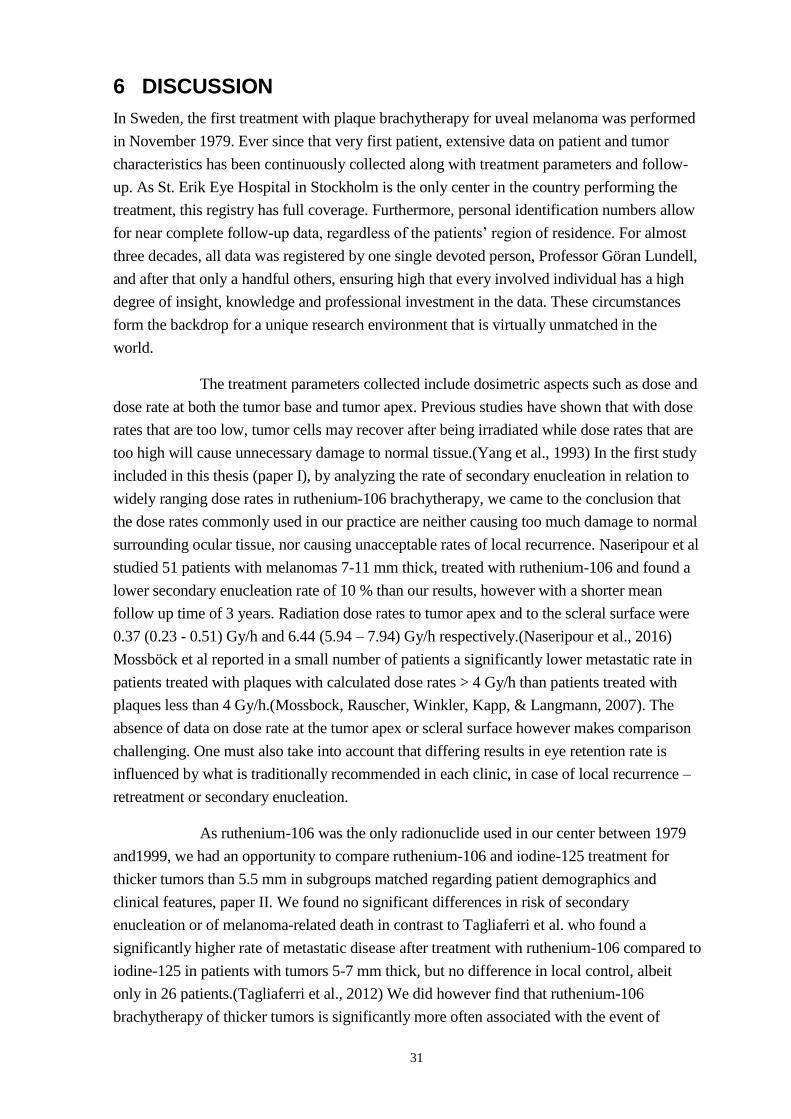
31
6 DISCUSSION
In Sweden, the first treatment with plaque brachytherapy for uveal melanoma was performed
in November 1979. Ever since that very first patient, extensive data on patient and tumor
characteristics has been continuously collected along with treatment parameters and follow-
up. As St. Erik Eye Hospital in Stockholm is the only center in the country performing the
treatment, this registry has full coverage. Furthermore, personal identification numbers allow
for near complete follow-up data, regardless of the patients’ region of residence. For almost
three decades, all data was registered by one single devoted person, Professor Göran Lundell,
and after that only a handful others, ensuring high that every involved individual has a high
degree of insight, knowledge and professional investment in the data. These circumstances
form the backdrop for a unique research environment that is virtually unmatched in the
world.
The treatment parameters collected include dosimetric aspects such as dose and
dose rate at both the tumor base and tumor apex. Previous studies have shown that with dose
rates that are too low, tumor cells may recover after being irradiated while dose rates that are
too high will cause unnecessary damage to normal tissue.(Yang et al., 1993) In the first study
included in this thesis (paper I), by analyzing the rate of secondary enucleation in relation to
widely ranging dose rates in ruthenium-106 brachytherapy, we came to the conclusion that
the dose rates commonly used in our practice are neither causing too much damage to normal
surrounding ocular tissue, nor causing unacceptable rates of local recurrence. Naseripour et al
studied 51 patients with melanomas 7-11 mm thick, treated with ruthenium-106 and found a
lower secondary enucleation rate of 10 % than our results, however with a shorter mean
follow up time of 3 years. Radiation dose rates to tumor apex and to the scleral surface were
0.37 (0.23 - 0.51) Gy/h and 6.44 (5.94 – 7.94) Gy/h respectively.(Naseripour et al., 2016)
Mossböck et al reported in a small number of patients a significantly lower metastatic rate in
patients treated with plaques with calculated dose rates > 4 Gy/h than patients treated with
plaques less than 4 Gy/h.(Mossbock, Rauscher, Winkler, Kapp, & Langmann, 2007). The
absence of data on dose rate at the tumor apex or scleral surface however makes comparison
challenging. One must also take into account that differing results in eye retention rate is
influenced by what is traditionally recommended in each clinic, in case of local recurrence –
retreatment or secondary enucleation.
As ruthenium-106 was the only radionuclide used in our center between 1979
and1999, we had an opportunity to compare ruthenium-106 and iodine-125 treatment for
thicker tumors than 5.5 mm in subgroups matched regarding patient demographics and
clinical features, paper II. We found no significant differences in risk of secondary
enucleation or of melanoma-related death in contrast to Tagliaferri et al. who found a
significantly higher rate of metastatic disease after treatment with ruthenium-106 compared to
iodine-125 in patients with tumors 5-7 mm thick, but no difference in local control, albeit
only in 26 patients.(Tagliaferri et al., 2012) We did however find that ruthenium-106
brachytherapy of thicker tumors is significantly more often associated with the event of

32
retreatment than iodine-125 brachytherapy. This alone we consider sufficient reason to avoid
ruthenium-106 for thicker tumors. The reason for retreatment is local recurrence and
occasionally insufficient regression. Even if we found no such relation in our study, local
recurrence has been associated with higher risk of metastatic disease in a large multicenter
study by the Ophthalmic Oncology Task Force.(Ophthalmic Oncology Task, 2016) In this
context, it should however be mentioned that the prognostic implications of tumor recurrence
may be caused by the fact that aggressive tumors recur more often, rather than that
insufficient brachytherapy cause increased aggressiveness. Consistent with our findings in
both paper I and II, Papageorgiou et al. didn’t find a correlation between dose rate and risk of
local recurrence, but indeed a higher risk for local recurrence for tumors >5 mm thick treated
with ruthenium-106.(Papageorgiou, Cohen, Bunce, Kinsella, & Hungerford, 2011) Tarmann
et al. analyzed 30 patients with tumors >7 mm thick that received ruthenium-106 treatment,
and found a significant correlation to secondary enucleation but not to tumor
recurrence.(Tarmann et al., 2015) Again, differing results are influenced by local decisions on
retreatment versus secondary enucleation in cases of tumor relapse. Not least, avoidance of
retreatment is of course desirable for the patient and also for economic and health care
reasons.
For thinner tumors, ruthenium-106 seems to be a better treatment choice. It
has a high local control rate,(B. Damato, Patel, et al., 2005; Takiar et al., 2015) and its
preferred usage for thinner tumors is supported by the findings by Takiar et al, who found a
significantly higher risk of unwanted side effects when comparing iodine-125 against
ruthenium-106 for tumors <5 mm thick.(Takiar et al., 2015)
As described earlier, men have been reported to have larger tumors than women
at presentation. (B. E. Damato & Coupland, 2012) In addition, men have been reported to
have a higher frequency and earlier debut of metastatic disease than women, (Zloto, Pe'er, &
Frenkel, 2013) and also a shorter survival rate after the diagnosis of metastatic disease.
(Rietschel et al., 2005) In paper III we analyzed tumor and treatment parameters in relation to
gender, as far as we know not investigated before from a treatment perspective. We found no
significant differences between men and women in survival or ocular outcomes that would
indicate that the treatment strategy should be adjusted in any way depending on the patient’s
sex.
The dose of 85 Gy to the tumor apex, at least for iodine-125, has traditionally
been accepted since the COMS trials, and in the same time period our clinic lowered the apex
dose from 100 Gy to 80 Gy for iodine-125, i.e 5 Gy lower than the American Brachytherapy
Society’s recommendations. Throughout the years, the great majority of patients have
received the intended dose, but for natural reasons, final doses have been lower and higher
than prescribed in some cases. In paper IV we found no adverse effects on treatment
effectivity or side effects from a dose range of 73-108 Gy. This finding is in concordance
with Perez et al. who did not find lower rates of local control, survival, metastases with
iodine-125 doses down to 63-69 Gy to the apex. They did however note significantly worse
visual acuity and radiation complications with higher doses up to 104 Gy (Perez et al., 2014)
and this report was included in a large meta-analysis of 15 retrospective and prospective

33
studies by Echegaray et al. who also concluded that there are no certain relation between
local recurrence and lower iodine-125 doses to the apex, opening up for randomized trials
investigating this further.(Echegaray, Bechrakis, Singh, Bellerive, & Singh, 2017) Quivey et
al found a correlation between lower dose and dose rate to the tumor apex, lower dose rate to
the base, long treatment duration and increased risk of local failure when analyzing 150
patients treated with iodine-125 retrospectively, however treatment dose to the tumor apex
ranged all the way down to 29.5 Gy.(Quivey, Augsburger, Snelling, & Brady, 1996)
In our study we included both ruthenium-106 and iodine-125, a decision based
on our finding in paper II, that the use of ruthenium-106 or iodine-125 is not an independent
predictor of melanoma-related death.
The fact that uveal melanoma can metastasize decades after primary treatment
raises concerns that it doesn’t really matter how and if we try to treat the tumor. However, it
is well known that larger tumors carry a worse prognosis.(Arnljots et al., 2018; Shields,
Furuta, Thangappan, et al., 2009; Shields, Kaliki, Furuta, et al., 2015) It has also been shown
that older age at treatment is associated with worse prognosis and most risk factors for tumor
spread. As the mean tumor diameter is larger for older patients, and larger tumors carry a
higher degree of for example monosomy 3, earlier treatment prevents not only local tumor
growth but also negative genetic alterations, dedifferentiation and metastatic spread.(B. E.
Damato, Heimann, Kalirai, & Coupland, 2014) Over the last years the importance of tumor
surrounding microenvironment for the metastatic process has been paid more and more
attention. Uveal melanoma primary tumors which express a high level of immune
checkpoints T cell Ig and ITIM domain (TIGIT) and indoleamine 2,3-dioxygenase (IDO)
carry a higher risk of metastatic spread,(Robertson et al., 2017; Stalhammar, Seregard, &
Grossniklaus, 2019) as well as tumors with a high rate of infiltrating macrophages and a
dense extracellular matrix, (Makitie, Summanen, Tarkkanen, & Kivela, 2001; Stalhammar,
See, Phillips, & Grossniklaus, 2019) all of which have more time and opportunity to develop
in the happening of insufficient and/or late treatment. That being said, brachytherapy for
uveal melanoma is a necessary treatment and our findings support that lower treatment doses
and dose rates are still effective, with the benefit of fewer ocular complications.

34
7 CONCLUSIONS
Uveal melanoma is a rare cancer, but still the most common primary malignant intraocular
tumor in adults. The prognosis is similar after enucleation and eye-preserving plaque
brachytherapy. A high proportion of patients develop metastases, after which the prognosis is
dismal.
Plaque brachytherapy of uveal melanoma with ruthenium-106 and iodine-125 is
well established and these radionuclides are widely used in most developed countries. The
aim of this thesis was to analyze different aspects of brachytherapy of uveal melanoma and its
correlation with ocular and patient outcomes. Both radionuclides have their place in the
treatment regime, with ruthenium-106 being more advantageous for thinner tumors and
iodine-125 for thicker tumors. The current dose and dose rate recommendations are effective
but our findings support that lower doses and dose rates are also safe and effective, which
opens up for further investigation in randomized trials. We found no indications that the
patient’s gender needs to be taken into account when planning the brachytherapy treatment,
or when estimating prognosis for the patient.

35
8 POINTS OF PERSPECTIVE
The introduction of brachytherapy as a treatment alternative for uveal melanoma has meant a
sight preserving alternative for countless patients. In the initial era of brachytherapy,
treatment was based mainly on consensus. Increasing number of studies analyzing the
optimal radionuclide, dose and dose rate as well as which patient features and genetic aspects
are most relevant for outcome, takes us further and further towards optimizing the
brachytherapy treatment for each individual patient. To be able to tell the patients that we are
giving the best treatment available not only based on experience, but also based on extensive
research, is of essence.
My studies are retrospective and not randomized. Even if a prospective
randomized trial is always preferable, the large number of patients that we are able to analyze
in our extensive BRUM directory reaching four decades back, makes our findings a very high
powered valuable contribution to the knowledge about uveal melanoma and brachytherapy
we have today.
Hopefully, one day brachytherapy will be a treatment that was used in the past.
Hopefully, as we understand more and more of the molecular pathways and the genetics of
uveal melanoma, a tumor specific treatment effective on both primary tumor and
micrometastases, not harmful for normal tissue, will become a reality. Until then, plaque
brachytherapy will be a mainstay in uveal melanoma treatment and consequently, further
studies in the field of brachytherapy in parallel with studies of uveal melanoma on a
molecular level are warranted.

36
9 ACKNOWLEDGEMENTS
There are a number of colleagues, friends and family which I would like to thank for making
this thesis possible.
Gustav Stålhammar, my main supervisor, for your wise, forceful and enthusiastic guidance
that has brought me to the finish line.
Stefan Seregard, my co-supervisor, for opening the door to the world of research for me and
for sharing your endless expertise.
Olle Larsson, my co-supervisor, for welcoming me to his lab, and the great researchers
working there,giving me invaluable insights on uveal melanoma on a molecular level.
Göran Lundell, and in loving memory of Marie Lundell, for inviting me to your home and
sharing your treasure of patient data with me, as well as your insightful thoughts.
Anna Lundvall, for being my public defense chairperson and, together with the colleagues
at the Department of pediatric ophthalmology and strabismus, always supporting me in
my clinical work and cheering me on in my research.
Eva Dafgård-Kopp, Hammurabi Bartuma and Tony Pansell, for taking the time and
effort to review and comment on my work in my half-time review.
Eric Trocmé, for warm-hearted collaboration and hard work on the projects.
Charlotta All-Ericsson, for teaching me the surgery of uveal melanoma and generously
sharing your clinical experience.
Frank Träisk, my supervisor during my residency and forever my mentor in spirit.
My resident colleagues at St. Erik Eye Hospital who shared the laughs and the agonies with
me during my residency.
Emma Lardner, for always staying positive and helpful in the histopathology lab.
Johan Nygren, for fast and high quality support with all computer issues during the way.
Daniel, my beloved husband, my life jacket who keeps me afloat at all times. The father of
my precious sons Didrik, Dominik and Dino. My four D:s, words can’t express how much I
love you.
Monika and Francesco, my parents, for setting the foundation of who I am and for always
being supportive.
My brothers – Andreas and Giovanni, for being by my side my whole life. You make me a
better person. And thank you for finding the best sisters (in law) – Elin and Jeanette.
Anna, for being my loyal best friend in good times and in bad.

37
10 REFERENCES
Abrams, M. J., Gagne, N. L., Melhus, C. S., & Mignano, J. E. (2016). Brachytherapy vs.
external beam radiotherapy for choroidal melanoma: Survival and patterns-of-care
analyses. Brachytherapy, 15(2), 216-223. doi:10.1016/j.brachy.2015.12.001
Accuracy of diagnosis of choroidal melanomas in the Collaborative Ocular Melanoma Study.
COMS report no. 1. (1990). Arch Ophthalmol, 108(9), 1268-1273.
doi:10.1001/archopht.1990.01070110084030
Al-Jamal, R. T., Cassoux, N., Desjardins, L., Damato, B., Konstantinidis, L., Coupland, S. E.,
. . . Kivela, T. T. (2016). The Pediatric Choroidal and Ciliary Body Melanoma Study:
A Survey by the European Ophthalmic Oncology Group. Ophthalmology, 123(4),
898-907. doi:10.1016/j.ophtha.2015.12.024
Algazi, A. P., Tsai, K. K., Shoushtari, A. N., Munhoz, R. R., Eroglu, Z., Piulats, J. M., . . .
Sullivan, R. J. (2016). Clinical outcomes in metastatic uveal melanoma treated with
PD-1 and PD-L1 antibodies. Cancer, 122(21), 3344-3353. doi:10.1002/cncr.30258
American Brachytherapy Society - Ophthalmic Oncology Task Force. Electronic address, p.
e. c., & Committee, A. O. (2014). The American Brachytherapy Society consensus
guidelines for plaque brachytherapy of uveal melanoma and retinoblastoma.
Brachytherapy, 13(1), 1-14. doi:10.1016/j.brachy.2013.11.008
Andreoli, M. T., Mieler, W. F., & Leiderman, Y. I. (2015). Epidemiological trends in uveal
melanoma. Br J Ophthalmol, 99(11), 1550-1553. doi:10.1136/bjophthalmol-2015-
306810
Arnljots, T. S., Al-Sharbaty, Z., Lardner, E., All-Eriksson, C., Seregard, S., & Stalhammar,
G. (2018). Tumour thickness, diameter, area or volume? The prognostic significance
of conventional versus digital image analysis-based size estimation methods in uveal
melanoma. Acta Ophthalmol, 96(5), 510-518. doi:10.1111/aos.13668
Aronow, M. E., Topham, A. K., & Singh, A. D. (2018). Uveal Melanoma: 5-Year Update on
Incidence, Treatment, and Survival (SEER 1973-2013). Ocul Oncol Pathol, 4(3),
145-151. doi:10.1159/000480640
Augsburger, J. J., Correa, Z. M., & Shaikh, A. H. (2009). Effectiveness of treatments for
metastatic uveal melanoma. Am J Ophthalmol, 148(1), 119-127.
doi:10.1016/j.ajo.2009.01.023
Augsburger, J. J., Gamel, J. W., Shields, J. A., Markoe, A. M., & Brady, L. W. (1987). Post-
irradiation regression of choroidal melanomas as a risk factor for death from
metastatic disease. Ophthalmology, 94(9), 1173-1177. doi:10.1016/s0161-
6420(87)33310-x
Baily, C., O'Neill, V., Dunne, M., Cunningham, M., Gullo, G., Kennedy, S., . . . Horgan, N.
(2019). Uveal Melanoma in Ireland. Ocul Oncol Pathol, 5(3), 195-204.
doi:10.1159/000492391
Bande Rodriguez, M. F., Fernandez Marta, B., Lago Baameiro, N., Santiago-Varela, M.,
Silva-Rodriguez, P., Blanco-Teijeiro, M. J., . . . Pineiro Ces, A. (2020). Blood
Biomarkers of Uveal Melanoma: Current Perspectives. Clin Ophthalmol, 14, 157-
169. doi:10.2147/OPTH.S199064
Beenakker, J. W., Ferreira, T. A., Soemarwoto, K. P., Genders, S. W., Teeuwisse, W. M.,
Webb, A. G., & Luyten, G. P. (2016). Clinical evaluation of ultra-high-field MRI for

38
three-dimensional visualisation of tumour size in uveal melanoma patients, with
direct relevance to treatment planning. MAGMA, 29(3), 571-577.
doi:10.1007/s10334-016-0529-4
Bergman, L., Nilsson, B., Lundell, G., Lundell, M., & Seregard, S. (2005). Ruthenium
brachytherapy for uveal melanoma, 1979-2003: survival and functional outcomes in
the Swedish population. Ophthalmology, 112(5), 834-840.
doi:10.1016/j.ophtha.2004.11.038
Bergman, L., Seregard, S., Nilsson, B., Lundell, G., Ringborg, U., & Ragnarsson-Olding, B.
(2003). Uveal melanoma survival in Sweden from 1960 to 1998. Invest Ophthalmol
Vis Sci, 44(8), 3282-3287. doi:10.1167/iovs.03-0081
Bergman, L., Seregard, S., Nilsson, B., Ringborg, U., Lundell, G., & Ragnarsson-Olding, B.
(2002). Incidence of uveal melanoma in Sweden from 1960 to 1998. Invest
Ophthalmol Vis Sci, 43(8), 2579-2583. Retrieved from
https://www.ncbi.nlm.nih.gov/pubmed/12147588
Bianciotto, C., Shields, C. L., Guzman, J. M., Romanelli-Gobbi, M., Mazzuca, D., Jr., Green,
W. R., & Shields, J. A. (2011). Assessment of anterior segment tumors with
ultrasound biomicroscopy versus anterior segment optical coherence tomography in
200 cases. Ophthalmology, 118(7), 1297-1302. doi:10.1016/j.ophtha.2010.11.011
Boker, A., Pilger, D., Cordini, D., Seibel, I., Riechardt, A. I., Joussen, A. M., & Bechrakis, N.
E. (2018). Neoadjuvant proton beam irradiation vs. adjuvant ruthenium brachytherapy
in transscleral resection of uveal melanoma. Graefes Arch Clin Exp Ophthalmol,
256(9), 1767-1775. doi:10.1007/s00417-018-4032-7
Burris, C. K. H., Azari, A. A., Kanavi, M. R., Dubielzig, R. R., Lee, V., Gottlieb, J. L., . . .
Albert, D. M. (2017). Is There an Increased Prevalence of Asteroid Hyalosis in Eyes
with Uveal Melanoma? A Histopathologic Study. Ocul Oncol Pathol, 3(4), 259-261.
doi:10.1159/000461586
Carvajal, R. D., Schwartz, G. K., Tezel, T., Marr, B., Francis, J. H., & Nathan, P. D. (2017).
Metastatic disease from uveal melanoma: treatment options and future prospects. Br J
Ophthalmol, 101(1), 38-44. doi:10.1136/bjophthalmol-2016-309034
Caujolle, J. P., Paoli, V., Chamorey, E., Maschi, C., Baillif, S., Herault, J., . . . Hannoun-Levi,
J. M. (2013). Local recurrence after uveal melanoma proton beam therapy: recurrence
types and prognostic consequences. Int J Radiat Oncol Biol Phys, 85(5), 1218-1224.
doi:10.1016/j.ijrobp.2012.10.005
Chew, A. L., Spilsbury, K., & Isaacs, T. W. (2015). Survival from uveal melanoma in
Western Australia 1981-2005. Clin Exp Ophthalmol, 43(5), 422-428.
doi:10.1111/ceo.12490
Chiam, P. J., Coupland, S. E., Kalirai, H., Groenewald, C., Heimann, H., & Damato, B. E.
(2014). Does choroidal melanoma regression correlate with chromosome 3 loss after
ruthenium brachytherapy? Br J Ophthalmol, 98(7), 967-971.
doi:10.1136/bjophthalmol-2013-304472
Chien, J. L., Sioufi, K., Surakiatchanukul, T., Shields, J. A., & Shields, C. L. (2017).
Choroidal nevus: a review of prevalence, features, genetics, risks, and outcomes. Curr
Opin Ophthalmol, 28(3), 228-237. doi:10.1097/ICU.0000000000000361

39
Choudhary, M. M., Gupta, A., Bena, J., Emch, T., & Singh, A. D. (2016). Hepatic
Ultrasonography for Surveillance in Patients With Uveal Melanoma. JAMA
Ophthalmol, 134(2), 174-180. doi:10.1001/jamaophthalmol.2015.4810
Coleman, D. J., Silverman, R. H., Chabi, A., Rondeau, M. J., Shung, K. K., Cannata, J., &
Lincoff, H. (2004). High-resolution ultrasonic imaging of the posterior segment.
Ophthalmology, 111(7), 1344-1351. doi:10.1016/j.ophtha.2003.10.029
Collaborative Ocular Melanoma Study, G. (2006). The COMS randomized trial of iodine 125
brachytherapy for choroidal melanoma: V. Twelve-year mortality rates and
prognostic factors: COMS report No. 28. Arch Ophthalmol, 124(12), 1684-1693.
doi:10.1001/archopht.124.12.1684
The coms randomized trial of iodine 125 brachytherapy for choroidal melanoma: V. twelve-
year mortality rates and prognostic factors: coms report no. 28. (2006). Archives of
Ophthalmology, 124(12), 1684-1693. doi:10.1001/archopht.124.12.1684
Damato, B. (2018). Predicting Choroidal Melanoma Regression after Brachytherapy.
Ophthalmology, 125(5), 755-756. doi:10.1016/j.ophtha.2018.01.031
Damato, B., Duke, C., Coupland, S. E., Hiscott, P., Smith, P. A., Campbell, I., . . . Howard, P.
(2007). Cytogenetics of uveal melanoma: a 7-year clinical experience.
Ophthalmology, 114(10), 1925-1931. doi:10.1016/j.ophtha.2007.06.012
Damato, B., Kacperek, A., Chopra, M., Campbell, I. R., & Errington, R. D. (2005). Proton
beam radiotherapy of choroidal melanoma: the Liverpool-Clatterbridge experience.
Int J Radiat Oncol Biol Phys, 62(5), 1405-1411. doi:10.1016/j.ijrobp.2005.01.016
Damato, B., Patel, I., Campbell, I. R., Mayles, H. M., & Errington, R. D. (2005). Local tumor
control after 106Ru brachytherapy of choroidal melanoma. Int J Radiat Oncol Biol
Phys, 63(2), 385-391. doi:10.1016/j.ijrobp.2005.02.017
Damato, B. E. (2012). Local resection of uveal melanoma. Dev Ophthalmol, 49, 66-80.
doi:10.1159/000328261
Damato, B. E., & Coupland, S. E. (2012). Differences in uveal melanomas between men and
women from the British Isles. Eye (Lond), 26(2), 292-299. doi:10.1038/eye.2011.272
Damato, B. E., Dukes, J., Goodall, H., & Carvajal, R. D. (2019). Tebentafusp: T Cell
Redirection for the Treatment of Metastatic Uveal Melanoma. Cancers (Basel), 11(7).
doi:10.3390/cancers11070971
Damato, B. E., Heimann, H., Kalirai, H., & Coupland, S. E. (2014). Age, survival predictors,
and metastatic death in patients with choroidal melanoma: tentative evidence of a
therapeutic effect on survival. JAMA Ophthalmol, 132(5), 605-613.
doi:10.1001/jamaophthalmol.2014.77
Davies, H., Bignell, G. R., Cox, C., Stephens, P., Edkins, S., Clegg, S., . . . Futreal, P. A.
(2002). Mutations of the BRAF gene in human cancer. Nature, 417(6892), 949-954.
doi:10.1038/nature00766
Decatur, C. L., Ong, E., Garg, N., Anbunathan, H., Bowcock, A. M., Field, M. G., &
Harbour, J. W. (2016). Driver Mutations in Uveal Melanoma: Associations With
Gene Expression Profile and Patient Outcomes. JAMA Ophthalmol, 134(7), 728-733.
doi:10.1001/jamaophthalmol.2016.0903

40
Design and methods of a clinical trial for a rare condition: the Collaborative Ocular
Melanoma Study. COMS Report No. 3. (1993). Control Clin Trials, 14(5), 362-391.
doi:10.1016/0197-2456(93)90052-f
Desjardins, L., Lumbroso-Le Rouic, L., Levy-Gabriel, C., Cassoux, N., Dendale, R., Mazal,
A., . . . Asselain, B. (2012). Treatment of uveal melanoma by accelerated proton
beam. Dev Ophthalmol, 49, 41-57. doi:10.1159/000328257
Diener-West, M., Earle, J. D., Fine, S. L., Hawkins, B. S., Moy, C. S., Reynolds, S. M., . . .
Collaborative Ocular Melanoma Study, G. (2001a). The COMS randomized trial of
iodine 125 brachytherapy for choroidal melanoma, II: characteristics of patients
enrolled and not enrolled. COMS Report No. 17. Arch Ophthalmol, 119(7), 951-965.
doi:10.1001/archopht.119.7.951
Diener-West, M., Earle, J. D., Fine, S. L., Hawkins, B. S., Moy, C. S., Reynolds, S. M., . . .
Collaborative Ocular Melanoma Study, G. (2001b). The COMS randomized trial of
iodine 125 brachytherapy for choroidal melanoma, III: initial mortality findings.
COMS Report No. 18. Arch Ophthalmol, 119(7), 969-982.
doi:10.1001/archopht.119.7.969
Diener-West, M., Reynolds, S. M., Agugliaro, D. J., Caldwell, R., Cumming, K., Earle, J. D.,
. . . Collaborative Ocular Melanoma Study Group, R. (2004). Screening for metastasis
from choroidal melanoma: the Collaborative Ocular Melanoma Study Group Report
23. J Clin Oncol, 22(12), 2438-2444. doi:10.1200/JCO.2004.08.194
Dogrusoz, M., Kroes, W. G., van Duinen, S. G., Creutzberg, C. L., Versluis, M., Bleeker, J.
C., . . . Jager, M. J. (2015). Radiation Treatment Affects Chromosome Testing in
Uveal Melanoma. Invest Ophthalmol Vis Sci, 56(10), 5956-5964.
doi:10.1167/iovs.15-17092
Dunavoelgyi, R., Dieckmann, K., Gleiss, A., Sacu, S., Kircher, K., Georgopoulos, M., . . .
Poetter, R. (2011). Local tumor control, visual acuity, and survival after
hypofractionated stereotactic photon radiotherapy of choroidal melanoma in 212
patients treated between 1997 and 2007. Int J Radiat Oncol Biol Phys, 81(1), 199-
205. doi:10.1016/j.ijrobp.2010.04.035
Earle, J., Kline, R. W., & Robertson, D. M. (1987). Selection of iodine 125 for the
Collaborative Ocular Melanoma Study. Arch Ophthalmol, 105(6), 763-764.
doi:10.1001/archopht.1987.01060060049030
Echegaray, J. J., Bechrakis, N. E., Singh, N., Bellerive, C., & Singh, A. D. (2017). Iodine-125
Brachytherapy for Uveal Melanoma: A Systematic Review of Radiation Dose. Ocul
Oncol Pathol, 3(3), 193-198. doi:10.1159/000455872
Edge, S. E., Bryd, D. R., Compton, C. A., Fritz, A. G., Greene, F. L., & Troti, A. (2010).
Ophthalmic Oncology Task Force. Malignant Melanoma of the Uvea. . In AJCC
Cancer Staging Manual.7th ed. (pp. 547-559). New York: Springer.
Fili, M., Trocme, E., Bergman, L., See, T. R. O., Andre, H., Bartuma, K., . . . Stalhammar, G.
(2020). Ruthenium-106 versus iodine-125 plaque brachytherapy of 571 choroidal
melanomas with a thickness of >/=5.5 mm. Br J Ophthalmol, 104(1), 26-32.
doi:10.1136/bjophthalmol-2018-313419
Finger, P. T. (2009). Radiation therapy for orbital tumors: concepts, current use, and
ophthalmic radiation side effects. Survey of Ophthalmology, 54(5), 545-568.
doi:10.1016/j.survophthal.2009.06.004

41
Finger, P. T., Chin, K. J., Duvall, G., & Palladium-103 for Choroidal Melanoma Study, G.
(2009). Palladium-103 ophthalmic plaque radiation therapy for choroidal melanoma:
400 treated patients. Ophthalmology, 116(4), 790-796, 796 e791.
doi:10.1016/j.ophtha.2008.12.027
Ghazawi, F. M., Darwich, R., Le, M., Rahme, E., Zubarev, A., Moreau, L., . . . Litvinov, I. V.
(2019). Uveal melanoma incidence trends in Canada: a national comprehensive
population-based study. Br J Ophthalmol, 103(12), 1872-1876.
doi:10.1136/bjophthalmol-2018-312966
Giuliari, G. P., Sadaka, A., Hinkle, D. M., & Simpson, E. R. (2011). Current treatments for
radiation retinopathy. Acta Oncologica, 50(1), 6-13.
doi:10.3109/0284186X.2010.500299
Glynn, R. J., Seddon, J. M., Gragoudas, E. S., Egan, K. M., & Hart, L. J. (1989). Evaluation
of tumor regression and other prognostic factors for early and late metastasis after
proton irradiation of uveal melanoma. Ophthalmology, 96(10), 1566-1573.
doi:10.1016/s0161-6420(89)32685-6
Gragoudas, E. S., Lane, A. M., Munzenrider, J., Egan, K. M., & Li, W. (2002). Long-term
risk of local failure after proton therapy for choroidal/ciliary body melanoma. Trans
Am Ophthalmol Soc, 100, 43-48; discussion 48-49. Retrieved from
https://www.ncbi.nlm.nih.gov/pubmed/12545676
Groenewald, C., Konstantinidis, L., & Damato, B. (2013). Effects of radiotherapy on uveal
melanomas and adjacent tissues. Eye (Lond), 27(2), 163-171.
doi:10.1038/eye.2012.249
Gunduz, K., Shields, C. L., Shields, J. A., Cater, J., Freire, J. E., & Brady, L. W. (1999a).
Plaque radiotherapy of uveal melanoma with predominant ciliary body involvement.
Archives of Ophthalmology, 117(2), 170-177. Retrieved from
http://www.ncbi.nlm.nih.gov/pubmed/10037560
Gunduz, K., Shields, C. L., Shields, J. A., Cater, J., Freire, J. E., & Brady, L. W. (1999b).
Radiation retinopathy following plaque radiotherapy for posterior uveal melanoma.
Arch Ophthalmol, 117(5), 609-614. doi:10.1001/archopht.117.5.609
Gupta, K., McCannel, C. A., Kamrava, M., Lamb, J., Almanzor, R. D., & McCannel, T. A.
(2016). Tumor-height regression rate after brachytherapy between choroidal
melanoma gene expression profile classes: effect of controlling for tumor height.
Graefes Arch Clin Exp Ophthalmol, 254(7), 1371-1378. doi:10.1007/s00417-016-
3305-2
Hall, E. J., & Giaccia, A. J. (2011). Radiobiology for the Radiologist. Philadelphia, UNITED
STATES: Wolters Kluwer Health.
Harbour, J. W., & Chao, D. L. (2014). A molecular revolution in uveal melanoma:
implications for patient care and targeted therapy. Ophthalmology, 121(6), 1281-
1288. doi:10.1016/j.ophtha.2013.12.014
Harbour, J. W., Meredith, T. A., Thompson, P. A., & Gordon, M. E. (2003). Transpupillary
thermotherapy versus plaque radiotherapy for suspected choroidal melanomas.
Ophthalmology, 110(11), 2207-2214; discussion 2215. doi:10.1016/S0161-
6420(03)00858-3

42
Harbour, J. W., Onken, M. D., Roberson, E. D., Duan, S., Cao, L., Worley, L. A., . . .
Bowcock, A. M. (2010). Frequent mutation of BAP1 in metastasizing uveal
melanomas. Science, 330(6009), 1410-1413. doi:10.1126/science.1194472
Hawkins, B. S., & Collaborative Ocular Melanoma Study, G. (2004). The Collaborative
Ocular Melanoma Study (COMS) randomized trial of pre-enucleation radiation of
large choroidal melanoma: IV. Ten-year mortality findings and prognostic factors.
COMS report number 24. Am J Ophthalmol, 138(6), 936-951.
doi:10.1016/j.ajo.2004.07.006
Histopathologic characteristics of uveal melanomas in eyes enucleated from the Collaborative
Ocular Melanoma Study. COMS report no. 6. (1998). Am J Ophthalmol, 125(6), 745-
766. doi:10.1016/s0002-9394(98)00040-3
Hong, M., Wei, W., Hua, L., Xu, X., & Shao, L. (2014). Clinical observation of local
resection or enucleation for uveal melanoma. Chin Med J (Engl), 127(19), 3459-3463.
Retrieved from https://www.ncbi.nlm.nih.gov/pubmed/25269914
Hrbacek, J., Mishra, K. K., Kacperek, A., Dendale, R., Nauraye, C., Auger, M., . . . Weber,
D. C. (2016). Practice Patterns Analysis of Ocular Proton Therapy Centers: The
International OPTIC Survey. Int J Radiat Oncol Biol Phys, 95(1), 336-343.
doi:10.1016/j.ijrobp.2016.01.040
Jager, M. J., Brouwer, N. J., & Esmaeli, B. (2018). The Cancer Genome Atlas Project: An
Integrated Molecular View of Uveal Melanoma. Ophthalmology, 125(8), 1139-1142.
doi:10.1016/j.ophtha.2018.03.011
Jampol, L. M., Moy, C. S., Murray, T. G., Reynolds, S. M., Albert, D. M., Schachat, A. P., . .
. Collaborative Ocular Melanoma Study, G. (2002). The COMS randomized trial of
iodine 125 brachytherapy for choroidal melanoma: IV. Local treatment failure and
enucleation in the first 5 years after brachytherapy. COMS report no. 19.
Ophthalmology, 109(12), 2197-2206. doi:10.1016/s0161-6420(02)01277-0
Jochems, A., van der Kooij, M. K., Fiocco, M., Schouwenburg, M. G., Aarts, M. J., van
Akkooi, A. C., . . . Kapiteijn, E. (2019). Metastatic Uveal Melanoma: Treatment
Strategies and Survival-Results from the Dutch Melanoma Treatment Registry.
Cancers (Basel), 11(7). doi:10.3390/cancers11071007
Jouhi, S., Jager, M. J., de Geus, S. J. R., Desjardins, L., Eide, N. A., Grange, J. D., . . . Kivela,
T. T. (2019). The Small Fatal Choroidal Melanoma Study. A Survey by the European
Ophthalmic Oncology Group. Am J Ophthalmol, 202, 100-108.
doi:10.1016/j.ajo.2019.01.031
Kaiserman, I., Anteby, I., Chowers, I., Blumenthal, E. Z., Kliers, I., & Pe'er, J. (2004). Post-
brachytherapy initial tumour regression rate correlates with metastatic spread in
posterior uveal melanoma. Br J Ophthalmol, 88(7), 892-895.
doi:10.1136/bjo.2003.036285
Kaliki, S., & Shields, C. L. (2017). Uveal melanoma: relatively rare but deadly cancer. Eye
(Lond), 31(2), 241-257. doi:10.1038/eye.2016.275
Kaliki, S., Shields, C. L., & Shields, J. A. (2015). Uveal melanoma: estimating prognosis.
Indian J Ophthalmol, 63(2), 93-102. doi:10.4103/0301-4738.154367
Khan, F. M., & Gibbons, J. P. (2014). Khan's the Physics of Radiation Therapy. Philadelphia,
UNITED STATES: Wolters Kluwer.

43
Khoja, L., Atenafu, E. G., Suciu, S., Leyvraz, S., Sato, T., Marshall, E., . . . Joshua, A. M.
(2019). Meta-analysis in metastatic uveal melanoma to determine progression free
and overall survival benchmarks: an international rare cancers initiative (IRCI) ocular
melanoma study. Ann Oncol, 30(8), 1370-1380. doi:10.1093/annonc/mdz176
Kivela, T. T. (2018). The first description of the complete natural history of uveal melanoma
by two Scottish surgeons, Allan Burns and James Wardrop. Acta Ophthalmol, 96(2),
203-214. doi:10.1111/aos.13535
Kivelä, T., Simpson, E., Grossniklaus, H., Jager, M. J., Singh, A., & Caminal, J. M. (2017).
Uveal Melanoma. In AJCC Cancer Staging Manual. (8:th ed., pp. 805-817): Chicago:
Springer.
Konstantinidis, L., Groenewald, C., Coupland, S. E., & Damato, B. (2014). Long-term
outcome of primary endoresection of choroidal melanoma. Br J Ophthalmol, 98(1),
82-85. doi:10.1136/bjophthalmol-2013-304022
Krohn, J., Chen, Y. C., Stabo-Eeg, N. O., & Hamre, B. (2020). Cherenkov Luminescence
Imaging for Assessment of Radioactive Plaque Position in Brachytherapy of Uveal
Melanoma: An In Vivo Feasibility Study. Transl Vis Sci Technol, 9(7), 42.
doi:10.1167/tvst.9.7.42
Krohn, J., Monge, O. R., Skorpen, T. N., Mork, S. J., & Dahl, O. (2008). Posterior uveal
melanoma treated with I-125 brachytherapy or primary enucleation. Eye (Lond),
22(11), 1398-1403. doi:10.1038/sj.eye.6702911
Kuk, D., Shoushtari, A. N., Barker, C. A., Panageas, K. S., Munhoz, R. R., Momtaz, P., . . .
Postow, M. A. (2016). Prognosis of Mucosal, Uveal, Acral, Nonacral Cutaneous, and
Unknown Primary Melanoma From the Time of First Metastasis. Oncologist, 21(7),
848-854. doi:10.1634/theoncologist.2015-0522
Laube, T., Fluhs, D., Kessler, C., & Bornfeld, N. (2000). Determination of surgeon's
absorbed dose in iodine 125 and ruthenium 106 ophthalmic plaque surgery.
Ophthalmology, 107(2), 366-368; discussion 368-369. doi:10.1016/s0161-
6420(99)00050-0
Lax, I. (1991). Dosimetry of 106Ru eye applicators with a p-type silicon detector. Phys Med
Biol, 36(7), 963-972. doi:10.1088/0031-9155/36/7/005
Lommatzsch, P. K. (1986). Results after beta-irradiation (106Ru/106Rh) of choroidal
melanomas: 20 years' experience. Br J Ophthalmol, 70(11), 844-851.
doi:10.1136/bjo.70.11.844
Lommatzsch, P. K., & Lommatzsch, R. (1991). Treatment of juxtapapillary melanomas. Br J
Ophthalmol, 75(12), 715-717. doi:10.1136/bjo.75.12.715
Lorigan, J. G., Wallace, S., & Mavligit, G. M. (1991). The prevalence and location of
metastases from ocular melanoma: imaging study in 110 patients. AJR Am J
Roentgenol, 157(6), 1279-1281. doi:10.2214/ajr.157.6.1950883
Mahendraraj, K., Lau, C. S., Lee, I., & Chamberlain, R. S. (2016). Trends in incidence,
survival, and management of uveal melanoma: a population-based study of 7,516
patients from the Surveillance, Epidemiology, and End Results database (1973-2012).
Clin Ophthalmol, 10, 2113-2119. doi:10.2147/OPTH.S113623
Maheshwari, A., & Finger, P. T. (2018). Regression patterns of choroidal melanoma: After
palladium-103 ((103)Pd) plaque brachytherapy. Eur J Ophthalmol, 28(6), 722-730.
doi:10.1177/1120672118776146

44
Makitie, T., Summanen, P., Tarkkanen, A., & Kivela, T. (2001). Tumor-infiltrating
macrophages (CD68(+) cells) and prognosis in malignant uveal melanoma. Invest
Ophthalmol Vis Sci, 42(7), 1414-1421. Retrieved from
https://www.ncbi.nlm.nih.gov/pubmed/11381040
Mallet, J. D., Gendron, S. P., Drigeard Desgarnier, M. C., & Rochette, P. J. (2014).
Implication of ultraviolet light in the etiology of uveal melanoma: A review.
Photochem Photobiol, 90(1), 15-21. doi:10.1111/php.12161
Martin, M., Masshofer, L., Temming, P., Rahmann, S., Metz, C., Bornfeld, N., . . .
Zeschnigk, M. (2013). Exome sequencing identifies recurrent somatic mutations in
EIF1AX and SF3B1 in uveal melanoma with disomy 3. Nat Genet, 45(8), 933-936.
doi:10.1038/ng.2674
Marwaha, G., Macklis, R., Singh, A. D., & Wilkinson, A. (2013). Brachytherapy.
Developments in Ophthalmology, 52, 29-35. doi:10.1159/000351053
Mashayekhi, A., Shields, C. L., Rishi, P., Atalay, H. T., Pellegrini, M., McLaughlin, J. P., . . .
Shields, J. A. (2015). Primary transpupillary thermotherapy for choroidal melanoma
in 391 cases: importance of risk factors in tumor control. Ophthalmology, 122(3),
600-609. doi:10.1016/j.ophtha.2014.09.029
Masoomian, B., Shields, J. A., & Shields, C. L. (2018). Overview of BAP1 cancer
predisposition syndrome and the relationship to uveal melanoma. J Curr Ophthalmol,
30(2), 102-109. doi:10.1016/j.joco.2018.02.005
McLean, I. W., Foster, W. D., Zimmerman, L. E., & Gamel, J. W. (2018). Modifications of
Callender's Classification of Uveal Melanoma at the Armed Forces Institute of
Pathology. Am J Ophthalmol, 195, lvi-lx. doi:10.1016/j.ajo.2018.08.025
Moore, R. F. (1930). Choroidal Sarcoma Treated by the Intraocular Insertion of Radon Seeds.
Br J Ophthalmol, 14(4), 145-152. doi:10.1136/bjo.14.4.145
Mossbock, G., Rauscher, T., Winkler, P., Kapp, K. S., & Langmann, G. (2007). Impact of
dose rate on clinical course in uveal melanoma after brachytherapy with ruthenium-
106. Strahlenther Onkol, 183(10), 571-575. doi:10.1007/s00066-007-1734-x
Mouriaux, F., Diorio, C., Bergeron, D., Berchi, C., & Rousseau, A. (2012). Liver function
testing is not helpful for early diagnosis of metastatic uveal melanoma.
Ophthalmology, 119(8), 1590-1595. doi:10.1016/j.ophtha.2012.01.045
Mruthyunjaya, P., Seider, M. I., Stinnett, S., Schefler, A., & Ocular Oncology Study, C.
(2017). Association between Tumor Regression Rate and Gene Expression Profile
after Iodine 125 Plaque Radiotherapy for Uveal Melanoma. Ophthalmology, 124(10),
1532-1539. doi:10.1016/j.ophtha.2017.04.013
Nag, S., Quivey, J. M., Earle, J. D., Followill, D., Fontanesi, J., Finger, P. T., & American
Brachytherapy, S. (2003). The American Brachytherapy Society recommendations for
brachytherapy of uveal melanomas. Int J Radiat Oncol Biol Phys, 56(2), 544-555.
doi:10.1016/s0360-3016(03)00006-3
Naseripour, M., Jaberi, R., Sedaghat, A., Azma, Z., Nojomi, M., Falavarjani, K. G., & Nazari,
H. (2016). Ruthenium-106 brachytherapy for thick uveal melanoma: reappraisal of
apex and base dose radiation and dose rate. J Contemp Brachytherapy, 8(1), 66-73.
doi:10.5114/jcb.2016.57818
Nath, R., Anderson, L. L., Luxton, G., Weaver, K. A., Williamson, J. F., & Meigooni, A. S.
(1995). Dosimetry of interstitial brachytherapy sources: recommendations of the

45
AAPM Radiation Therapy Committee Task Group No. 43. American Association of
Physicists in Medicine. Med Phys, 22(2), 209-234. doi:10.1118/1.597458
Nayman, T., Bostan, C., Logan, P., & Burnier, M. N., Jr. (2017). Uveal Melanoma Risk
Factors: A Systematic Review of Meta-Analyses. Curr Eye Res, 42(8), 1085-1093.
doi:10.1080/02713683.2017.1297997
Nichols, E. E., Richmond, A., & Daniels, A. B. (2016). Tumor Characteristics, Genetics,
Management, and the Risk of Metastasis in Uveal Melanoma. Semin Ophthalmol,
31(4), 304-309. doi:10.3109/08820538.2016.1154175
Onken, M. D., Worley, L. A., Ehlers, J. P., & Harbour, J. W. (2004). Gene expression
profiling in uveal melanoma reveals two molecular classes and predicts metastatic
death. Cancer Res, 64(20), 7205-7209. doi:10.1158/0008-5472.CAN-04-1750
Onken, M. D., Worley, L. A., Tuscan, M. D., & Harbour, J. W. (2010). An accurate,
clinically feasible multi-gene expression assay for predicting metastasis in uveal
melanoma. J Mol Diagn, 12(4), 461-468. doi:10.2353/jmoldx.2010.090220
Ophthalmic Oncology Task, F. (2016). Local Recurrence Significantly Increases the Risk of
Metastatic Uveal Melanoma. Ophthalmology, 123(1), 86-91.
doi:10.1016/j.ophtha.2015.09.014
Packer, S., & Rotman, M. (1980). Radiotherapy of choroidal melanoma with iodine-125.
Ophthalmology, 87(6), 582-590. doi:10.1016/s0161-6420(80)35194-4
Pagliara, M. M., Tagliaferri, L., Azario, L., Lenkowicz, J., Lanza, A., Autorino, R., . . . Blasi,
M. A. (2018). Ruthenium brachytherapy for uveal melanomas: Factors affecting the
development of radiation complications. Brachytherapy, 17(2), 432-438.
doi:10.1016/j.brachy.2017.11.004
Papageorgiou, K. I., Cohen, V. M., Bunce, C., Kinsella, M., & Hungerford, J. L. (2011).
Predicting local control of choroidal melanomas following (1)(0)(6)Ru plaque
brachytherapy. Br J Ophthalmol, 95(2), 166-170. doi:10.1136/bjo.2009.176198
Patel, M., Smyth, E., Chapman, P. B., Wolchok, J. D., Schwartz, G. K., Abramson, D. H., &
Carvajal, R. D. (2011). Therapeutic implications of the emerging molecular biology
of uveal melanoma. Clinical Cancer Research, 17(8), 2087-2100. doi:10.1158/1078-
0432.CCR-10-3169
Pe'er, J. (2012). Ruthenium-106 brachytherapy. Dev Ophthalmol, 49, 27-40.
doi:10.1159/000328254
Pepin, F., Julien, A. S., Fugaru, I., Lihimdi, N., Thariat, J., Landreville, S., & Mouriaux, F.
(2019). Regression rate of choroidal melanoma following iodine-125 brachytherapy is
not associated with metastatic spread. Melanoma Res, 29(3), 295-300.
doi:10.1097/CMR.0000000000000540
Perez, B. A., Mettu, P., Vajzovic, L., Rivera, D., Alkaissi, A., Steffey, B. A., . . . Kirsch, D.
G. (2014). Uveal melanoma treated with iodine-125 episcleral plaque: an analysis of
dose on disease control and visual outcomes. Int J Radiat Oncol Biol Phys, 89(1),
127-136. doi:10.1016/j.ijrobp.2014.01.026
Phillpotts, B. A., Sanders, R. J., Shields, J. A., Griffiths, J. D., Augsburger, J. A., & Shields,
C. L. (1995). Uveal melanomas in black patients: a case series and comparative
review. J Natl Med Assoc, 87(9), 709-714. Retrieved from
https://www.ncbi.nlm.nih.gov/pubmed/9583969

46
Quivey, J. M., Augsburger, J., Snelling, L., & Brady, L. W. (1996). 125I plaque therapy for
uveal melanoma. Analysis of the impact of time and dose factors on local control.
Cancer, 77(11), 2356-2362. doi:10.1002/(SICI)1097-
0142(19960601)77:11<2356::AID-CNCR26>3.0.CO;2-V
Radin, P. P., Lumbroso-Le Rouic, L., Levy-Gabriel, C., Dendale, R., Sastre, X., &
Desjardins, L. (2008). Scleral necrosis after radiation therapy for uveal melanomas:
report of 23 cases. Graefes Archive for Clinical and Experimental Ophthalmology,
246(12), 1731-1736. doi:10.1007/s00417-008-0920-6
Rashid, M., Heikkonen, J., & Kivela, T. (2015). Tumor Regression After Brachytherapy for
Choroidal Melanoma: Reduction of Thickness and Cross-Sectional Area by Shape
and Regression Pattern. Invest Ophthalmol Vis Sci, 56(4), 2612-2623.
doi:10.1167/iovs.14-16322
Rashid, M., Heikkonen, J., Singh, A. D., & Kivela, T. T. (2018). Clinical Predictors of
Regression of Choroidal Melanomas after Brachytherapy: A Growth Curve Model.
Ophthalmology, 125(5), 747-754. doi:10.1016/j.ophtha.2018.01.032
Reese, A. B., & Howard, G. M. (1967). Flat uveal melanomas. Am J Ophthalmol, 64(6),
1021-1028. doi:10.1016/0002-9394(67)93054-1
Reiter, J. G., Makohon-Moore, A. P., Gerold, J. M., Heyde, A., Attiyeh, M. A., Kohutek, Z.
A., . . . Nowak, M. A. (2018). Minimal functional driver gene heterogeneity among
untreated metastases. Science, 361(6406), 1033-1037. doi:10.1126/science.aat7171
Rietschel, P., Panageas, K. S., Hanlon, C., Patel, A., Abramson, D. H., & Chapman, P. B.
(2005). Variates of survival in metastatic uveal melanoma. J Clin Oncol, 23(31),
8076-8080. doi:10.1200/JCO.2005.02.6534
Rikner, G., & Grusell, E. (1987). General specifications for silicon semiconductors for use in
radiation dosimetry. Phys Med Biol, 32(9), 1109-1117. doi:10.1088/0031-
9155/32/9/004
Robertson, A. G., Shih, J., Yau, C., Gibb, E. A., Oba, J., Mungall, K. L., . . . Woodman, S. E.
(2017). Integrative Analysis Identifies Four Molecular and Clinical Subsets in Uveal
Melanoma. Cancer Cell, 32(2), 204-220 e215. doi:10.1016/j.ccell.2017.07.003
Sagoo, M. S., Shields, C. L., Mashayekhi, A., Freire, J., Emrich, J., Reiff, J., . . . Shields, J. A.
(2011). Plaque radiotherapy for juxtapapillary choroidal melanoma: tumor control in
650 consecutive cases. Ophthalmology, 118(2), 402-407.
doi:10.1016/j.ophtha.2010.06.007
Salvi, S. M., Aziz, H. A., Dar, S., Singh, N., Hayden-Loreck, B., & Singh, A. D. (2017).
Uveal Melanoma Regression after Brachytherapy: Relationship with Chromosome 3
Monosomy Status. Ocul Oncol Pathol, 3(2), 87-94. doi:10.1159/000451001
Scholes, A. G., Damato, B. E., Nunn, J., Hiscott, P., Grierson, I., & Field, J. K. (2003).
Monosomy 3 in uveal melanoma: correlation with clinical and histologic predictors of
survival. Invest Ophthalmol Vis Sci, 44(3), 1008-1011. doi:10.1167/iovs.02-0159
Seregard, S., & Damato, B. (2014). Uveal Malignant Melanoma: Management Options for
Brachytherapy. In B. Damato & A. D. Singh (Eds.), Clinical Ophthalmic Oncology:
Uveal Tumors (pp. 173-188). Berlin, Heidelberg: Springer Berlin Heidelberg.
Seregard, S., Pelayes, D. E., & Singh, A. D. (2013). Radiation therapy: posterior segment
complications. Developments in Ophthalmology, 52, 114-123.
doi:10.1159/000351088

47
Shain, A. H., Bagger, M. M., Yu, R., Chang, D., Liu, S., Vemula, S., . . . Kiilgaard, J. F.
(2019). The genetic evolution of metastatic uveal melanoma. Nat Genet, 51(7), 1123-
1130. doi:10.1038/s41588-019-0440-9
Shields, C. L., Bianciotto, C., Pirondini, C., Materin, M. A., Harmon, S. A., & Shields, J. A.
(2008). Autofluorescence of choroidal melanoma in 51 cases. Br J Ophthalmol, 92(5),
617-622. doi:10.1136/bjo.2007.130286
Shields, C. L., Bianciotto, C., Rudich, D., Materin, M. A., Ganguly, A., & Shields, J. A.
(2008). Regression of uveal melanoma after plaque radiotherapy and thermotherapy
based on chromosome 3 status. Retina, 28(9), 1289-1295.
doi:10.1097/IAE.0b013e31817f7b3e
Shields, C. L., Dalvin, L. A., Ancona-Lezama, D., Yu, M. D., Di Nicola, M., Williams, B. K.,
Jr., . . . Shields, J. A. (2018). CHOROIDAL NEVUS IMAGING FEATURES IN
3,806 CASES AND RISK FACTORS FOR TRANSFORMATION INTO
MELANOMA IN 2,355 CASES: The 2020 Taylor R. Smith and Victor T. Curtin
Lecture. Retina. doi:10.1097/IAE.0000000000002440
Shields, C. L., Furuta, M., Berman, E. L., Zahler, J. D., Hoberman, D. M., Dinh, D. H., . . .
Shields, J. A. (2009). Choroidal nevus transformation into melanoma: analysis of
2514 consecutive cases. Arch Ophthalmol, 127(8), 981-987.
doi:10.1001/archophthalmol.2009.151
Shields, C. L., Furuta, M., Thangappan, A., Nagori, S., Mashayekhi, A., Lally, D. R., . . .
Shields, J. A. (2009). Metastasis of uveal melanoma millimeter-by-millimeter in 8033
consecutive eyes. Arch Ophthalmol, 127(8), 989-998.
doi:10.1001/archophthalmol.2009.208
Shields, C. L., Kaliki, S., Cohen, M. N., Shields, P. W., Furuta, M., & Shields, J. A. (2015).
Prognosis of uveal melanoma based on race in 8100 patients: The 2015 Doyne
Lecture. Eye (Lond), 29(8), 1027-1035. doi:10.1038/eye.2015.51
Shields, C. L., Kaliki, S., Furuta, M., Fulco, E., Alarcon, C., & Shields, J. A. (2015).
American Joint Committee on Cancer Classification of Uveal Melanoma (Anatomic
Stage) Predicts Prognosis in 7,731 Patients: The 2013 Zimmerman Lecture.
Ophthalmology, 122(6), 1180-1186. doi:10.1016/j.ophtha.2015.01.026
Shields, C. L., Kaliki, S., Furuta, M., Mashayekhi, A., & Shields, J. A. (2012). Clinical
spectrum and prognosis of uveal melanoma based on age at presentation in 8,033
cases. Retina, 32(7), 1363-1372. doi:10.1097/IAE.0b013e31824d09a8
Shields, C. L., Kaliki, S., Hutchinson, A., Nickerson, S., Patel, J., Kancherla, S., . . . Shields,
J. A. (2013). Iris nevus growth into melanoma: analysis of 1611 consecutive eyes: the
ABCDEF guide. Ophthalmology, 120(4), 766-772. doi:10.1016/j.ophtha.2012.09.042
Shields, C. L., Kaliki, S., Livesey, M., Walker, B., Garoon, R., Bucci, M., . . . Shields, J. A.
(2013). Association of ocular and oculodermal melanocytosis with the rate of uveal
melanoma metastasis: analysis of 7872 consecutive eyes. JAMA Ophthalmol, 131(8),
993-1003. doi:10.1001/jamaophthalmol.2013.129
Shields, C. L., Kaliki, S., Shah, S. U., Luo, W., Furuta, M., & Shields, J. A. (2012). Iris
melanoma: features and prognosis in 317 children and adults. J AAPOS, 16(1), 10-16.
doi:10.1016/j.jaapos.2011.10.012
Shields, C. L., Naseripour, M., Cater, J., Shields, J. A., Demirci, H., Youseff, A., & Freire, J.
(2002). Plaque radiotherapy for large posterior uveal melanomas (> or =8-mm thick)

48
in 354 consecutive patients. Ophthalmology, 109(10), 1838-1849. doi:10.1016/s0161-
6420(02)01181-8
Shields, C. L., Pirondini, C., Bianciotto, C., Materin, M. A., Harmon, S. A., & Shields, J. A.
(2008). Autofluorescence of choroidal nevus in 64 cases. Retina, 28(8), 1035-1043.
doi:10.1097/IAE.0b013e318181b94b
Shields, C. L., Shields, J. A., & De Potter, P. (1995). Patterns of indocyanine green
videoangiography of choroidal tumours. Br J Ophthalmol, 79(3), 237-245.
doi:10.1136/bjo.79.3.237
Shields, C. L., Sioufi, K., Srinivasan, A., DiNicola, M., Masoomian, B., Barna, L. E., . . .
Shields, J. A. (2018). Visual Outcome and Millimeter Incremental Risk of Metastasis
in 1780 Patients With Small Choroidal Melanoma Managed by Plaque Radiotherapy.
JAMA Ophthalmol. doi:10.1001/jamaophthalmol.2018.3881
Singh, A. D., Bergman, L., & Seregard, S. (2005). Uveal melanoma: epidemiologic aspects.
Ophthalmol Clin North Am, 18(1), 75-84, viii. doi:10.1016/j.ohc.2004.07.002
Singh, A. D., Kalyani, P., & Topham, A. (2005). Estimating the risk of malignant
transformation of a choroidal nevus. Ophthalmology, 112(10), 1784-1789.
doi:10.1016/j.ophtha.2005.06.011
Singh, A. D., & Kivelä, T. CHAPTER 45 - Uveal malignant melanoma: COMS results. In
(pp. 267-271).
Singh, A. D., Pelayes, D. E., Seregard, S., Macklis, R., & Bandello, F. (2013). Ophthalmic
Radiation Therapy: Techniques and Applications (Vol. 52). Basel: S. Karger AG.
Singh, A. D., Rennie, I. G., Kivela, T., Seregard, S., & Grossniklaus, H. (2004). The
Zimmerman-McLean-Foster hypothesis: 25 years later. Br J Ophthalmol, 88(7), 962-
967. doi:10.1136/bjo.2003.029058
Singh, A. D., Rennie, I. G., Seregard, S., Giblin, M., & McKenzie, J. (2004). Sunlight
exposure and pathogenesis of uveal melanoma. Surv Ophthalmol, 49(4), 419-428.
doi:10.1016/j.survophthal.2004.04.009
Singh, A. D., Shields, C. L., & Shields, J. A. (2001). Prognostic factors in uveal melanoma.
Melanoma Res, 11(3), 255-263. doi:10.1097/00008390-200106000-00007
Singh, A. D., Turell, M. E., & Topham, A. K. (2011). Uveal melanoma: trends in incidence,
treatment, and survival. Ophthalmology, 118(9), 1881-1885.
doi:10.1016/j.ophtha.2011.01.040
Sisley, K., Rennie, I. G., Cottam, D. W., Potter, A. M., Potter, C. W., & Rees, R. C. (1990).
Cytogenetic findings in six posterior uveal melanomas: involvement of chromosomes
3, 6, and 8. Genes Chromosomes Cancer, 2(3), 205-209.
doi:10.1002/gcc.2870020307
Sisley, K., Rennie, I. G., Parsons, M. A., Jacques, R., Hammond, D. W., Bell, S. M., . . .
Rees, R. C. (1997). Abnormalities of chromosomes 3 and 8 in posterior uveal
melanoma correlate with prognosis. Genes Chromosomes Cancer, 19(1), 22-28.
doi:10.1002/(sici)1098-2264(199705)19:1<22::aid-gcc4>3.0.co;2-2
Sliney, D. H. (2002). How light reaches the eye and its components. Int J Toxicol, 21(6), 501-
509. doi:10.1080/10915810290169927
Stalhammar, G., See, T. R. O., Phillips, S. S., & Grossniklaus, H. E. (2019). Density of PAS
positive patterns in uveal melanoma: Correlation with vasculogenic mimicry, gene

49
expression class, BAP-1 expression, macrophage infiltration, and risk for metastasis.
Mol Vis, 25, 502-516. Retrieved from
https://www.ncbi.nlm.nih.gov/pubmed/31588174
Stalhammar, G., Seregard, S., & Grossniklaus, H. E. (2019). Expression of immune
checkpoint receptors Indoleamine 2,3-dioxygenase and T cell Ig and ITIM domain in
metastatic versus nonmetastatic choroidal melanoma. Cancer Med, 8(6), 2784-2792.
doi:10.1002/cam4.2167
Stallard, H. B. (1948). Radiotherapy of Malignant Intra-Ocular Neoplasms. Br J Ophthalmol,
32(9), 618-639. doi:10.1136/bjo.32.9.618
Stallard, H. B. (1966). Radiotherapy for malignant melanoma of the choroid. Br J
Ophthalmol, 50(3), 147-155. doi:10.1136/bjo.50.3.147
Stuart, E. A. (2010). Matching methods for causal inference: A review and a look forward.
Stat Sci, 25(1), 1-21. doi:10.1214/09-STS313
Tagliaferri, L., Smaniotto, D., Blasi, M., Pagliara, M., Villano, A., Balestrazzi, E., . . .
Valentini, V. (2012). PO-411 Brachytherapy with iodine 125 or ruthenium 106 for
treatment of choroidal melanomas measuring 5-7 mm in thickness. . Radiother Oncol,
103, 163-164.
Takiar, V., Voong, K. R., Gombos, D. S., Mourtada, F., Rechner, L. A., Lawyer, A. A., . . .
Beadle, B. M. (2015). A choice of radionuclide: Comparative outcomes and toxicity
of ruthenium-106 and iodine-125 in the definitive treatment of uveal melanoma. Pract
Radiat Oncol, 5(3), e169-e176. doi:10.1016/j.prro.2014.09.005
Tarmann, L., Wackernagel, W., Avian, A., Mayer, C., Schneider, M., Winkler, P., &
Langmann, G. (2015). Ruthenium-106 plaque brachytherapy for uveal melanoma. Br
J Ophthalmol, 99(12), 1644-1649. doi:10.1136/bjophthalmol-2015-306666
Tomizuka, T., Namikawa, K., & Higashi, T. (2017). Characteristics of melanoma in Japan: a
nationwide registry analysis 2011-2013. Melanoma Res, 27(5), 492-497.
doi:10.1097/CMR.0000000000000375
Triozzi, P. L., & Singh, A. D. (2011). Blood biomarkers of uveal melanoma metastasis. Br J
Ophthalmol, 95(1), 3-4. doi:10.1136/bjo.2010.193896
Triozzi, P. L., & Singh, A. D. (2012). Blood biomarkers for uveal melanoma. Future Oncol,
8(2), 205-215. doi:10.2217/fon.11.150
Triozzi, P. L., & Singh, A. D. (2014). Adjuvant Therapy of Uveal Melanoma: Current Status.
Ocul Oncol Pathol, 1(1), 54-62. doi:10.1159/000367715
Tunes da Silva, G., Logan, B. R., & Klein, J. P. (2009). Methods for equivalence and
noninferiority testing. Biol Blood Marrow Transplant, 15(1 Suppl), 120-127.
doi:10.1016/j.bbmt.2008.10.004
Wachtlin, J., Bechrakis, N. E., Schueler, A. O., Helbig, H., Bornfeld, N., & Foerster, M. H.
(2000). Phacoemulsification following treatment of choroidal melanoma. Graefes
Archive for Clinical and Experimental Ophthalmology, 238(12), 942-948. Retrieved
from http://www.ncbi.nlm.nih.gov/pubmed/11196355
Vader, M. J. C., Madigan, M. C., Versluis, M., Suleiman, H. M., Gezgin, G., Gruis, N. A., . . .
van der Velden, P. A. (2017). GNAQ and GNA11 mutations and downstream YAP
activation in choroidal nevi. Br J Cancer, 117(6), 884-887. doi:10.1038/bjc.2017.259

50
Vajdic, C. M., Kricker, A., Giblin, M., McKenzie, J., Aitken, J., Giles, G. G., & Armstrong,
B. K. (2003). Incidence of ocular melanoma in Australia from 1990 to 1998. Int J
Cancer, 105(1), 117-122. doi:10.1002/ijc.11057
Walpole, S., Pritchard, A. L., Cebulla, C. M., Pilarski, R., Stautberg, M., Davidorf, F. H., . . .
Hayward, N. K. (2018). Comprehensive Study of the Clinical Phenotype of Germline
BAP1 Variant-Carrying Families Worldwide. J Natl Cancer Inst, 110(12), 1328-
1341. doi:10.1093/jnci/djy171
van den Bosch, T., van Beek, J. G., Vaarwater, J., Verdijk, R. M., Naus, N. C., Paridaens, D.,
. . . Kilic, E. (2012). Higher percentage of FISH-determined monosomy 3 and 8q
amplification in uveal melanoma cells relate to poor patient prognosis. Invest
Ophthalmol Vis Sci, 53(6), 2668-2674. doi:10.1167/iovs.11-8697
Van Raamsdonk, C. D., Bezrookove, V., Green, G., Bauer, J., Gaugler, L., O'Brien, J. M., . . .
Bastian, B. C. (2009). Frequent somatic mutations of GNAQ in uveal melanoma and
blue naevi. Nature, 457(7229), 599-602. doi:10.1038/nature07586
Van Raamsdonk, C. D., Griewank, K. G., Crosby, M. B., Garrido, M. C., Vemula, S.,
Wiesner, T., . . . Bastian, B. C. (2010). Mutations in GNA11 in uveal melanoma. N
Engl J Med, 363(23), 2191-2199. doi:10.1056/NEJMoa1000584
Wang, Z., Nabhan, M., Schild, S. E., Stafford, S. L., Petersen, I. A., Foote, R. L., & Murad,
M. H. (2013). Charged particle radiation therapy for uveal melanoma: a systematic
review and meta-analysis. Int J Radiat Oncol Biol Phys, 86(1), 18-26.
doi:10.1016/j.ijrobp.2012.08.026
Weis, E., Vrouwe, S. Q., LeBaron, D. B., Parliament, M. B., Shields, J., & Shields, C. L.
(2019). Changes in Ultraviolet Radiation Exposure to the Ocular Region: A
Population-Based Study. Cancers (Basel), 11(5). doi:10.3390/cancers11050719
Wen, J. C., Oliver, S. C., & McCannel, T. A. (2009). Ocular complications following I-125
brachytherapy for choroidal melanoma. Eye (Lond), 23(6), 1254-1268.
doi:10.1038/eye.2009.43
Wilkinson, D. A., Kolar, M., Fleming, P. A., & Singh, A. D. (2008). Dosimetric comparison
of 106Ru and 125I plaques for treatment of shallow (<or=5 mm) choroidal melanoma
lesions. Br J Radiol, 81(970), 784-789. doi:10.1259/bjr/76813976
Willerding, G. D., Cordini, D., Moser, L., Krause, L., Foerster, M. H., & Bechrakis, N. E.
(2016). Neoadjuvant proton beam irradiation followed by transscleral resection of
uveal melanoma in 106 cases. Br J Ophthalmol, 100(4), 463-467.
doi:10.1136/bjophthalmol-2015-307095
Win, P. H., Robertson, D. M., Buettner, H., McCannel, C. A., & Bennett, S. R. (2006).
Extended follow-up of small melanocytic choroidal tumors treated with transpupillary
thermotherapy. Arch Ophthalmol, 124(4), 503-506. doi:10.1001/archopht.124.4.503
Virgili, G., Gatta, G., Ciccolallo, L., Capocaccia, R., Biggeri, A., Crocetti, E., . . . Group, E.
W. (2007). Incidence of uveal melanoma in Europe. Ophthalmology, 114(12), 2309-
2315. doi:10.1016/j.ophtha.2007.01.032
Vogelstein, B., Papadopoulos, N., Velculescu, V. E., Zhou, S., Diaz, L. A., Jr., & Kinzler, K.
W. (2013). Cancer genome landscapes. Science, 339(6127), 1546-1558.
doi:10.1126/science.1235122

51
Vrabec, T. R., Augsburger, J. J., Gamel, J. W., Brady, L. W., Hernandez, C., & Woodleigh,
R. (1991). Impact of local tumor relapse on patient survival after cobalt 60 plaque
radiotherapy. Ophthalmology, 98(6), 984-988. doi:10.1016/s0161-6420(91)32193-6
Yang, C. M., Olsen, K. R., Schwade, J. G., Houdek, P. V., Markoe, A. M., Pisciotta, V., &
Wu, X. (1993). Dose rate effect of 125I irradiation on normal rabbit eyes and
experimental choroidal melanoma. Exp Eye Res, 57(5), 577-585.
doi:10.1006/exer.1993.1162
Yu, G. P., Hu, D. N., & McCormick, S. A. (2006). Latitude and incidence of ocular
melanoma. Photochem Photobiol, 82(6), 1621-1626. doi:10.1562/2006-07-17-RA-
970
Zloto, O., Pe'er, J., & Frenkel, S. (2013). Gender differences in clinical presentation and
prognosis of uveal melanoma. Invest Ophthalmol Vis Sci, 54(1), 652-656.
doi:10.1167/iovs.12-10365