Effect of Silver Nanoparticles on Growth of Eukaryotic Green ...
Upload
independentCategory
view
1download
0
Archaeal aIF2B interacts with eukaryotic translation initiationfactors eIF2α and eIF2Bα: implications for aIF2B function andeIF2B regulation
Kamal Dev1, Thomas J. Santangelo2, Stefan Rothenburg1, Dante Neculai3, MadhusudanDey1, Frank Sicheri3, Thomas E. Dever1, John N. Reeve2, and Alan G. Hinnebusch1,*1Laboratory of Gene Regulation and Development, National Institute of Child Health and HumanDevelopment, Bethesda, MD 20892, USA2Department of Microbiology, Ohio State University, Columbus, OH 43210, USA3Samuel Lunenfeld Research Institute, Mount Sinai Hospital, Toronto, Canada
AbstractTranslation initiation is down-regulated in eukaryotes by phosphorylation of the α subunit of eIF2,which inhibits its guanine nucleotide exchange factor eIF2B. The N-terminal S1 domain ofphosphorylated eIF2α interacts with a subcomplex of eIF2B formed by the three regulatory subunits,α/GCN3, β/GCD7, and δ/GCD2, blocking the GDP-GTP exchange activity of the catalytic ε-subunitof eIF2B. These regulatory subunits have related sequences and also have sequences in commonwith many archaeal proteins, some of which are involved in methionine salvage and CO2 fixation.Our sequence analyses predicted however that members of one phylogenetically distinct and coherentgroup of these archaeal proteins (designated aIF2Bs) are functional homologues of the α, β and δsubunits of eIF2B. Three of these proteins, from different Archaea, have been shown to bind invitro to the α subunit of the archaeal aIF2 from the cognate Archaeon. In once case, the aIF2B proteinwas shown further to bind to the S1 domain of the α subunit of yeast eIF2 in vitro and to interactwith eIF2Bα/GCN3 in vivo in yeast. The aIF2B-eIF2α interaction was however independent ofeIF2α phosphorylation. Mass spectrometry has identified several proteins that copurify with aIF2Bfrom Thermococcus kodakaraensis and these include aIF2α, a sugar-phosphatenucleotidyltransferase with sequence similarity to eIF2Bε, and several large subunit (50S) ribosomalproteins. Based on this evidence that aIF2B has functions in common with eIF2B, the crystal structureestablished for an aIF2B was used to construct a model of the eIF2B regulatory subcomplex. In thismodel, the evolutionarily conserved regions and sites of regulatory mutations in the three eIF2Bsubunits in yeast are juxtaposed in one continuous binding surface for phosphorylated eIF2α.
KeywordsaIF2B; eIF2; eIF2B; Archaea; S. cerevisiae; T. kodakaraensis; translation initiation;phosphorylation; GCN3; GCD2; GCD7; GCD6; ribosomes
*Corresponding author: NIH, Building 6, Room 230, Bethesda, MD 20892, Tel: (301)496-4480; Fax: (301)496-6828;[email protected]'s Disclaimer: This is a PDF file of an unedited manuscript that has been accepted for publication. As a service to our customerswe are providing this early version of the manuscript. The manuscript will undergo copyediting, typesetting, and review of the resultingproof before it is published in its final citable form. Please note that during the production process errors may be discovered which couldaffect the content, and all legal disclaimers that apply to the journal pertain.
NIH Public AccessAuthor ManuscriptJ Mol Biol. Author manuscript; available in PMC 2010 September 25.
Published in final edited form as:J Mol Biol. 2009 September 25; 392(3): 701–722. doi:10.1016/j.jmb.2009.07.030.
NIH
-PA Author Manuscript
NIH
-PA Author Manuscript
NIH
-PA Author Manuscript
IntroductionProtein synthesis in eukaryotic cells is regulated by phosphorylation of serine 51 (Ser51) ofthe α-subunit of translation initiation factor 2. This converts the GDP-bound form of eIF2 intoa competitive inhibitor of eIF2B, the guanine nucleotide exchange factor (GEF) of eIF2. OnlyeIF2-GTP can form a ternary complex (TC) with methionyl initiator tRNA (Met-tRNAi) andassemble the preinitiation complex on the small (40S) ribosomal subunit, and so eIF2phosphorylation reduces the rate of general translation initiation. This inhibition of eIF2Boccurs during stress or starvation conditions when eIF2α is phosphorylated by the activationof eIF2α-specific protein kinases, reprogramming the cell's translational profile 1; 2.
The eIF2B is comprised of five non-identical subunits in 1:1:1:1:1 stoichiometry 3, four ofwhich are essential proteins in yeast4. Only the C-terminal segment of the largest (ε) subunit(GCD6 in yeast) is required for GEF activity in vitro, but this has a lower specific activity thanthe complete eIF2B complex 5. Three of the subunits (α/GCN3, β/GCD7, and δ/GCD2) notonly support substrate binding/catalysis by the ε subunit but also play regulatory roles asmediators of the inhibition of GEF activity by eIF2 phosphorylated on its α-subunit (eIF2[αP]). These eIF2B regulatory subunits have related sequences and form a stable subcomplexthat binds the α subunit of eIF2 in vitro, dependent on Ser51 phosphorylation 6; 7; 8; 9.Mutations in the regulatory subunits or in eIF2α that weaken this interaction abrogate theinhibition of eIF2B activity by phosphorylated eIF2 in vivo 9; 10, arguing that tight bindingof the phosphorylated α-subunit of eIF2 (eIF2α-P) to the regulatory subcomplex of eIF2B isnecessary to inhibit GEF activity. We have proposed that phosphorylation of eIF2α stabilizeseIF2 binding to eIF2B in a manner that prevents a productive interaction of the catalytic ε-subunit with the GDP binding pocket in eIF2γ 6. The GEF activity of eIF2B that lacks the α-subunit is resistant to inhibition by eIF2(αP) 6; 11; 12, and consistent with this, deletion of theα/GCN3 subunit has no deleterious effects on yeast except under starvation conditions wheneIF2B inhibition by eIF2(αP) is essential for survival 4.
Much of the information processing machinery found in eukaryotes appears to have originatedin the archaea 13, or the common ancestor to archaea and eukaryotes 14, whereas the eukaryoticoperational genes are most closely related to bacterial genes 15. There are structural andfunctional homologues of eIF2 in Archaea, designated aIF2, that in the GTP-bound state bindMet-tRNAi and transfer it to the small ribosomal subunit 4; 16; 17; 18; 19. However, Ser51 isnot conserved in archaeal aIF2α, and although Pyrococcus horikoshii aIF2α wasphosphorylated in vitro on Ser48 by the eukaryotic eIF2α kinase PKR, the significance of thishas not been determined 20. Archaeal genomes do not encode recognizable homologues of thecatalytic ε-subunit of eIF2B, and aIF2β lacks a region corresponding to the N-terminal portionof eIF2β that binds to the catalytic segment of eIF2Bε and stimulates nucleotide exchange 5;21. Given these observations, and that aIF2 from Sulfolobus solfataricus binds GDP and GTPwith equal affinity 16, it seems that there is no GEF for aIF2 nor a mechanism that regulatesGDP-GTP exchange on aIF2 in Archaea.
Archaeal genomes do, however, encode three families of proteins with sequences related tothe eIF2B regulatory subunits. A member of one of these families has been documented to bea ribose-1,5-biphosphate isomerase (RBPI) and to participate in CO2 fixation 22. Based onsequence and motif conservation, the second family of proteins is likely to comprise themethylthioribose-1-phosphate isomerases (MTNAs), which function in methionine salvage22; 23. Our detailed sequence analyses leads us to conclude that the third family is most closelyrelated to the eIF2B regulatory subunits, and the experiments reported here were thereforeundertaken to determine if members of this family (designated aIF2Bs) have functions incommon with the eIF2B regulatory subunits. We have established that aIF2Bs from severalspecies do bind to the α-subunits of their cognate aIF2s, and that one such aIF2B binds to yeast
Dev et al. Page 2
J Mol Biol. Author manuscript; available in PMC 2010 September 25.
NIH
-PA Author Manuscript
NIH
-PA Author Manuscript
NIH
-PA Author Manuscript
eIF2α but does so independently of Ser51 phosphorylation. When isolated directly fromThermococcus kodakaraensis aIF2B co-purified with aIF2α, with a protein that has sequencesin common with eIF2Bε, and with several ribosomal large-subunit proteins. With this supportfor aIF2B family members interacting with aIF2 and participating in translation initiation, weused the crystal structure established for aIF2B from Pyrococcus horikoshii to construct amodel of the regulatory subcomplex of yeast eIF2B. The model predicts that the threeregulatory subunits assemble to generate a complex that has one composite binding surface towhich eIF2α-P could bind and so regulate eIF2B activity.
ResultsAlignments of eIF2B-related sequences identify putative archaeal eIF2B homologs
To identify archaeal proteins likely to be homologs of the eIF2B regulatory subunits, ratherthan MTNA or RBPI enzymes, we conducted BLAST searches using the eIF2B-related proteinfrom Pyrococcus horikoshii PH0440, as the query sequence and constructed multiple sequencealignments of the 90 proteins with the highest BLAST scores. Bacterial and eukaryotic MTNAshave 8 highly conserved sequence motifs, designated mI to mVIII with 6 invariant residues(underlined in Fig. 1) that likely make contacts with the bound phosphate in the active site, orfunction as catalytic residues 23; 24. Of the 47 archaeal genomes analyzed, ∼90% encode oneprotein highly related to established MTNAs. These proteins contain all the conserved motifs,and almost all of the invariant residues, and so are very likely to be functional archaeal MTNAs(MTNA-like sequences in Fig. 1 & Table 1). Approximately half of the genomes that encodesuch a MTNA sequence also encode a protein that has sequences related to motifs mII, mIIIand mV-VIII but that is truncated at the N-terminus and so lacks mI and the N-terminal β-sheetcomponent of MTNAs 23. These proteins do retain the invariant Cys in mIV but have 8 residuesdeleted (residues GxxATxxx) from this motif. They include the RBPI encoded in T.kodakaraensis TK0185, and it seems likely therefore that these proteins are all RBPIs orproteins with related enzyme activities (RBPI-like sequences in Fig. 1 & Table 1).
Approximately 50% of the archaeal genomes encode a protein that lacks the N-terminal β-sheet of MTNAs and has highly degenerate versions of all the motifs except for, in some cases,mVI and mVIII These proteins lack both the conserved Cys in mIV and the 8 residues deletedfrom this motif in the RBPI family. Given that eIF2B regulatory subunits also lack most or allof the motifs conserved in MTNAs 23, this third family of archaeal proteins were the mostlikely candidates for aIF2Bs (aIF2B-like sequences in Fig. 1 & Table 1) and this includedPH0440, the protein that had already been crystallized and designated aIF2Bα 25.
Phylogenetic analyses of the 90 archaeal sequences together with 7 putative MTNAs and 24eIF2B regulatory subunits from plant, fungal or animal species confirmed, with high bootstrapvalues, that these proteins formed three phylogenetically coherent and discrete groups (Fig. 2).A dendrogram with similar topology was generated when the trees were rooted to the RBPIsequences (Fig. 2) or midpoint-rooted (not shown). All of the archaeal proteins designated asMTNA-like in Table 1 formed one monophyletic clade that also contained all 7 putativeeukaryotic MTNAs (Fig. 2), although some of these proteins are annotated in UniProt asmembers of the eIF2B family. All of the putative archaeal RBPIs formed a second group thatdid not contain any eukaryotic sequences, and all the putative archaeal aIF2Bs formed a thirdmonophyletic clade which also contained all 24 eIF2B sequences. Thus, the aIF2B sequencesare more closely related to the eukaryotic eIF2B subunits than to eukaryotic or archaeal MTNAsor archaeal RBPI sequences.
It is interesting that in the Euryarchaea aIF2B is encoded in all Thermococcales,Thermoplasmatales and Halobacteriales genomes but not in the genomes of class I or class IImethanogens (Table 1). Haloarcula marismortui is unique in having three different aIF2Bs.
Dev et al. Page 3
J Mol Biol. Author manuscript; available in PMC 2010 September 25.
NIH
-PA Author Manuscript
NIH
-PA Author Manuscript
NIH
-PA Author Manuscript
The single aIF2B present in P. horikoshii forms homodimers 25, but with three aIF2Bs presentin H. marismortui the opportunity exists to form heterodimers, or even a heterotrimer as formedby three eIF2B regulatory subunits in eukaryotes.
T. acidophilum aIF2B binds tightly to cognate aIF2α and the S1 domain of yeast eIF2αTo determine if the aIF2B proteins were functionally analogous to the eukaryotic eIF2Bregulatory subunits, we generated and purified recombinant aIF2B and aIF2α, tagged at theirN-termini with hexahistidine (His6) or GST, respectively, from three archaeal species (P.horikoshii, P. furiosus and T. acidophilum). These proteins were assayed for their ability toform complexes by GST pull-down assays. Western blot analyses revealed that all threeHis6-tagged aIF2B proteins (His-aIF2B) interacted significantly with the GST-aIF2α from thesame species, but did not bind to GST alone (Fig. 3A). The amount of His-aIF2B binding toGST-aIF2α was consistently greatest for the proteins from T. acidophilum and, based onCoomassie Blue staining, a molar excess of His-aIF2B bound to GST-aIF2α from this species,suggesting the formation of a stable complex between oligomeric His-aIF2B and GST-aIF2α(Fig. 3B). Complex formation by the two proteins from P. horikoshii could be enhanced,however, by addition of 200 mM NaCl to the binding reaction (Fig. 3C, lanes 4-11).
The same procedure was used to determine if His6-tagged aIF2B from T. acidophilum couldalso form a stable complex with recombinant yeast eIF2α/SUI2 fused to GST. Remarkably,His6-aIF2B bound to GST-eIF2α (Fig. 4B, lanes 1-4 vs. 17-20), and deletions from the C-terminus revealed that this interaction required only the ∼140 N-terminal residues of eIF2α inthe GST-eIF2α fusion (lanes 5-16). The subcomplex formed by the eIF2B regulatory subunitssimilarly binds to this region of eIF2α 9; 10, which includes the β-barrel “S1 domain” (residues1-87) and the Ser51 phosphorylation target 26 (Fig. 4A). When the N-terminal 39 or 44 aminoacid residues were removed from the eIF2α in the GST-eIF2α fusion, eliminating β-strands 1and 2, or 1, 2 and 3, respectively (Fig. 4A), binding by His-aIF2B was substantially reduced(Fig. 4C) consistent with aIF2B binding requiring an intact β-barrel in the S1 domain ofeIF2α.
We asked next whether the yeast eIF2B regulatory subcomplex resembles aIF2B in requiringan intact S1 domain in eIF2α for complex formation, as this was not fully established in ourprevious studies. To this end, we examined the effects of removing β1-β2 from the GST-eIF2α fusion on binding of the eIF2B regulatory subcomplex present in whole cell extracts(WCEs) of a yeast strain over-expressing all three regulatory subunits (α/GCN3, β/GCD7, δ/GCD2). Increased binding of the over-expressed subunits was observed previously in extractsof a strain overproducing all three subunits, but not in strains overproducing only a singlesubunit, thus establishing that a trimeric regulatory subcomplex was responsible for theincreased binding to GST-eIF2α. Furthermore, prior phosphorylation of GST-eIF2α on Ser51by the human eIF2α kinase PKR was required for this binding reaction 9. As expected, weobserved binding of α/GCN3, β/GCD7, and δ/GCD2 in the WCEs to GST-eIF2α only whenthe latter was phosphorylated by PKR in vitro (Fig. 4D, lanes 6-9). Also as seen previously,the C-terminally truncated fusion GST-eIF2α1–140 was not phosphorylated by PKR and didnot bind to the regulatory subunits (lanes 14-17).
PKR binds to the N-terminal region of eIF2α, making direct contact with residues in the β-barrel 27, but it additionally requires the adjacent helical domain of eIF2α (residues 88-182)for efficient phosphorylation of Ser51 10; 27 (Fig. 4A). This latter requirement can account forour finding that the C-terminally truncated fusion GST-eIF2α1–140 was not phosphorylated byPKR. Unexpectedly, we found that the fusion truncated from the N-terminus and lacking β1-β2, GST-eIF2α40–305, was still phosphorylated by PKR under these conditions, presumablyowing to the high substrate concentrations employed in the reactions (Fig. 4D, lanes 10-13).Nevertheless, the phosphorylated protein did not bind the eIF2B regulatory subcomplex. These
Dev et al. Page 4
J Mol Biol. Author manuscript; available in PMC 2010 September 25.
NIH
-PA Author Manuscript
NIH
-PA Author Manuscript
NIH
-PA Author Manuscript

last results suggest that an intact β-barrel in the eIF2α S1 domain is required for tight bindingto the eIF2B regulatory subcomplex, just as concluded above for aIF2B.
Our finding that the eIF2B regulatory subcomplex cannot bind to the C-terminally truncatedfusion GST-eIF2α1–140 (Fig. 4D) is ostensibly at odds with the fact that His-aIF2B bindsefficiently to this fusion (Fig. 4B). However, the eIF2B regulatory subcomplex binds only tophosphorylated GST-eIF2α, whereas His-aIF2B can bind the unphosphorylated protein.Hence, failure of the eIF2B regulatory subcomplex to interact with GST-eIF2α1–140 can beexplained simply by its inability to be phosphorylated, without the need to propose thateIF2α1–140 lacks a critical determinant for binding eIF2B that would be dispensable for bindingaIF2B.
Although aIF2B bound unphosphorylated GST-eIF2α, we investigated whether this interactionwas stimulated by Ser51 phosphorylation, given that phosphorylation of GST-eIF2α by PKRis required for its tight binding to the eIF2B regulatory subcomplex (Fig. 5A, lanes 1-8).However, we found that His-aIF2B bound equally well to un-phosphorylated andphosphorylated GST-eIF2α (lanes 9-16), consistent with phosphorylation of Ser51 notincreasing the affinity of aIF2B for eIF2α.
Some substitutions in the S1 domain of eIF2α reduce binding of phosphorylated GST-eIF2αto the eIF2B regulatory subcomplex in vitro 9; 10 and confer a Gcn- (general controlnoninducible) phenotype in yeast by impairing translational induction of the GCN4 gene.Phosphorylation of eIF2α by the kinase GCN2 in amino acid starved cells, with attendantreduction in eIF2·GTP formation, reduces general protein synthesis but specifically stimulatestranslation of GCN4 mRNA owing to its unique reinitiation mechanism. As GCN4 is atranscriptional activator of amino acid biosynthetic enzymes subject to general amino acidcontrol, its induction enables increased amino acid production (reviewed in 28). Gcn-
substitutions in eIF2α that weaken binding of eIF2α-P to the eIF2B regulatory subcomplexeliminate the inhibition of eIF2B by eIF2(αP) and thereby block induction of GCN4 and aminoacid biosynthetic genes under its control. Using the GST pull-down assay, we determined ifthe substitutions in two such representative Gcn- variants of eIF2α (R88T and L84F) affectedHis6-aIF2B binding. However, these substitutions did not reduce recombinant His6-aIF2Bbinding to GST-eIF2α (Fig. 5B) and, therefore, the aIF2B interaction with eIF2α is bothindependent of Ser51 phosphorylation and insensitive to substitutions that weaken binding ofphosphorylated eIF2α to the eIF2B regulatory subcomplex in yeast.
aIF2B binds eIF2Bα in vivo and destabilizes the eIF2B-eIF2 complexHaving found that T. acidophilum aIF2B binds tightly to GST-eIF2α in vitro we asked if itwould bind to eIF2, or interact with eIF2B regulatory subunits, in yeast cells. We expressedGST-aIF2B or GST alone from the galactose-inducible GAL1 promoter, precipitated the fusionprotein from WCEs with glutathione-agarose beads, and probed the co-precipitated proteinsfor eIF2B and eIF2 subunits. Although eIF2 was not detectable in the bound fraction, the eIF2Bsubunit α/GCN3 was co-precipitated with GST-aIF2B, but not with GST alone (Fig. 6A). Thesefindings suggest that aIF2B forms a stable complex with α/GCN3 in yeast, possibly aheterodimer resembling the PH0440 homodimer 25.
Consistent with previous findings 7; 29, when aliquots of WCE from cells expressing GSTalone were incubated with antibodies against eIF2B subunit ε/GCD6, the immunoprecipitatedcomplexes contained all five eIF2B subunits and eIF2α (Fig. 6B, lanes 1-2). However, theamounts of the three eIF2B regulatory subunits and eIF2α that coimmunoprecipitated with ε/GCD6 from WCE from cells over-expressing GST-aIF2B were greatly reduced as comparedwith extracts containing GST alone, whereas the amount of co-precipitating γ/GCD1 was notinfluenced by GST-aIF2B over-expression (Fig. 6B, lanes 1-4). One explanation would be that
Dev et al. Page 5
J Mol Biol. Author manuscript; available in PMC 2010 September 25.
NIH
-PA Author Manuscript
NIH
-PA Author Manuscript
NIH
-PA Author Manuscript
GST-aIF2B forms a heterodimer with α/GCN3, leading to dissociation of α/GCN3 from eIF2Band attendant loss of the other two regulatory subunits and eIF2 from the eIF2B·eIF2 complex.This would leave only γ/GCD1 tightly bound to ε/GCD6, ie. the two subunits of the eIF2Bcatalytic subcomplex 6. Consistent with this possibility, deleting GCN3 from the parental strainyields coimmunoprecipitation results almost indistinguishable from those obtained by over-expression of GST-aIF2B in yeast (Fig. 6B, lanes 3-6).
A gcn3Δ mutation has no detrimental effects on cell growth in nutrient rich media, in whichtranslational control of GCN4 is dispensable 30, and this is also the case for over-expressionof GST-aIF2B (data not shown). Apparently, neither gcn3Δ nor GST-aIF2B over-expressionsubstantially impairs the essential functions of eIF2B in translation initiation in vivo. Hence,as discussed further below, the disruption of eIF2-eIF2B complexes observed in thesesituations most likely occurs only in vitro during the stringent washes applied to the complexesimmobilized on protein A-Sepharose with ε/GCD6 antibodies or on glutathione-Sepharose byGST-aIF2B.
aIF2B interacts with aIF2α, a sugar-phosphate nucleotidyl transferase related to eIF2Bε, andto 50S ribosomal proteins in T. kodakaraensis
To determine if aIF2B interacts with aIF2 or other components of the translational machineryin archaeal cells, we replaced the gene encoding aIF2B in T. kodakaraensis (TK1047) with amodified allele tagged at the C-terminus with HA and His10 epitopes. As documented in Fig.2, the encoded amino acid sequence of TK1047 is very similar to that of P. horikoshii aIF2B(PH0440). Cultures of T. kodakaraensis H4 and H2 expressing tagged and untagged versionsof aIF2B, respectively, were grown as described 31, and WCEs prepared and subjected toNi+2-chelation chromatography. Column fractions from the H4 extract that contained HA-His10-aIF2B were identified by Western blot analyses and pooled. Fractions eluting under thesame conditions from the H2 WCE were also collected and pooled. Mass spectrometry usingmultidimensional protein identification technology (MudPit) was used to identify proteins thatco-purified with the HA-His10-aIF2B from T. kodakaraensis H4 using the search engineMascot (www.matrixscience.com). Table 2 lists the proteins with Mascot scores higher than100 for which two or more independent peptides were identified in the H4 sample (at a 5%false discovery rate) and that were not identified in the H2 sample. In addition to a large numberof peptides matching aIF2B itself, the H4 samples contained two peptides that match the αsubunit of aIF2 and peptides matching 9 large (50S) ribosomal subunit proteins, but no small(30S) subunit proteins. High Mascot scores were also recorded for the β and γ subunits of aIF2(293 and 515, respectively) reflecting identification of 4-6 unique peptides for these proteinsin the H4 samples. These sequences are not included in Table 2, however, as subsets of β andγ peptides were also present in the H2 samples, albeit with lower Mascot scores (191 and 246for aIF2β and aIF2γ, respectively). It appears that these two aIF2 subunits bind non-specificallyto the Ni+2 resin.
To substantiate the conclusion that aIF2α interacts with aIF2B, we purified HA-His10-aIF2Band limited the mass spectrometry analysis to co-purified material in gel slices that containedpolypeptides with the predicted mobility of aIF2α (31.6 kD). Again, there were two differentpeptides matching the aIF2α sequence in the gel slice from the H4 strain (Mascot score 219)but none in the corresponding gel slice from the H2 strain. This identification of two peptidesin a narrow molecular mass fraction greatly increases the likelihood that they derive fromaIF2α. Given the presence of aIF2α and multiple 50S subunit proteins exclusively in H4fractions, and the greater sequence coverage of aIF2β and aIF2γ peptides in the H4 versus H2fractions, it seems very likely that aIF2B interacts with aIF2 and 50S subunits in T.kodakaraensis cells.
Dev et al. Page 6
J Mol Biol. Author manuscript; available in PMC 2010 September 25.
NIH
-PA Author Manuscript
NIH
-PA Author Manuscript
NIH
-PA Author Manuscript

It is intriguing that aIF2B also interacted specifically with a sugar-phosphate nucleotidyltransferase (TK0955; Table 2) that is predicted to synthesize GDP-mannose from mannose-1-phosphate and GTP (KEGG orthology database entry K00966 athttp://www.genome.jp/kegg/ko.html). Both subunits of the eIF2B catalytic subcomplex, ε/GCD6 and γ/GCD1, resemble sugar-phosphate nucleotidyl transferases, harboring bothnucleotidyl transferase (NUCT) and isoleucine-rich hexapeptide repeat (I-patch) domains 32.The TK0955 sequence contains regions that conform to the consensus sequences for bothNUCT and I-patch domains, and can be aligned with both domains in ε/GCD6 (Fig. 6C anddata not shown). It is possible that aIF2B interacts with TK0955 in T. kokakarensis in a mannersimilar to the interaction of the regulatory subunits with catalytic subunits in the eIF2Bholoprotein.
Structural modeling of eIF2B regulatory subunits and mutationsTaken together, the biochemical results described above confirmed that the aIF2B proteins westudied exhibited three distinct interactions that argue for functional conservation with eIF2Bregulatory subunits. Namely, they form specific associations with aIF2α and eIF2α proteins,with a regulatory subunit of eIF2B (α/GCN3), and with a protein containing the NUCT and I-patch domains present in eIF2B catalytic subunits. Given this conservation, we took advantageof the crystal structure of P. horikoshii aIF2B to generate 3-D models of the yeast eIF2Bregulatory subunits and predict the locations of Gcn- regulatory mutations.
The crystal structure of the antiparallel homodimer of PH0440 25 is shown in a ribbonsdepiction in Fig. 7A using blue and magenta or red and yellow, for the α-helical and β-stands,respectively, of the two protomers. In the view shown in panel I (left-hand side of Fig. 7A),the same face of each protomer in the homodimer is visible, rotated 180° relative to the otherprotomer. The view in panel II of Fig. 7A is rotated 180° from that of panel I to show theopposite face of the homodimer.
To construct a model of the β/GCD7 regulatory subunit, we first aligned PH0440 with thesequences of eIF2Bβ from different plants, animals and fungi. PH0440 can be aligned witheIF2Bβ sequences throughout their lengths except for four inserts of variable length ineIF2Bβ, designated i-1 to i-4 (Fig. S1). We then projected the degree of sequence conservationat each residue (excluding the inserts) onto a surface representation of the PH0440 homodimerto produce a model of a hypothetical β/GCD7 homodimer that displays the predicted surface-conserved residues in the eukaryotic proteins (Fig. 7B, panels I & II). Interestingly, most ofthe highly conserved residues (in magenta) lay on the face of β/GCD7 visible in panel I, whereasthe opposite face in panel II is enriched in nonconserved residues.
We then asked whether the more highly conserved face of the β/GCD7 model is enriched inresidues that were implicated in the inhibition of eIF2B by eIF2(αP) by the isolation of Gcn-
substitutions at these positions that abrogate this regulation. We first identified residues inPH0440 that align with 15 spontaneous Gcn- mutations isolated previously in GCD77; 33 andprojected these residues on the 3-D model of β/GCD7. Eight such mutations alter predictedsurface-exposed residues, of which 6 map to the highly conserved face of β/GCD7. ThePH0440 residues altered by these GCD7 mutations (D178Y, R254C, P291S, V292A, I348Vand N357I) are colored in dark blue and labeled on the homodimer shown on the right-handpanel of Fig. 8A, and also indicated by stars on the left-hand panel to reveal their sequenceconservation among β/GCD7 homologs. (For convenience, we labeled some substitutions onone protomer and some on the other protomer.) The remaining two mutations alter surfaceresidues visible when the dimer is rotated by 90° (F82L, Fig. 8B) or on the nonconserved faceof the model (K329E, Fig. 8C).
Dev et al. Page 7
J Mol Biol. Author manuscript; available in PMC 2010 September 25.
NIH
-PA Author Manuscript
NIH
-PA Author Manuscript
NIH
-PA Author Manuscript

Six of the seven remaining Gcn- mutations in GCD7 alter residues located in one of the fourinserts, which do not align with PH0440 residues. However, all three inserts containing thesemutations are predicted to project from the conserved face of the protein, as shown by theresidues colored in black that immediately flank the inserts in the right-hand panels of Fig. 8A-B. Because the Y305C, P306L, and S359G mutations in this group alter amino acids within 3residues of the beginning of insert-3 or -4 (Fig. 8), they likely alter residues exposed on theconserved face of β/GCD7. Because the L117S, I118T and S119P Gcn- mutations in this groupaffect residues roughly in the middle of i-1 we cannot predict their locations. The final Gcn-
mutation (G218R) in β/GCD7 alters a residue buried below the surface of aIF2B (not shown).
In summary, 9 of 12 Gcn- mutations whose locations can be predicted alter surface-exposedresidues on the conserved face of the β/GCD7 model. We showed previously that one of theGcn- mutations mapping on the conserved face (D178Y, Fig. 8A), decreases eIF2α-P bindingto the eIF2B regulatory subcomplex in vitro 9. This suggests that binding determinants foreIF2α-P reside on the conserved face of β/GCD7. It is significant that D178Y and 6 otherGcn- mutations on the conserved face span ∼50% of the protein length but define a muchsmaller proportion of the total surface area in the predicted 3-D structure. Based on theproximity of these 7 residues in the folded protein, we propose that they define a contiguoussurface in β/GCD7 that is critical for tight binding of eIF2(αP) to the eIF2B regulatorysubcomplex.
Turning next to the δ/GCD2 subunit, the C-terminal ∼400 residues of δ/GCD2 from differenteukaryotic species can be aligned with aIF2B except for seven inserts and an extension at theC-terminus (Supplementary Fig. S2). Remarkably, most of the highly conserved residues inδ/GCD2 project to the same face of the molecule that contains the majority of highly conservedresidues in β/GCD7, whereas the opposite face is enriched with non-conserved residues (exceptfor the dimer interface) (Fig. 7C). Importantly, 9 of the 10 Gcn- mutations isolated in δ/GCD27 are also predicted to alter surface-exposed residues on the conserved face (dark blue residuesand stars in Fig. 9A, right and left panels, respectively), and the tenth (E377K) alters a residuelacking in aIF2B that corresponds to insert i-5 on the conserved face (black residues in Fig.9A, right).
Strikingly similar findings were obtained for the α/GCN3 subunit of eIF2B, with the mosthighly conserved residues identified by sequence alignment (Fig. S3) projected on the sameface of the model that harbors most of the conserved residues in the δ/GCD2 and β/GCD7models (Fig. 7D). Moreover, all seven Gcn- mutations we isolated in α/GCN3 7 alter surface-exposed residues (5 blue residues in Fig. 9C), or residues in small inserts only a few positionsremoved from a surface exposed residue on the conserved face (F73L and F240L/I; blackresidues in Fig. 9C).
Previously, we isolated α/GCN3 mutations with a distinct phenotype, known as Gcd- (generalcontrol derepressed), in which GCN4 translation is constitutively induced in the absence ofeIF2α phosphorylation by GCN2 34. A subset of these gcn3c mutations appear to mimic theinhibitory effect of eIF2α phosphorylation on eIF2B by increasing the binding ofunphosphorylated eIF2α to the eIF2B regulatory subunits, as their Gcd- phenotypes aresuppressed by Gcn- mutations in eIF2α that also suppress the effects of eIF2α phosphorylationon GCN4 expression and cell growth 33. If these gcn3c mutations increase binding of the eIF2Bregulatory subcomplex to unphosphorylated eIF2α, as we have proposed, then some of themshould map to the conserved face of α/GCN3 in proximity to Gcn- mutations that conferresistance to eIF2(αP). In fact, this prediction was met for 6 of the 7 gcn3c mutations that altersurface-exposed residues in the α/GCN3 model (orange residues in Fig. 9C), and also for theD71N gcn3c mutation that maps to insert i-1 on the conserved face (black residues). Ourfindings are consistent with the idea that Gcn- and Gcd- mutations affecting the conserved face
Dev et al. Page 8
J Mol Biol. Author manuscript; available in PMC 2010 September 25.
NIH
-PA Author Manuscript
NIH
-PA Author Manuscript
NIH
-PA Author Manuscript

of α/GCN3 delineate its contribution to the binding surface for eIF2(αP) in the eIF2B regulatorysubcomplex. Whereas Gcn- mutations are expected to weaken binding of eIF2(αP) as the meansof overcoming inhibition of eIF2B, certain gcn3c mutations in nearby residues likely inhibiteIF2B by strengthening an inhibitory mode of binding by unphosphorylated eIF2 to the eIF2Bregulatory subcomplex.
DiscussionArchaeal aIF2B is a structural homolog of eIF2B regulatory subunits
Archaeal genomes encode many proteins annotated as orthologs of eIF2B regulatory subunits.However, this designation overlooks the fact that the eIF2B regulatory subunits are also relatedto the metabolic enzymes MTNA and RBPI. Indeed, we predict that ∼50% of the 90 eIF2B-related archaeal proteins examined are MTNAs, based on (i) the presence of all 8 conservedsequence motifs present in bone fide MTNAs, including 6 presumptive active site residues23; 24, and (ii) a phylogenetic analysis (Fig. 2) that assigns these proteins exclusively to onemonophyletic clade. Most archaeal genomes encode one such predicted MTNA. A secondclade of eIF2B-related proteins, detected in ∼50% of the species, includes the one documentedRPBI from T. kodakaraensis These last sequences contain all of the predicted active siteresidues of MTNAs but lack the N-terminal β-sheet and an 8-residue segment of motif IV.Finally, ∼50% of the genomes encode a protein belonging to a third clade, which lack most ofthe conserved signature sequences of MTNAs and cluster on the phylogenetic dendrogramwith eukaryotic eIF2B regulatory subunits (α, β, and δ), leading to our prediction that theseproteins are archaeal orthologs of eIF2B subunits. Fortunately, this group includes the aIF2Bof P. horikoshii PH0440, whose crystal structure was reported as a structural model foreIF2Bα 25, although our phylogenetic analyses indicates no closer relationship of PH0440 toeIF2Bα than to eIF2Bβ or eIF2Bδ (Fig. 2).
Consistent with our prediction, we found that members of the third clade (designated aIF2B)from P. horikoshii, P. furiosus and T. acidophilum all bound specifically to their cognateaIF2α proteins in vitro and that the T. acidophilum aIF2B also bound yeast eIF2α. Binding ofT. acidophilum aIF2B to eIF2α in vitro required only the N-terminal S1 domain of eIF2α, andN-terminal deletions of this domain reduced binding of GST-eIF2α to both aIF2B and theeIF2B regulatory subcomplex from yeast. This is consistent with previous findings that Gcn-
mutations in the S1 domain of eIF2α reduce binding of phosphorylated GST-eIF2α to the eIF2Bregulatory subcomplex in vitro and reduce inhibition of eIF2B by eIF2(αP) in vivo 9; 10. Thus,it seems that the S1 domain of eIF2α is a key binding site for both the eIF2B regulatorysubcomplex and aIF2B. However, whereas eIF2B tightly binds only to Ser51-phosphorylatedGST-eIF2α, strong binding of T. acidophilum aIF2B to GST-eIF2α was independent ofphosphorylation and unaffected by Gcn- mutations that weaken binding of phosphorylatedGST-eIF2α to the yeast eIF2B regulatory subcomplex. Thus, there appear to be importantdifferences in the S1 domain contacts of aIF2B versus the eIF2B regulatory subcomplex, whichare relevant to the mechanism that renders the eIF2B interaction with eIF2α dependent onSer51 phosphorylation in eukaryotes.
Although GST-aIF2B from T. acidophilum bound eIF2α in vitro we found that over-expressingGST-aIF2B or untagged aIF2B did not complement the Gcn- phenotype conferred byeliminating the α/GCN3 subunit of eIF2B in a gcn3Δ mutant 34, nor the Gcd- or Slg- phenotypesof various GCD2 or GCD7 mutants with these phenotypes 35 (data not shown). Thus, it appearsthat aIF2B cannot functionally replace any of the three regulatory subunits of eIF2B in vivoInterestingly, however, GST-aIF2B does form a stable complex with α/GCN3 in yeast cellsand GST-aIF2B over-expression decreased the co-immunoprecipitation from WCEs of eIF2Bregulatory subunits and eIF2 with the catalytic subunit (ε/GCD6) of eIF2B, consistent withdestabilization of the eIF2B·eIF2 complex. As GST-aIF2B over-expression did not confer
Dev et al. Page 9
J Mol Biol. Author manuscript; available in PMC 2010 September 25.
NIH
-PA Author Manuscript
NIH
-PA Author Manuscript
NIH
-PA Author Manuscript
dominant Slg-, Gcn- or Gcd- phenotypes (data not shown), GST-aIF2B binding apparently didnot sequester significant amounts of α/GCN3 from eIF2B in living cells. Hence, we proposethat in vivo GST-aIF2B binds to α/GCN3 within the eIF2B·eIF2 complex without disruptingα/GCN3 interactions with other eIF2B subunits or eIF2α. We hypothesize that a GST-aIF2Bα/GCN3 heterodimer dissociates during the stringent washes applied to immobilizedeIF2B·eIF2 complexes isolated from WCEs and this, in turn, provokes dissociation of the otherregulatory subunits and eIF2 from the ε/GCD6-γ/GCD1 catalytic subcomplex. This issupported by the fact that the elimination of GCN3 in gcn3Δ cells evokes a similardestabilization of immunoprecipitated eIF2B·eIF2 complexes.
Additional support for a physiologically significant aIF2B interaction with aIF2α/eIF2α wasprovided by the finding that aIF2α co-purifies directly with His10-HA-tagged aIF2B from T.kodakaraensis cells. Although aIF2β and aIF2γ also bound non-specifically to the Ni+2 resinused to purify His10-tagged aIF2B, they also appeared to be present at higher abundance in thefractions containing His10-tagged versus untagged aIF2B. Several 50S ribosomal subunitproteins also co-purified with His10-aIF2B, findings arguing strongly that aIF2B functions inT. kodakaraensis cells in a complex involved in translation initiation. This is an interestingpossibility, since eIF2B has been proposed to replace GTP when eIF2-GDP is bound to 60Sribosomes 36.
It is intriguing that a sugar-phosphate nucleotidyltransferase (TK0955) also copurifiedspecifically with His10-tagged aIF2B from T. kodakaraensis cells, as ε/GCD6 and γ/GCD1eIF2B subunits have nucleotide-binding (NUCT) and isoleucine-repeat (I-patch) domains incommon with these enzymes32. The enzymes lack a critical domain found at the C-terminusof ε/GCD6 that is needed for GEF activity. Moreover, the β-subunit of aIF2 lacks the N-terminal region that in eIF2β binds eIF2Bε to promote nucleotide exchange 21. Hence, it isimprobable that TK0955 functions as the catalytic subunit of an archaeal GEF for aIF2. Theroles of the NUCT and I-patch domains in the eIF2B catalytic subunits have not beendetermined, but it was suggested that the NUCT domain binds sugar or nucleotide allostericregulators of eIF2B activity, and that the I-patch domain mediates heterodimerization betweenε/GCD6 and γ/GCD1 in the catalytic subcomplex 37. Our finding that aIF2B binds to TK0955further implies that the NUCT or I-patch domains in the eIF2B catalytic subunits participatein protein-protein interactions with one or more of the regulatory subunits in eIF2B.
What is the function of aIF2B in Archaea? The interactions with aIF2 and 50S ribosomalproteins suggest that it regulates some aspect of aIF2 activity in translation initiation.Interestingly, the C-terminal domain (CTD) of eIF2α is structurally similar to the CTD ofeEF1Bα, the GEF for translation elongation factor eEF1A, which functions analogously toeIF2 in the elongation phase of protein synthesis. It has been suggested that the CTD ofaIF2α might serve as an intrinsic GEF domain, and that the aIF2α N-terminal domain (NTD)could regulate this function 37; 38. If so, then binding of aIF2B to the NTD of aIF2α mightstimulate (or inhibit) the intrinsic GEF activity of aIF2α. This is an attractive possibility becauseaIF2B and its eukaryotic counterparts in eIF2B would then have analogous roles in modulatingthe respective GEFs for aIF2 and eIF2. We have deleted the gene (TK1047) that encodes aIF2Bfrom the T. kodakaraensis genome and the deletion mutant grows normally under laboratoryconditions (see supplementary material), demonstrating that aIF2B is not essential. This is notsurprising, however, given that many archaeal genomes do not encode aIF2B orthologs (Table1). Hence, if aIF2B regulates an intrinsic GEF activity of aIF2α, this would have to be a functionrequired only under certain physiological conditions, perhaps during stress or starvation.
Dev et al. Page 10
J Mol Biol. Author manuscript; available in PMC 2010 September 25.
NIH
-PA Author Manuscript
NIH
-PA Author Manuscript
NIH
-PA Author Manuscript

Proposed structural model for the eIF2α-P binding surface of the eIF2B regulatorysubcomplex
Our findings that aIF2B can interact with aIF2α/eIF2α in vitro, with aIF2 in T.kodakaraensis cells, with eIF2Bα/GCN3 in yeast cells, and with a T. kodakaraensis protein(TK0955) with NUCT and I-Patch domains found in eIF2Bε/GCD6, all suggest that the 3-Dstructure and surface properties of aIF2B are highly related to those of eIF2B regulatorysubunits. Accordingly, we used the crystal structure of the P. horikoshii aIF2B homodimer topredict the locations of evolutionarily conserved residues and regulatory mutations in 3-Dmodels of the eIF2B regulatory subunits. Our analysis predicts that each of the three subunitscontains the majority of its evolutionarily conserved, solvent-exposed residues on the sameface of the molecule, which also contains the majority of residues altered by Gcn- mutationsthat render eIF2B insensitive to inhibition by eIF2(αP) and (at least for some) reduce bindingof the eIF2B regulatory subcomplex to eIF2α-P 9. While spread over a large portion of thelinear sequences of these proteins, the Gcn- mutations demarcate limited regions of theconserved faces of the predicted C-terminal α/β domains, which are similar but non-identicalin the three regulatory subunits. We propose that these regions represent the contributions ofthe regulatory subunits to a composite binding surface for phosphorylated eIF2α.
How might eIF2Bα, eIF2Bβ and eIF2Bδ interact with one another in a heterotrimeric structureto arrange their conserved faces in one continuous surface? The crystal structure of the PH0440homodimer depicted in Fig. 7A does not offer an obvious answer to this question. However,when viewed along the crystallographic 3-fold axis, it was noted previously that a trimer ofPH0440 homodimers could interact to form a homohexamer (Fig. 10A), and it was proposedthat this quaternary structure might provide a model for the heterotrimeric eIF2B regulatorysubcomplex 25. The bulk of the available evidence indicates that the eIF2B·eIF2 complexcontains one molecule of each of the eight different subunits 39; 40; 41; 42, which would precludethe occurrence of homodimers of the eIF2B regulatory subunits. However, it seems possiblethat a heterotrimer of eIF2B α, β, and δ monomers could assemble using the same interfacesemployed in the predicted homohexameric structure, which would juxtapose the conservedfaces of these three subunits to produce a continuous multivalent binding surface harboringthe regulatory sites identified by Gcn- mutations (Fig. 10B). We envision that this putativeheterotrimer would be stabilized by interactions of its β and δ subunits with the ε and γ subunitsof the catalytic subcomplex of eIF2B (Fig. 10B). Moreover, as virtually all of the eIF2B inyeast occurs in the form of eIF2B·eIF2 42, interactions of eIF2α with the regulatorysubcomplex, and of eIF2β and eIF2γ with the catalytic subcomplex of eIF2B would furtherstabilize the whole assembly. We envision that α/GCN3's conserved surface interacts directlywith the portion of the S1 domain of eIF2α containing phosphorylated Ser51 (Fig. 10C).Interactions of the conserved surfaces in β/GCD7 and δ/GCD2 with other portions of the S1domain, or even other regions of eIF2α, would additionally be required for the tight bindingof eIF2α-P to the regulatory subcomplex that blocks productive interaction of the eIF2Bεcatalytic domain with the GDP-binding pocket in eIF2γ.
This model has several attractive features. First, it can accommodate the fact that eIF2Bα/GCN3 is a nonessential eIF2B subunit in yeast, as its removal might weaken, but should notdisrupt the remaining seven-subunit eIF2B·eIF2 co-complex. By envisioning directinteractions between the aIF2B-related domains in the β and δ regulatory subunits with the εand γ catalytic subunits of eIF2B, the model incorporates our finding of interaction betweenaIF2B and the sugar-phosphate nucleotidyltransferase TK0955 in Archaea. The model alsoprovides for a surface on α/GCN3 that would be available for interaction with aIF2B when thelatter is overexpressed in yeast, as aIF2B could form a heterodimer with α/GCN3 in a mannermimicking aIF2B homodimerization (Fig. 10C). This interaction would not disrupt α/GCN3'sinteractions with other eIF2B subunits or eIF2α in vivo, explaining the lack of Gcn-, Gcd-, or
Dev et al. Page 11
J Mol Biol. Author manuscript; available in PMC 2010 September 25.
NIH
-PA Author Manuscript
NIH
-PA Author Manuscript
NIH
-PA Author Manuscript
Slg- phenotypes associated with aIF2B overexpression in yeast. However, the α/GCN3·GST-aIF2B heterodimer could dissociate from the rest of the complex during the stringent washesapplied to immobilized eIF2B·eIF2 complexes, as proposed above. To evaluate these and otherpredictions of our model, it will be necessary to determine the 3-D structure of a complexbetween the eIF2B regulatory subcomplex and eIF2α-P. The results presented here indicatethat analyzing the binary complex between aIF2B and aIF2α could be a more tractableintermediate undertaking that could provide valuable insights into this intriguing structuralproblem.
Materials and MethodsPlasmids and yeast strains
The names of all plasmids used in this study, with brief descriptions and sources, are listed inSupplementary Table S1. Their constructions employed standard molecular biology techniquesand were verified by restriction digests and sequencing, as described in the SupplementaryMethods. The sequences of all primers employed are given in Table S2, except for primers6009 and 6010 that were described previously 31. The genes encoding aIF2α and aIF2B werePCR amplified from samples of Pyrococcus horikoshii, Pyrococcus furiosus, andThermoplasma acidophilum genomic DNA purchased from the ATCC (http://www.atcc.org).
Yeast strains H4 (MATα leu2-3 leu2-112 ura3-52) 30 and H1331 (MATα leu2-3 leu2-112ura3-52 gcn3∷LEU2) 34 were described previously, and transformed with plasmid DNA bystandard methods 43.
GST pull-down assays with recombinant proteins expressed in Escherichia coliSynthesis of GST-aIF2α and His6-aIF2B in E. coli BL21(DE3) (Novagen) harboringpPH0440-1, pPF0475-1, pTa0910-1, pPH096-1, pPF1140-1-1 or pTa1203-2 (Table S1) wasinduced by addition of 1 mM IPTG to 25 ml cultures growing in LB medium containingappropriate antibiotics at an OD600 of 0.6-0.8. Incubation was continued at 37°C for 4 to 5 hto allow accumulation of His6-aIF2B or GST-aIF2α. Cells were harvested, resuspended in lysisbuffer (50 mM Tris-HCl [pH 8.0], 150 mM KCl, 2.5 mM MgCl2, 0.1 mM EDTA, 1 mM DTT)containing protease inhibitors (Roche) and lysed by sonication (eight times for 30 s with 1 minintervals on ice). Whole cell-extracts (WCEs) were clarified by centrifugation at 13,000g for20 min at 4°C.
For pull-down assays, glutathione-Sepharose beads (GE-Healthcare) were prewashed withbinding buffer (BB; lysis buffer containing 0.1% Triton X-100; 5 mM NaF) and 30 μl of a 50%slurry were mixed and incubated with a WCE containing a GST fusion protein for 2 h at 4°Con a rotator. The beads were washed three times with 500 μl of ice-cold BB, and then incubatedwith a bacterial WCE containing a His6-aIF2B protein for 4 h at 4°C in BB. To investigate saltdependency, NaCl was included in the binding buffer as indicated. After washing three timeswith 500 μl of BB, the beads were resuspended in 20 μl of 2× Laemmli sample buffer. Thepolypeptides present were separated by electrophoresis through 4 to 20% SDS-polyacrylamidegradient gels (Biorad) and then transferred to a nitrocellulose membrane (Biorad) by electro-blotting at 25 V for 2 h. The membrane was incubated with the appropriate antiserum, and theimmune complexes formed were then visualized by incubation with a horseradish peroxidase-conjugated anti-rabbit secondary antibody and an enhanced chemiluminescence system (GE-Healthcare). For assays using phosphorylated GST-eIF2α, the fusion proteins bound toglutathione-Sepharose beads were incubated with or without recombinant human PKR inkinase buffer, and the pull-down assays were performed as described previously 10.
Dev et al. Page 12
J Mol Biol. Author manuscript; available in PMC 2010 September 25.
NIH
-PA Author Manuscript
NIH
-PA Author Manuscript
NIH
-PA Author Manuscript
GST pull-down and co-immunoprecipitation assays using yeast WCEsThe yeast WCEs used for GST pull-down assays were prepared as described previously 6except that the cells were broken by vortexing with acid-washed glass beads five times for 1min each, with 1 min intervals on ice, in 75 mM Tris-HCl (pH 7.5), 1 mM EDTA, with completeprotease inhibitor tablets added to the buffer. Co-immunoprecipitation with anti-GCD6antibodies was conducted as described previously44; 7, except that 500 μg of WCE were usedinstead of the ribosomal salt wash fraction.
Epitope-tagging and deletion of TK1047 in T. kodakaraensisA 2079 bp DNA fragment from the T. kodakaraensis chromosome containing the 3′ terminusof TK1046 was PCR-amplified using primers B and C (Table S2). This DNA was digestedwith EcoRI and BamHI, and ligated with EcoRI-and BamHI-digested plasmid pUMT2downstream of trpE to produce plasmid pH-1. A 2034 bp DNA fragment containing TK1047,TK1048 and the 5′ terminus of TK1049 was amplified with primers D and E (Table S2),digested with PstI and HindIII, and ligated into PstI-and HindIII-digested pH-1 upstream oftrpE to produce pH-2. QuikChange XL mutagenesis (Stratagene) was used to generateplasmids pH-3 and pH-4 from pH-2. Primers ΔTK1047-F and ΔTK1047-R were used to deleteTK1047 from pH-2 to generate pH-3, and primers tagTK1047-F and tagTK1047-R were usedto add HA-epitope and His10 tag encoding sequences in-frame to the 3′ terminus of TK1047to generate pH-4. Aliquots of pH-2, pH-3 or pH-4 DNA were used to transform T.kodakaraensis strain KW128 (ΔpyrF; ΔtrpE∷pyrF) as previously described 45. Transformantswere selected by growth on plates lacking tryptophan, and were then grown in liquid MA-YT-S° medium overnight. Genomic DNA was isolated, and PCR amplification using primer pairsA and 6010, and F and 6009, were used to confirm that the desired integration had occurredinto the T. kodakaraensis genome 31. All constructs and derivatives were further verified byDNA sequencing. Representative T. kodakaraensis strains were designated H2, H3 and H4,respectively, that contained trpE inserted between TK1046 and TK1047, trpE inserted andTK1047 deleted, and trpE inserted plus TK1047-HA-his10.
Computational methodsSequence alignments (Fig. 1) were generated using Clustal W at http://www.uniprot.org, andmultiple sequence alignments (Fig. 2) were generated as described 46. Phylogenetic analyseswere carried out using the neighbor-joining method with nodal support assessed viabootstrapping (1000 replicates) as implemented in PAUP 47. The alignments in supplementaryFigs. S1-S3 were obtained using Clustal W X version 2.048. ConSurf 49 and PyMOL 50 wereused to obtain the sequence conservation scores and generate the surface representations ofsequence conservation, respectively, shown in Figs. 7, 8 and 9.
Supplementary MaterialRefer to Web version on PubMed Central for supplementary material.
AcknowledgmentsWe thank Ernie Hannig for GCD11 antibodies, Hongfang Qiu and Graham Pavitt for assistance and advice to K.D.,and Loubna Tazi for comments on the manuscript. This work was supported in part by the Intramural Research Programof the NIH, NIH grant GM53185 to J.N.R., and by the Canadian Cancer Society for F.S. and D.N.
References1. Ron, D.; Harding, HP. elf2α Phosphorylation in Celluar Stress Responses and Disease. In: Mathews,
M.; Sonenberg, N.; Hershey, JWB., editors. Translational Control in Biology and Medicine. ColdSpring Harbor Laboratory Press; Cold Spring Harbor: 2007. p. 345-368.
Dev et al. Page 13
J Mol Biol. Author manuscript; available in PMC 2010 September 25.
NIH
-PA Author Manuscript
NIH
-PA Author Manuscript
NIH
-PA Author Manuscript
2. Dever, TE.; Dar, AC.; Sicheri, F. The elF2α Kinases. In: Mathews, M.; Sonenberg, N.; Hershey, JWB.,editors. Translational Control in Biology and Medicine. Cold Spring Harbor Laboratory Presspublication; 2007. p. 319-344.
3. Kito K, Ota K, Fujita T, Ito T. A synthetic protein approach toward accurate mass spectrometricquantification of component stoichiometry of multiprotein complexes. J Proteome Res 2007;6:792–800. [PubMed: 17269735]
4. Hinnebusch, AG.; Dever, TE.; Asano, K. Mechanism of Translation Initiation in the YeastSaccharomyces cerevisiae. In: Mathews, M.; Sonenberg, N.; Hershey, JWB., editors. TranslationalControl in Biology and Medicine. Vol. 1. Cold Spring Harbor Laboratory Press; Cold Spring Harbor:2007. p. 225-268.
5. Gomez E, Mohammad SS, Pavitt GD. Characterization of the minimal catalytic domain within eIF2B:the guanine-nucleotide exchange factor for translation initiation. EMBO J 2002:5292–5301. [PubMed:12356745]
6. Pavitt GD, Ramaiah KVA, Kimball SR, Hinnebusch AG. eIF2 independently binds two distinct eIF2Bsubcomplexes that catalyze and regulate guanine-nucleotide exchange. Genes Dev 1998;12:514–526.[PubMed: 9472020]
7. Pavitt GD, Yang W, Hinnebusch AG. Homologous segments in three subunits of the guanine nucleotideexchange factor eIF2B mediate translational regulation by phosphorylation of eIF2. Mol Cell Biol1997;17:1298–1313. [PubMed: 9032257]
8. Yang W, Hinnebusch AG. Identification of a regulatory subcomplex in the guanine nucleotideexchange factor eIF2B that mediates inhibition by phosphorylated eIF2. Mol Cell Biol 1996;16:6603–6616. [PubMed: 8887689]
9. Krishnamoorthy T, Pavitt GD, Zhang F, Dever TE, Hinnebusch AG. Tight binding of thephosphorylated α subunit of initiation factor 2 (eIF2α) to the regulatory subunits of guanine nucleotideexchange factor eIF2B is required for inhibition of translation initiation. Mol Cell Biol 2001;21:5018–5030. [PubMed: 11438658]
10. Dey M, Trieselmann B, Locke EG, Lu J, Cao C, Dar AC, Krishnamoorthy T, Dong J, Sicheri F, DeverTE. PKR and GCN2 kinases and guanine nucleotide exchange factor eukaryotic translation initiationfactor 2B (eIF2B) recognize overlapping surfaces on eIF2alpha. Mol Cell Biol 2005;25:3063–3075.[PubMed: 15798194]
11. Fabian JR, Kimball SR, Heinzinger NK, Jefferson LS. Subunit assembly and guanine nucleotideexchange activity of eukaryotic initiation factor-2B expressed in Sf9 cells. J Biol Chem1997;272:12359–12365. [PubMed: 9139680]
12. Kimball SR, Fabian JR, Pavitt GD, Hinnebusch AG, Jefferson LS. Regulation of guanine nucleotideexchange through phosphorylation of eukaryoitc initiation factor eIF2α. Journal of BiologicalChemistry 1998;273:12841–12845. [PubMed: 9582312]
13. Cavicchioli, R., editor. Archaea-Molecular and Cellular Biology. ASM Press; Washington, DC: 2007.14. Woese CR, Kandler O, Wheelis ML. Towards a natural system of organisms: proposal for the domains
Archaea, Bacteria, and Eucarya. Proc Natl Acad Sci U S A 1990;87:4576–9. [PubMed: 2112744]15. Rivera MC, Jain R, Moore JE, Lake JA. Genomic evidence for two functionally distinct gene classes.
Proc Natl Acad Sci U S A 1998;95:6239–44. [PubMed: 9600949]16. Pedulla N, Palermo R, Hasenohrl D, Blasi U, Cammarano P, Londei P. The archaeal eIF2 homologue:
functional properties of an ancient translation initiation factor. Nucleic Acids Res 2005;33:1804–1812. [PubMed: 15788752]
17. Yatime L, Schmitt E, Blanquet S, Mechulam Y. Functional molecular mapping of archaeal translationinitiation factor 2. J Biol Chem 2004;279:15984–15993. [PubMed: 14761973]
18. Yatime L, Mechulam Y, Blanquet S, Schmitt E. Structure of an archaeal heterotrimeric initiationfactor 2 reveals a nucleotide state between the GTP and the GDP states. Proc Natl Acad Sci U S A2007;104:18445–50. [PubMed: 18000047]
19. Nikonov O, Stolboushkina E, Nikulin A, Hasenohrl D, Blasi U, Manstein DJ, Fedorov R, Garber M,Nikonov S. New insights into the interactions of the translation initiation factor 2 from archaea withguanine nucleotides and initiator tRNA. J Mol Biol 2007;373:328–36. [PubMed: 17825838]
Dev et al. Page 14
J Mol Biol. Author manuscript; available in PMC 2010 September 25.
NIH
-PA Author Manuscript
NIH
-PA Author Manuscript
NIH
-PA Author Manuscript

20. Tahara M, Ohsawa A, Saito S, Kimura M. In vitro phosphorylation of initiation factor 2 alpha (aIF2alpha) from hyperthermophilic archaeon Pyrococcus horikoshii OT3. J Biochem 2004;135:479–85.[PubMed: 15115772]
21. Asano K, Krishnamoorthy T, Phan L, Pavitt GD, Hinnebusch AG. Conserved bipartite motifs in yeasteIF5 and eIF2Bε, GTPase-activating and GDP-GTP exchange factors in translation initiation,mediate binding to their common substrate eIF2. EMBO J 1999;18:1673–1688. [PubMed: 10075937]
22. Sato T, Atomi H, Imanaka T. Archaeal type III RuBisCOs function in a pathway for AMP metabolism.Science 2007;315:1003–6. [PubMed: 17303759]
23. Bumann M, Djafarzadeh S, Oberholzer AE, Bigler P, Altmann M, Trachsel H, Baumann U. Crystalstructure of yeast Ypr118w, a methylthioribose-1-phosphate isomerase related to regulatory eIF2Bsubunits. J Biol Chem 2004;279:37087–37094. [PubMed: 15215245]
24. Sekowska A, Denervaud V, Ashida H, Michoud K, Haas D, Yokota A, Danchin A. Bacterial variationson the methionine salvage pathway. BMC Microbiol 2004;4:9. [PubMed: 15102328]
25. Kakuta Y, Tahara M, Maetani S, Yao M, Tanaka I, Kimura M. Crystal structure of the regulatorysubunit of archaeal initiation factor 2B (aIF2B) from hyperthermophilic archaeon Pyrococcushorikoshii OT3: a proposed structure of the regulatory subcomplex of eukaryotic IF2B. BiochemBiophys Res Commun 2004;319:725–732. [PubMed: 15184043]
26. Nonato MC, Widom J, Clardy J. Crystal structure of the N-terminal segment of human eukaryotictranslation initiation factor 2alpha. J Biol Chem 2002;277:17057–17061. [PubMed: 11859078]
27. Dar AC, Dever TE, Sicheri F. Higher-order substrate recognition of eIF2alpha by the RNA-dependentprotein kinase PKR. Cell 2005;122:887–900. [PubMed: 16179258]
28. Hinnebusch AG. Translational regulation of GCN4 and the general amino acid control of yeast. AnnuRev Microbiol 2005;59:407–450. [PubMed: 16153175]
29. Cigan AM, Bushman JL, Boal TR, Hinnebusch AG. A protein complex of translational regulators ofGCN4 is the guanine nucleotide exchange factor for eIF-2 in yeast. Proc Natl Acad Sci USA1993;90:5350–5354. [PubMed: 8506384]
30. Hannig EM, Hinnebusch AG. Molecular analysis of GCN3, a translational activator of GCN4:evidence for posttranslational control of GCN3 regulatory function. Mol Cell Biol 1988;8:4808–4820. [PubMed: 3062370]
31. Santangelo TJ, Cubonova L, James CL, Reeve JN. TFB1 or TFB2 is sufficient for Thermococcuskodakaraensis viability and for basal transcription in vitro. J Mol Biol 2007;367:344–57. [PubMed:17275836]
32. Koonin EV. Multidomain organization of eukaryotic guanine nucleotide exchange translationinitiation factor eIF-2B subunits revealed by analysis of conserved sequence motifs. Protein Science1995;4:1608–1617. [PubMed: 8520487]
33. Vazquez de Aldana CR, Hinnebusch AG. Mutations in the GCD7 subunit of yeast guanine nucleotideexchange factor eIF-2B overcome the inhibitory effects of phosphorylated eIF-2 on translationinitiation. Mol Cell Biol 1994;14:3208–3222. [PubMed: 8164676]
34. Hannig EH, Williams NP, Wek RC, Hinnebusch AG. The translational activator GCN3 functionsdownstream from GCN1 and GCN2 in the regulatory pathway that couples GCN4 expression toamino acid availability in Saccharomyces cerevisiae. Genetics 1990;126:549–562. [PubMed:2249755]
35. Harashima S, Hinnebusch AG. Multiple GCD genes required for repression of GCN4, a transcriptionalactivator of amino acid biosynthetic genes in Saccharomyces cerevisiae. Mol Cell Biol 1986;6:3990–3998. [PubMed: 3540603]
36. Shaun N, Thomas B, Matts RL, Levin DH, London IM. The 60S ribosomal subunit as a carrier ofeukaryotic initiation factor 2 and the site of reversing factor activity during protein synthesis. J BiolChem 1985;260:9860–9866. [PubMed: 3926772]
37. Marintchev A, Wagner G. Translation initiation: structures, mechanisms and evolution. Q RevBiophys 2004;37:197–284. [PubMed: 16194295]
38. Ito T, Marintchev A, Wagner G. Solution structure of human initiation factor eIF2alpha revealshomology to the elongation factor eEF1B. Structure 2004;12:1693–704. [PubMed: 15341733]
Dev et al. Page 15
J Mol Biol. Author manuscript; available in PMC 2010 September 25.
NIH
-PA Author Manuscript
NIH
-PA Author Manuscript
NIH
-PA Author Manuscript

39. Panniers R, Henshaw EC. A GDP/GTP exchange factor essential for eukaryotic initiation factor 2cycling in Ehrlich ascites tumor cells and its regulation by eukaryotic initiation factor 2phosphorylation. J Biol Chem 1983;258:7928–34. [PubMed: 6553052]
40. Konieczny A, Safer B. Purification of the eukaryotic initiation factor 2-eukaryotic initiation factor2B complex and characterization of its guanine nucleotide exchange activity during protein synthesisinitiation. J Biol Chem 1983;258:3402–3408. [PubMed: 6826566]
41. Salimans M, Goumans H, Amesz H, Beene R, Voorma HO. Regulation of protein synthesis ineukaryotes. Mode of action of eRF, an eIF-2-recycling factor from rabbit reticulocytes in GDP/GTPexchange. Eur J Biochem 1984;145:91–98. [PubMed: 6101245]
42. Cigan AM, Foiani M, Hannig EM, Hinnebusch AG. Complex formation by positive and negativetranslational regulators of GCN4. Mol Cell Biol 1991;11:3217–3228. [PubMed: 2038327]
43. Gietz RD, Willems AR, Woods RA. Studies on the transformation of intact yeast cells by the LiAc/SS-DNA/PEG procedure. Yeast 1995;11:355–560. [PubMed: 7785336]
44. Dever TE, Yang W, Åström S, Byström AS, Hinnebusch AG. Modulation of tRNAiMet, eIF-2 and
eIF-2B expression shows that GCN4 translation is inversely coupled to the level of eIF-2.GTP.Met-tRNAi
Met ternary complexes. Mol Cell Biol 1995;15:6351–6363. [PubMed: 7565788]45. Santangelo TJ, Cubonova L, Matsumi R, Atomi H, Imanaka T, Reeve JN. Polarity in archaeal operon
transcription in Thermococcus kodakaraensis. J Bacteriol 2008;190:2244–8. [PubMed: 18192385]46. Rothenburg S, Deigendesch N, Dey M, Dever TE, Tazi L. Double-stranded RNA-activated protein
kinase PKR of fishes and amphibians: varying the number of double-stranded RNA binding domainsand lineage-specific duplications. BMC Biol 2008;6:12. [PubMed: 18312693]
47. Swofford, D. Phylogenetic Analysis Using Parsimony (and other methods) version 4. SinauerAssociates, Inc.; Sunderland, MA: 2002.
48. Larkin MA, Blackshields G, Brown NP, Chenna R, McGettigan PA, McWilliam H, Valentin F,Wallace IM, Wilm A, Lopez R, Thompson JD, Gibson TJ, Higgins DG. Clustal W and Clustal Xversion 2.0. Bioinformatics 2007;23:2947–8. [PubMed: 17846036]
49. Landau M, Mayrose I, Rosenberg Y, Glaser F, Martz E, Pupko T, Ben-Tal N. ConSurf 2005: theprojection of evolutionary conservation scores of residues on protein structures. Nucleic Acids Res2005;33:W299–302. [PubMed: 15980475]
50. DeLano, WL. The PyMOL Molecular Graphics System. Palo Alto, CA: 2002.
Dev et al. Page 16
J Mol Biol. Author manuscript; available in PMC 2010 September 25.
NIH
-PA Author Manuscript
NIH
-PA Author Manuscript
NIH
-PA Author Manuscript

FIG. 1. Alignment of archaeal sequences related to eIF2B regulatory subunits demonstratingdistinct protein familiesA multiple sequence alignment was constructed using ClustalW for 24 archaeal sequencesretrieved in a BLAST search using PH0440 as the query sequence. The sequences are identifiedon the far left by the abbreviations of their species of origin (listed below) followed by theUniProtKB/Swiss-Prot accession numbers. As explained in the text, the sequences have beengrouped according to their classification as RBPI-like (top 8), MTNA-like (middle 9), oraIF2B-like (bottom 7) sequences, based on the presence (or absence) of the following sequencemotifs (mI to mVIII) which are highly conserved in eukaryotic and bacterial MTNAs:[QxxLP]I; [VRGAPxI]II; [LxxRPTA]III; [TxCNxGxxATxxxGTA]IV; [ETRPxxQGxxLTxxE
Dev et al. Page 17
J Mol Biol. Author manuscript; available in PMC 2010 September 25.
NIH
-PA Author Manuscript
NIH
-PA Author Manuscript
NIH
-PA Author Manuscript

(x)12D]V; [GAD(x)5GDxANKxGTxxLA(x)9F]VI; [GxxIxxExRxxxE(x)5G]VII; and[FDxTPxxLI(x)7G]VIII
23; 24 The locations of mI to mVIII are indicated on the line immediatelybelow the MTNA-like sequences. The underlined residues in the motifs are proposed to makecontacts with the bound phosphate in yeast MTNA 23. Residues in the archaeal sequencesmatching the motifs are shown in red. Positions within these motifs that are identical, highlyconserved or moderately conserved in all 24 archaeal sequences are indicated below the aIF2B-like sequences with asterisks, colons or periods, respectively. The species abbreviations are:ARCFU, Archaeoglobus fulgidus; HALMA, Haloarcula marismortui; METJA,Methanococcus jannaschii; META3, Methanococcus aeolicus; METHJ, Methanospirillumhungatei; PYRFU, Pyrococcus furiosus; PYRHO, Pyrococcus horikoshii; TKO,Thermococcus kodakaraensis; PYRAB, Pyrococcus abyssi; THEAC, Thermoplasmaacidophilum.
Dev et al. Page 18
J Mol Biol. Author manuscript; available in PMC 2010 September 25.
NIH
-PA Author Manuscript
NIH
-PA Author Manuscript
NIH
-PA Author Manuscript
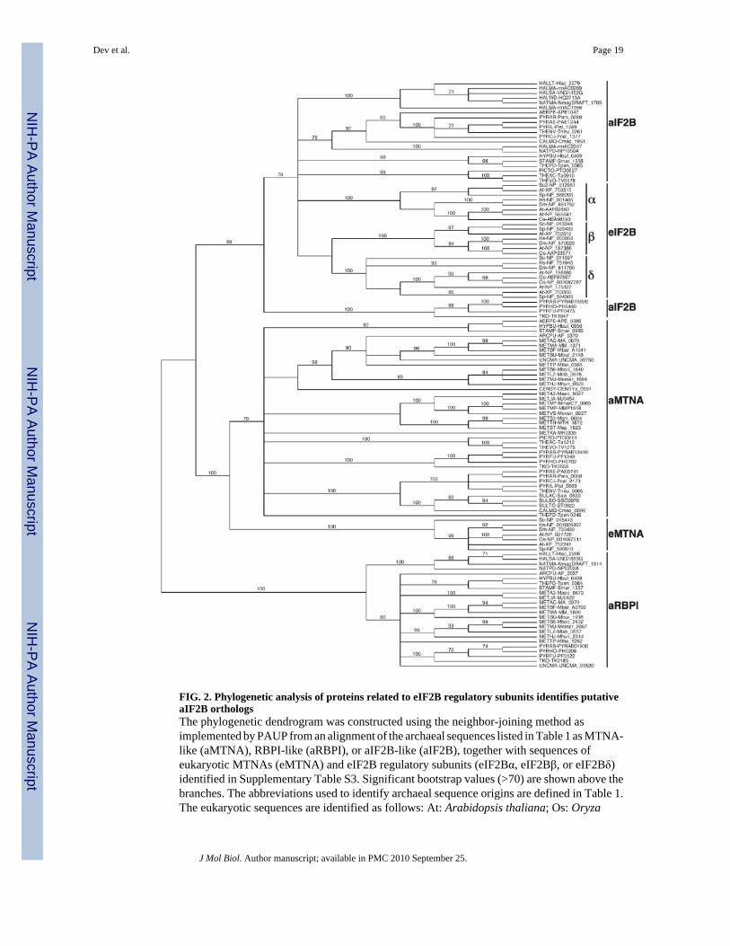
FIG. 2. Phylogenetic analysis of proteins related to eIF2B regulatory subunits identifies putativeaIF2B orthologsThe phylogenetic dendrogram was constructed using the neighbor-joining method asimplemented by PAUP from an alignment of the archaeal sequences listed in Table 1 as MTNA-like (aMTNA), RBPI-like (aRBPI), or aIF2B-like (aIF2B), together with sequences ofeukaryotic MTNAs (eMTNA) and eIF2B regulatory subunits (eIF2Bα, eIF2Bβ, or eIF2Bδ)identified in Supplementary Table S3. Significant bootstrap values (>70) are shown above thebranches. The abbreviations used to identify archaeal sequence origins are defined in Table 1.The eukaryotic sequences are identified as follows: At: Arabidopsis thaliana; Os: Oryza
Dev et al. Page 19
J Mol Biol. Author manuscript; available in PMC 2010 September 25.
NIH
-PA Author Manuscript
NIH
-PA Author Manuscript
NIH
-PA Author Manuscript

sativa; Sc: Saccharomyces cerevisiae; Sp: Schizosaccharomyces pombe; Af: Aspergillusfumigatus; Dm: Drosophila melanogaster; Hs: Homo sapiens.
Dev et al. Page 20
J Mol Biol. Author manuscript; available in PMC 2010 September 25.
NIH
-PA Author Manuscript
NIH
-PA Author Manuscript
NIH
-PA Author Manuscript

FIG. 3. Archaeal aIF2α proteins bind their cognate aIF2Bs in vitro(A) Aliquots of E. coli WCE containing GST-aIF2α from T. acidophilum (300 and 400 μg,lanes 2 and 3), P. horikoshii (200, 300 or 400 μg, lanes 8-10), or P. furiosus (200, 300, or 400μg, lanes 16-18) or equal amounts of GST alone (lanes 5 and 6, 12-14, 20-22) were incubatedwith glutathione-Sepharose beads to immobilize the GST proteins. After washing, the beadswere incubated with 400 μg bacterial WCE containing the His-aIF2B of the correspondingspecies. After extensive washing, the bound polypeptides (P, pellet) were separated by SDS-PAGE and analyzed by Western blotting using antibodies against GST or the hexa-histidinetag. Lanes 1, 4, 7, 11, 15, and 19 contain 5% of the total input (I) amounts of WCEs used forthe reactions. (B) T. acidophilum GST-aIF2α or GST alone from 400 μg of bacterial WCE
Dev et al. Page 21
J Mol Biol. Author manuscript; available in PMC 2010 September 25.
NIH
-PA Author Manuscript
NIH
-PA Author Manuscript
NIH
-PA Author Manuscript

were immobilized on glutathione-Sepharose beads and incubated with 400 μg WCE from cellscontaining the vector plasmid (P, lane 4) or a plasmid expressing His6-aIF2B (P, lane 5), as in(A). Bound proteins were resolved by SDS-PAGE and visualized by Coomassie Blue (CBB-R250) staining. Lanes 1-3 contain 10% of the input (I) fractions for GST alone, GST-aIF2α,and His-aIF2B respectively. Stars indicate the respective full-length proteins, as confirmed byWestern blotting. Molecular size markers (kDa) are indicated on the right. (C) GST-aIF2α fromP. horikoshii (lanes 4-11) or GST alone (lanes 12-17) from 300 or 400 μg of E. coli WCEswere immobilized on glutathione-Sepharose beads and incubated with 400 μg bacterial WCEcontaining P. horikoshii His-aIF2B in the presence of 100, 200 or 300 mM NaCl, as indicated.Bound proteins (Pellet) were analyzed as in (A). Lanes 1-3 contain 5% of the input (I) amountsof WCEs used for the reactions.
Dev et al. Page 22
J Mol Biol. Author manuscript; available in PMC 2010 September 25.
NIH
-PA Author Manuscript
NIH
-PA Author Manuscript
NIH
-PA Author Manuscript

FIG. 4. N-terminal S1 domain of yeast eIF2α is involved in the interaction with T. acidophilumaIF2B and the yeast eIF2B regulatory subcomplex(A) Ribbons depiction of the co-crystal structure of human PKR (orange) and the N-terminalamino acids 3-175 of region of yeast eIF2α (PDB ID 2a19). Strands β1-β5 of the S1domain ofeIF2α are colored blue, black, green, red, and yellow, respectively. The locations of Gcn-
mutants R88T and L84F, analyzed in (Fig. 5B) are indicated. (B) Full-length wild-type GST-eIF2α (WT 1-304 aa) and the indicated derivatives truncated at the C terminus (designated bythe amino acids [aa] remaining) were immobilized on glutathione-Sepharose beads and assayedfor binding T. acidophilum His-aIF2B as described for Fig. 3A, except that 100, 200, or 300μg of WCE containing the GST fusions were employed. Lanes 1, 5, 9, 13, and 17 contain 5%of total input (for the highest amounts of WCE containing GST fusions) used in the assays (C)GST pull down assays conducted as in (B) except using 200 or 300 μg of WCE containing WTGST-eIF2α or the indicated derivatives truncated at the N terminus (designated by the aminoacids remaining). The star indicates GST produced by partial degradation of the GST-eIF2αfusions visible in lanes 1-6. (D) WT GST-eIF2α (lanes 6-9), the indicated truncations describedin (B or C) (lanes 10-17), or GST alone (lanes 18 and 19) in two different amounts (200 and400 μg) of bacterial extract were immobilized on glutathione-Sepharose beads and incubated
Dev et al. Page 23
J Mol Biol. Author manuscript; available in PMC 2010 September 25.
NIH
-PA Author Manuscript
NIH
-PA Author Manuscript
NIH
-PA Author Manuscript
with (+) or without (-) purified PKR in kinase assay buffer. After washing, immobilizedproteins were incubated with 500 μg of WCE from yeast strain H4 over-expressing all threeeIF2B regulatory subunits from a high-copy plasmid (HC 2/7/3). After washing, the boundproteins (Pellet, lanes 6-19) were analyzed by Western blotting using antibodies against GCD2,GCD7, GCN3, GST, or Ser51-phosphorylated eIF2α, as indicated. Lane 1 contains 10% of theinput yeast WCE, whereas lanes 2-5 contain 5% of the total input (I) amounts of bacterialWCEs containing the GST proteins. Species in lanes 12-14 detected with GCD2 and GCD7antibodies do not exhibit the same mobilities as GCD2 and GCD7 in lanes 6-7 and, hence, areregarded as non-specific, cross-reacting species.
Dev et al. Page 24
J Mol Biol. Author manuscript; available in PMC 2010 September 25.
NIH
-PA Author Manuscript
NIH
-PA Author Manuscript
NIH
-PA Author Manuscript

FIG. 5. Neither phosphorylation of Ser-51 by PKR nor Gcn- mutations affect interaction ofeIF2α with T. acidophilum aIF2B(A) Wild-type GST-eIF2α in three different amounts (200, 300 and 400 μg) of bacterial WCEwere immobilized on glutathione-Sepharose beads and phosphorylated by PKR (+), or leftunphosphorylated (-), before incubating with either 500 μg of yeast WCE containing the over-expressed eIF2B regulatory subunits or 400 μg bacterial WCE containing T. acidophilum His-aIF2B, and the bound proteins were detected by Western blot analysis, all as described in Figs.4B and D. Lanes headed by P contain the bound fractions. Lanes 1 and 5 contain 10%, whereaslanes 9 and 13 contain 5%, of the input (I) WCEs. (B) Pull-down assays were done as describedin Fig. 4B, except that 200 or 300 μg of WCEs containing either WT GST-eIF2α, GST-eIF2α X-R88T, GST-eIF2α-L84F, or GST alone and 400 μg of WCE containing T.acidophilum His-aIF2B were used. Lanes 1-5 contain 5% of the input (I) amounts of WCE.
Dev et al. Page 25
J Mol Biol. Author manuscript; available in PMC 2010 September 25.
NIH
-PA Author Manuscript
NIH
-PA Author Manuscript
NIH
-PA Author Manuscript

FIG. 6. T. acidophilum aIF2B interacts with yeast eIF2Bα/GCN3 in vivo and destabilizes theeIF2·eIF2B complex in vitro(A) Transformants of yeast strain H4 harboring plasmids pGAL-Ta0910 or pEGKT encoding,respectively, T. acidophilum GST-aIF2B or GST alone under the GAL promoter, were grownat 30°C to A600 of ∼ 0.8 in SC medium containing 2% raffinose as carbon source and shiftedto SC medium containing 2% raffinose and 2% galactose and an additional 4 h of incubation.The GST-aIF2B or GST proteins in three different amounts (200, 300 and 400 μg) of yeastWCE were immobilized on glutathione-Sepharose beads. After extensive washing, boundproteins (Pellet, lanes 2-4 and 6-8) were analyzed by Western blotting using antibodies againsteIF2B subunits GCD6, GCD1, GCD2, GCD7, and GCN3, eIF2α, eIF2β, eIF2γ, and GST, as
Dev et al. Page 26
J Mol Biol. Author manuscript; available in PMC 2010 September 25.
NIH
-PA Author Manuscript
NIH
-PA Author Manuscript
NIH
-PA Author Manuscript

indicated. Lane 1 and 5 contains 10% of the input (I) amounts of WCE. (B) Yeast WCE (500μg) from gcn3Δ strain H1331 grown in SC medium and the WCEs from (A) wereimmunoprecipitated using polyclonal antibodies against GCD6 and protein A Sepharose beads.After extensive washing, immunocomplexes (P, lanes 2, 4 and 6) were analyzed by Westernblotting using antibodies against GCD6, GCD1, GCD2, GCD7, GCN3, and eIF2α. Lanes 1, 3and 5 represent 10% of the input (I) amounts. (C) Alignment of the NUCT domains ineIF2Bε/GCD6, TK0955, and the consensus NUCT domain sequence defined previously 32
using the following symbols: (.) any residue allowed; U or u, highly or moderately conservedbulky hydrophobic residue, respectively; O or o, highly or moderately conserved small residue;J or j, highly or moderately conserved positively charged residue; $, Ser or Thr. Identities (|)and conservative replacements (*) are indicated.
Dev et al. Page 27
J Mol Biol. Author manuscript; available in PMC 2010 September 25.
NIH
-PA Author Manuscript
NIH
-PA Author Manuscript
NIH
-PA Author Manuscript
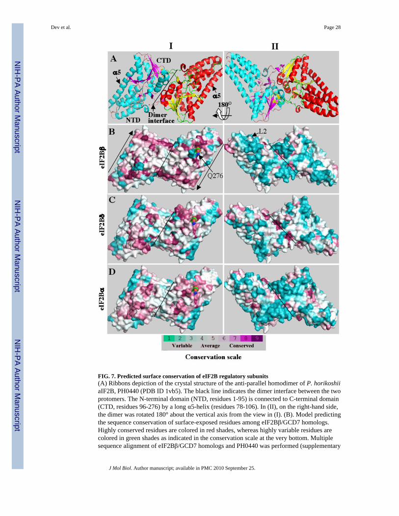
FIG. 7. Predicted surface conservation of eIF2B regulatory subunits(A) Ribbons depiction of the crystal structure of the anti-parallel homodimer of P. horikoshiiaIF2B, PH0440 (PDB ID 1vb5). The black line indicates the dimer interface between the twoprotomers. The N-terminal domain (NTD, residues 1-95) is connected to C-terminal domain(CTD, residues 96-276) by a long α5-helix (residues 78-106). In (II), on the right-hand side,the dimer was rotated 180° about the vertical axis from the view in (I). (B). Model predictingthe sequence conservation of surface-exposed residues among eIF2Bβ/GCD7 homologs.Highly conserved residues are colored in red shades, whereas highly variable residues arecolored in green shades as indicated in the conservation scale at the very bottom. Multiplesequence alignment of eIF2Bβ/GCD7 homologs and PH0440 was performed (supplementary
Dev et al. Page 28
J Mol Biol. Author manuscript; available in PMC 2010 September 25.
NIH
-PA Author Manuscript
NIH
-PA Author Manuscript
NIH
-PA Author Manuscript
Fig. S1) and the degree of sequence conservation at amino acid positions that can be alignedwith the PH0440 sequence is projected on the surface of the PH0440 homodimer. Highlyconserved residues (red) are clustered on one surface (I), whereas most of the variable residues(blue) are on the opposite face (II). (C) and (D). Models predicting the sequence conservationof surface-exposed residues among eIF2Bδ/GCD2 (C) and eIF2Bα/GCN3 homologs,constructed from the alignments shown in Figs. S2 and S3, respectively, just as in (B).
Dev et al. Page 29
J Mol Biol. Author manuscript; available in PMC 2010 September 25.
NIH
-PA Author Manuscript
NIH
-PA Author Manuscript
NIH
-PA Author Manuscript
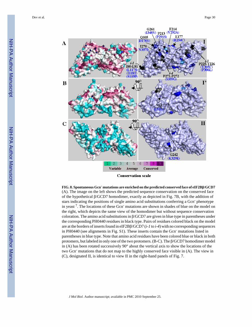
FIG. 8. Spontaneous Gcn- mutations are enriched on the predicted conserved face of eIF2Bβ/GCD7(A). The image on the left shows the predicted sequence conservation on the conserved faceof the hypothetical β/GCD7 homodimer, exactly as depicted in Fig. 7B, with the addition ofstars indicating the positions of single amino acid substitutions conferring a Gcn- phenotypein yeast 7. The locations of these Gcn- mutations are shown in shades of blue on the model onthe right, which depicts the same view of the homodimer but without sequence conservationcoloration. The amino acid substitutions in β/GCD7 are given in blue type in parentheses underthe corresponding PH0440 residues in black type. Pairs of residues colored black on the modelare at the borders of inserts found in eIF2Bβ/GCD7 (i-1 to i-4) with no corresponding sequencesin PH0440 (see alignments in Fig. S1). These inserts contain the Gcn- mutations listed inparentheses in blue type. Note that amino acid residues have been colored blue or black in bothprotomers, but labeled in only one of the two protomers. (B-C). The β/GCD7 homodimer modelin (A) has been rotated successively 90° about the vertical axis to show the locations of thetwo Gcn- mutations that do not map to the highly conserved face visible in (A). The view in(C), designated II, is identical to view II in the right-hand panels of Fig. 7.
Dev et al. Page 30
J Mol Biol. Author manuscript; available in PMC 2010 September 25.
NIH
-PA Author Manuscript
NIH
-PA Author Manuscript
NIH
-PA Author Manuscript

FIG. 9. Spontaneous Gcn- mutations are enriched on the predicted conserved faces of eIF2Bδ/GCD2 and eIF2Bα/GCN3, as are Gcd- mutations in eIF2Bα/GCN3(A-B) The locations of spontaneous Gcn- mutations in yeast δ/GCD2 7 are depicted in viewsI and II, identical to those shown in Fig. 7C, following the same conventions described in Fig.8 for β/GCD7 mutations. (C-D) The locations of Gcn- (blue) and Gcd- (orange) mutations inyeast α/GCN3 7; 34 are depicted in views I and II, identical to those shown in Fig. 7D, followingthe same conventions described in Fig. 8 for β/GCD7 mutations. Surface conservation on theleft panel is as shown in Fig. 7D. The amino acid residues of GCN3 which produce Gcd-
phenotype are colored in orange shades, whereas the one that correspond to Gcn- phenotypeare colored in blue (right panel). The aIF2B amino acid residues are numbered in black,
Dev et al. Page 31
J Mol Biol. Author manuscript; available in PMC 2010 September 25.
NIH
-PA Author Manuscript
NIH
-PA Author Manuscript
NIH
-PA Author Manuscript

followed by yeast mutations in parenthesis. (D) The dimer has been rotated 180° about thevertical axis in opposite direction showing the least conserved face.
Dev et al. Page 32
J Mol Biol. Author manuscript; available in PMC 2010 September 25.
NIH
-PA Author Manuscript
NIH
-PA Author Manuscript
NIH
-PA Author Manuscript
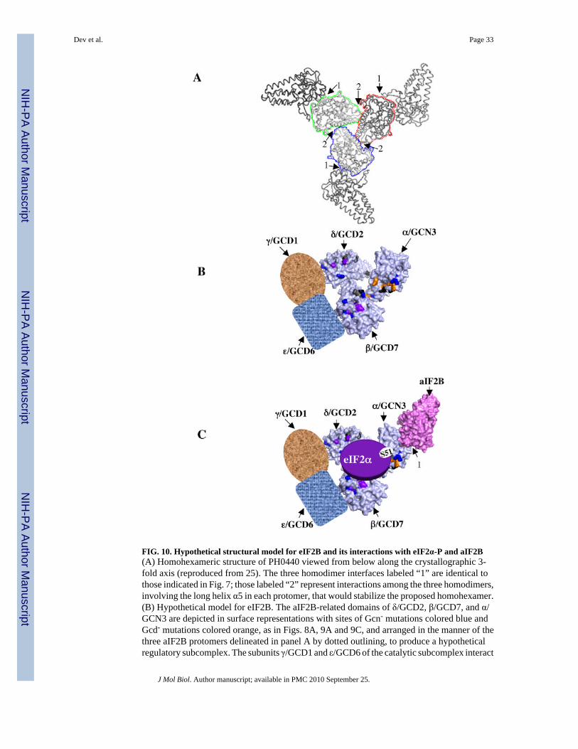
FIG. 10. Hypothetical structural model for eIF2B and its interactions with eIF2α-P and aIF2B(A) Homohexameric structure of PH0440 viewed from below along the crystallographic 3-fold axis (reproduced from 25). The three homodimer interfaces labeled “1” are identical tothose indicated in Fig. 7; those labeled “2” represent interactions among the three homodimers,involving the long helix α5 in each protomer, that would stabilize the proposed homohexamer.(B) Hypothetical model for eIF2B. The aIF2B-related domains of δ/GCD2, β/GCD7, and α/GCN3 are depicted in surface representations with sites of Gcn- mutations colored blue andGcd- mutations colored orange, as in Figs. 8A, 9A and 9C, and arranged in the manner of thethree aIF2B protomers delineated in panel A by dotted outlining, to produce a hypotheticalregulatory subcomplex. The subunits γ/GCD1 and ε/GCD6 of the catalytic subcomplex interact
Dev et al. Page 33
J Mol Biol. Author manuscript; available in PMC 2010 September 25.
NIH
-PA Author Manuscript
NIH
-PA Author Manuscript
NIH
-PA Author Manuscript
with the δ/GCD2 and β/GCD7 subunits of the regulatory subcomplex. (C) The same model inpanel B but showing the predicted binding surface for eIF2α on the conserved face of the eIF2Bregulatory subcomplex and the proposed heterodimer formation between aIF2B and α/GCN3using the dimer interface “1” defined in (A). (see text for further details.)
Dev et al. Page 34
J Mol Biol. Author manuscript; available in PMC 2010 September 25.
NIH
-PA Author Manuscript
NIH
-PA Author Manuscript
NIH
-PA Author Manuscript
NIH
-PA Author Manuscript
NIH
-PA Author Manuscript
NIH
-PA Author Manuscript
Dev et al. Page 35
Tabl
e 1
Pred
icte
d pr
otei
n se
quen
ces s
imila
r to
eIF
2B r
egul
ator
y su
buni
ts in
var
ious
arc
haea
l spe
cies
1
Spec
ies
Arc
haea
l lin
eage
2M
TN
A-li
keR
BPI
-like
aIF2
B-li
ke
Ther
mof
ilum
pen
dens
(TH
EPD
)C
A1R
WS6
(Tpe
n_02
46)
AlR
X63
(Tpe
n_03
84)
A1R
X64
(Tpe
n_03
85)
Hyp
erth
erm
us b
utyl
icus
(HY
PBU
)C
A2B
LE3
(Hbu
t_09
56)
A2B
K49
(Hbu
t_04
98)
A2B
K50
(Hbu
t_04
99)
Stap
hylo
ther
mus
mar
inus
(STA
MF)
CA
3DN
77 (S
mar
_098
9)A
3DP8
8 (S
mar
_135
7)A
3DP8
9 (S
mar
_135
8)
Aero
pyru
m p
erni
x (A
ERPE
)C
Q9Y
E84
(APE
_068
6)—
Q9Y
D65
(APE
1047
)
Cal
divi
rga
maq
uilin
gens
is (C
ALM
Q)
CA
8MB
P9 (C
maq
_039
6)—
A8M
BN
4 (C
maq
_195
1)
Pyro
bacu
lum
ars
enat
icum
(PY
RA
R)
CA
4WG
V8
(Par
s_00
08)
—A
4WH
N4
(Par
s_02
88)
Pyro
bacu
lum
aer
ophi
lum
(PY
RA
E)C
Q8Z
YJ8
(PA
E074
1)—
Q8Z
XJ8
(PA
E124
4)
Pyro
bacu
lum
isla
ndic
um (P
YR
IL)
CA
1RSA
9 (P
isl_
0663
)—
A1R
U20
(Pis
l_12
89)
Pyro
bacu
lum
cal
idifo
ntis
(PY
RC
J)C
A3M
Y71
(Pca
l_21
73)
—A
3MV
Y1
(Pca
l_13
77)
Ther
mop
rote
us n
eutr
ophi
lus (
THEN
V)
CB
1Y9P
1 (T
neu_
0005
)—
B1Y
B83
(Tne
u_02
61)
Cen
arch
aeum
sym
bios
um (C
ENSY
)C
A0R
V57
(CEN
SYa_
0591
)—
—
Sulfo
lobu
s sol
fata
ricu
s (SU
LSO
)C
Q97
UM
6 (S
SO29
78)
——
Sulfo
lobu
s tok
odai
i (SU
LTO
)C
Q97
3H9
(ST0
922)
——
Sulfo
lobu
s aci
doca
ldar
ius (
SULA
C)
CQ
4JB
92 (S
aci_
0539
)—
—
Pyro
cocc
us fu
rios
us (P
YR
FU)
EQ
8U17
8 (P
F134
9)Q
8U4G
6 (P
F012
2)Q
8U3J
1 (P
F047
5)
Pyro
cocc
us h
orik
oshi
i (PY
RH
O)
EO
5843
3 (P
H07
02)
O57
947
(PH
0208
)O
5818
5 (P
H04
40)
Ther
moc
occu
s kod
akar
aens
is (T
KO
)E
Q5J
F43
(TK
0556
)Q
5JFM
9 (T
K01
85)
Q5J
DZ9
(TK
1047
)
Pyro
cocc
us a
byss
i (PY
RA
B)
EQ
9UZ1
6 (P
YR
AB
1341
0)Q
9V28
1(PY
RA
B01
930)
Q9U
YB
6(PY
RA
B15
920)
Met
hano
cocc
us ja
nnas
chii
(MET
JA)
EQ
5789
6 (M
J045
4)Q
5758
6 (M
J012
2)—
Met
hano
cocc
us a
eolic
us (M
ETA
3)E
A6U
SZ8
(Mae
o_00
27)
A6U
UT5
(Mae
o_06
73)
—
Met
hano
culle
us m
aris
nigr
i (M
ETM
J)E
A3C
W71
(Mem
ar_1
694)
A3C
X93
(Mem
ar_2
067)
—
Met
hano
regu
la b
oone
i (M
ETB
6)E
A7I
9E4
(Mbo
o_18
40)
A7I
B35
(Mbo
o_24
32)
—
Met
hano
saet
a th
erm
ophi
la (M
ETTP
)E
A0B
634
(Mth
e_03
65)
A0B
8L7
(Mth
e_12
62)
—
Met
hano
sarc
ina
acet
ivor
ans (
MET
AC
)E
Q8T
UJ1
(MA
_007
6)Q
8TTP
7(M
A_0
379)
—
Met
hano
corp
uscu
lum
labr
eanu
m (M
ETLZ
)E
A2S
S42
(Mla
b_09
78)
A2S
QX
5 (M
lab_
0557
)—
Met
hano
sarc
ina
bark
eri (
MET
BF)
EQ
46D
N3
(Mba
r_A
1041
)Q
46EC
2 (M
bar_
A07
93)
—
Met
hano
sarc
ina
maz
ei (M
ETM
A)
EQ
8PX
51 (M
M_1
371)
Q8P
WI0
(MM
_160
6)—
Met
hano
cocc
oide
s bur
toni
i (M
ETB
U)
EQ
12U
85 (M
bur_
2118
)Q
12U
Q7
(Mbu
r_19
38)
—
Arch
aeog
lobu
s ful
gidu
s (A
RC
FU)
EO
2987
7 (A
F_03
70)
O28
242
(AF_
2037
)—
J Mol Biol. Author manuscript; available in PMC 2010 September 25.

NIH
-PA Author Manuscript
NIH
-PA Author Manuscript
NIH
-PA Author Manuscript
Dev et al. Page 36
Spec
ies
Arc
haea
l lin
eage
2M
TN
A-li
keR
BPI
-like
aIF2
B-li
ke
Met
hano
spir
illum
hun
gate
i (M
ETH
J)E
Q2F
N46
(Mhu
n_06
23)
Q2F
SY5
(Mhu
n_23
14)
—
Met
hano
bact
eriu
m th
erm
oaut
otro
phic
um (M
ETTH
)E
O27
900
(MTH
_187
2)—
—
Met
hano
brev
ibac
ter s
mith
ii (M
ETS3
)E
A5U
LDl(M
sm_0
804)
——
Met
hano
cocc
us m
arip
alud
is (M
ETM
P)E
Q6L
WT8
(MM
P161
8)—
—
Met
hano
spha
era
stad
tman
ae (M
ETST
)E
Q2N
FJ6
(Msp
_102
3)—
—
Met
hano
cocc
us m
arip
alud
is (M
ETM
7)E
A6V
HK
6 (M
mar
C7_
0865
)—
—
Met
hano
pyru
s kan
dler
i (M
ETK
A)
EQ
8TY
G4
(MK
0335
)—
—
Met
hano
cocc
us v
anni
elii
(MET
VS)
EA
6UQ
Q9
(Mev
an_0
927)
——
Ther
mop
lasm
a ac
idop
hilu
m (T
HEA
C)
EQ
9HIW
4 (T
a121
2)—
Q9H
JQ7
(Ta0
910)
Ther
mop
lasm
a vo
lcan
ium
(TH
EVO
)E
Q97
8Y9
(TV
1275
)—
Q97
A32
(TV
0978
)
Picr
ophi
lus t
orri
dus (
PIC
TO)
EQ
6L2F
6 (P
TO02
61)
—Q
6L1P
0(PT
O05
27)
Hal
obac
teri
um sa
linar
ium
(HA
LSA
)E
—Q
9HP1
1 (V
NG
1853
G)
Q9H
PV9
(VN
G14
52G
)
Hal
orub
rum
lacu
spro
fund
i (H
ALL
T)E
—B
9LR
X1
(Hla
c_22
69)
B9L
RY
1 (H
lac_
2279
)
Nat
rial
ba m
agad
ii (N
ATM
A)
E—
B9Z
FW1
(Nm
agD
RA
FT_1
314)
B9Z
EY5
(Nm
agD
RA
FT_1
705)
Nat
rono
mon
as p
hara
onis
(NA
TPD
)E
—Q
3IQ
A4
(NP3
202A
)Q
3ITB
8(N
P105
0A)
Hal
oarc
ula
mar
ism
ortu
i (H
ALM
A)
E—
—Q
5V0X
9 (r
rnA
C19
56)
Q5V
557
(rrn
AC
0289
)Q
5V07
5 (r
rnA
C22
47)
Hal
oqua
drat
um w
alsb
yi (H
ALW
D)
E—
—Q
18G
S5 (H
Q27
13A
)
Unc
ultu
red
met
hano
geni
c ar
chae
on (U
NC
MA
)E
Q0W
2L6
(UN
CM
A_0
8750
)Q
0W0H
9 (U
NC
MA
_015
20)
—
1 A B
LAST
sear
ch o
f the
arch
aeal
gen
ome d
atab
ase w
as co
nduc
ted
usin
g P.
hor
ikos
hii a
IF2B
(PH
0440
; Sw
iss-
Prot
O58
185)
and
the 8
9 se
quen
ces w
ith th
e hig
hest
BLA
ST sc
ores
wer
e sub
ject
ed to
mul
tiple
sequ
ence
alig
nmen
ts u
sing
Clu
stal
W an
d cl
assi
fied
acco
rdin
g to
the p
rese
nce
or a
bsen
ce o
f MTN
A si
gnat
ure
mot
ifs 3
6 , la
bele
d m
I-m
VIII
, as i
llust
rate
d fo
r a su
bset
of t
he se
quen
ces i
n Fi
g. 1
. MTN
A-li
ke se
quen
ces c
onta
in p
erfe
ct o
r nea
rly p
erfe
ct m
atch
es to
all
eigh
t mot
ifs. R
BPI
-like
sequ
ence
s als
o co
ntai
n pe
rfec
t mat
ches
to th
e M
TNA
mot
ifs e
xcep
t for
an N
-term
inal
trun
catio
n th
at re
mov
es m
I and
an
8- re
sidu
e de
letio
n in
mIV
. The
aIF
2B-li
ke p
rote
ins a
lso
exhi
bit t
hese
two
diff
eren
ces f
rom
MTN
A-li
ke se
quen
ces a
nd a
lso
cont
ain
dege
nera
te v
ersi
ons o
f the
oth
er m
otifs
. (Se
e Fi
g. 1
and
text
for m
ore
deta
ils.)
Sequ
ence
s are
desi
gnat
ed b
y th
eir S
wis
s-Pr
ot a
cces
sion
num
bers
and
ord
ered
locu
s num
bers
(sho
wn
in p
aren
thes
is).
2 E =
Eury
arch
aea;
C =
Cre
narc
haea
J Mol Biol. Author manuscript; available in PMC 2010 September 25.
NIH
-PA Author Manuscript
NIH
-PA Author Manuscript
NIH
-PA Author Manuscript
Dev et al. Page 37
Table 2MudPit analysis of proteins co-purifying with T. kodakaraensis His10-HA-tagged aIF2B1
Orderd locus no. Interacting protein MASCOT score no. of peptides matched (% sequence coverage)
TK1047 Translation initiation factor IF-2B alpha 1209 28 (60)
TK1557 Dehydrogenase 444 12 (13)
TK1502 50S ribosomal protein L18e 404 10 (33)
TK0137 Nucleic acid binding protein 404 12 (47)
TK0955 Sugar-phosphate nucleotidyltransferase 283 8 (11)
TK1417 50S ribosomal protein L1P 254 5 (30)
TK1100 Translation initiation factor IF-2 alpha 236 9 (8)
TK1522 50S ribosomal protein L18P 224 6 (21)
TK1520 50S ribosomal protein L30P 200 5 (16)
TK1410 DNA primase 192 6 (9)
TK1541 50S ribosomal protein L4P 170 4 (6)
TK2200 Hypothetical protein 167 6 (10)
TK1528 50S ribosomal protein L5P 163 7 (20)
TK2304 50S ribosomal protein L2P 162 4 (7)
TK1542 50S ribosomal protein L3P 151 4 (12)
TK0477 HAD superfamily hydrolase 148 4 (10)
TK0019 ABC transporter ATPase 136 2 (3)
TK1636 Putative RNA-associated protein 115 2 (4)
TK1418 50S ribosomal protein L11P 111 2 (7)
1Proteins with MASCOT scores of greater than 100 identified in the eluate containing His10-HA-tagged aIF2B from the H4 (tagged) strain of T.
kodakarensis are listed in descending order, along with the number of peptides matched to the relevant protein and the percentage of its amino acidsequence covered by the matching peptides. None of the listed proteins were detected in equivalent column fractions prepared from the untagged H2strain (See text for further details).
J Mol Biol. Author manuscript; available in PMC 2010 September 25.
Copyright © 2022 FDOKUMEN