Apoptosis and Autophagy in Breast Cancer Cells following Exemestane Treatment
10
Apoptosis and Autophagy in Breast Cancer Cells following Exemestane Treatment Cristina Amaral 1,2 , Margarida Borges 1,2 , Soraia Melo 1,3 , Elisia ´ rio Tavares da Silva 4 , Georgina Correia-da- Silva 1,2 , Nate ´ rcia Teixeira 1,2 * 1 Laboratory of Biochemistry, Department of Biological Sciences, Faculty of Pharmacy, University of Porto, Porto, Portugal, 2 Institute for Molecular and Cell Biology (IBMC), University of Porto, Porto, Portugal, 3 Department of Zoology, Faculty of Sciences and Technology, University of Coimbra, Coimbra, Portugal, 4 Center of Pharmaceutical Studies, Pharmaceutical Chemistry Laboratory, Faculty of Pharmacy, University of Coimbra, Coimbra, Portugal Abstract Aromatase inhibitors (AIs), which block the conversion of androgens to estrogens, are used for hormone-dependent breast cancer treatment. Exemestane, a steroidal that belongs to the third-generation of AIs, is a mechanism-based inhibitor that binds covalently and irreversibly, inactivating and destabilizing aromatase. Since the biological effects of exemestane in breast cancer cells are not totally understood, its effects on cell viability, cell proliferation and mechanisms of cell death were studied in an ER-positive aromatase-overexpressing breast cancer cell line (MCF-7aro). The effects of 3-methyladenine (3-MA), an inhibitor of autophagy and of ZVAD-FMK, an apoptotic inhibitor, in exemestane treated cells were also investigated. Our results indicate that exemestane induces a strong inhibition in MCF-7aro cell proliferation in a dose- and time-dependent manner, promoting a significant cell cycle arrest in G 0 /G1 or in G 2 /M phases after 3 and 6 days of treatment, respectively. This was accompanied by a decrease in cell viability due to activation of cell death by apoptosis, via mitochondrial pathway and the occurrence of autophagy. Inhibition of autophagy by the autophagic inhibitor, 3-MA, resulted in a reduction of cell viability and activation of caspases. All together the results obtained suggest that exemestane induced mitochondrial-mediated apoptosis and autophagy, which act as a pro-survival process regulating breast cancer cell apoptosis. Citation: Amaral C, Borges M, Melo S, Silva ETd, Correia-da-Silva G, et al. (2012) Apoptosis and Autophagy in Breast Cancer Cells following Exemestane Treatment. PLoS ONE 7(8): e42398. doi:10.1371/journal.pone.0042398 Editor: Srikumar P. Chellappan, H. Lee Moffitt Cancer Center & Research Institute, United States of America Received March 19, 2012; Accepted July 5, 2012; Published August 13, 2012 Copyright: ß 2012 Amaral et al. This is an open-access article distributed under the terms of the Creative Commons Attribution License, which permits unrestricted use, distribution, and reproduction in any medium, provided the original author and source are credited. Funding: Cristina Amaral is a recipient of a PhD grant of Fundac ¸a ˜o para a Cie ˆncia e Tecnologia (FCT) (SFRH/BD/48190/2008). This work was funded by FEDER Funds through the Operational Competitiveness Program- COMPETE and by National Funds through FCT- Fundac ¸a ˜ o para a Cie ˆ ncia e Tecnologia under the project FCOMP-01-0124-FEDER-020970 (PTDC/QUI-BIQ/120319/2010). The funders had no role in study design, data collection and analysis, decision to publish, or preparation of the manuscript. Competing Interests: The authors have declared that no competing interests exist. * E-mail: [email protected] Introduction Breast cancer is the most common cause of cancer death in women worldwide. Among breast cancer patients, 60% of pre- menopausal and 70–80% of post-menopausal women have hormone-dependent (estrogen receptor positive [ER + ]) tumors [1,2]. As estrogens play a crucial role in stimulating ER + tumor growth, the suppression of their effects is considered an important therapeutic target for breast cancer treatment. Two main approaches have been successfully applied. One targets the ER directly through the use of selective estrogen receptor modulators (SERM), such as tamoxifen, or of selective estrogen receptor down-regulators (SERD), like fulvestrant. The other is achieved by the use of aromatase inhibitors (AIs) that inhibit aromatase, the enzyme responsible by the last step of estrogen synthesis, blocking the conversion of androgens to estrogens [1,3]. Over the past three decades AIs became an effective alternative to tamoxifen, showing clinical benefits with high specificity and reduced recurrence rates [4]. The third-generation of AIs includes non-steroidal triazole derivates, anastrozole and letrozole, that act as competitive inhibitors and one steroidal derivate of androstene- dione, exemestane [4,5]. Exemestane is a mechanism-based inhibitor that is catalytically converted into chemically reactive intermediates These molecules bind covalently and irreversibly to the substrate-binding pocket of the enzyme, inactivating and producing suicide aromatase inhibition [1,6,7]. Wang and Chen (2006) found that exemestane destabilizes aromatase and induces its degradation by the proteosome after its irreversible inactivation [8]. On the other hand, exemestane and its principal metabolite, 17-hydroexemestane, exhibit androgenic effects as it binds with high affinity to the androgen receptor, causing in that way, lower bone loss [2,6,7]. The efficacy of hormonal therapy in breast cancer is based on the fact that estrogens play an important role in cancer cell survival and proliferation, essentially affecting cell cycle [9] and inducing expression of growth factors and cytokines [10,11]. It has also been reported that estrogen deprivation causes a decrease in cell proliferation and induces apoptosis in MCF-7 cells [12,13] and in MCF-7 xenografts [14,15]. SERMs [13,16,17] and antagonists of estrogen receptor [18] induce inhibition of cell proliferation and apoptosis in breast cancer cell lines. Although recent reports showed that tamoxifen and 4-hydroxytamoxifen (4-OHT) induced autophagy [19,20], others referred that tamoxifen treatment is associated with both types of cell death [21,22]. It has also been reported that some AIs, like letrozole, anastrozole and formestane inhibit proliferation of breast cancer cells by inducing cell cycle PLOS ONE | www.plosone.org 1 August 2012 | Volume 7 | Issue 8 | e42398
Transcript of Apoptosis and Autophagy in Breast Cancer Cells following Exemestane Treatment
Apoptosis and Autophagy in Breast Cancer Cellsfollowing Exemestane TreatmentCristina Amaral1,2, Margarida Borges1,2, Soraia Melo1,3, Elisiario Tavares da Silva4, Georgina Correia-da-
Silva1,2, Natercia Teixeira1,2*
1 Laboratory of Biochemistry, Department of Biological Sciences, Faculty of Pharmacy, University of Porto, Porto, Portugal, 2 Institute for Molecular and Cell Biology
(IBMC), University of Porto, Porto, Portugal, 3 Department of Zoology, Faculty of Sciences and Technology, University of Coimbra, Coimbra, Portugal, 4 Center of
Pharmaceutical Studies, Pharmaceutical Chemistry Laboratory, Faculty of Pharmacy, University of Coimbra, Coimbra, Portugal
Abstract
Aromatase inhibitors (AIs), which block the conversion of androgens to estrogens, are used for hormone-dependent breastcancer treatment. Exemestane, a steroidal that belongs to the third-generation of AIs, is a mechanism-based inhibitor thatbinds covalently and irreversibly, inactivating and destabilizing aromatase. Since the biological effects of exemestane inbreast cancer cells are not totally understood, its effects on cell viability, cell proliferation and mechanisms of cell deathwere studied in an ER-positive aromatase-overexpressing breast cancer cell line (MCF-7aro). The effects of 3-methyladenine(3-MA), an inhibitor of autophagy and of ZVAD-FMK, an apoptotic inhibitor, in exemestane treated cells were alsoinvestigated. Our results indicate that exemestane induces a strong inhibition in MCF-7aro cell proliferation in a dose- andtime-dependent manner, promoting a significant cell cycle arrest in G0/G1 or in G2/M phases after 3 and 6 days of treatment,respectively. This was accompanied by a decrease in cell viability due to activation of cell death by apoptosis, viamitochondrial pathway and the occurrence of autophagy. Inhibition of autophagy by the autophagic inhibitor, 3-MA,resulted in a reduction of cell viability and activation of caspases. All together the results obtained suggest that exemestaneinduced mitochondrial-mediated apoptosis and autophagy, which act as a pro-survival process regulating breast cancer cellapoptosis.
Citation: Amaral C, Borges M, Melo S, Silva ETd, Correia-da-Silva G, et al. (2012) Apoptosis and Autophagy in Breast Cancer Cells following ExemestaneTreatment. PLoS ONE 7(8): e42398. doi:10.1371/journal.pone.0042398
Editor: Srikumar P. Chellappan, H. Lee Moffitt Cancer Center & Research Institute, United States of America
Received March 19, 2012; Accepted July 5, 2012; Published August 13, 2012
Copyright: � 2012 Amaral et al. This is an open-access article distributed under the terms of the Creative Commons Attribution License, which permitsunrestricted use, distribution, and reproduction in any medium, provided the original author and source are credited.
Funding: Cristina Amaral is a recipient of a PhD grant of Fundacao para a Ciencia e Tecnologia (FCT) (SFRH/BD/48190/2008). This work was funded by FEDERFunds through the Operational Competitiveness Program- COMPETE and by National Funds through FCT- Fundacao para a Ciencia e Tecnologia under the projectFCOMP-01-0124-FEDER-020970 (PTDC/QUI-BIQ/120319/2010). The funders had no role in study design, data collection and analysis, decision to publish, orpreparation of the manuscript.
Competing Interests: The authors have declared that no competing interests exist.
* E-mail: [email protected]
Introduction
Breast cancer is the most common cause of cancer death in
women worldwide. Among breast cancer patients, 60% of pre-
menopausal and 70–80% of post-menopausal women have
hormone-dependent (estrogen receptor positive [ER+]) tumors
[1,2]. As estrogens play a crucial role in stimulating ER+ tumor
growth, the suppression of their effects is considered an important
therapeutic target for breast cancer treatment. Two main
approaches have been successfully applied. One targets the ER
directly through the use of selective estrogen receptor modulators
(SERM), such as tamoxifen, or of selective estrogen receptor
down-regulators (SERD), like fulvestrant. The other is achieved by
the use of aromatase inhibitors (AIs) that inhibit aromatase, the
enzyme responsible by the last step of estrogen synthesis, blocking
the conversion of androgens to estrogens [1,3].
Over the past three decades AIs became an effective alternative
to tamoxifen, showing clinical benefits with high specificity and
reduced recurrence rates [4]. The third-generation of AIs includes
non-steroidal triazole derivates, anastrozole and letrozole, that act
as competitive inhibitors and one steroidal derivate of androstene-
dione, exemestane [4,5]. Exemestane is a mechanism-based
inhibitor that is catalytically converted into chemically reactive
intermediates These molecules bind covalently and irreversibly to
the substrate-binding pocket of the enzyme, inactivating and
producing suicide aromatase inhibition [1,6,7]. Wang and Chen
(2006) found that exemestane destabilizes aromatase and induces
its degradation by the proteosome after its irreversible inactivation
[8]. On the other hand, exemestane and its principal metabolite,
17-hydroexemestane, exhibit androgenic effects as it binds with
high affinity to the androgen receptor, causing in that way, lower
bone loss [2,6,7].
The efficacy of hormonal therapy in breast cancer is based on
the fact that estrogens play an important role in cancer cell
survival and proliferation, essentially affecting cell cycle [9] and
inducing expression of growth factors and cytokines [10,11]. It has
also been reported that estrogen deprivation causes a decrease in
cell proliferation and induces apoptosis in MCF-7 cells [12,13] and
in MCF-7 xenografts [14,15]. SERMs [13,16,17] and antagonists
of estrogen receptor [18] induce inhibition of cell proliferation and
apoptosis in breast cancer cell lines. Although recent reports
showed that tamoxifen and 4-hydroxytamoxifen (4-OHT) induced
autophagy [19,20], others referred that tamoxifen treatment is
associated with both types of cell death [21,22]. It has also been
reported that some AIs, like letrozole, anastrozole and formestane
inhibit proliferation of breast cancer cells by inducing cell cycle
PLOS ONE | www.plosone.org 1 August 2012 | Volume 7 | Issue 8 | e42398
arrest in G0/G1 phase and cell death by apoptosis [13,23].
Recently, we demonstrated that the steroidal AIs 5a-androst-3-en-
17-one and 3a,4a-epoxy-5a-androstan-17-one, previously synthe-
sized in our laboratory [24], inhibit cell proliferation in various
tumour cell lines [25] and induce apoptosis and autophagy in
MCF-7aro cell line [26]. Nevertheless, the effects of exemestane in
breast cancer cells are not totally understood. In this way, it was
evaluated the biological effects of this steroidal AI in an ER-
positive aromatase-overexpressing breast cancer cell line (MCF-
7aro) and studied the mechanisms of cell death induced by
exemestane.
Results
Morphological studiesTo investigate the morphological alterations induced by
exemestane, MCF-7aro cells, were cultured with or without
exemestane during 3, 6 (Fig. 1) and 9 days and examined by phase
contrast microscopy, Giemsa and Hoechst staining. After 3 days of
exemestane treatment, few membrane blebbings and chromatin
fragmentation were observed (data not shown). After 6 and 9 days,
cells showed marked morphological alterations, like membrane
blebbings, chromatin condensation and fragmentation, cytoplasm
vacuolization and the presence of non-adherent cells. A decrease
in cell density was also observed after 9 days of treatment (data not
shown). These features were more evident for the highest
concentration of exemestane and increased with the time of
treatment.
Cell viability and cell proliferationTo evaluate the effects of exemestane (2.5–15 mM) in MCF-
7aro cells viability and cytotoxicity, 3-(4,5-dimethylthiazol-2-yl)-
2,5-diphenyltetrazolium (MTT) and lactate dehydrogenase (LDH)
assays were performed for 2, 3, 6 and 9 days. After 2 days of
exemestane treatment no effects in cell viability were observed.
Although, after 3, 6 and 9 days and as shown in Fig. 2A,
exemestane induced a reduction in cell viability that was dose- and
time-dependent. The lower concentrations of exemestane (2.5–
5 mM) did not affect cell viability, except in the case of 9 days of
treatment. However, for the higher concentrations (10–15 mM),
exemestane induced a significant decrease (p,0.05; p,0.001) in
cell viability. A significant increase (p,0.05) in LDH release was
only observed for exemestane at 15 mM after 9 days of treatment
(Fig. 2B).
To analyse the exemestane effects in cell proliferation,
thymidine incorporation assay was performed. As shown in
Fig. 2C, exemestane induced a dramatic decrease in cell
proliferation in a dose- and time- dependent manner. Contrary
to the effect on cell viability, all concentrations and times of
incubation caused a statistically significant decrease (p,0.05;
p,0.001) in the rate of DNA synthesis.
Cell cycle analysisTo identify the underlying mechanism associated with the anti-
proliferative effects of exemestane, it was evaluated the effect on
cell cycle progression by measuring DNA content by flow
cytometry. After 3 days of treatment, different concentrations of
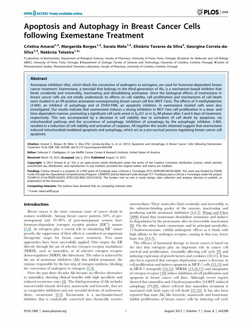
Figure 1. Effects of exemestane on MCF-7aro cells morphology. Phase contrast microscopy (A, D and G), Giemsa staining (B, E and H) andHoechst staining (C, F and I). MCF-7aro cells were examined in the absence (A, B and C) or in the presence of 10 mM (D, E and F) or 15 mM (G, H and I)of exemestane during 6 days. Treated cells presented, cytoplasm vacuolization (black arrows) in phase contrast microscopy and Giemsa staining,chromatin condensation (yellow filled arrows) and chromatin fragmentation (yellow open arrows) in Hoechst staining.doi:10.1371/journal.pone.0042398.g001
Exemestane Induce Apoptosis and Autophagy
PLOS ONE | www.plosone.org 2 August 2012 | Volume 7 | Issue 8 | e42398
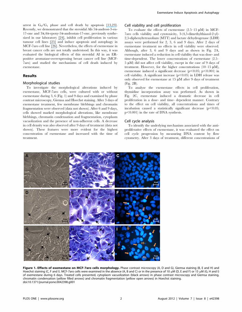
exemestane (5–10 mM) caused a significant cell cycle arrest in G0/
G1 phase and a decrease in the percentage of cells in S and G2/M
cell cycle phases (Fig. 2D) in a dose-dependent manner. An
accumulation of cells in G0/G1 phase of 75.3961.25%,
81.9460.25% (p,0.001) and 90.3061.68% (p,0.001) was
observed for 2.5, 5 and 10 mM, respectively, when comparing to
the control (70.4961.37%). However, after 6 days of treatment it
was detected an arrest in G2/M phase, 20.4361.05% (p,0.05)
and 34.4762.67% (p,0.001) for 5 and 10 mM, respectively, when
comparing to the control 14.5660.34% (Fig. 2E).
Analysis of cell deathTo investigate the type of cell death induced by exemestane in
MCF-7aro cells, assays for apoptosis and autophagy assessment
were performed. For the apoptotic analysis, translocation of PS to
the outer surface of plasma membrane was evaluated by Annexin
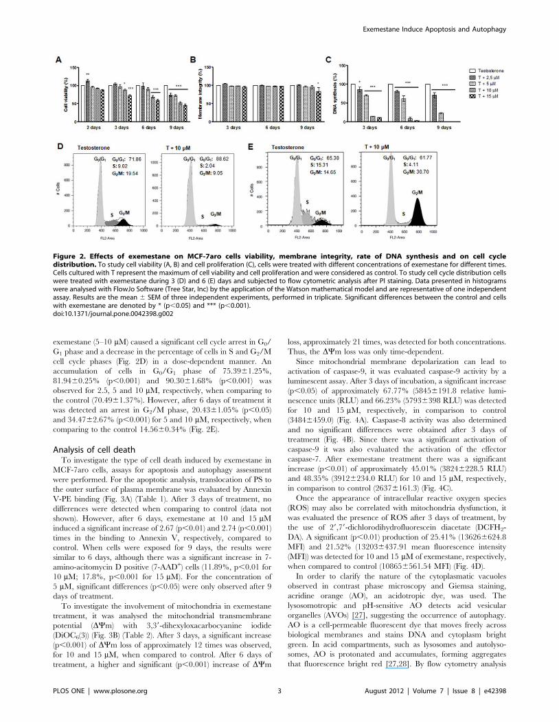
V-PE binding (Fig. 3A) (Table 1). After 3 days of treatment, no
differences were detected when comparing to control (data not
shown). However, after 6 days, exemestane at 10 and 15 mM
induced a significant increase of 2.67 (p,0.01) and 2.74 (p,0.001)
times in the binding to Annexin V, respectively, compared to
control. When cells were exposed for 9 days, the results were
similar to 6 days, although there was a significant increase in 7-
amino-acitomycin D positive (7-AAD+) cells (11.89%, p,0.01 for
10 mM; 17.8%, p,0.001 for 15 mM). For the concentration of
5 mM, significant differences (p,0.05) were only observed after 9
days of treatment.
To investigate the involvement of mitochondria in exemestane
treatment, it was analysed the mitochondrial transmembrane
potential (DYm) with 3,39-dihexyloxacarbocyanine iodide
(DiOC6(3)) (Fig. 3B) (Table 2). After 3 days, a significant increase
(p,0.001) of DYm loss of approximately 12 times was observed,
for 10 and 15 mM, when compared to control. After 6 days of
treatment, a higher and significant (p,0.001) increase of DYm
loss, approximately 21 times, was detected for both concentrations.
Thus, the DYm loss was only time-dependent.
Since mitochondrial membrane depolarization can lead to
activation of caspase-9, it was evaluated caspase-9 activity by a
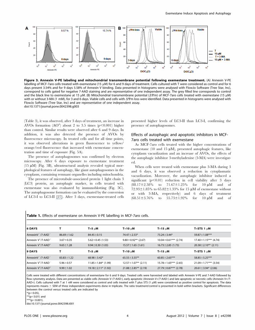
luminescent assay. After 3 days of incubation, a significant increase
(p,0.05) of approximately 67.77% (58456191.8 relative lumi-
nescence units (RLU) and 66.23% (57936398 RLU) was detected
for 10 and 15 mM, respectively, in comparison to control
(34846459.0) (Fig. 4A). Caspase-8 activity was also determined
and no significant differences were obtained after 3 days of
treatment (Fig. 4B). Since there was a significant activation of
caspase-9 it was also evaluated the activation of the effector
caspase-7. After exemestane treatment there was a significant
increase (p,0.01) of approximately 45.01% (38246228.5 RLU)
and 48.35% (39126234.0 RLU) for 10 and 15 mM, respectively,
in comparison to control (26376161.3) (Fig. 4C).
Once the appearance of intracellular reactive oxygen species
(ROS) may also be correlated with mitochondria dysfunction, it
was evaluated the presence of ROS after 3 days of treatment, by
the use of 29,79-dichlorodihydrofluorescein diacetate (DCFH2-
DA). A significant (p,0.01) production of 25.41% (136266624.8
MFI) and 21.52% (132036437.91 mean fluorescence intensity
(MFI)) was detected for 10 and 15 mM of exemestane, respectively,
when compared to control (108656561.54 MFI) (Fig. 4D).
In order to clarify the nature of the cytoplasmatic vacuoles
observed in contrast phase microscopy and Giemsa staining,
acridine orange (AO), an acidotropic dye, was used. The
lysosomotropic and pH-sensitive AO detects acid vesicular
organelles (AVOs) [27], suggesting the occurrence of autophagy.
AO is a cell-permeable fluorescent dye that moves freely across
biological membranes and stains DNA and cytoplasm bright
green. In acid compartments, such as lysosomes and autolyso-
somes, AO is protonated and accumulates, forming aggregates
that fluorescence bright red [27,28]. By flow cytometry analysis
Figure 2. Effects of exemestane on MCF-7aro cells viability, membrane integrity, rate of DNA synthesis and on cell cycledistribution. To study cell viability (A, B) and cell proliferation (C), cells were treated with different concentrations of exemestane for different times.Cells cultured with T represent the maximum of cell viability and cell proliferation and were considered as control. To study cell cycle distribution cellswere treated with exemestane during 3 (D) and 6 (E) days and subjected to flow cytometric analysis after PI staining. Data presented in histogramswere analysed with FlowJo Software (Tree Star, Inc) by the application of the Watson mathematical model and are representative of one independentassay. Results are the mean 6 SEM of three independent experiments, performed in triplicate. Significant differences between the control and cellswith exemestane are denoted by * (p,0.05) and *** (p,0.001).doi:10.1371/journal.pone.0042398.g002
Exemestane Induce Apoptosis and Autophagy
PLOS ONE | www.plosone.org 3 August 2012 | Volume 7 | Issue 8 | e42398
(Table 3), it was observed, after 3 days of treatment, an increase in
AVOs formation (AO+) about 2 to 3.5 times (p,0.001) higher
than control. Similar results were observed after 6 and 9 days. In
addition, it was also detected the presence of AVOs by
fluorescence microscopy. In treated cells and for all time points,
it was observed alterations in green fluorescence to yellow/
orange/red fluorescence that increased with exemestane concen-
tration and time of exposure (Fig. 5A).
The presence of autophagosomes was confirmed by electron
microscopy. After 6 days exposure to exemestane treatment
(15 mM) (Fig. 5B), ultrastructural analysis revealed typical mor-
phological features of autophagy, like giant autophagosomes in the
cytoplasm, containing remnant organelles including mitochondria.
The presence of microtubule-associated protein 1 light chain 3
(LC3) protein, an autophagic marker, in cells treated with
exemestane was also evaluated by immunoblotting (Fig. 5C).
The autophagosome formation can be evaluated by the conversion
of LC3-I to LC3-II [27]. After 3 days, exemestane-treated cells
presented higher levels of LC3-II than LC3-I, confirming the
presence of autophagosomes.
Effects of autophagic and apoptotic inhibitors in MCF-7aro cells treated with exemestane
As MCF-7aro cells treated with the higher concentrations of
exemestane (10 and 15 mM), presented autophagic features, like
cytoplasm vacuolization and an increase of AVOs, the effects of
the autophagic inhibitor 3-methyladenine (3-MA) were investigat-
ed.
When cells were treated with exemestane plus 3-MA during 3
and 6 days, it was observed a reduction in cytoplasmatic
vacuolization. Moreover, the autophagic inhibitor induced a
significant (p,0.01) reduction in cell viability after 3 days
(88.1762.58% to 71.6761.25% for 10 mM and of
72.9361.85% to 65.8261.33% for 15 mM of exemestane without
or with 3-MA, respectively) and 6 days of treatment
(68.5163.76% to 55.7361.92% for 10 mM and of
Figure 3. Annexin V-PE labeling and mitochondrial transmembrane potential following exemestane treatment. (A) Annexin V-PElabelling of MCF-7aro cells treated with exemestane (15 mM) for 6 and 9 days of treatment. Cells cultured with T were considered as control and for 6days present 3.54% and for 9 days 5.58% of Annexin V binding. Data presented in histograms were analysed with FlowJo Software (Tree Star, Inc),correspond to cells gated for negative 7-AAD staining and are representative of one independent assay. The grey filled line corresponds to controland the black line to exemestane at 15 mM. (B) Mitochondrial transmembrane potential (DYm) of MCF-7aro cells treated with exemestane (15 mM)with or without 3-MA (1 mM), for 3 and 6 days. Viable cells and cells with DYm loss were identified. Data presented in histograms were analysed withFlowJo Software (Tree Star, Inc) and are representative of one independent assay.doi:10.1371/journal.pone.0042398.g003
Table 1. Effects of exemestane on Annexin V-PE labelling in MCF-7aro cells.
6 DAYS T T+5 mM T+10 mM T+15 mM T+STS 1 mM
AnnexinV2/7-AAD2 86.6961.62 84.4560.15 74.4762.53* 73.2463.44* 59.8761.08***
Annexin V+/7-AAD2 3.6760.35 5.6260.45 (1.53) 9.8060.92** (2.67) 10.0460.61*** (2.74) 17.4061.15*** (4.74)
Annexin V+/7-AAD+ 9.6561.28 9.9460.30 (1.03) 15.5761.65 (1.61) 16.7362.85 (1.73) 20.3862.10** (2.11)
9 DAYS T T+5 mM T+10 mM T+15 mM T+STS 1 mM
AnnexinV2/7-AAD2 83.8361.22 68.9863.42* 65.5563.55** 60.8562.65*** 58.8565.25***
Annexin V+/7-AAD2 5.9660.57 11.8561.84* (1.99) 12.5761.07** (2.11) 15.7861.65*** (2.65) 21.0961.71*** (3.54)
Annexin V+/7-AAD+ 9.9961.02 19.1862.11* (1.92) 21.8862.85** (2.19) 27.7960.87*** (2.78) 20.6163.94* (2.06)
Cells were treated with different concentrations of exemestane for 6 and 9 days. Treated cells were harvested and labeled with Annexin V-PE and 7-AAD followed byflow cytometry analysis. Data are presented as viable cells (Annexin V-/7-AAD-), early apoptotic (Annexin V+/7-AAD-) and late apoptotic or necrotic cells (Annexin V+/7-AAD+). Cells cultured with T at 1 nM were considered as control and cells treated with T plus STS (1 mM) were considered as positive control for apoptosis. The datarepresents means 6 SEM of three independent experiments done in triplicate. The ratio treatment/control is presented in bold within brackets. Significant differencesbetween the control versus treated cells are indicated by*(p,0.05),**(p,0.01) and***(p,0.001).doi:10.1371/journal.pone.0042398.t001
Exemestane Induce Apoptosis and Autophagy
PLOS ONE | www.plosone.org 4 August 2012 | Volume 7 | Issue 8 | e42398
59.2863.79% to 46.7562.94% for 15 mM of exemestane without
or with 3-MA, respectively).
By the analysis of Annexin V-PE assay, after 6 days, any
difference between exemestane with or without 3-MA (data not
show) was observed, suggesting that 3-MA, in the conditions used,
did not affect the translocation of PS to the outer surface of plasma
membrane caused by exemestane at that time point. However, 3-
MA induced a decrease in the DYm loss of cells treated with
exemestane (Table 2) (Fig. 3B). Comparing the ratio treatment/
control of DYm loss for both days, it was noted that this reduction
was only dependent on time and not on exemestane concentra-
tion. In addition, in our conditions, 3-MA only induced a slight
decrease of approximately 10% in the caspase-9 activity after 3
days of exemestane treatment (Fig. 4A). However, exemestane plus
3-MA induced a significant increase (p,0.05) of 37.52%
(34896235.8 RLU) and 39.62% (36156306.3 RLU) in caspase-
8 activity for 10 and 15 mM, respectively (Fig. 4B) and also a
significant increase (p,0.001) in caspase-7 activity of 72.30%
(4628686.02 RLU) and 69.69% (45586142.5 RLU) for 10 and
15 mM, respectively (Fig. 4C), when compared to control. The
cells treated with exemestane plus 3-MA presented a significant
increase (p,0.05) in caspase-8 and caspase-7 activity (Fig. 4B, 4C),
when comparing to exemestane-treated cells.
The cells with exemestane plus 3-MA showed a significant
increase of ROS of approximately 65.84% and 73.76% for 10 and
15 mM, respectively, when compared to control (p,0.001)
(Fig. 4D) and to cells treated only with exemestane (p,0.05).
Inhibition of autophagy at day 6 by 3-MA almost completely
abolished the AO positive staining induced by exemestane. When
cells were exposed to 3-MA plus exemestane, the AVOs formation
decreased to 19.6161.79% and 27.5261.17% versus
59.3562.66% and 74.2161.62% for only exemestane, respec-
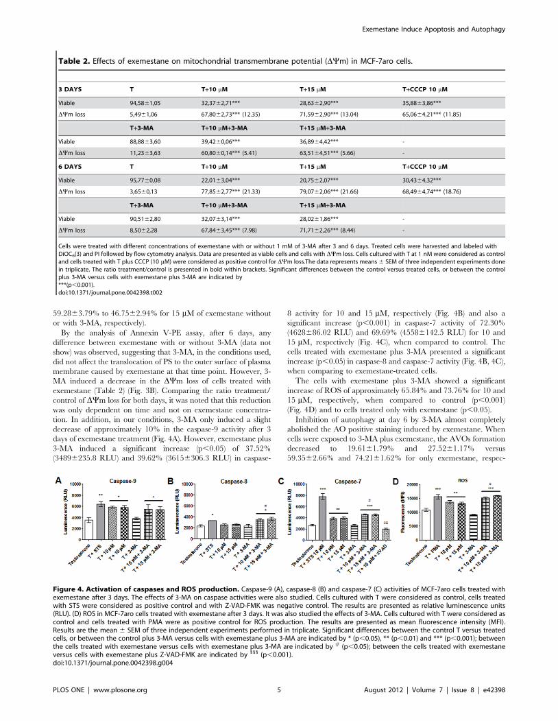
Table 2. Effects of exemestane on mitochondrial transmembrane potential (DYm) in MCF-7aro cells.
3 DAYS T T+10 mM T+15 mM T+CCCP 10 mM
Viable 94,5861,05 32,3762,71*** 28,6362,90*** 35,8863,86***
DYm loss 5,4961,06 67,8062,73*** (12.35) 71,5962,90*** (13.04) 65,0664,21*** (11.85)
T+3-MA T+10 mM+3-MA T+15 mM+3-MA
Viable 88,8863,60 39,4260,06*** 36,8964,42*** -
DYm loss 11,2363,63 60,8060,14*** (5.41) 63,5164,51*** (5.66) -
6 DAYS T T+10 mM T+15 mM T+CCCP 10 mM
Viable 95,7760,08 22,0163,04*** 20,7562,07*** 30,4364,32***
DYm loss 3,6560,13 77,8562,77*** (21.33) 79,0762,06*** (21.66) 68,4964,74*** (18.76)
T+3-MA T+10 mM+3-MA T+15 mM+3-MA
Viable 90,5162,80 32,0763,14*** 28,0261,86*** -
DYm loss 8,5062,28 67,8463,45*** (7.98) 71,7162,26*** (8.44) -
Cells were treated with different concentrations of exemestane with or without 1 mM of 3-MA after 3 and 6 days. Treated cells were harvested and labeled withDiOC6(3) and PI followed by flow cytometry analysis. Data are presented as viable cells and cells with DYm loss. Cells cultured with T at 1 nM were considered as controland cells treated with T plus CCCP (10 mM) were considered as positive control for DYm loss.The data represents means 6 SEM of three independent experiments donein triplicate. The ratio treatment/control is presented in bold within brackets. Significant differences between the control versus treated cells, or between the controlplus 3-MA versus cells with exemestane plus 3-MA are indicated by***(p,0.001).doi:10.1371/journal.pone.0042398.t002
Figure 4. Activation of caspases and ROS production. Caspase-9 (A), caspase-8 (B) and caspase-7 (C) activities of MCF-7aro cells treated withexemestane after 3 days. The effects of 3-MA on caspase activities were also studied. Cells cultured with T were considered as control, cells treatedwith STS were considered as positive control and with Z-VAD-FMK was negative control. The results are presented as relative luminescence units(RLU). (D) ROS in MCF-7aro cells treated with exemestane after 3 days. It was also studied the effects of 3-MA. Cells cultured with T were considered ascontrol and cells treated with PMA were as positive control for ROS production. The results are presented as mean fluorescence intensity (MFI).Results are the mean 6 SEM of three independent experiments performed in triplicate. Significant differences between the control T versus treatedcells, or between the control plus 3-MA versus cells with exemestane plus 3-MA are indicated by * (p,0.05), ** (p,0.01) and *** (p,0.001); betweenthe cells treated with exemestane versus cells with exemestane plus 3-MA are indicated by # (p,0.05); between the cells treated with exemestaneversus cells with exemestane plus Z-VAD-FMK are indicated by 111 (p,0.001).doi:10.1371/journal.pone.0042398.g004
Exemestane Induce Apoptosis and Autophagy
PLOS ONE | www.plosone.org 5 August 2012 | Volume 7 | Issue 8 | e42398
tively for 10 and 15 mM (Table 3). Statistical significant differences
were also observed between cells with exemestane and cells with
exemestane plus 3-MA (p,0.001). The fluorescence microscopy
showed that cells treated with exemestane plus 3-MA presented
less yellow/orange/red fluorescence (Fig. 5A) than exemestane-
treated cells, confirming the reduction of AVOs formation.
In cells treated with exemestane during 3 days, it was detected
the presence of LC3-II. In the control and treated cells with
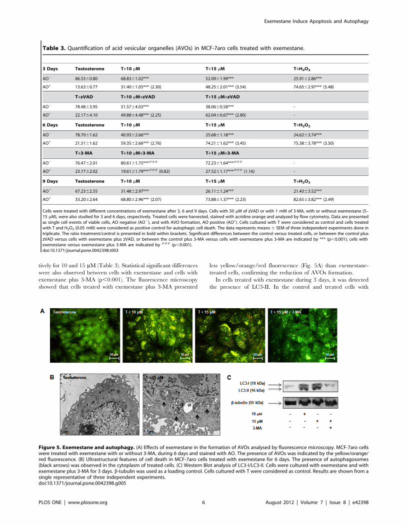
Table 3. Quantification of acid vesicular organelles (AVOs) in MCF-7aro cells treated with exemestane.
3 Days Testosterone T+10 mM T+15 mM T+H2O2
AO2 86.5360.80 68.8361.02*** 52.0961.99*** 25.9162.86***
AO+ 13.6360.77 31.4061.05*** (2.30) 48.2562.01*** (3.54) 74.6562.97*** (5.48)
T+zVAD T+10 mM+zVAD T+15 mM+zVAD
AO2 78.4863.95 51.5764.03*** 38.0660.58*** -
AO+ 22.1764.10 49.8864.48*** (2.25) 62.0460.67*** (2.80) -
6 Days Testosterone T+10 mM T+15 mM T+H2O2
AO2 78.7061.62 40.9362.66*** 25.6861.18*** 24.6263.74***
AO+ 21.5161.62 59.3562.66*** (2.76) 74.2161.62*** (3.45) 75.3863.78*** (3.50)
T+3-MA T+10 mM+3-MA T+15 mM+3-MA
AO2 76.4762.01 80.6161.75***### 72.2361.64***### -
AO+ 23.7762.02 19.6161.79***### (0.82) 27.5261.17***### (1.16) -
9 Days Testosterone T+10 mM T+15 mM T+H2O2
AO2 67.2362.55 31.4862.97*** 26.1161.24*** 21.4363.52***
AO+ 33.2062.64 68.8062.96*** (2.07) 73.8861.37*** (2.23) 82.6563.82*** (2.49)
Cells were treated with different concentrations of exemestane after 3, 6 and 9 days. Cells with 50 mM of zVAD or with 1 mM of 3-MA, with or without exemestane (5–15 mM), were also studied for 3 and 6 days, respectively. Treated cells were harvested, stained with acridine orange and analyzed by flow cytometry. Data are presentedas single cell events of viable cells, AO negative (AO2), and with AVO formation, AO positive (AO+). Cells cultured with T were considered as control and cells treatedwith T and H2O2 (0.05 mM) were considered as positive control for autophagic cell death. The data represents means 6 SEM of three independent experiments done intriplicate. The ratio treatment/control is presented in bold within brackets. Significant differences between the control versus treated cells, or between the control pluszVAD versus cells with exemestane plus zVAD, or between the control plus 3-MA versus cells with exemestane plus 3-MA are indicated by *** (p,0.001); cells withexemestane versus exemestane plus 3-MA are indicated by ### (p,0.001).doi:10.1371/journal.pone.0042398.t003
Figure 5. Exemestane and autophagy. (A) Effects of exemestane in the formation of AVOs analysed by fluorescence microscopy. MCF-7aro cellswere treated with exemestane with or without 3-MA, during 6 days and stained with AO. The presence of AVOs was indicated by the yellow/orange/red fluorescence. (B) Ultrastructural features of cell death in MCF-7aro cells treated with exemestane for 6 days. The presence of autophagosomes(black arrows) was observed in the cytoplasm of treated cells. (C) Western Blot analysis of LC3-I/LC3-II. Cells were cultured with exemestane and withexemestane plus 3-MA for 3 days. b-tubulin was used as a loading control. Cells cultured with T were considered as control. Results are shown from asingle representative of three independent experiments.doi:10.1371/journal.pone.0042398.g005
Exemestane Induce Apoptosis and Autophagy
PLOS ONE | www.plosone.org 6 August 2012 | Volume 7 | Issue 8 | e42398
15 mM of exemestane plus 3-MA, LC3-II was absent and only
LC3-I was detected (Fig. 5C).
Since it has been referred a cross-talk between apoptosis and
autophagy, it was also studied the effect of Z-VAD-FMK in AVOs
formation, after 3 days of exemestane treatment. As shown in
Table 3, no alteration in AVOs formation was detected. As
expected, Z-VAD-FMK inhibited the activation of caspase-7
induced by exemestane (Fig. 4D).
Discussion
This study explored the in vitro effects of exemestane on MCF-
7aro cell proliferation, cell cycle progression and induction of cell
death. This cell line is considered an important tool to study
growth responses to aromatase inhibitors, as it is a breast cancer
ER+ cell line stably transfected with the aromatase gene, that
express high aromatase levels [23]. In the present work,
exemestane induced a decrease in MCF-7aro cell proliferation
and viability in a dose- and time-dependent manner, with no
effects on cell membrane integrity, except for the higher
concentration and for prolonged times of exposure. Our results
revealed that the anti-proliferative effects of exemestane are
essentially due to the retention in G0/G1 phase, which blocks the
G1/S phase transition of cell cycle, preventing cells to enter in the
S phase that occurs when cells are exposed to exemestane for a
short period of time. However, for longer times, exemestane
caused retention in G2/M cell cycle phase that has been referred
to be associated with enhanced apoptosis and cytotoxicity [29].
Like exemestane, non-steroidal AIs, letrozole and anastrozole, also
induced a decrease in cell proliferation and cell cycle arrest in
MCF-7aro cells [13,23].
In addition to inhibit cell cycle progression, exemestane caused
a reduction in cell viability. This was accompanied by morpho-
logical alterations, such as membrane blebbings, chromatin
condensation and fragmentation, which suggested the occurrence
of cell death by apoptosis. However, the appearance of cytoplasm
vacuolization, as well as the presence of AVOs also indicated the
occurrence of autophagy.
To clarify the mechanism of cell death involved, it was further
studied the effect of exemestane on the exposure of PS to the outer
leaflet of cell membrane and on the DYm. Our results revealed
that exemestane induced a significant increase in the binding of
annexin V and a significant increase in DYm loss. In the intrinsic
pathway of apoptosis, a loss of DYm is associated with the release
of cytochrome c in the cytosol and formation of apoptosome,
leading to the activation of caspase-9 and of effector caspases [30].
After 3 days of treatment, cells presented a significant increase in
caspase-9 activity and a significant production of intracellular
ROS, suggesting the induction of apoptosis through the activation
of the intrinsic pathway. However, as it has been referred that
non-steroidal AIs like letrozole and anastrozole induced, respec-
tively, a down-regulation and up-regulation of caspase-8 expres-
sion in MCF-7aro cells [23], the activity of this enzyme was also
evaluated, though no significant increase was detected. The
activation of the effector caspase-7 by exemestane confirms the
induction of apoptosis.
In addition, by flow cytometry it was observed that exemestane
induced a significant increase in AVOs with the time of exposure.
Moreover, electron microscopy revealed the presence of autopha-
gosomes engulfing cytoplasmatic fractions and organelles, such as
mitochondria. During autophagy, the pro-LC3 is proteolytically
converted in the cytosolic form of LC3-I, that is lipidated and
translocated to autophagosome membranes to the form LC3-II,
which is associated to the maturation of autophagosomes [27,31].
The AI also induced the turnover of LC3, which is associated to
the conversion of LC3-I in LC3-II. All together, our findings
confirmed the existence of autophagosomes and suggest the
occurrence of autophagy that seems to increase with time of
treatment.
As it is referred in the literature that there is a cross-talk between
these two types of cell death and some features found in apoptosis,
like DYm loss, chromatin condensation and PS exposure to the
outer leaflet of cell membrane, may also occur in the autophagic
process [32,33], it was evaluated the effects of 3-MA in exemestane
treated cells. 3-MA is an inhibitor of autophagy, which blocks the
formation of autophagosomes, by controlling class I and class III
phosphatidylinositol 3-kinases (PI3K) [27,31]. As expected, the
autophagic inhibitor caused a reduction in the cytoplasm
vacuolization. 3-MA did neither affect the translocation of PS to
the outer surface of plasma membrane nor the increase in the
caspase-9 activity induced by exemestane. 3-MA did not
completely abolish the drop of DYm, it only reduced the
mitochondrial membrane depolarization observed with exemes-
tane, indicating that this phenomenon is due to both processes. On
the other hand, cells treated with exemestane plus 3-MA presented
an increase in intracellular ROS when comparing to exemestane
treated cells. Thus, autophagy may be a defense mechanism
against the accumulation of ROS. Autophagy may remove ROS-
generating mitochondria, therefore decreasing ROS production,
acting as a self-protective mechanism [34]. Moreover, the
inhibition of the autophagic process induced a decrease in cell
viability and an increment on caspase-8 activity, when compared
to exemestane treated cells. Hou et al. (2011) showed that in
cytoprotective autophagy, active caspase-8 is sequestered by
autophagosomes and degraded by lysosomes [35]. The activity
of caspase-8 controls the switch from protective to destructive role
of autophagy [32]. Some reports, have demonstrate that activation
of caspase-8 may be independent of death receptors activation and
occur downstream of mitochondria by a mechanism not totally
understood [36,37]. The cytoprotective role of autophagy was
confirmed by the significant increase in caspase-7 activity observed
in cells treated with exemestane plus 3-MA. Thus, the use of the 3-
MA, as shown by a dramatic reduction of AVOs and LC3 II
formation, affected the machinery of autophagy, reducing the
appearance of the typical features of this process and not the
apoptotic markers, like Annexin V or caspase-9. However, it
increased the production of ROS and the activation of caspase-8
and caspase-7 in treated cells.
On the other hand, the use of Z-VAD-FMK did not affect the
autophagic process. Supporting these observations, Boya et al.
(2005) have also demonstrated that Z-VAD-FMK did not affect
the formation of autophagic vacuoles by hydroxychloroquine [38].
Different interactions between apoptosis and autophagy have
been proposed. They may act as partners to induce efficient cell
death in a coordinated or cooperative manner, but in this case, if
one pathway of cell death is blocked the other assume or may only
be activated if the other fails. Autophagy may act as an antagonist
to block apoptotic cell death by promoting cell survival and
stabilizing genome integrity. Autophagy may also act as enabler of
apoptosis, as it does not lead to death but participates in
morphologic and cellular events that occur during apoptosis that
should be prevented if autophagy is inhibited [39,40]. In our
study, and in the conditions used, when apoptosis is inhibited there
is no exacerbation or activation of the other mechanism. However,
autophagy inhibition reduced cell viability, increased ROS
production and induced activation of caspase-8 and caspase-7.
Thus, it appears that autophagy induced by exemestane may act
as a pro-survival process. Other authors have demonstrated that
Exemestane Induce Apoptosis and Autophagy
PLOS ONE | www.plosone.org 7 August 2012 | Volume 7 | Issue 8 | e42398
tamoxifen induced both processes of cell death and that autophagy
acts as a mechanism of cell pro-survival [20,21]. Recent works in
breast cancer cell lines, also showed that autophagy induced by
epirubicin [41] or sulforahane [42] acts as pro-survival mecha-
nism, protecting cells from apoptotic cell death. Moreover, Abedin
et al. (2007) suggested that the role of autophagy in delaying
apoptosis or prolonging survival is characteristic of noninvasive
breast tumor cells [43].
In cells with caspase-dependent apoptosis, autophagy can be
activated by mitochondrial membrane potential loss and cyto-
crome c redistribution [44,45], eliminating damaged mitochondria
and thereby limiting ROS production and tumor cell death by
apoptosis [34,46], being together with apoptosis important for
tumor suppression [47].
This is the first study that documents the biological effects and
mechanisms of cell death induced by the steroidal AI exemestane,
in a breast cancer cell line. The main effect of exemestane on
MCF-7aro cells is on cell proliferation due to a significant cell
cycle arrest in G0/G1 phase and G2/M. Moreover in the
conditions used, autophagy and mitochondrial-mediated apoptosis
occur simultaneously. In addition, the mitochondria have an
important role in the occurrence of cell death, but autophagy may
act as a pro-survival process regulating or controlling breast cancer
cells from apoptosis. This study may also suggest the use of
inhibitors of autophagy with exemestane in a combination therapy
to sensitize breast cancer cells to death, being a promising
approach for the treatment of hormone-dependent breast cancers.
Materials and Methods
Cell cultureThe ER-positive aromatase-overexpressing human breast can-
cer cell line, MCF-7aro, prepared by stable transfection of MCF-7
cells with the human placental aromatase gene and Geneticin
selection [48] [49] [50], was kindly provided by Dr. Shiuan Chen
(Beckman Research Institute, City of Hope, Duarte, CA, U.S.A.).
Cells were maintained with Eagles’s minimum essential medium
(MEM) supplemented with Earle’s salts and 1 mmol/L sodium
pyruvate, 1% penicillin-streptomycin-amphotericin B, 700 ng/ml
G418 and 10% heat-inactivated fetal bovine serum (FBS) (Gibco)
in 5% CO2 atmosphere at 37uC. To avoid the interference of
steroids present in FBS and of the estrogenic effects of phenol-red
[51], three days before starting the experiments, cells were
cultured in an E2-free MEM medium without phenol-red
containing 5% pre-treated charcoal heat-inactivated fetal bovine
serum (CFBS). All the experiments were performed according to
these conditions, with 1 nM of testosterone (T) (Sigma-Aldrich
Co.), which was used as aromatase substrate and proliferation
inducing agent and with or without exemestane (Sequoia Research
Products Ltd.). The medium and drugs were refreshed every 3
days. Cells incubated with 1 nM of T plus 0.05% of DMSO
(Sigma-Aldrich Co.) were used as control. All the assays were
performed in triplicate in three independent experiments.
Morphological studiesThe morphological alterations induced by exemestane were
evaluated by phase contrast microscopy, Giemsa and Hoechst
staining. After treatment, cells were fixed with 4% of paraformal-
dehyde (Sigma-Aldrich Co.). For Hoechst staining, cells were
exposed to 0.5 mg/ml Hoechst 33258 (Sigma-Aldrich Co.) for
20 min and mounted with vectashield mounting medium. The
nuclear morphology was examined under a fluorescence micro-
scope (Eclipse E400, Nikon), equipped with an excitation filter
with maximum transmission at 360/400 nm, and processed by
Nikon ACT-2U image software. The Giemsa (Merck) stained cells
were observed under the microscope Eclipse E400, Nikon
equipped with image analysis software LeicaQwin.
Cell viability and cell proliferationCell viability was assessed by tetrazolium salt, 3-(4,5-di-
methylthiazol-2-yl)-2,5-difenyltetrazolium (MTT) assay and by
measuring the lactate dehydrogenase (LDH) release. MCF-7aro
cells were cultured in 96-well plates and incubated with different
concentrations of exemestane during 2, 3, 6 and 9 days. Cells were
also treated with 1 mM of 3-methyladenine (3-MA, (Sigma-
Aldrich Co.)), an inhibitor of autophagy. After MTT (0.5 mg/ml)
(Sigma-Aldrich Co.) addition, formazan was quantified spectro-
photometrically. LDH release was measured using CytoTox 96
nonradioactive cytotoxity assay kit (Promega Corporation) ac-
cording to the manufacturer’s protocol.
To study the effects of exemestane on DNA synthesis, 3H-
thymidine incorporation assay was performed. At each exposure
time, 3H-thymidine (0.5 mCi) (Amersham) was added and incu-
bated for the last 8 hours. Cells were harvested and, after addition
of scintillation cocktail, 3H-thymidine incorporation was deter-
mined in a scintillation counter (LS 6500, Beckman Instruments).
Results are expressed as relative percentage of the untreated
control cells (100%).
Cell cycle analysisTo investigate the anti-proliferative effects of exemestane, cell
cycle analysis was performed by flow cytometry. Cells were
incubated with exemestane (2.5–10 mM) during 3 and 6 days, fixed
with 70% cold ethanol and resuspended in 0.5 ml of DNA staining
solution (5 mg/ml Propidium Iodide (PI), 0.1% Triton X-100 and
200 mg/ml DNase-free RNase A in PBS) (Sigma-Aldrich Co.) for
30 min. The three fluorescence channels (FL-1, FL-2 and FL-3)
were set on a linear scale. The antiproliferative effect was indicated
by the percentage of cells in G0/G1, S and G2/M phases of the cell
cycle. Flow cytometric analysis was always based on the
acquisition of 20 000 events in a Becton Dickinson FACSCalibur
equipped with CELLQuest Pro software.
Analysis of apoptosisTo evaluate the translocation of phosphatidylserine (PS) to the
cell surface, Annexin V-PE apoptosis detection Kit (BD Biosci-
ences Pharmingen) was used. Mitochondrial transmembrane
potential (DYm) loss was studied using 3,3-dihexyloxacarbocya-
nine iodide (DiOC6(3) (Gibco) and flow cytometry analysis. Cells
were cultured in 6-well plates and treated with exemestane (5–
15 mM) during 3, 6 and 9 days. Adherent and non-adherent cells,
after being pooled were incubated with the corresponding dye.
Cells were stained with Annexin V-PE and 7-amino-acitomycin
(7-AAD), according to the manufacturer’s instructions. As positive
control, cells were incubated with staurosporine (STS) (1 mM)
(Sigma-Aldrich Co.) for 14 hours. Detectors for all three
fluorescence channels (FL-1, FL-2 and FL-3) were set on a
logarithmic scale. Bivariant analysis of Annexin-PE fluorescence
(FL-2) and 7AAD fluorescence (FL-3) distinguished different cell
populations, Annexin V2/7-AAD2 were designated as viable
cells; Annexin V+/7-AAD2 as apoptotic and Annexin V+/7-
AAD+ as late apoptotic and/or necrotic cells.
For DYm, cells were treated with exemestane or with 3-MA
(1 mM) with or without exemestane, during 3 and 6 days. As
positive control, cells were incubated with 10 mM of the
mitochondrial depolarizant agent carbonyl cyanide m-chlorophe-
nylhydrazone (CCCP) (Sigma-Aldrich Co.) and stained with
DiOC6(3) (10 nM) for 30 min. PI (5 mg/ml) was added prior to
Exemestane Induce Apoptosis and Autophagy
PLOS ONE | www.plosone.org 8 August 2012 | Volume 7 | Issue 8 | e42398
FACS analysis to discriminate among live cells that stain only with
DiOC6(3), early apoptotic cells that lost the ability to accumulate
DiOC6(3), and late apoptotic/necrotic cells that stain only with PI.
Detectors were set on logarithmic scale, FL-1 was used to measure
DiOC6(3) at green fluorescence and FL2 and FL-3 to measure PI
red fluorescence.
Caspase-GloH 9, Caspase-GloH 8 and Caspase-GloH 3/7
(Promega Corporation) are homogeneous luminescent assays that
were used according to the manufacturer’s instructions. Cells were
incubated with exemestane (10–15 mM) and with 3-MA (1 mM)
plus exemestane for 3 days. As positive control, cells were
incubated with STS (10 mM) for 3 hours and as negative control,
cells with exemestane plus Z-VAD-FMK (50 mM). The resultant
luminescence was measured in a 96-well Microplate Luminometer
(BioTek Instruments) and presented as relative light units (RLU). It
must be noted that as MCF-7 cells are known to be caspase-3
deficient [52], the use of Caspase-GloH 3/7 kit only evaluates the
activation of caspase-7.
Intracellular reactive oxygen species (ROS) measurementTo detect the levels of intracellular ROS the 29,79-dichlorodi-
hydrofluorescein diacetate (DCFH2-DA) method was used.
DCFH2-DA is a lipophilic non-fluorescent compound that crosses
cell membrane and is oxidized to the fluorescent compound 29,79-
dichlorofluorescein (DCF) [53]. Cells were incubated with
exemestane (10–15 mM) and with 3-MA (1 mM) plus exemestane
for 3 days. As positive control, cells were incubated with phorbol
12-myristate 13-acetate (PMA) (Sigma-Aldrich Co.) at 25 ng/ml
for 2 hours. Cells were labeled with DCFH2-DA (50 mM) (Sigma-
Aldrich Co.) for 1 hour at 37uC and fluorescence was measured
using an excitation wavelength of 480 nm and an emission filter of
530 nm in a 96-well Microplate Luminometer and is presented as
mean fluorescence intensity (MFI).
Detection of acid vesicular organellesAcridine orange (AO) (Sigma-Aldrich Co.) was used to evaluate
and quantify the formation of acid vesicular organelles (AVOs), by
fluorescence microscopy and flow cytometry. AO is an acidotropic
fluorescent dye that stain DNA and cytoplasm bright green (AO2)
and when protonated in the presence of acid compartments it
fluorescences bright red (AO+). MCF-7aro cells treated with
exemestane (10–15 mM) were cultured for 3, 6 and 9 days. As
positive control, cells were incubated with H2O2 (0.05 mM)
(Sigma-Aldrich Co.) for 14 hours. Cells with and without
exemestane were also treated with Z-VAD-FMK (50 mM) (BD
Biosciences Pharmingen), a pan caspase inhibitor, during 3 days or
with 3-MA (1 mM) during 6 days. After incubation, cells were
tripsinized and incubated with AO at 0.5 mg/ml. Green (510–
530 nm) and red (.650 nm) fluorescence emission with blue
(488 nm) excitation light was measured with detectors for
fluorescence channels FL-1 and FL-3 set on a linear scale.
For fluorescence microscopy, cells were stained with AO at
0.1 mg/ml during 15 min. The presence of AVOs was indicated
by the yellow/orange/red fluorescence, analysed in the fluores-
cence microscope equipped with a 490 nm band-pass blue
excitation filters and a 515-nm long pass-barrier filter.
Analysis of intracellular vacuoles by electron microscopyCells were cultured in 6-well plates and treated with exemestane
(15 mM) during 6 days. Cells were fixed with 2% glutaraldehyde/
4% paraformaldehyde (Sigma-Aldrich Co.) and post-fixed in 1%
osmium tetroxide. Ultrathin sections (60 nm) were collected and
stained with uranyl acetate and lead citrate, and examined using a
Zeiss EM902 transmission electron microscope (Carl Zeiss
Oberkochen). Images were digitally recorded using a Gatan SC
1000 ORIUS CCD camera (Warrendale).
Western-Blot analysisCells treated with exemestane (10–15 mM) and with or without
3-MA were cultured during 3 days in 6-well plates. After
incubation, cells were lysed with cold TNTE lysis buffer (20 mM
Tris-HCl, 150 mM NaCl, 0.3% Triton X-100 and 5 mM EDTA)
(Sigma-Aldrich Co.), pH 7.5 containing appropriate protease
inhibitors (Sigma-Aldrich Co.), and centrifuged at 18 8006g for
5 min at 4uC. Protein concentrations were determined using a
Bradford assay kit (Bio-Rad). A total of 100 mg of protein per
sample were subjected to 4–20% SDS-PAGE and transferred to
nitrocellulose membranes. Immunodetection was performed using
rabbit polyclonal antibody anti-LC3 (1:250) (Medical & Biological
Laboratories) and the secondary peroxidase goat anti-rabbit
antibody (1:5000) (Vector Laboratories). Immunoreactive bands
were visualized using a chemiluminescent substrate Super Signal
West Pico (Pierce). Membranes were then stripped and incubated
with rabbit monoclonal anti-b-tubulin antibody (1:500) (Santa
Cruz) to control loading variations.
Statistical analysisThe data presented are expressed as the mean 6 SEM.
Statistical analysis of data was performed using analysis of variance
(ANOVA) followed by Bonferroni and Dunnet post-hoc tests for
multiple comparisons. Values of P,0.05 were considered as
statistically significant.
Acknowledgments
We thank Dr. Shiuan Chen, City of Hope, Duarte, CA, USA for kindly
supplying MCF-7aro cells and Dr. Rui Fernandes from IBMC for his help
in electron microscopy studies.
Author Contributions
Conceived and designed the experiments: CA GCdS NT. Performed the
experiments: CA SM. Analyzed the data: CA MB GCdS NT. Contributed
reagents/materials/analysis tools: CA GCdS NT. Wrote the paper: CA
GCdS NT. Revised the manuscript: ETdS GCdS NT. Read and approved
the manuscript for publication: CA MB SM ETdS GCdS NT.
References
1. Macedo LF, Sabnis G, Brodie A (2009) Aromatase inhibitors and breast cancer.
Ann N Y Acad Sci 1155: 162–173.
2. Gluck S (2010) Exemestane as first-line therapy in postmenopausal women with
recurrent or metastatic breast cancer. Am J Clin Oncol 33: 314–319.
3. Fabian CJ (2007) The what, why and how of aromatase inhibitors: hormonal
agents for treatment and prevention of breast cancer. Int J Clin Pract 61: 2051–
2063.
4. Lonning PE (2009) Lack of complete cross-resistance between different
aromatase inhibitors; a real finding in search for an explanation? Eur J Cancer
45: 527–535.
5. Dutta U, Pant K (2008) Aromatase inhibitors: past, present and future in breast
cancer therapy. Med Oncol 25: 113–124.
6. Miller WR, Bartlett J, Brodie AM, Brueggemeier RW, di Salle E, et al. (2008)
Aromatase inhibitors: are there differences between steroidal and nonsteroidal
aromatase inhibitors and do they matter? Oncologist 13: 829–837.
7. Deeks ED, Scott LJ (2009) Exemestane: a review of its use in postmenopausal
women with breast cancer. Drugs 69: 889–918.
8. Wang X, Chen S (2006) Aromatase destabilizer: novel action of exemestane, a
food and drug administration-approved aromatase inhibitor. Cancer Res 66:
10281–10286.
Exemestane Induce Apoptosis and Autophagy
PLOS ONE | www.plosone.org 9 August 2012 | Volume 7 | Issue 8 | e42398
9. Doisneau-Sixou SF, Sergio CM, Carroll JS, Hui R, Musgrove EA, et al. (2003)
Estrogen and antiestrogen regulation of cell cycle progression in breast cancercells. Endocr Relat Cancer 10: 179–186.
10. Gross JM, Yee D (2003) The type-1 insulin-like growth factor receptor tyrosine
kinase and breast cancer: biology and therapeutic relevance. Cancer MetastasisRev 22: 327–336.
11. Nicholson RI, McClelland RA, Gee JM, Manning DL, Cannon P, et al. (1994)Epidermal growth factor receptor expression in breast cancer: association with
response to endocrine therapy. Breast Cancer Res Treat 29: 117–125.
12. Kyprianou N, English HF, Davidson NE, Isaacs JT (1991) Programmed celldeath during regression of the MCF-7 human breast cancer following estrogen
ablation. Cancer Res 51: 162–166.13. Thiantanawat A, Long BJ, Brodie AM (2003) Signaling pathways of apoptosis
activated by aromatase inhibitors and antiestrogens. Cancer Res 63: 8037–8050.14. Detre S, Salter J, Barnes DM, Riddler S, Hills M, et al. (1999) Time-related
effects of estrogen withdrawal on proliferation- and cell death-related events in
MCF-7 xenografts. Int J Cancer 81: 309–313.15. Truchet I, Jozan S, Guerrin M, Mazzolini L, Vidal S, et al. (2000)
Interconnections between E2-dependent regulation of cell cycle progressionand apoptosis in MCF-7 tumors growing on nude mice. Exp Cell Res 254: 241–
248.
16. Kallio A, Zheng A, Dahllund J, Heiskanen KM, Harkonen P (2005) Role ofmitochondria in tamoxifen-induced rapid death of MCF-7 breast cancer cells.
Apoptosis 10: 1395–1410.17. Nazarewicz RR, Zenebe WJ, Parihar A, Larson SK, Alidema E, et al. (2007)
Tamoxifen induces oxidative stress and mitochondrial apoptosis via stimulatingmitochondrial nitric oxide synthase. Cancer Res 67: 1282–1290.
18. Lim KB, Ng CY, Ong CK, Ong CS, Tran E, et al. (2001) Induction of apoptosis
in mammary gland by a pure anti-estrogen ICI 182780. Breast Cancer ResTreat 68: 127–138.
19. Samaddar JS, Gaddy VT, Duplantier J, Thandavan SP, Shah M, et al. (2008) Arole for macroautophagy in protection against 4-hydroxytamoxifen-induced cell
death and the development of antiestrogen resistance. Mol Cancer Ther 7:
2977–2987.20. Qadir MA, Kwok B, Dragowska WH, To KH, Le D, et al. (2008)
Macroautophagy inhibition sensitizes tamoxifen-resistant breast cancer cellsand enhances mitochondrial depolarization. Breast Cancer Res Treat 112: 389–
403.21. de Medina P, Payre B, Boubekeur N, Bertrand-Michel J, Terce F, et al. (2009)
Ligands of the antiestrogen-binding site induce active cell death and autophagy
in human breast cancer cells through the modulation of cholesterol metabolism.Cell Death Differ 16: 1372–1384.
22. de Medina P, Silvente-Poirot S, Poirot M (2009) Tamoxifen and AEBS ligandsinduced apoptosis and autophagy in breast cancer cells through the stimulation
of sterol accumulation. Autophagy 5: 1066–1067.
23. Itoh T, Karlsberg K, Kijima I, Yuan YC, Smith D, et al. (2005) Letrozole-,anastrozole-, and tamoxifen-responsive genes in MCF-7aro cells: a microarray
approach. Mol Cancer Res 3: 203–218.24. Cepa MM, Tavares da Silva EJ, Correia-da-Silva G, Roleira FM, Teixeira NA
(2005) Structure-activity relationships of new A,D-ring modified steroids asaromatase inhibitors: design, synthesis, and biological activity evaluation. J Med
Chem 48: 6379–6385.
25. Cepa M, Correia-da-Silva G, Tavares da Silva EJ, Roleira FM, Hong Y, et al.(2008) Molecular mechanisms of aromatase inhibition by new A, D-ring
modified steroids. Biol Chem 389: 1183–1191.26. Cepa M, Correia-da-Silva G, da Silva EJ, Roleira FM, Borges M, et al. (2008)
New steroidal aromatase inhibitors: suppression of estrogen-dependent breast
cancer cell proliferation and induction of cell death. BMC Cell Biol 9: 41.27. Chen Y, Azad MB, Gibson SB (2010) Methods for detecting autophagy and
determining autophagy-induced cell death. Can J Physiol Pharmacol 88: 285–295.
28. Paglin S, Hollister T, Delohery T, Hackett N, McMahill M, et al. (2001) A novel
response of cancer cells to radiation involves autophagy and formation of acidicvesicles. Cancer Res 61: 439–444.
29. DiPaola RS (2002) To arrest or not to G(2)-M Cell-cycle arrest : commentary re:A. K. Tyagi et al., Silibinin strongly synergizes human prostate carcinoma
DU145 cells to doxorubicin-induced growth inhibition, G(2)-M arrest, andapoptosis. Clin. cancer res., 8: 3512–3519, 2002. Clin Cancer Res 8: 3311–3314.
30. Kroemer G, Galluzzi L, Brenner C (2007) Mitochondrial membrane
permeabilization in cell death. Physiol Rev 87: 99–163.31. Klionsky DJ, Abeliovich H, Agostinis P, Agrawal DK, Aliev G, et al. (2008)
Guidelines for the use and interpretation of assays for monitoring autophagy in
higher eukaryotes. Autophagy 4: 151–175.32. Madden DT, Egger L, Bredesen DE (2007) A calpain-like protease inhibits
autophagic cell death. Autophagy 3: 519–522.33. Lemasters JJ, Qian T, He L, Kim JS, Elmore SP, et al. (2002) Role of
mitochondrial inner membrane permeabilization in necrotic cell death,
apoptosis, and autophagy. Antioxid Redox Signal 4: 769–781.34. Dewaele M, Maes H, Agostinis P (2010) ROS-mediated mechanisms of
autophagy stimulation and their relevance in cancer therapy. Autophagy 6: 838–854.
35. Hou W, Han J, Lu C, Goldstein LA, Rabinowich H (2010) Autophagicdegradation of active caspase-8: a crosstalk mechanism between autophagy and
apoptosis. Autophagy 6: 891–900.
36. Ferreira CG, Span SW, Peters GJ, Kruyt FA, Giaccone G (2000) Chemotherapytriggers apoptosis in a caspase-8-dependent and mitochondria-controlled
manner in the non-small cell lung cancer cell line NCI-H460. Cancer Res 60:7133–7141.
37. de Vries JF, Wammes LJ, Jedema I, van Dreunen L, Nijmeijer BA, et al. (2007)
Involvement of caspase-8 in chemotherapy-induced apoptosis of patient derivedleukemia cell lines independent of the death receptor pathway and downstream
from mitochondria. Apoptosis 12: 181–193.38. Boya P, Gonzalez-Polo RA, Casares N, Perfettini JL, Dessen P, et al. (2005)
Inhibition of macroautophagy triggers apoptosis. Mol Cell Biol 25: 1025–1040.39. Eisenberg-Lerner A, Bialik S, Simon HU, Kimchi A (2009) Life and death
partners: apoptosis, autophagy and the cross-talk between them. Cell Death
Differ 16: 966–975.40. Eisenberg-Lerner A, Kimchi A (2009) The paradox of autophagy and its
implication in cancer etiology and therapy. Apoptosis 14: 376–391.41. Sun WL, Chen J, Wang YP, Zheng H (2011) Autophagy protects breast cancer
cells from epirubicin-induced apoptosis and facilitates epirubicin-resistance
development. Autophagy 7: 1035–1044.42. Kanematsu S, Uehara N, Miki H, Yoshizawa K, Kawanaka A, et al. (2010)
Autophagy inhibition enhances sulforaphane-induced apoptosis in human breastcancer cells. Anticancer Res 30: 3381–3390.
43. Abedin MJ, Wang D, McDonnell MA, Lehmann U, Kelekar A (2007)Autophagy delays apoptotic death in breast cancer cells following DNA damage.
Cell Death Differ 14: 500–510.
44. Yan CH, Yang YP, Qin ZH, Gu ZL, Reid P, et al. (2007) Autophagy is involvedin cytotoxic effects of crotoxin in human breast cancer cell line MCF-7 cells.
Acta Pharmacol Sin 28: 540–548.45. Lemasters JJ (1998) The mitochondrial permeability transition: from biochem-
ical curiosity to pathophysiological mechanism. Gastroenterology 115: 783–786.
46. Zhivotovsky B, Orrenius S (2010) Cell death mechanisms: cross-talk and role indisease. Exp Cell Res 316: 1374–1383.
47. Gozuacik D, Kimchi A (2004) Autophagy as a cell death and tumor suppressormechanism. Oncogene 23: 2891–2906.
48. Zhou DJ, Pompon D, Chen SA (1990) Stable expression of human aromatasecomplementary DNA in mammalian cells: a useful system for aromatase
inhibitor screening. Cancer Res 50: 6949–6954.
49. Sun XZ, Zhou D, Chen S (1997) Autocrine and paracrine actions of breasttumor aromatase. A three-dimensional cell culture study involving aromatase
transfected MCF-7 and T-47D cells. J Steroid Biochem Mol Biol 63: 29–36.50. Masri S, Phung S, Wang X, Wu X, Yuan YC, et al. (2008) Genome-wide
analysis of aromatase inhibitor-resistant, tamoxifen-resistant, and long-term
estrogen-deprived cells reveals a role for estrogen receptor. Cancer Res 68:4910–4918.
51. Berthois Y, Katzenellenbogen JA, Katzenellenbogen BS (1986) Phenol red intissue culture media is a weak estrogen: implications concerning the study of
estrogen-responsive cells in culture. Proc Natl Acad Sci U S A 83: 2496–2500.
52. Kurokawa H, Nishio K, Fukumoto H, Tomonari A, Suzuki T, et al. (1999)Alteration of caspase-3 (CPP32/Yama/apopain) in wild-type MCF-7, breast
cancer cells. Oncol Rep 6: 33–37.53. Chen X, Zhong Z, Xu Z, Chen L, Wang Y (2010) 29,79-Dichlorodihydro-
fluorescein as a fluorescent probe for reactive oxygen species measurement:Forty years of application and controversy. Free Radic Res 44: 587–604.
Exemestane Induce Apoptosis and Autophagy
PLOS ONE | www.plosone.org 10 August 2012 | Volume 7 | Issue 8 | e42398