Brain volumes in familial and non-familial schizophrenic probands and their unaffected relatives
Upload
independentCategory
view
2download
0
Schizophrenia Research 39 (1999) 19–29www.elsevier.com/locate/schres
Antipsychotic drug effects on motor activationmeasured by functional magnetic resonance imaging
in schizophrenic patientsDieter F. Braus *, Gabriele Ende, Wolfgang Weber-Fahr, Alexander Sartorius,
Andreas Krier, Petra Hubrich-Ungureanu, Matthias Ruf, Sabine Stuck,Fritz A. Henn
Central Institute of Mental Health (ZI), NMR-Research PO Box 122 120, D-68072 Mannheim, Germany
Received 6 August 1998; accepted for publication 10 February 1999
Abstract
Brain function and laterality in schizophrenia were investigated by means of a simple motor task with a self-generated left-hand sequential finger opposition (SFO) using a whole-brain high-speed (100 ms per slice) functionalimaging technique. Neuroleptic-naıve, acutely ill schizophrenic patients were compared to schizophrenic patientsunder stable neuroleptic medication and matched controls. The goal was to evaluate both the motor function in first-episode patients and possible effects of different neuroleptic treatments on functional MRI results. Forty patientssatisfying ICD 10 criteria (F20.x) for schizophrenia and sex- and age-matched healthy volunteers participated in thisstudy. All subjects underwent fMRI examinations on a conventional 1.5 T MR unit. The primary sensorimotor cortexand the high-order supplementary motor area (SMA) were evaluated. There was a close similarity in the activationof the primary and high-order (SMA) sensorimotor areas between first-episode schizophrenic patients and controls.In contrast, a significant reduction in the overall blood oxygen level dependent (BOLD) response was seen insensorimotor cortices (contra- and ipsilateral ) in schizophrenic patients under stable medication with typicalneuroleptics. This effect was not present in patients treated with atypical antipsychotics. Both antipsychotic treatments,however, led to a significant reduction in activation of the SMA region compared to controls and neuroleptic-naıvesubjects. Thus, the present study provides no evidence for the localized involvement of the primary motor cortex orthe SMA as a relatively stable vulnerability marker in schizophrenia. There is, however, strong evidence thatneuroleptics themselves influence fMRI activation patterns and that there are major differences between typicalneuroleptics and atypical antipsychotics. © 1999 Elsevier Science B.V. All rights reserved.
Keywords: fMRI; Medication; Motor task; Schizophrenia
1. Introduction described Dementia Praecox. Even simple tasks,such as finger-tapping or trail-making, have beenreported to show disturbances in motor functionA variety of motor deficits have been describedin schizophrenic patients (Manschreck et al., 1985;in schizophrenia since Kraepelin (1919) initiallyHeinrichs and Buchanan, 1988; Ismail et al., 1998).Perhaps even more intriguing is the report that* Corresponding author. Tel.: +49-621-1703-672;children having a high genetic risk for schizo-fax: +49-621-1703-673.
E-mail address: [email protected] (D.F. Braus) phrenia show a variety of subtle neurological signs
0920-9964/99/$ – see front matter © 1999 Elsevier Science B.V. All rights reserved.PII: S0920-9964 ( 99 ) 00032-8

20 D.F. Braus et al. / Schizophrenia Research 39 (1999) 19–29
( Woods et al., 1986; Erlenmeyer-Kimling and 2. PurposeCornblatt, 1987), and patients with chronic schizo-
The purpose of our study was to examine thephrenia were demonstrated to have subtle neuro-motor system with a left-hand motor sequentiallogical stigmata as children ( Walker, 1994; Walkerfinger opposition (SFO) task in neuroleptic-naıve,et al., 1994). This suggests that motor functionacutely ill schizophrenic patients in comparison tomay not only be disturbed in schizophrenia butschizophrenic patients under stable neurolepticmay be altered prior to the onset of psychoticmedication and matched controls using a whole-symptoms. The nature of the motor dysfunctionbrain high-speed (100 ms per slice) EPI technique.is not understood, and the steps in the initiation ofThe goal was to evaluate possible effects of neuro-motor activity that are pathological have not yetleptic treatments on fMRI results.been established.
With the introduction of in-vivo functionalimaging methods, such as single photon emission
3. Materials and methodscomputed tomography (SPECT) and positronemission tomography (PET) (Buchsbaum et al.,
3.1. Subjects1984; Guenther et al. 1991, 1994), there is someevidence that the brains of schizophrenics might
Forty patients living in the community ofhave difficulties recruiting neuronal activity forMannheim and satisfying DSM-III-R (Americaneven very simple tasks.Psychiatric Association, 1987) as well as ICD 10PET and SPECT studies are not without contro-criteria for schizophrenia and 15 sex- and age-versy because of the low spatial and temporalmatched healthy volunteers participated in thisresolution of these methods.study. All but two were right-handed, as evaluatedFunctional magnetic resonance imaging (fMRI)on the Edinburgh Handedness Inventory (Oldfield,techniques provide a non-invasive neuroimaging1971). Twenty-six out-patients had been clinically
alternative with considerable potential to tacklestable for at least 6 months and had no medication
these problems. Recent hardware advances have changes during a period of at least 3 months (n=made it possible to visualize and measure cortical 10 receiving 25–450 mg of clozapine per day, n=activity with enhanced spatial and temporal reso- 3 risperidon 4–6 mg per day, n=17 received typicallution (Stehling et al., 1991; Belliveau et al., 1991; neuroleptics). Of these outpatients, 19 revealedBandettini et al., 1992; Kwong et al., 1992). In deficits in the Wisconsin Card Sorting Testneuroscience research, most commonly blood ( WCST) (Robinson et al., 1980) and Continuousoxygen level-dependent contrast (BOLD effect, Performance Task (CPT) (Rosvold et al., 1956) inOgawa et al., 1990, 1992) is used for fMRI experi- comparison with controls.ments. The BOLD contrast depends on changes Fourteen first-admission in-patients were neuro-in both oxygen consumption and perfusion, where leptic-naıve, first-episode schizophrenic patientsthe latter is the dominant effect. (ICD 10, F20.0, mean BPRS: 46±5, n=3 with
As an indicator of regional brain activity, fMRI negative symptoms). Particular care was taken tooffers several advantages over other functional exclude patients with a history of neurologicalimaging methods. It can localize functional activity disorders or substance abuse.to specific neuroanatomic regions since both ana- Fifteen age- and sex-matched healthy volunteerstomic as well as functional data are collected were recruited from hospital associates andwithin the same session. Better spatial resolution acquaintances. Each participant underwent acan be achieved with fMRI in comparison to structured screening interview; a history of signifi-SPECT and PET. Functional MRI is non-invasive cant medical, neurological, psychiatric illnesses orand does not require the injection of radioactive substance abuse was excluded. Table 1 shows thetracers, and so there are no safety considerations clinical features of the study groups. Written
informed consent was obtained after the purposelimiting repeated measurements in a given subject.

21D.F. Braus et al. / Schizophrenia Research 39 (1999) 19–29
Table 1Characteristics of patients and controls
First-episode Patients under stable Patients under stable Healthydrug-free medication with medication with subjects
atypical antipsychotics typical neuroleptics
Age (mean±s.d.)a 26.8±5.3 years 35±11.4 years 36.7±8.8 years 31.4±4.8 yearsMale n=7 n=7 n=7 n=8Female n=7 n=6 n=6 n=7Age of onset 26.7±0.6 years 24.4±6.6 years 24.2±4.4Antipsychotics Naıve Clozapine n=10 Fluphenthixol n=7; Clopenthixol n=1; Naıve
(average 180.6 mg) Haloperidol n=4; Bromperidol n=1Risperidon n=3 (average 182 mg chlorpromazine(average 5 mg) equivalent)
Duration of illnessb 4.2±8 months 128±94 months 144±88
a Age: first-episode vs. healthy subjects: ANOVA, p<0.03. Patients under stable medication vs. healthy subjects: ANOVA, p=0.084.b Duration of illness: typical versus atypical: ANOVA, p=0.67.
of the study had been explained to all participants. 220 mm). Slices were 3 mm thick with a gap factorof 0.3. Thus, the voxel size was 3.4×3.4×3 mm.The study was approved by the university ethics
committee. For anatomical reference, 19 T1-weighted imagesat identical positions were acquired. Slices wereoriented axially parallel to the AC-PC line accord-3.2. fMRI paradigming to Talairach and Tournoux (1988). Shimmingwas optimized with an automated Siemens shimThe focus of the present study was the evalua-
tion of the sensorimotor cortex response to the adjust (MAPSHIM). Each functional T21 slice wasimaged 100 times for a total period of 400 s.activation task. Sequential finger opposition task
(SFO) is a simple, easily performed motor para-digm that has been used in PET and SPECT 3.4. Data analysisstudies.
Controls and patients practised the SFO task Data analysis was performed using custom soft-ware (BrainVoyager 2.5; Goebel, 1996). Prior to(2–3 Hz) outside the magnet prior to the fMRI
experiment. Then, the paradigm started in the statistical analysis, the time series of the functionalimages was aligned for each slice in order tomagnet with alternating rest periods of 40 s fol-
lowed by 40 s of activation. The subjects minimize the effects of head movement. For eachslice, the third recorded functional image was usedunderwent five rest/activation cycles. The start of
each rest/activation cycle was induced by speech as a reference image to which all other images ofthe slice time series were registered. Movement(‘start’ and ‘stop’), and the performance of finger
movement was visually controlled. A vacuum pad artifacts were quantified; four subjects (10% of allschizophrenics) with excessive movement (exceed-was used to improve head fixation and to minimize
any involuntary head movement. ing 0.4 voxels) were excluded from further analysis(Sartorius et al., 1997). The images were trans-formed into the standard space corresponding to3.3. Imagingthe atlas of Talairach and Tournoux (1988). Inaddition, spatial and temporal Gaussian smooth-Imaging was performed on a standard clinical
1.5T MRI Scanner (Siemens VisionA). For func- ing, removal of linear trend and rigorous nearest-neighbor cluster analysis (n=12) were performedtional MRI, we used a 19 slice EPI-Sequence
(TR=1.8 ms, TE=66 ms, a=90°) with an (Forman et al., 1995). In order to statisticallyevaluate the differences between stimulation condi-in-plane resolution of 64×64 pixels (FoV

22 D.F. Braus et al. / Schizophrenia Research 39 (1999) 19–29
tions, cross-correlation analysis was applied. For Qualitative evaluation of the structural imagesby a radiologist blind to subject status revealed nothe computation of correlation maps, the stimula-
tion protocol served as a reference function reflect- major structural abnormalities.All subjects showed an increased BOLD signaling the temporal sequence of stimulation and
control conditions. On a pixel-by-pixel basis, the within the predefined regions of interest relative tobaseline during the motor task. As expected, theresignal time course was cross-correlated with the
respective reference function (Bandettini et al., was a very strong contra- versus ipsilateral effectwith the contralateral side showing a significantly1992). Based on the statistical estimates for the
optimal threshold (Noll et al., 1997), pixels were greater evidence of activation than the ipsilateralside (paired t-test, p<0.01) in all study groups.included in the statistical map when the obtained
correlation value was greater than 0.55 and given When compared with healthy subjects, theneuroleptic-naıve, first-episode patients showed nolag values of 1 (corresponding to a 2–4-s response
delay after the beginning of a stimulation significant differences in the volume of activatedneural tissue (number of voxels) in the overallcondition).
Three regions of interest (ROIs) were anatomi- activation ( p=0.76), in the predefined motor area(M1 S1, premotor area: contralateral; p=0.85, orcally defined a priori for each participant with
reference to the coregistered structural 3D images ipsilateral; p=0.63), or in the supplementarymotor cortex (SMA, p=0.39) (Fig. 1, Table 2).[ipsi- and contralateral sensorimotor cortex M1S1
and medial premotor cortex (SMA)], according to There was no significant difference in the lateralityindex as a sign of disturbed hemispheric asymmetrythe atlas of Talairach and Tournoux (1988). Based
on these data, the spatial extent (SE) of activated ( p=0.89). The coefficient of variance, however,was 0.8 in comparison to 0.4 in healthy subjects,voxels in these three predefined ROIs for each
subject and condition were computed. which implies a more heterogeneous group ofsubjects.
When compared with healthy controls, the3.5. Statistical analysisschizophrenics under stable medication with typi-cal neuroleptics showed a highly significantThe numbers of activated voxels per ROI were
analyzed by post-hoc comparisons (ANOVA, ( p<0.001) overall diminished BOLD response aswell as a reduced activation in the ipsilateralp<0.05) using the diagnosis as a within-group
factor. Lateralization was defined as the lateraliza- ( p<0,001) and contralateral sensorimotor cortex(M1S1 region, premotor area) ( p<0.01) (Fig. 1,tion index (LI), which was computed as the differ-
ence between right and left hemispheric activation Table 2). In addition, this study group had asignificantly ( p<0.05) different laterality index,per global activation [LI = (r−l )/(r+l ), ipsi- and
contralateral sensorimotor cortex]. indicating a greater asymmetry. By comparison,patients under stable medication with atypicaldrugs (mostly clozapine) did not differ in theoverall activation, in the activation pattern of the4. Resultsprimary sensorimotor cortex (ipsi- and contralat-eral ) or in the laterality index from controls. ThePatients under stable medication and controls
did not differ with respect to age and gender. difference between typical and atypical treatmentschizophrenic groups was significant ( p<0.02).There was no significant difference between
patients in the atypical medication group in com- However, both schizophrenic medication groupshad a significantly decreased activation of theparison to those with typical neuroleptics regarding
age of onset, duration of illness, cognitive impair- SMA region ( p<0.05).Among the schizophrenic patients under stablement with WCST and CPT as well as gender. As
expected, patients under stable medication and medication, age, years of school education, theduration of illness, severity of illness (total BPRScontrols were significantly older ( p<0.03) in com-
parison to first-episode patients. score, cognitive impairment), and daily dosage of

23D.F. Braus et al. / Schizophrenia Research 39 (1999) 19–29
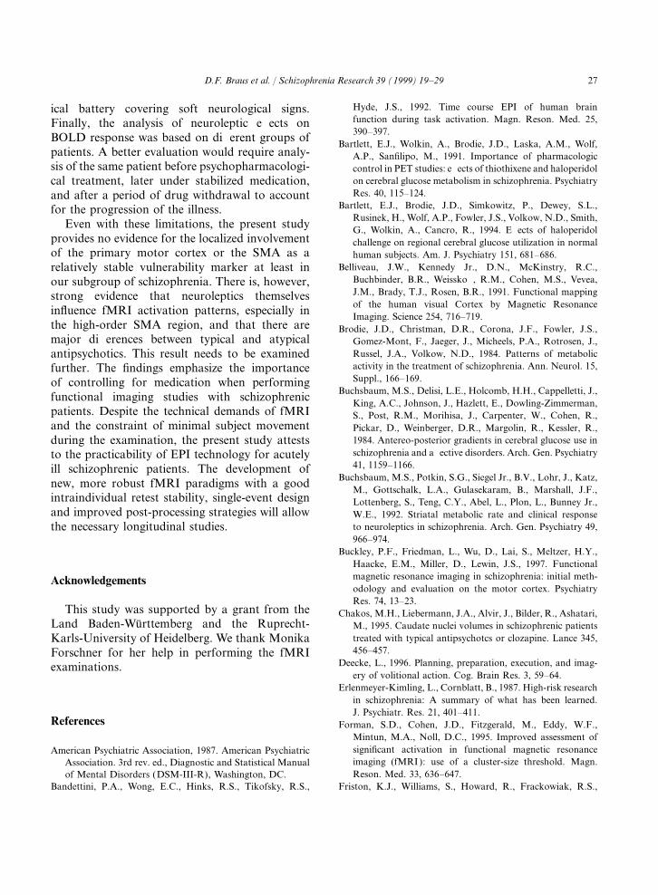
Fig. 1. Paradigm: self-paced sequential finger opposition left hand. Legend: contra=right sensorimotor cortex; ipsi=left sensorimotorcortex; SMA=Supplementary Motor Cortex LI=Laterality Index.
Table 2Significance testing of regional spatial extent of activation as well as laterality and medicationa,b
ROI spatial extent of Drug-free first-episode Patients under stable atypical Patients under stable medicationactivation patients vs. healthy subjects antipsychotic vs. healthy subjects with typical neuroleptics vs.
healthy subjects
Total BOLD response F=0.09 F=0.41 F=14.3p=0.76 p=0.53 p<0.001
Contralateral sensorimotor F=0.04 F=0.13 F=16.6cortex p=0.85 p=0.72 p<0.001
Ipsilateral sensorimotor F=0.24 F=0.003 F=7.6cortex p=0.63 p=0.98 p=0.01
SMAc F=0.75 F=4.4 F=7.4p=0.39 p=0.047 p=0.01
LateralityLI d F=0.02 F=0.12 F=4.25
p=0.89 p=0.73 p=0.049
a Paradigm: Self-paced sequential finger opposition task of the left hand.b Statistics: One-way ANOVA.c SMA: Supplementary Motor Area.d LI: Laterality Index.
neuroleptics were not significantly correlated with no difference in the visually controlled perfor-mance during the fMRI. Contrary to controls andany activation measures. We could not detect any
significant differences in the quantified motion first-episode schizophrenics, patients under stablemedication, especially patients treated with typicalartifacts between patients and controls. There was

24 D.F. Braus et al. / Schizophrenia Research 39 (1999) 19–29
neuroleptics, had to be encouraged more fre- necessary for the performance (Halsey et al., 1979)and, especially in fMRI experiments, movementquently to maintain motor performance in the
course of the experiment. artifacts (Friston et al., 1996). The maximum headmotion for each participant was evaluated, anddata sets exceeding 0.4 voxels motion were dis-carded in accordance with the literature (Just5. Discussionet al., 1996).
To our knowledge, this is the first fMRI studyusing high-speed EPI technology to examine under 5.2. First-episode schizophrenic patientsstandardized conditions the motor system inneuroleptic-naıve schizophrenic patients as well as When compared with healthy controls, the first-
episode, neuroleptic-naıve schizophrenic patientsin patients under stable medication.Our study illustrates that: showed no significant differences in overall activa-
tion, activation pattern in the predefined ROIs and1. SFO is a robust fMRI paradigm with reliableBOLD activation. laterality index in our study (Figs. 1 and 2,
Table 2). According to the coefficient of variance,2. There is a close similarity in the activation ofthe primary and high-order (SMA) sensorimo- the patient group is more heterogeneous than the
controls. After discarding two patients because oftor areas between first-episode schizophrenicpatients and controls. severe motion artifacts, there were no significant
differences between the study groups in the degree3. There is a significant reduction in the overallBOLD response as well as less activated voxels of motion.
Clinically, and as evaluated by neurophysiologi-in the sensorimotor cortices (contra- and ipsilat-eral ) in schizophrenic patients under stable cal examinations, co-ordination and sequencing of
movements (Manschreck et al., 1985; Heinrichsmedication with typical neuroleptics in compar-ison to controls, neuroleptic-naıve first-episode and Buchanan, 1988; Malla et al., 1995) seem to
be impaired in patients with negative symptoms,schizophrenics, and patients treated with atypi-cal drugs. but not in patients with delusions and hallucina-
tions. Paulman et al. (1990) and Guenther et al.4. Independent of the type of antipsychotic medi-cation, there is a significant reduction in the (1991) both examined a population of never-
medicated patients displaying positive symptomsvolume of activation in the SMA region com-pared to controls and neuroleptic-naıve (e.g. formal thought disorder, hallucinations, delu-
sions) using dynamic single-photon emission com-subjects.puterized tomography (SPECT). Both authorsreported a bilateral trend toward increased activa-tion in the positive symptom patients in contrast5.1. SFO paradigm in healthy subjectsto non-reactivity of the rCBF in negative symptompatients during motor activity. Thus, our fMRIThe activation pattern in healthy subjects
obtained with echo-planar functional magnetic res- results in first-episode schizophrenics are in linewith these previous studies. They do not supportonance imaging is in agreement with the findings
from several groups achieved with PET and fMRI an abnormal localized deficit in primary or high-order motor areas in neuroleptic-naıve schizo-using different quantification methods ( Kim et al.,
1993; Shibasaki et al., 1993; Rao et al., 1996). phrenic patients with predominantly positivesymptoms.Thus, the SFO motor task is a robust paradigm,
it can easily be controlled, and it usually yields asufficient signal increase in M1 S1 as well as SMA. 5.3. Schizophrenics under stable medicationThe observed interindividual variability in controlsmay be influenced by the frequency of the para- Guenther et al. (1994) investigated 13 patients
(n=9 under haloperidol medication), with a his-digm (Sabatini et al., 1991), the effort that is

25D.F. Braus et al. / Schizophrenia Research 39 (1999) 19–29
Seq
uent
ial fi
nger
opp
ositio
n left h
and:
Cro
ss c
orre
latio
n 0.
55
Sig
nal in
tens
ity: T
ime
cour
se S
MA
regi
on
Fig
.2.
FM
RI
acti
vati
onpa
tter
n:A
ntip
sych
otic
trea
tmen
t(l
eft)
led
toa
sign
ifica
ntre
duct
ion
inac
tiva
tion
ofth
eSM
Are
gion
com
pare
dto
(rig
ht)
am
atch
edco
ntro
lor
ane
urol
epti
c-na
ıve
first
-epi
sode
pati
ent.

26 D.F. Braus et al. / Schizophrenia Research 39 (1999) 19–29
tory of chronic schizophrenia and 14 healthy con- effects on brain metabolism (Brodie et al., 1984;Bartlett et al., 1991; Buchsbaum et al., 1992). Thetrols in a PET study with 11C-deoxyglucose as
tracer during a complex motor task. In this study, typical neuroleptic haloperidol results in decreasedwhole-brain and prefrontal metabolism, butschizophrenic patients showed a reduced activation
in the contralateral sensorimotor cortex and in increased basal ganglia metabolism (Bartlett et al.,1991, 1994). A PET study (Holocomb et al.,SMA. The authors found no evidence that this
lack of activation is related to either task accuracy 1996) showed a striatal metabolic increase withhaloperidol, which disappeared after theor task speed. An influence of medication or effort,
however, could not be ruled out. They concluded withdrawal of the drug. Chakos et al. (1995)showed that striatal metabolism increases withthat schizophrenics may have a brain dysfunction
that limits their capacity to produce a focal meta- classical neuroleptics and decreases with the atypi-cal clozapine. Striatal D2-receptor blockade couldbolic response to stimulation in functionally dis-
tinct brain regions. lead to increased inhibition of the thalamusthrough increased activity in gamma-aminobu-In an fMRI study using a FLASH sequence on
10 schizophrenic patients and seven healthy volun- tyric acid (GABA) projections. Reduced outputfrom the thalamus to the neocortex could contrib-teers ( Wenz et al., 1994, Schroder et al., 1995), a
highly significant lower activation value in the ute to reduced neocortical activity, which isreflected in reduced cerebral blood flow and bloodsensorimotor cortex and in the SMA region has
been described. In addition, Schroder et al. (1995) volume in the neocortex. Our FMRI results areconsistent with this hypothesis.found that the laterality ratio was reversed in
schizophrenics. This study had several important The significant difference in the laterality indexbetween the study group of typical neurolepticsmethodological differences from the current study
as they used single slice FLASH sequences, a and all other groups could be a cut-off artifact. Areduced overall activation in combination with adifferent post-processing procedure, no adequate
performance control and no movement evaluation conservative cut-off is increasing the lateralityindex artificially.and correction. These two studies suggest that
there may be differences in the bilateral activation The apparent differences between our study andthose of Schroder et al. (1995) and Wenz et al.of the motor cortex in schizophrenic patients.
Another recent fMRI study (Buckley et al., 1997) (1994) as well as Guenther et al. (1994) couldreflect the medication status and a different cut-could not replicate the reduced activation pattern
in the sensimotor cortex. off in the post-processing procedure or a differencein patient selection with greater negativeOur study showed reduced activation in the
SMA region in the atypical treatment patients as symptomatology.We are aware that this investigation has severalwell as an overall decreased activation pattern in
patients under typical neuroleptics in comparison limitations. First, the number of subjects studiedwas small. The second limitation of this study isto controls and neuroleptic naıve patients (Figs. 1
and 2, Table 2). The medial premotor cortex the inability to monitor effort and motivationobjectively, variables that have been shown to(SMA) represents a higher-order motor system
that is active as a programmer of motor subrou- affect the motor system (McAdam and Seal, 1969).In addition, the ‘resting condition’ is by no meanstines in the initiation and performance of repeti-
tive, self-generated movements (Deecke, 1996). uniform since diverse thoughts and emotions mayinterfere with the subject’s mental status and, atThis region is less sensitive to performance effects
(Sabatini et al., 1991) than the primary motor least in part, could affect regional cerebral bloodflow and consequently BOLD response. Thus, wecortex.
PET studies addressing the effect of antipsy- cannot rule out minor differences in the experimen-tal task between the groups. Furthermore, ourchotics on cerebral blood flow and metabolism
have demonstrated that neuroleptics do have study groups were not characterized by a neurolog-
27D.F. Braus et al. / Schizophrenia Research 39 (1999) 19–29
Hyde, J.S., 1992. Time course EPI of human brainical battery covering soft neurological signs.function during task activation. Magn. Reson. Med. 25,Finally, the analysis of neuroleptic effects on390–397.BOLD response was based on different groups of
Bartlett, E.J., Wolkin, A., Brodie, J.D., Laska, A.M., Wolf,patients. A better evaluation would require analy- A.P., Sanfilipo, M., 1991. Importance of pharmacologicsis of the same patient before psychopharmacologi- control in PET studies: effects of thiothixene and haloperidol
on cerebral glucose metabolism in schizophrenia. Psychiatrycal treatment, later under stabilized medication,Res. 40, 115–124.and after a period of drug withdrawal to account
Bartlett, E.J., Brodie, J.D., Simkowitz, P., Dewey, S.L.,for the progression of the illness.Rusinek, H., Wolf, A.P., Fowler, J.S., Volkow, N.D., Smith,Even with these limitations, the present study G., Wolkin, A., Cancro, R., 1994. Effects of haloperidol
provides no evidence for the localized involvement challenge on regional cerebral glucose utilization in normalof the primary motor cortex or the SMA as a human subjects. Am. J. Psychiatry 151, 681–686.
Belliveau, J.W., Kennedy Jr., D.N., McKinstry, R.C.,relatively stable vulnerability marker at least inBuchbinder, B.R., Weisskoff, R.M., Cohen, M.S., Vevea,our subgroup of schizophrenia. There is, however,J.M., Brady, T.J., Rosen, B.R., 1991. Functional mappingstrong evidence that neuroleptics themselvesof the human visual Cortex by Magnetic Resonanceinfluence fMRI activation patterns, especially in Imaging. Science 254, 716–719.
the high-order SMA region, and that there are Brodie, J.D., Christman, D.R., Corona, J.F., Fowler, J.S.,major differences between typical and atypical Gomez-Mont, F., Jaeger, J., Micheels, P.A., Rotrosen, J.,
Russel, J.A., Volkow, N.D., 1984. Patterns of metabolicantipsychotics. This result needs to be examinedactivity in the treatment of schizophrenia. Ann. Neurol. 15,further. The findings emphasize the importanceSuppl., 166–169.of controlling for medication when performing
Buchsbaum, M.S., Delisi, L.E., Holcomb, H.H., Cappelletti, J.,functional imaging studies with schizophrenicKing, A.C., Johnson, J., Hazlett, E., Dowling-Zimmerman,
patients. Despite the technical demands of fMRI S., Post, R.M., Morihisa, J., Carpenter, W., Cohen, R.,and the constraint of minimal subject movement Pickar, D., Weinberger, D.R., Margolin, R., Kessler, R.,
1984. Antereo-posterior gradients in cerebral glucose use induring the examination, the present study attestsschizophrenia and affective disorders. Arch. Gen. Psychiatryto the practicability of EPI technology for acutely41, 1159–1166.ill schizophrenic patients. The development of
Buchsbaum, M.S., Potkin, S.G., Siegel Jr., B.V., Lohr, J., Katz,new, more robust fMRI paradigms with a goodM., Gottschalk, L.A., Gulasekaram, B., Marshall, J.F.,
intraindividual retest stability, single-event design Lottenberg, S., Teng, C.Y., Abel, L., Plon, L., Bunney Jr.,and improved post-processing strategies will allow W.E., 1992. Striatal metabolic rate and clinical response
to neuroleptics in schizophrenia. Arch. Gen. Psychiatry 49,the necessary longitudinal studies.966–974.
Buckley, P.F., Friedman, L., Wu, D., Lai, S., Meltzer, H.Y.,Haacke, E.M., Miller, D., Lewin, J.S., 1997. Functionalmagnetic resonance imaging in schizophrenia: initial meth-Acknowledgementsodology and evaluation on the motor cortex. PsychiatryRes. 74, 13–23.
This study was supported by a grant from the Chakos, M.H., Liebermann, J.A., Alvir, J., Bilder, R., Ashatari,Land Baden-Wurttemberg and the Ruprecht- M., 1995. Caudate nuclei volumes in schizophrenic patients
treated with typical antipsychotcs or clozapine. Lance 345,Karls-University of Heidelberg. We thank Monika456–457.Forschner for her help in performing the fMRI
Deecke, L., 1996. Planning, preparation, execution, and imag-examinations.ery of volitional action. Cog. Brain Res. 3, 59–64.
Erlenmeyer-Kimling, L., Cornblatt, B., 1987. High-risk researchin schizophrenia: A summary of what has been learned.J. Psychiatr. Res. 21, 401–411.
References Forman, S.D., Cohen, J.D., Fitzgerald, M., Eddy, W.F.,Mintun, M.A., Noll, D.C., 1995. Improved assessment ofsignificant activation in functional magnetic resonanceAmerican Psychiatric Association, 1987. American Psychiatricimaging (fMRI ): use of a cluster-size threshold. Magn.Association. 3rd rev. ed., Diagnostic and Statistical ManualReson. Med. 33, 636–647.of Mental Disorders (DSM-III-R), Washington, DC.
Bandettini, P.A., Wong, E.C., Hinks, R.S., Tikofsky, R.S., Friston, K.J., Williams, S., Howard, R., Frackowiak, R.S.,

28 D.F. Braus et al. / Schizophrenia Research 39 (1999) 19–29
Turner, R., 1996. Movement-related effects in fMRI time- Noll, D.C., Genovese, C.R., Nystrom, L.E., Vazquez, A.L.,Forman, S.D., Eddy, W.F., Cohen, J.D., 1997. Estimatingseries. Magn. Reson. Med. 35, 346–355.test–retest reliability in functional MR imaging II: applica-Goebel, R., 1996. Brainvoyager: A program for analysing andtion to motor and cognitive activation studies. Magn.visualizing functional and structural MRI data sets. Neuroi-Reson. Med. 38, 508–517.mage 3, 604.
Ogawa, S., Lee, T.M., Kay, A.R., Tank, D.W., 1990. BrainGuenther, W., Petsch, R., Steinberg, R., Moser, E., Streck, P.,magnetic resonance imaging with contrast dependent onHeller, H., Kutz, G., Hippius, H., 1991. Brain dysfunctionblood oxygenation. Proc. Natl. Acad. Sci. USA 87,during motor activation and corpus callosum alterations in9868–9872.schizophrenia measures by cerebral blood flow and magnetic
Ogawa, S., Tank, D.W., Menon, R., Ellermann, J.M., Kim,resonance imaging. Biol. Psychiatry 29, 535–555.S.G., Merkle, H., Ugurbil, K., 1992. Intrinsic signal changesGuenther, W., Brodie, J.D., Bartlett, E.J., Dewey, S.L., Henn,accompanying sensory stimulation: functional brain map-F.A., Volkow, N.D., Alper, K., Wolkin, A., Cancro, R.,ping using MRI. Proc. Natl. Acad. Sci. USA 89, 5951–5955.Wolf, A.P., 1994. Diminished cerebral metabolic response
Oldfield, R.D., 1971. The assessment and analysis of handed-to motor stimulation in schizophrenics: a PET study. Eur.ness: the Edinburgh inventory. Neuropsychologia 9, 97–113.Arch. Psychiatry Clin. Neurosci. 244, 115–125.
Paulman, R.G., Devous, M.D., Gregory, R.R., et al., 1990.Halsey, J.H., Blauenstein, U.W., Wilson, E.M., Wills, E.H.,Hypofrontality and cognitive impairment in schizophrenia:1979. Regional cerebral blood flow comparison of right andDynamic SPECT and neuropsychological assessment ofleft hand movement. Neurology 29, 21–28.schizophrenic brain function. Biol. Psychiatry 27, 377–399.Heinrichs, D.W., Buchanan, R.W., 1988. Significance and
Rao, S.M., Bandettini, P.A., Binder, J.R., Bobholz, J.A.,meaning of neurological soft signs in schizophrenia. Am.Hammeke, T.A., Stein, E.A., Hyde, J.S., 1996. Relation-J. Psychiatry 145, 11–18.ship between finger movement rate and functional magneticHolocomb, H.H., Cascella, M.D., Thaker, G.K., Medhoff,resonance signal change in human primary motor cortex.D.R., Dannals, R.F., Tamminga, C.A., 1996. FunctionalJ. Cereb. Blood Flow Metab. 16, 1250–1254.sites of neuroleptic drug action in the human brain: PET/
Robinson, A., Heaton, R.K., Lehmann, R.A.W., Stilson, D.W.,FDG studies with and without haloperidol. Am. J. Psychi-1980. The utility of the Wisconsin card sorting test in detect-atry 153, 41–49.ing and localization frontal lobe lesions. J. Consult. Clin.
Ismail, B., Cantor-Graae, E., McNeil, T.F., 1998. NeurologicalPsychol. 48, 605–614.
abnormalities in schizophrenic patients and their siblings.Rosvold, H.E., Mirsky, A., Sarason, I., Bransome, E.D., Beck,
Am. J. Psychiatry 155, 84–89.L.H., 1956. A continuous performance test of brain damage.
Just, M.A., Carpenter, P.A., Keller, T.A., Eddy, W.F.,J. Consult. Psychol. 20, 343–350.
Thulborn, K.R., 1996. Brain activation modulated by Sabatini, U., Chollet, F., Rascol, O., Celsis, P., Rascol, A.,sentence comprehension. Science 274, 114–116. Lenzi, G.L., Marc-Vergnes, J.-P., 1991. Effect of side and
Kim, S.G., Ashe, J., Hendrich, K., Ellermann, J.M., Merkle, rate of stimulation on cerebral blood flow changes in motorH., Ugurbil, K., Georgopoulos, A.P., 1993. Functional areas during finger movements in humans. J. Cerebral Bloodmagnetic resonance imaging of motor cortex: hemispheric Flow Metab. 13, 639–645.asymmetry and handedness. Science 261, 615–617. Sartorius, A., Braus, D.F., Ende, G., Krier, A., Henn, F.A.,
Kraepelin, E., 1919. In: Robertson, G.M. (Ed.), Dementia Prae- 1997. Simple and Objective Evaluation of Motion Artifactscox and Paraphrenia. Krieger, New York, pp. 77–83. in 2D functional EPI Data, Proc. International Society ofTranslated by R.M. Barclay. Magnetic Resonances in Medicine, Vancouver.
Kwong, K.K., Belliveau, J.W., Chesler, D.A., Goldberg, I.E., Schroder, J., Wenz, F., Schad, L.R., Baudendistel, K., Knopp,Weisskoff, R.M., Poncelet, B.P., Kennedy, D.N., Hoppel, M.V., 1995. Sensorimotor cortex and supplementary motorB.E., Cohen, M.S., Turner, R., et al., 1992. Dynamic mag- area changes in schizophrenia: A study with functional mag-netic resonance imaging of human brain activity during pri- netic resonance imaging. Br. J. Psychiatry 167, 197–201.mary sensory stimulation. Proc. Natl. Acad. Sci. USA 89, Shibasaki, H., Sadato, N., Lyshkow, H., Yonekura, Y., Honda,5675–5679. M., Nagamine, T., Suwazono, S., Magata, Y., Ikeda, A.,
McAdam, D.W., Seal, D.M., 1969. Bereitschaftspotential Miyazaki, M., et al., 1993. Both primary motor cortex andenhancement with increased level of motivation. supplementary motor area play an important role in com-Electroencephal. Clin. Neurophysiol. 27, 73–75. plex finger movement. Brain 116, 1387–1398.
Malla, A.K., Norman, R.G.M., Aguilar, O., Carnahan, H., Stehling, M.K., Turner, R., Mansfield, P., 1991. Echo-planarCortese, L., 1995. Relationship between movement planning imaging: magnetic resonance imaging in a fraction of aand psychopathology profiles in schizophrenia. Br. J. Psych- second. Science 254, 43–50.iatry 167, 211–215. Talairach, J., Tournoux, P., 1988. Co-planar Stereotactic Atlas
Manschreck, T.C., Maher, B.A., Waller, N.G., Ames, D., of the Human Brain. Thieme, Stuttgart.Latham, C.A., 1985. Deficient motor synchrony in schizo- Walker, E.F., 1994. Developmentally moderated expression ofphrenic disorders: clinical correlates. Biol. Psychiatry 20, the neuropathology underlying schizophrenia. Schizophr.
Bull. 20, 453–480.990–1002.

29D.F. Braus et al. / Schizophrenia Research 39 (1999) 19–29
Walker, E.F., Savole, T., Davis, D., 1994. Neuromotor precur- schizophrenic patients receiving neuroleptic medication.Magn. Reson. Imaging 12, 975–982.sors of schizophrenia. Schizophr. Bull. 20, 441–451.
Wenz, F., Schad, L.R., Knopp, M.V., Baudendistel, K.T., Woods, B.T., Kinney, D.K., Yurgelun-Todd, D., 1986. Neuro-logical abnormalities in schizophrenic patients and theirFlomer, F., Schroder, J., van Kaick, G., 1994. Functional
magnetic resonance imaging at 1.5 T: activation pattern in families. Arch. Gen. Psychiatry 43, 657–663.
Copyright © 2022 FDOKUMEN