Effectiveness of Whey Protein Supplement in Resistance Trained Individuals
Upload
independentCategory
view
4download
0
ORIGINAL PAPER
Antioxidant properties of whey protein hydrolysates as measuredby three methods
Adriena Dryakova • Anne Pihlanto • Pertti Marnila •
Ladislav Curda • Hannu J. T. Korhonen
Received: 13 October 2009 / Revised: 17 January 2010 / Accepted: 3 February 2010 / Published online: 19 February 2010
� Springer-Verlag 2010
Abstract Four microbial proteases (Alcalase, Flavour-
zyme, Neutrase and Protamex) were used for the prepara-
tion of whey protein hydrolysates. The aim of this research
was to find out whether these hydrolysates can be used as a
source of whey derived antioxidants. Hydrolyzed samples,
including their unhydrolyzed protein solutions were tested
by the ABTS (2,20-azinobis-(3-ethylbenzothiazoline-6-sul-
fonic acid) decolorization assay, by the total radical-trap-
ping potential method and by the assay of liposomes
peroxidation (fluorescence photometry). Antioxidant
properties were enhanced by hydrolysis in most of cases.
Alcalase hydrolysates were found as the most effective
antioxidants as determined by ABTS assay (*50% of
antioxidant activity at 0.1 mg ml-1 of hydrolysate in
reaction) and fluorescence photometry. Liposomes were
oxidized *50% less (1.1 lM of a-tocopherol equivalent)
with Alcalase hydrolysates additive (at 5.85 mg ml-1 of
hydrolysate in reaction). Hydrolysates did not inhibit
the oxidation of liposomes at concentrations below
1.0 mg ml-1 in reaction. On the contrary, results of total
trapping potential method did not agree with findings
observed in other tests. In this assay, Neutrase hydrolysates
showed the best antioxidant properties. Pro-oxidant prop-
erties were observed in solutions containing (prior to the
enzyme Protamex addition only) intact whey protein as
determined by the measurement of the liposome peroxi-
dation. The ABTS assay was optimized for the evaluation
of the antioxidant activity in whey protein hydrolysates.
The reaction time should be prolonged to avoid underes-
timation of the antioxidant activity.
Keywords Whey � Hydrolysates � ABTS � Liposomes �Chemiluminescence
Introduction
The importance of oxidation processes in the body and
foodstuffs has been widely recognized. The oxidative stress
caused by reactive oxygen species plays a significant role
in a number of age-specific diseases and neurodegenerative
disorders [1]. In foods, oxidative reactions cause deterio-
rations in food quality, unacceptable taste or texture, color,
loss of nutritive value at whole and shortening of shelf life.
Compounds which are responsible for destructive and
lethal cellular effects are lipid peroxides and low molecular
weight compounds produced during late stage of the oxi-
dative reactions. The main targets of these reactions are
lipids, proteins, DNA and enzymes. To prevent foods from
undergoing deteriorations and to provide protection against
processes promoting the development of age-specific dis-
eases like cancer, atherosclerosis and diabetes, it is very
important to inhibit the lipid peroxidation occurring in
foodstuffs and the living body [2]. The potential adverse
effects of synthetic antioxidant additives, such as butylated
hydroxyanisole and butylated hydroxytoluene, have stim-
ulated their replacement by natural antioxidants derived
from dietary sources. One practical approach to solve this
problem is the use of antioxidants from natural sources,
mainly non-protein compounds from plants. Some proteins
from certain foods have also been reported to have the
ability to scavenge active oxygen species. For example,
A. Dryakova (&) � L. Curda
Institute of Chemical Technology, Technicka 5,
166 28 Prague 6, Czech Republic
e-mail: [email protected]
A. Pihlanto � P. Marnila � H. J. T. Korhonen
MTT Agrifood Research, 31600 Jokioinen, Finland
123
Eur Food Res Technol (2010) 230:865–874
DOI 10.1007/s00217-010-1231-9

porcine myofibrillar protein [3], soy and gelatin [4], potato
[5], milk proteins of which caseins [6, 7] and finally whey
proteins [8–11] have been shown to have the antioxidant
effect. In addition to proteins, protein hydrolysates have
also been found to exhibit the antioxidant activity. During
protein hydrolysis, protein’s overall antioxidant activity is
enhanced as its tertiary structure is disrupted and the sol-
vent accessibility of released amino acids increases.
Enzymatic digests of whey proteins and its use as potential
antioxidants have been also demonstrated in several stud-
ies: [12–15].
Most of authors have used for the evaluation of the
antioxidant activity (measurement of lipid oxidation)
methods based on measurement of thiobarbituric acid
reactive substances and assays based on the ability of
scavenging of different type radicals: either stable chro-
mogen 1,1-diphenyl-2-picrylhydrazyl radical (DPPH�) or
short-live radicals, such as fluorescein radical in oxygen
radical absorbance capacity (ORAC). The goal of this
research was to contribute to a number of previous studies
dealing with the antioxidant activity of whey protein
hydrolysates, and to find out whether these hydrolysates,
prepared by commercially available microbial proteases
(Alcalase, Flavourzyme Protamex and Neutrase), can be
used as a potential source of naturally derived antioxidants
and broaden the information about the mechanism of the
antioxidant effect. This effect was investigated in different
phase systems, using different principles: scavenging of the
2,20-azinobis (3-ethylbenzenthiazoline-6-sulfonic acid)
radical, by the assay of liposome peroxidation induced by
ferric ion or by the rate of trapping the luminol radical
induced by 2,2-azo-bis(2-methylpropionamidine) dichlo-
ride thermolysis. These methods are rarely used for the
determination of the antioxidant activity of whey protein
hydrolysates.
Materials and methods
Materials
The powdered whey protein concentrate (WPC), containing
60.8% of protein was supplied by Promil (Czech Republic).
Enzymes used for hydrolysis of WPC: Alcalase 2.4L (ALC);
Flavourzyme 500 MG (FLA); Protamex (PRO) and Neutrase
(NEU) were obtained from Novo Nordisk A/S (Denmark).
2,2-Azo-bis(2-methylpropionamidine) dichloride (ABAP)
was purchased from Acros Organics (Belgium). o-Phthaldi-
aldehyde (OPA) and N-acetylcysteine (NAC) were purchased
from Merck (Germany). Sodium dodecyl sulfate (SDS) was
purchased from Lachema (Czech Republic). (2,20-azinobis
(3-ethylbenzenthiazoline-6-sulfonic acid) diammonium salt
and 6-hydroxy-2,5,7,8-tetramethylchroman-2-carboxylic
acid (Trolox; water soluble analogue of vitamin E) and other
chemicals were obtained from Sigma Chemicals (Germany).
Preparation of enzymatic hydrolysates
The WPC was reconstituted in demineralized water to 4%
(w/v) concentration of protein solution and pH was
adjusted by 2 M NaOH or HCl to obtain the optimal value
for enzyme. Solutions were preheated in temperature-
controlled water bath to 45 �C. Protein content in enzymes
was determined as described by Lowry et al. [16]. The
hydrolysis parameters were used as follows: enzyme/sub-
strate ratio, 1/100 (w/w); pH 8 for ALC, pH 7 for FLA and
NEU and pH 6 for PRO, temperature 45 �C. The hydrolysis
was conducted for 3 h with continuous stirring. Samples
were withdrawn at 15; 30; 60; and 180 min from each
proteolytic mixture and were immediately heated at 85 �C
for 10 min to inactivate the enzyme. The unhydrolyzed
sample was taken before the enzyme addition. A small
amount of the freshly prepared hydrolysates was used to
determine the degree of hydrolysis and the rest was freeze-
dried for later use.
Degree of hydrolysis
The degree of hydrolysis (DH) was determined as descri-
bed by Spellman et al. [17] with slight modifications. The
OPA-NAC reagent was prepared by mixing of 1 ml of
0.3 M OPA solution (in methanol); 25 ml of 0.1 M of
Na2B407; 2.5 ml of 20% (w/v) of SDS and 0.05 g of NAC
(diluted in demineralized water). This mixture was diluted
to the final volume of 50 ml with demineralized water. The
reagent was prepared every day fresh. To dissolve the
possible precipitate in hydrolysate, created after inactiva-
tion of enzyme, 1 ml of 1 M NaOH was added to 1 ml of
hydrolysate. Further, hydrolysates were diluted with
demineralized water and then mixed with the OPA reagent
in ratio 1/100 and let to react in dark for 2 min. The
absorbance was measured at 335 nm using spectropho-
tometer (Cary 50, Varian, Australia). DH values were
calculated on the base of totally hydrolyzed WPC that
represented 100% of DH (data not shown) with subtraction
of DH calculated for unhydrolyzed sample (blank). The
average chain length (ACL) was determined as described
by Adler-Nissen [18], where ACL=100/DH. All samples
were measured in duplicates.
Antioxidant activity (ABTS decolorization assay)
The antioxidant activity of hydrolysates (scavenging of the
ABTS radical (ABTS•?) 2,20-azinobis-(3-ethylbenzothia-
zoline-6-sulfonic acid)) was measured using the decolor-
ization assay as described by Re et al. [19] and Virtanen
866 Eur Food Res Technol (2010) 230:865–874
123

et al. [20]. Briefly, 1ml of diluted ABTS•1 solution in
5 mM phosphate-buffered saline (PBS) was added to 10 ll
of sample, or Trolox in ethanol (standard) or PBS (blank),
and an absorbance (734 nm) reading (Lambda 25, Perkin
Elmer, USA) was taken at 30 �C (exactly 1 min after initial
mixing and up to 180 min at 1 min intervals). Experiments
showed that the dependence of the antioxidant activity on
time was linear up to the first 6-min reaction time.
Accordingly, 6-min reaction time was used for all
hydrolysates. All determinations were duplicated. The
antioxidant activity was measured in hydrolysate recon-
stituted in PBS to 5–10 mg ml-1 of initial sample con-
centration (dry matter) that is 0.05–0.1 mg ml-1 in
reaction. The antioxidant activity (%) was calculated with
the following equation: AA (%) = [(A743 blank–A743
sample)/A743 blank] 9 100%. The AA was calculated at
each minute of reaction time and plotted as a function of
the time to calculate the antioxidant activity as a single
quantity, so the antioxidant activity was expressed in terms
of the total contribution of the sample to the antioxidant
activity.
Total radical-trapping potential method (TRAP)
The effect of hydrolysates and the unhydrolyzed material
on total radical trapping was assessed as described by Lissi
et al. [21]. In the assay, 175 ll of sample (5.8–0.4 mg ml-1
of freeze-dried hydrolysate in reaction) or 50 ll Trolox
(20–80 lM) was incubated with 25 ll of luminol (0.02 M
5-amino-2,3-dihydro-1,4-phthal-azinedione) and 50 ll of
buffer (0.01 M sodium phosphate buffer with 0.9% NaCl
pH 7.8) in a micro plate for 10 min at 37 �C. After incu-
bation, 50 ll of ABAP solution (90 mM) was added.
A chemiluminescence reading was taken immediately after
addition and up to 2.5 h using luminometer (Luminoscan
EL1, ThermoScientific, Finland). All determinations were
carried out in duplicate, at each separate concentration of
the sample. Samples and standard were reconstituted in
buffer (0.01 M sodium phosphate buffer with 0.9% NaCl
pH 7.8). The chemiluminescence peak time was plotted as
a function of sample dry matter. Total antioxidant potential
(TRAP)—trapping activity—was expressed as the equiva-
lent of Trolox concentration by regression analysis of
plotted chemiluminescence peak time versus sample dry
matter.
Assay of lipid peroxidation in liposomes
To determine the susceptibility of lipid to peroxidation
with hydrolysates additive, a liposomal system was used.
Lipid peroxidation was tested using the fluorescence plate
reader as previously described by Tirmenstein et al. [22]
with slight modifications. Phosphatidyl choline liposomes
were prepared according to the procedures of Ursini et al.
[23]. a-Tocopherol treated liposomes were used as standard
compounds. Liposomes were stored at 4 �C for at least
1 week prior to use to increase levels of lipid hydroper-
oxides. In assay, 10 ll of liposomes (with standard or not)
and 50 ll of hydrolysates reconstituted in potassium
phosphate–glycine–ascorbate buffer (at concentrations
0.14–5.85 mg ml-1 in reaction; dry matter) were loaded
into the wells of a microplate. One hundred and twenty
microliter of potassium phosphate (50 mM)–glycine
(100 mM)–ascorbate (0.45 mM) buffer (pH 7.4) was
added, and the oxidation was initiated by adding 20 ll of
25 lM ferric chloride/1 mM ADP. The microplates were
incubated at 37 �C, shaken, and fluorescence (EX360nm,
EM460nm) was measured using plate reader (Wallac
Viktor 2, PerkinElmer, Finland). The measurement was
performed at 15-min intervals for total 4 h. Blank (fluo-
rescence360/460 in well containing buffer and ferric
chloride only) was subtracted before calculating the lipo-
some oxidation. At least three independent experiments
were carried out for each sample. The liposome oxidation
(LO) (after 4 h of incubation) was calculated. The lipo-
some oxidation was expressed as the equivalent of Trolox
concentration by regression analysis of plotted LO versus
sample dry matter.
Statistical analysis
Dose dependencies of tested properties were evaluated by
means of regression analysis. Correlation coefficient was
used for the evaluation of the optimization of ABTS assay.
Data were analyzed using the t-test or by multifactor
analysis of variance.
Results and discussion
Degree of hydrolysis
The DH of WPC ranged from 6 to 35%, depending on
protease specificity and substrate affinity (Table 1). Inter-
estingly, ACL of ALC and FLA hydrolysates was quite
similar (9 for ALC; 3 for FLA after 180 min of hydrolysis).
On the contrary, calculated ACL of PRO and NEU
hydrolysates were higher (18 for NEU and 16 for PRO after
180 min hydrolysis) (Table 1). As expected, DH generally
increased and correspondingly to that, the ACL of
hydrolysates decreased, with the course of hydrolysis. The
significant difference (p \ 0.05) of the calculated DH
between FLA hydrolysates and ALC, NEU and PRO
hydrolysates was observed, which is caused by different
nature of enzyme reaction. Flavourzyme (from Aspergillus
oryzae) is a crude enzyme formula probably with both endo
Eur Food Res Technol (2010) 230:865–874 867
123

and exopeptidases activities. The highest DH, i.e. with
highest amount of released amino groups (or peptide with
shortest amino group chain length), was found in Fla-
vourzyme hydrolysates. The specificity of Neutrase (from
Bacillus amyloliquifaciens) and Protamex is not clearly
known. Hydrolysates digested with these enzymes showed
clearly lower DH and contained peptides with longer
amino acid chain length than those prepared by Flavour-
zyme digestion.
Antioxidant activity (ABTS decolorization assay)
The ABTS decolorization assay is applicable to both
lipophilic and hydrophilic compounds and has been widely
used to the assessment of antioxidant activity [24]. All
samples demonstrated antioxidant activity ranking from 7
to 54% (at 6 min reaction time at 0.10 mg ml-1 concen-
tration of hydrolysate in reaction). The dependence of
antioxidant activity on sample concentration (0.05–
0.10 mg ml-1) was found linear (6 min reading) in all
samples (R2 [ 0.9). Before hydrolysis, all samples (unhy-
drolyzed whey protein) showed low antioxidant activity
(7–19.8%) and during 180 min of hydrolysis, the values
increased notably with all tested hydrolysates (40–54.2%).
The development of the antioxidant activity in relation to
hydrolysis time is shown in Fig. 1. Alcalase hydrolysates
were the most effective in scavenging the ABTS•?.
Already, after 15 min of hydrolysis time a 48% antioxidant
activity was found, whereas in Protamex hydrolysates not
even 180 min of hydrolysis produced such activity. Lower
antioxidant activity in Neutrase and Protamex hydrolysates
can be attributed to the specific enzyme cleavage which did
not produce peptides able to scavenge ABTS•?, taking into
the account the relatively lower affinity of enzyme to
substrate (Table 1). It can be also said that peptide mix-
tures of higher chain lengths ([16 of amino groups in
Table 1 Degree of hydrolysis and average chain length calculated in individual whey protein hydrolysates
Time of hydrolysis (min) ALC FLA NEU PRO
DH (%) ACL DH (%) ACL DH (%) ACL DH (%) ACL
0 0 – 0 – 0 – 0 –
15 7.7 13 4.5 22 0.8 125 0.9 111
30 9.0 11 15.2 7 3.8 26 2.5 40
60 10.9 9 20.7 5 3.9 26 4.7 21
180 11.3 6 35.2 3 5.6 18 6.1 16
Time of hydrolysis indicates the time of the hydrolytic reaction when the hydrolysates was taken at in minutes
DH degree of hydrolysis, ACL average chain length, ALC alcalase, FLA flavourzyme, NEU neutrase, PRO protamex
0
10
20
30
40
50
60
Alcalase Flavourzyme Neutrase Protamex
AA
[%
]
0 min 15 min 30 min 60 min 180 min
Fig. 1 Development of
antioxidant activity (ABTS
decolorization assay) during
hydrolysis; AA, antioxidant
activity (%); time indicates the
time of hydrolysis when the
sample was withdrawn at (min);
sample concentration (dry
matter) = 0.10 mg ml-1;
reading 6 min; data are
displayed as means ± SD
868 Eur Food Res Technol (2010) 230:865–874
123
chain) were not as effective in scavenging the ABTS•? as
hydrolysates with shorter chain lenghts (Table 1). The
activity found in unhydrolyzed samples confirms the gen-
erally known fact that whey protein itself exhibits some
antioxidant properties [25]. The differences observed
among individual unhydrolyzed samples might have been
explained by different tertiary structure of the protein due
to initial pH and heat treatment of the protein solution prior
to hydrolysis.
The antioxidant activity correlated with degree of
hydrolysis of ALC hydrolysates (r = 0.95). In this study,
lower correlation of DH and antioxidant activity was
observed in other hydrolysates (FLA r = 0.90; NEU
r = 0.75; PRO r = 0.70). A strong correlation between
DPPH scavenging activity and DH of ALC hydrolysates of
porcine plasma was observed [26]. Virtanen et al. [20]
found that during fermentation of milk some lactic acid
bacteria produced ABTS radical scavenging activity that
correlated with DH. OPA method determines the content of
primary a and e-amino groups of free amino acids or in
peptides [27]. High degree of hydrolysis thus means high
content of released amino groups and it should presumably
show also good antioxidant activity in FLA hydrolysates.
Nevertheless, the antioxidant activity of hydrolysates was
not found to be directly attributed to DH, suggesting that
the activity is inherent to specific amino acid sequence.
Inconsistent data are reported for antioxidant activity of
whey proteins or their digests. Some authors relate the
antioxidant activity to low molecular fractions of whey,
[14, 28] on the contrary, others report higher antioxidant
activities [15, 25] in relation to high molecular weight
compounds. Pena-Ramos and Xiong [14] detected highest
antioxidant activity in FLA hydrolysate, and Hogan et al.
[29] detected highest antioxidant activity in milk protein
hydrolysates UF fraction (\3 kDa) prepared by microbial
protease (also from Aspergillus oryzae) when compared to
other hydrolysates prepared by alkaline and neutral prote-
ases. Since in this study, hydrolysis by ALC produced
highest antioxidant activity, it should be emphasized that
results of these studies were carried out with different
materials; (e.g. whey protein concentrate/isolate; different
content of non-protein compounds which produce thio-
barbituric acid reactive substances with protein upon
heating) and the methods and evaluation of results often
greatly varies which makes comparison difficult. Finally,
the uses of concrete enzyme/substrate combination and
conditions of hydrolysis will influence the final antioxidant
potential of hydrolysates.
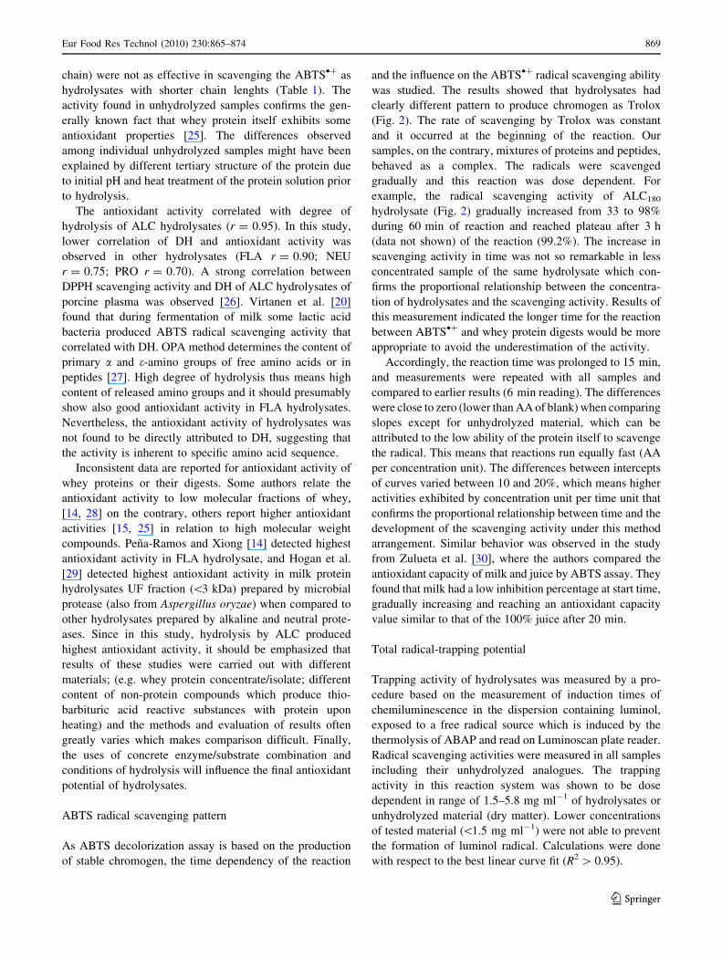
ABTS radical scavenging pattern
As ABTS decolorization assay is based on the production
of stable chromogen, the time dependency of the reaction
and the influence on the ABTS•? radical scavenging ability
was studied. The results showed that hydrolysates had
clearly different pattern to produce chromogen as Trolox
(Fig. 2). The rate of scavenging by Trolox was constant
and it occurred at the beginning of the reaction. Our
samples, on the contrary, mixtures of proteins and peptides,
behaved as a complex. The radicals were scavenged
gradually and this reaction was dose dependent. For
example, the radical scavenging activity of ALC180
hydrolysate (Fig. 2) gradually increased from 33 to 98%
during 60 min of reaction and reached plateau after 3 h
(data not shown) of the reaction (99.2%). The increase in
scavenging activity in time was not so remarkable in less
concentrated sample of the same hydrolysate which con-
firms the proportional relationship between the concentra-
tion of hydrolysates and the scavenging activity. Results of
this measurement indicated the longer time for the reaction
between ABTS•? and whey protein digests would be more
appropriate to avoid the underestimation of the activity.
Accordingly, the reaction time was prolonged to 15 min,
and measurements were repeated with all samples and
compared to earlier results (6 min reading). The differences
were close to zero (lower than AA of blank) when comparing
slopes except for unhydrolyzed material, which can be
attributed to the low ability of the protein itself to scavenge
the radical. This means that reactions run equally fast (AA
per concentration unit). The differences between intercepts
of curves varied between 10 and 20%, which means higher
activities exhibited by concentration unit per time unit that
confirms the proportional relationship between time and the
development of the scavenging activity under this method
arrangement. Similar behavior was observed in the study
from Zulueta et al. [30], where the authors compared the
antioxidant capacity of milk and juice by ABTS assay. They
found that milk had a low inhibition percentage at start time,
gradually increasing and reaching an antioxidant capacity
value similar to that of the 100% juice after 20 min.
Total radical-trapping potential
Trapping activity of hydrolysates was measured by a pro-
cedure based on the measurement of induction times of
chemiluminescence in the dispersion containing luminol,
exposed to a free radical source which is induced by the
thermolysis of ABAP and read on Luminoscan plate reader.
Radical scavenging activities were measured in all samples
including their unhydrolyzed analogues. The trapping
activity in this reaction system was shown to be dose
dependent in range of 1.5–5.8 mg ml-1 of hydrolysates or
unhydrolyzed material (dry matter). Lower concentrations
of tested material (\1.5 mg ml-1) were not able to prevent
the formation of luminol radical. Calculations were done
with respect to the best linear curve fit (R2 [ 0.95).
Eur Food Res Technol (2010) 230:865–874 869
123

Results of this measurement were not parallel with those
obtained by ABTS decolorization assay (Figs. 1, 3).
Trapping radicals by samples in this reaction system was
improved by hydrolysis almost in all cases. Statistical
testing showed significant differences in trapping activity
between hydrolysates and unhydrolyzed protein in all tes-
ted hydrolysates (p \ 0.05). Generally, best trapping
activity was observed in hydrolysates, withdrawn after
180 min of hydrolysis. Best trapping activity among all
samples was found in NEU hydrolysate withdrawn after
180 min of hydrolysis (7.7 mM of Trolox equivalent).
PRO hydrolysate withdrawn after 15 min of hydrolysis
(4.5 mM of Trolox equivalent) had the highest activity
among all PRO hydrolysates. Alcalase hydrolysates were
not effective as Protamex hydrolysates; similar values
(4.5 mM of Trolox equivalent) were found in samples
hydrolyzed by Alcalase for 180 min in comparison with
Protamex hydrolysates where this activity was seen in
sample withdrawn already at 15 min of hydrolysis.
Hydrolysis influenced the trapping activity of FLA
hydrolysates least among all samples. FLA hydrolysates
exhibited the lowest activity in comparison with all
hydrolyzed samples. Released amino groups (shortest
peptide chain lengths—see Table 1) in FLA hydrolysates
were probably either oxidized themselves or they were not
able to prevent the formation of radicals and by that extend
the chemiluminescence peak time.
Samples showed a different pattern from those obtained
by Trolox in this reaction system. Addition of samples
(hydrolyzed or not) did not produce a measurable induction
time (lag phase) but a very moderate decrease in observed
chemiluminescence intensity. This was also observed in the
study from Lissi et al. [21] on human albumin, where the
negative surface charge of albumin was denoted to reduce
the reaction between the reactive center and the luminol-
derived radical-anion. Furthermore, protein solutions prior
to FLA and NEU hydrolysis (equal concentrations) showed
remarkably higher chemiluminescence maxima at peak
induction time than other unhydrolyzed solutions (data not
shown). This might have explained higher chemilumines-
cence peak time observed after the addition of these sam-
ples. Protein treatment prior to hydrolysis may have
resulted in the premature oxidation of some amino acid
residues or alterations in the protein’s tertiary structure that
affected its free radical scavenging activity [12].
Inconsistent results obtained by ABTS assay and TRAP
method may be accounted to the differences and particu-
larly the variation in analytical procedures. Arnao [24]
states, that in the case of natural antioxidants, they may be
multifunctional. The mechanism that is operative or
0
10
20
30
40
50
60
70
80
90
100
0 10 20 30 40 50 60
AA
[%]
t [min]
Fig. 2 Time dependency of the antioxidant activity (ABTS decolor-
ization assay) of selected samples; AA = antioxidant activity;
t = time of reading (the activity was measured during first 60 min;
immediately after adding the sample into the reaction); filled squareALC hydrolysate withdrawn after 180 min of hydrolysis; sample
concentration in reaction (ABTS assay) = 0.10 mg ml-1 filled dia-mond ALC hydrolysate withdrawn before the enzyme addition;
sample concentration in reaction (ABTS assay) = 0.10 mg ml-1
open triangle Trolox; concentration in reaction (ABTS
assay) = 2.5 lg ml-1 open circle ALC hydrolysate withdrawn after
180 min of hydrolysis; sample concentration in reaction (ABTS
assay) = 2.5 lg ml-1 closed circle ALC hydrolysate withdrawn
before the enzyme addition; sample concentration in reaction (ABTS
assay) = 2.5 lg ml-1
870 Eur Food Res Technol (2010) 230:865–874
123

dominant in particular situation is dependent on conditions
and yet this will affect the kinetics and hence the antioxi-
dant activity.
Inhibition of lipid peroxidation
The development of lipid peroxidation inhibition was
studied in all samples including unhydrolyzed material
using Fe–ADP system by measuring the inhibition of
phosphatidylcholine liposome peroxidation induced by
ferric ion. Results were expressed as a-tocopherol equiva-
lent. Samples did not inhibit the oxidation of liposomes at
concentrations below 1.0 mg ml-1 in reaction. Inhibition
of liposome oxidation was observed at concentrations of
5.85 mg ml-1 of the hydrolysate in reaction. Hydrolysis
influenced antioxidant properties of tested hydrolysates
depending on the enzyme used (Fig. 4). The decrease in
liposome oxidation in comparison with blank (liposomes
with no additives) was observed in all tested samples
(p \ 0.0003), except the unhydrolyzed sample withdrawn
before hydrolysis by PRO (–0.5 lM of a-tocopherol).
Liposomes containing this sample were oxidized 20%
more than blank (liposomes with no additives). Significant
effect of hydrolysis was most markedly seen in PRO
hydrolysates (p \ 0.05); however, liposomes with these
additives showed highest rates of liposome oxidation
against blank (0.4–0.7 lM of a-tocopherol). On the con-
trary, the lowest rates of liposome oxidation were found in
samples containing ALC hydrolysates; the lipid oxidation
was inhibited by approximately 50% (corresponds to
*1 lM of a-tocopherol) in comparison with blank, as
observed in the sample containing ALC15 and remained
stable in relation to the time of hydrolysis. However, the
influence of hydrolysis on improving ALC hydrolysates
antioxidant properties was not found significant (p \ 0.05).
Low rates of liposome oxidation treated with ALC
hydrolysates can be attributed to the ALC cleavage speci-
ficity for aromatic residues (Phe, Trp, Tyr) which are
potential radical scavengers [31]. Significant difference
between the influence of the unhydrolyzed protein and
NEU and FLA hydrolysates addition on the liposome
oxidation was found (p \ 0.01). Hydrolysis did not induce
the antioxidant activity in NEU hydrolysates: liposomes
were oxidized even more with NEU hydrolysates additives,
in comparison with the liposomes containing their unhy-
drolyzed analogues. Rival et al. [7] suggested a theory,
supported by results from measurements of the antioxidant
activity of caseins and casein peptides, that casein and their
digests might be a preferred target over fatty acid free
radical intermediate(s) and can either initiate further
unwanted radical-mediated side reactions or terminate
chain reactions depending on the stability of the resulting
protein/peptide radical. Furthermore, proteins/peptides can
be oxidized during the process, according to a site or
sequence-specific mechanism. NEU hydrolysates, in this
reaction system, were less effective to slow down the
development of lipid peroxidation in liposomes. Hydroly-
sate, withdrawn after 3 h of hydrolysis by FLA was the
most effective among all FLA hydrolysates (1.0 of lM of
a-tocopherol). Other FLA hydrolysates were less effective,
and 15 min of hydrolysis by FLA even led to the forma-
tion of prooxidant properties (0.2 of lM of a-tocopherol).
0.0
1.0
2.0
3.0
4.0
5.0
6.0
7.0
8.0
9.0
10.0
Alcalase Flavourzyme Neutrase Protamex
Tro
lox
[mM
]
0 min 15 min 30 min 60 min 180 min
Fig. 3 Development of the
trapping activity (Total radical-
trapping potential method)
during hydrolysis; trapping
activity was expressed as the
Trolox equivalent (mM), on
equal concentration of samples
4 mg ml-1 in reaction—dry
matter; time indicates the time
of hydrolysis when the sample
was withdrawn at (min)
Eur Food Res Technol (2010) 230:865–874 871
123

Pena-Ramos and Xiong [14] investigated the antioxidant
activity of whey hydrolysates measured by TBARS in Fe-
ascorbate liposomal system. Whey protein isolate hydro-
lyzed by commercial crude enzyme protease F (exo and
enprotease from Aspergillus oryzae) was found to be most
effective. Liposomes containing these hydrolysates were
shown to have lowest amount of TBARS among tested
hydrolysates.
Noticeably different behavior of PRO unhydrolyzed
sample (also observed when measured by ABTS decolor-
ization assay) remains unclear. The solution (containing
intact whey protein) prior to adding PRO enzyme was
adjusted to the pH 6 in comparison with other enzymes
where the hydrolysis run at relatively neutral pH (or
alkaline). In contrast to this observation, Shon and Haque
[32] studied the antioxidant ability of native and thermized
sour whey in oxidation-catalyzed systems. The antioxidant
ability increased with decreasing pH (pH 3–7) in terms of
decreased TBARS and peroxide values. Lower lipid oxi-
dation rates at pH values below the pI of the whey powder
in the presence of iron are seem to be due to the electro-
static repulsion between iron and droplet surfaces [33].
However, the treatment prior to hydrolysis might have
negatively affected the tertiary structure of the protein and
influence its ability to slow down the development of lipid
oxidation. Elias et al. [34] studied the influence of native
and denatured (50–95 �C) b-lactoglobulin on oxidative
stability of stabilized menhaden oil-in water emulsions at
pH 7. Conformational changes were seen in heated b-lac-
toglobulin as detected by amount of exposed tryptophan
residues. Also, solubility of major whey protein, b-lacto-
globulin, was observed to decrease upon heating [35]. The
combination of lower pH and heating may lead to forma-
tion of insoluble protein aggregates [36], which in conse-
quence may lead to the worse access of active compounds
to the surface of liposomes.
Conclusions
Antioxidant activity as measured by three different meth-
ods (ABTS, TRAP and inhibition of lipid peroxidation in
liposomal system) was detected in all whey protein
hydrolysates. Antioxidant properties were enhanced by
hydrolysis in most of cases. The best antioxidant properties
were found in ALC hydrolysates as determined by ABTS
decolorization assay and by the measurement of the lipid
peroxidation inhibition. ALC hydrolysates scavenged the
ABTS radical most effectively. FLA, NEU and PRO did
not exhibit so good antioxidant activity in this reaction
system. The highest total trapping potential was observed
in NEU and PRO hydrolysates among all tested hydrolyzed
samples. ALC hydrolysates inhibited most effectively the
lipid oxidation development in liposomes. Hydrolysis of
whey protein by NEU did not improve the ability of
samples to inhibit the development of lipid oxidation in
liposomal system.
The differences among tested samples may be appointed
to the different reaction system used. Used enzymes pro-
duced digests that exhibited different scavenging charac-
teristics which shows that combination of enzyme/substrate
is the key parameter in searching for compounds having
antioxidant properties in protein hydrolysates. Protein
treatment (heating, pH adjustment) prior to hydrolysis
-0.5
-0.3
-0.1
0.1
0.3
0.5
0.7
0.9
1.1
1.3
1.5
Alcalase Flavourzyme Neutrase Protamex
α-T
ocop
hero
l [M
]µ
0 min 15 min 30 min 60 min 180 min
Fig. 4 Development of the
liposome peroxidation (Assay
of lipid peroxidation in
liposomes) during hydrolysis;
LO was expressed as the
a-tocopherol equivalent (lM),
on equal concentration of
sample 5.85 mg ml-1 in
reaction—dry matter; time
indicates the time of hydrolysis
when the sample was withdrawn
at (min)
872 Eur Food Res Technol (2010) 230:865–874
123

should be also considered as this influence the protein
folding which can lead to the worsening of the antioxidant
properties. Unhydrolyzed protein solutions adjusted to pH
6 prior to hydrolysis were not effective in scavenging of
ABTS and even promoted a lipid oxidation in liposomal
system. The special care should be taken when optimizing
hydrolysis, especially when related to the pH. The rela-
tionship between pH and measured antioxidant activities
remained to be unclear. ALC whey protein hydrolysates
were shown to have a certain antioxidant potential and can
be used as a source of naturally derived antioxidants e.g. in
food; pharmaceutical industry or cosmetics.
Acknowledgments This work was supported by the Ministry of
Education, Youth and Sports of the Czech Republic (research project
MSM 6046137305) and by the Socrates—Erasmus program.
References
1. Cui K, Luo X, Xu K, Ven Murthy MR (2004) Role of oxidative
stress in neurodegeneration: recent developments in assay
methods for oxidative stress and Nutraceutical antioxidants.
Progr Neuro Psychopharmacol Biol Psychiatr 28:771–799
2. Halliwell B, Murcia MA, Chirico S, Arumoma OI (1995) Free
radicals and antioxidants in food and in vivo: what they do and
how they work. Crit Rev Food Sci Nutr 35:7–20
3. Saiga A, Tanabe S, Nishimura T (2003) Antioxidant activity of
peptides obtained from porcien myofibrillar proteins by protease
treatment. J Agr Food Chem 51:3661–3667
4. Park EY, Murakami H, Mori T, Matsumura Y (2005) Effects of
protein and peptide addition on lipid oxidation in powder model
system. J Agr Food Chem 53:137–144
5. Pihlanto A, Akkanen S, Korhonen HJ (2008) ACE-inhibitory and
antioxidant properties of potato (Solanum tuberosum). Food
Chem 109:104–112
6. Rival SG, Boeriu CG, Wichers HJ (2001) Caseins and casein
hydrolysates. 2. Antioxidative properties and relevance to
lipoxygenase inhibition. J Agr Food Chem 49:295–302
7. Rival SG, Fornaroli S, Boeriu CG, Wichers HJ (2001) Caseins
and casein hydrolysates. 1. Lipoxygenase inhibitory properties.
J Agr Food Chem 49:287–294
8. Hu M, McClements DJ, Decker EA (2003) Impact of whey
protein emulsifiers on the oxidative stability of salmon oil-in-
water emulsions. J Agr Food Chem 51:1435–1439
9. Hu M, McClements DJ, Decker EA (2003) Lipid oxidation in
corn in oil-in-water emulsions stabilized by casein, whey protein
isolate, and soy protein isolate. J Agric Food Chem 51:1696–
1700
10. Lajoie N, Gauthier SF, Pouliot Y (2001) Improved storage sta-
bility of model infant formula by whey peptides fractions. J Agr
Food Chem 49:1999–2007
11. Pena-Ramos EA, Xiong YL (2003) Whey and soy protein inhibit
lipid oxidation in cooked pork patties. Meat Sci 64:259–263
12. Elias RJ, McClements DJ, Bridgewater JD, Vachet RW, Waraho
T, Decker EA (2006) Antioxidant mechanism of enzymatic
hydrolysates of b-lactoglobulin in food lipid dispersions. J Agr
Food Chem 54:9565–9572
13. Hernandez-Ledesma B, Davalos A, Bartolome B, Amigo L
(2005) Preparation of antioxidant enzymatic hydrolysates from
a-lactalbumin and b-lactoglobulin. Identification of active pep-
tides by HPCL-MS. J Agr Food Chem 53:588–593
14. Pena-Ramos EA, Xiong YL (2001) Antioxidative activity of
whey protein hydrolysates in a liposomal system. J Dairy Sci
84:2577–2583
15. Pena-Ramos EA, Xiong YL, Arteaga GE (2004) Fractionation
and characterization for antioxidant activity of hydrolysed whey
protein. J Sci Food Agr 84:1908–1918
16. Lowry OH, Rosebrough NJ, Farr AL, Randall RJ (1951) Protein
measurement with folin phenol reagent. J Biol Chem 193:265–
275
17. Spellman D, McEvoy E, O’Cuinn GO, Fitzgerald RJ (2003)
Proteinase and exopeptidase hydrolysis of whey protein: Com-
parison of the TNBS, OPA and pH stat methods for quantification
of degree of hydrolysis. Int Dairy J 13:447–453
18. Adler-Nissen J (1986) Some fundamental aspects of food protein
hydrolysis. In: Enzymic hydrolysis of food proteins. Elsevier,
New York, pp 9–19
19. Re R, Pellegrini N, Proteggente A, Pannala A, Yang M, Rice-
Evans C (1999) Antioxidant activity applying an improved ABTS
radical cation decolorization assay. Free Radical Biol Med
26:1231–1237
20. Virtanen T, Pihlanto A, Akkanen S, Korhonen H (2007) Devel-
opment of antioxidant activity in milk whey during fermentation
with lactic acid bacteria. J Appl Microbiol 102:106–115
21. Lissi E, Hanna MS, Pascual C, Del Castillo MD (1995) Evalu-
ation of total antioxidant potential (TRAP) and total antioxidant
reactivity from luminol/enhanced chemiluminescence measure-
ments. Free Radical Biol Med 18:153–158
22. Tirmenstein MA, Pierce CA, Leraas TL, Fariss MW (1998) A
fluorescence plate reader assay for monitoring the susceptibility
of biological samples to lipid peroxidation. Anal Biochem
265:246–252
23. Ursini F, Maiorino M, Valente M, Ferri L, Gregolin C (1982)
Purification from pig liver of a protein which protects liposomes
and biomembranes from peroxidative degradation and exhibits
glutathione peroxidase activity on phosphatidylcholine hydro-
peroxides. Biochim Biophys Acta 710:197–211
24. Arnao MB (2000) Some methodological problems in the deter-
mination of antioxidant activity using chromogen radicals: a
practical case. Trends Food Sci Technol 11(11):419–421
25. Tong LM, Sasaki S, McClements DJ, Decker EA (2000) Mech-
anisms of the antioxidant activity of a high molecular weight
fraction of whey. J Agr Food Chem 48(5):1473–1478
26. Liu Q, Kong B, Xiong YL, Xia X (2009) Antioxidant activity and
functional properties of porcine plasma protein hydrolysate as
influenced by the degree of hydrolysis. Food Chem 118(2):403–
410
27. Molnar-Perl I (2001) Derivatization and chromatographic
behavior of the o-phthaldialdehyde amino acid derivatives
obtained with various SH-group-containing additives. J Chrom A
913:283–302
28. Colbert LB, Decker EA (1991) Antioxidant activity of an ultra-
filration permeate from acid whey. J Food Sci 56:1248–1250
29. Hogan S, Zhang L, Li J, Wanga H, Zhou K (2009) Development
of antioxidant rich peptides from milk protein by microbial
proteases and analysis of their effects on lipid peroxidation in
cooked beef. Food Chem 117:438–443
30. Zulueta A, Esteve MJ, Frıgola A (2009) ORAC and TEAC assays
comparison to measure the antioxidant capacity of food products.
Food Chem 114:310–316
31. Doucet D, Otter DE, Gauthier SF, Foegeding EA (2003) Enzyme-
induced gelation of extensively hydrolyzed whey proteins by
Alcalase: peptide identification and determination of enzyme
specificity. J Agr Food Chem 51:6300–6308
32. Shon J, Haque ZU (2007) Antioxidative ability of native and
thermized sour whey in oxidation-catalysed model systems. Int
J Dairy Technol 60(2):143–148
Eur Food Res Technol (2010) 230:865–874 873
123

33. Donnelly JL, Decker EA, McClements DJ (1998) Iron-catalyzed
oxidation of menhaden oil as affected by emulsifiers. J Food Sci
63:997–1000
34. Elias RJ, McClements DJ, Decker EA (2007) Impact of thermal
processing on the antioxidant mechanisms of continuous phase
b-lactoglobulin in oil-in-water emulsions. Food Chem 104:
1402–1409
35. Sava N, Van der Plancken I, Clayes W, Hendrickx M (2005) The
kinetics of heat induced structural changes of ß-lactoglobulin. J
Dairy Sci 88:1646–1653
36. Hidalgo J, Gamper E (1977) Solubility and heat stability of whey
protein concentrates. J Dairy Sci 60:1515–1518
874 Eur Food Res Technol (2010) 230:865–874
123
Copyright © 2022 FDOKUMEN