Antimicrobial properties and action of galangal ( Alpinia galanga Linn.) on Staphylococcus aureus
7
LWT 39 (2006) 1214–1220 Antimicrobial properties and action of galangal (Alpinia galanga Linn.) on Staphylococcus aureus Jirawan Oonmetta-aree a , Tomoko Suzuki b , Piyawan Gasaluck a, , Griangsak Eumkeb c a School of Food Technology, Institute of Agricultural Technology, Suranaree University of Technology, Nakhon Ratchasima 30000, Thailand b Laboratory of Ecological Circulation, Faculty of Bioresources, Mie University, Tsu-city 514-8507, Japan c School of Biology, Institute of Science, Suranaree University of Technology, Nakhon Ratchasima 30000, Thailand Received 13 December 2004; received in revised form 20 June 2005; accepted 23 June 2005 Abstract The ethanol extracts of the Zingiberaceae family (galangal, ginger, turmeric and krachai) were evaluated for antimicrobial action on Staphylococcus aureus 209P and Escherichia coli NIHJ JC-2 by using an agar disc diffusion assay. The galangal extract had the strongest inhibitory effect against S. aureus. The minimum inhibitory concentration (MIC) of the galangal extract was 0.325 mg/ml and the minimum bactericidal concentration (MBC) at 1.3 mg/ml using the broth dilution method. Transmission electron microscopy clearly demonstrated that the galangal extract caused both outer and inner membrane damage, and cytoplasm coagulation. The disruption of the cytoplasmic membrane properties was determined by the releasing of cell materials including nucleic acid which absorbed UV/VIS spectrophotometer at 260 nm. The major compound of the extract was D,L-1 0 -acetoxychavicol acetate which was identified by GC-MS and NMR. r 2005 Published by Elsevier Ltd. on behalf of Swiss Society of Food Science and Technology. Keywords: Galangal; Antimicrobial activity; Membrane damage; Staphylococcus aureus 1. Introduction At present, food safety is a fundamental concern to both consumers and food industries in particular as there are an increasing number of reported cases of food associated infections. Most consumers prefer high quality, nutritious and long shelf-life food products with no preservative agents. Food preservation, how- ever, is the basis of some of the largest and the most modern food industries in the world. Spices and their essential oils have been widely used as natural food preservatives to make the processed foodstuff safe for consumers. Rhizomes of some members of the Zingiberaceae family such as galangal [Alpinia galanga (Linn.) Stuntz], ginger (Zingiber officinalis Roscoe), turmeric (Curcuma longa Linn.) and krachai [Boesenbergia pandurata (Roxb.) Schltr] have been extensively used as condiment for flavoring and local medicines for the stomachache, carminative and treating diarrhea. They are known to contain various antimicrobial agents. Galangal has characteristic fragrance as well as pungency; hence, its rhizomes are widely used as a condiment for foods in Thailand. Galangal is also used as a medicine for curing stomachache in China and Thailand (Yang & Eilerman, 1999). It has been shown that essential oils from both fresh and dried rhizomes of galangal have antimicrobial activities against bacteria, fungi, yeast and parasite (Farnsworth & Bunyapraphatsara, 1992). Janssen and Scheffer (1985) have reported that terpinen-4-ol, one of the monoterpenes in the essential oil from fresh galangal rhizomes, contains an antimicrobial activity against Trichophyton mentagrophytes. Acetoxychavicol acetate (ACA), a compound isolated from an n-pentane/diethyl ether-soluble extract of dried rhizomes, is active against ARTICLE IN PRESS www.elsevier.com/locate/lwt 0023-6438/$30.00 r 2005 Published by Elsevier Ltd. on behalf of Swiss Society of Food Science and Technology. doi:10.1016/j.lwt.2005.06.015 Corresponding author. Tel.: +66 44223262; fax: +66 44224150. E-mail address: [email protected] (P. Gasaluck).
-
Upload
independent -
Category
Documents
-
view
0 -
download
0
Transcript of Antimicrobial properties and action of galangal ( Alpinia galanga Linn.) on Staphylococcus aureus

ARTICLE IN PRESS
0023-6438/$30.0
doi:10.1016/j.lw
�CorrespondE-mail addr
LWT 39 (2006) 1214–1220
www.elsevier.com/locate/lwt
Antimicrobial properties and action of galangal(Alpinia galanga Linn.) on Staphylococcus aureus
Jirawan Oonmetta-areea, Tomoko Suzukib, Piyawan Gasalucka,�, Griangsak Eumkebc
aSchool of Food Technology, Institute of Agricultural Technology, Suranaree University of Technology, Nakhon Ratchasima 30000, ThailandbLaboratory of Ecological Circulation, Faculty of Bioresources, Mie University, Tsu-city 514-8507, Japan
cSchool of Biology, Institute of Science, Suranaree University of Technology, Nakhon Ratchasima 30000, Thailand
Received 13 December 2004; received in revised form 20 June 2005; accepted 23 June 2005
Abstract
The ethanol extracts of the Zingiberaceae family (galangal, ginger, turmeric and krachai) were evaluated for antimicrobial action
on Staphylococcus aureus 209P and Escherichia coli NIHJ JC-2 by using an agar disc diffusion assay. The galangal extract had the
strongest inhibitory effect against S. aureus. The minimum inhibitory concentration (MIC) of the galangal extract was 0.325mg/ml
and the minimum bactericidal concentration (MBC) at 1.3mg/ml using the broth dilution method. Transmission electron
microscopy clearly demonstrated that the galangal extract caused both outer and inner membrane damage, and cytoplasm
coagulation. The disruption of the cytoplasmic membrane properties was determined by the releasing of cell materials including
nucleic acid which absorbed UV/VIS spectrophotometer at 260 nm. The major compound of the extract was D,L-10-acetoxychavicol
acetate which was identified by GC-MS and NMR.
r 2005 Published by Elsevier Ltd. on behalf of Swiss Society of Food Science and Technology.
Keywords: Galangal; Antimicrobial activity; Membrane damage; Staphylococcus aureus
1. Introduction
At present, food safety is a fundamental concern toboth consumers and food industries in particular asthere are an increasing number of reported cases of foodassociated infections. Most consumers prefer highquality, nutritious and long shelf-life food productswith no preservative agents. Food preservation, how-ever, is the basis of some of the largest and the mostmodern food industries in the world. Spices and theiressential oils have been widely used as natural foodpreservatives to make the processed foodstuff safe forconsumers.
Rhizomes of some members of the Zingiberaceaefamily such as galangal [Alpinia galanga (Linn.) Stuntz],ginger (Zingiber officinalis Roscoe), turmeric (Curcuma
0 r 2005 Published by Elsevier Ltd. on behalf of Swiss Soc
t.2005.06.015
ing author. Tel.: +6644223262; fax: +6644224150.
ess: [email protected] (P. Gasaluck).
longa Linn.) and krachai [Boesenbergia pandurata
(Roxb.) Schltr] have been extensively used as condimentfor flavoring and local medicines for the stomachache,carminative and treating diarrhea. They are known tocontain various antimicrobial agents. Galangal hascharacteristic fragrance as well as pungency; hence, itsrhizomes are widely used as a condiment for foods inThailand. Galangal is also used as a medicine for curingstomachache in China and Thailand (Yang & Eilerman,1999). It has been shown that essential oils from bothfresh and dried rhizomes of galangal have antimicrobialactivities against bacteria, fungi, yeast and parasite(Farnsworth & Bunyapraphatsara, 1992). Janssen andScheffer (1985) have reported that terpinen-4-ol, one ofthe monoterpenes in the essential oil from fresh galangalrhizomes, contains an antimicrobial activity againstTrichophyton mentagrophytes. Acetoxychavicol acetate(ACA), a compound isolated from an n-pentane/diethylether-soluble extract of dried rhizomes, is active against
iety of Food Science and Technology.

ARTICLE IN PRESSJ. Oonmetta-aree et al. / LWT 39 (2006) 1214–1220 1215
some bacteria and many dermatophyte species (Janssen& Scheffer, 1985). In addition, the ability of ACA to actas an antiulcer and antitumor agents as well as aninhibitor of chemically induced carcinogenesis is event(Murakami, Toyota, Ohura, Koshimizu, & Ohigashi,2000).
The staphylococcal food poisoning or food intoxica-tion syndrome was first studied in 1894 (Jay, 2000).Staphylococcal gastroenteritis is caused by ingestion ofenterotoxins produced by some staphylococcal speciesand strains. Staphylococcus aureus is nonspore formingGram-positive cocci, which in some strains, are able toproduce an enterotoxin (Vanderzant & Splittstoesser,1992). In processed foods in which S. aureus should havebeen destroyed by processing, the reappearance of thisparticular bacterium can cause damages to foodindustries as it is a vector of food poisoning. It may beinferred that sanitation or temperature control or bothwere inadequate. There is no guarantee that foodstuff issafe enough for consumption, although only a traceamount of S. aureus is present. Natural preservativessuch as spices and plant essential oils are used asadditives, instead of chemical preservatives because foodremains safe for consumers while S. aureus is eliminated.
The purpose of this study is to: (1) compare theantimicrobial effect of four genus in Zingiberaceaefamily against S. aureus and Escherichia coli; (2) todescribe the primary mode of actions of galangalethanol extract; and (3) to identify the main componentof the galangal ethanol extract.
2. Materials and methods
2.1. Spice extracts preparation
Rhizomes of galangal, ginger, turmeric and root ofkrachai were purchased from a local market in NakhonRatchasima, Thailand. The fresh rhizomes were slicedand dried in a tray-dryer oven at 50 1C for 24 h, afterwhich they were ground in a blender (National, MX-T2GN, Taiwan) to make powder.
2.2. Microorganisms preparation
Stock cultures of S. aureus 209P and E. coli NIHJ JC-2, used throughout this study, were kindly provided bythe Laboratory of Ecological Circulation, Faculty ofBioresources, Mie University, Japan. Staphylococcus
epidermidis TISTR No. 518, Bacillus cereus TISTR No.687, Bacillus megaterium, Streptococcus lactis, Salmo-
nella sp., Enterobacter aerogenes, Pseudomonas aerugi-
nosa, and Saccharomyces cerevisiae were used forantimicrobial galangal extract activity. They weremaintained on Trypticase soy agar (TSA, Difco, USA)slants at 4 1C. For use in experiments, the bacteria were
grown separately in Trypticase soy broth (TSB, Difco,USA) and incubated at 35 1C for 18–24 h. Each bacterialsuspension was subsequently streaked on TSA platesand incubated at 35 1C for 48 h. A single colony wastransferred to 10ml of TSB and incubated at 35 1C for18–24 h. This culture was used for the antibacterialassay.
2.3. Extraction of ethanol extracts from test spices
Ten grams powder of the spices were extracted with100ml of 100% ethanol and left at room temperatureovernight. The extracts were dried by using a rotaryevaporator (BUCHI Rotavapor R-114, Switzerland)and kept at 4 1C until use (Achararit, Panyayong, &Rachatagomut, 1983; Caichompoo, 1999; Harborne,Mabry, & Mabry, 1975 with some modifications).
2.4. Antimicrobial activities of spice ethanol extracts
2.4.1. Screening spice ethanol extracts
Antimicrobial activity was determined by modifiedagar disc diffusion method and broth dilution methodusing Kirby and Bauer (Lennette, Barilows, Hausler, &Shadony, 1991). Single colonies of the respective testbacteria were transferred into TSB and incubatedovernight. Three milliliters of each culture were mixedwith 100ml of melted Mueller Hinton Agar (MHA,Difco, USA) at about 45 1C and poured onto thesurfaces of an agar plate containing 2 g agar/100mldistilled water.
Ethanol extracts (80 ml) were loaded onto sterile filterpaper discs (+ 8mm, Advantec, Tokyo, Japan) twicewith air-drying in between. Control discs were similarlyprepared using distilled water and 100% ethanol. Eachof the discs was placed on the aforementioned bacterialculture plates and incubated at 35 1C for 18–24 h.Inhibition zones (including the diameter of disc) weremeasured and recorded.
2.4.2. Antimicrobial activities of galangal ethanol extract
against other microorganisms
Galangal ethanol extract was selected for determiningantimicrobial activity against other microorganisms: S.
epidermidis TISTR No. 518, B. cereus TISTR No. 687,B. megaterium, S. lactis, Salmonella sp., E. aerogenes, P.
aeruginosa and S. cerevisiae by using modified agar discdiffusion method as previously described.
2.4.3. Minimum inhibitory concentration (MIC)
S. aureus was cultured into 5ml of TSB, andincubated in a shaker incubator at 35 1C for 18–24 h.The optical density (OD) of the bacteria was adjusted tothe standard of McFarland No. 0.5 with 0.85–0.9 gsodium chloride/100ml sterile solution to achieve aconcentration of approximately 108 cfu/ml. The final

ARTICLE IN PRESSJ. Oonmetta-aree et al. / LWT 39 (2006) 1214–12201216
concentration of the cell number of approximately106 cfu/ml was obtained by diluting 100 times withsterile sodium chloride solution. One milliliter of thebacterial suspension at 106 cfu/ml was transferred into1ml of MHB.
The galangal ethanol extract was diluted in 100%ethanol to 1:1, 1:2, 1:4, 1:8, 1:16 (v/v), and 80 ml wasloaded onto sterile filter paper discs (+ 8mm,Advantec, Tokyo, Japan) twice with air-drying inbetween. Infiltrated paper disc (160 ml) was submergedinto 1ml of MHB, after which 1ml of S. aureus (ca.106 cfu/ml) was added and incubated with shaking at35 1C for 18–24 h. The MIC of the ethanol extracts wasregarded as the lowest concentration of extracts thatwere not permitted for any turbidity of the testedmicroorganism (Lennette et al., 1991; Lorian, 1995).
2.4.4. Minimum bactericidal concentration (MBC)
All the tubes were used in the MIC studies that didnot show any turbidity of the bacteria and the last tubewith turbidity were determined for MBC. An Aliquot of0.1ml of the suspension was spread onto TSA andincubated at 35 1C, 18–24 h. The MBC was the lowestconcentration which the initial inoculum was killed as99.9% or more (Lennette et al., 1991; Lorian, 1995).
2.4.5. Effect of galangal ethanol extract on cell
membrane of S. aureus
S. aureus was cultured into 100ml TSB and incubatedat 35 1C for 18–24 h. After incubation, the bacterial cellswere separated by centrifugation at 10,000g for 5min,and resuspended in sterile sodium chloride solution(0.85–0.9 g/100ml). Suspensions were adjusted toachieve a concentration of approximately 1010 cfu/ml.Spice ethanol extracts at 0.8mg/ml were loaded ontosterile filter paper discs. One disc was submerged intoeach test tube of 4ml of the above bacterial suspension.This suspension was incubated at 35 1C for 0, 2, 6, 16and 24 h. Additional extracts and bacterial pellets wereremoved after incubating as described previously bycentrifugation at 10,000g for 5min. Low molecularweight metabolites known to leak from cells includenucleotides and their component structures (purines,pyrimidines, pentose and inorganic phosphate), aminoacids and inorganic ions. The levels of purines,pyrimidines and their derivatives in supernatant weredetermined by measuring the OD at 260 nm using UV/VIS spectrophotometer (GBC Scientific equipment Pty.Ltd, Australia) by the modified method used by Woo,Rhee, and Park (2000) and Carson, Mee, and Riley(2002).
2.4.6. Transmission electron microscopy (TEM)
The method of TEM was modified from Horiuchiet al. (2001).
S. aureus was cultured in TSB at 37 1C for 18–24 h.Twenty microliters of the bacterial suspension weretransferred to 3ml of TSB. Filter paper discs (+ 8mm,Advantec, Tokyo, Japan) were infiltrated with 160 ml ofthe extract and air-dried for 2 h. One paper disc wassubmerged into one test tube of the 2 and 4 h culture ofbacterial suspension. This suspension was incubated at37 1C for 18–24 h. Bacterial cells were collected bycentrifugation at 10,000 g for 5min. They were fixed inthe mixture of 3 g glutaraldehyde/100ml solution at 4 1Cfor 2 h. Cells were washed three times with 0.1mol/lphosphate buffer solution, pH 7.2 and post-fixed with2 g OsO4/100ml solution in 0.1mol/l phosphate buffersolution at room temperature overnight. The pelletswere embedded in 2 g/100ml melted agar and thendehydrated through a serial concentration of ethanol(35, 50, 70, 95, 100ml in 100ml distilled water,respectively, for 15min). They were infiltrated in apropylene oxide for 15min twice, working mixture(propylene oxide: Spurr’ resin, 1:1) for 30min, followedby Spurr’ resin for 60min. They were kept in 100%Spurr’ resin and polymerized in a hot air oven at 60 1Cfor 72 h. The polymerized samples were sliced with anultramicrotome (MTX 75500, RMC, USA) and ob-served using a transmission electron microscope (JEOL,JEM 2010, Japan).
2.5. Identification of galangal ethanol extract compound
2.5.1. Gas chromatography-mass spectrometry
(GC-MS)
The compound and structure of some ingredientsfrom major galangal ethanol extract were analysed byGC-MS (Network GC System, 6890N, USA). Achromatographic column of 30.0mm was used (Agilent19091S-433), and a capillary column with a diameter250.00 mm and a film thickness of 0.25 mm. Chromato-graphic conditions were as follows: split ratio 10:1;carrier gas:helium; initial oven temperature 100 1C;injection volume 1 ml. Mass spectrometer condition(Agilent 5973 Network Mass selective detector): ionsource 230, threshold 100 eV.
The percentage of each compound was calculated asthe ratio of the peak area to the total chromatographicarea. The identification of the major peak was comparedto the reference compound (Mitsui, Kobayashi, Naga-hori, & Ogiso, 1976; Noro et al., 1988).
2.5.2. Nuclear magnetic resonance spectroscopy (NMR)
The structure of some active ingredients from majorgalangal ethanol extract was confirmed by 1H-NMR,13C-NMR compared to standard reference (D,L-10-ACA;LKT Laboratories, Inc., USA) (Mitsui et al., 1976;Noro et al., 1988).

ARTICLE IN PRESS
Table 2
Antibacterial properties of ethanol galangal extract against various
microorganisms using agar disc diffusion method
Microorganisms Galangal extract Ethanol Deionized water
B. cereus 11.0070.00a 0 0
B. megaterium 11.5070.00a 0 0
S. lactis 9.0070.00a 0 0
S. epidermidis 19.6770.58a 0 0
Salmonella sp. 0 0 0
E. aerogenes 0 0 0
P. aeruginosa 0 0 0
S. cerevisiae 21.6771.16a 0 0
Interpretation into categories of susceptibility is achieved by compar-
ing zone sizes of test organisms with appropriate control organisms on
the same test plate.R, resistant: p9mm; I, intermediate: X10–13mm;
S, susceptible: X14mm (Lorian, 1995).aValues for zone of growth inhibition are presented as mean7SD.
J. Oonmetta-aree et al. / LWT 39 (2006) 1214–1220 1217
3. Results and discussion
3.1. Antimicrobial activities of spice ethanol extracts
The growth inhibition zones measured by using agardisc diffusion assay are presented in Table 1. In general,S. aureus (Gram-positive bacteria) is more sensitive toextracts than E. coli (Gram-negative bacteria). Theseresults are in substantial agreement with many previousstudies (Burt, 2004; Maillard, 2002). Turmeric showedless inhibition in growth of both S. aureus and E. coli.Galangal extract exhibited the strongest inhibitoryactivity against S. aureus. Extract of galangal, krachaiand ginger had no effect against E. coli. All of theseresults demonstrate that extract of galangal should beused for further studies about antimicrobial activityagainst other microorganisms, MIC and MBC ofgalangal extract against S. aureus and electron micro-scopy.
Antimicrobial activities of galangal extracts arepresent in Table 2. The results indicate that S.
epidermidis and S. cerevisiae were susceptible to theextract. The extract was less antimicrobial activityagainst B. cereus and B. megaterium and there was noantimicrobial activity against some Gram-negativebacteria: Salmonella sp., E. aerogenes and Pseudomonas
aeruginosa. S. cerevisiae was a primary representative ofeukaryotic microorganisms that showed the suscept-ibility to the galangal extract as the same as the previousstudy; Janssen and Scheffer (1985) indicated that ACAhad inhibitory effect against T. mentagrophytes.
Galangal extract was selected to study MIC andMBC by using the broth dilution method. MIC ofgalangal extract against S. aureus at a concentration of0.325mg/ml showed bacteriostatic compound and theMBC or bactericidal effects against S. aureus was1.3mg/ml.
Table 1
Antibacterial properties of ethanol spice extracts against various
microorganisms using agar disc diffusion method
Spices Zone of growth inhibition (mm)
S. aureus E. coli
Galangal 22.3370.58a 0
Krachai 11.0070.00a 0
Ginger 11.0070.00a 0
Turmeric 10.0070.00a 10.0070.00a
Ethanol 0 0
Distilled water 0 0
Interpretation into categories of susceptibility is achieved by compar-
ing zone sizes of test organisms with appropriate control organisms on
the same test plate.R, resistant: p9mm; I, intermediate: X10–13mm;
S, susceptible: X14mm (Lorian, 1995).aValues for zone of growth inhibition are presented as mean7SD.
3.2. Identification of galangal ethanol extract compound
The major compound of galangal ethanol extract wasanalysed by GC-MS (Network GC System, 6890N,USA) and 1H-NMR, 13C-NMR was used to identify itschemical structure. The main compound of galangalextract were essential oil; D,L-10-ACA (76.49%) whichare defined as a group of odorous principles, of whichthe major constituent that derived from galangalrhizome and seed extract (Mitsui et al., 1976; Noro etal., 1988; Prasit, Chalermpol, Prasat, & Kamolchanok,2002). The minor compounds of crude extract were alsoidentified by GC-MS; p-coumaryl diacetate (7.96%),palmitic acid (3.19%), acetoxyeugenol acetate (3.06%),9-octadecenoic acid (2.28%), eugenol, b-bisabolene,b-farnesene and sesquiphellandrene.
3.3. Effects of galangal ethanol extracts on growth of
S. aureus
3.3.1. TEM
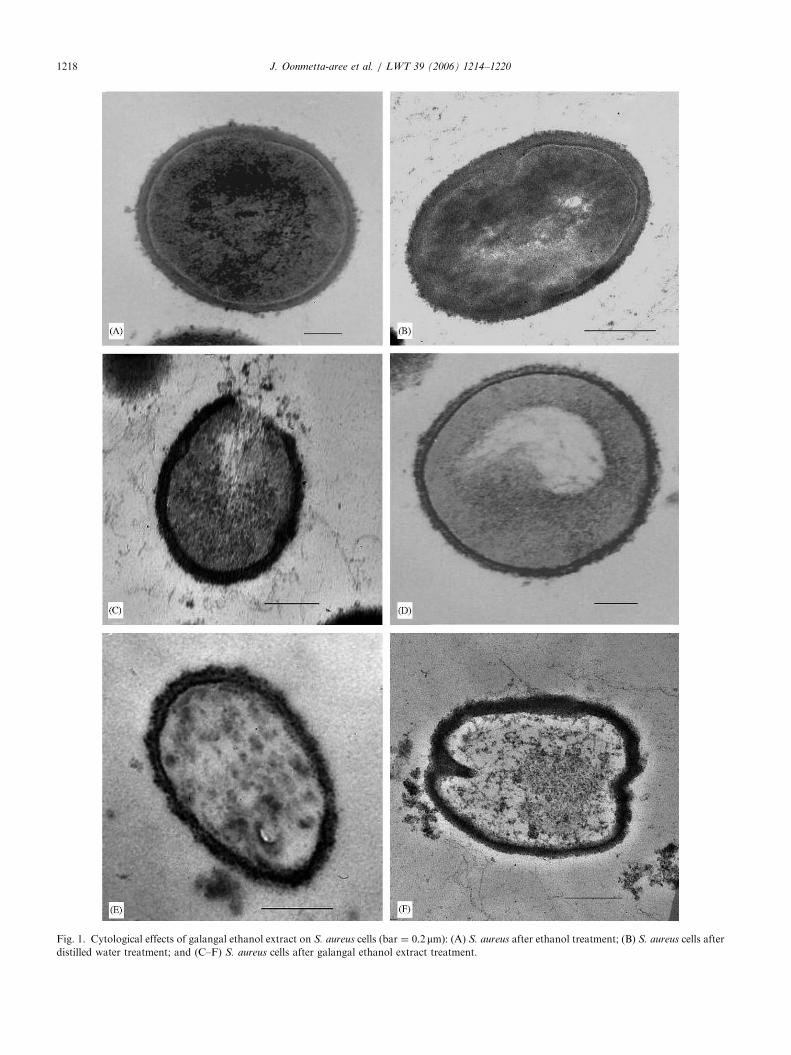
Galangal extract caused dramatic cytological mod-ifications to S. aureus cell in Figs. 1C–E compare tonormal cells (Figs. 1A and B). Galangal extract treatedS. aureus showed alteration in outer membrane’sintegrity with cell membranes and cell walls beingdisrupted and damaged resulting in a release of cellmaterials in cytoplasm (Fig. 1C). Fig. 1D showed theintegrity of the membrane and cytoplasmic material incytoplasm was coagulated and aggregated near cellmembrane. The intracellular components of cells, suchas nucleic acids, proteins, ribosomes aggregated throughthe cell (Figs. 1C, E and F). Figs. 1E and F also showedthat cell contents were coagulated. The results fromTEM showed that the galangal extract might affect thecell osmolarity, ribosomes and cell coagulation com-pounds.
ARTICLE IN PRESS
Fig. 1. Cytological effects of galangal ethanol extract on S. aureus cells (bar ¼ 0.2 mm): (A) S. aureus after ethanol treatment; (B) S. aureus cells after
distilled water treatment; and (C–F) S. aureus cells after galangal ethanol extract treatment.
J. Oonmetta-aree et al. / LWT 39 (2006) 1214–12201218

ARTICLE IN PRESSJ. Oonmetta-aree et al. / LWT 39 (2006) 1214–1220 1219
3.3.2. Effect of galangal ethanol extract on cell
membrane of S. aureus
The TEM results in Fig. 1 illustrated that the galangalextract ruptured the outer membrane of S. aureus. As aresult, there was a loss of cell permeability propertieswhich was correlated with the OD260 results. Theleakage of cytoplasmic membrane was analysed bydetermining the release of cell materials includingnucleic acid, metabolites and ions which was absorbedat 260 nm into the suspensions. Higher extract, theexposure times results in higher cell materials whichshow higher OD260. Fig. 2 shows that the OD260 ofbacterial cell materials were 1.09, 1.36 and 1.45 after S.
aureus suspensions were treated with 0.27mg/ml galan-gal extract at 2, 6, 16 h, respectively. The results fromreleasing of cellular material indicate that the higherexposure time leads to higher cell leakage that appearsto lose nucleic acid, critical molecules and ions.
Crude extract of galangal was the lipophilic com-pounds, soluble in ethanol which is a property ofessential oils. Most major and minor compounds wereidentified by GC-MS as ACA, p-coumaryl diacetate,palmitic acid, acetoxyeugenol acetate, eugenol, b-bisa-bolene, b-farnesene and sesquiphellandrene were thephenolic compounds, phenolic derivative compounds,the ester of weak acid, fatty acid, terpenes and others.Since the large number of different chemical compoundspresented in this crude extract therefore its mechanismof action can affect multiple target sites against thebacterial cells (Figs. 1C–F). b-bisabolene, b-farneseneand sesquiphellandrene are terpenes in the essential oilsof spices that mechanism of action should be similar to
0
0.2
0.4
0.6
0.8
1
1.2
1.4
1.6
1.8
2 6 16 24Exposure times (h)
Op
tica
l den
sity
at
260
nm
Galangal extractControl
Fig. 2. Measuring absorbance of the cell materials contents at 260 nm
releasing from S. aureus cells after treatment with galangal extract
(gray bars) at 2, 6, 16 and 24 h, compared to S. aureus control
suspension (white bars). The data are expressed as means7standard
deviations.
other terpenes and other phenolic compounds in thiscrude extract as indicated as involving disruption of thecytoplasmic membrane (Fig. 1C) and coagulation of cellcontents (Figs. 1D–F) (Burt, 2004; Davidson & Naidu,2000). The essential oils are hydrophobicity whichenables them to a partition in the lipids of the bacterialcell membrane, disturbing the structures and renderingthem more permeable. Besides, the essential oils are ableto induce the leakage of ions and other cell contents.The results from TEM and the loss of 260 nm absorbingmaterials suggested that the extract affected thecytoplasmic membrane of S. aureus and induced theloss of nucleic acids and ions (Burt, 2004; Carson et al.,2002; Woo et al., 2000). ACA; main compound in thiscrude extract, is ester of acetic acid which have similarmechanisms in the bacterial cells (Freese, Sheu, &Galliers, 1973). Huang, Forsberg, and Gibbins (1986)and Freese et al. (1973) have been demonstrated thatacetate acts as other organic acids, having membranegradient neutralization and denaturing of proteins insidethe cell. Antimicrobial activity of acetic acid is related topH, and the undissociated form of the acid is primarilyresponsible for antimicrobial activity. When this ester isdissolved in solution, undissociated acetic acid canpenetrate the cell membrane lipid bilayer of bacteriaand yeasts and release protons into the cytoplasmbecause the cell interior has a higher pH than theexterior (Booth, 1985; Davidson, 2001; Marquis, Clock,& Mota-Meira, 2003). Excess intracellular protons canacidify the cytoplasm and cause protein denaturationand energy loss due to activation of ATP-dependentproton pumps located in the cell membrane (Booth,1985; Marshall, Cotton, & Bal’a, 2000). The methylester is able to penetrate to the hydrophobic regions ofthe membranes and the carboxyl groups pass throughthe cell membrane, perturbing in the lowering of internalpH and denaturing of proteins inside the cell whichcoagulation of cell contents (Figs. 1E and F) (Huanget al., 1986; Marquis et al., 2003).
The galangal extract could not inhibit the growth ofE. coli (Gram-negative bacteria) because the extractcould not penetrate through the outer membrane whichwas composed of a lipopolysaccharide monolayersurrounding the cell wall that restricts diffusion ofhydrophobic compounds (Burt, 2004; Vaara, 1992).
4. Conclusions
The major constituent of galangal ethanol extract wasD,L-10-ACA, which was found in some plants in theZingiberaceae family, especially galangal. This galangalextract had the greatest inhibitory effect againstS. aureus among ginger, turmeric and krachai. SomeGram-positive bacteria and yeast were susceptible to theextract, especially Staphylococcal species. The data

ARTICLE IN PRESSJ. Oonmetta-aree et al. / LWT 39 (2006) 1214–12201220
obtained from this study could be described as theantimicrobial effect of the extract which depended onthe exposure time and the bacterial cell concentration.Galangal ethanol extract caused the internal pHchanging and denaturing of proteins inside the cell andalso disrupted the cytoplasmic membrane function of S.
aureus cells which resulted in a loss of cytoplasmicconstituents and ions. It leads to understanding the rolesof the chemical compounds in galangal extract inmicrobial physiology and gain more information ontheir actions, including information on mechanisms.This should lead to effective application of the spiceextracts as natural antimicrobial agents to controlfoodborne pathogens, food spoilage organisms in foodindustries.
Acknowledgements
This work was supported by a research Grant fromSuranaree University of Technology, Thailand.
References
Achararit, C., Panyayong, W., & Rachatagomut, E. (1983). Study of
antifungal of some Thai herbs. Research project, Mahidol Uni-
versity, Bangkok.
Booth, I. R. (1985). Regulation of cytoplasmic pH in bacteria.
Microbiological Reviews, 49(4), 329–378.
Burt, S. (2004). Essential oils: Their antibacterial properties and
potential applications in foods-a review. International Journal of
Food Microbiology, 94, 223–253.
Caichompoo, W. (1999). Antimicrobial activities of volatile oil and
curcuminoids from Curcuma longa. Master’s Thesis, Mahidol
University, Thailand.
Carson, C. F., Mee, B. J., & Riley, T. V. (2002). Mechanism of action
of Melaleuca alternifolia (Tea Tree) oil on Staphylococcus aureus
determined by time-kill, lysis, leakage, and salt tolerance assays
and electron microscopy. Antimicrobial Agents and Chemotherapy,
46, 1914–1920.
Davidson, P. M. (2001). Chemical preservatives and natural anti-
microbial compounds. In M. P. Doyle, L. R. Beuchat, & T. J.
Montville (Eds.), Food microbiology: Fundamental and frontiers,
(2nd ed) (pp. 593–627). Washington, DC: ASM Press.
Davidson, P. M., & Naidu, A. S. (2000). Phyto-phenols. In A. S.
Naidu (Ed.), Natural food antimicrobial systems (pp. 265–294).
Boca Raton, FL, USA: CRC Press.
Farnsworth, N. R., & Bunyapraphatsara, N. (1992). Thai medicinal
plants. Recommended for primary health care system. Bangkok:
Prachachon.
Freese, E., Sheu, C. W., & Galliers, E. (1973). Function of lipophilic
acids as antimicrobial food additives. Nature, 241, 321–325.
Harborne, J. B., Mabry, T. J., & Mabry, H. (1975). The flavonoids.
Great Britain: Chapman & Hall.
Huang, L., Forsberg, C. W., & Gibbins, L. N. (1986). Influence of
external pH and fermentation products on Clostridium acetobuty-
licum intracellular pH and cellular distribution of fermentation
products. Applied and Environmental Microbiology, 51, 1230–1234.
Horiuchi, Y., Onoe, T., Noguchi, M., Okumoto, K., Takemura, K.,
Fukushima, H., & Komoya, H. (2001). Single mixing fixation using
glutaraldehyde and osmium tetroxide for improved ultrastructure
of bacteria. Journal of Electron Microscopy Technology Medical
Biology, 16, 19–25.
Janssen, A. M., & Scheffer, J. J. C. (1985). Acetoxychavicol acetate, an
antifungal component of Alpinia galanga. Planta Medica, 6,
507–511.
Jay, J. M. (2000). Modern food microbiology (6th ed). Maryland: Asean
Publishers.
Lennette, T. H., Barilows, A., Hausler, W. J., Jr., & Shadony, H. J.
(1991). Manual of clinical microbiology (5th ed). Washington, DC:
American Society for Microbiology.
Lorian, V. (1995). Antibiotics in laboratory medicine. In J. F. Acar, &
F. W. Goldstein (Eds.), Disk susceptibility test, (4th ed) (p. 1).
London: Williams & Walkins Awaverly.
Maillard, J. Y. (2002). Bacterial target sites for biocide action. Journal
of Applied Microbiology Symposium Supplement, 92, 16S–27S.
Marquis, R. E., Clock, S. A., & Mota-Meira, M. (2003). Fluoride and
organic weak acids as modulators of microbial physiology. FEMS
Microbiology Reviews, 26, 493–510.
Marshall, D. L., Cotton, L. N., & Bal’a, F. A. (2000). Acetic acid. In
A. S. Naidu (Ed.), Natural food antimicrobial systems (pp.
661–688). Boca Raton, FL, USA: CRC Press.
Mitsui, S., Kobayashi, S., Nagahori, H., & Ogiso, A. (1976).
Constituents from seeds of Alpinia galanga wild and their
anti-ulcer activities. Chemical & Pharmaceutical Bulletin, 24,
2377–2382.
Murakami, A., Toyota, K., Ohura, S., Koshimizu, K., & Ohigashi, H.
(2000). Structure–activity relationships of (10S)-10-Acetoxychavicol
acetate, a major constituent of a southeast Asian condiment plant
Languas galangal, on the inhibition of tumor-promoter-induced
Epstein–Barr virus activation. Journal of Agricultural and Food
Chemistry, 48, 1518–1523.
Noro, T., Seikiya, T., Katoh, M., Oda, Y., Miyase, T., Kuroyanagi,
M., Ueno, A., & Fukushima, S. (1988). Inhibitors of xanthine
oxidase from Alpinia galanga. Chemical & Pharmaceutical Bulletin,
36, 244–248.
Prasit, P., Chalermpol, K., Prasat, K., & Kamolchanok, R. (2002). 10-
Acetoxychavicol acetate for tuberculosis treatment. United States
Patent Application: 0020192262.
Vaara, M. (1992). Agents that increase the permeability of the outer
membrane. Microbiological Reviews, 56, 395–411.
Vanderzant, C., & Splittstoesser, D. F. (1992). Staphylococcal
enterotoxins. In R. W. Bennett, S. Notermans, & S. R. Tatini
(Eds.), Compendium of methods for the microbiological examination
of foods, (3rd ed) (pp. 5–51). American Public Health Association,
WA: Print-Edwards Brothers.
Woo, I.-S., Rhee, I.-K., & Park, H.-D. (2000). Differential damage in
bacterial cells by microwave radiation on the basis of cell wall
structure. Applied and Environmental Microbiology, 66,
2243–2247.
Yang, X., & Eilerman, R. G. (1999). Pungent principle of Alpinia
galanga (L.) Swartz and its applications. Journal of Agricultural and
Food Chemistry, 47, 1657–1662.