Hydrochromic Conjugated Polymers for Human Sweat Pore Mapping

BRIEF REPORTS
CHANGE IN ROTAVIRUS EPIDEMIOLOGY INNORTHEAST FLORIDA AFTER THE INTRODUCTION
OF ROTAVIRUS VACCINE
Haidee Custodio, MD,* Carmen Masnita-Iusan, MS,†Peter Wludyka, PhD,† and Mobeen H. Rathore, MD, CPE*‡
Abstract: Retrospective analysis done at a children’s hospital showedsignificant decrease in infections and hospitalizations caused by rotavirusin northeast Florida after the introduction of rotavirus vaccines in 2006.The rotavirus season was delayed in onset by 8 months and durationprolonged by 2–3 months in 2008, and no definite season occurred in 2009.
Key Words: gastroenteritis, diarrhea, rotavirus vaccine, vaccine effect,Florida
Accepted for publication March 1, 2010.From the *Division of Pediatric Infectious Diseases and Immunology, Depart-
ment of Pediatrics, University of Florida, Jacksonville, FL; and †Depart-ment of Mathematics and Statistics, and ‡Wolfson Children’s Hospital,University of North Florida, Jacksonville, FL.
Mobeen Rathore, MD, is on the speakers’ bureau and advisory boards ofMerck and GlaxoSmithKline.
Address for correspondence: Mobeen H. Rathore, MD, CPE, Departmentof Pediatrics, 653-1 West 8th Street, L-13, 3rd Floor/LRC, Jackson-ville, FL 32209. E-mail: [email protected].
Supplemental digital content is available for this article. Direct URLcitations appear in the printed text and are provided in the HTML andPDF versions of this article on the journal’s Web site (www.pidj.com).
Copyright © 2010 by Lippincott Williams & WilkinsDOI: 10.1097/INF.0b013e3181dbf256
The number of deaths attributable to rotavirus in the UnitedStates is low; however, rotavirus infections still cause signifi-
cant morbidity, resulting in hospitalizations and outpatient vis-its.1,2 The annual rotavirus season in the United States typicallyoccurs in the cooler months, starting in the fall in the southwestand ending in the northeast by spring.3
In the United States, 2 rotavirus vaccines, RotaTeq (Merckand Company, Whitehouse Station, NJ) approved in February2006 and Rotarix (GlaxoSmithKline, Research Triangle Park, NC)approved in April 2008, are recommended for use by the AdvisoryCommittee on Immunization Practices.4 These 2 vaccines wereexpected to significantly decrease the number and severity ofrotavirus infections in the United States. We conducted a retro-spective analysis to examine the epidemiology of rotavirus infec-tions and hospitalizations in northeast Florida before and after theintroduction of rotavirus vaccines.
METHODSWe reviewed records at the Wolfson Children’s Hospital
(WCH), the only regional tertiary care center serving children innortheast Florida. WCH has 192 beds, 8000–9000 annual admis-sions, and a catchment area of 3 million. The study period wasbetween January 1, 2004, and December 31, 2009, and includedpatients aged �18 years who were tested for and those who werehospitalized because of rotavirus infection. Prevaccine period wasdefined as 2004–2006, and vaccine period as 2007–2009. Thestudy was approved by the institutional review board.
Rotavirus vaccine uptake was indirectly estimated by deter-mining the number of vaccine doses distributed in northeastFlorida. The number of doses distributed in the public sector wasobtained from the Bureau of Immunization, Florida Department ofHealth, and in the private sector from the 2 vaccine manufacturers.Data of live births for the study period were obtained from theFlorida state census online.5
Laboratory records were reviewed to identify patients inwhom rotavirus tests were ordered on a stool specimen. Rotavirustests were done only on bulk stool specimens. It was assumed thatclinicians ordered rotavirus tests only on patients who had diar-rhea, and patients were assumed to have rotavirus infection if thetest result was positive.
Hospitalization because of rotavirus infection was used as asurrogate for the severity of the infection. The number of hospi-talizations because of rotavirus was determined and compared withthe overall number of hospitalized rotavirus-infected patients andalso with all annual hospitalizations at WCH from 2004 to 2009.Children admitted secondary to rotavirus infection were identifiedusing International Classification of Diseases, Ninth Revision,Clinical Modification code 008.61 used only as the primary orsecondary discharge diagnosis.
We determined the seasonal pattern (onset, peak, andend) of rotavirus infections in northeast Florida during theprevaccine period (2004 –2006) and vaccine (2007–2009) peri-ods. Season onset was defined as �10% positive tests for atleast 2 consecutive months, and season end as �10% positivetests for at least 2 consecutive months. Season peak was definedas the month(s) with the highest percentage of positive tests.
Statistical analyses were done comparing prevaccine period(2004–2006) with vaccine period (2007–2009) for rotavirus infec-tions and hospitalizations using SAS 9.1 software (SAS Institute,Cary, NC). Categorical comparisons were done using �2 tests.Means were compared using 2-sample t tests.
RESULTSShown in Table 1 are live births, rotavirus tests done,
rotavirus tests positive, total hospitalization, rotavirus hospitaliza-tions, and vaccine doses distributed for each of the 6 study years.Monthly distribution of rotavirus tests ordered and positive resultsin the study period are depicted in Figure, Supplemental DigitalContent 1, http://links.lww.com/INF/A440.
The number of tests performed decreased by 12.1% from anaverage of 758 in the prevaccine period to an average of 666 in thevaccine period (P � 0.28, t test statistic). There was a 57.8%decline in the absolute number of positive rotavirus tests from anannual average of 207 in the prevaccine period to 87.3 in thevaccine period (P � 0.03, t test statistic). The proportion ofpositive tests decreased significantly from 27.3% in the prevaccineperiod to 13.11% in the vaccine period (P � 0.0001, �2). Apositive test was 2.08 times more likely in the prevaccine periodcompared with the vaccine period.
There was a decrease in rotavirus hospitalizations relative tototal hospitalizations in the vaccine period (0.20%) compared withthe prevaccine period (0.71%; P � 0.0001, �2). Among those whohad rotavirus infections, the absolute number of hospitalizationsdecreased by 72% from an annual average 63.3 in the prevaccineperiod to 18 in the vaccine period (P � 0.01, t test statistic). Theproportion of patients hospitalized because of rotavirus infectiondecreased significantly from 30.6% in the prevaccine period to20.6% in the vaccine period (P � 0.0024, �2).
The rotavirus season in northeast Florida started in Januaryand ended in May or June, with a peak in March or April for theprevaccine years and 2007. However, in 2008, the season started inSeptember, peaked in October, and ended in April 2009 (Fig.,Supplemental Digital Content 1, http://links.lww.com/INF/A440).No defined rotavirus season was appreciated in 2009.
DISCUSSIONWe observed a decrease in infections and hospitalizations
because of rotavirus and a change in the rotavirus season after the
The Pediatric Infectious Disease Journal • Volume 29, Number 8, August 2010766 | www.pidj.com

introduction of rotavirus vaccines in 2006. This is similar to thepreviously reported data.6,7 However, the change in the seasonalpattern of rotavirus infection in northeast Florida after the intro-duction of the vaccine was different from what has recently beenreported from other parts of the United States.6
As has been reported for the southern US,6 our studyshowed that both the total number of rotavirus tests performed andreported positive decreased. We also observed that during thevaccine period there was a decrease in the proportion of positivetests relative to total rotavirus tests performed.
Our study also showed a decrease in the severity of rotavi-rus infections as reflected by the decline in hospitalizations. This issimilar to what has been reported in a postlicensure evaluation ofthe effectiveness of the rotavirus vaccine.7
The introduction of rotavirus vaccines not only decreasedthe number of infections and hospitalizations but also changed theannual seasonality of rotavirus infection. National reports showedthat the onset of 2007–2008 rotavirus season was delayed by 11weeks but the duration was shorter than 2000–2006 season,whereas 2008–2009 season had an earlier season onset but longerduration compared with 2007–2008 season.6 According to ourdata, the rotavirus season onset in northeast Florida during theprevaccine years and 2007 was in January, with the peak occurringin March or April and the season ending in May or June. Theduration of rotavirus season in our region during the prevaccineyears and 2007 was 5–6 months. However, in 2008, the durationof the season was 8 months, starting in September, peaking inOctober, and ending in April 2009. Compared with the prevaccineperiod, the onset of rotavirus season in 2008 was delayed by 8months, the peak occurred sooner (within 1 month of seasononset), and the duration was 2–3 months longer. No rotavirusseason was observed in 2009. Secular variability has been reportedafter introduction of rotavirus vaccine.8 Prolonged follow-up ofseason patterns will clarify this issue. We continue to monitorrotavirus infections and hospitalizations in northeast Florida.
There are several limitations to our study. This is a retro-spective analysis subject to numerous confounders and biases.Although we believe that there were no changes in admission ortesting policies at our institution, changes in clinical practice overthe years may have some influences on the changes we observed.Some patients with acute gastroenteritis might not have been testedfor rotavirus infection and some may have been tested at outsidelaboratories, leading to a lower detection rate. Individual patientinformation was not available before 2008, which may haveresulted in counting some positive rotavirus results more thanonce. However, this was not very likely because we did notobserve any duplication of results in 2008 and 2009 when patient-level information became available. Additionally, vaccine uptakein our study was assessed indirectly using vaccine distribution,which may not accurately represent actual vaccine use. However,vaccine uptake has been steadily increasing, with 40%–64% first-
dose coverage reported for children aged 3 months.9 In addition,use of International Classification of Diseases, Ninth Revision,Clinical Modification codes often underestimate incidence of ro-tavirus-related admissions.10 However, the difference in infectionsand hospitalizations because of rotavirus between the prevaccineand vaccine periods is significant and seems to be temporallyrelated to rotavirus vaccine availability.
In conclusion, our study showed a decrease in infections andhospitalizations because of rotavirus infection temporally associ-ated with the introduction of rotavirus vaccine. In addition, theseason seems to be evolving from delayed onset and prolongedduration in 2008 to no appreciable “season” in 2009. Continuedsurveillance over the next several years will be critical to deter-mine whether the decrease in rotavirus infection and the seasonaldifferences in northeast Florida are sustained over time.
ACKNOWLEDGMENTSThe authors acknowledge the contributions of Page McKitrick,
Diane Halstead, PhD, and Nizar Maraqa, MD.
REFERENCES1. Kilgore PE, Holman RC, Clarke MJ, et al. Trends of diarrheal disease-
associated mortality in U.S. children, 1968 through 1991. JAMA. 1995;274:1143–1148.
2. Charles MD, Holman RC, Curns AT, et al. Hospitalizations associated withrotavirus gastroenteritis in the United States, 1993–2002. Pediatr Infect Dis J.2006;25:489–493.
3. Torok TJ, Kilgore PE, Clark MJ, et al. Visualizing geographic and temporaltrends in rotavirus activity in the United States, 1991 to 1996. Pediatr InfectDis J. 1997;16:941–946.
4. Cortese MM, Parashar UD. Prevention of rotavirus gastroenteritis amonginfants and children: recommendations of the Advisory Committee onImmunization Practices (ACIP). MMWR Recomm Rep. 2009;58:1–25.
5. Florida vital statistics annual reports �Florida Department of Health Website�. Available at: http://www.flpublichealth.com/VSBOOK/VSBOOK.aspx. Accessed January 15, 2010.
6. National Respiratory and Enteric Virus Surveillance System, Panozzo CA,Tate JE, et al. Reduction in rotavirus after vaccine introduction—UnitedStates, 2000—2009. MMWR Recomm Rep. 2009;58:1146–1149.
7. Boom JA, Tate JE, Sahni LC, et al. Effectiveness of pentavalent rotavirusvaccine in a large urban population in the United States. Pediatrics.2010;125:e199–e207.
8. Payne DC, Szilagyi PG, Staat MA, et al. Secular variation in United Statesrotavirus disease rates and serotypes. Pediatr Infect Dis J. 2009;28:948–953.
9. Clayton H, Cortese M, Payne D, et al. Uptake of rotavirus vaccine(RotaTeq®) among infants in immunization registries and adherence to agerecommendations for vaccine administration �CDC NIP/NIC Web site�.Available at: http://cdc.confex.com/cdc/nic2008/webprogram/Paper15710.html. Accessed October 6, 2009.
10. Chang HH, Glass RI, Smith PF, et al. Disease burden and risk factors forhospitalizations associated with rotavirus infection among children in NewYork State, 1989 through 2000. Pediatr Infect Dis J. 2003;22:808–814.
TABLE 1. Live Births, Rotavirus Tests, Hospitalizations, and Vaccine Doses by Year
Year Live Births Rotavirus Tests Positive RotavirusTests (%)
TotalHospitalizations
RotavirusHospitalizations
Vaccine DosesDistributed
2004 17,453 707 223 (31.5) 8757 70 N/A2005 18,154 760 200 (26.3) 8983 58 N/A2006 19,024 807 198 (24.5) 8915 62 8,1382007 19,268 795 159 (20) 8567 38 34,1352008 18,720 646 67 (10.4) 8279 9 37,9102009 N/A* 557 36 (6.4) 8595 7 42,220
*2009 Live birth information will not be available until January 2011.
The Pediatric Infectious Disease Journal • Volume 29, Number 8, August 2010 Rotavirus Epidemiology
© 2010 Lippincott Williams & Wilkins www.pidj.com | 767
ANTIBODY PERSISTENCE 12 MONTHS AFTER ABOOSTER DOSE OF MENINGOCOCCAL-CCONJUGATED VACCINE IN THE SECOND
YEAR OF LIFE
Javier Diez-Domingo, MD, PhD,*†M. Victoria Planelles-Cantarino, MD,† Jose M. Baldo-Torrenti, MD,†Isabel Ubeda-Sansano, MD, PhD,† Angels Jubert-Rosich, MD,†Joan Puig-Barbera, MD, PhD,*and M. Victoria Gutierrez-Gimeno, PhD*
Abstract: We report on the results of the 12-month follow-up of childrenaged 14 to 18 months who received primary and booster vaccinations witheither a meningococcal-C vaccine conjugated to tetanus toxoid or CRM197.
Seroprotection (92.8%) and geometric mean titers/serum bactericidalactivity (410.5; 95% CI: 273.4–616.3) were higher in children receivingthe meningococcal serogroup C tetanus toxoid conjugate, compared with61.5% and serum bactericidal antibody geometric mean titer of 45.1 (95%CI: 28.5–71.3) when MenC-CRM197 conjugate was used.
Key Words: Neisseria meningitidis, serotype C, immunization,secondary, meningococcal vaccinesAccepted for publication February 17, 2010.From the *Centro Superior de Investigacion en Salud Publica (CSISP), Valen-
cia, Spain; and †Vaccine Institute of Valencia (VIVA), Valencia, Spain.Supported by Direccion General de Salud Publica, Valencia, Spain. EudraCT
2006–003525–82, and Clinicaltrials.Gov identifier NCT00392808.Address for correspondence: Javier Diez-Domingo, MD, PhD, Centro Supe-
rior de Investigacion en Salud Publica (CSISP), Valencia, Avda Catalunya20, 46021 Valencia, Spain. E-mail: [email protected].
Supplemental digital content is available for this article. Direct URLcitations appear in the printed text and are provided in the HTML andPDF versions of this article on the journal’s Web site (www.pidj.com).
DOI: 10.1097/INF.0b013e3181d9e653
Waning protection after immunization with serogroup C me-ningococcal vaccines in infancy and early childhood has
been associated with a fall in serum bactericidal antibody concen-trations. Despite the immunologic memory induced by these vac-cines, rapid progression of meningococcal disease would not allowthe organism to mount an anamnestic immune response.1,2 Pro-tection correlates with the circulating antibody concentration. Abooster dose, introduced in the second year of life, induces asubstantial rise in bactericidal antibody titers.3,4
Seroepidemiologic studies have shown that 3–6 years aftera single dose of conjugated meningococcal serogroup C (MenC)vaccine, the percentage of subjects protected with serum bacteri-cidal antibody (SBA) titers �1:8 decreases with the age when thevaccine was given.5,6
The kinetics of the seroprotection after a booster dose is notwell known. There are no long-term studies to assess the persistenceof antibodies. In 1 clinical trial,3 the SBA geometric mean titers(GMTs) decreased by more than 97% in the first 18 months after thebooster dose. A higher rate of persisting protective SBA-MenC titerswas observed in those subjects who had higher SBA-MenC GMTsafter booster vaccination.3
We have recently described the immune response to a boosterdose in children primed and boosted with 2 MenC vaccines.4 Theantibody titers reached were strongly depended on the vaccine usedfor priming; GMTs were 3.5 times higher if the vaccine used wasconjugated with tetanus toxoid (MenC-TT) compared with CRM197
(MenC-CRM) conjugated vaccine formulation. Although the MenCvaccine used for priming has been described to affect the response toHaemophilus influenzae type b (Hib) vaccines given concomintantly,7 wedid not find any difference after boosting with either MenC vaccine.4
The objective of this study was to assess the persistence ofthe seroprotection against MenC and Hib 1 year after the boosterdose and to analyze the factors that might influence seroprotection.
METHODSThis study was designed as a multicenter, randomized,
open-label, clinical trial, described in a previous publication.4
Children aged 14 to 18 months who completed the primaryvaccination series before their 8th month with either MenC-TT orMenC-CRM were randomized to receive one of these vaccines asa booster. The exclusion criteria were having an acute or severechronic disease or hypersensitivity to any of the vaccine compo-nents including antibiotics, a history of invasive disease and beingtreated with any immunosuppressant drug.
The Ethics Committee of DGSP/CSISP, Valencia, approvedthe study, and informed written consent was signed by the parentsor guardians before entering in the study.
A blood sample was collected before the booster dose, and1 month and 12 months (�3 weeks) after the booster. The resultsof this last sample are described here.Serologic Studies. Sera were analyzed at the Manchester VaccineEvaluation Department of the Health Protection Agency, UK.Functional MenC antibody titers were determined using the SBAassay. The SBA target strain was MENC11 (C:16:P1.7–1,1), andthe complement source was baby rabbit sera (Pel-Freeze Incorpo-rated, Rodgerson, AZ). SBA titers are expressed as the reciprocalof the final serum dilution equivalent to 50% killing at 60 minutes.Hib specific antibodies (IgG) were quantified using standardizedELISA at the Immunoassay Laboratory, Health ProtectionAgency, Porton Down, UK. The standard sera used were theInternational anti-Hib Quality Control Serum Center for Biologicsand Evaluation Research (CBER) 1983.
Antibody titers were log transformed; geometric mean titers(GMT) and concentrations (GMC) with 95% confidence intervalswere calculated. Mann-Whitney U test was used to evaluate signifi-cant differences between antibody assays at each time point. Childrenwere considered seroprotected when MenC SBA titers were �1:8,and anti-Hib antibodies were �0.15 �g/mL.
RESULTSOf the 389 subjects randomized in the clinical trial, 334
(85.9%) had blood drawn 12 months after the booster dose. Onemonth after the booster dose all but 2 children had reached seropro-tection titers (Table 1). One year later, 61.5% of subjects primed withMenC-CRM who received a booster with the same vaccine retainedseroprotective values, whereas 92.8% of subjects primed and boostedwith MenC-TT retained seroprotective titers. SBA titers of �1:128were measured in 49.5% and 91.3% of subjects, respectively. Theseroprotection rate was dependent not only on which vaccine wasused for priming (MenC-CRM versus MenC-TT), but also on whichvaccine was used for the booster dose. Sixty-nine percent of thechildren primed with MenC-CRM had SBA titers �1:8, comparedwith 87.2% of those primed with MenC-TT (P � 0.01), with GMTsof 60.0 (95% CI: 43.8–82.2) and 283.8 (95% CI: 204.3–394.3),respectively.
In children initially primed with MenC-CRM, 61.5% of thosewho received a booster with the same vaccine, and 75.5% of thoseboosted with MenC-TT had seroprotective SBA titers (P � 0.05) 12months after the booster dose. Among children who were primed withMenC-TT, 92.8% of those boosted with the same vaccine, and 81.9%of those boosted with MenC-CRM (P � 0.055) were still seropro-tected 12 months after the vaccine was administered.
The SBA GMTs among groups also varied depending onwhich vaccine was used for booster. Children primed with MenC-
Diez-Domingo et al The Pediatric Infectious Disease Journal • Volume 29, Number 8, August 2010
© 2010 Lippincott Williams & Wilkins768 | www.pidj.com

CRM had SBA GMTs of 45.1 (95% CI 28.5–71.3) if boosted withthe same vaccine and 77.4 (95% CI: 50. 1–119.7) if boosted withMenC-TT (P � 0.066). Among children primed with MenC-TT,the SBA GMTs were 199.3 (95% CI: 119.8–331.5) if boosted withMenC-CRM compared with 410.5 (95% CI: 273.4–616.3) ifboosted with MenC-TT (P � 0.05) (Fig., Supplemental DigitalContent 1, http://links.lww.com/INF/A426).
Three of the 283 subjects from whom a blood sample wasdrawn for Hib antibodies did not maintain anti-Hib concentra-tions �0.15, which is considered a correlation of protection, 12months post booster. One such case occurred in 3 of the 4treatment groups. There was no difference in anti Hib GMCbetween children initially vaccinated with MenC-CRM (3.4�g/mL; 95% CI: 2.8 – 4.1) versus those primed with MenC-TT(3.6 �g/mL; 95% CI: 2.9 – 4.4), nor was the anti-Hib concen-tration dependent of which vaccine was used for booster (Table,Supplemental Digital Content 2, http://links.lww.com/INF/A427).
DISCUSSIONIn the 12 months after administration of a Men C booster
vaccine to toddlers (aged 14–18 months), a rapid decrease in SBAtiters was observed. In blood samples taken 12 months (�3 weeks)after the booster vaccination, 1 in 4 children were not seropro-tected as measured by SBA.
A booster vaccination was recommended to children whohad been primed during their first year of life, as it had previouslybeen shown that immunogenicity decreased in these children aftertheir first birthday.1,2 It was estimated that a single booster dosewould protect children for a longer period and that additionalvaccinations would not be required.
In a previous study we showed the immunologic effect 4weeks after the booster dose,4 in the current article we describe thefollow-up of these children. One month after the booster vaccine,most of the subjects had seroconverted to SBA �1:128 and hadattained high SBA titers, with differences depending on whichvaccine had been used for the primary vaccination series; subjectswho had been primed with the MenC-TT vaccine attained SBAtiters 3.5 times higher than those primed with the MenC-CRMvaccine.4 One year later, the SBA titers were shown to have
decreased substantially, although the SBA GMTs remained higherthan before the booster vaccination and a large percentage ofsubjects retained seroprotective levels. A decrease in the percent-age of children with SBA �1:8 might reflect a decrease in thenumber of children protected with advancing age.
At 12 months postbooster vaccination, seroprotection ratesand SBA titers were dependent on which vaccine had been usedfor the primary and booster vaccinations. The highest percentageof subjects was seroprotected when MenC-TT vaccines had beenused for all (prime and booster) vaccinations.
The decrease in seroprotection has also been described inanother study in which the investigators used a combination vaccine(Hib-MenC3) and found that SBA titers fell up to 95% in the first 18months after administration of a booster dose given in the second yearof life. Also, although 91% to 100% of the children who received asingle vaccine dose in their second year of life8 became seroprotectedwith an SBA titer of �1:8, at 4 years of age the seroprotection ratesin these children were low.9 These findings seem to indicate thatseroprotection decreases over time after a single dose of MenCvaccine in preschool-aged (1–2-year-old) children.
Findings from this study may influence future MenC vaccina-tion policies. If SBA titers and seroprotection rates decrease in thisway, a large proportion of young children could be at risk fordeveloping MenC infection. However, if herd immunity is estab-lished, this will not translate into increased infection rates. Most of theadolescents in Europe, in countries where a catch-up program wasestablished, are still seroprotected6; however, as children vaccinatedin infancy, with or without a booster dose, reach adolescence, apossible resurgence of MenC circulation among this group is possible,as the protective herd immunity disappears. A booster dose duringearly adolescence could be required6; however, this was not predicted ina mathematical model indicating that high levels of indirect protectionagainst meningococcal group C diseases are likely to persist even if thevaccine only provides 3 years of protection against carriage.10
ACKNOWLEDGMENTSThe authors thank all the participants in the clinical trial
and the investigators collecting data.
TABLE 1. Seroprotection and SBA GMTs in the 4 Study Groups Depending on the VaccineUsed for Priming and Boosting
Booster Vaccine
Priming Vaccination
MenC-CRM MenC-TT
MenC-CRM MenC-TT MenC-CRM MenC-TT
SBA �1281 month post
N 100 107 86 81n (%) 100 (100) 106 (99.1) 86 (100) 80 (98.8)
12 months postN 91 102 72 69n (%) 45 (49.5) 60 (58.8) 55 (76.4) 63 (91.3)
SBA �81 month post
N 100 107 86 81n (%) 100 (100) 106 (99.1) 86 (100) 80 (98.8)
12 months postN 91 102 72 69n (%) 56 (61.5) 77 (75.5) 59 (81.9) 64 (92.8)
SBA GMT (95% CI)Prebooster 8.6 (6.1–11.9) 13.2 (8.9–19.5) 7.6 (5.1–11.2) 9.7 (6.4–14.9)1 month post 1746 (1378–2213) 2061 (1599–2627) 6278 (4841–8144) 6786 (5023–9167)12 months post 45.1 (28.5–71.3) 77.4 (50.1–119.7) 199.3 (119.8–331.5) 410.5 (273.4–616.3)
The Pediatric Infectious Disease Journal • Volume 29, Number 8, August 2010 Meningococcal Vaccine Booster Dose
© 2010 Lippincott Williams & Wilkins www.pidj.com | 769

REFERENCES1. Trotter CL, Andrews NJ, Kaczmarski EB, et al. Effectiveness of meningo-
coccal serogroup C conjugate vaccine 4 years after introduction. Lancet.2004;364:365–367.
2. Larrauri A, Cano R, Garcia M, et al. Impact and effectiveness of menin-gococcal C conjugate vaccine following its introduction in Spain. Vaccine.2005;23:4097–4100.
3. Tejedor JC, Moro M, Merino JM, et al. Immunogenicity and reactogenicityof a booster dose of a novel combined Haemophilus influenzae typeb-Neisseria meningitidis serogroup C-tetanus toxoid conjugate vaccinegiven to toddlers of 13–14 months of age with antibody persistence up to 31months of age. Pediatr Infect Dis J. 2008;27:579–588.
4. Diez Domingo J, Planelles Cantarino V, Baldo Torrentí J, et al. A random-ized, multicentre, open-label clinical trial to assess the immunogenicity ofa meningococcal C vaccine booster dose administered to children aged 14to 18 months, as well as the importance of priming. Pediatr Infect Dis J.2010;29:148–152.
5. de Voer RM, van der Klis FR, Engels CW, et al. Kinetics of antibodyresponses after primary immunization with meningococcal serogroup Cconjugate vaccine or secondary immunization with either conjugate orpolysaccharide vaccine in adults. Vaccine. 2009.
6. Snape MD, Kelly DF, Lewis S, et al. Seroprotection against serogroup Cmeningococcal disease in adolescents in the United Kingdom: observa-tional study. BMJ. 2008;336:1487–1491.
7. Southern J, Crowley-Luke A, Borrow R, et al. Immunogenicity of one, twoor three doses of a meningococcal C conjugate vaccine conjugated totetanus toxoid, given as a three-dose primary vaccination course in UKinfants at 2, 3 and 4 months of age with acellular pertussis-containingDTP/Hib vaccine. Vaccine. 2006;24:215–219.
8. Richmond P, Borrow R, Goldblatt D, et al. Ability of 3 different menin-gococcal C conjugate vaccines to induce immunologic memory after asingle dose in UK toddlers. J Infect Dis. 2001;183:160–163.
9. Snape MD, Kelly DF, Green B, et al. Lack of serum bactericidal activity inpreschool children two years after a single dose of serogroup C meningo-coccal polysaccharide-protein conjugate vaccine. Pediatr Infect Dis J.2005;24:128–131.
10. Trotter CL, Gay NJ, Edmunds WJ. Dynamic models of meningococcalcarriage, disease, and the impact of serogroup C conjugate vaccination.Am J Epidemiol. 2005;162:89–100.
CONCOMITANT RESPIRATORY VIRAL INFECTIONSIN CHILDREN WITH KAWASAKI DISEASE
Alejandro Jordan-Villegas, MD, Michael L. Chang, MD,Octavio Ramilo, MD, and Asuncion Mejías, MD
Abstract: The role of respiratory viruses in the pathogenesis of Kawasakidisease (KD) remains controversial. In this study, we showed that 8.8% ofpatients with KD had documented respiratory viral infections. Patients withconcomitant viral infections had a higher frequency of coronary arterydilatations and were significantly more often diagnosed with incompleteKD. The presence of a concomitant viral infection should not exclude thediagnosis of KD.
Key Words: Kawasaki disease, incomplete, respiratory virusesAccepted for publication March 1, 2010.From the Division of Pediatric Infectious Diseases, University of Texas South-
western Medical Center and Children’s Medical Center Dallas, Dallas, TX.Address for correspondence: Asuncion Mejias, MD, The Research Institute
at Nationwide Children’s Hospital, The Ohio State University Collegeof Medicine, 700 Children’s Drive, WA 4022, Columbus, OH 43205.E-mail: [email protected].
DOI: 10.1097/INF.0b013e3181dba70b
Kawasaki disease (KD) is an acute self-limited multisystemvasculitis that represents the most common cause of acquired
heart disease in children in the developed world.1,2 Although theepidemiology and clinical features of KD suggest an infectious
origin, and many viral infections share similar clinical character-istics, its etiology remains unknown.3–5
The diagnosis of KD requires the presence of fever for 5days, 4 of 5 characteristic clinical features, and the lack of anotherdiagnosis that could explain the findings.6,7 In this context, theidentification of a microbial pathogen in children with suspectedKD could delay diagnosis and treatment.8 The present study wasdesigned (1) to determine the frequency of concomitant respiratoryviral infections in children with KD, and (2) to compare theclinical features and outcomes of KD patients with and withoutdocumented viral respiratory infections.
METHODSMedical records from patients hospitalized at Children’s
Medical Center Dallas with the diagnosis of KD from January 1,1999, through December 31, 2008, were identified by InternationalClassification of Diseases-9 codes (446.1). Records were reviewedto determine which patients with KD had been tested for respira-tory viruses. Subsequently, KD patients were classified as casesand controls according to the results of their virology tests. Caseswere defined as patients with KD in whom a viral respiratorypathogen was identified, and controls were those KD patients whotested negative for those viruses. Each case was matched with 2controls for age, sex, and date of hospitalization (�3 weeks). Viraltesting was performed at the discretion of the attending physicianin all patients by direct fluorescent-antibody (DFA) assay for 7respiratory viruses (respiratory syncytial virus �RSV�, parainflu-enza virus �PIV� types 1, 2, and 3, influenza virus A and B, andadenovirus). If the DFA showed negative results, a viral culturewas automatically performed. Patients’ demographic characteris-tics, clinical presentation, course, response to treatment with in-travenous immunoglobulin (IVIG), laboratory results, and echo-cardiographic findings were compared between the groups.Refractory disease was defined by the presence of persistent orrecrudescent fever 36 hours after completion of IVIG infusion.6,9
Incomplete KD was defined by fever for at least 5 days, and lessthan 4 of classic KD clinical criteria at presentation documented bythe attending physician, and before any echocardiographic studieswere performed.6 The study was approved by the InstitutionalReview Board of the University of Texas Southwestern MedicalCenter at Dallas (IRB #052009-004).
Descriptive analyses were performed using frequency dis-tributions and rates. Means (�SD) or medians (percentiles 25th to75th) were used to summarize patient’s demographic and baselinecharacteristics. Groups were compared using Student t test orMann-Whitney U test for continuous variables. �2 tests with Yatescorrection for continuity or �-test were used for associationsbetween categorical variables and proportions. A 2-tailed P value�0.05 was considered significant. Sigma Stat 2003 software(SPSS Science, San Rafael, CA) was used for analyses.
RESULTSDuring the study period, 394 patients were hospitalized at
Children’s Medical Center Dallas with KD. Viral testing wasperformed in 251 (63.7%) patients. The proportion of patientstested for respiratory viruses significantly increased from 24% in1999 to 84% in 2008 (P � 0.0001). From 1999 to 2001, 24%–43%of KD patients were tested for respiratory viruses, all with negativeresults. Thereafter, the percentage of KD patients tested increasedas well as the percentage of positive viral results ranging from6.6% in 2006 to 16.6% in 2004 (overall rate of detection 11.4% peryear). Twenty-two (8.8%) patients had a respiratory virus identi-fied, including rhinovirus (n � 6), adenovirus (n � 6), influenza Aor B (n � 5), PIV 1–3 (n � 3), and RSV (n � 2). Although RSV
Jordan-Villegas et al The Pediatric Infectious Disease Journal • Volume 29, Number 8, August 2010
© 2010 Lippincott Williams & Wilkins770 | www.pidj.com

(2 of 2) and influenza (4 of 5) were mostly identified by DFA, allrhinovirus isolates (6 of 6) and the majority of PIV (2 of 3) andadenoviruses (4 of 6) were negative by DFA and diagnosed byviral culture.
There were no significant differences in age, sex, and raceor ethnicity between KD patients with viral infections (cases) andthose who tested negative for viruses (controls). The median(75%–25% percentiles) age of cases and controls were 3.4 (1.2–4.5) versus 2.7 (1.2–5.7) years, respectively (P � 0.76). Sixtypercent (13 of 22) of cases and 68% (30 of 44) of controls weremale, and white (63% of cases vs. 52% of controls) followed byHispanic (23% in each group), black (5% vs. 16%), and other races(9% in each group) were the most common cases and controls’race/ethnic groups.
The clinical criteria, clinical course, and outcomes of casesand controls are summarized in Table 1. Except for emesis, whichwas documented more frequently in KD patients with viral infec-tions; the rest of factors evaluated, including fever for at least 5days, the classic clinical features of KD,6 and the presence ofrespiratory and gastrointestinal symptoms at the time of hospital-ization, were similar between groups. On the other hand, childrenwith viral infections were diagnosed more often with incompleteKD by the attending physician before echocardiographic studieswere performed than were those with negative viral results (cases36% �8 of 22� vs. controls 11% �5 of 44�; P � 0.036).
Except for serum alanine aminotransferase values beforeIVIG administration, which were significantly lower in cases thanin controls, the rest of laboratory indices evaluated, includingwhite blood cell and platelet counts, serum albumin concentra-tions, other liver function tests (aspartate aminotransferase,
�-glutamyl transpeptidase), and pyuria, were not significantlydifferent between the groups. Acute-phase reactants (erythro-cyte sedimentation rate and C-reactive protein) showed a trendtoward lower values in children with KD and concomitant viralinfections (Table 2).
Regarding clinical outcomes, there were no differences indays of fever before hospitalization or days of fever to IVIGadministration, frequency of refractory disease, and length ofhospitalization between the groups. Overall, echocardiographicexaminations performed during the course of the acute hospital-ization showed similar frequency of cardiac abnormalities betweenthe groups, with the exception of coronary artery dilations definedby Z-scores, which were more frequently diagnosed in KD patientswho tested positive for respiratory viruses and were independent ofthe timing of IVIG administration (cases 42% �9 of 22� vs. controls14% �6 of 44�; P � 0.02; Table 1).
There was follow-up information for 16 (72%) cases and 35(79%) controls. One patient in each group with previous normalechocardiograms developed cardiac abnormalities at the first fol-low-up visit 2 weeks after discharge. One was a patient withadenovirus infection who developed a right coronary artery sac-cular aneurysm and left coronary artery dilation, and the other wasa control patient who developed left anterior descending coronaryartery dilation.
DISCUSSIONThe purpose of our study was to determine the frequency
and the clinical impact of respiratory viral infections in almost 400children diagnosed with KD during a 10-year period. We foundthat the detection of a respiratory virus by DFA and culture wasnot uncommon in children with KD. Children with KD and aconcomitant viral infection were diagnosed significantly moreoften with incomplete KD at presentation and had a higher fre-quency of coronary artery dilations than those KD patients whotested negative. This observation was independent of the timing ofIVIG administration after onset of fever.
Multiples studies have evaluated the role of various infec-tious pathogens as potential agents for KD10–13; however, despitemore than 40 years of active research, the cause of KD remainsunknown. Because of its seasonality, different studies have at-
TABLE 2. Laboratory Values on Admission of Casesand Controls
Laboratory Values,Median (Interquartile
Range)Cases (n � 22) Controls (n � 44) P*
WBC count, 103/�L 12.7 (8.1–14.5) 13.2 (10.4–18.7) 0.1PMN, % 52.5 (41–67) 59.5 (45.5–72) 0.36Bands, % 10 (3–12) 6 (1–16.5) 0.29Lymphocytes, % 20.5 (15–32) 21 (12–30.5) 0.6
Platelet count,103/�L
299 (233–362) 331 (278.25–396.75) 0.09
ESR, mm/h 54.5 (32–68) 71 (58.25–91) 0.05CRP, mg/dL 7.9 (5.5–14.2) 11.05 (6.8–17.7) 0.32Albumin, g/dL 2.75 (2.25–3.65) 2.9 (2.8–3.2) 0.71ALT, U/L 36 (27–78.5) 66 (33–127.5) 0.03AST, U/L 31.5 (25–66) 46 (26.75–80.25) 0.31GGT, U/L 54 (15.5–112) 77 (20.5–210.5) 0.22Pyuria (�10 WBCs
per hpf), %15 (14.5–22.5) 25 (21.5–48.25) 0.38
*Mann-Whitney U test.WBC indicates white blood cell; PMN, polymorphonuclear leukocytes; ESR, eryth-
rocyte sedimentation rate; CRP, C-reactive protein; ALT, alanine aminotransferase;AST, aspartate aminotransferase; GGT, �-glutamyl transpeptidase.
TABLE 1. Demographics, Clinical Characteristics,and Outcomes of Cases and Controls
Variable Cases(n � 22)
Controls(n � 44) P
Clinical criteria and nonspecificsymptoms, n (%)
Bilateral conjunctival injection 16 (73) 41 (93) 0.06*Mucosal changes† 16 (73) 37 (84) 0.46*Polymorphous rash 17 (77) 37 (84) 0.72*Changes of extremities§ 6 (27) 19 (43) 0.32*Cervical lymphadenopathy 10 (45) 20 (45) 0.79*Rhinorrhea 10 (46) 19 (43) 0.97*Cough 8 (36) 13 (30) 0.83*Diarrhea 5 (23) 5 (11) 0.35*Emesis 9 (41) 3 (7) 0.02*Sore throat 0 2 (5) 0.73*
Clinical characteristics andoutcomes
Days of illness at presentation,median (IQ range)
5.5 (5–7) 5 (4–7) 0.13‡
Days of fever before IVIG,median (IQ range)
7 (5–8) 6 (5–7) 0.12‡
Length of stay, median (IQ range) 4 (3–5) 3 (3–5) 0.48‡
IVIG resistance, n (%) 4 (18) 8 (18) 1*Abnormal echocardiography
findings, n (%)12 (55) 18 (41) 0.41*
Coronary aneurysms 2 (9) 4 (9) 0.64*Coronary dilation¶ 9 (42) 6 (14) 0.02*Coronary ectasia 1 (4) 4 (9) 0.81*Pericardial effusion 4 (9) 0.37*
*Z-test for proportions.†Strawberry tongue or dry, cracked, erythematous lips.‡Mann-Whitney U test.§Change of extremities, including induration of hands and feet with erythematous
palms and soles and periungual desquamation.¶Z-scores.
The Pediatric Infectious Disease Journal • Volume 29, Number 8, August 2010 Respiratory Viruses and Kawasaki Disease
© 2010 Lippincott Williams & Wilkins www.pidj.com | 771

tempted to establish a link between KD and specific respiratoryviruses, including adenovirus, human coronavirus, and humanbocavirus, with inconclusive results.10,12,14 In agreement withShike et al14 we did not find a predominant virus associated withthe disease; rather, a similar proportion of children with KD whohad a variety of respiratory viruses, including adenovirus, rhino-virus, PIV, or influenza. In a study conducted during 2 outbreaksof KD in the United States during the 1980s, a preceding respira-tory illness documented by questionnaire was found in 83% and56% of children with KD, respectively.15 The investigators mea-sured convalescent-phase antibodies to 33 microbial agents, butthere was no information as to whether the presence of a concom-itant viral infection was associated with differences in clinicalpresentation and outcomes.15
Currently, in the absence of a specific test, the diagnosis ofKD relies on clinical characteristics, experience of the clinician,6
and exclusion of other illnesses that could mimic the disease.7,16 Inan earlier retrospective study conducted in an emergency depart-ment to assess the burden of adenovirus infections in children,treatment with IVIG was withheld and no echocardiograms wereperformed in 4 of 5 children with adenovirus infection and sus-pected KD.8 In our study, 3 of 6 patients with KD and adenovirusinfections had abnormal echocardiograms initially, and 1 of the 6patients developed cardiac abnormalities on follow-up examina-tion. Moreover, children with KD and a concomitant viral infec-tion had coronary artery dilations significantly more frequentlythan those in the control group. Except for this observation, therewere no other significant differences in terms of outcomes betweencases and controls. In addition, 18% of patients in each group didnot respond to the first dose of IVIG, and 9% in each groupdeveloped coronary artery aneurysms. Evidence suggests thatdelaying therapy for KD is associated with increased risk oftreatment failure and for development of coronary artery aneu-rysms.17,18 Our results underscore the need for considering IVIGtherapy in children with a high index of suspicion for KD even inthe absence of all classic symptoms and with documented respi-ratory viral infections.
Our study has a number of limitations. Its retrospectivedesign makes it difficult to draw definitive conclusions. We cannotdemonstrate that all patients diagnosed with incomplete KD and aconcomitant viral infection had indeed KD, or that the diagnosis ofincomplete KD itself prompted clinicians to order viral testingmore frequently, which may have biased our results. Nevertheless,this is an inherent limitation of this disease, whose cause is stillunknown. In addition, the fact that nonmolecular techniques wereused for the diagnosis of respiratory viruses most likely underes-timated the real incidence and contribution of respiratory viralinfections in these patients and warrants future prospective studiesto address the role of respiratory viruses in the pathogenesis of KD.
REFERENCES1. Burns JC, Glode MP. Kawasaki syndrome. Lancet. 2004;364:533–544.
2. Kawasaki T, Kosaki F, Okawa S, et al. A new infantile acute febrilemucocutaneous lymph node syndrome (MLNS) prevailing in Japan. Pedi-atrics. 1974;54:271–276.
3. Ruuskanen O, Meurman O, Sarkkinen H. Adenoviral diseases in children:a study of 105 hospital cases. Pediatrics. 1985;76:79–83.
4. Fimbres AM, Shulman ST. Kawasaki disease. Pediatr Rev. 2008;29:308–315; quiz 315–316.
5. Burgner D, Harnden A. Kawasaki disease: what is the epidemiology tellingus about the etiology? Int J Infect Dis. 2005;9:185–194.
6. Newburger JW, Takahashi M, Gerber MA, et al. Diagnosis, treatment, andlong-term management of Kawasaki disease: a statement for health profes-sionals from the Committee on Rheumatic Fever, Endocarditis and Ka-wasaki Disease, Council on Cardiovascular Disease in the Young, Ameri-can Heart Association. Circulation. 2004;110:2747–2771.
7. Dajani AS, Taubert KA, Gerber MA, et al. Diagnosis and therapy ofKawasaki disease in children. Circulation. 1993;87:1776–1780.
8. Rocholl C, Gerber K, Daly J, et al. Adenoviral infections in children: theimpact of rapid diagnosis. Pediatrics. 2004;113:e51–e56.
9. Tremoulet AH, Best BM, Song S, et al. Resistance to intravenous immu-noglobulin in children with Kawasaki disease. J Pediatr. 2008;153:117–121.
10. Okano M, Thiele GM, Sakiyama Y, et al. Adenovirus infection in patientswith Kawasaki disease. J Med Virol. 1990;32:53–57.
11. Culora GA, Moore IE. Kawasaki disease, Epstein-Barr virus and coronaryartery aneurysms. J Clin Pathol. 1997;50:161–163.
12. Esper F, Shapiro ED, Weibel C, et al. Association between a novel humancoronavirus and Kawasaki disease. J Infect Dis. 2005;191:499–502.
13. Rowley AH, Baker SC, Shulman ST, et al. RNA-containing cytoplasmicinclusion bodies in ciliated bronchial epithelium months to years after acuteKawasaki disease. PLoS ONE. 2008;3:e1582.
14. Shike H, Shimizu C, Kanegaye JT, et al. Adenovirus, adeno-associatedvirus and Kawasaki disease. Pediatr Infect Dis J. 2005;24:1011–1014.
15. Bell DM, Brink EW, Nitzkin JL, et al. Kawasaki syndrome: description oftwo outbreaks in the United States. N Engl J Med. 1981;304:1568–1575.
16. Baker AL, Lu M, Minich LL, et al. Associated symptoms in the ten daysbefore diagnosis of Kawasaki disease. J Pediatr. 2009;154:592.e2–595.e2.
17. Tse SM, Silverman ED, McCrindle BW, et al. Early treatment withintravenous immunoglobulin in patients with Kawasaki disease. J Pediatr.2002;140:450–455.
18. Wilson N, Heaton P, Calder L, et al. Kawasaki disease with severe cardiacsequelae: lessons from recent New Zealand experience. J Paediatr ChildHealth. 2004;40:524–529.
MYCOBACTERIAL INFECTIONS IN TEXASCHILDREN
A 5-YEAR CASE SERIES
Andrea T. Cruz, MD, MPH,*† Lydia T. Ong, PA,*and Jeffrey R. Starke, MD*
Abstract: The epidemiology of nontuberculous mycobacterial infections ispoorly understood, particularly in regions where tuberculosis (TB) isendemic. In 5 years, 75 children had nontuberculous mycobacterial disease(30 lymphadenopathy, 17 pulmonary, 17 soft tissue, and 11 bacteremia)and 30 had TB. Divergent antibiotic susceptibility profiles and the persis-tence of disease caused by TB emphasize the importance of microbiologicdiagnosis for suspected mycobacterial disease.
Key Words: nontuberculous mycobacteria, tuberculosis, child
Accepted for publication February 17, 2010.From the Sections of *Infectious Diseases and †Emergency Medicine, Depart-
ment of Pediatrics, Baylor College of Medicine, Houston, TX.Address for correspondence: Andrea T. Cruz, MD, MPH, Texas Children’s
Hospital, 6621 Fannin Street, Suite A210. MC 1-1481, Houston, TX77030. E-mail: [email protected].
Supplemental digital content is available for this article. Direct URLcitations appear in the printed text and are provided in the HTML andPDF versions of this article on the journal’s Web site (www.pidj.com).
DOI: 10.1097/INF.0b013e3181da5795
Nontuberculous mycobacteria (NTM) are ubiquitous environ-mental organisms long dismissed as nonpathogenic. Knowl-
edge of NTM epidemiology, diagnosis, and treatment has beenhampered by geographic variation in species distribution,1 vari-ability in laboratory growth requirements, interspecies variation inantimicrobial susceptibility patterns,2 and a lack of clinical trialsdemonstrating efficacy of individual treatment regimens. Dataregarding the epidemiology of NTM species can guide empirictherapy in immunocompromized patients. This series is from a
Cruz et al The Pediatric Infectious Disease Journal • Volume 29, Number 8, August 2010
© 2010 Lippincott Williams & Wilkins772 | www.pidj.com
region of the United States that manages large numbers of cases ofchildren with both NTM and tuberculosis (TB) disease.
METHODSThis was a retrospective case series of all patients with
culture-proven mycobacterial disease seen at Texas Children’sHospital in Houston, TX, from June 1, 2003, to May 31, 2008.Antimicrobial susceptibility testing by minimal inhibitory concen-trations for NTM isolates, ordered at physician discretion, wasperformed by the University of Texas Health Center at Tyler andfor all M. tuberculosis isolates was performed at the City ofHouston Mycobacteriology Laboratory. Medical, radiologic, andmicrobiologic records were reviewed after the approval by ourInstitutional Review Board. Statistical analyses were performedusing STATA 10 (Stata, Inc., College Station, TX).
American Thoracic Society guidelines2 were used to eval-uate NTM respiratory tract isolates. Clinical criteria for NTMpulmonary disease included pulmonary symptoms with radio-graphic findings (eg, nodules, cavities, and multifocal bronchiec-tasis). Microbiologic criteria included positive cultures from �2separate upper respiratory tract specimens or at least 1 lowerrespiratory tract specimen. For other sites, disease was defined asa single positive culture from an ordinarily sterile site.
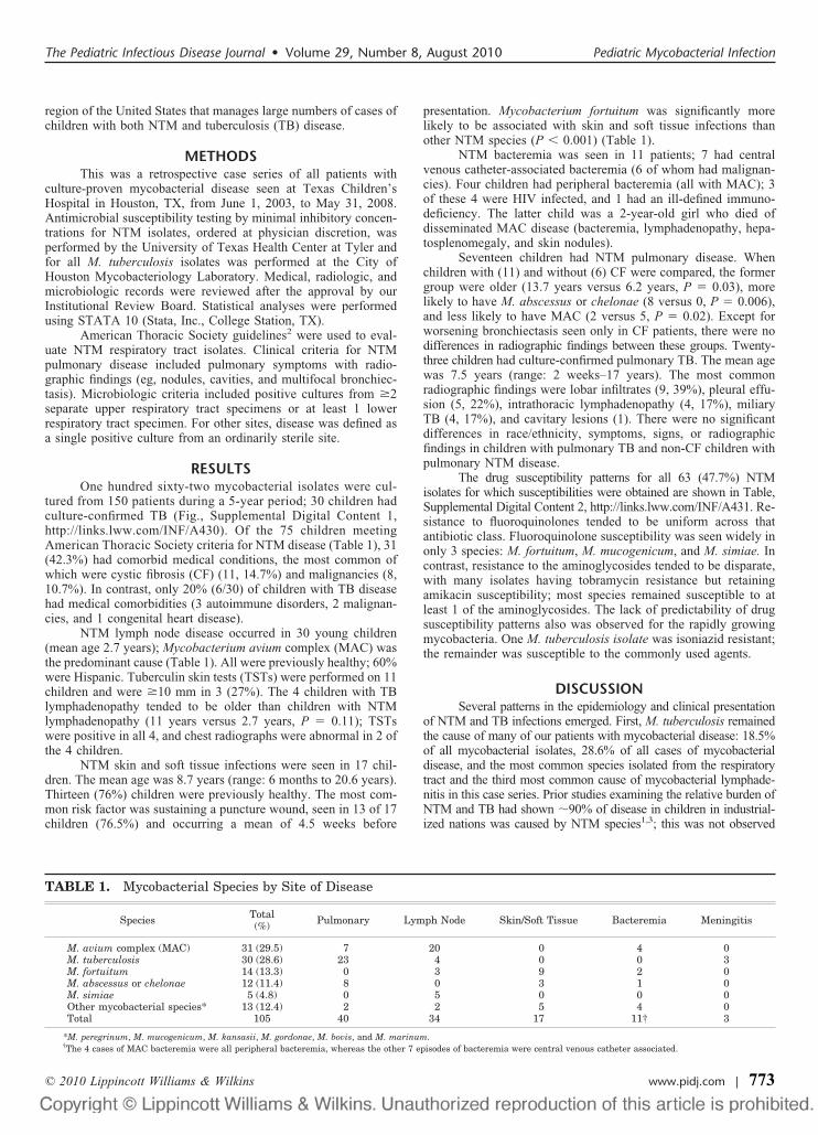
RESULTSOne hundred sixty-two mycobacterial isolates were cul-
tured from 150 patients during a 5-year period; 30 children hadculture-confirmed TB (Fig., Supplemental Digital Content 1,http://links.lww.com/INF/A430). Of the 75 children meetingAmerican Thoracic Society criteria for NTM disease (Table 1), 31(42.3%) had comorbid medical conditions, the most common ofwhich were cystic fibrosis (CF) (11, 14.7%) and malignancies (8,10.7%). In contrast, only 20% (6/30) of children with TB diseasehad medical comorbidities (3 autoimmune disorders, 2 malignan-cies, and 1 congenital heart disease).
NTM lymph node disease occurred in 30 young children(mean age 2.7 years); Mycobacterium avium complex (MAC) wasthe predominant cause (Table 1). All were previously healthy; 60%were Hispanic. Tuberculin skin tests (TSTs) were performed on 11children and were �10 mm in 3 (27%). The 4 children with TBlymphadenopathy tended to be older than children with NTMlymphadenopathy (11 years versus 2.7 years, P � 0.11); TSTswere positive in all 4, and chest radiographs were abnormal in 2 ofthe 4 children.
NTM skin and soft tissue infections were seen in 17 chil-dren. The mean age was 8.7 years (range: 6 months to 20.6 years).Thirteen (76%) children were previously healthy. The most com-mon risk factor was sustaining a puncture wound, seen in 13 of 17children (76.5%) and occurring a mean of 4.5 weeks before
presentation. Mycobacterium fortuitum was significantly morelikely to be associated with skin and soft tissue infections thanother NTM species (P � 0.001) (Table 1).
NTM bacteremia was seen in 11 patients; 7 had centralvenous catheter-associated bacteremia (6 of whom had malignan-cies). Four children had peripheral bacteremia (all with MAC); 3of these 4 were HIV infected, and 1 had an ill-defined immuno-deficiency. The latter child was a 2-year-old girl who died ofdisseminated MAC disease (bacteremia, lymphadenopathy, hepa-tosplenomegaly, and skin nodules).
Seventeen children had NTM pulmonary disease. Whenchildren with (11) and without (6) CF were compared, the formergroup were older (13.7 years versus 6.2 years, P � 0.03), morelikely to have M. abscessus or chelonae (8 versus 0, P � 0.006),and less likely to have MAC (2 versus 5, P � 0.02). Except forworsening bronchiectasis seen only in CF patients, there were nodifferences in radiographic findings between these groups. Twenty-three children had culture-confirmed pulmonary TB. The mean agewas 7.5 years (range: 2 weeks–17 years). The most commonradiographic findings were lobar infiltrates (9, 39%), pleural effu-sion (5, 22%), intrathoracic lymphadenopathy (4, 17%), miliaryTB (4, 17%), and cavitary lesions (1). There were no significantdifferences in race/ethnicity, symptoms, signs, or radiographicfindings in children with pulmonary TB and non-CF children withpulmonary NTM disease.
The drug susceptibility patterns for all 63 (47.7%) NTMisolates for which susceptibilities were obtained are shown in Table,Supplemental Digital Content 2, http://links.lww.com/INF/A431. Re-sistance to fluoroquinolones tended to be uniform across thatantibiotic class. Fluoroquinolone susceptibility was seen widely inonly 3 species: M. fortuitum, M. mucogenicum, and M. simiae. Incontrast, resistance to the aminoglycosides tended to be disparate,with many isolates having tobramycin resistance but retainingamikacin susceptibility; most species remained susceptible to atleast 1 of the aminoglycosides. The lack of predictability of drugsusceptibility patterns also was observed for the rapidly growingmycobacteria. One M. tuberculosis isolate was isoniazid resistant;the remainder was susceptible to the commonly used agents.
DISCUSSIONSeveral patterns in the epidemiology and clinical presentation
of NTM and TB infections emerged. First, M. tuberculosis remainedthe cause of many of our patients with mycobacterial disease: 18.5%of all mycobacterial isolates, 28.6% of all cases of mycobacterialdisease, and the most common species isolated from the respiratorytract and the third most common cause of mycobacterial lymphade-nitis in this case series. Prior studies examining the relative burden ofNTM and TB had shown 90% of disease in children in industrial-ized nations was caused by NTM species1,3; this was not observed
TABLE 1. Mycobacterial Species by Site of Disease
Species Total(%) Pulmonary Lymph Node Skin/Soft Tissue Bacteremia Meningitis
M. avium complex (MAC) 31 (29.5) 7 20 0 4 0M. tuberculosis 30 (28.6) 23 4 0 0 3M. fortuitum 14 (13.3) 0 3 9 2 0M. abscessus or chelonae 12 (11.4) 8 0 3 1 0M. simiae 5 (4.8) 0 5 0 0 0Other mycobacterial species* 13 (12.4) 2 2 5 4 0Total 105 40 34 17 11† 3
*M. peregrinum, M. mucogenicum, M. kansasii, M. gordonae, M. bovis, and M. marinum.†The 4 cases of MAC bacteremia were all peripheral bacteremia, whereas the other 7 episodes of bacteremia were central venous catheter associated.
The Pediatric Infectious Disease Journal • Volume 29, Number 8, August 2010 Pediatric Mycobacterial Infection
© 2010 Lippincott Williams & Wilkins www.pidj.com | 773

here. Houston/Harris County has 1 of the highest rates of TB in theUnited States, with a case rate of 8.5 per 100,000, almost double thenational rate.4 The high rate of TB disease in this population madeempiric therapy and public health considerations for mycobacteriallymph node and pulmonary disease challenging.
Second, a number of children with puncture wounds grew aNTM species, with M. fortuitum being the most associated withdisease after skin/soft tissue inoculation. NTM species should beconsidered in infected puncture wound refractory to conventionalantibiotics. Empiric therapy for suspected mycobacterial skin/softtissue infections should target the rapidly growing mycobacterial species.
Third, children with NTM lymphadenopathy tended to beyounger than children with TB lymphadenopathy. In a preschool-agedchild lacking TB risk factors, an enlarged peripheral lymph node incombination with a positive TST was much more likely to be causedby a NTM species. This epidemiologic pattern has public health andclinical importance. As NTM species are not transmitted person-to-person, no contact investigation is needed for a child with NTMdisease. Excision of the involved node is usually curative for NTM,5
but systemic antibiotic therapy is necessary for tuberculous lymphad-enopathy. Additionally, empiric antibiotic selection would differmarkedly based on whether TB or NTM was suspected.
Fourth, pulmonary disease was the second most commonsite of NTM disease. In contrast to children with NTM lymphad-enopathy or skin/soft tissue infections, almost all children withNTM pulmonary disease had comorbid medical conditions. Thismade differentiating colonization from true infection difficult, asmany of these children had underlying pulmonary pathology.6
Relying on a radiographic criterion was difficult because differen-tiating worsening CT findings due to NTM infection versus pro-gression of underlying disease was challenging. Further compli-cating clinical management was that children with CF tended tohave disease with NTM species (M. abscessus or chelonae), whichwere less susceptible to oral agents than non-CF children withNTM pulmonary disease.
Finally, the importance of securing a microbiologic diagno-sis was emphasized by the divergent drug susceptibility patterns.Although lack of standardization in susceptibility testing and smallnumbers of isolates for certain species precluded a powered sta-tistical analysis, resistance to fluoroquinolones tended to be uni-form, whereas resistance to aminoglycosides tended to be dispar-ate. The relatively large burden of M. tuberculosis in thispopulation also made empiric therapy more challenging for pul-monary and lymphatic disease, as few NTM species apart from M.kansasii are susceptible to the same antibiotics as M. tuberculosis.
There were limitations to this study. First, the informationwas collected retrospectively; consequently, not all data wereevaluable for all subjects, and the diagnostic and treatment regi-mens were not standardized. Second, this study was conducted ina tertiary care pediatric facility with many immunocompromizedchildren, a group not reflective of the general population. Third,there was selection bias as to which children with suspected TBwere admitted for cultures; cultures are sought more often in sickerchildren or children in whom the source case is unknown. Fourth,using only culture-proven cases of TB disease in children under-estimated the true disease burden in this population.7 The issue oflow-culture yield is applicable to NTM species as well—negativecultures are common; therefore, a review of only positive culturecases might introduce bias, because children who have positivecultures may not be reflective of all cases.
Tuberculosis comprised a large proportion of mycobacterialdisease in this series of children and was the most commonspecimen isolated from respiratory tract isolates. Wide variationin antimicrobial susceptibility patterns among NTM species,
together with the large percentage of disease caused by TB,emphasizes the importance of obtaining a microbiologic diag-nosis, particularly in children presenting with pulmonary find-ings and/or lymphadenopathy.
REFERENCES1. Haverkamp MH, Arend SM, Lindeboom JA, et al. Nontuberculous myco-
bacterial infection in children: a 2-year prospective surveillance study in theNetherlands. Clin Infect Dis. 2004;39:450–456.
2. Griffith DE, Aksamit T, Brown-Elliott BA, et al. ATS MycobacterialDiseases Subcommittee; American Thoracic Society; Infectious DiseasesSociety of America. An official ATS/IDSA statement: diagnosis, treatment,and prevention of nontuberculous mycobacterial diseases. Am J Respir CritCare Med. 2007;175:367–416.
3. Joshi W, Davidson PM, Jones PG, et al. Non-tuberculous mycobacteriallymphadenitis in children. Eur J Pediatr. 1989;148:751–754.
4. CDC. Reported Tuberculosis in the United States, 2006. Atlanta, GA: U.S.Department of Health and Human Services, CDC;2007.
5. Lindeboom JA, Kuijper EJ, Bruijnesteijn van Coppenraet ES, et al. Surgicalexcision versus antibiotic treatment for nontuberculous mycobacterial cervi-cofacial lymphadenitis in children: a multicenter, randomized, controlledtrial. Clin Infect Dis. 2007;44:1057–1064.
6. Razvi S, Quittell L, Sewall A, et al. Respiratory microbiology of patientswith cystic fibrosis in the United States, 1995 to 2005. Chest. 2009;136:1554–1560.
7. Marais BJ, Gie RP, Schaaf HS, et al. Childhood pulmonary tuberculosis: oldwisdom and new challenges. Am J Respir Crit Care Med. 2006;173:1078–1090.
ASSOCIATION BETWEEN LIPODYSTROPHY ANDLEPTIN IN HUMAN IMMUNODEFICIENCYVIRUS-1-INFECTED CHILDREN RECEIVINGLOPINAVIR/RITONAVIR-BASED THERAPY
Salvador Resino, PhD,* Claudia Palladino, PhD,†‡Raquel Lorente, BSc,†§ Dariela Micheloud, PhD, MD,*Jose Maria Bellon, MSc,¶ Beatriz Larru, PhD, MD,†Maria Dolores Gurbindo Gutierrez, PhD, MD,�**Maria Isabel de Jose, PhD, MD,†† Rosa Polo, MD,‡‡and Maria Angeles Munoz-Fernandez, PhD, MD†**; on Behalfof the Spanish Group of Pediatric HIV Infection
Abstract: Highly active antiretroviral therapy might lead to the developmentof dyslipidemia and lipodystrophy (LD) syndrome. We carried out a multi-center prospective study of 22 human immunodeficiency virus (HIV)-1-infected children treated during 48 months with lopinavir/ritonavir-basedhighly active antiretroviral therapy to evaluate the trend of serum lipids andadipokines. Increase in plasma leptin levels and leptin/adiponectin ratio wasassociated with LD. These adipokines may be surrogate markers of LD.
Key Words: protease inhibitor, adipokines, lipids, antiretroviral therapy,adverse effects
Accepted for publication February 27, 2010.From the *Laboratorio de Epidemiología Molecular de Enfermedades Infeccio-
sas, Centro Nacional de Microbiología, Instituto de Salud Carlos III,Majadahonda, Madrid, Spain; †Laboratorio de Inmuno-Biología Molecular,Hospital General Universitario “Gregorio Maranon,” Madrid, Spain; ‡Isti-tuto Pasteur, Fondazione Cenci-Bolognetti, Universita degli Studi di Roma“La Sapienza,” Rome, Italy; §Servicio de Medicina Interna, Hospital Gen-eral Universitario “Gregorio Marañón,” Madrid, Spain, ¶Unidad de Inves-tigacion, Fundacion para la Investigacion Biomedica del Hospital GregorioMaranon, Madrid, Spain; �Inmuno-Pediatría, Hospital General Universitario“Gregorio Maranon,” Madrid, Spain; **Unidad Asociada de RetrovirologíaHumana, HGUGM-CSIC, Madrid, Spain; ††Servicio Infecciosas Infantil,Hospital Universitario La Paz, Madrid, Spain; and ‡‡Ministerio de Sanidady Asuntos Sociales, Madrid, Spain.
Resino et al The Pediatric Infectious Disease Journal • Volume 29, Number 8, August 2010
© 2010 Lippincott Williams & Wilkins774 | www.pidj.com

The authors do not have commercial or other associations that might posea conflict of interest.
Address for correspondence: Salvador Resino, Laboratorio de EpidemiologíaMolecular de Enfermedades Infecciosas, Centro Nacional de Microbi-ología, Instituto de Salud Carlos III, Carretera Majadahonda-Pozuelo, Km2.2, 28220 Majadahonda (Madrid), Spain. E-mail: [email protected]. orMaria Angeles Munoz-Fernandez, Laboratorio de Inmuno-Biología Mo-lecular, Hospital General Universitario “Gregorio Maranon,” Madrid,Spain. E-mail: [email protected].
Supplemental digital content is available for this article. Direct URL citationsappear in the printed text and are provided in the HTML and PDF versionsof this article on the journal’s Web site (www.pidj.com).
DOI: 10.1097/INF.0b013e3181db741b
Highly active antiretroviral therapy (HAART) has achievedreduction in mortality and morbidity in human immunodefi-
ciency virus (HIV)-infected children.1 Nevertheless, long-termHAART exposure could lead to the development of dyslipidemiaand lipodystrophy (LD) syndrome.2–4 In particular, lopinavir/ritonavir (LPV/r; Kaletra; Abbott Laboratories, Abbott Park, IL), acombination of HIV-1 protease inhibitors (PIs), has shown potentantiviral activity and clinical effectiveness5,6 but its benefits mustbe balanced against potential adverse events.7
LD is characterized by adipose tissue redistribution and met-abolic disorders (hypercholesterolemia and insulin resistance �IR�),8,9
both being associated with increased proinflammatory cytokine val-ues. Moreover, HAART and inflammatory cytokines are associatedwith a decrease in adiponectin and an increase in leptin.10,11
Leptin regulates proinflammatory immune responses by con-trolling tumor necrosis factor-� production and macrophage activa-tion12; tumor necrosis factor-� and interleukin-6 are capable ofstimulating adipocyte leptin production.12 Moreover, adiponectin hasbeen found to be inversely associated with metabolic syndrome,11 andthis adipokine has anti-inflammatory activity,11 which could be in-volved in a compensatory mechanism to cushion the inflammatoryeffect induced by leptin and resistin. To date, few studies on plasmakinetics of these adipocyte-secreted hormones (adipokines) in HIV-infected children have been published. This study evaluates the trendof serum lipids and adipokines in HIV-1-infected children treatedwith LPV/r-based HAART as salvage therapy.
PATIENTS AND METHODSA multicenter prospective study of vertically HIV-1-in-
fected children of the HIV Spanish Pediatric Cohort on LPV/r-based salvage therapy was done. The study was approved by thehospitals’ ethical committees. The inclusion criteria were as fol-lows: (a) �2 years of follow-up, (b) age �1 year, (c) previouslytreated with antiretroviral therapies (ARTs) and having records ofvirologic failure with PI or non-nucleoside analog, and (d) startingsalvage HAART with LPV/r.
Children were monitored every 3 months with interviews,physical examinations, and blood sample collection for percentageof CD4� and CD8� T-cells and HIV-1 RNA measurements.Fasting serum samples were frozen at 70°C for further assays.Total cholesterol, triglycerides, high-density lipoprotein choles-terol, low-density lipoprotein cholesterol, and glucose concentra-tions were available for routine clinical use. The lipid panel wasperformed on fasting specimens. Several biologic samples wereprovided by the Spanish HIV BioBank belonging to the SpanishAIDS Research Network.13
Pediatricians administered the appropriate ART followinginternational guidelines.14 The ART previous LPV/r-based therapywas classified as monotherapy of a nucleoside reverse transcriptioninhibitor, on combined-therapy of 2 nucleoside reverse transcrip-
tion inhibitors, or HAART (combinations of 3 or more drugs).Adherence was evaluated recording the dose taken and interview-ing the parents or tutors during follow-up, and it was summarizedas a single percentage for each patient. LPV/r-based HAARTinitiation was defined as the first time children took LPV/r with 2or more antiretrovirals. Subsequent regimen changes were ignoredin terms of statistical analysis whereas it still included LPV/r.
LD was diagnosed by clinical examination at the mostrecent visit15 and was standardized across participating hospitals.The degree of lipoatrophy or lipohypertrophy in every part of thebody was categorized as absent (score of 0), mild (score 1),moderate (score 2), or severe (score 3). Patients with scores �2were classified in the LD group and patients with scores �2 in theno-LD (N-LD) group. Height, weight, and body mass index wereexpressed as z-scores and were adjusted for age and sex.
The adipokines panel was performed from fasting serumsamples frozen at 70°C. Multiplex suspension bead array immu-noassay was performed using the Luminex 100 analyzer (LuminexCorporation, Austin, TX) Multiplex kit (LINCOplex; LINCOResearch, St. Charles, MO) to identify protein expression inplasma. The degree of IR was estimated using the homeostaticmodel assessment (HOMA) method using the formula: plasmaglucose (mmol/L) � serum insulin (mU/L)/22.5.
The 2-sided Fisher exact test was used to compare categor-ical variables. Continuous variables were compared longitudinallyeither within groups in comparison with baseline (Wilcoxon test)or between groups (Mann-Whitney U test). Medians were esti-mated with interquartile range; SPSS (v.12; SPSS Inc., Chicago,IL) was used. P values were 2-tailed, and significance was definedas P � 0.05.
RESULTSWe recruited 22 vertically HIV-1-infected children in 2
tertiary hospitals, and they were followed up from October 1,2000, until January 1, 2003. Eleven (50%) patients were assignedto the LD group and 11 (50%) to the N-LD group. One patient hadmoderate lipohypertrophy; 1 had mild lipoatrophy and lipohyper-trophy; 1 had mild lipoatrophy and moderate lipohypertrophy; 4had moderate lipoatrophy; 3 had moderate lipoatrophy and mod-erate lipohypertrophy; and 1 had moderate lipoatrophy and severelipohypertrophy. The 2 groups (N-LD and LD) showed similarage, anthropometric values, adherence to antiretrovirals, as well asclinical and immunologic status (Table, Supplemental DigitalContent 1, http://links.lww.com/INF/A438). Neither gender norage at baseline was associated with LD, but LD patients wereslightly older than N-LD patients. Patients had been similarlyexposed to antiretrovirals previous LPV/r administration but ashorter duration of d4T administration as part of the salvageregimen was observed in the LD group (P � 0.021) (Table,Supplemental Digital Content 1, http://links.lww.com/INF/A438).
The medians of CD4�, CD8�, HIV-1 RNA, and lipids werecomparable between groups during follow-up (data not shown).The N-LD group had an increase in adiponectin values, but it wasnot statistically significant (Fig. 1A). The LD group had a signif-icant increase in leptin concentrations at weeks 24, 36, and 48 andhad higher values of leptin than in the N-LD-group (P � 0.016;Fig. 1B). The leptin/adiponectin ratio was similar between groupsat baseline; the LD group had higher values than the N-LD groupat weeks 36 and 48 (Fig. 1C). Patients of the 2 groups had similarresistin, C-peptide, and HOMA values, which slightly increasedduring follow-up in both groups (data not shown).
The Pediatric Infectious Disease Journal • Volume 29, Number 8, August 2010 Leptin and Lipodystrophy
© 2010 Lippincott Williams & Wilkins www.pidj.com | 775

DISCUSSIONHIV-associated LD syndrome (HIV-LS) has been associ-
ated with the use of PIs in children.2–4,15 Although it is wellcharacterized in adults,16 there are few pediatric data available onHIV-LS, and the role of adipokines has been previously high-lighted only in cross-sectional studies. Verkauskiene et al17 andKim et al18 found decreased adiponectin levels in HIV-infectedchildren with LD, whereas Papaevangelou et al19 and Dzwonek etal4 found no difference in leptin values between HAART-treatedand untreated patients. The main goal of our longitudinal studywas to monitor plasma metabolic changes and adipokines levelsassociated with LD in HIV-infected children on LPV/r-basedHAART as salvage therapy.
All patients reached virologic suppression during 48 weeksof follow-up, consistent with previously reported evaluation ofLPV/r effectiveness.6,20 Nevertheless, virologic failure was ob-served in some patients without LD at the end of the study. Despitethis, the %CD4� values were similar in both study groups.
Unlike other studies, we did not find a significant increase incholesterol, triglycerides, low-density lipoprotein, and high-den-sity lipoprotein; however, only a small percentage of children inour cohort had significant hyperlipidemia during follow-up. Theprevalence of LD was higher than in other reports,3,4,21 probablybecause of previous long-term ART exposure.
Overall, 50% of the patients experienced LD and showedchanges in plasma adipokines that have been found to be associatedwith IR.10,22 The increase in leptin levels may be the direct effect of
PI on adipose tissue, which may contribute to an overall adiposeimbalance and the development of LD and metabolic syndrome inHIV patients.23 Serum leptin concentrations reflect body fat contentand are associated with IR.11 Moreover, the increase in leptin that weobserved might be explained by the so called leptin resistance24 andmight support the hypothesis that leptin (as a marker for body fat)could play a key role in the pathogenesis of LD in HIV-infectedchildren. By contrast, children without LD had an increase in adi-ponectin levels and a decrease in leptin/adiponectin ratio, which mightrepresent a compensatory response to antiretroviral-induced meta-bolic syndrome.10,25 Adiponectin has been found to be inverselyassociated with metabolic syndrome11 and changes in adipose tissuemay lead to hypoadiponectinemia in the later stages of metabolicsyndrome or LD in HIV-infected children.11,18
Disturbance of lipid metabolism was observed in all patientsand included moderate hypercholesterolemia and hypertriglyceri-demia but all the children had HOMA and C-peptide values belowthe lower limit of normal, this being associated with absence oftype II diabetes. The high leptin values observed in patients withLD might suggest that these subjects meet criteria close to the IR.
The limitation of our study arises from the small number ofpatients enrolled. Moreover, a significant percentage of children hadalready received PI-based HAART before the salvage regimen, whichcould have played an important role in metabolic syndrome and LD.In conclusion, the increases in plasma leptin concentrations and in theleptin/adiponectin ratio were associated with LD, and these adi-pokines may be surrogate markers for the risk of diabetes and
FIGURE 1. Trend of plasma adiponectin (A), leptin (B), and adiponectin/leptin (C) levels during follow-up of vertically HIV-1-infected children on salvage HAART with LPV/r. Values are expressed as median. *Significant differences (P � 0.05) be-tween groups by Mann-Whitney U test. †Significant differences (P � 0.05) within group by Wilcoxon test. LD indicateschildren with lipodystrophy; N-LD, children without lipodystrophy.
Resino et al The Pediatric Infectious Disease Journal • Volume 29, Number 8, August 2010
© 2010 Lippincott Williams & Wilkins776 | www.pidj.com

cardiovascular disease in HIV-infected children on HAART. Futureprospective studies that involve larger cohorts of children on LPV/rtreatment as salvage therapy are necessary to confirm our findings andto further elucidate the complexity of HIV-LS.
ACKNOWLEDGMENTSThe authors acknowledge the patients in this study for their
participation, and the HIV BioBank belonging to the Spanish AIDSResearch Network (RIS) and the collaborating centers for theclinical samples provided. This work was supported by Fundacionpara la Investigacion y la Prevencion del SIDA en Espana grantFIPSE 36650/07, Fondo de Investigacion Sanitaria (FIS) of Min-isterio de Ciencia e Innovacion grants PI07/90201 and PI08/0738,and Instituto de Salud Carlos III grant UIPY 1467/07 (to S.R.);Fundacion para la Investigacion y la Prevencion del SIDA enEspana grants FIPSE 24534/05 and 24632/07, Fondo de Investi-gacion Sanitaria (FIS) of Ministerio de Ciencia e Innovacion FISgrants PI052476, PI061479, and Red RIS RD06-0006-0035, Fun-dacion Caja Navarra, Comunidad de Madrid grant S-SAL-0159-2006, and Task Force in Europe for Drug Development for theYoung (TEDDY) (to M.A.M.F.); and grant from Istituto Pasteur,Fondazione Cenci-Bolognetti, Universita degli Studi di Roma “LaSapienza” (to C.P.).
REFERENCES1. Resino S, Resino R, Bellon JM, et al. Clinical outcomes improve with
highly active antiretroviral therapy in vertically HIV type-1-infected chil-dren. Clin Infect Dis. 2006;43:243–252.
2. Beregszaszi M, Dollfus C, Levine M, et al. Longitudinal evaluation and riskfactors of lipodystrophy and associated metabolic changes in HIV-infectedchildren. J Acquir Immune Defic Syndr. 2005;40:161–168.
3. Guillen S, Ramos JT, Resino R, et al. Impact on weight and height with theuse of HAART in HIV-infected children. Pediatr Infect Dis J. 2007;26:334–338.
4. Dzwonek AB, Lawson MS, Cole TJ, et al. Body fat changes and lipodys-trophy in HIV-infected children: impact of highly active antiretroviraltherapy. J Acquir Immune Defic Syndr. 2006;43:121–123.
5. Resino S, Bellon JM, Ramos JT, et al. Positive virological outcome afterlopinavir/ritonavir salvage therapy in protease inhibitor-experienced HIV-1-infected children: a prospective cohort study. J Antimicrob Chemother.2004;54:921–931.
6. Larru B, Resino S, Bellon JM, et al. Long-term response to highly activeantiretroviral therapy with lopinavir/ritonavir in pre-treated vertically HIV-infected children. J Antimicrob Chemother. 2008;61:183–190.
7. Kosalaraksa P, Bunupuradah T, Engchanil C, et al. Double boosted proteaseinhibitors, saquinavir, and lopinavir/ritonavir, in nucleoside pretreated chil-dren at 48 weeks. Pediatr Infect Dis J. 2008;27:623–628.
8. Taylor P, Worrell C, Steinberg SM, et al. Natural history of lipid abnor-malities and fat redistribution among human immunodeficiency virus-infected children receiving long-term, protease inhibitor-containing, highlyactive antiretroviral therapy regimens. Pediatrics. 2004;114:e235–e242.
9. Wohl DA, McComsey G, Tebas P, et al. Current concepts in the diagnosisand management of metabolic complications of HIV infection and itstherapy. Clin Infect Dis. 2006;43:645–653.
10. Samaras K, Wand H, Law M, Emery S, Cooper D, Carr A. Prevalence ofmetabolic syndrome in HIV-infected patients receiving highly active anti-retroviral therapy using International Diabetes Foundation and Adult Treat-ment Panel III criteria: associations with insulin resistance, disturbed bodyfat compartmentalization, elevated C-reactive protein, and hypoadi-ponectinemia. Diabetes Care. 2007;30:113–119.
11. Krause JC, Toye MP, Stechenberg BW, et al. HIV-associated lipodystrophyin children. Pediatr Endocrinol Rev. 2005;3:45–51.
12. Antuna-Puente B, Feve B, Fellahi S, et al. Adipokines: the missing linkbetween insulin resistance and obesity. Diabetes Metab. 2008;34:2–11.
13. Garcia-Merino I, de Las Cuevas N, Jimenez JL, et al. The Spanish HIVBioBank: a model of cooperative HIV research. Retrovirology. 2009;6:27.
14. Sharland M, Gibb D, Giaquinto C. Current evidence for the use ofpaediatric antiretroviral therapy—a PENTA analysis. Paediatric European
Network for the Treatment of AIDS Steering Committee. Eur J Pediatr.2000;159:649–656.
15. Hartman K, Verweel G, de Groot R, et al. Detection of lipoatrophy inhuman immunodeficiency virus-1-infected children treated with highlyactive antiretroviral therapy. Pediatr Infect Dis J. 2006;25:427–431.
16. Sweeney LL, Brennan AM, Mantzoros CS. The role of adipokines inrelation to HIV lipodystrophy. AIDS. 2007;21:895–904.
17. Verkauskiene R, Dollfus C, Levine M, et al. Serum adiponectin and leptinconcentrations in HIV-infected children with fat redistribution syndrome.Pediatr Res. 2006;60:225–230.
18. Kim RJ, Carlow DC, Rutstein JH, et al. Hypoadiponectinemia, dyslipide-mia, and impaired growth in children with HIV-associated facial lipoatro-phy. J Pediatr Endocrinol Metab. 2007;20:65–74.
19. Papaevangelou V, Papassotiriou I, Vounatsou M, et al. Changes in leptinserum levels in HIV-infected children receiving highly active antiretroviraltherapy. Scand J Clin Lab Invest. 2007;67:291–296.
20. Resino S, Bellon JM, Ramos JT, et al. Salvage antiretroviral therapy inHIV-infected children: advantages of lopinavir-ritonavir. Pediatr Infect DisJ. 2004;23:923–930.
21. Ene L, Goetghebuer T, Hainaut M, et al. Prevalence of lipodystrophy inHIV-infected children: a cross-sectional study. Eur J Pediatr. 2007;166:13–21.
22. Yamauchi T, Kamon J, Minokoshi Y, et al. Adiponectin stimulates glucoseutilization and fatty-acid oxidation by activating AMP-activated proteinkinase. Nat Med. 2002;8:1288–1295.
23. Cianflone K, Zakarian R, Stanculescu C, et al. Protease inhibitor effects ontriglyceride synthesis and adipokine secretion in human omental and sub-cutaneous adipose tissue. Antivir Ther. 2006;11:681–691.
24. Rosicka M, Krsek M, Matoulek M, et al. Serum ghrelin levels in obesepatients: the relationship to serum leptin levels and soluble leptin receptorslevels. Physiol Res. 2003;52:61–66.
25. Tsiodras S, Mantzoros C. Leptin and adiponectin in the HIV associatedmetabolic syndrome: physiologic and therapeutic implications. Am J InfectDis. 2006;2:141–152.
NATURALLY FLUCTUATING LOW INCIDENCE OFINVASIVE PNEUMOCOCCAL INFECTIONS NOT
AFFECTED BY LARGE-SCALE HAEMOPHILUSINFLUENZAE TYPE B VACCINATION
Heikki Peltola, MD,* Merja Helminen, MD,†and Risto Vuento, MD‡
Background: Since many pathogens colonize the child’s oro/nasopharynxin a similar manner, elimination of diseases such as those caused byHaemophilus influenzae type b (Hib) has a potential of augmenting otherserious infections. Streptococcus pneumoniae is an agent of special interestnow that Hib conjugates have been used widely for more than 2 decades.Patients and Methods: All blood and cerebrospinal fluid isolations of Hiband S. pneumoniae were collected prospectively from 85,000 Finnishchildren at age 0–15 years by one central laboratory during 27 years.Results: Hib vaccination, launched in 1986–1988, led to a quick decline ofcases until the last was detected in 1991. In the next few years, theincidence of bacteremic S. pneumoniae infections increased, but now for15 years, the numbers of cases have been slowly declining. This finding isnot explained by less active sample-taking because the number of bloodcultures have almost doubled in the past years.Conclusions: Large-scale Hib vaccination does not increase the incidenceof pneumococcal diseases which continue their year-to-year fluctuation atlow levels. Only a years-long follow-up permits conclusions on a vacci-nation’s potential influence on the epidemiology of other diseases.
Key Words: Streptococcus pneumoniae, pneumococcus, Haemophilusinfluenzae, incidence, epidemiology, vaccinationAccepted for publication March 1, 2010.From the *Helsinki University Central Hospital, Hospital for Children and
Adolescents, Helsinki, Finland; †Department of Pediatrics; and ‡TampereUniversity Hospital, Centre for Laboratory Medicine, Tampere, Finland.
The Pediatric Infectious Disease Journal • Volume 29, Number 8, August 2010 Hib Vaccine Vs. Pneumococcal Diseases
© 2010 Lippincott Williams & Wilkins www.pidj.com | 777

Address for correspondence: Prof. H. Peltola, HUCH Hospital for Children andAdolescents, PO Box 281 (11 Stenback St.), 00029 HUS, Helsinki, Finland.E-mail: [email protected].
Supplemental digital content is available for this article. Direct URL citationsappear in the printed text and are provided in the HTML and PDF versionsof this article on the journal’s Web site (www.pidj.com).
DOI: 10.1097/INF.0b013e3181dbd820
Because certain competition likely prevails among the bacteriaresiding in the human oro/nasopharynx, an introduction of
large-scale vaccination affecting the colonization may potentiallyincrease the incidence of infections caused by other respiratorypathogens.1 The rationale is that some less-common pathogensmay find a new niche because of the changed balance in carriage.2
For Haemophilus influenzae type b (Hib), this concern wasraised along with conjugate vaccines and diseases such as thosecaused by the non-type b H. influenzae strains3–5 and Streptococ-cus pneumoniae.6,7 Replacement of Hib infections by other severeH. influenzae infections seems unlikely,8 whereas for the nonva-ccine-type S. pneumoniae strains, this phenomenon is well docu-mented in several countries.9–16 However, few prospective long-term data have been available to address the question whether aninteraction exists between Hib vaccination and invasive pneumo-coccal infections.
In 1995, it was reported that bacteremic S. pneumoniaeinfections were becoming more common in Finland,7 likely be-cause large-scale vaccination had eliminated Hib diseases. How-ever, an association does not prove causation,17 and the follow-upwas only 3 years. We now have continued it prospectively for 15more years and are convinced that the fear was unfounded.
PATIENTS AND METHODSThe Pirkanmaa region, located 100 miles north from Helsinki
(capital), has a population of almost half a million, of which 85,000are children aged 0 to 15 years. No major fluctuation has occurredover the last years. The district is served by 1 tertiary hospital(Tampere) whose central laboratory is the only 1 in the region andobtains all bacterial isolates. The center handles more than 30,000blood cultures a year. Because of its large size, Pirkanmaa is repre-sentative of the whole country in which the interregional differencesin primary health care are remarkably small.
All blood and cerebrospinal fluid (CSF) isolations of Hib orS. pneumoniae were collected and computerized during the 27-year period of 1983 to 2009. Following the concept of the earlier
report,7 we examined children treated in the Tampere UniversityHospital. Through years, the clinicians were encouraged to takethe blood and, if necessary, CSF cultures to disclose the causativeagent of all potentially invasive infections. Standard techniqueswere used to identify Hib and S. pneumoniae.
RESULTSBetween 1995 and 2009, the number of blood
cultures varied (Table, Supplemental Digital Content 1,http://links.lww.com/INF/A439). Fewer CSF cultures were ob-tained, but only 1 child (in 1997) with S. pneumoniae menin-gitis did not yield the agent also from the blood. Almost 3000cultures per year have been performed in the past several years.
The last Hib isolation in 1991 was soon followed by anincrease in bacteremic S. pneumoniae cases (Fig. 1). However,none of the years during the last 15 years has reached the peak of1994 when 23 cases were identified. Pneumococcal infections, ifanything, have been slowly declining towards the pre-Hib vacci-nation levels (Fig. 1), and this favorable trend has not been due toless active sample-taking.
In this population, as in Finland in general,18 bacterialmeningitis has become rare; 4 to 8 cases per year in the wholecountry. Annually, 11 bacteremic S. pneumoniae cases and �1pneumococcal meningitis have been diagnosed in Pirkanmaa in thelast 5 years. No other agent has replaced Hib or S. pneumoniaeisolations. Neisseria meningitidis has been even a rarer one; 19cases from 1995 to 2009, of which 13 were meningitis (all except1 were of group B).
DISCUSSIONOur 27-year follow-up of a large childhood population in
Finland elucidated 2 important issues. First, the incidence ofinvasive S. pneumoniae infections has been low, despite thedisappearance of Hib diseases,19 and despite these 2 agents per-form rather similarly in the respiratory tract. Our epidemiologicdata fit well with those from the whole country (population 5.4million) in which the annual incidence of all invasive S. pneu-moniae infections at age 0 to 4 years was 23.5/100,000. Not all ofthese cases are preventable with the existing vaccines.20
Second, the incidence of pneumococcal (and many other)infections fluctuate from year to year. In Pirkanmaa, it first lookedas invasive S. pneumoniae infections would explode once Hib
FIGURE 1. Annual cases of invasive H. influenzae type b and S. pneumoniae infections in 0- to 15-year-old children duringthe 27-year period of 1983 to 2009 in the district of Pirkanmaa, Finland.
Peltola et al The Pediatric Infectious Disease Journal • Volume 29, Number 8, August 2010
© 2010 Lippincott Williams & Wilkins778 | www.pidj.com

diseases were eliminated,7 until a follow-up of many more yearsshowed that this was not the case. Conclusions on epidemiologictrends should be drawn only after a long enough (several years)monitoring of the situation.
In the United States, pediatric vaccination has surprisinglyreduced invasive pneumococcal diseases of the elderly people byherd immunity.21 This effect has not been seen in Europe (vagueevidence comes from Catalonia, Spain).22 The obvious reason isthat the prevaccination incidence in western Europe was approx-imately one sixth of that in the United States; the annual rate at age0 to 1 years was 20 to 35 versus 160 to 180 cases per 100,000children in Europe23 and the United States,21 respectively. With adifference of this magnitude, one cannot expect the US experienceto repeat in most of Europe, although the vaccine-type infectionshave expectedly declined.24,25 On the other hand, nonvaccine-typeinfections have increased alarmingly in countries such as Spain,France, and United Kingdom.11,12,14,16,22
In the developing world, and among risk populations andindustrialized countries (the United States), where the incidence ofpneumococcal infections is or was high, swift introduction of vacci-nation is well justified—despite it doubles the costs of pediatricvaccines. In the industrialized low-incidence countries, the situation isless clear, specially that the replacement phenomenon has become anissue.9,14 Some 40 affluent countries have included pneumococcalvaccination in their calendar, and Finland joins the orchestra in2010. As a consequence, 10 to 15 severe and many more nonse-vere S. pneumoniae infections per year will likely be prevented,but these figures should be projected against the overall costs andthe 180,000 vaccinations which the 3-dose schedule requires. If thenew conjugate is also capable of preventing otitis media,26 thescenario changes drastically.
Our data demonstrate that the launch of large-scale Hibvaccination does not increase the incidence of pneumococcaldiseases. However, local epidemiology, and the severity of man-ifestations20 should also be taken into account. If severe manifes-tations are rare, as they are in most of industrialized Europe, abalance between the clinically meaningful benefits, costs, and therisks of augmenting more serious nonvaccine-type diseases14,16,22
should be sought. Warning signs11,12,14,16,27 have been signaled.
REFERENCES1. Nikoskelainen J, Lahtonen E, Leino A, et al. Is group-specific meningo-
coccal vaccination resulting in epidemics caused by other groups of virulentmeningococci? Lancet. 1978;312:403–405.
2. Takala A, Eskola J, Leinonen M, et al. Reduction of oropharyngeal carriageof Haemophilus influenzae type b (Hib) in children immunized with an Hibconjugate vaccine. J Infect Dis. 1991;164:982–986.
3. Waggoner-Fountain LA, Hendley JO, Cody EJ, et al. The emergence ofHaemophilus influenzae types e and f as significant pathogens. Clin InfectDis. 1995;21:1322–1324.
4. Urwin G, Krohn JA, Deaver-Robinson K, et al, the Haemophilus influenzaeStudy Group. Invasive disease due to Haemophilus influenzae serotype f:clinical and epidemiological characteristics in the H. influenzae serotype bvaccine era. Clin Infect Dis. 1996;22:1069–1076.
5. Campos J, Roman F, Perez-Vazquez M, et al. Infection due to Haemophilusinfluenzae serotype: microbiological, clinical, and epidemiological features.Clin Infect Dis. 2003;37:841–845.
6. Foster JA, McGowan KL. Rising rate of pneumococcal bacteremia at theChildren’s Hospital of Philadelphia. Pediatr Infect Dis J. 1994;13:1143–1144.
7. Baer M, Vuento R, Vesikari T. Increase in bacteraemic pneumococcalinfections in children. Lancet. 1995;345:661.
8. Ward JI. Editorial response: invasive infections due to Haemophilus influ-enzae serotype f (Hif)—is Hif an emerging pathogen? Clin Infect Dis.1996;22:1077–1078.
9. Kilpi T, Åhman H, Jokinen J, et al. Protective efficacy of a secondpneumococcal conjugate vaccine against pneumococcal acute otitis mediain infants and children: randomized, controlled trial of a 7-valent pneumo-
coccal polysaccharide-meningococcal outer membrane protein complexvaccine in 1666 children. Clin Infect Dis. 2003;37:1155–1164.
10. Pelton SI. Replacement pneumococcal disease in perspective. Clin InfectDis. 2008;46:1353–1355.
11. Calbo E, Diaz A, Canadell E, et al. Invasive pneumococcal disease amongchildren in a health district of Barcelona; early impact of pneumococcalconjugate vaccine. Clin Microbiol Infect. 2006;12:867–872.
12. Obando I, Arroyo LA, Sanchez-Tatay D, et al. Molecular epidemiology ofpaediatric invasive pneumococcal disease in southern Spain after theintroduction of heptavalent pneumococcal conjugate vaccine. Clin Micro-biol Infect. 2006;13:347–348.
13. Messina AF, Katz-Gaynor K, Barton T, et al. Impact of the pneumococcalconjugate vaccine on serotype distribution and antimicrobial resistance ofinvasive Streptococcus pneumoniae isolates in Dallas, TX, children from1999 through 2005. Pediatr Infect Dis J. 2007;26:461–467.
14. Munoz-Almagro C, Jordan I, Gene A, et al. Emergence of invasivepneumococcal disease caused by nonvaccine serotypes in the era of 7-valentconjugate vaccine. Clin Infect Dis. 2008;6:174–182.
15. Bender JM, Ampofo K, Daly J, et al. Pneumococcal necrotizing pneu-monia in Utah: does serotype matter? Clin Infect Dis. 2008;46:1346 –1352.
16. Lepoutre A, Varon E, Georges S, et al. Impact of infant pneumococcalvaccination on invasive pneumococcal diseases in France, 2001–2006. EuroSurveill. 2008;28:13(35).
17. Booy R, Heath P, Willocks L, et al. Invasive pneumococcal infections inchildren. Lancet. 1995;345:1245–1246.
18. Klemets P, Lyytikainen O, Ruutu P, et al. Trends and geographical variationin invasive pneumococcal infections in Finland. Scand J Infect Dis. 2008;40:621–628.
19. Peltola H, Salo E, Saxen H. Incidence of Haemophilus influenzae type bmeningitis during 18 years of vaccine use: observational study using routinehospital data. BMJ. 2005;330:18–19.
20. Koskikallio E, Salo E, Kaijalainen T, et al. Clinical severity of invasivepneumococcal infections of children. An analysis of 85 cases in Finland.J Finn Med Ass. 2004;59:1893–1898 �In Finnish, abstract in English�.
21. Whitney CG, Farley MM, Hadler J, et al. Decline in invasive pneumococcaldisease after the introduction of protein-polysaccharide conjugate vaccine.N Engl J Med. 2003;348:1737–1746.
22. Ardanuy C, Tubau F, Pallares R, et al. Epidemiology of invasive pneumo-coccal disease among adult patients in Barcelona before and after pediatric7-valent pneumococcal conjugate vaccine introduction, 1997–2007. ClinInfect Dis. 2009;48:57–64.
23. Jefferson T, Ferroni E, Curtale F, et al. Streptococcus pneumoniae inwestern Europe: serotype distribution and incidence in children less than 2years old. Lancet Infect Dis. 2006;6:405–410.
24. Vestereim DF, Løvoll Ø, Aaberge IS, et al. Effectiveness of a 2�1 doseschedule pneumococcal conjugate vaccination programme on invasivepneumococcal diseases among children in Norway. Vaccine. 2008;26:3277–3281.
25. Casado-Flores J, Rodrigo C, Arístegui J, et al. Decline in pneumococcalmeningitis in Spain after introduction of the heptavalent pneumococcalconjugate vaccine. Pediatr Infect Dis J. 2008;27:1020–1021.
26. Schuerman L, Borys D, Hoet B, et al. Prevention of otitis media: now areality? Vaccine. 2009;27:5748–5754.
27. Veenhoven R, Bogaert D, Uiterwaal C, et al. Effect of conjugatepneumococcal vaccine followed by polysaccharide pneumococcal vac-cine on recurrent otitis media: a randomised study. Lancet. 2003;361:2189 –2195.
POLYMORPHISM IN THE P2X7 GENE INCREASESSUSCEPTIBILITY TO EXTRAPULMONARYTUBERCULOSIS IN TURKISH CHILDREN
Deniz Tekin, PhD Student,* Zeliha Kayaalti, PhD,*Nazan Dalgic, MD,† Erkan Cakir, MD,‡Tulin Soylemezoglu, PhD,* Busra Isin Kutlubay, MD,†and Betul Aydin Kilic, MD†
Abstract: The P2X7 gene polymorphisms have been linked to increasedrisk for pulmonary and extrapulmonary tuberculosis in some popula-
The Pediatric Infectious Disease Journal • Volume 29, Number 8, August 2010 Hib Vaccine Vs. Pneumococcal Diseases
© 2010 Lippincott Williams & Wilkins www.pidj.com | 779
tions. In this study, the genotype and allelic frequencies 1513A3Cvariant within the P2X7 gene was significantly higher than in thehealthy controls (P � 0.035, P � 0.041). This is the first studydemonstrating that the 1513A3C polymorphism is associated withextrapulmonary tuberculosis in children.
Key Words: childhood, extrapulmonary tuberculosis, single nucleotidepolymorphism
Accepted for publication February 17, 2010.*From the *Institute of Forensic Medicine, Ankara University, Ankara;
†�Si�sli Etfal Training and Research Hospital, Division of PediatricInfectious Diseases, Istanbul; and ‡Sureyyapasa Chest Diseases andThoracic Surgery Teaching Hospital, Division of Pediatric Pulmonol-ogy, Istanbul.
Address for correspondence: Deniz Tekin, Ozdogukent sitesi, E-Blok,Daire32 Dikmen-Ankara, Turkey. E-mail: [email protected].
DOI: 10.1097/INF.0b013e3181d9932e
Tuberculosis in children and adults are 2 distinct geneticdisease. Childhood tuberculosis has been relatively ne-
glected and remains a public health priority, particularly indeveloping countries. Mycobacterium tuberculosis is a faculta-tive intracellular pathogen. Macrophages are the principal hostcells for intracellular replication of mycobacteria and are re-sponsible for killing internalized bacilli through the generationof reactive nitrogen and oxygen intermediates and the destruc-tive action of lysosomal enzymes. The P2X7 gene is highlyexpressed on human macrophages, and it is activated mainly byadenosine-5-triphosphate. Activation of P2X7 opens a cationchannel causing induction of apoptosis and the activation of thephospholipase D(PLD) promoting phagosome-lysosome fusionand thus leading to mycobacterial death. Several single nucle-otide polymorphisms (SNPs) in P2X7 lead to loss of receptorfunction. The most common of these, the 1513A3C polymor-phism, leads to almost complete loss of the P2X7 function withlack of ATP-induced mycobacterial killing in these individu-als.1 On the other hand, the 762T3C SNP in the promoterregion of P2X7 has been reported to have a protective effectagainst tuberculosis in the Gambian population.2 In contrast, nosignificant association of the P2X7 7623C SNP polymor-phism with tuberculosis has been found in different popula-tions.3,4 To determine whether polymorphisms of P2X7 confersusceptibility or resistance to extrapulmonary tuberculosis inTurkish children, we performed a case-control study comparingthe frequency of polymorphisms in P2X7 in children withextrapulmonary tuberculosis and healthy controls.
MATERIALS AND METHODSThe study population consisted of 74 extrapulmonary pa-
tients admitted to Sisli Etfal Training and Research Hospital andSureyyapasa Chest Diseases and Thoracic Surgery Teaching Hos-pital. Extrapulmonary tuberculosis was diagnosed by isolation ofM. tuberculosis in culture or through pathologic evidence oftuberculosis disease in biopsy materials and radiographic findings.The control group of 192 unrelated subjects was assembled fromthe outpatient clinic by random sampling, and sex, age, ethnicancestry, and regional distribution information was matched withthat of patients with tuberculosis. The current study was approvedby the institutional ethics committee. The 1513A3C and762T3C polymorphisms in the P2X7 gene were investigated inthe patients with extrapulmonary tuberculosis and in controls bythe Institute of Forensic Medicine at Ankara University.Genomic DNA Preparation. Genomic DNA was extracted fromethylenediaminetetraacetic acid anticoagulant peripheral wholeblood samples employing the QIAmp blood DNA mini-kit.
Determination of P2X7 15133AC Polymorphism. The 1513A/Csingle nucleotide polymorphism was genotyped by PCR-restric-tion fragment length polymorphism with the following primers:5�-AGACCTGCGATGGACTTCACAG-3� (forward) and 5�-GC-CAGGTGGCGTAGCACC TG-3� (reverse).3 Amplification wascarried out on a Techne Tc 512 PCR System in a 50-�L reactionmixture containing 200 �M of dNTPs, 20 pmol each of forward(F) and reverse (R) primers, 1 U Hot Star TaqDNA polymerase(Qiagen, Hilden, Germany), 1X PCR buffer (Qiagen), and 100 ngof genomic DNA. The PCR products were digested at 37°C for 3hours with 5.0 U of HaeII (Promega, Madison, WI).Determination of P2X7 –762T3C Polymorphism. The 762T/Cpolymorphism was genotyped using an allele-specific PCRassay.4 Two outer primers—P2X73 (5�-GAAACAGGGCCCT-GGGTCC TC-3�, forward) and P2X74 (5�-TGGTGGGGGTG-GAGGGGC-30, reverse)—and the 2 inner primers—P2X75(5�-GGTGTCCCTCACTGAATAGGTCAAT-3�, forward) andP2X76 (5�-GGCAGTCCAACAAAGTTAGGTTTG-3�, re-verse)—were used. The 2 outer primers amplified a 373-bpfragment in all cases. For the 762T allele, a 186-bp fragmentwas amplified using primers P2X74 and P2X75; for the 762Callele, a 235-bp fragment was amplified using primers P2X73and P2X76. The amplified PCR fragments were subjected toelectrophoresis in 2.0% standard agarose gels.Statistical Analyses. Statistical analyses were carried out usingSPSS software, version 16.0. Categorical variables were comparedby the �2 test. P � 0.05 was accepted to be significant. Odds ratios(ORs) with 95% confidence intervals (95% CI) were also calcu-lated for these comparisons. P � 0.05 was considered statisticallysignificant. The frequencies of P2X7 1513A3C and 762T3Calleles and genotypes in patients with tuberculosis and controlswere obtained by direct count, and the departure from the Hardy-Weinberg equilibrium was assessed using Haldane exact test.
RESULTSThe mean (�SD) age was 91.7 (�59.5) months for
patients with extrapulmonary tuberculosis and 93 (�59) monthsfor controls. The proportion of female patients was 55% in thepatient group and 55% in the control group. Abdominal tuber-culosis was diagnosed in 16 (21.6%) cases, central nervoussystem tuberculosis in 13 (17.6%), tuberculosis lympadenitis in13 (17.6%), miliary tuberculosis in 8 (10.8%), bone tuberculo-sis in 8 (10.8%), renal tuberculosis in 6 (8.1%), tuberculosispericarditis in 5 (6.8%), and pleural tuberculosis in 5 (6.8%).Analysis of the polymorphism at position 1513 revealed astatistically significant difference in the genotype distributionfor the 2 groups (P � 0.05). A closer look at the genotypedistribution showed that the frequency of the CC homozygousand AC heterozygous genotype for the 1513A3C SNP in thepatient with extrapulmonary tuberculosis was significantlyhigher than in healthy controls (P � 0.035, P � 0.041). Thewild-type AA homozygotes were significantly more prevalentin healthy controls (P � 0.002). In addition 1513C allelefrequency was significantly higher (26.4% versus 14.6%, OR2.096, 95% CI 1.319 –3.29, P � 0.001) in patients with ex-trapulmonary tuberculosis when compared with healthy con-trols (Table 1). In contrast, no significant association of theP2X7 –762T3C polymorphism and extrapulmonary tubercu-losis was detected.
DISCUSSIONHere we describe a genetic association study aiming to
obtain insights into the role of the human P2X7 receptor genein host susceptibility to extrapulmonary tuberculosis. Our re-
Tekin et al The Pediatric Infectious Disease Journal • Volume 29, Number 8, August 2010
© 2010 Lippincott Williams & Wilkins780 | www.pidj.com

sults suggest that the 1513A3C polymorphism of P2X7 wassignificantly associated with extrapulmonary tuberculosis inchildren. To our knowledge, this is the first study demonstratingthat 1513A3C polymorphism has been associated with ex-trapulmonary tuberculosis in children. Although previous stud-ies have revealed genetic associations with pulmonary tubercu-losis, only a small number of genes have been associated withan increase in susceptibility to extrapulmonary tuberculosis,including mannose binding lectin, TLR2 and TIRAP withtuberculous meningitis, SLC11A1 with pleurisy, and IL-1 Rawith extrapulmonary tuberculosis.5–9 Researchers from Austra-lia found an association between the 1513C allele and extrapul-monary tuberculosis in adults.10 Children are particularly vul-nerable to extrapulmonary tuberculosis following an infection,which is a major cause of morbidity and mortality. The mech-anism of predisposition to extrapulmonary disease in individu-als with the 1513C allele probably lies at the macrophagelevel.10 The alveolar macrophage is the first line of defense inthe innate immune response to tuberculosis. Stimulation ofinfected macrophages through P2X7 receptor induces a range ofcellular changes, which results in phagosome-lysosome fusionand the killing of M. tuberculosis. The 1513A3C polymor-phism leads to reduced receptor function on the macrophagesurface, which reduces the response of P2X7 to ATP, andreduces ATP-mediated mycobacterial killing.1 After inhalationof M. tuberculosis, initial control of infection in alveolarmacrophages may rely in part on the activation of P2X7receptors.10 A deficiency of P2X7-mediated control of myco-bacterial infection within macrophages in the lung may causedissemination to extrapulmonary sites in children. While manycases of primary tuberculosis infection in children are asymp-tomatic, an alternative explanation is that signaling throughP2X7 plays a more important role in the control of reactivatedtuberculosis infection than in the initial containment of M.tuberculosis within the lung. After reactivation of dormantbacilli, the absence of P2X7-mediated killing of mycobacteriain pulmonary macrophages could then result in spread toextrapulmonary sites. In addition, Fernando et al. showed thatpatients with latent infection who harbor the 1513 SNP devel-oped extrapulmonary tuberculosis.10 They examined the asso-ciation of the 1513C allele with tuberculosis in 2 independentcase-control studies. In the Liverpool cohort, univariate analy-sis of genotyping data revealed a significant association be-tween the 1513C allele and extrapulmonary disease with an ORof 4.0 (95% CI, 1.7–9.3; P � 0.001). In the Sydney cohort, asin the Liverpool cohort, the association was only evident forextrapulmonary disease with an OR of 3 (95% CI, 1.5– 6.1; P �0.01). Our study showed that the frequency of the CC homozy-
gous and AC heterozygous genotype for the 1513A3C SNPand C allele was significantly higher in patients with extrapul-monary tuberculosis than in healthy controls (P � 0.035, P �0.041 and P � 0.001). We also found that the CC homozygousgenotype for the 1513A3C SNP increases susceptibility toextrapulmonary tuberculosis in children with an OR of 3.907(95% CI, 1.199 –12.730; P � 0.041). The wild-type AA ho-mozygotes were significantly more prevalent in healthy controls(P � 0.002). Although no data are available regarding thefrequency of 1513C allele in the healthy Turkish population,1513C allele is a risk factor for extrapulmonary tuberculosis(P � 0.035, 95% CI, OR � 1.932, 1.087–3.433) with delete-rious action manifested even in the heterozygous state (P �0.035, 95% CI, OR � 1.932, 1.087–3.433).
In summary, we found that 1513A3C SNP in the P2X7gene showed a significant association with extrapulmonary tuber-culosis, further supporting evidence that this receptor plays arelevant role in the pathogenesis of childhood tuberculosis. Incontrast, no significant association of the P2X7 762 gene poly-morphism with extrapulmonary tuberculosis was detected.
REFERENCES1. Fernando SL, Saunders BM, Sluyter R, et al. Gene dosage determines the
negative effects of polymorphic alleles of the P2X7 receptor on adenosinetriphosphate-mediated killing of mycobacteria by human macrophages.J Infect Dis. 2005;192:149–155.
2. Li CM, Campbell SJ, Kumararatne DS, et al. Association of a polymor-phism in the P2X7 gene with tuberculosis in a Gambian population. JID.2002;186:1458–1462.
3. Moreno-Nino P, Portales-Perez D, Hernandez-Castro B, et al. P2X7 andNRAMP 1/SLC11 A1 gene polymorphisms in Mexican Mestizo patientswith pulmonary tuberculosis. Clin Exp Immunol. 2007;148:469–477.
4. Makrousov I, Sapozhnikova N, Narvskoya O. Mycobacterium tuberculosisco-existence with humans: making an imprint on the macrophage P2X7receptor gene? J Med Microbiol. 2008;57(Pt. 5):581–584.
5. Hoal- Van Helden EG, Epstein J, Victor TJ, et al. Mannose-binding proteinB allele confers protection against tuberculous meningitis. Pediatr Res.1999;45:459–464.
6. Thuong NT, Hawn TR, Thwaites GE, et al. A polymorphism in humanTLR2 is associated with increased susceptibility to tuberculous meningitis.Genes Immun. 2007;8:422–428.
7. Hawn TR, Dunstan SJ, Thwaites GE, et al. A polymorphism in toll-interleukin1 receptor domain containing adaptor protein is associated with susceptibility tomeningeal tuberculosis. J Infect Dis. 2006;194:1127–1134.
8. Kim JH, Lee SY, Lee SH, et al. NRAMP1 genetic polymorphisms as arisk factor of tuberculous pleurisy. Int J Tuberc Lung Dis. 2003;7:370 –375.
9. Wilkinson RJ, Patel P, Llewelyn M, et al. Influence of polymorphism in thegenes for the interleukin(IL)-1 receptor antagonist and IL-1 beta on tuber-culosis. J Exp Med. 1999;189:1863–1874.
TABLE 1. Distribution of P2X7 1513 Adenin3Cytosine Polymorphism and Odds Ratios in Patients WithTuberculosis and the Control Group
Patients With Tuberculosis Control GroupP Odds Ratio (95% CI)
Subjects, n (%) Subjects, n (%)
Genotype distributionsAA 39 (52.7) 141 (73.4) 0.002 0.403 (0.231–0.704)AC 28 (37.8) 46 (24) 0.035 1.932 (1.087–3.433)CC 7 (9.5) 5 (2.6) 0.041 3.907 (1.199–12.730)Total 74 (100) 192 (100) — —
Allele frequenciesA 109 (73.6) 328 (85.4) 0.001 0.477 (0.300–0.758)C 39 (26.4) 56 (14.6) 0.001 2.096 (1.319–3.329)Total 148 (100) 384 (100) — —
n indicates number of individuals; A, adenin; C, cytosine; CI, confidence interval.
The Pediatric Infectious Disease Journal • Volume 29, Number 8, August 2010 P2X7 Gene Polymorphism and Tuberculosis
© 2010 Lippincott Williams & Wilkins www.pidj.com | 781

10. Fernando SL, Saunders BM, Sluyter R, et al. A polymorphism in the P2X7gene increases susceptibility to extrapulmonary tuberculosis. Am J CritCare Med. 2007;175:360–366.
PERICARDIAL EFFUSION COMPLICATING NOVELINFLUENZA A (H1N1) INFECTION IN AN INFANT
Katalin Koranyi, MD,* Dustin Yontz, MD,† Zachary Rohrer, BS,‡Amy Leber, PhD,§ and Octavio Ramilo, MD*
Abstract: We report the case of a 3-month-old boy with novel influenza A(H1N1) infection complicated by pericardial effusion. The patient wastreated with pericardial drainage, oseltamivir, and ibuprofen and improved.Pericarditis and pericardial effusion have been occasionally associated withinfluenza A infections. To our knowledge, this is the first case of pericar-dial effusion reported during the current novel influenza A (H1N1) pan-demic.
Key Words: pericardial effusion, H1N1 influenza, infantAccepted for publication March 11, 2010.From the *College of Medicine, The Ohio State University, Nationwide Children’s
Hospital, Columbus, OH; †Grant Medical Center, OhioHealth, Columbus;‡Ohio University, College of Osteopathic Medicine, Athens, OH; and §Nation-wide Children’s Hospital, Columbus, OH.
Address for correspondence: Katalin Koranyi, MD, 700 Children’s Dr.,Columbus, OH 43205. E-mail [email protected].
DOI: 10.1097/INF.0b013e3181de4952
A novel recombinant influenza A (H1N1) infection was first iden-tified in Mexico in April 2009 and rapidly spread globally.1 The
full spectrum of clinical manifestations of this novel virus is not yetfully known but it seems to be similar to seasonal influenza. Althoughthe infection is generally mild, severe complications and mortalityhave been reported in children, young adults, pregnant women, andthose with underlying medical conditions. Common complications ofseasonal influenza and presumably of novel influenza A (H1N1)include viral pneumonia, exacerbation of asthma, otitis media, andbacterial pneumonia. Rarely, cardiac complications including pericar-ditis and pericardial effusion have also been recognized to complicateseasonal influenza infections. Pericarditis has long been associatedwith several viral infections, and it is assumed that 90% of cases of“idiopathic” pericarditis are of viral origin.2 We report the first case ofpericardial effusion in a young infant, resulting from novel influenzaA (H1N1) infection.
CASE HISTORYA 3-month-old boy was admitted to the hospital because of
fever, cough, and cardiomegaly. He was well until 4 days beforeadmission when he developed nasal congestion and dry cough. He fedless than usual and was irritable. On the day of admission in October2009, his temperature rose to 38°C. He was taken to an urgent carefacility where a chest radiograph revealed moderate cardiomegaly,and he was admitted to the hospital.
He was born full term and delivered by cesarean sectionbecause of breech presentation. His father had cough and nasalcongestion beginning 1 week before the illness of the patient.
On presentation to the hospital, his temperature was 37.2°C,blood pressure 92/71 mm Hg, and respiratory rate of 56 beats perminute, and heart rate 166 beats per minute. He was pale, irritable, andhad grunting respirations. He had subcostal and intercostal retractions,but the lungs were clear to auscultation. Heart tones were normal, andhe did not have heart murmur or gallop rhythm. His abdomen wasslightly distended and tender. Liver edge was palpable at 5 cm below
the right costal margin. Cardiac echo/Doppler study on the day ofadmission demonstrated moderate to severe pericardial effusion. Ul-trasound-guided pericardiocentesis performed about 16 hours afteradmission and yielded 70 mL of light yellow fluid. The same dayinfluenza A test from nasal pharyngeal swab was reported as positive,and 5 day course of oseltamivir and 28 day course of ibuprofen werestarted. Additional 15 mL of fluid was obtained by a pericardial drain,which was removed 2 days later. Tachycardia and tachypnea gradu-ally improved, and he was discharged after 5 days of hospitalization.
On admission, the white blood cell count in blood was 13,400cell/mm3 with 58% segmented neutrophils, 33% lymphocytes, and9% monocytes. The hemoglobin was 13.4 g/dL. Pericardial fluid had6410 red blood cells/mm3, 2090 white blood cells/ mm3, protein of3.2 g/dL, albumin of 1.6 g/dL, and lactate of 654 U/L. Nasopharyn-geal swab tested positive for influenza A by in-house direct fluores-cent antibody testing and influenza A subtype 2009 H1N1 (FocusDiagnostics). In-house polymerase chain reaction testing from throat,adenovirus from eye, and rhinovirus, adenovirus, enterovirus, humanmetapneumovirus, and parainfluenza virus from the pericardial fluidwere all negative. Bacterial, acid fast bacteria, fungal, and viralcultures in the pericardial fluid were sterile. Influenza A by in-housedirect fluorescent antibody and 2009 H1N1 influenza A by real-timepolymerase chain reaction (Focus Diagnostics) tests in the pericardialfluid were negative.
On follow-up visit 11 days later, the parents reported that hewas feeding normally and acting well. Examination revealed a wellappearing baby with heart rate of 130 beats per minute and respiratoryrate of 44 breaths per minute. At the apex of his heart, a grade II/VIpericardial friction rub was heard. The liver edge was not palpable.Cardiac echo/Doppler study showed a small rim of pericardial effu-sion with normal wall motion of all 4 heart chambers.
DISCUSSIONAcute pericarditis has infectious and noninfectious causes. It is
often asymptomatic but may present with chest pain or life-threaten-ing pericardiac tamponade. Purulent pericarditis is generally causedby Staphylococcus aureus. In the postvaccine-era pericarditis causedby Haemophilus influenzae type b is very rare in vaccinating coun-tries. Idiopathic or “benign” pericarditis is most often of viral etiology.Among the viruses, enterovirus (coxsackie B), adenovirus, cytomeg-alovirus, herpes simplex virus, Epstein-Barr virus, and more recentlyhuman immunodeficiency virus have been implicated. Although in-fluenza virus, especially influenza A virus, is mentioned in textbooksas a cause of myopericarditis, pericarditis, and pericardial effusion,few cases have been reported in the medical literature.2,3 An adultsuspected of acute myocardial infarction was found to have pericar-dial effusion and perimyocarditis. Rise of influenza A antibodysupported the diagnosis of viral cause.4 Asian influenza virus wasisolated from the pericardial fluid of a 5-year-old child with pericar-dial tamponade. The child underwent pericardiocentesis twice andrecovered.5 An adult with acute effusive-constrictive pericarditis be-cause of influenza A infection was reported from Israel in 1997. Thispatient was treated with corticosteroids and pericardiectomy.6
The pathogenesis of influenza pericarditis is not clear. Directviral invasion with growth of influenza virus from the pericardial fluidhas been described.5 Immune-mediated pathology in case of infec-tious pericarditis has been suspected, and antimyolemmal and anti-sarcolemmal antibodies have been identified.7 Collection of largepericardial fluid or rapid accumulation of pericardial fluid may lead topericardial tamponade. Hypotension and muffled heart tones, presentin cases of large pericardial effusion or tamponade were absent in ourpatient. Pulsus paradoxicus and jugular venous distention in young
Koranyi et al The Pediatric Infectious Disease Journal • Volume 29, Number 8, August 2010
© 2010 Lippincott Williams & Wilkins782 | www.pidj.com

children may be difficult to detect by physical examination andwere not found in our patient. Based on the presence of cough,grunting respirations, and tachycardia, the diagnosis of bron-chiolitis was made on admission. Because his chest radiographshowed cardiomegaly, diagnosis of viral myocarditis or peri-cardial effusion were also considered. Cardiac echo/Dopplerobtained shortly after admission revealed the presence of sig-nificant pericardial effusion.
The current pandemic caused by novel influenza A (H1N1)first reported in April 2009 in Mexico spread rapidly and has beenreported worldwide. Although the illness is generally self-limited andmild, cases of severe disease and death have been reported in previ-ously healthy individuals, especially in young adults and childrenpossibly because of lack of cross-reacting antibodies.8 Here, we reportfrom the United States the first case of pericardial effusion in a healthyyoung infant infected with novel influenza A (H1N1) virus. Afterpericardiocentesis and drainage of the pericardial fluid, the infantmade a good recovery. Although a very rare complication, pericardialeffusion needs to be considered when assessing an individual with“flu-like” symptoms and cardiomegaly.
REFERENCES1. Centers for Disease Control and Prevention. Outbreak of swine-origin
influenza A (H1N1) virus infection-Mexico, March–April 2009. MMWRMorb Mortal Wkly Rep. 2009;58:467–470.
2. Hilinski JA. Pericarditis. In: Long SS, Pickering LK, Prober CG, eds.Principles and Practice of Pediatric Infectious Diseases. New York:Churchill Livingstone; 2008:280–283.
3. Kaplan SL, Friedman RA. Infectious pericarditis. In: Feigin RD, Cherry JD,Demmler GJ, et al, eds. Textbook of Pediatric Infectious Diseases. Philadel-phia: Saunders; 2004:380–390.
4. Franzen D, Mertens T, Waidner T, et al. Perimyocarditis in influenza Ainfection. Klin Wochenschr. 1991;69:404–408.
5. Hildebrandt HM, Maassab HF, Willis PW. Influenza virus pericarditis.Report of a case with isolation of Asian influenza virus from pericardial fluid.Am J Dis Child. 1962;104:579–582.
6. Reichman N, Kaufman N, Flatau E. Acute effusive-constrictive pericarditisin influenza A. Harefuah. 1997;132:89–90.
7. Maisch B, Maisch S, Kochsiek K. Immune reactions in tuberculous andchronic constrictive pericarditis. Clinical data and diagnostic significance ofantimyocardial antibodies. Am J Cardiol. 1982;50:1007–1013.
8. Dawood FS, Jain S, Finelli L, et al. Emergence of a novel swine-origin influenzaA (H1N1) virus in humans. N Engl J Med. 2009;360:2605–2615.
The Pediatric Infectious Disease Journal • Volume 29, Number 8, August 2010 Pericardial Effusion With Influenza A (H1N1)
© 2010 Lippincott Williams & Wilkins www.pidj.com | 783
Copyright © 2022 FDOKUMEN