Antibody-Based Techniques to Distinguish Proteins and Identify Sturgeon Glue in Works of Art
13
Proceedings of Symposium 2011 – Adhesives and Consolidants for Conservation 1 ______________________________________________________ Julia Schultz and Karin Petersen (biographies and contact information for authors can be found at the end of this paper) Abstract The use of an indirect Enzyme-Linked Immunosorbent Assay (ELISA) method for the identification, down to a species level, of proteinaceous binding media and adhesives in works of art is presented. The production and application of a custom-made specific antibody to sturgeon glue is described and challenges of species specificity, detection limits, as well as localization of the protein in the sample stratigraphy are discussed through results obtained from analysis of replica and artwork samples. The custom-made antibody has proven highly specific to sturgeon species only and is suitable for the identification of sturgeon glue in works of art. ELISA screening for ovalbumin, casein, and collagen (unspecific) demonstrates the identification of proteins even in mixtures. Titre et Résumé Utilisation de techniques d’anticorps pour déterminer la nature des protéines et identifier la colle d’esturgeon dans les œuvres d’art Le présent article traite de l’utilisation d’une méthode indirecte du type ELISA (méthode immunoenzymatique fondée sur la fixation d’un anticorps) pour identifier, à une échelle aussi précise que celle des espèces, les adhésifs et les médiums adhésifs de nature protéique présents dans les œuvres d’art. Il comporte la description de l’élaboration sur mesure et de l’application d’un anticorps propre à la colle d’esturgeon, ainsi qu’une discussion portant sur les défis associés à la spécificité des espèces, aux limites de détection et à la localisation de la protéine dans la séquence stratigraphique de l’échantillon; les sujets de discussion sont fondés sur les résultats des analyses d’échantillons d’œuvres d’art et de reproductions. L’anticorps élaboré sur mesure présente une spécificité élevée particulière pour les espèces d’esturgeon et il constitue un outil adéquat pour l’identification de la colle d’esturgeon dans les œuvres d’art. Les résultats de la sélection préliminaire, au moyen de la méthode ELISA, de l’ovalbumine, de la caséine et du collagène (non spécifique) démontrent que l’identification de protéines est possible, et ce, même dans des mélanges. Introduction The identification of binding media and adhesives in works of art can provide important information regarding materials and techniques, history of use, trade routes and authenticity, and can also help in assessing deterioration processes and making informed treatment decisions. Antibody-Based Techniques to Distinguish Proteins and Identify Sturgeon Glue in Works of Art
-
Upload
abk-stuttgart -
Category
Documents
-
view
5 -
download
0
Transcript of Antibody-Based Techniques to Distinguish Proteins and Identify Sturgeon Glue in Works of Art

Proceedings of Symposium 2011 – Adhesives and Consolidants for Conservation 1
______________________________________________________
Julia Schultz and Karin Petersen (biographies and contact information for authors can be found at the end of this paper)
Abstract
The use of an indirect Enzyme-Linked Immunosorbent Assay (ELISA) method for the identification, down to a species level, of proteinaceous binding media and adhesives in works of
art is presented. The production and application of a custom-made specific antibody to sturgeon glue is described and challenges of species specificity, detection limits, as well as localization of the protein in the sample stratigraphy are discussed through results obtained from analysis of replica and artwork samples. The custom-made antibody has proven highly specific to sturgeon species only and is suitable for the identification of sturgeon glue in works of art. ELISA screening for ovalbumin, casein, and collagen (unspecific) demonstrates the identification of proteins even in mixtures.
Titre et Résumé
Utilisation de techniques d’anticorps pour déterminer la nature
des protéines et identifier la colle d’esturgeon dans les œuvres
d’art
Le présent article traite de l’utilisation d’une méthode indirecte du type ELISA (méthode immunoenzymatique fondée sur la fixation d’un anticorps) pour identifier, à une échelle aussi précise que celle des espèces, les adhésifs et les médiums adhésifs de nature protéique présents dans les œuvres d’art. Il comporte la description de l’élaboration sur mesure et de l’application d’un anticorps propre à la colle d’esturgeon, ainsi qu’une discussion portant sur les défis associés à la spécificité des espèces, aux limites de détection et à la localisation de la protéine dans la séquence stratigraphique de l’échantillon; les sujets de discussion sont fondés sur les résultats des analyses d’échantillons d’œuvres d’art et de reproductions. L’anticorps élaboré sur mesure présente une spécificité élevée particulière pour les espèces d’esturgeon et il constitue un outil adéquat pour l’identification de la colle d’esturgeon dans les œuvres d’art. Les résultats de la sélection préliminaire, au moyen de la méthode ELISA, de l’ovalbumine, de la caséine et du collagène (non spécifique) démontrent que l’identification de protéines est possible, et ce, même dans des mélanges.
Introduction
The identification of binding media and adhesives in works of art can provide important
information regarding materials and techniques, history of use, trade routes and authenticity,
and can also help in assessing deterioration processes and making informed treatment decisions.
Antibody-Based Techniques to
Distinguish Proteins and Identify
Sturgeon Glue in Works of Art
Proceedings of Symposium 2011 – Adhesives and Consolidants for Conservation 2
Among naturally occurring organic substances, proteins have been widely used throughout
history as binding media, adhesives or varnishes. Egg, milk and animal glue - with their main
distinctive proteins ovalbumin (egg white), casein and collagen respectively - are the most
prominent proteinaceous binding media. They can not only be encountered in a variety of
objects but some have also been used as conservation materials, often as consolidants.
Current traditional analytical methods for the identification of proteins in works of art such as
staining methods, chromatographic and spectroscopic techniques can be limited by sample size,
material mixtures, the natural degradation of materials as well as microbiological
contaminations (Schultz et al. 2009, ref. 14-25). In addition the biological sources of the
proteins usually remain unknown. Immunological methods are based on the specific reaction of
antibodies with antigens and were originally developed in the fields of medicine and
biotechnology. These techniques are able to distinguish between different protein types
(ovalbumin, casein or collagen) and can for example further refine the biological source of
collagen down to genus levels (e.g. collagen from rabbit, bovine or sturgeon). In comparison,
molecular genetic techniques should be able to identify the species level and in ideal cases even
the individual. The Enzyme-linked Immunosorbent Assay (ELISA), a common immunological
technique employs enzymatic labelled antibodies for the immunological signal (Engvall &
Perlmann 1971, Crowther 2001). It is characterized by minimal sample preparation and high
sample throughput. The major benefit, however, is its acute specificity and high sensitivity,
which allows unambiguous identification and differentiation of closely related binding media —
even in complex mixtures — on a relatively small sample. Recent research has successfully
demonstrated the use of ELISA for the identification of proteins and gums in works of art
(Heginbotham et al. 2004, Mazurek 2006, Scott et al. 2009, Arslanoglu et al. 2010, Schultz et
al. 2010).
This paper presents the production and application of a custom-made specific antibody to
sturgeon collagen and describes the use of an indirect ELISA technique to simultaneously
screen for ovalbumin, casein and collagen, and further differentiate into unspecific collagen,
fish collagen and collagen specific to sturgeon. The advantages and limitations of the ELISA
technique in general as well as the identification of proteins on a genus specific level in works
of art are discussed through results obtained from analysis of replica and authentic samples
from artworks.
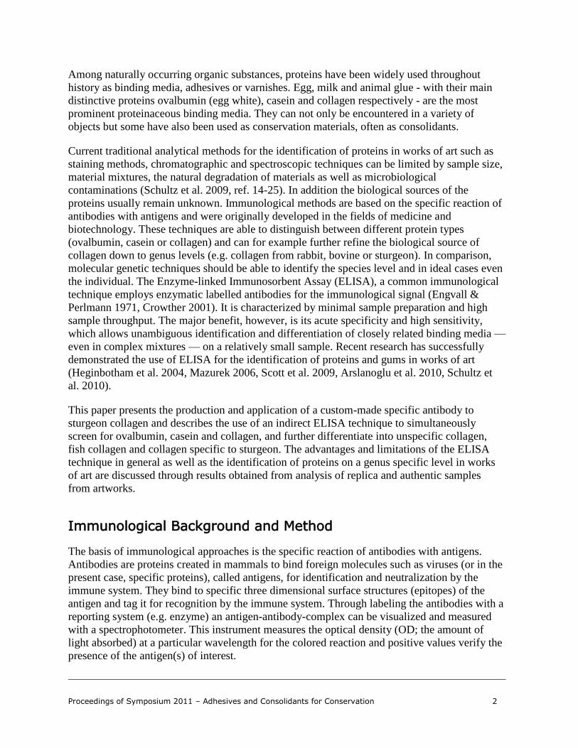
Immunological Background and Method
The basis of immunological approaches is the specific reaction of antibodies with antigens.
Antibodies are proteins created in mammals to bind foreign molecules such as viruses (or in the
present case, specific proteins), called antigens, for identification and neutralization by the
immune system. They bind to specific three dimensional surface structures (epitopes) of the
antigen and tag it for recognition by the immune system. Through labeling the antibodies with a
reporting system (e.g. enzyme) an antigen-antibody-complex can be visualized and measured
with a spectrophotometer. This instrument measures the optical density (OD; the amount of
light absorbed) at a particular wavelength for the colored reaction and positive values verify the
presence of the antigen(s) of interest.

Proceedings of Symposium 2011 – Adhesives and Consolidants for Conservation 3
There are two general categories of antibodies: mono- and polyclonal. Whereas monoclonal
antibodies – those derived from a single cell line of the host - recognize and bind only to one
single epitope, polyclonal antibodies are a serum product containing many different antibodies
that can recognize several epitopes of the specific antigen. Only polyclonal antibodies were
used in this study and they are typically produced by immunization of a suitable mammal such
as rabbit or goat.
In direct ELISA, in simple terms, an unknown amount of antigen is extracted from a sample.
The extracted solution is placed into the wells of a commonly used 96-well microtiter plate
where the antigen binds to the plastic surface of the wells by adsorption. The antigen,
immobilized on the surface, is then incubated with a solution of specific antibody, which can
bind to the antigen (if present). The antibody is conjugated to an enzyme, and in a final step a
substrate is added such that the enzyme converts to yield a detectable colorimetric signal. If the
target protein is present the solution in the wells turns green, otherwise they remain colourless.
The colour is depending on the enzyme and substrate used for the assay. The indirect ELISA
adds another step and is based on the use of a non-conjugated primary antibody and an enzyme-
conjugated secondary antibody. The addition of a secondary antibody increases the sensitivity
of the assay which is desirable if sample sizes are limited. An in depth discussion of the
principles of ELISA techniques applied to the analysis of artworks can be found elsewhere
(Schultz et al. 2009). Indirect ELISA experiments reported here were developed at the HAWK
Hildesheim (Schultz 2006) and further optimized at the Metropolitan Museum of Art as part of
the author’s PhD dissertation (forthcoming). This technique was used for the identification of
different proteins in paint samples from a replica panel painting prepared by the author in 2005,
species specific investigations of glues, and the detection of fixatives in pastel paintings.
Figure 1: Commonly used 96-well microtiter plate for
ELISA showing positive detection by green colour of a
target protein (casein)
Proceedings of Symposium 2011 – Adhesives and Consolidants for Conservation 4
Results and Discussion
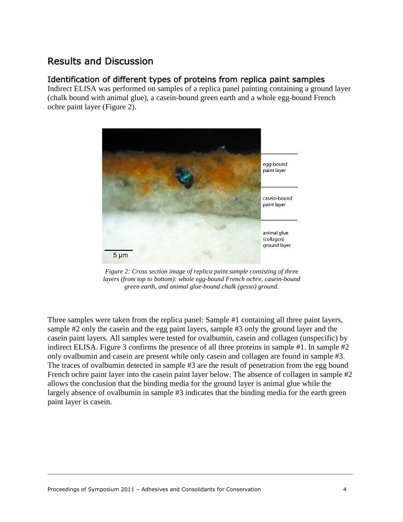
Identification of different types of proteins from replica paint samples Indirect ELISA was performed on samples of a replica panel painting containing a ground layer
(chalk bound with animal glue), a casein-bound green earth and a whole egg-bound French
ochre paint layer (Figure 2).
Figure 2: Cross section image of replica paint sample consisting of three
layers (from top to bottom): whole egg-bound French ochre, casein-bound
green earth, and animal glue-bound chalk (gesso) ground.
Three samples were taken from the replica panel: Sample #1 containing all three paint layers,
sample #2 only the casein and the egg paint layers, sample #3 only the ground layer and the
casein paint layers. All samples were tested for ovalbumin, casein and collagen (unspecific) by
indirect ELISA. Figure 3 confirms the presence of all three proteins in sample #1. In sample #2
only ovalbumin and casein are present while only casein and collagen are found in sample #3.
The traces of ovalbumin detected in sample #3 are the result of penetration from the egg bound
French ochre paint layer into the casein paint layer below. The absence of collagen in sample #2
allows the conclusion that the binding media for the ground layer is animal glue while the
largely absence of ovalbumin in sample #3 indicates that the binding media for the earth green
paint layer is casein.

Proceedings of Symposium 2011 – Adhesives and Consolidants for Conservation 5
0,000
0,100
0,200
0,300
0,400
0,500
0,600
0,700
0,800
0,900
1,000
1,100
1,200
1,300
1,400
ovalbumin
(egg)
casein collagen
(unspecific)
ovalbumin
(egg)
casein collagen
(unspecific)
ovalbumin
(egg)
casein collagen
(unspecific)
#1 whole sample, all 3 paint layers #2 only casein and egg-bound paint
layers
#3 only collagen (ground) and casein-
bound paint layers
Op
tica
l De
nsi
ty O
D (
41
4n
m)
Figure 3: ELISA results for the identification of proteins in replica paint samples. The concentration for all
samples shown here is 30 µg/ml of pigmented sample in aqueous buffer solution.
It is advised not to relate optical density (OD) value intensities with protein quantification when
dealing with art samples, due to protein degradation, extraction efficiency and pigment
interference. Furthermore, each antibody has a variable response to its antigen which also
results in a variation in colorimetric response relative to concentration. ELISA can easily
identify complex mixtures of binding media but only gives qualitative information on the
protein(s) present in the whole sample. However, in some cases with appropriate sample
preparation, the spatial distribution of the protein(s) can be determined within the sample. This
ELISA was designed to definitively confirm the presence of those proteins that were part of the
assay screening. But a comparison of intensities (semi-quantitative) is allowable when the
layers/parts analyzed are from the same sample and were tested on the same plate and in the
same experiment.
Species specific investigation of sturgeon glue In some cases, not only the differentiation of the protein types is of interest, but also the
biological source. Species specificity may be important for determining geographical
provenance or mapping of trade routes, or could aid in the understanding of the history of use of
specific materials or the verification of historical recipes. This seems to be of particular interest
in the case for sturgeon glue, which is obtained from sturgeons´ swim bladders.

Proceedings of Symposium 2011 – Adhesives and Consolidants for Conservation 6
The use of sturgeon glue is mentioned in historic documents from the medieval times for the
illumination of books, where it was used as a size on which pigments or gold leaf were laid and
occasionally as a medium. However, historic sources do not always specify which fish species
or part of the fish was used to make the glue. Sturgeon glue was also used for sizing paper, for
gilding of glass and wood, and as a fixative for pastel drawings (Petukhova 1989). It has been
traditionally used as an adhesive in painting and icon conservation and represents nowadays an
essential conservation material for consolidation purposes. The exclusive use and acclaimed
properties of sturgeon glue make its identification often desirable.
A vast number of antibodies are commercially available on the market but almost no species-
specific ones for the needs and requirements of conservation science exist. At present only
monoclonal sturgeon collagen antibodies are offered. Using a monoclonal antibody is
advantageous as it is more likely to identify a species of origin of the protein of interest.
However, if the antigen is damaged and the specific epitope is lost, for example due to
degradation, the antigen will not be detected. Therefore, we decided to use a custom made
polyclonal sturgeon antibody. Polyclonal antibodies are used to obtain general information of
the protein, and due to their greater variety, often increase the chance of detection at the
expense of specificity. This is particularly important in works of art, where the state of the
protein is unknown due to degradation over time.
The specificity of an antibody depends on the quality and purity of the antigen, among other
factors. Usually, little information is known about the exact origin or species, manufacturing
processes, pre-treatments and additives of commercially-available swim bladders, which can
have an impact on the glues’ quality but more significantly influence the antibody production.
Therefore, the use of commercial sturgeon bladder was eliminated and fresh bladder material of
a known sturgeon species (Siberian sturgeon) from a controlled fish farm was used as antigen.
The quality of any antibody is particularly dependent on its specificity. Upon receipt the
specificity of the custom-made sturgeon collagen antibody was extensively evaluated by
indirect ELISA. The results confirmed that the polyclonal sturgeon antibody is not specific to
the Siberian sturgeon from which it was derived, but to sturgeon species in general. The
detection limit determined for sturgeon glue was below 1 µg/ml. No cross reactivity was
detected with other fish, fish bladders, or fish and mammal glues, both commercial glues and
those made from known species (Figure 4).

Proceedings of Symposium 2011 – Adhesives and Consolidants for Conservation 7
0,000
0,100
0,200
0,300
0,400
0,500
0,600
0,700
0,800
0,900
1,000O
pti
cal
De
ns
ity
OD
(4
14
nm
)sturgeon bladder from a farm
sturgeon bladder (com)
sturgeon bladder, cheap (com)
Eel
Hering
Salmon
carp bladder from a farm
pike bladder from a farm
trout glue*
carp glue*
liquid fish glue (com)
gelatine (com)
hide glue from cow*
rabbit skin glue (com)
rabbit skin glue*
bone glue from pig*
parchment glue from goat*
Figure 4: ELISA evaluation of the new custom made polyclonal sturgeon bladder antibody: Specificity and cross
reactivity. (com) stands for commercial product; *glue from known species prepared by the author.
The concentration used for all samples was 10 µg/ml of collagen based material in aqueous buffer solution.
OD values below 0.05 cannot be considered as positive detection.
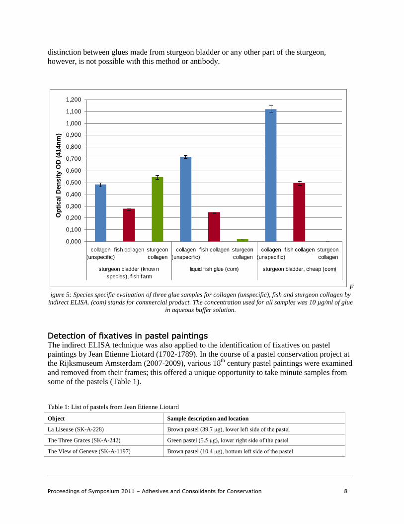
However, one of the commercial sturgeon glues tested negative. The appearance and the low
price of this glue differed greatly from all other tested sturgeon glues. In a separate ELISA
experiment, the cheap sturgeon bladder was tested together with a sturgeon bladder of known
origin and species and a commercial fish glue for collagen (species-unspecific), fish collagen
and sturgeon collagen (Figure 5). The presence of collagen was verified in all three glues, but
the origins differed. The result of the fish glue confirms the presence of fish collagen but not
sturgeon; whereas the sturgeon bladder from the fish farm was positive for fish and for sturgeon
collagen. The result from the cheap sturgeon glue was only positive for fish and not for sturgeon
collagen. Reasons for the negative result can be manifold but most likely indicate that the cheap
glue tested does not contain any sturgeon at all and/or is chemically modified such that the
sturgeon specific epitopes are lost and no antibody binding is possible. The glue does contain
fish collagen, but whether degraded sturgeon collagen or any other fish species were used
cannot be answered with this ELISA set up. The result demonstrates the problematic nature of
commercial trade names, but more importantly it underlines the significance of a well chosen
antigen for any antibody production (known origin and species).
The polyclonal custom-made sturgeon collagen antibody has proven highly specific to sturgeon
species only and seems suitable for the identification of sturgeon glue in works of art. A
Proceedings of Symposium 2011 – Adhesives and Consolidants for Conservation 8
distinction between glues made from sturgeon bladder or any other part of the sturgeon,
however, is not possible with this method or antibody.
0,000
0,100
0,200
0,300
0,400
0,500
0,600
0,700
0,800
0,900
1,000
1,100
1,200
collagen
(unspecif ic)
f ish collagen sturgeon
collagen
collagen
(unspecif ic)
f ish collagen sturgeon
collagen
collagen
(unspecif ic)
f ish collagen sturgeon
collagen
sturgeon bladder (know n
species), f ish farm
liquid f ish glue (com) sturgeon bladder, cheap (com)
Op
tical
Den
sit
y O
D (
414n
m)
F
igure 5: Species specific evaluation of three glue samples for collagen (unspecific), fish and sturgeon collagen by
indirect ELISA. (com) stands for commercial product. The concentration used for all samples was 10 µg/ml of glue
in aqueous buffer solution.
Detection of fixatives in pastel paintings The indirect ELISA technique was also applied to the identification of fixatives on pastel
paintings by Jean Etienne Liotard (1702-1789). In the course of a pastel conservation project at
the Rijksmuseum Amsterdam (2007-2009), various 18th
century pastel paintings were examined
and removed from their frames; this offered a unique opportunity to take minute samples from
some of the pastels (Table 1).
Table 1: List of pastels from Jean Etienne Liotard
Object Sample description and location
La Liseuse (SK-A-228) Brown pastel (39.7 μg), lower left side of the pastel
The Three Graces (SK-A-242) Green pastel (5.5 μg), lower right side of the pastel
The View of Geneve (SK-A-1197) Brown pastel (10.4 μg), bottom left side of the pastel

Proceedings of Symposium 2011 – Adhesives and Consolidants for Conservation 9
Relevant literature reports that fish glue mixed with equal quantities of water and spirit of wine
was a commonly used fixative in the 18th
century (Shelley 2005). Apparently, Liotard also
refers to those formulas but it is unknown if he made use of them (Trivas 1941; Anderson
1994). Three samples from Liotard’s La Liseuse, The Three Graces and The View of Geneve
were tested for fish and sturgeon collagen by indirect ELISA. Neither fish nor sturgeon collagen
was detected in any of the artwork samples.
However, to determine whether ELISA can be applied to the identification of fixatives, replica
samples with and without pastel or fixative, non-aged and artificially aged (15 days at 80°C and
65% RH) were prepared (Courtesy Cécile Gombaud, conservator in the pastel conservation
project of the Rijksmuseum Amsterdam) and evaluated by ELISA. The fixative was prepared as
follows: 0.5% percent (w/v) sturgeon glue in a water/ethanol mixture (1:1 v/v). The experiments
were repeated several times with decreasing amount of sample (milligrams to micrograms) to
establish detection limits (Table 2).
Table 2: List of pastel replica samples and tested sample amounts
Replica sample# Sample amounts* tested by ELISA
#1 parchment, sturgeon glue fixative, non-aged 49.1 μg 567.2 μg - 2.3 mg
#2 parchment, pastel (iron oxide), sturgeon glue fixative, non-
aged
66.6 μg 175.5 μg - 2.5 mg
#3 parchment, sturgeon glue fixative, artificially aged 68.7 μg 92.1 μg 135.6 μg 2.6 mg
#4 parchment, pastel (iron oxide), sturgeon glue fixative,
artificially aged
57.7 μg 143.3 μg 277.1 μg 4 mg
#5 parchment, pastel (iron oxide), no fixative, artificially aged 90.2 μg 129 μg - 2.8 mg
* total sample amount including parchment, pigment and fixative (where applied)
Out of five replica samples only the non-aged pigmented sample (#2) with the largest sample
amount tested positively for fish and sturgeon collagen. The result indicates that:
there is a higher chance of protein detection for a pigmented sample because it contains
a greater amount of binding media due to the increased surface
the protein retains immunoactive after the addition of ethanol used in the fixative
preparation (see recipe above). Ethanol is known for degrading proteins.
aging has a devastating effect on the already initially low-concentrated protein
but most importantly, the overall concentration of the fixative is too low
In order to get above the detection limit one would need an inappropriate amount of sample.
Thus, in case of the pastel paintings, it is possible that no fixative or a fixative other than fish-
based was used by Liotard or that the amount of protein in the fixative was below the detection
limit of the ELISA technique.

Proceedings of Symposium 2011 – Adhesives and Consolidants for Conservation 10
Conclusion
ELISA has proven effective in distinguishing proteins such as ovalbumin, casein and collagen
(various sources). It is highly sensitive and provides qualitative information on the proteins,
most importantly in mixtures. Still, quantification of proteins in art samples remains challenging
and may never be possible by ELISA due to difficulties in extraction efficiency, protein
degradation and pigment interference.
Immunological methods can only detect proteins that are part of the assay; of course, other
proteins may be present. Moreover, a negative ELISA result does not always mean that the
target protein is not present in the sample. Other possibilities may be that the protein content in
the sample is below the detection limit of the ELISA due to low concentration, insufficient
extraction, or degradation of the protein.
Species specificity is not easy to achieve when used for the identification of organic material in
conservation science due to the similarity of related species, the inconsistent compositions of
proteinaceous materials (modern and historical) and most importantly, the loss of
immunoreactivity due to protein degradation and material mixtures. However, the presented
results are encouraging because of the ability to narrow down the biological source of collagen
to unspecific collagen, fish collagen or collagen specifically from sturgeon. Antibody-based
techniques - like every analytical technique - have limitations but are clearly poised to take a
significant and complementary role in the analysis of proteins in works of art in the near future.
Acknowledgements
The authors wish to thank The Metropolitan Museum of Art, New York, especially Julie
Arslanoglu (Associate Scientist at the Department of Scientific Research) and the Andrew W.
Mellon Foundation for the financial support. Special thanks to Idelette van Leeuwen (Head of
Paper Conservation of the Rijksmuseum) and Cécile Combaud (freelance paper conservator) for
their contribution to the pastel analysis and Beth Edelstein (Assistant Conservator at The
Sherman Fairchild Center for Objects Conservation at The Metropolitan Museum of Art) for her
comments.

Proceedings of Symposium 2011 – Adhesives and Consolidants for Conservation 11
References
Anderson, J. “Fixing Pastels: A Letter from Liotard to the 2nd Earl of Bessborough in 1763”. The Burlington Magazine, 13 (1994), pp. 23-25. Arslanoglu, J., J. Schultz, J. Loike, K. Petersen “Immunology and Art: Using antibodies-based Techniques to Identify Proteins and Gums in Artworks”. Journal of Bioscience, 35 (2010), pp. 3-10. Crowther, J.R. The ELISA Guidebook. Humana Press, New Jersey, 2001. Engvall, E. and P. Perlmann “Enzyme-linked Immunosorbent Assay (ELISA) quantitative Assay of Immunoglobulin G”. Immunochemistry, 8 (1971), pp. 871-875. Heginbotham, A., V. Millay, and M. Quick “The Use of Immunofluorescence Microscopy (IFM) and Enzyme-linked Immunosorbent Assay (ELISA) as Complementary Techniques for Protein Identification in Artists’ Materials”. Journal of the American Institute for Conservation, 45 (2006), pp. 89-105. Mazurek, J. Identification of Proteins in Works of Art by Enzyme-linked Immunosorbent Assay (ELISA). M.A. thesis, University of California Davis, 2006. Unpublished. Petukhova, T. “Potential application of isinglass adhesive for paper conservation”. The Book and Paper Group Annual, (AIC), 8 (1989), pp. 58-61. Schultz, J. „Immunologische Methoden zur Analytik tierischer Bindemittel. Möglichkeiten und Grenzen“. M.A. thesis HAWK University of Applied Sciences and Arts of Hildesheim, Holzminden, Göttingen, Germany, 2006. Unpublished. Schultz, J., J. Arslanoglu, C. Tavzes, K. Petersen “Immunological Techniques: A different Approach to the Analysis of Proteins in Cultural Heritage. Part I: the basics explained“. Zeitschrift für Kunsttechnologie und Konservierung 23 (2009), pp. 129-139. Schultz, J., J. Arslanoglu and K. Petersen “The use of EL ISA for the identification of proteinaceous binding media from an eighteenth-century Damascene reception room”. p. 269 in Preprints of Papers presented at the “Conservation and the Eastern Mediterranean” Congress of the International Institute for Conservation of Historic and Artistic Works, Istanbul, 20-24. September 2010. London: International Institute for conservation of Historic
and Artistic Works, 2010 Schultz, J. „Immunologische Methoden zur Analytik tierischer Bindemittel an Kunst- und Kulturgut“ (working title). PhD disseration, Stuttgart State Academy of Art and Design, Germany (forthcoming). Scott, D.A., S. Warmlander, J. Mazurek, S. Quirke “Examination of some Pigments, Grounds and Media from Egyptian Cartonnage Fragments in the Petrie Museum, University College London”. Journal of Archaeological Science, 36 (2009), pp. 923-932. Shelley, M. “Pastelists at Work: Two Portraits at the Metropolitan Museum by Maurice Quentin de La Tour and Jean Baptiste Perronneau”. Metropolitan Museum Journal, 40 (2005), pp. 105-120. Trivas, N.S. “Two Formulas by Liotard”. Technical Studies, 10 (1941), pp. 29-32.

Proceedings of Symposium 2011 – Adhesives and Consolidants for Conservation 12
Materials and suppliers
Falcon 96-well flexible PVC assay plates, flat bottom, #353912, Becton Dickinson: http://www.bd.com/ Newborn calf serum (NCS) #SH30118.02, HyClone, Citric acid, #A940 10xPBS liquid Concentrate, #BP3994 Tween 20, #BP337 Thermo Fisher Scientific: http://www.fishersci.com/ Sodium azide, #S2002, Sigma-Aldrich: http://www.sigmaaldrich.com/ Rb-anti-ovalbumin, #AB1225, Millipore: http://www.millipore.com/ Rb-anti-bovine casein, #RCAS-10A, Immunology Consultants Laboratory, Inc.:http://www.icllab.com/ Rb-anti-collagen type I, #ab34710, Abcam: http://www.abcam.com/ Rb-anti-fish collagen type I, #T89171R, Meridian Life Science, Inc.: http://meridianlifescience.com/ Gt-anti-rabbit-HRP, IgG, #4050-05 ABTS substrate powder, #0202 Southern Biotech: http://www.southernbiotech.com/

Proceedings of Symposium 2011 – Adhesives and Consolidants for Conservation 13
Author Biographies and Contact Information
Biographies et coordonnées des auteurs
Julia Schultz was trained as a conservator specializing in furniture and wooden objects. She received a Diploma from the University of Applied Sciences and Arts Hildesheim (HAWK), Germany in 2003, with a focus on historical woodworking tools and microbiologically contaminated objects. After a postgraduate internship in the Department of Decorative Arts and Sculpture Conservation at the J. Paul Getty Museum in California (2003–2004), she obtained an MA in Conservation from HAWK, researching antibody-based techniques for the identification of proteinaceous binding media in works of art (2006). She is currently a PhD student at the State Academy of Art and Design in Stuttgart, Germany and, after 2 years as an Andrew W. Mellon Fellow at the Department of Scientific Research at The Metropolitan Museum of Art in New York, she is now a research assistant at HAWK where she continues her work on the application of immunological techniques to the study of works of art.
Contact Information: Faculty of Preservation of Cultural Heritage, Microbiology University of Applied Sciences and Arts Hildesheim (HAWK) Bismarckplatz 10-11, 31135 Hildesheim, Germany E-mail: [email protected]
Julia Schultz a suivi une formation spécialisée en restauration, des meubles et objets de bois. Elle a obtenu un diplôme en 2003 de la University of Applied Sciences and Arts Hildesheim (HAWK), en Allemagne, ses études portant principalement sur les anciens outils à bois et les objets contaminés par des micro-organismes. Après un stage de formation postdoctorale au département de restauration d’œuvres d’art décoratif et de sculptures du J. Paul Getty Museum, en Californie (2003-2004), elle obtient une maîtrise en conservation-restauration à la HAWK, où elle approfondit les techniques à base d’anticorps utilisées pour identifier les liants protéiniques dans les œuvres d’art (2006). Elle est actuellement doctorante à la Stuttgart State Academy of Art and Design, en Allemagne, et après deux ans, elle est devenue boursière de la fondation Andrew W. Mellon au service de la recherche scientifique du Metropolitan Museum of Art de New York, et elle est désormais adjointe à la recherche à la HAWK, où elle poursuit ses travaux sur l’emploi des techniques immunologiques pour analyser les œuvres d’art.
Coordonnées : Faculty of Preservation of Cultural Heritage, Microbiology University of Applied Sciences and Arts Hildesheim (HAWK) Bismarckplatz 10-11, 31135 Hildesheim, Allemagne Courriel : [email protected]
Karin Petersen has a PhD in Plant Physiology from the Christian Albrechts University in Kiel, Germany (1984). She is presently a Professor of Microbiology in the field of conservation at the University of Applied Sciences and Arts Hildesheim (HAWK). Her research focus is biodeterioration and health care.
Contact Information: Faculty of Preservation of Cultural Heritage,
Microbiology University of Applied Sciences and Arts Hildesheim (HAWK) Bismarckplatz 10-11, 31135 Hildesheim, Germany E-mail: [email protected]
Karin Petersen détient un doctorat en physiologie végétale de la Christian Albrechts University, à Kiel en Allemagne (1984). Elle est actuellement professeure de microbiologie dans le domaine de la conservation- restauration à la University of Applied Sciences and Arts Hildesheim (HAWK). Ses recherches portent sur la biodétérioration et les soins de santé.
Coordonnées : Faculty of Preservation of Cultural Heritage, Microbiology University of Applied Sciences and Arts Hildesheim (HAWK) Bismarckplatz 10-11, 31135 Hildesheim, Allemagne Courriel : [email protected]