Adenocarcinoma of the Third and Fourth Portions of the Duodenum
Upload
up-dilimanCategory
view
2download
0
International Journal of Cancer Research 11 (2): 80-92, 2015ISSN 1811-9727 / DOI: 10.3923/ijcr.2015.80.92© 2015 Academic Journals Inc.
Anti-Cancer Effect and Mechanisms of Action of Mikania cordataPlant Extract on MCF-7 Human Breast Adenocarcinoma Cells
1T.G.C. Uy, 1A.M. Licuanan, 1G.E.D. Angeles, 1M.L.C.C. Bote, 1,2E.A.B. Macauyag,2C.C. Hernandez, 1S.D. Jacinto and 1R.M. Guzman-Genuino1Institute of Biology, University of the Philippines Diliman, Quezon City, 1101, Philippines2Institute of Chemistry, University of the Philippines, Diliman, Quezon City, 1101, Philippines
Corresponding Author: R.M. Guzman-Genuino, Institute of Biology, National Science Complex, University of the Philippines,Diliman, Quezon City, 1101, Philippines Tel: +632-9205471
ABSTRACTMikania cordata is a medicinal plant traditionally used as an antibacterial, anti-ulcer,
anti-inflammatory, antihelmintic and analgesic agent. The potential of M. cordata as ananti-cancer agent has not been explored thus far. This study aims to assess M. cordata as a possibleanti-cancer agent, specifically against MCF-7 breast cancer cells. Ethanol extract-vacuum liquidchromatography fractions inhibited proliferation of the MCF-7 cells via different mechanisms.Fraction 6 induces non-specific cytotoxicity via apoptosis and oxidative pathways demonstrated bythe TUNEL and DPPH assays. Fractions 7, 10 and 11 exhibit selective cytotoxicity against MCF-7breast cancer cells but does so through mechanisms other than apoptosis and oxidation. The resultssuggest that novel mechanisms of action may be at work, imparting cancer-specific cytotoxicityproperties of these fractions. Phytochemical screening suggests terpenes and saponins as thepossible anti-cancer agent in M. cordata.
Key words: Mikania cordata, breast cancer, MCF-7, apoptosis, oxidation
INTRODUCTIONBreast cancer is the second most commonly diagnosed cancer worldwide, comprising 11.9% of
all new cancers diagnosed. It is currently the leading cause of cancer-related death among women,accounting for 522,000 deaths in 2012 alone (Ferlay et al., 2013). Despite the advances in diagnosisand treatment strategies, the trend established over the past decade continues to show increasingincidence and mortality rates. A major reason for treatment failure has been attributed to thedevelopment of resistance to the chemotherapeutic drugs used for therapy (Moreno-Aspitia andPerez, 2009; Marquette and Nabell, 2012). This highlights the need for discovery of new andeffective drugs to treat breast cancer.
There has been growing interest in the anticancer activities of medicinal plant extracts, due tothe presence of secondary metabolites that produce definite pharmacological actions on the humanbody with lesser side effects (Briskin, 2000). This approach has given rise to the identification ofvinca alkaloids, vinblastine and podophyllotoxins as anticancer compounds (Cragg and Newman,2005).
Mikania cordata (Burm. f.) B.L. Robinson is a creeping woody perennial of the family Ateraceae,widely distributed across Southeast Asia and Eastern Africa. It is more popularly known asheartleaf hempvine, climbing hempvine or mile-a-minute plant and has been described in the
80
Int. J. Cancer Res., 11 (2): 80-92, 2015
compendium of the world’s worst weeds (Mercado, 1994; Nayeem et al., 2011). The plant hastraditionally been used as herbal medicine to treat pain, inflammations and other infectiousdisease. Various studies have reported the antibacterial activity of its ethanol leaf extract, analegsicactivity of its sesquiterpene dilactone, antiulcer activity of its alkaloidal ethanolic fraction, aswell as its use in phytoremediation (Paul et al., 2000; Ahmed et al., 2001; Mahmud et al., 2008;Ali et al., 2011). Its sesquiterpene lactone scandenolide has also been found to inhibit inflammatorymediators such as leukotrienes and platelet activating factor synthesis in leukocytes (Ysrael andCroft, 1990). It also causes enhancement of drug-detoxifying enzymes in the liver and increasedrate of hepatic proteins synthesis (Mandal et al., 1992; Bishayee and Chatterjee, 1995). Although,anti-tumor activity against Hela and K562 cell lines has been demonstrated for Mikania micranthaplant extract of same genus, no anticancer studies have been done on Mikania cordata speciesdespite the identification of significant chemical constituents in a Gas Chromatographic MassSpectrometry analysis (Dou et al., 2014; Patar and Yahaya, 2012). Thus, research testing theanticancer activity of Mikania cordata is warranted.
This study hypothesized that M. cordata exerts anti-cancer effects on human breast cancer cellswhile reducing toxicity to normal cells and that its anti-cancer activity is mediated through theinduction of apoptosis and oxidation. Based on this hypothesis, the study aimed to explore thepotential of Vacuum-Liquid Chromatography (VLC)-fractionated ethanolic extracts of leaves ofM. cordata as anti-cancer agents against MCF-7 breast cancer cell line, evaluate its cytotoxicactivity to normal cells using J774A.1 mouse macrophage cell line and brine shrimp lethality assay,study its possible mechanisms of action through apoptosis and oxidation and identify its activecomponents through phytochemical screening and Thin Liquid Chromatography (TLC).
MATERIALS AND METHODSSolvent extraction of M. cordata plant extract: Harvested fresh leaves (396 g) were cleaned,cut into small pieces, air-dried for 7 days and homogenized by a blender (Osterizer). The resultingpowder was soaked in distilled methanol for 24 h and filtered using 20-25 μm pore sized Whatmanfilter paper. The filtrate was concentrated in a rotary evaporator at 45°C (IKA digital evaporator)and the brown condensate was left to air-dry. The dried brown condensate was dissolved in distilledwater (AMT) and sonicated (Skymen) at Room Temperature (RT) before performing solventextraction. Fractions were obtained in increasing polarity using hexane, ethyl acetate and distilledwater, accordingly. To obtain hexane fraction, extract was vigorously mixed in 2 parts hexane: 1part distilled water solvent in a separating funnel. The hexane layer was collected and concentratedin rotary evaporator (45°C, IKA digital evaporator). The hexane distillate obtained from thisprocess was used to exhaust the aqueous layer until it became colorless, after which the clear,organic upper layer was collected as ‘hexane fraction’. To obtain ethyl acetate fraction, the aqueouslayer from the hexane partitioning was vigorously mixed with 2 parts equivalent ethyl acetate ina separating funnel. The ethyl acetate layer was similarly collected and concentrated in rotaryevaporator (45°C, IKA digital evaporator) and used to exhaust the aqueous layer, after which brownlayer was collected as ‘ethyl acetate fraction’. The final aqueous layer was concentrated bylyophilization (Labconco) to obtain the ‘aqueous fraction’. The hexane, ethyl acetate and aqueousfractions were then tested for cytotoxic activity against MCF-7 breast cancer cells.
Vacuum liquid chromatography of M. cordata ethyl acetate fraction: The ethyl acetatefraction was further subjected to Vacuum Liquid Chromatography (VLC) to obtain 13 fractions. The
81
Int. J. Cancer Res., 11 (2): 80-92, 2015
stationary phase was prepared by evenly suspending the ethyl acetate fraction (6.00 g) in silicapowder (200 g) and minimal ethyl acetate. This was added to the top of a pre-vacuum pressed silicain a column (5 cm diameter, 20 cm height). The eluent for each mobile phase (250 mL) wasconcentrated by rotary evaporator (45°C, IKA digital evaporator) and air-dried to solid form. Eachplant extract fraction was prepared as 4 mg mLG1 in dimethyl sulfoxide (DMSO; Amresco),filter-sterilized through 0.2 μm Whatman filter and tested for cytotoxic activity against MCF-7breast cancer cells.
Cell lines: Homo sapiens breast adenocarcinoma cell line MCF-7 and Mus musculus normalmacrophage cell line J774A.1 were purchased from American Type Culture Collection (ATCC).MCF-7 was cultured in Minimum Essential Media (MEM; Gibco, Life Technologies), supplementedwith 10% Fetal Bovine Serum (FBS; Gibco Life Technologies), 0.01 mg mLG1 human recombinantinsulin (Gibco Life Technologies) and 1% penicillin-streptomycin (Gibco Life Technologies) in T-25flasks (Corning). J774A.1 was cultured in Dulbecco’s Minimum Essential Media, high glucose withL-glutamine (DMEM; Gibco, Life Technologies), supplemented with 1% v/v 1 mM sodiumpyruvate, 1% v/v 48% D(+)-glucose solution, 2% v/v 7.5% sodium bicarbonate, 10 FBS and1% penicillin-streptomycin (Gibco, Life Technologies) in T-25 flasks. All cells were incubated at37°C in a humidified atmosphere of 5% CO2.
Resazurin cell proliferation assay: To rapidly determine which solvent-extracted fraction ofM. cordata exerted cytotoxic activity against breast cancer cells, the resazurin cell proliferationassay was performed. MCF-7 cells (1.9×10⁴ cells/well) were seeded into 96 well plates using freshculture medium for 24 h at 37°C, 5% CO2 incubating condition. Seeded cells were confirmed viableby microscopic examination, then treated with filter-sterilized hexane, ethyl acetate or aqueousfraction. Positive control was 8 M hydrogen peroxide while negative control was DMSO diluent.After incubation for 72 h, 20 μL of resazurin dye (Life Technologies) was added, followed by 24 hincubation at RT. Absorbance was measured at 570 nm using a microplate reader(LabeximLedetect 96). Relative cytotoxicity was computed as the ratio of the absorbance of sampleto the absorbance of the negative control or untreated sample:
(1)spl
NC
ACyt 100
A
MTT cell proliferation assay: The effect of VLC-fractionated ethyl acetate extract of M. cordataon MCF-7 and J774A.1 cell viability was determined using the MTT (3-(4,5-dimethylthiazol-2-yl)-2,5-diphenyltetrazolium bromide) cell proliferation assay. The cells were seeded into 96 well platesat 1.14×10⁴ cells/well for MCF-7 and 7.6×10⁴ cells/well for J774A.1, using fresh culture medium for24 h at 37°C, 5% CO2 incubating condition. Seeded cells were confirmed viable by microscopicexamination, then treated with increasing concentrations of filter-sterilized plant extract(50, 25, 12.5, 6.25 μg mLG1) in triplicate. Positive control was doxorubicin (DBL), while negativecontrol was DMSO (Amresco) diluent. After incubation for 72 h, 20 μL of 5 mg mLG1 MTT (Amresco)was added, followed by 4 h incubation at 37°C, 5% CO2 until microscopic examination of purpleprecipitate. Then, 150 μL of dimethyl sulfoxide was added, after which plate was incubated in thedark at RT for 10 min. The formazan product of MTT was measured as absorbance at 570 nm using
82
Int. J. Cancer Res., 11 (2): 80-92, 2015
a microplate reader (Labexim Ledetect 96). The IC50 was determined as the concentration oftreatment showing 50% cell growth inhibition in comparison to the control cell growth.
Brine shrimp lethality assay: Cytotoxicity of plant extract was tested on organismal level usingthe Brine Shrimp Lethality Assay (BSLA). Artemia salina, brine shrimp eggs were grown in abeaker with artificial sea water, prepared by dissolving 38 g sea salt in 1 L distilled water. Thebeaker was aerated under lamp for 48 h to hatch and mature eggs as nauplii (larva). Ten brineshrimps were introduced into each well of 24 well plates. Filter-sterilized plant extract was addedto duplicate wells to make concentrations of 1000, 100 and 10 μg mLG1, using artificial seawater toadjust volume to 500 μL per well. Positive control was 10% ethanol, while DMSO was the negativecontrol. The plates were left uncovered under lamp for 24 h, after which the number of survivingshrimps was counted. Percent death was calculated by dividing the number of dead nauplii by thetotal number and then multiplied by 100. Using probit analysis, the Lethality Concentration (LC50) was assessed at 95% confidence interval. Following Meyer et al. (1982) a plant extract withLC50 value less than 1000 μg mLG1 was considered toxic, while LC50 value greater than1000 μg mLG1 was non-toxic (Meyer et al., 1982).
TUNEL assay: To test if mechanism for anticancer activity involved apoptosis, terminaldeoxynucleotidyl transferase-dUTP nick end labeling, TUNEL assay was performed. MCF-7(1.14×10⁴ cells/well) were seeded into 96 well plates (Corning Costar) using fresh culture mediumfor 24 h at 37°C, 5% CO2 incubating condition. After 24 h, seeded cells were confirmed viable bymicroscopic examination, then treated with increasing concentrations of filter-sterilized plantextract (50, 25, 12.5, 6.25 μg mLG1) in triplicate. Positive control was doxorubicin, whilenegative control was DMSO. After 72 h, the Click-iT® TUNEL Alexa Fluor® imaging assay(Life Technologies) was then conducted according to manufacturer’s instructions. Finally, the platewas imaged under Axio Observer inverted microscope (Carl Zeiss), using excitation wavelength of350 nm and emission wavelength of 460 nm to view Hoechst-33342 and excitation wavelength of495 nm and emission wavelength of 519 nm to view Alexa-Fluor® 488.
DPPH oxidation assay: To test if mechanism for anticancer activity involved oxidation, a DPPH(1,1-diphenyl-2-picrylhydrazyl) assay was used. DPPH solution (0.12 mg mLG1) (Sigma-Aldrich) wasprepared in absolute ethanol in a tube covered with aluminum foil, to prevent reacting with light(Blois, 1958). In each well of a 96-well plate, 90 μL of DPPH solution was dispensed, followed byaddition of 10 μL of filter sterilized plant extract to final concentration of 400 μg mLG1, in triplicate.Positive control was gallic acid (Sigma), while negative control was distilled water. This wasperformed in the dark to prevent light-induced discoloration. The plate was covered with parafilmand aluminum foil, mixed laterally for 30 sec and incubated at 37°C for 30 min, after whichabsorbance was measured at 570 nm using a microplate reader (Labexim Ledetect 96). Free radicalscavenging activity (%) was computed by Eq. 2, where, AH2O is the absorbance of distilled water, Aspl
is absorbance of sample and APC is absorbance of positive control of ascorbic acid:
(2)H2O spl
H2O PC
A AFRSA(%) 100
A A
Phytochemical screening: The phytochemical screening for tannins, saponins, terpenoids,flavonoids, cardiac glycosides, phenolic compounds, steroids, alkaloids and triterpenes used
83
Int. J. Cancer Res., 11 (2): 80-92, 2015
MCI
100
90
80
70
60
50
40
30
20
10
0MC2 MC3 MC4 MC5 MC6 MC7 MC8 MC9 MC10 MC11 MC12 DOXO
Prol
ifer
atio
n (%
)
Treatment (25 µg mL¯ )1
procedures with slight modifications from Harborne (1984), Edeoga et al. (2005) andOnwukaeme et al. (2007). Each plant extract fraction was prepared in DMSO (100 μg uLG1) andused as ‘plant sample’ for qualitative testing of phytochemical constituents.
Thin liquid chromatography: Each plant extract fraction (4 mg mLG1) was dissolved in asufficient amount of ethyl acetate and was plated to a 6×4 cm silica gel thin layer chromatographyplate. This was placed in a saturated gas chamber with shallow layer of 50% ethyl acetate and 50%hexane solvent. The solvent was allowed to rise till the 6 cm mark on the plate then removed forreading. The Retention factor values were obtained through the Iodine staining and visualizationunder ultraviolet light (254 nm).
Statistical analyses: Results were analyzed in SPSS (IBM SPSS Version 20) using one-wayanalysis of variance (ANOVA) and Duncan’s multiple range test as post hoc analysis. Differenceswere considered statistically significant at the level of p-value<0.05.
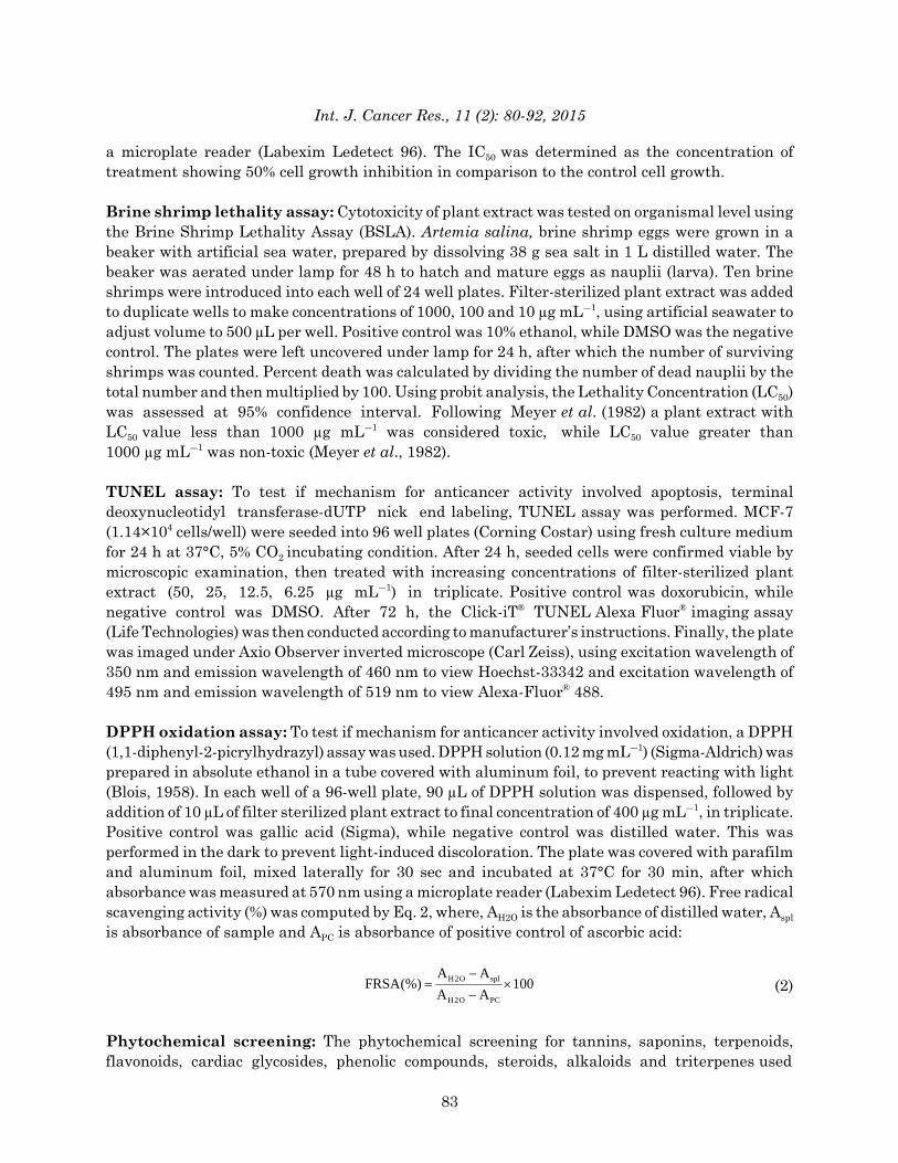
RESULTSMikania cordata fractions 6, 7, 10 and 11 inhibited proliferation of MCF-7 breast cancercells: To rapidly determine which solvent extracted fraction of M. cordata exerted anticanceractivity against breast cancer cells, resazurin cell proliferation assay was performed. Figure 1shows the relative percentage cytotoxicity of M. cordata hexane, ethyl acetate and aqueousfractions on MCF-7 cell line. The fractions were compared with hydrogen peroxide (H2O2) which isknown to cause oxidative stress-mediated cytotoxicity in MCF-7 cells (Alarifi, 2011). The ethylacetate fraction significantly inhibited the proliferation of MCF-7 cells. Furthermore, as with
Fig. 1: Effect of VLC-fractionated ethyl acetate M. cordata fractions on cell proliferation. Cellswere cultured in 96-well plates and treated with 4 mg mLG1 VLC fraction for 72 h. Cellviability was measured by MTT assay. Data represent the Mean±SD of three replicates.Statistical differences were analyzed with one-way ANOVA and Tukey’s range test
84
Int. J. Cancer Res., 11 (2): 80-92, 2015
0 10 20 30 40 50 60
100
90
80
70
60
50
40
30
20
10
0
Cel
l pr
oli
fera
tio
n (
%)
Concentration (µg mLG1)
DOXO
MC6
MC7
MC10
MC11
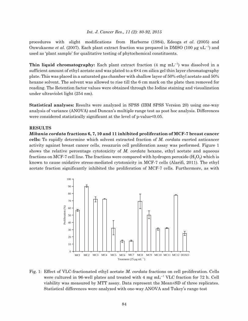
Fig. 2: Anti-proliferative effect of M. cordata on human breast cancer cells. Dose response curveof M. cordata treatment in MCF-7 cells. Cells were cultured in 96-well plates and treatedwith indicated concentrations of M. cordata VLC fractions for 72 h. Cell viability wasmeasured by MTT assay. Data represent the Mean±SD of three replicates. Statisticaldifferences were analyzed with one-way ANOVA and Tukey’s range test
untreated cells, treatment with 1% DMSO diluent had no significant cytotoxic effect on cells;therefore, the cytotoxic effect on MCF-7 was mediated by the plant extract treatment itself.
The ethyl acetate fraction of M. cordata was further partitioned by vacuum liquidchromatography. To determine the effect of the M. cordata VLC fractions on the proliferation ofbreast cancer cells, MTT cell proliferation assay was performed. Figure 1 shows the percent cellproliferation of MCF-7 breast cancer cells after treatment with different fractions. The fractionswere compared with doxorubicin (DBL), a widely used chemotherapeutic agent for breast cancer(Wang et al., 2004). M. cordata VLC fractions 6, 7, 10 and 11 significantly inhibited cellproliferation, at levels statistically similar to positive control doxorubicin. Their cytotoxicity curveson Fig. 2 show that inhibition was dosage-dependent. Although, cells treated with M. cordatagenerally had higher level of proliferation compared to positive control doxorubicin, % proliferationwas lower in cells treated with 25 μg mLG1 of fractions 6 and 7. Results suggest that M. cordatafractions 6 and 7 become more potent than doxorubicin but only at higher concentrations.Figure 3 compares the IC50 values of all fractions on MCF-7 cells which is shown in Table 1. Halfmaximal Inhibitory Concentration (IC50) values are commonly used to evaluate the potency of acompound, in which the lower the IC50 value, the compound is more potent.
Mikania cordata fractions 10 and 11 showed less cytotoxicity to normal J774A.1 murinemacrophages: To assess the effect of M. cordata fractions on normal cells, MTT cell proliferationassay was performed on a normal cell type, the J774A.1 murine macrophage cells (Blasi et al.,1987). The cytotoxicity curves of M. cordata fractions 6, 7, 10 and 11 in normal murinemacrophages in comparison with breast cancer cells. M. cordata fractions 6 and 7 inhibitedproliferation of both normal and cancer cells, indicative of non-specific cytotoxicity. M. cordata
85

Int. J. Cancer Res., 11 (2): 80-92, 2015
MCI
50
45
40
35
30
25
20
15
10
5
0MC2 MC3 MC4 MC5 MC6 MC7 MC8 MC9 MC10 MC11 MC12 DOXO
IC (
µg
mL
¯)
50
1
Treatments
* *
Fig. 3: Half maximal inhibitory concentration (IC50) values on MCF-7 for each VLC fraction. Theceiling value of 50 μg mLG1 was estimated, when IC50 could not be interpolated by linearregression in MC fractions 2 and 4
Table 1: Half maximal inhibitory concentration values on MCF7 and J774A.1 cells for each VLC fractionIC50 (μg mLG1)----------------------------------------------------------------------------------------------------------
Treatments MCF7 cells J774A.1 cellsMC1 30.297 24.510MC2 50.00* 40.640MC3 48.188 23.034MC4 50.00* 36.354MC5 20.965 10.899MC6 7.506 3.631MC7 9.185 4.890MC8 34.585 29.240MC9 25.210 38.942MC10 18.377 24.124MC11 17.449 26.540DOXO 2.041 1.997*Ceiling value of 50 μg mLG1 was estimated, when IC50 could not be interpolated by linear regression in MC fractions 2 and 4
fractions 10 and 11, however, inhibited proliferation of cancer cells more than normal cells.Accordingly, as shown in Table 1, IC50 was higher for normal cells, compared to the cancer cells.
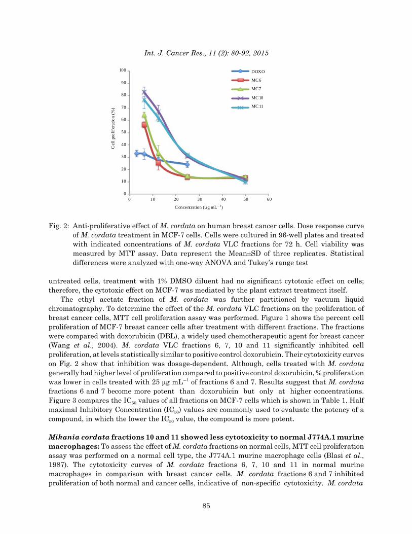
Mikania cordata fractions 10 and 11 showed less toxicity to brine shrimps: To screen fortoxicity to zoologic systems, M. cordata fractions were tested on hatched brine shrimp nauplii. TheVLC fractions all showed brine shrimp larvicidal activity. The Lethality Concentrations (LC50) areshown in Table 2. Since all the LC50 values were less than 1000 μg mLG1, the M. cordatafractions were considered toxic by standard Meyer et al. (1982). The brine shrimp lethalitywas dosage-dependent, with maximum mortality (100%) observed at 1000 μg mLG1 concentration.However, M. cordata fractions 10 and 11 are relatively less cytotoxic to brine shrimp, than fractions6 and 7.
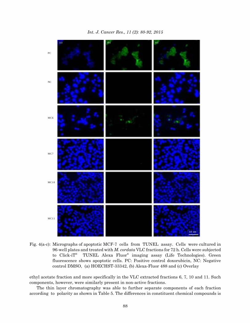
Mikania cordata fraction 6 induced apoptosis in MCF-7 cells: To determine if anti-canceractivity is mediated by apoptosis, TUNEL assay was performed. Figure 4 shows the apoptoticactivity of M. cordata fraction 6 on MCF-7 cells, similar to that of positive control, doxorubicin. In
86
Int. J. Cancer Res., 11 (2): 80-92, 2015
Table 2: Cytotoxic effect of M. cordata VLC fractions on brine shrimpPercent deaths at (24 h)--------------------------------------------------------------------------------------------------1000 100 10 LC50
Treatments ----------------------------------------------------------------(μg mLG1)---------------------------------------------------------------DMSO 100 45 10 83.621MC 1 100 80 40 17.298MC 3 100 70 45 17.205MC 4 100 70 30 29.162MC 5 100 80 35 20.454MC 6 100 70 50 13.719MC 7 100 85 45 13.257MC 8 100 60 35 30.818MC 9 100 60 30 36.044MC 10 100 65 30 32.388MC 11 100 75 35 22.539MC 12 100 50 20 58.828
Table 3: Phytochemical screening of solvent extracted M. cordataSolvent fraction Tannins Saponins Terpenoids Cardiac glycosides Phenolics Steroids Alkaloids FlavonoidsHexane - - - - - - - -Ethyl acetate - + + - - - - -Aqueous + + + - - - - +
Table 4: Phytochemical screening of vacuum liquid chromatography extracted- ethyl acetate fractions of M. cordataVLC fraction Tannins Saponins Terpenoids Cardiac glycosides Phenolics Steroids Alkaloids Flavonoids1 - + + - - - - -2 - + + - - - - -3 - + + - - - - -4 - + + - - - - -5 - + + - - - - -6 - + + - - - - -7 - + + - - - - -8 - + + - - - - -9 - + + - - - - -10 - + + - - - - -11 - + + - - - - -12 - - + - - - - -13 - - + - - - - -
contrast, more similar to the negative control DMSO, M. cordata fractions 7, 10 and 11 did notexert any apoptotic activity on the cells. The anti-cancer activity of M. cordata fraction 6 istherefore, mediated by apoptosis.
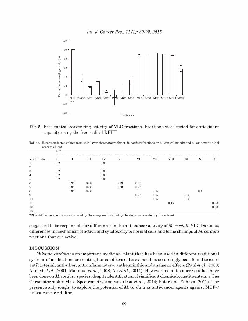
Mikania cordata fraction 6 induced oxidation in MCF-7 cells: To determine if anti-canceractivity is mediated by oxidation, DPPH assay was performed. Figure 5 shows the Free RadicalScavenging Activity (FRSA) values for each fraction. Fractions were compared to gallic acid which has established antioxidant properties (Bhadoriya et al., 2012). No oxidative activity was detectedin M. cordata fractions 7, 10 and 11; in fact, these fractions enhanced free radical scavengingactivity at a level similar to positive control gallic acid. Anti-cancer activity in fractions 7, 10 and11 is therefore, not mediated by oxidation. In contrast, oxidative activity was detected in M. cordatafraction 6 which indicates that its anti-cancer activity may also be mediated by oxidation.
Terpenes and saponins are candidates responsible for anti-cancer activity: Phytochemicalscreening was performed to identify possible active components responsible for the anti-canceractivity. Table 3 and 4 show the presence of saponins and terpenoids in the active M. cordata
87
Int. J. Cancer Res., 11 (2): 80-92, 2015
PC
NC
MC6
MC7
MC10
MC11
(a) (b) (c)
50 nm
Fig. 4(a-c): Micrographs of apoptotic MCF-7 cells from TUNEL assay. Cells were cultured in96-well plates and treated with M. cordata VLC fractions for 72 h. Cells were subjectedto Click-iT® TUNEL Alexa Fluor® imaging assay (Life Technologies). Greenfluorescence shows apoptotic cells. PC: Positive control doxorubicin, NC: Negativecontrol DMSO, (a) HOECHST-33342, (b) Alexa-Fluor 488 and (c) Overlay
ethyl acetate fraction and more specifically in the VLC extracted fractions 6, 7, 10 and 11. Suchcomponents, however, were similarly present in non-active fractions.
The thin layer chromatography was able to further separate components of each fractionaccording to polarity as shown in Table 5. The differences in constituent chemical compounds is
88
Int. J. Cancer Res., 11 (2): 80-92, 2015
MCI
120
100
80
60
40
20
0
-20
-40
MC2 MC3 MC4 MC5 MC6 MC7 MC8 MC9 MC10 MC11 MC12
Free
rad
ical
sca
veng
ing
acti
vity
(%
)
Treatments
DMSOGallicacid
Fig. 5: Free radical scavenging activity of VLC fractions. Fractions were tested for antioxidantcapacity using the free radical DPPH
Table 5: Retention factor values from thin layer chromatography of M. cordata fractions on silicon gel matrix and 50:50 hexane ethylacetate eluent
Rf*---------------------------------------------------------------------------------------------------------------------------------------------------------------
VLC fraction I II III IV V VI VII VIII IX X XI1 5.2 0.872 3 5.2 0.87 4 5.2 0.87 5 5.2 0.87 6 0.97 0.88 0.83 0.75 7 0.97 0.88 0.83 0.75 8 0.97 0.88 0.5 0.19 0.75 0.5 0.1310 0.5 0.13 11 0.17 0.0812 0.0813 *Rf is defined as the distance traveled by the compound divided by the distance traveled by the solvent
suggested to be responsible for differences in the anti-cancer activity of M. cordata VLC fractions,differences in mechanism of action and cytotoxicity to normal cells and brine shrimps of M. cordatafractions that are active.
DISCUSSIONMikania cordata is an important medicinal plant that has been used in different traditional
systems of medication for treating human disease. Its extract has accordingly been found to exertantibacterial, anti-ulcer, anti-inflammatory, anthelminthic and analgesic effects (Paul et al., 2000;Ahmed et al., 2001; Mahmud et al., 2008; Ali et al., 2011). However, no anti-cancer studies havebeen done on M. cordata species, despite identification of significant chemical constituents in a GasChromatographic Mass Spectrometry analysis (Dou et al., 2014; Patar and Yahaya, 2012). Thepresent study sought to explore the potential of M. cordata as anti-cancer agents against MCF-7breast cancer cell line.
89

Int. J. Cancer Res., 11 (2): 80-92, 2015
The results show that some of the Vacuum-Liquid Chromatography (VLC)-fractionated ethylacetate extracts of leaves of M. cordata have anticancer effects. M. cordata VLC fractions 6, 7, 10and 11 were found to significantly inhibit MCF-7 cell proliferation at levels statistically similar toimportant breast cancer drug doxorubicin. Inhibition was in a dosage-dependent manner and at25 μg mLG1, M. cordata fractions 6 and 7 began to inhibit cell proliferation more than doxorubicin,suggesting that these fractions become more potent than doxorubicin at higher concentrations. Thisreports a remarkable find with great potential for the cancer drug discovery program.
After establishing anticancer effects, the study sought to study its possible mechanisms of actionthrough apoptosis and oxidation and to evaluate each fraction for cytotoxicity to normal cells usingJ774A.1 mouse macrophage cell line and brine shrimp lethality assay. It found that the identifiedfractions exert their anticancer activities through different mechanisms. Fractions also differed inthe specificity of their cytotoxicity.
Mikania cordata fraction 6 exerted anticancer effects through both apoptotic and oxidativepathways. The TUNEL assay showed the occurrence of DNA fragmentation in the MCF-7 cellstreated with fraction 6. The DPPH assay also showed oxidation of the 1,1-diphenyl-2-picrylhydrazylreagent treated with fraction 6. This fraction, however, exerts non-specific cytotoxicity, inhibitingproliferation of both normal and cancer cells, as shown in cytotoxicity estimates to normal J774A.1murine macrophages and toxicity estimates to brine shrimps.
Mikania cordata fractions 10 and 11 show very interesting finds. They exert cytotoxicity morespecific for cancer cells. They inhibited proliferation of the MCF-7 cells more than the J774A.1 cellsand exhibited reduced mortality to brine shrimps.
Many chemotherapeutic regimens have been problematic due to their cytotoxicity to normalcells which cause debilitating side effects in patients that use them. This can be mitigated by theuse of chemicals that have more specific cytotoxicity for only the cancer cells. Such treatments canimprove clinical outcomes, since, clinicians can give higher concentrations for longer periods oftime, without significantly affecting quality of life of the patients.
The mechanism by which fractions 10 and 11 exert their cancer-specific cytotoxicity, however,is not mediated through either apoptotic or oxidative pathways. Studying their mechanism of actioncould therefore, elucidate pathways for targeted therapy.
Mikania cordata fraction 7, though showing non-specific cytotoxicity, also exerts anticancereffects through pathways other than apoptosis and oxidation. Such findings become significant inlight of the current problem of resistance. Breast cancer cells have been able to develop resistanceto current regimens that work by common mechanisms. Novel mechanisms of action couldtherefore, be potential treatments for resistant breast cancers.
The phytochemical screening pointed to terpenes and saponins as possible candidatesresponsible for exerting anticancer activities. On a further TLC analysis, the terpenes and saponinspresent in each fraction were found to be different in polarity which points to the presence of many,not just a single, active component in Mikania cordata, that is able to exert anticancer effects.Further dissection and analysis of individual components could lead to purified chemotherapeuticdrugs with increased potency and novel mechanisms of action.
The results from the present study clearly validate the anticancer potential of Mikania cordataextract. The findings become very important in light of the problem of current chemotherapeuticregimens being very expensive. Mikania cordata is a widely available plant, that is typically treatedas unwanted weeds, even being included in the compendium of world’s worst weeds. The studyshows that the weed could be a potential cheap source of treatment for cancer patients.
90
Int. J. Cancer Res., 11 (2): 80-92, 2015
In conclusion, Mikania cordata shows anti-cancer effects in MCF-7 human breastadenocarcinoma cells. Mikania cordata ethanol extract-vacuum liquid chromatography fractions6, 7, 10 and 11 inhibited proliferation of MCF-7 breast cancer cells. The anticancer effects of eachfraction act through different mechanisms of action and differ in specificity of cytotoxicity.Mikania cordata fraction 6 has non-specific cytotoxicity and acts through apoptosis and oxidativepathways. Mikania cordata fraction 7 also exerts non-specific cytotoxicity but acts throughmechanisms other than apoptosis and oxidation. Mikania cordata fractions 10 and 11 exertcytotoxicity more specific for cancer cells and acts through mechanisms other than apoptosis andoxidation. Phytochemical screening identified terpenes and saponins in the fractions that could becandidates responsible for the anti-cancer activity.
ACKNOWLEDGMENTSThis study was funded by the Institute of Biology, University of the Philippines-Diliman. The
group would like to acknowledge research assistants, Carlo Limbo, Cielo Marquez and ReginaFerrer, from the Mammalian Cell Culture Lab, Institute of Biology, University of the Philippines,as well as Leo Argulla and Lovely Kris Acuram, from the Natural Products Lab, Institute ofChemistry, University of the Philippines, for their assistance in the performance of experiments.
REFERENCESAhmed, M., M.T. Rahman, M. Alimuzzaman and J.A. Shilpi, 2001. Analgesic sesquiterpene
dilactone from Mikania cordata. Fitoterapia, 72: 919-921.Alarifi, S., 2011. Assessment of MCF-7 cells as an in vitro model system for evaluation of chemical
oxidative stressors. Afr. J. Biotechnol., 10: 3872-3879.Ali, S., S. Islam, M. Rahman, R. Islam, M.A. Sayeed and R. Islam, 2011. Antibacterial and cytotoxic
activity of ethanol extract of Mikania cordata (burm.f.) B.L. Robinson leaves. J. Basic Clin.Pharm., 2: 103-107.
Bhadoriya, U., P. Sharma and S.S. Solanki, 2012. In vitro free radical scavenging activity of gallicacid isolated from Caesalpinia decapetala wood. Asian Pac. J. Trop. Dis., 2: S833-S836.
Bishayee, A. and M. Chatterjee, 1995. Anticarcinogenic biological response of Mikania cordata:Reflections in hepatic biotransformation systems. Cancer Lett., 81: 193-206.
Blasi, E., D. Radzioch, S. Durum and L. Varesio, 1987. A murine macrophage cell line,immortalized by v-raf and v-myc oncogenes, exhibits normal macrophage functions. Eur. J.Immunol., 17: 1491-1498.
Blois, M.S., 1958. Antioxidant determinations by the use of a stable free radical. Nature,181: 1199-1200.
Briskin, D.P., 2000. Medicinal plants and phytomedicines. Linking plant biochemistry andphysiology to human health. J. Plant Physiol., 24: 507-514.
Cragg, G.M. and D.J. Newman, 2005. Plants as a source of anti-cancer agents. J. Ethnoharmacol.,100: 72-79.
Dou, X., Y. Zhang, N. Sun, Y. Wu and L. Li, 2014. The anti-tumor activity of Mikania micranthaaqueous extract in vitro and in vivo. Cytotechnology, 66: 107-117.
Edeoga, H.O., D.E. Okwu and B.O. Mbaebie, 2005. Phytochemical constituents of some Nigerianmedicinal plants. Afr. J. Biotechnol., 4: 685-688.
Ferlay, J., I. Soerjomataram, M. Ervik, R. Dikshit and S. Eser et al., 2013. GLOBOCAN 2012 v1.0,cancer incidence and mortality worldwide. IARC Cancer Base No. 11 [Internet], InternationalAgency for Research on Cancer, Lyon, France.
91

Int. J. Cancer Res., 11 (2): 80-92, 2015
Harborne, J.B., 1984. Phytochemical Methods: A Guide to Modern Techniques of Plant Analysis.2nd Edn., Chapman and Hall Ltd., London, UK., ISBN-13: 9780412255502, pp: 149-188.
Mahmud, R., N. Inoue, S.Y. Kasajima and R. Shaheen, 2008. Assessment of potential indigenousplant species for the phytoremediation of arsenic-contaminated areas of Bangladesh. Int. J.Phytoremed., 10: 117-130.
Mandal, P.K., A. Bishayee and M. Chatterjee, 1992. Stimulation of hepatic protein synthesis inresponse to Mikania cordata root extract in carbon tetrachloride-induced hepatotoxicity in mice.Italian J. Biochem., 41: 345-351.
Marquette, C. and L. Nabell, 2012. Chemotherapy-resistant metastatic breast cancer. Curr. Treat.Options Oncol., 13: 263-275.
Mercado, B.T., 1994. Notes on some growth characteristics of Mikania cord at a (Burm. F.)B.L. Robinson. J. Biotropia, 7: 30-40.
Meyer, B.N., N.R. Ferrigni, J.E. Putnam, L.B. Jacobsen, D.E. Nichols and J.L. McLaughlin, 1982.Brine shrimp: A convenient general bioassay for active plant constituents. Planta Med.,45: 31-34.
Moreno-Aspitia, A. and E.A. Perez, 2009. Treatment options for breast cancer resistant toanthracycline and taxane. Mayo Clin. Proc., 84: 533-545.
Nayeem, A.A., A. Khatun, M.S. Rahman and M. Rahman, 2011. Evaluation of phytochemical andpharmacological properties of Mikania cordata (Asteraceae) leaves. J. PharmacognosyPhytother., 3: 118-123.
Onwukaeme, D.N., T.B. Ikuegbvweha and C.C. Asonye, 2007. Evaluation of phytochemicalconstituents, antibacterial activities and effect of exudates of Pycanthus Angolensis weld warb(Myristicaceae) on corneal ulcers in rabbits. Trop. J. Pharm. Res., 6: 725-730.
Patar, A.A. and B.H. Yahaya, 2012. The analysis of aquoues and ethanolic extracts of MalaysianMikania cordata leaves towards the potential for medicinal substances. Eur. J. Sci. Res.,73: 434-440.
Paul, R.K., A. Jabbar and M.A. Rashid, 2000. Antiulcer activity of Mikania cordata. Fitoterapia,71: 701-703.
Wang, S., E.A. Konorev, S. Kotamraju, J. Joseph, S. Kalivendi and B. Kalyanaraman, 2004.Doxorubicin induces apoptosis in normal and tumor cells via distinctly different mechanisms.Intermediacy of H(2)O(2)-and p53-dependent pathways. J. Biol. Chem., 279: 25535-25543.
Ysrael, M.C. and K.D. Croft, 1990. Inhibition of leukotriene and platelet activating factor synthesisin leucocytes by the sesquiterpene lactone scandenolide. Planta Med., 56: 268-270.
92
Copyright © 2022 FDOKUMEN