Aniptodera salsuginosa, a new mangrove-inhabiting ascomycete, with observations on the effect of...
6
Mycol. Res. 98 (8): 931-936 (1994) Printed in Great Britain 931 Aniptodera salsuginosa, a new mangrove-inhabiting ascomycete, with observations on the effect of salinity on ascospore appendage morphology AKIRA NAKAGIRI AND T ADA YOSHI ITO Institute for Fermentation, Osaka, 17-85, ]uso-honmachi 2-chome, Yodogawa-ku, Osaka 532, Japan Aniptodera salsuginosa sp. nov. from intertidal decomposing mangrove wood is described, and its unique ascospore appendages and ascus apical apparatus are illustrated. The ascospore appendages were functional only when they were submerged in brackish water. The ascus tip is composed of a thickened apical ring (disc) with a pore, which is nonfunctional for ascospore release. Ascospores were discharged through a fissure in the ascus wall at the margin of the apical disc. Evolution and adaptation of the new fungus and related mangrove ascomycetes to aquatic environments from terrestrial habitats are briefly discussed. In a study of mangrove fungi from Iriomote Is., Okinawa, Japan, an unknown taxon similar to Aniptodera chesapeakensis Shearer & M. Miller and Halosarpheia minuta W. F. Leong, was found inhabiting decomposing mangrove wood [Nakagiri (1993) as 'Halosarpheia sp. 1']. This new fungus differs in ascoma, ascus and ascospore morphology from A. chesa- peakensis and H. minuta and has ascospores with stiff appendages at both ends. In distilled water, the ascospore appendages are immediately detached and the spores appear to be non-appendaged. However, in 20-30%0 seawater or in Shear's solution, they remain in close contact with the ascospore surface. Examination of appendage structure under different salinity levels revealed that they unravel immediately when they are suspended in brackish water (3-10%0)' The ascal ring, composed of a thickened apical disc with a pore, was also found to be nonfunctional. This is the first report of ascospore appendages responding to salinity levels. MATERIALS AND METHODS Collection and observation techniques follow those of Nakagiri (1993). Ascospore appendage morphology at various salinity levels (0, 3, 5, 10, 20 and 30%0) was observed under a microscope at regular intervals. RESULTS Aniptodera salsuginosa Nakagiri & Tad. Ito, sp. nov. (Figs 1-21). Etym.: From the Latin salsuginosa = growing in brackish or salt water, referring to the habitats. Ascomata 240-280 diametro, 92-112 alta, ellipsoidea vel applanata convexa, axe principali horizontali, cum collo curvato, nigra ad partem superam, hyalina ad partem inferam, immersa in epidermide cortice, ostiolata, papillata. Peridium 6-10 crassum, membranaceum. Colla 280-520 longa super superficiem hospitem, 100-160 longa in hospite, 35-48 diametro, periphysata. Paraphyses absentes. Catenophyses praesentes. Asci 76-90 x 12-14 octospori, cylindrici vel clavati, pedunculati, unitunicati, leptodermi, persistentes, cum apparatu apicalio. Ascosporae (12-) 14-20 (-23) x 4- 7 (x = 16'2 x 5'7 ellipsoideae vel fusiformes, hyalinae, uniseptatae. Ascosporarum appendices bipolares, in initio rigidae et tegentes super sporam, 9-14 longae, postea membranaceae in aqua salsa. Substratum, cortex mangrovium immersum. Holotype: IFO H-12161, from decomposing wood of Bruguiera gymnorrhyza (Shi-O-1), 26 Nov. 1991, from the Shiira River, Iriomote Is., Japan, deposited in the herbarium of the Institute for Fermentation, Osaka (IFO). Other material examined: IFO H-12162, from decomposing wood of Rhiwphora stylosa (Shi-Ya-6), 26 Nov. 1991, from the Shiira River, Iriomote Is.; Shi-O-2, from decomposing wood of B. gymnorrhyza, 26 Nov. 1991, from the Shiira River; Nak-O-3, from decomposing wood of B. gymnorrhyza, 26 Nov. 1991, from the Nakama River, Iriomote Is., Japan. Ascomata 240-280 IJIll diam., 92-112 IJm high, ellipSOidal to flattened cortex, main axis horizontal, black upper side, hyaline bottom, solitary, immersed just below the epidermis of bark, ostiolate, papillate (Figs 1, 2). Peridium 6-10 IJm thick, membranous, composed in the upper region of black elongate cells, interspersed among host cells; in the lower region of hyaline flat cells. Necks lateral, bending upward, 280-520 IJm long above the wood surface, 100-160 IJm long in the wood, 35-48 IJm diam., cylindricaL hyaline to subhyaline in the upper region, black in the lower region, with periphyses. Paraphyses absent. Catenophyses present. Asci 76-90 x 12-14 IJm, 8-spored, cylindrical to clavage, pedunculate, unitunicate, thin-walled, persistent, apex truncate with an apical thickening (ring) with a pore, with cytoplasm retracted in the subapical region. Asci develop in a hymenium at the base of the 59-2
-
Upload
independent -
Category
Documents
-
view
0 -
download
0
Transcript of Aniptodera salsuginosa, a new mangrove-inhabiting ascomycete, with observations on the effect of...

Mycol. Res. 98 (8): 931-936 (1994) Printed in Great Britain 931
Aniptodera salsuginosa, a new mangrove-inhabiting ascomycete,with observations on the effect of salinity on ascosporeappendage morphology
AKIRA NAKAGIRI AND TADAYOSHI ITO
Institute for Fermentation, Osaka, 17-85, ]uso-honmachi 2-chome, Yodogawa-ku, Osaka 532, Japan
Aniptodera salsuginosa sp. nov. from intertidal decomposing mangrove wood is described, and its unique ascospore appendages andascus apical apparatus are illustrated. The ascospore appendages were functional only when they were submerged in brackish water.The ascus tip is composed of a thickened apical ring (disc) with a pore, which is nonfunctional for ascospore release. Ascosporeswere discharged through a fissure in the ascus wall at the margin of the apical disc. Evolution and adaptation of the new fungus andrelated mangrove ascomycetes to aquatic environments from terrestrial habitats are briefly discussed.
In a study of mangrove fungi from Iriomote Is., Okinawa,Japan, an unknown taxon similar to Aniptodera chesapeakensisShearer & M. Miller and Halosarpheia minuta W. F. Leong, wasfound inhabiting decomposing mangrove wood [Nakagiri(1993) as 'Halosarpheia sp. 1']. This new fungus differs inascoma, ascus and ascospore morphology from A. chesapeakensis and H. minuta and has ascospores with stiffappendages at both ends. In distilled water, the ascosporeappendages are immediately detached and the spores appearto be non-appendaged. However, in 20-30%0 seawater or inShear's solution, they remain in close contact with theascospore surface. Examination of appendage structure underdifferent salinity levels revealed that they unravel immediatelywhen they are suspended in brackish water (3-10%0)' Theascal ring, composed of a thickened apical disc with a pore,was also found to be nonfunctional. This is the first report ofascospore appendages responding to salinity levels.
MATERIALS AND METHODS
Collection and observation techniques follow those of Nakagiri(1993). Ascospore appendage morphology at various salinitylevels (0, 3, 5, 10, 20 and 30%0) was observed under amicroscope at regular intervals.
RESULTS
Aniptodera salsuginosa Nakagiri & Tad. Ito, sp. nov.(Figs 1-21).
Etym.: From the Latin salsuginosa = growing in brackish orsalt water, referring to the habitats.
Ascomata 240-280 ~m diametro, 92-112 ~m alta, ellipsoidea velapplanata convexa, axe principali horizontali, cum collo curvato,nigra ad partem superam, hyalina ad partem inferam, immersa inepidermide cortice, ostiolata, papillata. Peridium 6-10 ~m crassum,
membranaceum. Colla 280-520~ longa super superficiem hospitem,100-160 ~m longa in hospite, 35-48 ~m diametro, periphysata.Paraphyses absentes. Catenophyses praesentes. Asci 76-90 x 12-14 ~,octospori, cylindrici vel clavati, pedunculati, unitunicati, leptodermi,persistentes, cum apparatu apicalio. Ascosporae (12-) 14-20 (-23) x 47~ (x = 16'2 x 5'7 ~m), ellipsoideae vel fusiformes, hyalinae,uniseptatae. Ascosporarum appendices bipolares, in initio rigidae ettegentes super sporam, 9-14 ~m longae, postea membranaceae inaqua salsa. Substratum, cortex mangrovium immersum.
Holotype: IFO H-12161, from decomposing wood of Bruguiera
gymnorrhyza (Shi-O-1), 26 Nov. 1991, from the Shiira River, IriomoteIs., Japan, deposited in the herbarium of the Institute for Fermentation,Osaka (IFO).
Other material examined: IFO H-12162, from decomposing wood ofRhiwphora stylosa (Shi-Ya-6), 26 Nov. 1991, from the Shiira River,
Iriomote Is.; Shi-O-2, from decomposing wood of B. gymnorrhyza, 26
Nov. 1991, from the Shiira River; Nak-O-3, from decomposingwood of B. gymnorrhyza, 26 Nov. 1991, from the Nakama River,
Iriomote Is., Japan.
Ascomata 240-280 IJIll diam., 92-112 IJm high, ellipSOidal toflattened cortex, main axis horizontal, black upper side,hyaline bottom, solitary, immersed just below the epidermisof bark, ostiolate, papillate (Figs 1, 2). Peridium 6-10 IJm thick,membranous, composed in the upper region of black elongatecells, interspersed among host cells; in the lower region ofhyaline flat cells. Necks lateral, bending upward, 280-520 IJmlong above the wood surface, 100-160 IJm long in the wood,35-48 IJm diam., cylindricaL hyaline to subhyaline in theupper region, black in the lower region, with periphyses.Paraphyses absent. Catenophyses present. Asci 76-90 x 12-14IJm, 8-spored, cylindrical to clavage, pedunculate, unitunicate,thin-walled, persistent, apex truncate with an apical thickening(ring) with a pore, with cytoplasm retracted in the subapicalregion. Asci develop in a hymenium at the base of the
59-2

A. Nakagiri and T. Ito 932
Figs 1-7. Light micrographs (Figs 1-4) and SEM micrographs (Figs 5-7) of Aniptodera salsuginosa. Fig. 1. Habit on mangrove wood.Necks (arrows) rise from ascomata immersed below the epidermis of the bark. Note the slight swelling of the bark which indicates thepresence of an ascoma. Fig. 2. Vertical section of ascoma immersed in bark. Fig. 3. Young and mature asci. Note the retraction of thecytoplasm in the subapical region of the asci (arrows). Fig. 4. A mature ascus showing a thickened apical ring. Fig. 5. SEM micrographof a mature ascus. Fig. 6. An apical ring (disc) of an ascus showing concentric undulations at the edge of the disc and around the centralpore. Fig. 7. An apical ring detaching from the ascus wall. Scale bars: 1, 2 = 100 IJm; 3-5 = 10 IJm; 6, 7 = 11Jm.
ascocarp and are easily detached from the base and releasedinto the ascocarp venter (Figs 3-7). Ascospores (12-) 14-20(-23) x 4-7 IJm (x = 16'2 x 5'7 IJm, n = 43), ellipsoidal tofusiform, hyaline, 2-celled, not constricted at the septum, withapical appendages (Figs 8, 9). Ascospore appendages attachedapically at both ends, initially stiff, 9-14 !-lm long, unfurling in
seawater into a membranous structure (Figs 8, 9, 20). SEMobservation revealed they are wavy bundles composed offilamentous threads (Figs 12, 13). A small projection to whichthe appendage is attaching is present at each end of theascospore (Figs IS, 16).

A new mangrove ascomycete, Aniptodera salsuginosa 933
Figs 8-16. Light micrographs (Figs 8, 9) and SEM micrographs (Figs 10-16) of ascospore appendages of Aniptodera salsuginosa. Figs8, 9. Ascospores with unfurling appendages submerged in IO'Yoo seawater. Figs 10, 11. Before unfurling, ascospore appendages areattached along the spore wall forming a ridge. Figs 12, 13. Unfurling appendages. The appendage is composed of a wavy bundle offibrous material. After submergence the bundle separates from the spore wall and separates into filamentous threads, then assumes aflaring membranous structure. Fig. 14. Ascospores submerged in distilled water. Most appendages are detached. Figs IS, 16. A smallprojedion (arrow) at the ascospore apex, showing a connedion with the appendage. Scale bars: 8, 9 = 10 !Jm; 10, 12 = 5 1Jffi; 11,13-16 = 1 1Jffi.

A. Nakagiri and T. Ito
Figs 17-21. Appendage structure of ascospores of Aniptoderasalsuginosa immediately after submergence at different salinity levels.Fig. 17. Seawater (30 %0): appendages were firmly attached to theascospore wall. Fig. 18. Brackish water (10%0): appendages of oneascospore began unravelling, but those of the other spore still folded.Fig. 19. Brackish water (5~oo): appendages of most ascospores beganunravelling. Fig. 20. Brackish water (3 %0): appendages fully unfurled.Fig. 21. Distilled water: all ascospores lost their appendages. Arrowsindicate the released appendages. Scale bars: 17 (same scale as 18-20)& 21 = 10 ~m.
Habitat: Bark of submerged intertidal wood of R. stylosa andB. gymnorrhyza.
Colonies on SWCMA slow growing (6-8 mm diam. in40 d at 25 °C), dark olive green. Single ascospore isolates, IFO32577 (AN-1273) derived from the holotype, and IFO 32578(AN-1202) derived from the specimen IFO H-12162, weredeposited in IFO culture collection.
Observations on the effect of salinity on ascosporeappendage morphology
Observation of appendage structure under various salinitiesrevealed differences in the degree of unravelling. In seawater(20 or 30 %0)' appendages remained in close contact with theascospore wall for more than 10 min (Fig. 17). Even followingpressing or rough agitation, the appendages remained in close
934
contact with the ascospore wall. After approximately 15 minthe ends began to separate from the ascospore wall. After 40min of submergence most ascospores had fully expandedappendages. In brackish water (10 %0) about half of theascospores examined had partly or fully unfolded appendageswithin 5 min (Fig. 18), while most ascospores had fullyunfolded appendages at both ends and some of the appendageswere detached within 17-25 min. In 5 ~oo seawater, mostascospores began unravelling immediately after submergence(Fig. 19). The appendages fully unravelled within 5 min, andsome were lost. When submerged in 3 %0 seawater, almost allappendages immediately unfurled (Fig. 20). In distilled water,however, all ascospores immediately lost their appendages(Fig. 21).
Scanning electron microscopy of ascospores submerged in10 ~oo seawater revealed appendages to be attached at firstalong the ascospore wall and composed of a wavy ridge offibrous material (Figs. 10, 11). The wavy bundle separatedfrom the spore wall soon after submergence (Fig. 12) andloosened into filamentous threads (Fig. 13). In distilled waterappendages immediately detached from the ascospores(Fig. 14).
Ascus apical apparatus
The ascus has apical thickening (ring) with a pore and underSEM it appears as a disc of 4-6 ~m diam. with doubleconcentric undulations at the edge and around the central pore(Fig. 6). It is easily separable from the ascus (Fig. 7).Ascospores were released from the asci through a fissure madeby splitting of the ascus wall at the margin of the apical discand not through the apical pore. The apical pore, therefore,could be considered nonfunctional for ascospore discharge.
DISCUSSION
This new fungus can be included in Aniptodera Shearer &
M. Miller or Halosarpheia Kohlm. & E. Kohlm. Seven specieshave been described in Aniptodera (Shearer & Miller, 1977;Hyde, Farrant & Jones, 1986; Shearer, 1989; Hyde, 1990,1992). Aniptodera chesapeakensis closely resembles A. salsuginosain having an apical ring with a pore in the asci and bicelledascospores with appendages at both ends [but originallydescribed as non-appendaged (Shearer & Miller, 1977)],though the appendage structure and development of theformer species have not clearly been clarified yet (Farrant,1986). However, shape of ascomata (globose to subglobose)and size of asci and ascospores differ from those of A. salsuginosa (Table 1). Aniptodera mangrovei K. D. Hyde is described ashaving an apical ring with a pore at the ascus apex andappendaged ascospores (Hyde et al., 1986), but theirmorphology differs from those of A. salsuginosa and A.chesapeakensis. This species also differs from the latter twospecies in that it lacks the cytoplasm retraction in the subapicalregion of ascus (Hyde et aI., 1986; Hyde, 1992). Ascosporesof A. lignatilis K. D. Hyde have similar appendages to those ofA. salsuginosa, but its ascus structure and larger ascospore aredifferent (Hyde, 1992). Aniptodera longispora K. D. Hyde, A.
A new mangrove ascomycete, Aniptodera salsuginosa
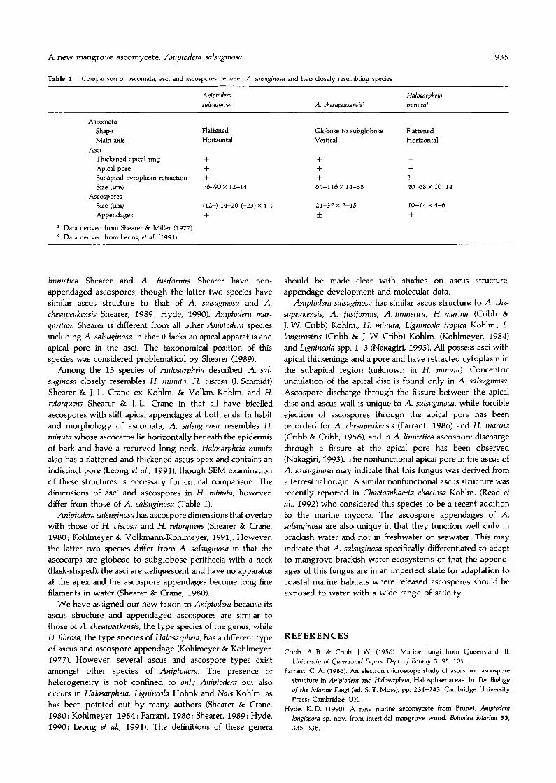
Table 1. Comparison of ascomata, asci and ascospores between A. salsuginosa and two closely resembling species
935
AscomataShapeMain axis
AsciThickened apical ringApical poreSubapical cytoplasm retractionSize (\JJl)
AscosporesSize (\JJl)
Appendages
1 Data derived from Shearer & Miller (1977).2 Data derived from Leong et al. (1991).
Aniptoderasalsuginosa
FlattenedHorizontal
+++7~90x 12-14
(12-) 14-20 (-23) X4-7
+
A. chesapeakensis1
Globose to subgloboseVertical
+++64-116 x 14-38
21-37 x 7-15
±
Halosarpheiaminuta2
FlattenedHorizontal
++
4D-68 x 10-14
10-14 x 4--6
+
limnetica Shearer and A. fusiformis Shearer have nonappendaged ascospores, though the latter two species havesimilar ascus structure to that of A. salsuginosa and A.chesapeakensis Shearer, 1989; Hyde, 1990), Aniptodera margarition Shearer is different from all other Aniptodera speciesincluding A. salsuginosa in that it lacks an apical apparatus andapical pore in the asci. The taxonomical position of thisspecies was considered problematical by Shearer (1989).
Among the 13 species of Halosarpheia described, A. salsuginosa closely resembles H. minuta, H. viscosa (1. Schmidt)Shearer & J. L. Crane ex Kohlm. & Volkm,-Kohlm. and H.retorquens Shearer & J. L. Crane in that all have bicelledascospores with stiff apical appendages at both ends. In habitand morphology of ascomata, A. salsuginosa resembles H.minuta whose ascocarps lie horizontally beneath the epidermisof bark and have a recurved long neck. Halosarpheia minutaalso has a flattened and thickened ascus apex and contains anindistinct pore (Leong et al., 1991), though SEM examinationof these structures is necessary for critical comparison. Thedimensions of asci and ascospores in H. minuta, however,differ from those of A. salsuginosa (Table I).
Aniptodera salsuginosa has ascospore dimensions that overlapwith those of H. viscosa and H. retorquens (Shearer & Crane,1980; Kohlmeyer & Volkmann-Kohlmeyer, 1991). However,the latter two species differ from A. salsuginosa in that theascocarps are globose to subglobose perithecia with a neck(flask-shaped), the asci are deliquescent and have no apparatusat the apex and the ascospore appendages become long finefilaments in water (Shearer & Crane, 1980).
We have assigned our new taxon to Aniptodera because itsascus structure and appendaged ascospores are similar tothose of A. chesapeakensis, the type species of the genus, whileH. fibrosa, the type species of Halosarpheia, has a different typeof ascus and ascospore appendage (Kohlmeyer & Kohlmeyer,1977). However. several ascus and ascospore types existamongst other species of Aniptodera. The presence ofheterogeneity is not confined to only Aniptodera but alsooccurs in Halosarpheia, Lignincola H6hnk and Nais Kohlm. ashas been pointed out by many authors (Shearer & Crane,1980; Kohlmeyer, 1984; Farrant, 1986; Shearer, 1989; Hyde,1990; Leong et aI., 1991). The definitions of these genera
should be made clear with studies on ascus structure,appendage development and molecular data.
Aniptodera salsuginosa has similar ascus structure to A. chesapeakensis, A. fusiformis, A. limnetica, H. marina (Cribb &
J. W. Cribb) Kohlm., H. minuta, Lignincola tropica Kohlm., L.longirostris (Cribb & J. W. Cribb) Kohlm. (Kohlmeyer, 1984)and Lignincola spp. 1-3 (Nakagiri, 1993). All possess asci withapical thickenings and a pore and have retracted cytoplasm inthe subapical region (unknown in H. minuta). Concentricundulation of the apical disc is found only in A. salsuginosa.Ascospore discharge through the fissure between the apicaldisc and ascus wall is unique to A. salsuginosa, while forcibleejection of ascospores through the apical pore has beenrecorded for A. chesapeakensis (Farrant, 1986) and H. marina(Cribb & Cribb, 1956), and in A. limnetica ascospore dischargethrough a fissure at the apical pore has been observed(Nakagiri, 1993). The nonfunctional apical pore in the ascus ofA. salsuginosa may indicate that this fungus was derived froma terrestrial origin. A similar nonfunctional ascus structure wasrecently reported in Chaetosphaeria chaetosa Kohlm. (Read etal., 1992) who considered this species to be a recent additionto the marine mycota. The ascospore appendages of A.salsuginosa are also unique in that they function well only inbrackish water and not in freshwater or seawater. This mayindicate that A. salsuginosa specifically differentiated to adaptto mangrove brackish water ecosystems or that the appendages of this fungus are in an imperfect state for adaptation tocoastal marine habitats where released ascospores should beexposed to water with a wide range of salinity.
REFERENCES
Cribb. A. B. & Cribb, ). W. (1956). Marine fungi from Queensland. II.University of Queensland Papers. Dept. of Botany 3. 95-105.
Farrant, C. A. (1986). An electron microscope study of ascus and ascosporestructure in Aniptodera and Halosarpheia, Halosphaeriaceae. In The Biologyof the Marine Fungi (ed. S. T. Moss), pp. 231-243. Cambridge UniversityPress: Cambridge, UK.
Hyde, K. D. (1990). A new marine ascomycete from Brunei. Aniptoderalongispora sp. nov. from intertidal mangrove wood. Bofanica Marina 33,335-338.

A. Nakagiri and T. Ito
Hyde, K. D. (1992). Tropical Australian freshwater fungi. I. Some Ascomycetes.Aust. Syst. Bot. 5, 109-116.
Hyde, K. D., Farrant, C. A. & Jones, E. B. G. (1986). Marine fungi fromSeychelles. III. Aniptodera mangrouii sp. nov. from mangrove wood.Canadian Journal of Botany 64, 2989-2992.
Kohlmeyer, J. (1984). Tropical marine fungi. P.s.Z.N.l: Marine Ecology 5,329-378.
Kohlmeyer, J. & Kohlmeyer, E. (1977). Bermuda marine fungi. Transactions ofthe British Mycological Society 68, 207-219.
Kohlmeyer, J. & Volkmann-Kohlmeyer, B. (1991). Illustrated key to thefilamentous higher marine fungi. Botanica Marina 34, 1-61.
Leong, W. F.. Tan, T. K., Hyde, K. D. & Jones, E. B. G. (1991). Halosarpheiaminuta sp. nov., an ascomycete from submerged mangrove wood. CanadianJournal of Botany 69, 883-886.
(Accepted 28 January 1994)
936
Nakagiri, A. (1993). Intertidal mangrove fungi from Iriomote Island. [FO
Research Communications 16, 24--62.Read, S. ).. Jones, E. B. G., Moss, S. T. & Johnson, R. G. (1992). Ultrastructural
observations of the marine Ascomycotina Corollospora angusta, Corollosporacolossa, Corollospora lacera and Chaetosphaeria chaetosa. Botanica Marina 35,553-560.
Shearer, C. A. (1989). Aniptodera (Halosphaeriaceae) from wood in freshwaterhabitats. Mycologia 81, 139-146.
Shearer, C. A. & Crane, J. L. (1980). Fungi of the Chesapeake Bay and itstributaries. VIII. Ascomycetes with unfurling appendages. Botanica Marina23, 607--615.
Shearer, C. A. & Miller, M. (1977). Fungi of the Chesapeake Bay and itstributaries. V. Aniptodera chesapeakensis gen. et sp. nov. Mycologia 69,887-898.