
and y- Cbains Of The High-Affinity Receptor Com - CORE
189
Assessing the Role of Amino Acid Residues in The Transmembrane Domains Of The a- and y- Cbains Of The High-Affinity Receptor Complex For Immunoglobulin E In Signal Transduction By Amir Rashid Thesis submitted in part fulfilment of the requirement for the degree of Doctor of Philosophy August2009 Department of Molecular Biology and Biotechnology, University of Sheffield
-
Upload
khangminh22 -
Category
Documents
-
view
0 -
download
0
Transcript of and y- Cbains Of The High-Affinity Receptor Com - CORE

Assessing the Role of Amino Acid Residues in The Transmembrane Domains Of The a- and yCbains Of The High-Affinity Receptor Complex For Immunoglobulin E In Signal Transduction
By
Amir Rashid
Thesis submitted in part fulfilment of the requirement for the degree of
Doctor of Philosophy
August2009
Department of Molecular Biology and Biotechnology, University of Sheffield

•
To my fa111ily

•
ABSTRACT
Abstract
The dramatic rise in the incidence of allergic/asthmatic disorders in the past three
decades has placed a major socio-economic burden on global health care. Exocytosis
of mediators causing allergic responses follows the binding, and subsequent receptor
cross-linking by cognate allergen of immunoglobulin (lg) E antibodies to Fe-receptors
(FcERI), expressed predominantly on mast cells and basophils.
Fe-receptors have an invariant predominantly hydrophobic ammo acid motif
(LFAVDTGL) in their transmembrane (TM) domain but contain a hydrophilic
aspartic acid residue (D194). The function of this potentially energetically unstable
residue in the TM of the human (hu) Fci,Rla subunit was targeted by transfecting the
Rat Basophilic Cell line (RBL-2H3.1) with cDNA constructs encoding the gene for
native and mutant huFcERia subunits and assessing receptor expression and signalling
events. RBL-2H3 transfected with cDNA constructs encoding a medium-sized polar
residue hufci,Rla (D194T) demonstrated the formation of a functional rat/human
chimeric receptor complex which, when activated via hulgE and antigen, supports
mediator release, intracellular calcium mobilisation and tyrosine phosphorylation of y
chain and Syk kinase. Transfection with mutant huFci,Rla subunit cDNA constructs
encoding non-polar Ile (D1941) and V (D194V), larger sized polar Arg (D194R),
smaller sized polar Ser (D194S) and non-polar Ala (D194A) abrogated surface
expression of huFcERia and degranulation. An established RBL y-chain deficient cell
line served as a model for assessing a- and y-chains interactions through the effect of
mutations introduced into the y-chain. Compared to parental RBL-2H3 cells, the RBL
y-chain deficient cells supported reduced levels of FcERI expression and mediator
release. Transfection of RBL y-chain deficient cells with wild-type and mutant (T22A
and T22S) y-chain cDNA constructs restored FctRI expression to levels observed in
the parental cell line, but only partially restored mediator release, indicating a
defective secretory response.
Collectively this study identifies D194 in FctRia as a potential target for developing
future anti-allergic drugs that act by inhibiting the FctRI signalling cascade.
iii

i
j
' I
ACKNOWLEDGEMENTS
Acknowledgements
I am grateful to my supervisor, Dr Birgit Helm, firstly for giving me the opportunity
to work in her lab and for taking up the enormous task of converting a medic into a
researcher. I am indebted to her for the assistance she provided to me at every critical
juncture (of which there were plenty) in my stay here. I am thankful to her for not
only understanding but also helping me overcome the problems I encountered within
and outside the scope of research.
I would like to thank Dr Qaiser Sheikh for the guidance which he provided to me on
innumerable occasions. He went out of the way to assist me in my research for which
I am grateful.
I am grateful to Dr Jonathan Housden for sharing the practical tips involved in lab
work/calculations and troubleshooting at every step ofmy research. I would also like
to thank my advisor Dr Arthur Moir who was always available for providing very
useful and constructive advice.
I would like to thank all members of the lab: Dr Mike Hunter, Mark Street, HongTu
Ye, Esmaeil Saddrodiny and Sari Saban for their cooperation and for maintaining a
cordial atmosphere in the lab. It was an honour and pleasure working with them. I am
also grateful to previous members of the group namely Dr Marco Iodice and Dr
Kathleen Carroll who were always very helpful when contacted for advice. I would
also like to mention the undergraduate project students Alison, Mary, Demetris and
Vaidas who worked with me and were a great help in my research.
I am grateful to Dr Lynda Partridge, Dr Sandrine Soubes and all my colleagues in the
department especially, Rachel, Noha, Arunya, Mike and Sameer for the cooperation
extended to me during my stay here.
I would like to thank Dr Mark Jones for training me in flowcytometry. Sue and Kay at
flowcytometry facility at Medical School also have my gratitude for being patient and
accommodating me at very short notice on many occasions.
I am thankful to Professor David Homby and Ms Linda Harris for the support
extended to me particularly at times when I needed it most.
Last but in no way least I would like to thank my family for their sacrifice and bearing
with me in this particularly difficult time.
iv

LIST OF ABBREVIATIONS
List of Abbreviations
•
AM Acetoxymethyl ester
APC Antigen presenting cell
BCR B cell receptor
BMMC Bone marrow-derived mast cells
BS Buffered solution
BSS Balanced salt solution
BSA Bovine serum albumin
BtK Bruton's tyrosine kinase
ca+2 Calcium ion
Cbp Csk-binding protein
CD Cluster of differentiation
cDNA Complementary DNA
CDS Cell dissociation solution
CRACs Calcium release activated channels
CsK COOH-tenninal Src kinase
0194 Wild type human FctRia subunit
0194N Human FctRia subunit with Asp-+Asn
D194L Human FctRia subunit with Asp-+Leu
D194T Human FctRia subunit with Asp-+Thr
V

LIST OF ABBREV/A TIO NS
D194S Human Fc£Rla subunit with Asp---+Ser
• D194R Human FccRla subunit with Asp---+Arg
D194I Human FccRia subunit with Asp---+Ile
D194V Human FccRla subunit with Asp---+Val
DAG 1, 2-diacylglycerol
DCs Dendritic cells
dH20 distilled water
DMEM Dulbeco's Modified Eagles media
DMSO Dimethyl Sulphoxide
DNP-HSA Dinitophenol-human serum albumin
DOK Downstream ofKinases
DPBS Dulbeco's phosphate buffered saline
DTT Di-thiothreitol
EC Extracellular
ECL Enhanced chemiluminescence
EDTA Ethylenediaminetetraacetic acid
EGF Epidermal growth factor
ELISA Enzyme-linked immunosorbent assay
ER Endoplasmic reticulum
ERK extracellular regulated kinase
FACS Flourescent activated cell sorting
FCS Foetal calf serum
FcaRI High-affinity IgA receptor
Fc£Rl High-affinity lgE receptor
Vl

FcERia
• FcERI~
FcR-y
FcyRs
FcyRI
FcyRII
FcyRIII
FcyRIIIa
FITC
fMLP
FS
GM-CSF
GPI
Grb-2
HRP
humanayy
huFcERia
huigE
!CAM
IFN-y
lg
IgE/IgM/IgG
IL
IP
IP3
LIST OF ABBREVIATIONS
High-affinity IgE receptor alpha subunit
High-affinity IgE receptor beta subunit
Fe gamma subunit
IgG receptor
High-affinity IgG receptor
Low-affinity IgG receptor II
Low-affinity IgG receptor III
Low-affinity IgG receptor III alpha-subunit
Fluorescein isothiocyanate
n-formyl-Met-Leu-Phe
Forward scatter
Granulocyte-macrophage colony-stimulating factor
Glycosylphosphatidylinositol
Growth factor receptor-bound protein 2
Horseradish peroxidise
Chimera with huFcERia EC, rodent yTM and CT domains
Human FcERI alpha subunit
Human IgE
Intracellular cell adhesion molecules
Interferon- y
Immunoglobulin
Immunoglobulin E/Immunoglobulin Ml immunoglobulin G
Interleukin
Immunoprecipitation
Inositol-I, 4, 5-triphosphate
vu

LIST OF ABBREVIATIONS
IR Insulin receptor
• ITAM Immunoreceptor tyrosine-based activation motif
ITIM Immunoreceptor tyrosine-based inhibitory motif
JNK Jun amino-terminal kinase
LAB Linker for activation of B cells
LAT Linker for activation of T cells
LPS Lipopolysaccharide
LT Leukotrine
MAFA Mast cell function-associated antigen
MFI Mean fluorescence intensity
MHC Major histocompatability complex
mAb Monoclonal antibody
MAPK p-38 mitogen-activated protein kinase
mlg Membrane immunoglobulin
mlgE Mouse IgE
mlgM Membrane-bound immunoglobulin M
N2 Nitrogen
NIP-HSA 4-hydroxy-5-iodo-3-nitrophenyl-human serum albumin
NK Natural Killer
NTAL Non-T-cell activation linker
PAF Platelet activating factor
PAMP Pathogen-associated molecular pattern
PC Poorly cytokinergic
PH Pleckstrin homology
P13K Phosphatidylinositol 3-kinase
Vlll

PIP2
• PIP3
PKC
PLA2
PLC
PRR
PTK
PVDF
RBL-2H3
RBL-y-
RBL-y+
rFcRy
RIPA
SCF
SDS
1-D SOS-PAGE
SH21SH3
She
SHIP
SHP
SIT
SLP-76
SNAREs
SOCCs
sos
LIST OF ABBREV/A TIONS
Phosphatidylinositol-4, 5-biphosphate
Phosphatidylinositol-1, 4, 5-triphosphate
Protein kinase C
Phospholipase A2
Phospholipase C
Pathogen recognising receptor
Protein tyrosine kinase
Polyvinyledene difluoride
Rat Basophilic Leukaemia sub-line 2H3
Rat Basophilic Leukaemia gamma chain deficient mutant sub-line
RBL-y- cell line transfected with wild-type gamma subunit
Rodent gamma construct
Radioimmunoprecipitation assay
Stem cell factor
Sodium dodecyl sulphate
One-dimensional SDS-polyacrylamide gel electrophoresis
Src homology 2/Src homology 3
SH2-containing proto-oncogene
SH2-containing inositol-5 '-phosphatase
SH2-containing protein tyrosine phosphatase
specific immunotheapy
SH2-containing leukocyte specific protein of 76 kDa
Soluble NSF-attached protein receptors
Store operated calcium channels
Son of sevenless
ix

ss
• Syk
T22A
T22S
TCR
TE
Tho,Th1/Th2
TLR
TM
TNF
WB
LIST OF ABBREV/A TIONS
Side scatter
Spleen tyrosine kinase
RBL-y- cell line transfected with mutant gamma subunit Thr---+Ala
RBL-y- cell line transfected with mutant gamma subunit Thr---+Ser
T cell receptor
Trypsin/EDT A
T helper subsets
Toll-like receptor
Transmembrane
Tumor necrosis factor
Western blot
X
• TABLE OF CONTENTS
Abstract
Acknowledgements
Abbreviations
Table of Contents
List of Tables and Figures
CHAPTER 1 - INTRODUCTION
1.1 The Allergy Epidemic
1.2 Immune System: An Overview
1.2.1 The Innate System
1.2.2 The Adaptive System
1.2.3 The Immune Cells
Dendiritic Cells
NKCells
The Phagocytic Cells-Neutrophils and Macrophages
1.3 An Overview of Allergic Response
1.4 Mast Cells
1.4.1 Mast Cells Activation
1.4.2 Mast Cell Degranulation
1.5 Immunoglobulins
1.5.1 Immunoglobulin E, IgE
1.6 The High Affinity IgE Receptor, FcERI
1.6.1 The Ligand -binding Subunit- FcERia
1.6.2 The Signal amplifying Subunit- FcR~
1.6.3 The Signal Transducing Subunit- FcRy
1.6.4 Interaction of IgE with FcERI
TABLE OFCONTENTS
111
IV
V
XI
XIX
1
2
4
5
6
7
7
7
7
8
9
10
14
15
15
16
17
17
18
18
1.7 The Role of Conserved Charged Residues in Transmembrane Domains of
XI

TABLE OF CONTENTS
Immunoreceptors on Receptor Assembly, Expression and
Function 21
1.7.1 The Role ofTransmembrane Charged Residues of FcERI 21
1.7.2 The Role ofTransmembrane Charged Residues oflgA Receptor. 22
1.7.3 The role of the Transmembrane Charged Residues ofT cell receptor 22
1.7.4 The role of the Transmembrane Charged Residues ofB cell Receptor 23
1.8 Current Therapeutic Strategies for Allergy 24
1.8.l Conventional Therapies 24
1.8.2 Immunomodulators 25
1.8.3 Mast Cell Therapeutics 26
1.9 Aim of study 28
CHAPTER 2 - l\1ATERIALS AND l\1ETHODS 33
2.1 Materials and Equipment 34
2.1.1 Cell Culture Reagents 34
2.1.2 Protein Manipulation 34
2.1.3 Antibodies Used 35
2.1.4 Molecular Biology Reagents 36
2.1.5 Buffers 36
2.1.6 Equipment 37
2.2 Cell Lines 38
2.3 Mammalian Cell Culture Techniques 38
2.3.1 Culturing RBL cell Line 38
2.3.2 Cryogenic Preservation 39
2.3.3 Thawing of Cells 39
Xll

TABLE OF CONTENTS
• 2.4 ~ -Hexosaminidase Assay: Measurement of Secretory Output from RBL Cell lines in
Response to an Immunological Stimulus 39
2.5 Cell lysate Preparation for 1 D- PAGE 40
2.5.1 Sample preparation 40
2.5.2 Bradford's Method for Protein Estimation 41
2.5.3 Immunoprecipitation 42
2.5.4 One-dimensional Polyacrylamide Gel Electrophoresis 42
2.5.5 Western transfer of proteins to PVDF membrane 43
2.5.6 Immunodetection of transferred proteins 44
2.5.7 Detection of Proteins with Enhanced Chemilurninescence(ECL) 45
2.6 Assessing FcERI Expression in Transfected Cell lines by Flow Cytometry 45
2. 7 F ACS cell sorting of transfected cell lines by Flow Cytometry 46
2.8 Assesment of Intracellular Calcium Mobilisation 47
2.9 Molecular Biology Methods 48
2.9.1 Bacterial Strain 48
2.9.2 E.Coli Growth Media 48
2.9.3 Vectors 49
2.9.4 Preparation of Competent Bacterial Cells 49
2.9.5 Transformation of Competent Bacterial Cells 50
2.9 .6 Isolation of Plasmid DNA 50
2.9.6.1 QIAprep Spin Miniprep Kit 50
2.9.6.2 QIAGEN Plasmid Purification System 50
2.9.7 Agarose Gel Electrophoresis 51
2.9.8 DNA manipulation 52
2.9.8.1 Restriction Enzyme Digests 52
2.9.8.2 Dephosphorylation 52
2.9.8.3 Ligation 52
2.9.8.4 DNA Purification from Agarose Gel 53
2.9.8.5 Site Directed Mutagenesis 53
2.9.8.6 DNA sequencing 54
Xlll

•
TABLE OF CONTENTS
2.10 Transfection Mammalian Cells 54
CHAPTER 3 Mutagenesis of Human High Affinity lgE
Receptor Alpha Chain subunit 56
3.1 Conserved Motif-FctRla 57
3 .1.1 Residues Targeted for Site Directed Mutagenesis 57
3.1.2 Primer Design 58
3.2 Vectors 62
3.2.1 pUC18 62
3.2.2 Mammalian expression vector 62
3.3 Cloning into pUC18 62
3.4 Site-Directed Mutagenesis 63
3.5 Sequencing 63
· 3.6 Subcloning to pEE6 66
3.7 Expression of the cDNA huFccRia constructs 69
3.7.1 Transfection into Rat Basophilic Leukemia Cell line (RBL-2H3.l) 69
CHAPTER 4 Identification of huFctRla expression and
Assessment of l\fediator Release via the
Transfected Receptor
4.1 Introduction
4.1.1 RBL-2H3 cell line
4.2 Results
70
72
72
73
4.2.1 Flow cytometric analysis oftransfected mutant hufctRla receptor subunits 73
4.2.2 13-hexosaminidase Release Assays 77
4.2.3 Intracellular Calcium Mobilisation 80
XIV

TABLE OF CONTENTS
4.2.4 FcR-y and Syk Kinase Phosphorylation
4.2.4.1 lmmunoprecipitation Studies Results
4.3 Summary of Results
CHAPTER 5 High Affinity lgE Receptor a. and y Subunit
83
84
86
Interactions 88
5.1 Introduction 89
5.2 Human a.yy Construct 89
5.2.1 Results 91
5.2.1.1 Primer Design 91
5.2.1.2 Site-Directed Mutagenesis 92
5.2.1.3 Subcloning to pEE6 92
5.2.1.4 Transfection into Rat Basophilic Leukemia Cell line (RBL-2H3.l) 95
5.2.1.5 Flow cytometric analysis of transfected RBL-2H3 cells for huFcERla
receptor subunits
5.2.1.6 13-hexosaminidase Release Assays
5.2.1. 7 Intracellular calcium mobilization
5.2.1.8 Discussion
5.3 RBL Gamma Chain Deficient Cell Line
5.3.1 Subcloning to pEE6
5.3.2 Transfection into RBL gamma Deficient Cell Line
5.3.3 Flow cytometric analysis of transfected RBL-y-cells for FcERla receptor
subunits expression
5.3.413-hexosaminidase Release Assays
5.3.5 Intracellular Calcium mobilisation
95
96
98
99
100
101
103
104
104
105
xv

TABLE OF CONTENTS
CHAPTER 6 DISCUSSION
6.1 Introduction
6.2 Detection ofhuFcERI Expression and Mediator Release in Transfected
110
111
RBL.2H3.1 Cells 112
6.3 Transfection ofRBL Gamma Chain Deficient Cell with Wild-type and Mutant rFcERy Constructs 114
6.4 Future Work 115
6.4.1 Mutational analysis of FcR-y Subunit 115
6.4.2 Alternative Model System 116
6.5 Conclusions 116
REFRENCES 118
APPENDIX I 146
xvi

TABLE OF CONTENTS
LIST OF TABLES AND FIGURES
Figure 1.1 Overview of allergic response 12
Figure 1.2 FcERI mediated signalling pathways in mast cells 13
Figure 1.3 Isoforms ofFcERI 16
Figure 1.4 Model oftetrameric high affinity receptor 20
Figure 1.5 Modelling oftransmembrane helices 30
Figure 3.la Sequence Verification oftransmembrane domains of Wild
Type (D 194) and mutant (D 194N and D 194L) huFctRia constructs 60
Figure 3.lb Sequence Verification oftransmembrane domains of mutants
(Dl94T, D194I, D194V, DI 94R, and DI 94S) huFctRia constructs 61
Figure 3.2 Bacterial expression vector pUC 18 64
Figure 3.3 Mammalian Expression Vector pEE6 64
Figure 3.4 Cloning into pUC 18 65
Figure 3.5 Site directed mutagenesis 66
Figure 3.6 Subcloning into pEE6 68
Figure 3.7 Test digestion ofmidiprep plasmid DNA 69
Figure 4.1 Assesment of cell surface expression in DI 94 (wild type), O194T, D194V
and D1941 transfected cell lines by flowcytometry 76
Figure 4.2 Assesment of cell surface expression in D 194S and D 194R transfected cell
lines by flowcytometry 77
Figure 4.3 Release of J3-hexosaminidase through FceRicx transfected receptors in RBL-
2H3 and RBL-2H3 transfected cell lines D194T, D194R, D1941, D194S and
D 194V cell lines in response to antigenic stimulus 79
Figure 4.4 Assessment oflntracellular calcium level ofRBL-2H3 transfected cell lines
D194 (wild-type), DI 94T, O194V, D1941, DI 94R and D194S transfected cell
lines following activation in the absence of extracellular calcium 82
xvii

TABLE OF CONTENTS
Figure 4.S Phosphorylation state of FcR-y in parental RBL-2H3.land RBL-2H3.1 cells
transfected with the mutant huFcERla cDNA constructs (DI 94 and D194T) 85
Figure 4.6 Phosphorylation state ofSyk kinase in parental RBL-2H3.land RBL-2H3.l
cells transfected with the mutant huFccRia cDNA constructs (DI 94 and
D194T
Figure S.1 Test digestion ofminiprep plasmid DNA
Figure S.2 Test digestion ofmidiprep plasmid DNA
86
93
93
Figure S.3 Assessment of cell surface expression of huFccRia transfected receptors in
parental RBL-2H3 transfected with humana.yy construct by flowcytometry 95
Figure S.4 Release of 13-hexosaminidase through FctRia. transfected receptors in parental
RBL-2H3 human a.yy construct cell lines in response to antigenic stimulus 96
Figure S.S Assessment oflntracellular calcium level of RBL-2H3 transfected cell lines
cell line following activation in the absence of extracellular calcium 98
Figure S.6 Test digestion ofminiprep plasmid DNA 102
Figure S.7 Test digestion ofmidiprep plasmid DNA
Figure S.8 Assesment of cell surface expression of mouse FctRia. receptors in
RBL-2H3, RBL-y- cell line, RBL-y- cell line transfected with wild type
rFcRy (RBL-y+) and mutant rFcRy construct (T22A and T22S) and sham
103
transfection by flowcytometry 106
Figure S.9 Release of 13-hexosaminidase in RBL-2H3, RBL-y- cell line, RBL-y- cell line
transfected with wild type (RBL-y+) and mutant rFcRy construct (T22A and
T22S) and sham transfection in response to antigenic stimulus 107
Figure S.10 Assesment of intracellular calcium level in RBL-2H3, RBL-y- cell line,
RBL-y- cell line transfected with wild type (RBL-y+) and mutant rFcRy
construct (T22A and T22S) and sham transfection following activation in
the absence of extracellular calcium 108
xviii

TABLE OF CONTENTS
Table 1.1: Mast cell and mast-product directed therapeutics 27
Appendix I 146
Rashid et al., 2009 (Manuscript submitted to Molecular immunology)
xix

CHAPTER1
CHAPTER]
INTRODUCTION
INTRODUCTION
1

CHAPTER1
Chapter 1
I11troductio11
1.1: The Allergy "Epidemic"
INTRODUCTION
Type I hypersensitivity or immunoglobulin (lg) E-mediated allergy is a broad term
describing undesirable immune responses that follows the encounter with seemingly
non-pathogenic antigens referred to as allergens. The first historical reference to an
allergic response dates to 2640B.C and depicts, on a pylon in Luxor, Egypt, the death
of a Pharaoh from an anaphylactic shock following a bee sting
[http://www.slideshare.net/inemet/anaphylactic-reactions]. Immunoglobulin E and
mast cells/basophils are now recognised key players in the allergic response
(Williams and Galli, 2000). Immune response of the IgE isotype play an important
role in the response to and elimination of parasites from intestines of the host, a
function that has been authenticated by various studies in which it has been
demonstrated that chronic/heavy helminthic infestation, characterised by high levels
of parasite specific IgE, protects against allergy (Yazdanbakhsh and Matricardi,
2004). The Viennese paediatrician Clemens von Pirquet in 1906 coined the term
allergy from the Greek words a//os meaning other and ergon meaning work after he
noticed the fact that some of his patients were sensitive to apparently innocuous
entities like dust, pollen and certain foods while the term anaphylaxis was first used
by Portier and Richard in 1902 to explain an unexpected effect that occurred during
immunization of dogs.
The various ailments associated with allergyare atopic dermatitis, urticaria, allergic
rhinitis, asthma and the potentially fatal condition of anaphylaxis (Corry and
Kheradmand, 1999). The incidence of allergic diseases is on the · rise globally
(Pawankar et al., 2008) specially in industrialized countries where 10-30% of the
population suffer from diverse allergic symptoms (ISAAC Committee, 1998, Sole et
al., 2001) The rise .has been rapid and started between l 960~ 1970 with an ongoing
increase witnessed in the 1980s and 90s to levels that have attained almost epidemic
proportions such that allergic diseases are being labelled as the epidetnic of the 21 st
2

CHAPTER1 INTRODUCTION
century (lsolauri et al.. 2004). The increase witnessed in westernized societies is in the
range of 75-100% in cases of asthma (Mannino et al.. 2002) and 150% in atopic
diseases (Wichmann, 1996). Similar trends are now being observed in the most
populous continent Asia that is home to fast emerging economies. According to recent
epidemiological data by ISAAC III (International Study of the Asthma and Allergies
in Childhood) the incidence of Asthma and allergic rhinitis is also on the rise in low
and middle income countries (Bjorksten et al., 2008) with incidence for asthma
increasing from 0.8% to 29% and for allergic rhinitis from 5% to 45% of the
population in many Asian countries (Pawankar et al., 2008).
The pathology of allergic diseases is not fully understood but it is believed that both
genetic and environmental factors have a role to play (Gould et al.. 2003). Genetic
predisposition is believed to contribute at least to half of the susceptibility to asthma
(Duffy et al., 1990). The list of allergens includes house dust mite and cockroach
emanations, pollens, and fungal spores (Machado et al., 1996, Helm et al., 2000).
Food hypersensitivities which have been documented include cow milk, egg, peanut.
soya, wheat. shellfish, tree nuts and fish (Sampson, 1999, Sampson, 2004). Allergic
reaction to latex components now commonly referred to as the 'latex-fruit' syndrome
are directed against a conserved chitin binding domain in plant host defense proteins
with diverse enzymatic functions referred to as the hevein symphony. This immune
response represents a major problem for health professionals, patients and members of
the general public using latex products (Toraason et al., 2000, Agarwal and
Gawkrodger, 2002, Pilon and Radman, 2006).
Most of these environmental allergens have been present for millennia and current
allergy epidemic even in industrialised countries with genetically stable pool such as
Japan point to the fact that changes in environmental factors are probably responsible
for the current epidemic (Cookson and Moffatt, 1997). Diesel exhaust particles
(DEPs) enhance the production of pro-allergenic cytokines from cells present in nasal
mucosa and have been held accountable for the increase in airway allergic diseases
(Diaz-Sanchez et al., 1996). Chronic respiratory diseases are highly prevalent in
workers who are in employment in motor garages where they are exposed to motor
vehicle exhaust emission and diesel engine exhausts (Bener et al., 1998). Many
components in environmental tobacco smoke [ETS] are chemically similar to DEP,
and have been shown to stimulate ongoing lgE synthesis (Smyth et al., 2000). These
3

CHAPTERl INTRODUCTION
chemicals are also implicated in increased airway hypersensitivity and sensitisation to
common aero-allergens in young children and held accountable for the increased
incidence in allergy and respiratory symptoms (Magnusson and Johansson, 1986,
Andrae et al., 1988).
Reunification of Germany provided a case study of the impact of life style on allergic
diseases. A study carried out by von Mutius et al (1998) indicated a significant rise in
the incidence of hay fever amongst children in East Germany who, following the
reunification of the country adapted to a more Westernized way of life. Likewise
obesity, a major and ever growing problem in children of industrialized countries, is
positively linked with the onset of adult-onset asthma (Camargo et al., 1999). The
"Hygiene Hypothesis" that has been much quoted as a probable cause of increased
incidence in allergic diseases in affiuent Western societies was originally presented by
epidemiologist Dr Strachan (1989). The hypothesis stated that there is an inverse
relationship between family size and allergic diseases (Strachan, 1989). Strachan
proposed that the increase in atopic disorders might be due to a decreased occurrence
in early infancy of infections that are commonly transmitted through contact with
elder siblings. A consensus about this concept is now evolving and it is postulated that
elementary changes in life style have deprived human beings from developing
immunoregulatory mechanisms brought about by exposure to certain micro-organisms
and viral infections thus contributing to increase in the prevalence of allergic diseases
in the last few decades (Pawankar et al., 2008).
1.2: Immune System: An Overview
Immunity is the universal potential of the host to recognize and fight with the aim of
eliminating the predatory microbes, which are pathogenic to it and, if allowed to live,
will produce noxious effects on the body. It involves both molecular and cellular
agents acting in cohesion and involves the coordinated interaction between
components of the innate and adaptive immune systems.
Broadly defined, the immune system depends on a combination of the non-specific
but immediate acting force of innate system and the more target oriented slowly
responding system of the acquired/adaptive immune system with an extent of overlap
4

CHAPTER1 INTRODUCTION
present (Basset et al., 2003). The Toll-like receptors (TLRs) expressed by antigen
presenting cells and mast cells recognize and differentiate self from pathogens and
bring about the commencement and subsequent steering of adaptive immune
responses (Orinska et al., 2005, Kaisho and Alcira, 2006). The microbes in their bid to
gain entry to the body face first resistance in the shape of skin and mucosal membrane
lining the gastrointestinal, respiratory and urogenital tract, which in addition to acting
as a physical barrier to the microbes in the external environment, harbour mast cells at
a position where they are ideally placed to combat any incoming pathogens (Kitamura
et al., 1977, Metcalfe et al., 1997). The mast cells are thus not only on the interface of
external and internal environment but form a bridge between both the innate and
adaptive immunity due to their established place as a component of the innate system.
At the same time, due to the expression of Fe receptors they mediate responses to
immunoglobulins, and as a result mediate allergic sensitization. The origin of allergy
may be found at this interface and better comprehension of the complex mechanisms
related to the role of mast cells in the immune system may lead to developing more
efficient intervention strategies to combat the growing menace of allergic diseases.
1.2.1: The Innate System
The innate system is comprised of mechanical, chemical and cellular elements. The
mechanical component includes the physical barrier of intact epidermis and mucosa
and associated physiological roles such as cilial motility, desquamation and mucus
secretion (Basset et al., 2003). The initial and all important task of recognising the
pathogen is carried out by cell surface expressed pathogen recognising receptors
(PRRs), found on the surface of many cells including epithelial cells,
macrophages/monocytes, granulocytes, mast cells and dendiritic cells, that identify a
wide array of pathogen associated molecular patterns (PAMPs) (Janeway and
Medzhitov, 2002). The PAMPs recognition, which include lipopolysaccharide (LPS),
peptidoglycan, lipoproteins and oligosaccharides, leads to activation of the
inflammatory response to a wide variety of the pathogenic stimuli (Iwasaki and
Medzhitov, 2004). The chemical mediators involved in the innate system can be sub
grouped into pattern recognition molecules, the proteins and peptides that eradicate
the micro-organisms and last but not the least depend on cytokines and chemokines
generated following activation of innate immune responses that have a crucial
s

CHAPTERl INTRODUCTION
regulatory role in the steering of adaptive immunity (Hedges et al., 1995, Machado et
al., 1996, Pitman and Blumberg, 2000, Janeway and Medzhitov, 2002, Wynn, 2009).
Mast cells, dendiritic cells, NK cells, macrophages and epithelial cells all play a role
in the innate system (Basset et al., 2003).
1.2.2: The Adaptive System
Adaptive immunity. also known as acquired immunity is more specialised than the
innate system and builds on the work already performed by its more primitive
counterpart. As the name implies a person has to have had a previous exposure to the
agent to be able to mount a response against it. The initial encounter with the allergen
(immunization) brings about a complex and intricate chain of events leading to
production of antibodies by lymhocytes (Owen. 2007). Any subsequent exposure to
the same antigen then makes the host capable of combating and eradicating the
pathogen through the presence of immunological memory. The adaptive system is
very specific and target oriented as it possesses the sel£'non-self recognition
mechanism through the expression of T cell receptor (TCR). B cell receptor (BCR)
and Major-histocompatability complexes (MHC) making it a very well organized
immune system (Hoebe et al., 2004). The activated T helper cells (Th), through the
effects of secreted cytokines and by the binding of CD40 on B cells to CD40 ligand
on T cells, promote the production of immunoglobulins by the B cells (Owen, 2007).
There are subsets of Th cells. Thl and Th2, derived from Th0, depending on the
cytokines secreted and the target organisms (Gould et al.. 2003). Thl cells secrete IL-
2 and interferon -y in response to challenge by bacteria and viruses, while Th2 cells
secrete IL-4, IL-5, IL-6 and IL-10 when challenged by helminth and allergen
(Finkelman et al., 1997, Romagnani, 2001). It is Th2 phenotype cells which are
responsible for isotype switching ofB cells to lgE production which is brought about
by cytokines IL-4. IL-13 via signals through CD40 and CD21 (Gauchat et al., 1993,
Jeppson et al., 1998). It is interesting to note in this context that activated mast cells
express a similar cytokine pattern to Th2 cells and via the expression of CD40L have,
in theory, the capability to induce class-switching to the lgE isotype (Machado et al.,
1996). Another subset T regulatory cell (Treg) mediate self-tolerance and prevent
autoimmune diseases (Steinke and Borish, 2006).
6

CHAPTER1 INTRODUCTION
1.2.3: The Immune Cells
Dendiritic Cells
The dendiritic cells are antigen-presenting cells, arising from the bone marrow, and
are generally considered to be the link between innate and acquired immunity due to
their property of presenting antigens in the tissues and then circulating to regional
lymph nodes where they mature (Banchereau and Steinman, 1998, Zitvogel, 2002).
These cells initiate the immune response, have PRRs on their surface that recognise
P AMPs, and are widely distributed in both lymphoid and non-lymphoid tissues
making them ideally located to be at the interface of the host and environment to
perform their role (Janeway and Medzhitov, 2002).
NK cells
These cells are also part of the innate system and are derived from the lymphoid cell
line. They also have an immunomodulatory role as they activate macrophages by
releasing TNFa and IFN-y and are responsible for killing virally infected and tumour
cells during cell-to-cell contact by releasing perforin and granzyme (Middleton et al.,
2002).
The Phagocytic Cells-Neutrophils and Macrophages
These are effector cells of the innate immunity that destroy the invading pathogens
before they can carry out their deleterious effects, but these cells also initiate the
adaptive response by presenting the antigens to T-cells. The first cells to come into
action are the resident macrophages but they are rapidly joined by the circulating
neutrophils if the need arises (Zhang et al., 2000). Macrophages and neutrophils
possess several PRRs that make possible the recognition through direct and indirect
means of the opsonised micro-organisms (Janeway and Medzhitov, 2002). The
invading pathogen is then internalised in a phagosome, which fuses with endosomes
and lysosomes to form phagolysosome leading to killing of micro-organism by using
oxidative and non-oxidative mechanisms (Burg and Pillinger, 2001). The recruitment
of the neutrophils to the damaged tissue is brought about through the chemotactic
action of cytokines such as IL-113, TNF-a, G-CSF, and chemokines such as fMLP and
LPS and substances from the damaged tissue (Philpott et al., 2001). The neutrophils
seem to have an immunoregulatory role characterised by the release of certain
7

CHAPTER1. INTRODUCTION
chemokines, as well as the release of substantial amounts of TNF and IL-I (Chaplin,
2003).
1.3: An Overview of Allergic Response
The symptoms of immediate hypersensitivity are caused by the release of preformed
and newly synthesized mediators that are released from mast cells and basophils in
response to a variety of stimuli (Blank and Rivera, 2004). The best characterized
event involves the sensitization of mast cells and basophils by lgE binding to high
affinity lgE receptors; subsequent cell activation follows encounter with cognate
allergen representing currently the best defined route leading to the regulated
secretion of the pharmacologically active mediators responsible for the symptoms of
class I hypersensitivity responses (Nadler et al., 2000). It is however known that mast
cells and basophils can be activated by a number of diverse mechanisms, including
initial exposure to enzymatically active allergens, which induce the secretion of
diverse mediators, including IL-4/13; the importance of these processes is only
beginning to emerge. It still remains to be demonstrated if this is in fact the first in a
series of steps leading to the synthesis of antibodies of the IgE isotype, although it is
generally accepted that mast cells and basophils express similar ligands and cytokine
to Th2 cells known to steer B-cells responses to class-switching to the lgE isotype
(Dudler et al., 1995, Machado et al., 1996).
The sequence of events involved in the allergy mechanism involves a complex
interaction of cells and chemicals. Although the list of allergens is long and varied
the commonly encountered allergens are pollen grains, house dust mite, cockroach
emanations, mould spores, latex, soya and nuts (Machado et al., 1996, Helm et al.,
2000). The current generally accepted mechanism proposes that after exposure these
allergens on entry into the body come in contact with the APCs such as basophils and
DCs at the mucosa! surfaces (Zitvogel, 2002, Wynn, 2009). The antigenic region of
these allergens in association with the MHC class II are presented on the cell surface
after being internalised and processed by the DCs. The presented peptides then bind
to narve T cells in the lymph nodes leading to Th2-differentiation (Owen, 2007). This
brings about release of characteristic Th2 cytokines, IL-4, IL-13 causing B cell IgE
class switching and secretion of antigen-specific IgE into the circulatory system
8
·----

CHAPTERl INTRODUCTION
(Platts-Mills, 2001). IgE leaves the circulation and significant amount bind to the high
affinity IgE receptors (FcsRI) present on the surface of mast cells, basophils,
eosinophils and other effector cells (Wang et al., 1992, Maurer et al., 1994, Gounni et
al., 1994, Kinet, 1999). Following this initial priming/sensitisation phase any
subsequent exposure to allergens causes cross-linking of adjacent FcsRI receptors
present on the surface of mast cells which initiates a downstream signalling cascade
culminating in mast cell degranulation causing the rapid exocytosis of pre-formed and
stored mediators present in the mast cells granules (Theoharides et al., 2007). This is
labeled as the acute phase of the allergy and is characterised by symptoms such as
sneezing, coughing, wheezing, itching and tissue swelling that are brought about by
action of mediators like histamine on tissues, which causes increased vascular
permeability and smooth-muscle contraction (Galli et al., 1999, Wedemeyer and
Galli, 2000, Gould et al., 2003). The acute phase is followed by the late-phase that
occurs 4-6 hours after the initial response and is brought about by other effector cells
including neutrophils, eosinophils, macrophages, lymphocytes and basophils and may
last for 1-2 days (Kay et al., 1997, Williams and Galli, 2000). An overview of the
allergic response is shown in the figure 1.1.
1.4: Mast Cells
There is much evidence for the role of mast cells in allergy and asthma (Bradding et
al., 2006) along with their role in different disease and immunological functions such
as tissue remodeling, wound healing, pathological fibrosis, arthritis, angiogenesis and
body response to neoplasia (Benoist and Mathis, 2002). Mast cells were initially
described by Paul Ehrlich in 1878 as a type of granular cell of the connective tissue
and the name "Mastzellen" was coined (Riley, 1954). The initial focus of the research
was on the intracellular granules of mast cells containing histamine (Riley and West,
19S2, Riley, 1954) and the first role of mast cells to come into attention was their
involvement with the potentially fatal condition of anaphylaxis (Keller, 1962). Mast
cells play a prominent role in allergy (Williams and Galli, 2000) and express high
affinity immunoglobulin (lgE) receptors on their cell surface (Metzger, 1992, Turner
and Kinet, 1999). It is now established that mast cells are the prime effector cells
involved in host defence mechanisms against parasitic worms, bacteria and
9

CHAPTER1 INTRODUCTION
intracellular protozoan parasites by acting in synergy with imrnunoglobulin E
(Watanabe et al., 1994, Maurer et al., 1998, Marshall, 2004) and are of utmost
importance to host defence and survival during and after a bacterial infection
(Echtenacher et al., 1996, Malaviya et al., 1996). Mast cells arise from CD34+
pluripotent stem cells in the bone marrow (Nabel et al., 1981) and are not a
component of the connective tissue as was initially believed (Kitamura et al., 1977).
They circulate in the blood in immature form and finally reside in the tissues where
they mature under the influence of stem cell factor (SCF) and their numbers are
regulated by concentrations of local cytokines like IL-4, IL-6, IL-9, IL-10 and TNF
(Padawer, 1974). IL-3 is known to stimulate the proliferation and differentiation of
mast cells (Lantz et al., 1998).
1.4.1: Mast Cells Activation
Antigen-mediated aggregation of the lgE bound to its high -affinity receptor on mast
cells and basophils initiates a downstream signalling cascade leading to exocytosis of
the preformed and denovo synthesised mediators (Siraganian, 2003). Mediator release
is influenced by cytokines, growth factors and microenvironmental conditions. IL-4 is
believed to enhance FceRI-mediated reactions from human mast cells (Bischoff et al.,
1999).The aggregation of FceRI receptors causes phosphorylation, usually by Lyn
(Siraganian, 2003), of tyrosine residues in imrnunoreceptor tyrosine- based activation
motif (ITAM) of both the p and the 'Y subunits of FceRI (Reischl et al., 1999, Rivera,
2002, Siraganian, 2003, Galli et al., 2005). The tyrosine phosphorylated IT AMs then
act as scaffolds for the binding of the additional signaling molecules with Src
homology domain 2 (SH2) like the cytoplasmic protein tyrosine kinase Syk, which
binds mainly to the 'Y subunit of the receptor through its two SH2 domains bringing
about conformational change of Syk leading to its activation and autophosphorylation
(Costello et al., 1996, Zhang et al., 1996). The activated Syk kinase through direct or
indirect means phosphorylates several proteins, including linker for activation of T
cells (LAT) (Saitoh et al., 2000), SH2 containing leukocyte-specific protein (SLP-76),
Vav, phospholipase C-yl (PLC-yl) and PLC-y2 (Blank and Rivera, 2004).
The mast cells and basophils on being sensitised by lgE produced by B cells, release a
wide array of mediators. These mediators are responsible for immediate
10

CHAPTER1 INTRODUCTION
hypersensitivity causing acute phase symptoms. Another protein tyrosine kinase, Fyn,
is also constitutively bound to the ~ chain and following the receptor aggregation
causes tyrosine phosphorylation of Grb2-associated binder like protein 2 (Gab2),
which then binds the p85 subunit of phosphatidylinositol 3-kinase (PDK)
(Parravicini et al., 2002). The PDK then catalyses the conversion of
phosphatidylinosito-4, 5-biphosphate (PIP2) to phosphatidylinosito-3, 4, 5-
triphosphate (PIP3). The PIP3 in the membrane attracts to the membrane many proteins
containing pleckstrin homology (PH) domains like BtK, PLC-yl, PLC-y2 and
phosphoinostide-dependent protein kinase! (Siraganian, 2003). The tyrosine
phosphorylated PLC-yl and PLC-y2 catalyse the hydrolysis of PIP2 resulting in the
generation of inositol-1, 4,5-triphosphate (IP3) and 1, 2-diacylglycerol, these second
messengers then bring about release of Ca+2 from internal stores by binding ofIP3 to
specific intracellular receptors (Smith et al., 2001) and activation of protein kinase C
(PKC) respectively. After the initial rise of Ca +2 there is depletion of the Ca +2 stores
which necessitates the need for entry from the extracellular medium which is brought
about by activation of calcium release activated channels (CRACS also known as
store operated Ca channels; SOCCs) on the plasma membranes that allow the
response to be maintained (Scharenberg and Kinet, 1998). In order to produce signals
for a sustained Ca+2 influx the tyrosine phosphorylation and activation of Bruton's
tyrosine kinase (BtK), SLP-76, LAT and PLC-y are mandatory (Siraganian, 2003).
These early events are followed subsequently by activation of other enzymes and
adaptors including Vav, She, Grb2 and SOS, which in turn stimulate small GTPases
such as Rae, Ras and Rho. These pathways lead to activation of the extracellular
signal-regulated kinase (ERK), Jun amino-terminal kinase (JNK) and p38 mitogen
activated protein (MAP) kinase pathways, histamine release, phosphorylation of
transcription factors that induce the synthesis of new cytokines, and activation of
phospholipase A2 (cPLA2) to release arachidonic acid (Siraganian, 2003).
lgE-independent activation can take place through complement receptors or the Toll
like receptor (TLR) and mast cells activators include SCF, complement factors (C3a,
C5a), neuropeptides (substance P), adenosine, TLR and scavenger receptors (Dawicki
and Marshall, 2007, Brown et al., 2008). A simplified diagrammatic representation of
mast cell signalling via FceRI is given in figure 1. 2.
11

CHAPTER1
7 •• ~ • • 4 ... . .. -~
Endop~-..aaes ---- -------------GM,CSF •• L
TNFcf • L • ~ • II&~ • L~(IL,.l,4',5, ti,l~
Br~,/ . L ~ • • EC -,\ •
Pro~
i·
Iininediate h :n> er sen sith i.tv
•'
Br(IJl(holp MWUffe<rdion
Va90clilation Oedana
Ai:fi.ation and recruitment of
infhmrna:tory cells
~
A~ l\ylatic Shock Hayf.ever Urocaria
__ J
Figure 1.1: Overview of Allergic Response
INTRODUCTION
Target Cell (m.ast cell or
basophil)
' \ I I
'
Co-re.r;u]ation of IgE~u
~
. , '
, , ,
, ,>
Kilinard Pu-asitef?
Deriruction of tumour cells?
A
I Ly.01omalmzym.es
Pla.nninoren activucr Ill. Le'li<ouitnef PAK &mwpwilpKoxidue Oxnalmetaholite1 Cyt,o<ichl :nu,ctiMd,,n
l hlfl·umn.-itiou
DeL'lyed h:,r1>e1~en-.iti\oify
The mast cells and basophils on being sensitised by IgE, produced by B cells, release
a wide array of mediators. These mediators are responsible for immediate
hypersensitivity causing acute phase symptoms (Adapted from Cain, 1997).
12

CHAPTERl INTRODUCTION
LAT LA.T
SHIP
I Histamine release I
Figure 1.2: FctRI Mediated Signalling Pathways in Mast Cells
A simplified diagrammatic representation of mast cell signalling via FceRI. The
cross-linking of the high affinity IgE receptor crosslinking in response to appropriate
antigenic stimuli initiates a downstream signalling cascade. The aggregation of Fe&RI
receptors cause phosphorylation, usually by Lyn, of the tyrosine residues in
immunoreceptor tyrosine- based activation motif (IT AM) of both the f3 and the y
subunits of FceRI. This is followed by recruitment and activation of Syk kinaset
multiple adaptor proteins are activated leading to mast cell degranulation, cytokine
Production and arachidonic acid production (Adapted from Siraganian, 2003).
13

CHAPTERl INTRODUCTION
1.4.2: Mast Cell Degranulation
The fundamental step involved in mast cell response in both innate and acquired
immunity is the exocytosis of the contents of its mediators in response to stimulants
(Blank and Rivera, 2004). The various symptoms of the atopic phenomena are elicited
by the profound quantities of the vesicular contents released in a single stimulatory
episode by a process of compound or regulated exocytosis (where the membranes of
secretory granules fuse with each other and the plasma membrane) to ensure a
maximal biological response which is contrary to what is seen in some other secretory
cells (Blank and Rivera, 2004). Mast cells produce a variety of intra and extra-cellular
mediators that are produced in response to different stimuli, and they are able to
control the kind and the strength of response (Theoharides et al., 2007). Mast cells
secretory mediators have key functions and can be divided into three groups
(Williams and Galli, 2000)
1) Preformed substances
2) Newly synthesized metabolites
3) Chemokines or Cytokines
Preformed mediators include a biologic amine, typically histamine, proteoglycans,
heparin and a wide variety of proteases such as tryptase and chymase (Stevens and
Austen, 1989, Huang et al., 1998). Newly generated mediators typically produced
during IgE-mediated activation include arachidonic acid metabolites, principally
leukotriene C4 (LTC4), leukotrine B4 (LTB4) and prostaglandin D2 (PGD2)
(Murakami et al., 1995) along with platelet-activating factor (Longphre et al., 1996).
The leukotrines and prostglandins are responsible for bronchoconstriction and
vasodilatation in addition to recruiting neutrophils and activating eosinophils
(Henderson et al., 1996, Rothenberg et al., 1999). Most allergens stimulate mast cells
degranulation through cross-linking of the lgE sensitised receptors. The local and
systemic consequences of mast cell degranulation produce the pleiotropic effects
associated with the diverse allergic manifestations (Costa et al., 1997). Furthermore
they also regulate the allergic responses thus indicating an immuoregulatory role in
addition to its effector cell role (Williams and Galli, 2000).
14

CHAPTER1 INTRODUCTION
The wide spectrum of diverse pro-inflammatory mediators released from IgE
activated target also plays an important role in the up-regulation of MHC class II and
FceRI molecules and the potential enhancement of lgE synthesis (Williams and Galli,
2000) by a feedback mechanism that amplifies the response to allergen. The
chemotactic effect of mast cells mediators in the recruitment of inflammatory cells
like eosinophils, monocytes, neutrophils and platelets is a hallmark of the late phase
response, which occurs some 6-12 h after mast cell degranulation (Helm, 1994).
1.5: Immunoglobulins
There are nine antibody classes (isotypes) in humans: IgM, IgD, IgGl, IgG2, IgG3,
IgG4, IgAl, IgA2 and IgE all having similar structure consisting of heavy (H) and
light (L) chains with variable (V) and constant (C) regions made up of lg domains
(Gould et al., 2003). H-chains in lgA, lgD and IgG have 3 CH domains each while in
IgM and IgE there are 4. A pair of identical antigen-binding sites is present in the V
regions of the L- and H-chains. The Fab region of the antibody is made up of these
together with the adjacent CH domain pair (Gould et al., 2003). The Fe region, which
contains the FcR binding sites, of the antibody is comprised of the pair of remaining
lg domains.
1.5.1: Immunoglobulin E, IgE
The discovery of a new antibody isotype called IgE was reported in 1967 by Ishizaka
and Ishizaka (lshizaka and Ishizaka, 1967). lmmunoglobulin E plays a key role in the
early-phase allergic inflammation response but may also have a part to play in the
late-phase allergic response (Williams and Galli, 2000) besides being involved in
immune response to parasitic infections (Y azdanbakhsh et al., 2001, Maize ls and
Y azdanbakhsh, 2003). A direct correlation between serum IgE levels and asthma has
been established (Burrows et al., 1989; Sunyer et al., 1995). Plasma cells in the
mucosa-associated lymphoid tissue are responsible for the production of IgE (Geha et
al, 2003). In humans the level of serum IgE is the lowest of all antibodies and is in the
range of~ 150ng/ml (King et al., 1991 ). The lgG has three heavy chain domains while
IgE molecule is composed of 4 constant heavy chain domains (Cd - Ce4) and 1
variable heavy chain domain with the additional domain CE:2 replacing the hinge
region (Gould et al., 2003).
15

CHAPTER1. INTRODUCTION
1.6: The High Affinity IgE Receptor, FctRI
The largest class of Fe receptors belongs to the immunoglobulin superfamily and
includes IgG specific FcyRI, FcyRII and FcyRIII receptors and lgE specific FcERI
receptor (Kinet, 1999). FcERI has the highest affinity of all immunoglobulin receptors
with a binding constant in the 10-9 to 10-10 M range for its ligand, IgE (Daeron, 1997). It
exists as a tetramer on the surface of human mast cells and basophils consisting of a a
chain, a P-chain and a disulphide-linked dimer of y-chains, while on Langerhans cells,
dendritic cells and monocytes the FcERI is present as a trimer and is devoid of the Pchain and has only one a-chain and two y-chains (Turner and Kinet, 1999). However in
rodents its expression is limited to the surface of mast cells and basophils and only the
tetrameric isoform is present (Kinet, 1999). The two isoforms of FcERI are shown in
figure 1.3.
FceRI FceRI
y y y y
A. B.
Figure 1.3: Isoforms of FcERI
The two isoforms of FcERI. The tetramer (aJ3yy) shown in A and trimer (ayy) shown in
B (adapted from Gould et al., 2003).
16

CHAPTER1 INTRODUCTION
1.6.1: The Ligand -binding Subunit- FcERla
The a-chain crosses the cell membrane with a single putative helical segment. The
extracellular domain of the a-chain binds IgE with high affinity (Hakimi et al., 1990,
Blank et al., 1991, Ra et al., 1993).The a-chain is comprised of two extracellular
immunoglobulin-related domains, one transmembrane domain and a short cytoplasmic
tail (Kinet et al., 1987, Kochan et al., 1988, Shimizu et al., 1988, Ra et al., 1989). The a
chain is heavily glycosylated and appears as a heterogenous band centered around the
45-kDa marker when resolved by standard SOS-PAGE; however the weight of the a
chain protein core is 27kDa (Kinet, 1999). The a-chain has seven N-linked
glycosylation sites which are thought to interact in the ER with the ER folding machine
(Letourneur et al., 1995b ); however as part of the ER quality control mechanism these
glycosylation moieties are trimmed before the subunit can be exported from the ER,
failing which the receptor may be retained within the ER (Albrecht et al., 2000).
Another system to differentiate between assembled and non-assembled receptors in the
ER is the presence of a dilysine ER retention signal in the cytoplasmic domain of the
FcrRia subunit the steric masking of which is done by the cytoplasmic domain of the
FcRy subunit to ensure the exportation of the assembled receptor to th~ cell surface from
the ER (Letourneur et al., 1995a). The human FcrRia subunit has a considerable
homology with human FcyRIII (Ravetch and Kinet, 1991).
1.6.2: The Signal Amplifying Subunit- FcRp
The FcRP. is alsp found as a subunit of the low affinity IgG receptor (FcyRIII)
(Kurosaki et al., 1992). The P-chain has been computed to span the cell membrane 4
times and it has been proposed that this subunit amplifies the signal transduction
tenfolds (Lin et al., 1996, Dombrowicz et al., 1998, Donnadieu et al., 2000a) but is
not mandatory for a functional receptor complex (Scharenberg and Kinet, 1997,
Kinet, 1999). FcrRIP. also enhances the cell surface expression bY. early association
with th~ FcrRI~ subunit during the process of receptor assembly thus ensuring better
receptor exportation and maturation (Donnadieu et al., 2000b ). This second
amplification effect when added upon the first amplification make the downstream
signalling events 12-30 fold greater in tetrameric FceRI. comple?{es compared to the
trimeric FctRI receptor. The P-chain, like the y-chain has intracellular tyrosine
17

CHAPTER! INTRODUCTION
activation motifs (ITAMs) that are phosphorylated immediately following receptor
cross-linking (Romeo et al., 1992, Samelson and Klausner, 1992, Cambier, 1995,
Pawson, 1995) and play a pivotal role in activation of downstream signaling from the
receptor complex which culminates in the exocytic release of the mediators of the
allergic response.
1.6.3: The Signal Transducing Subunit- FcRy
FcR1. is found as a homo and hetro-dimer in association with a number of receptors
including FccxRI, FceRI, FcyRI (Ernst et al., 1993), FcyRIII, and T-cell receptor
(TCR) (Orloff et al., 1990). They-chain is an integral membrane protein consisting of
a short 5 amino acid extracellular domain, a 21 amino acid transmembrane domain
and a 42 amino acid cytoplasmic domain (Kuster et al., 1990). y-chains have
intracellular tyrosine activation motifs (ITAMs), which is important in initiating the
downstream signaling (Romeo et al., 1992, Samelson and Klausner, 1992, Cambier,
1995, Pawson, 1995) . FcRy_ exists as a disulphide linked dimer with a disulphide
bond between the cystine residues at the N-terminus region (Varin-Blank and
Metzger, 1990). It belongs to the same family of molecules as the t.; and T) chains of
the TCR complex. It has been shown that FcRy an9 ~ chain ofTCR in their capability
to mediate T cell development and function are interchangeable although the native
TCR~ subunit is far more efficient (Shores et al., 1997).
1.6.4: Interaction of IgE with FcERI
Immunoglobulin E in the body is found mostly bound to FceRI (Geha et al., 2003) and
the half-life of FceRI bound lgE has been measured to be about 14 days in the skin
(Tada et al., 1975, Hunt et al., 2005). lgE remains bound to FceRI even in the absence of
antigens; thus the receptor adopts the antigenic specificity of the prevalent lgE repertoire
(Gannan et al., 2000). The lgE- FceRI interaction exhibits a 1: 1 stoichiometry (Garman
et al., 2000). It has been found that monomeric lgE can stimulate mast cell survival
through signalling pathways distinct from those initiated by receptor cross-linking (Asai
et al., 2001). In vitro experiments with monomeric lgE and bone-marrow derived mast
cells have demonstrated that a decrease in the concentration of both free and FctRI -
18

CHAPTERl INTRODUCTION
bound IgE might lead to a decrease in the mast cell numbers (Kalesnikoff et al., 2001,
Asai et al., 2001 ).
Variable regions of heavy and light chains of the immunoglobulin molecules have an
allergen-binding site. The Ce3 domain of Fe fragment oflgE is able to bind to two types
of immunoglobulin Fe receptors, the high-affinity lgE receptor (FceRI) and the low
affinity lgE receptor (FceRII) (Vercelli et al., 1989, Nissim et al., 1993, Presta et al.,
1994).
19

CHAPTER!
D194 Membrane
Cytoplasm
AG
l,V,L
W,c,A
T,S,G
P,Y
INTRODUCTION
FreRla subunit
FcsRifJ subunit
FcR-y subunits
Figure 1.4: Model of Tetrameric High Affinity Receptor
Model of tetrameric high affinity receptor first suggested by Blank et al (1989) and
later modified by Kinet and Metzger (1990). The residues have been colour coded
20
Th22

•·
CHAPTER1. INTRODUCTION
according to their hydrophobicity/hydrophilicity score (see scale). The putative
transmembrane domain is predominantly hydrophobic, with the FceRia subunit
transmembrane domain containing a hydrophilic aspartic acid residue (D 194, shown
in blue)
1.7: The Role of Conserved Charged Residues in
Transmembrane Domains of Immunoreceptors on
Receptor Assembly, Expression and Function
The importance of conserved charged residues in the transmembrane domains (TMs)
of the immune receptors on receptor subunit assembly, expression and function has
been highlighted in various studies over the last two decades.
1.7.1: The Role ofTransmembrane Charged Residues ofFccRI
Varin-Blank and Metzger (1990) carried out mutations in a, ~. and y subunits of
FcsRI and analysed the effects of these genetically engineered modifications on FcsRI
surface expression. The study concluded that transmembrane domains are crucial for
!he accurate receptor assemblage and transport to the cell surface while the
cytoplasmic domains affect the surface expression of the receptors although their role
is not mandatory. The COS-7 cell line was used by Varin-Blank and Metzger for
transient transfections of the mutated subunits ofFcsRI. A conserved charged aspartic
acid residue is present in both the a and y subunits of FcsRI of rat, mouse and human
and the , chain of TCR of mouse and human (Kuster et al., 1990). This arrangement
of a charged hydrophilic residue in a predominantly hydrophobic environment of lipid
bilayer is energetically unstable. The mutations introduced in this residue by Varin
Blank and Metzger were D195A in the FcsRia subunit and D1 lA in the FcR-y
subunit. They were then co-transfected with the appropriate wild-type rat FcsRI
subunit cDNAs. The results indicated a decrease in the cell surface expression with
the most adversely affected being the double mutation D195ND11A. The importance
ofy-subunit in the surface expression of the FcsRia subunit was highlighted when the
mutation D1 IA lead to complete abrogation of the human FcsRia (wild-type) cell
surface expression. The conserved nature coupled with the presence in the
21

..
CHAPTER1 INTRODUCTION
transmembrane region where the subunits of the receptor interact with each other
make this residue a potential target for studies to elucidate the mechanisms involved
in receptor subunits interactions .
1.7.2: The Role ofTransmembrane Charged Residues oflgA
Receptor
The transmembrane lgA immunoglobulin receptor (FcaRI, CD89) is expressed alone
or in association with FcR-y subunit on the surface of monocytes, macrophages,
neutrophils and eosinophils. Studies carried out on this receptor to understand the
relationship of the FcaRI and FcR-y subunit, by introducing site directed mutations,
revealed that the positively- charged arginine residue at position 209 within the FcaRI
TM domain is needed for the functional association with the FcR-y subunit (Morton et
al., 1995). Another mutation (Y25F) in the FcR-y subunit located at the
cytoplasmic/transmembrane interface had no affect on the FceRI-FcR-y expression
but did show pronounced affects on FcaRI-FcR-y arrangement by substantially
decreasing ( ~ 10%) the cell surface expression, subunit association and signal
transduction capability in the mutant transfected cell line as compared to the wild
type FcaRI-FcR-y transfected cell line (Wines et al., 2004).
1.7.3: The Role of the Transmembrane Charged Residues of T cell
Receptor (TCR)
The TCR is a multi-subunit complex having 8 transmembrane proteins each of which
has a charged residue in the transmembrane region (Weiss, 1991). TCR consists of
two heterodimeric glycoproteins (TCRa and TCRP) which together with CD3y, -B, -e,
and -l; form the CD3 complex. The CD3B, CD3e, and CDl; subunits have an aspartic
acid residue while the TCRa subunit has conserved positively charged lysine/arginine
residues, TCRP subunit has lysine residue and CD3y has glutamic acid residue. TCR
assembly takes place in the ER (Minami et al., 1987) and only intact TCR complexes
are efficiently transported to the plasma membrane (Sussman et al., 1988). TCR has
been widely studied and it was found that charged TM residues of a and p subunit are
needed for TCR subunit interaction. Studies involved transfection of both a or p
subunit cDNA in a or P deficient T cell lines which do not express TCR on their cell
22

CHAPTERl INTRODUCTION
surface. One of these studies showed that the a. and P subunits do form heterodimers
and were unable to associate with CD3 (Alcover et al., 1990). When the lysine residue
of P subunit was mutated the cell surface expression of TCR did not take place even
when the mutation introduced was of the same charge/structure (Alcover et al., 1990,
Morley et al., 1988). A study by Blumberg et al (1990) on TCRa. TM residues by
carrying out single and double mutations showed abrogation of cell surface
expression in case of double mutations (Rl 18G/K123I). The importance of polarity of
the transmembrane residues in signal transduction in TCR was highlighted in a study
by Fuller-Espie et al (1998) where four polar residues located within the TCRP CART
(conserved antigen receptor transmembrane) motif were replaced by non-polar
residues of similar size and shape. Polar to non-polar TM mutations of the conserved
tyrosines within the TCRP subunit greatly reduced the expression level and functional
capability of TCR indicating the importance of polar TM residues (Kunjibettu et al.,
2001).
1.7.4: The Role of the Transmembrane Charged Residues ofB Cell
Receptor (BCR)
BCR is composed of a membrane-bound ligand-binding immunoglobulin (mlg) and
signal transducing heterodimer, Ig-a./lg-p. Any mlg that fails to become associated
with the Ig-a./Ig-p heterdimer is retained by ER due to the presence of an ER retention
signal in the TM domain of mlg. mlg has 26 amino acid a. helical transmembrane
domains with 13 out of these residues being conserved in 7 out of the 8 isotypes of
mlg and 10 of the 26 having a hydroxyl residue. Those residues predominantly
present on one side of the a. helix were targeted for mutation studies. Various studies
(Shaw et al., 1990, Grupp et al., 1993, Sanchez et al., 1993, Stevens et al., 1994)
showed that single mutations had no affect on the receptor expression and function
but double mutations affect the lg-a./lg-p heterodimer association with Y 465 and
S466 TM residues identified as playing significant function in lg-a./lg-p association
and receptor expression/function.
23

CHAPTER1 INTRODUCTION
1.8: Current Therapeutic Strategies for Treatment of
Allergy
Traditionally people have been advised to avoid exposure to the allergens, something
that is not always possible. Currently available therapeutic strategies available for
combating allergic diseases are inefficient and are frequently associated with
debilitating side effects. It is hoped that advances in gene therapy vaccines and drug
delivery will provide the basis for the development of more efficient therapeutics
using a more target oriented approach that will produce less undesirable side-effects.
1.8.1: Conventional Therapies
The various medications currently available are not effective on their own and are
usually used in combinations that provide symptomatic treatment rather than targeting
the underlying immunological pathology. The first-line of treatment conventionally
has been the administration of oral antihistamines that act by countering the effects of
histamine released by the mast cells (Meltzer, 1998). These drugs are however
associated with side effects the most significant of which has been sedation, which
necessitated the introduction of non-sedating antihistamines. Topical corticosteroids
that reduce the inflammation are the most effective treatment for asthma and rhinitis
and are extensively being used worldwide. Corticosteroids reduce inflammation by
increasing the transcription of anti-inflammatory genes and more decisively
suppressing the transcription of inflammatory genes (Pawankar et al., 2008). Another
drug, which is time tested in treating the life-threatening situation of anaphylaxis, is
epinephrine (Kemp et al., 2008).
Allergen immunotherapy introduced in 1911, targets the specific allergen to which a
person is sensitised and has proven to be a potent long-term therapeutic tool capable
of reducing the symptoms of allergic rhinitis and asthma, and in children treated for
allergic rhinitis decreases the subsequent development of asthma (Barnes, 1999).
Demerits associated with this strategy include multiple injections over a period of
months and associated risk of anaphylaxis or additional sensitisation (Lewis, 2002).
Recently success has been declared by a Cambridge group using this approach for
desensitisation to peanut flour (Clark et al., 2009). The drugs used for treating asthma
24

CHAPTERl INTRODUCTION
may be grouped as either relievers or controllers. The relievers or the bronchodilators
include P2 - agonists, theophylline and anticholinergics while the controllers or the
anti-inflammatory treatments include corticosteroids, sodium cromoglycate and anti
leukotrines. p2 -agonists produce bronchodilation by acting directly on the p2
receptors in smooth muscles lining the airways (Barnes, 1999).
1.8.2: lmmunomodulators
An ideal therapeutic strategy for allergic and immunological diseases would be one
that can bring about induction of immune tolerance by changing the aberrant immune
response and thus provide a long-lasting cure with minimal side effects. For this
purpose as the knowledge of pathogenesis of allergic and immunological diseases
improve and the underlying molecular mechanisms are elucidated, novel therapeutic
agents called immunomodulators are being developed (Casale and Stokes, 2008). The
therapies included in this category are Toll like receptor (TLRs) 4 and 9 agonists,
immunosimulatory oligodeoxynucleotides, oral and perenterally administered
cytokine blockers, and specific cytokine receptor antagonists.
TLRs play an important role in activating the APCs of the immune system; agonists
acting on TLRs can alter the allergic phenomena by changing the Thi and Th2
~ytokine balance thus raising concerns about risk/benefit ratio of such broad spectrum
therapies. CXR-675 (aqueous formulation of monophosphoryl lipid A) is a TLR4
agonist currently under trials for treating seasonal allergic rhinitis, (Casale et al.,
2006) which acts by binding with LPSs and endotoxins present on the cell surface,
while TLR9 agonists such as Tolamba act by binding to unmethylated cytosine
phosphate guanine (CpG) motifs, most commonly found in bacterial pathogens
(Krieg, 2006). In order to develop targeted therapies with lesser side-effects, the Th2
cytokines have emerged as potential targets with strategies targeting single or multiple
related cytokines. Therapies against IL-4, IL-5, and IL-13 are at various stages of
evaluation. Similarly, oral cytokine (IL-4 and IL-5) synthesis inhibitors are being
developed with A VP-13358 showing suppression of lgE, CD23 and Th2 cytokine
responses in ex vivo and in vitro mouse and human assays (Richards et al., 2004).
Other strategies that are currently explored include inhibition of Syk kinase, although
these may be associated with severe side-effects since syk is an almost ubiquitous
25

..
CHAPTERl INTRODUCTION
signal transducer (section 1.4.1). Various syk-inhibitors, Rl 12 and R-343, are under
evaluation for efficacy and side-effects (Casale and Stokes, 2008).
Another drug category to combat asthma is the leukotrine receptor antagonists group
of drugs which has proven to be less effective than the inhaled corticosteroids in the
control of asthma (Capra et al., 2006). Cysteinyl-leukotrines (cys-LTs) are potent
bronchoconstrictors to counter the effects of which cys-LT1 receptor antagonists,
montelukast and zafirlukast have been developed (Israel et al., 1993).
1.8.3: Mast Cell Therapeutics
Mast cells are now known to play a crucial part in the pathogenesis of allergic
diseases by not only inducing an immediate response but also contributing towards
the late-phase reaction (Williams and Galli, 2000). As the understanding of the
mechanisms underlying mast cell signalling and release of mediators increase, novel
therapeutic modalities are being developed. The drugs devised to combat allergic
diseases by acting on mast cells as the target can be broadly grouped into those
directed at the cell membrane targets (membrane receptors), to intracellular targets
( cell signalling, gene expression) or to extracellular targets. These drugs are in
various stages of development (Table 1.1) and the treatment options might depend on
using them in combination as there is variable response in patients and allergic
diseases. One drug which has shown promise is passive immunisation with the anti
lgE humanized monoclonal antibody, omalizumab which targets the binding domain
of circulating lgE thus blocking its binding to FcERI on the inflammatory cells leading
to the indirect down-regulation of FcERI expression on the cell membrane (Beck et
al., 2004).
26

•·
CHAPTER1 INTRODUCTION
St1ge of
Mast cell target Therapeuric d~s Mecha~m of :rrion developmem
Cell membrane Chromones Potenrial disruption ofCa2• intlux,chloride ion 1ranspo1t andexocyric Clinical use processes
Pi agonisis Increase cytoso lie cAMP levels through bi!Kli ng of P2 receptors Clinical use
Omalizumab Monoclonal antibody to free fgE resulting in decreased foil membrane Clinical use expression
CCRl antagonisrs Block chemotaxis and degranuliion Clinical trials
ct• and t• channel Jlguption of ion influx with anenuarion of degrangularion and Pre-clinical
antagonists chemoraxis Anri-CD63 anribody Monodonal antibody to ~3 which interferes with cellula- adhesion to Pre-clinical
~I imegrins and blocks FccRI-induced degranularion via impairment
of Gab2-Pllk pathway lnll':rell ula- Glucocorticoids Regulate tmscriprion of numerous inflammatory genes Clinical use
Syk kinase inhibitors Block IgE-FccRI-mediied dov.nstream signaling lphosphorylarion) Clinical trials
MAPK inhibitois Block phosphoryliion of muhi~e intracellular proteim ftnduding Clinical trials 1ra1scriprion factors) that are involved in cellular proliferarion, differenriiion, survival and chronic int1ammarion
PDE4 inhibitors Block hydrolysis of cAMP to S' AMP Clinical trials
Extracellular S-L0 inhibitor Blocks the conversion of arachidonit acid to LTA. which subsequendy Clinical use prevenis CytT formarion
Tiyptase inhibitors Block dte prote~e activity of riyptast Pre-d inical
CytTRI ant1gonists Block the binding to and cffeclS of Cyst! on t1rget cells Clinical me
Hi-, rerepror anragonislS Block the binding to and cffeclS of hisramine on target cells H,nti: Clinical use H3: Clinical trials
"4: Pre-dinical PAR-2antagonisrs Block PAR-2 receptor signaling following activiion by proteases Pre-dinical
le.g.. nyptast)
IF and Cmf-2 receptor Block the binding to and cffeclS of PGD2 on target cells Pre-dinical antagoni1s
*Representative strategies in each iarget class are pr~nred. MAPK, mitogen-activared protein kin~; POK, phosphoinosiride-3 kina\e; PDE, ph~hodieste~e; S-LO, S-lipoxygenase; PAR-2, proteinast• irrivated receptor 2; CRTH-2, chemoartractant receptor homologue on T helper type 2 cells; IF, D p~ianoid.
Table 1.1: Mast Cell and Mast-product Directed Therapeutics
The drugs devised to combat allergic diseases by acting on mast cells as the target can
be broadly grouped into those directed at the cell membrane targets (membrane
receptors), to intracellular targets (cell signalling, gene expression) or to extracellular
targets (Adapted from Brown et al., 2008).
27

CHAPTERl INTRODUCTION
1.9: Aim of Study
Our group is exploring the relationships between the structure of the FctRI and its
ability to mediate transmembrane signalling. The methodology employed was to carry
out site-directed mutations in the alpha and gamma subunits of the FctRI. When
compared to other multisubunit membrane receptors the FctRI is characterised by
weak subunit interactions (Kinet et al., 1985) so that even minor modifications of one
or more of the subunits can lead to significant overall changes. Immunoreceptors have
conserved transmembrane motifs that are believed to play a crucial role in receptor
assembly, expression and function.
Transmembrane domains of FctRia and FcyRIIIa subunits have an invariant eight
amino acid motif (LFAVDTGL), which has been shown to be conserved in mice, rats
and humans. This predominantly hydrophobic motif has a hydrophilic aspartic acid
residue (Dl94) that in theory is energetically unstable (Ravetch and Kinet 1991). The
aim of the present study was to examine the role of the D 194 residue within the TM
domain of the huFctRia subunit on cell surface expression and FctRI -mediated
signaling. Rat Basophilic Cells (RBL-2H3.1) originating from rats treated with ~
ethylamine were chosen for the study due to their identical phenotype to the immature
human mast cells (Eccleston et al., 1973). The RBLs are functionally homologous to
human mast cells in FctRI cell surface expression, granular content and mediator
release and represent an accepted model system to study mast cell/basophil
physiology. The ligand-binding domain of the rodent FctRI complex does not
recognise primate or human IgE. Previous studies have shown that in RBL-2H3 cells
the transfected huFceRia subunit forms a functional chimeric rat/human receptor
complex with endogenous FcR-y and FcR-~ subunits. These rat/human chimeric
receptors can be specifically activated via a human IgE (hulgE) mediated cross
linking stimulus to mediate cell secretion (Gilfillan et al., 1992, Wilson et al., 1993)
and conclusions to be drawn from the outcome of these experiments are more relevant
to the assessment of mast cell signaling pathways than earlier studies based on COS
cell transfection (Varin-Blank and Metzger 1990). The outcome of preliminary
mutagenesis studies by Iodice (2006) led to the proposal of a model structure for
FctRI by Professor P.Artimyuk (UoS), which proposed an interaction between the
D 194 residue of the alpha subunit and Thr22 residue of the gamma chain as shown in
figure 1.6 (Iodice, 2006). This model differed significantly from the one proposed
28

CHAPTER1. INTRODUCTION
from the one proposed previously by (Farber and Sears, 1991) which suggested an
interaction between positively charged K29 (FcR-y) which corresponds to K30 in
FcR-y subunit, with negatively charged D203 (rat FcyRIIIa) homologous to D194 in
huFceRia. In the model proposed by our group there is a 2:1 stoichiometry between
the FcR-y and FceRia subunits rather than a 1:1 association predicted in the earlier
model (Farber and Sears, 1991). The other difference between the two models
predicted that K29, found in a relatively hydrophilic sequence RLKIQV, is located in
the cytoplasmic rather than the TM region, as proposed by Farber and Sears (1991).
Thus our model considered only the interaction between the minimal essential
receptor subunits ofFceRia and y involved in FceRI-mediated signalling.
In order to assess the proposed model and to elucidate the FceRia and FcR-y subunits
interactions it was necessary that mutations be carried out in the FcR-y subunits. For
this purpose a subline of RBL-2H3, which was reported to be defective in the
expression of FceRI, RBL y-chain deficient cell line (RBL-y-) (section 2.2), a gift
from Professor Draber, was used to make a distinction between cells responses
mediated by the endogenous FcRy from those of FcERI. The RBL y-chain deficient
cell line was prepared (Draberova and Draber, 1995) using chemical mutagenesis
suppressing y-chain expression. The y subunit, is essential for the cell surface
expression of FceRI and FcyRIII (Kinet, 1992) and the resultant y-chain deficient cell
line expresses a lower level of the lgE receptor. Following transfection of y-chain
deficient RBLs with an expression plasmid encoding the rodent gamma construct
demonstration of cell surface expression of the receptor complex could be
demonstrated in the y-chain transfected cells. The cells could be sensitized with
mouse lgE and challenged with antigen to assess the release of mediators (Bocek et
al., 1995). This suggested that the RBL y-chain deficient cell line could be employed
as a host for the transfection of native and mutant y-chain constructs for the
assessment of a structure/function relationship in FceRia/y interaction. This
investigation formed the basis for the second part of the project since transfection of
gene construct encoding native and mutant forms of FceRiy should facilitate, through
site-directed mutagenesis, an investigation into the role of Thr22 in supporting surface
expression and lgE mediated, antigen induced cell activation.
29

CHAPTERl
Putative TM
Figurel.6: Modeling of Transmembrane Helices
INTRODUCTION
The transmembrane regions of both the FcaRia and FcR-y subunits were identified
from NCBI annotations and confirmed by hydropathy plots (Kyte and Doolittle,
1982). The proposed model envisages two FcR-y subunits linked at Cys7 residue by
disulphide bond and a central located single FcaRia helix all with inter-helical angles
of approximately -25°. The aim was to expose most of the hydrophobic side chains on
the side to permit a favorable interaction with lipid tail groups in the membrane. The
model positioned D194 of the FcaRia subunit in the hydrogen bonding distance from
the Thr22 residues present in the both FcR-y subunits (indicated by arrows). The
model was subjected to energy minimization using the X-PLOR program (Brunger et
al., 1987) in order to improve stereochemistry and to optimize contacts from the
original model (adapted from Iodice, 2006).
30

CHAPTERl INTRODUCTION
Another approach employed was to create a chimera (humanayy) usmg the
extracellular (EC) domain of the human high affinity lgE receptor alpha subunit
(huFcERla) spliced onto the rodent gamma TM and cytoplasmic domain (CD) and
transfect them into the RBL cell line in order to assess the possibility of selectively
activating cells transfected with this single pass construct for antigen induced
mediator release. This strategy might facilitate assessment of the surface expression of
the huF cERla ligand binding domain independent of the endogenous rodent F cERla.
As mentioned earlier the rodent FcERI requires a ~ subunit in addition to two y
subunits for its expression in contrast to the human, which can exist as a trimer on the
surface of some but not all cells (Section 1.6). The yTM and CD contain important
residues that warrant investigation by site directed mutagenesis which can be
activated independent of the endogenous expressed subunits by stimulating through
hulgE and antigen as the hulgE is specific for the huFcERla chain. Repetto et al
(1996) prepared chimeric receptors of the EC and cytoplasmic (CT) domains of the
FcERI and the IL-2R p55 subunit (I) were constructed and stably transfected into
RBL-2H3 cells. The two constructs prepared by Repetto et al were o./y/y and 1/y/y and
results demonstrated that FcERla EC and FcERy CT domains are mandatory for
signalling process leading to the response characteristically seen after the receptor
aggregation {Repetto et al., 1996).
This ongoing study should provide significant information regarding the importance
of conserved polar amino acids in the TM domain of multi-subunit cell surface
receptors. Identification of molecular interactions that occur at contact sites between
TM domains of the subunits of the high-affinity lgE receptor by mutational analysis
may reveal targets for the development of therapeutic agents aimed specifically at
inhibition of FcERl-mediated cell signaling. The identification of TM amino acid
residues implicated in the down stream signalling may reveal useful targets in the
treatment of the IgE-mediated hypersensitivity through the introduction of a peptide
sequence which contains the amino acid which has the ability to inhibit
transmembrane interaction in receptor complexes thus interrupting the downstream
signalling cascade at an early stage. This approach has been successfully applied to
the development of "molecular wedge" anti-cancer drugs, by developing TM-derived
peptides that inhibit neu oncogene receptor aggregation thus retarding growth of neu
transformed cells in monolayers and tumors in nude mice (Lofts et al., 1993). This
31

CHAPTER1 INTRODUCTION
approach was also used to modulate the T-cell receptor signaling and may have wider
applications for the development of selective inhibitors of T cell-mediated diseases.
Manolios et al (1997) showed that a specific synthetic peptide (core peptide), based
on a conserved motif present in the TM region of TCRa. subunit which contained
positively-charged residues interacting with similarly negatively-charged residues of
the CD3 complex (Clevers and Ferrier, 1998), entered and suppressed T cell function.
This novel therapeutic agent has shown promise in inhibiting T cell functions in vitro
in T cell hybridoma (2B4. l l) and in reducing clinical signs of three T cell mediated
diseases in in vivo studies in animal models of immunoreceptor-mediated pathologies
(Manolios et al., 1997). These core peptides are not only anchor proteins but appear to
function by inhibiting downstream signalling by blocking receptor subunit
interactions in the membrane. The most interesting aspects of these peptides is their
ability to internalize to many cell types without any complications (Huynh et al.,
2003).
The current study aimed at a better comprehension of mast cell and lgE receptor
complex signalling to pave the way for development of efficient proactive preventive
measures and therapeutic tools to combat allergic disease thus relieving the sufferings
of millions of human beings world wide.
32

CIIAPTER2 MATERIALS AND METHODS
CHAPTER2
MATERIALS AND METHODS
33

CHAPTER2 MATERIALS AND METHODS
Chapter 2
Materials and Methods
2.1: Materials and Equipment
All chemicals were purchased from SIGMA unless stated otherwise. Deionised water was
used as solvent.
All solutions were formulated as described by Sambrook (2001).
2.1.1 Cell Culture Reagents
1.2 ml Cryogenic Vials
50 ml Polypropylene tubes
100 mm tissue culture dishes
CDS
DMEM
DPBS
FCS
Geneticin (G-418 sulphate)
Pencillin/streptomycin solution
Trypsin/EDT A
Universal tubes
2.1.2: Protein Manipulation
Acrylamide/Bis (30%)
Bradford protein assay reagent
Bovine serum albumin (BSA, Fraction V)
Developer/fixer solution
Nalge Nunc Inc.
Greiner Bio-One
Iwaki
Sigma-Aldrich
Sigma-Aldrich
Sigma-Aldrich
Autogen Bioclear
Invitrogen
Sigma-Aldrich
Sigma-Aldrich
Bibby Sterilin
Sigma-Aldrich
Bio-Rad
Fischer Scientific
Kodak
34

CHAPTER2
Dried semi -skimmed milk
Enhanced Chemiluminescence detection kit
Full Range molecular weight marker
Low Range molecular weight marker
Nitrocellulose membrane (HybondC+)
Protease Inhibitor Cocktail Tablets
Protein A agarose, fast flow
MATERIALS AND METHODS
TEMED (N·N·N·N·-tetra methyl ethylenediamine)
X-ray film
Somerfield
Amersham Biosciences
Fermentas
Amersham Biosciences
Amersham Biosciences
Roche
Chemicon
Bio-Rad
Kodak
2.1.3: Antibodies Used
Anti-phosphotyrosine( Clone 4G 1 0,mouse)
DNP-specific mouse IgE (mlgE)
Upstate
SPE-7 supernatant developed
at The Antibody Resource Centre,University Of Sheffield
Goat anti-human IgE biotinylated
Goat anti-mouse IgE FITC-conjugated
HumanigE (Chimeric NIP-specific)
Mouse anti-syk-[01]
NIP-specific human IgE (huigE)
Rabbit anti FcR-y
Rabbit anti-syk
Streptavidin R-phycoerythrin
Swine anti-rabbit HRP-conjugated
Vector Laboratories
Bethyl Laboratories
Serotec
Abeam
JW8 developed at The Antibody
Resource Centre, University Of Sheffield
Upstate
Santa Cruz Biotechnology
Sigma-Aldrich
Dakocytomation
35
- . -- . - --- - - ·----·-----· ·-- - ---··------ - ------ - -

CHAPTER2 MATERIALS AND METHODS
2.1.4: Molecular Biology Reagents
Agar (Bacto) Difeo
Agarose (SeaKem) FMC
Ampicillin Roche
Calf Alkaline Phosphatase Autogen Bioclear
DNA Markers ( 1 Kb) Fermentas
Plasmid Midi Kit Qiagen
METAFECTENE™ PRO Biontex
Restriction enzymes Promega
T4 DNA Ligase Promega
Tryptone Unipath
DNA gel extraction kit Bio-Rad
Plasmid miniprep kit Qiagen
Yeast extract Difeo
2.1.5: Buffers
Dulbecco 's phosphate buffered saline (DP BS) was purchased from Sigma and
sterilization was done by autoclaving.
Tissue culture Trypsin/EDTA solution (0.5% trypsin, 0.2% EDTA) was prepared by
diluting the Trypsin-EDT A (TE) 1 Ox stock (Sigma-Aldrich).
Tissue culture DMEM was supplemented with 10% FCS. Media used for transfected cell
lines was further supplemented with Geneticin (G-418) 400µg/ml, was initially
reconstituted in 20ml of DMEM and then added to FCS supplemented media via a 0.2µm
filter. 5ml of 1 00u/ml penicillin and I 00µg/ml streptomycin solution (Sigma-Aldrich)
was added to both types of media.
NIP-HSA was prepared according to manufacturer,s protocol.
36

CHAPTER2
2.1.6: Equipment
37° C Incubator
37° C Incubator (Galaxy R CO2)
Agarose gel electrophoresis equipment
Balances (AE200, P 13000)
96- Well plate reader (Milenia Kinetics Analyser)
Centrifuge (Minispin, <2ml)
Centrifuge (Centaur II, <50ml)
Cryomed liquid nitrogen container
FACSVantage flow cytometer
FACSCalibur flow cytometer
F ACSMoFlo flow cytometer
F ACSCyan ADP cytometer
FACSort flow cytometer
Electrophoresis (Mini Protean II)
Electroporator-Gene pulse
Fluorchem ™ Imaging System
Haemocytometer
Heat block
Microwave
Laminar Flow cabinets
pH Meter ( Toledo 320)
Pipettes (p 10, p20, p200, p 1000)
Pipettes multichannel
Wheel Rotator
Spectrophotometer (PU 8625)
UV Transilluminator . Vortexer (whirlmixer)
Water Bath
MATERIALS AND METHODS
LEEC
SLS
Bio-Rad
Mettler
Diagnostic Product Corporated
Eppendorf
MSE
Forma Scientific
BD Biosciences
BD Biosciences
Dakocytomation
Dakocytomation
Dakocytomation
Bio-Rad
Bio-Rad
Alpha Innotech Corporation
Weber Scientific
Grant
Goodmans
BioMat
Mettler
Gilson
Anachem limited
Stuart Scientific
Philips
UVP
Fisions
Clifton/ Nickle electrode
37

CHAPTER2 MATERIALS AND METHODS
2.2: Cell Lines
Rat Basophilic Leukaemia (RBL) cell line RBL-2H3. l employed in the study was
derived from rats treated with P -<:hlorethylamine that developed basophilic leukaemia
(Eccleston et al., 1973). The cell line has been extensively used in high affinity JgE
receptor studies due to its identical phenotype to immature human mast cells. The RBLs
have similarities to immature human mast cells in FcERI cell surface expression, granular
content and mediator release and thus represent an easily maintained mast cell-like model
for the study of IgE-mediated, antigen induced mediator secretion (Blank and Varin
Blank, 2004).
The other cell line used was RBL gamma chain deficient, a subline of RBL-2H3, which
was defective in the expression of FcERI and was a gift from Professor Draber of
University of Prague. RBL y-chain deficient cell line was isolated from RBL-2H3 cell
chemically mutagenised by 5 successive cycles of negative immunomagnetic separation
followed by cloning of FcERI deficient cells using limiting dilution (Draberova and
Draber, 1995) causing it to have an FcERI, which is devoid of the y subunit, a subunit
which is essential for the cell surface expression of FcERI and FcyRIII (Kinet, 1992).
2.3: Mammalian Cell Culture Techniques
2.3.1: Culturing RBL Cell Line
RBL-2H3. l cells were grown at 37° C in a humidified atmosphere of 5% CO2, as
monolayer cultures in I 001145mm plates in DMEM supplemented with I 0% FCS. The
transfected cell lines were grown in media supplemented with Geneticin (G-418). The
cells on reaching a confluency of 80-90% were harvested by trypsinisation. The media
was aspirated and cells washed in DPBS to remove FCS. Trypsin/EDT A was added to
each plate and incubated at 37° C for 5 minutes. Cells were lifted from the plate by gentle
washing with a PIO00 Gilson and added to an equal volume of media containing FCS .
(inactivating the trypsin). Cells were recovered by centrifugation (180g for 3 minutes)
and re-suspended in 1ml of the appropriate media. Aliquots according to the size of tissue
culture plates were added to the appropriate volume of media allowing confluency 2 days
later.
38
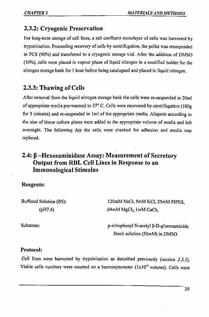
CHAPTER2 MATERIALS AND METHODS
2.3.2: Cryogenic Preservation
For long-term storage of cell lines, a sub confluent monolayer of cells was harvested by
trypsinisation. Proceeding recovery of cells by centrifugation, the pellet was resuspended
in FCS (90%) and transferred to a cryogenic storage vial. After the addition of DMSO
(10%), cells were placed in vapour phase of liquid nitrogen in a modified holder for the
nitrogen storage bank for I hour before being catalogued and placed in liquid nitrogen.
2.3.3: Thawing of Cells
After removal from the liquid nitrogen storage bank the cells were re-suspended in 20ml
of appropriate media pre-warmed to 37° C. Cells were recovered by centrifugation (180g
for 3 minutes) and re-suspended in 1ml of the appropriate media. Aliquots according to
the size of tissue culture plates were added to the appropriate volume of media and left
overnight. The following day the cells were checked for adhesion and media was
replaced.
2.4: fl -Hexosaminidase Assay: Measurement of Secretory Output from RBL Cell Lines in Response to an Immunological Stimulus
Reagents:
Buffered Solution (BS):
(pH7.4)
120mM NaCl, 5mM KCI, 25mM PIPES,
.04mM MgCh, lmM CaCh
Substrate: p-nitrophenyl N-acetyl ~-D-glucosaminide
Stock solution (50mM) in DMSO
Protocol:
Cell lines were harvested by trypsinisation as described previously (section 2.3.1).
Viable cells numbers were counted on a haemocytometer (lx104 volume). Cells were
39

CHAPTER2 MATERIALS AND METHODS
resuspended at a cell density of 0.5x 10⁶ /ml and sensitized with DNP-specific mlgE
(SPE-7 1/500) or NIP-specific hulgE (JW8 1/500). Aliquots (100µ1) were plated out in
triplicate on 96-well plates and incubated at 37°C for 16 hours. All solutions from this
point were pre-warmed to 37°C. Next day, 96 well plates were examined for cell
confluency before the media was removed and cells washed twice with DPBS. DNP
HSA and NIP-HSA dilutions (0.1-lO000ng/ml) and Triton -Xl00 (1%) were prepared in
BS. BS (100µ1) was added to the first set of three wells (blank), followed by 100µ1 of
Triton-Xl00 (total) to the last set of triplicate wells, and addition of DNP-HSA/NIP-HSA
dilutions to the remaining wells as appropriate. Following incubation for 20-30 minutes
at 37°C, 50µ1 of supernatant from each well was transferred to an identically labelled 96-
well plate. Substrate solution was diluted in 0.2M citrate buffer (pH4.5) to a final
concentration of 2mM. 50µ1 of substrate solution was added to each well containing
supernatant prior to 2-3 hours incubation at 37°C allowing time for the reaction to take
place. The reaction was terminated by the addition of 150µ1 lM Tris-HCI (pH 9.0). Plates
were analyzed using an ELISA reader set at 405nm. Analysis of plates allowed
percentage release for each DNP-HSA / NIP-HSA dilution to be calculated using the
following equation.
(Average of test triplicate)- (average of blank triplicates) x 100
(Average of total triplicates)
2.5: Cell Lysate Preparation for 1D- PAGE
2.5.1: Sample Preparation
Reagents:
RIPA Lysis buffer 150mMNaCl
1 % lgepal CA-630 (Nonidet P-40)
0.5% Sodium deoxycholate
0.1 % SDS, 50mM Tris-HCI (pH 8)
All lysis buffers were supplemented with protease inhibitor cocktail tablets (Roche), I 0-
100 µl PMSF and lmM Sodium orthovanadate.
40

CHAPTER2 MATERIALS AND METHODS
Protocol
Cells were cultured as monolayer on 145 mm plates and sensitized for 16 hours with
either DNP specific mlgE (SPE-7, 1/500) or NIP specific hulgE (JW8), 1/500) at 37°C.
Solutions and universal tubes were all pre-warmed to 37°C. The following day cells were
washed with pre-warmed PBS and harvested in pre-warmed CDS. This was followed by
recovery by centrifugation (180g for 3 minutes). The cell pellet obtained was
resuspended in I ml of pre warmed BS. Cell numbers were determined and re-suspension
was carried out at 5 x 10⁶/ml. The cells were divided equally into universal tubes
dependent on the number of samples required. The appropriate sample was activated for
2 minutes with the corresponding cross-linking agent (IOOng/ml, optimal concentration
determined from ~ -Hexosaminidase assay). The reaction was halted by addition of I 0ml
of ice cold PBS to each universal tube and the tubes were then kept on ice for 2 minutes.
The cells were recovered by centrifugation (180g for 3 minutes) followed by re
suspension in I ml ofDPBS and centrifugation (180g for 3 minutes) at room temperature.
The pellets were resuspended in 1ml RIPA Lysis Buffer (to which sodium
orthovanadate and I 0-100µ1 PMSF was added) transferred to cooled micro centrifuge
tubes, vortexed and left on rotating for 60 minutes at 4°C. From this point onwards all
procedures were carried out at 4°C. The tubes were then centrifuged (10000g for 15
minutes) and cell lysate obtained transferred to new micro centrifuge tubes and assayed
for protein content employing Bradford's method.
2.5.2: Bradford's Method for Protein Estimation
The need for carrying out protein quantification prior to immunoprecipitation is to ensure
uniform protein concentrations in all samples under study. The principle of Bradford
Assay is that binding of Coomassie Blue G-250 dye in the Bradford reagent to protein
causes a shift in absorption from 465 to 595 nm. A standard curve was prepared using
BSA. 900µ1 of Bradford Reagent (1/4 dilution, Bio Rad), 95µ1 dH2O and 5µ1 of cell
lysate were mixed in a 1.5ml cuvette to achieve a final volume of I ml. After a lapse of 5
minutes readings were taken on spectrophotometer set at 595nm.

CHAPTER2 MATERIALS AND METHODS
2.5.3: Immunoprecipitation
Protein A Agarose beads (Chemicon) were used in all immunoprecipitations. All pipette
tips were cut off when working with protein A to avoid bead degradation. All steps were
performed at 4 °C unless otherwise stated.
Protocol:
Protein A was prepared following the manufacturer's instructions (Chemicon). Cell
lysate was incubated with protein A (I 00µ1) of 50% slurry for 30 minutes ( end-to-end
rotation). This process is known as pre clearing which reduces any non-specific binding
by proteins within the sample, which may interfere with later analysis. Lysates were
centrifuged (10000g for 15 minutes) and the supernatant was transferred to new micro
centrifuge tubes being careful not to disrupt protein A pellet. Protein estimation of sample
was carried out using the Bradford protein assay allowing sample normalisation. At this
point, 15-25µ1 of cell lysate was taken from the samples to be used in anti
phosphotyrosine immunoprecipitation for the later purpose of FcR-y quantification. An
equal amount of appropriate antibody was added to each sample following the
manufacturer's instructions. Lysates were incubated overnight with end-to-end rotation to
allow antigen-antibody complex formation. The following day, protein A (100µ1) was
added to each sample as appropriate prior to 2-4 hour incubation with end-to-end rotation
allowing immune complex capture. Immunoprecipitates were pulsed in a centrifuge
(10000g), the supernatant removed and washed three times with appropriate ice-cold lysis
buffer. After the last washing all of the supernatant was removed from the
immunoprecipitates using a Gelsaver pipette microtip. Immunoprecipitates were
resuspended in 60µ1 of Laemmli's 1-D SDS-PAGE loading buffer (50mM Tris-HCI, pH
6.8, 2% SDS, I 0% Glycerol, I OOmM DTT) and boiled for 5 minutes. Samples were
centrifuged ( 10000g for 5 seconds) and resolved on a 12.5-17% SDS-P AGE gel.
2.5.4: One-dimensional Polyacrylamide Gel Electrophoresis
ProJeins were resolved by polyacrylamide gel electrophoresis using the Mini-Protean II
electrophoresis system (Bio-Rad). Acrylamide gels (12.5-17%) were made using a multi
gel casting chamber following the manufacturer's instructions. Discontinuous acrylamide
42

CHAPTER2 MATERIALS AND METHODS
gels were used consisting of a stacking gel (5% acrylamide in Tris-HCI pH 6.8) allowing
uniform entry of sample into the gel followed by a separating gel (12.5 -17% acrylamide
in Tris-HCI pH 8.8) for the resolution of proteins according to molecular weight.
Following the addition of 60µ1 of 1-D SDS sample buffer, samples were boiled for 5
minutes and were pulsed in a centrifuge (10000g). The samples were then allowed to cool
to room temperature prior to loading in parallel with a molecular marker (Fermentas) to
allow estimation of relative molecular weights of the resolved proteins. Electrophoresis
was performed in a Tris-Glycine buffer (2SmM Tris, 250mM Glycine pH 8.3, 0.1 %
SDS). Proteins were resolved at SOY for 20 minutes to allow proteins to enter the
stacking gel followed by I 00-120V until the dye front reached the end of separating gel.
On the completion of electrophoresis, gels were used for Western transfer to PVDF
membrane.
1 kDa I i I _,... .. 170- ........ .
- L- .. 130~ ...,, ... ,,.. J l ....._. ....... 100--~ i I
~ '""',. ~ -70 - : . I J , ....... - i -55 I .... i
~ - .. 40 __ -· . I I ""·,.._ 1 .. 35 ~
-~ L-2s ,..... ; !
□(::_ :i .___i
.c-m Tris~~SC&PAGE
Protein Ladder (10-250kDa) (Fermentas)
2.5.5: Western Transfer of Proteins to PVDF Membrane .
Electrophoretic transfer of proteins to PVDF (Amersham Biosciences) was performed
using the Mini Trans-Blot system (Bio-Rad) according to the manufacturer's instructions
43

CHAPTER2 MATERIALS AND METHODS
in a Tris-Glycine buffer 25mM Tris, 250mM Glycine pH 8.3, 20% methanol). The PVDF
membrane required a pre-soaking step in methanol for I minute followed by washing in
distilled water for 5 minutes before transfer buff er equilibration for IO minutes. The
assembly consisted of the gel and membrane in direct contact, sandwiched between two
sheets of filter paper and sponge on either side and held together in a transfer cassette.
The cassette was placed in the Trans-Blot System checking for correct orientation.
Electrophoretic transfer was carried overnight at 30V ( 4 °C).
2.5.6: Immunodetection of Transferred Proteins
Membranes were removed from the Mini Trans Blot system and placed in universal
tubes/50ml polypropylene tubes depending on the size of membrane (protein side facing
the lumen). All steps were carried out on a roller platform mixer (Denley Spiramix) to
ensure even distribution of solution across the membrane and minimize antibody usage.
Depending on the primary antibody used blocking of the non-specific binding sites on the
membrane was carried out by immersing the membrane either in 3% Bovine serum
albumin (anti-phosphotyrosine, clone 4G I 0) or 5% dried semi-skimmed milk (all other
antibodies) in DPBS-Tween (lxDPBS supplemented with 0.1% tween-20, Sigma
Aldrich) for 60-90 minutes at room temperature (alternatively it may be left overnight in
blocking solution in refrigerator at 2-8°C). The prepared blocking solutions were kept
aside for antibody dilutions. The membranes were rinsed three times in DPBS-Tween.
Membranes were incubated for 45-60 minutes at room temperature with the appropriate
primary antibody diluted in appropriate blocking solution. Following primary antibody
incubation the membranes were rinsed three times and washed in DPBS-Tween in four
steps of lxl5minutes and 3 x 05minutes. Dilution of the secondary horseradish
peroxidase (HRP) antibodies was made in appropriate blocking solution and the
membranes were incubated for 45-60 minutes. Membranes were washed as described
previously and prepared for protein detection using Enhanced Chemiluminescence (ECL)
according to manufacturer's instructions.
44

CHAPTER2 MATERIALS AND METHODS
2.5.7: Detection of Proteins with Enhanced Chemiluminescence (ECL)
Membranes were drained of excess DPBS-Tween and incubated with equal volumes
(enough to cover the membrane) of both ECL reagents for 1 minute at room temperature.
Membranes were placed in a photographic cassette (protein side up) and covered with
cling-film and taken to dark room. A sheet of autoradiography film was placed on top of
the membrane in the cassette and exposure of film for varying lengths of times dependent
on signal strength was carried out followed by developing and fixing of the film
(KODAK).
2.6: Assessing FceRI Expression in Transfected Cell lines
by Flow Cytometry
Reagents:
Wash buffer
Primary antibody
Secondary antibody
Flurochrome antibody
DPBS suplemented with 1% FCS (4°C)
Mouse IgE (migE) (UoS)/ human IgE (Serotec)
Biotinylated goat anti human IgE (Vector Laboratories)
Streptavidin R-phycoerythrin (Sigma-Aldrich)
Goat anti-mouse lgE FITC-conjugated (Bethyl Laboratories)
Protocol:
Transfected cell lines were grown in 100mm tissue culture plates to cell confluency for
the days of FACS analysis. Cells were washed with pre-warmed PBS and harvested in
pre-warmed CDS. This was followed by recovery by centrifugation (180g for 3minutes).
The cell pellet obtained was washed twice in 10ml wash buff er and resuspended at a
density of 5X10⁶ cells/assay in ice-cold wash buffer. 50µ1 of cell suspension was added to
non-sterile polypropylene FACS tube as un-labelled negative control and placed on ice
till cell analysis. Another 50µ1 cell suspension was added to appropriately labelled non
sterile polypropylene FACS tube as labelled control. The appropriate 1 ° antibody {mlgE / .
hulgE , 1 µg) was added to 100µ1 cell suspension in appropriately labelled non-sterile
polypropylene F ACS tube labelled as sample and incubated on ice for 30 minutes. After
45

CHAPTER2 MATERIALS AND METHODS
the 30 minutes incubation period the polypropylene F ACS tubes containing the labelled
control and the sample were washed two times with ice cold wash buffer before finally
resuspending the pellet in 1ml cold wash buffer. Biotinylated goat anti human IgE (1/400
dilution in wash buffer) / goat anti-mouse IgE FITC-conjugated (1/500) was added and
mixed to each tube and left on ice for 30 minutes. The cells were recovered by
centrifugation and washing as described above. Streptavidin R-Phycoerythin antibody
(1/50 dilution in wash buffer) was added (RBL transfected cells) and mixed to both tubes
(labelled control and sample) and again incubated on ice for 30 minutes. Two final
washes were carried out to remove excess Streptavidin R-Phycoerythin / goat anti-mouse
lgE FITC-conjugated antibody as described above and the cells were resuspended in
200µ1 wash buffer and kept on ice till the analysis. Samples were analysed using a
FASort or a FACS™ CY AN ADP flowcytometer (Dakocytomation).
2.7: FACS Cell Sorting of Transfected Cell Lines by Flow
Cytometry
Reagents same as above.
Transfected cell lines were grown in 1001145mm tissue culture plates to cell confluency
(~12X106cells/plate) for the days of FACS sort. Cells were washed with pre-warmed
PBS and harvested in pre-warmed CDS. This was followed by recovery by centrifugation
(180g for 3minutes). The cell pellet obtained was washed two times in 10ml wash buffer
and resuspended in 1 ml ice-cold wash buffer. Cell suspension (50µ1) was added to non
sterile polypropylene FACS tube as unlabelled negative control and was placed on ice till
cell sorting. Another 50µ1 cell suspension was added to non-sterile polypropylene F ACS
tube as labelled negative control and was placed on ice. Human lgE / mlgE (IOµg) was
added and mixed to the remaining cell suspension (sample) which was incubated on ice
for 30 minutes. Labelled control and sample were centrifuged (180g for 3minutes) at
4°C; the supernatant was removed and two washes with ice cold wash buffer were carried
o~t before finally resuspending the pellet in 1ml cold wash buffer. Biotinylated goat anti
human IgE (1/400 dilution in wash buffer) / goat anti-mouse lgE FITC-conjugated
(1/500) was added and mixed to each tube and left on ice for 30 minutes. The cells were
46

CHAPTER2 MATERIALS AND METHODS
recovered by centrifugation and washed as described above. Streptavidin R-Phycoerythin
antibody (1/50 dilution in wash buffer) was added (RBL transfected cells) and mixed to
both the tubes (labelled control and sample) and were kept on ice for 30 minutes. Final
two washes were carried out as described above to remove excess Streptavidin R
Phycoerythin I goat anti-mouse lgE FITC-conjugated antibody and cells were
resuspended in 200-500µ1 of wash buffer and were kept on ice till the time of cell sorting.
Samples were F ACS cell sorted using a F ACSAria or a F ACS Mo Flo flowcytometer
(Dakocytomation). The sorted cells were collected in the appropriate media and plated in
100mm plates in the appropiate media (supplemented with Penicillin lO0U/ml and
streptomycin 1 00µglml to avoid contamination) as described in section 2.3.1.
2.8: Assesment of Intracellular Calcium Mobilisation
Intracellular Ca2+ mobilization brought about by antigenic stimulus {NIP/DNP) was
assessed using the Ca2+ indicator Fluo-3AM , 4-( 6-Acetoxymethoxy-2, 7-dichloro-3-oxo-
9-xanthenyl)-4 • -methyl-2,2'( ethylenedioxy) dianiline-N' ,N' ,N' ,N' - tetraacetic acid
tetrakis (acetoxymethyl) ester. Developed by Minta et al (1989), Fluo 3 remains non
fluorescent until bound with Ca2+ resulting in a 40-fold increase in mean fluorescence.
Analysis was carried out in the visible light range with excitation at -488nm and
emission at~ 525nm. Acetoxylmethyl ester was added to give an uncharged molecule that
can diffuse across the cell membrane allowing entry into the cell cytoplasm.
Acetoxymethyl ester is hydrolysed by action of cytoplasmic esterase yielding Fluo 3
within cell {Tsien, 1981 ).
Reagents:
Balanced Salt Solution (BSS)
Fluo-3-AM
fonomycin
l 48mM NaCl, 49mM KCL, 63mM
D-sorbitol, 2.63mM K2HPO4,
lmM KH2PO4, 10.lmM HEPES
Sigma-Aldrich, 500 µM stock in DMSO
Sigma-Aldrich, lmglml stock in DMSO
47

CHAPTER2 MATERIALS AND METHODS
Protocol:
Cells were cultured as a monolayer on 145mm plate and passively sensitized for 16 hours
with either DNP-specific mlgE (SPE-&, 1/500) or NIP-specific hulgE (JW8, 1/500) at
37°C. The cells were washed in DPBS and harvested with CDS and centrifuged (180g for
3 minutes). Cells were counted and resuspended in BSS at lxl06/ml. For the purpose of
background reading 0.5 ml of each cell suspension was taken. This was followed by
addition of Fluo 3-AM (5µM) and incubation at room temperature for 30 minutes was
carried out in the dark. Cells were pelleted and resuspended in DPBS (containing no
additional CaCii or MgCb) at a density of 2xl06/ml. IgE sensitized cells loaded with
Fluo-3AM were analyzed using a FACSort flow cytometer preset for Fluo-3AM studies.
Data was recorded in the form of a density plot of mean fluorescence against time. After
an initial background reading of 30-50 seconds, samples were activated with the
appropriate cross-linking agent (NIP/DNP I 00ng/ml). In the event where no calcium
mobilization was witnessed ionomycin (IOµM) (a calcium ionophore) was used as a
positive control to ensure that the sample has been loaded with Fluo-3AM.
2.9: Molecular Biology Methods
2.9.1: Bacterial Strain
All cloning work was carried out using the XL I-blue E.coli strain (Stratagene).
Genotype - recAI end AI gyrA96 thi-1 hsdR17 supE44 re/AI lac [F'pro AB /acfl
ZM115 Tn IO (Tet1)].
2.9.2: E.Coli Growth Media
Bacterial cultures were grown in LB-broth or LB-agar as required supplemented with the
antibiotic (Ampicillin I 0µg/ml).
LB Broth- MQ water, Tryptone (IOg/L), Yeast extract (5g/l), Sodium Chloride (IOg/L).
LB Agar: LB Broth; Bacteriological Agar (15g/L).
48

CHAPTER2 MATERIALS AND METHODS
2.9.3: Vectors
pEE6 (Celltech)
This vector was used previously to generate the RBLhuFcERia transfected cell line
(Section 1.11) (Wilson et al., 1993). The vector confers resistance to ampicillin and
neomycin, allowing selection of transformants in E.Coli and mammalian cells using
G418.
pUC18
pUC18 is a 2686bp vector. The vector has a J3-lactamase resistance gene conferring
ampicillin resistance and lacZ operon, which allows selection of the successfully ligated
clones.
2.9.4: Preparation of Competent Bacterial Cells
Buffers
TFBJ- 30mM KCH3COO, IOmM CaCh. 50mM MnCh, IO0mM RbCh,
15% Glycerol pH 5.8 with CH3COOH
TFB2-10mM PIPES, 75mM CaCh. 50mM MnCh, lOmM RbCh.15% Glycerol.
pH 6.5 with KOH
Protocol:
A single colony from a freshly streaked LB plate was cultured overnight in 2.5ml LB
medium at 37°C with shaking (approximately 225rpm). The following day the entire
culture was used to inoculate 250ml LB medium containing 20mM MgSO4 in a 1 L flask.
Cells were cultured at 37°C with shaking until the OD was between 0.4 and 0.6 (upto 6
hours). Cells were centrifuged (4500g for 5 minutes) at 4°C and the pellet was gently
resuspended in 100ml ice-cold TFBl. After incubation on ice for 5 minutes at 4°C, the
c~lls were centrifuged as before and gently resuspended in 10ml TFB2. After incubation
on ice for 15-60 minutes the cells were divided into aliquots (200µ1) and quick-frozen in
a dry ice/ isopropanol bath for storage at -80°C.
49

CHAPTER2 MATERIALS AND METHODS
2.9.5: Transformation of Competent Bacterial Cells
Competent cells were thawed slowly on ice. Approximately 1 Ong plasmid DNA was
added to 200µ1 cells and mixed by swirling with a pipette tip. After incubation on ice for
30 minutes, cells were heated in a water bath at 42°C for 45-60 seconds then cooled on
ice for 2 minutes. LB medium (3ml) was added and the cells were incubated for 45
minutes with shaking (approximately 150rpm) at 37°C. 50- 200µ1 of mix was spread on
plates containing the selection antibiotic (ampicillin). The plates were incubated
overnight at 37°C and examined for colony growth the next day.
2.9.6: Isolation of Plasmid DNA
2.9.6.1: QIAprep Spin Miniprep Kit
The QIAprep miniprep kit was employed when small amount of DNA was required. The
kit contained all the required solutions and the protocol provided with the kit ~as
followed. Cells were isolated from a 3ml overnight culture by centrifugation. The kit is
based on alkaline lysis of bacterial cells followed by adsorption of DNA onto silica in the
presence of high salt. The procedure consists of following three basic steps
• Preparation and clearing of a bacterial lysate
• Adsorption of DNA onto the QIAprep membrane
• Washing and elution of plasmid DNA
Plasmid yield with the QIAprep miniprep system was variable and dependent on plasmid
copy number per cell, the individual insert in the plasmid, factors that affect growth of
the bacterial culture, the elution volume, and the elution incubation time. A 1.5ml
overnight culture yielded between 5 and 15µg of plasmid DNA.
2.9.6.2: QIAGEN Plasmid Midi Kit
When large quantities of high purity plasmid DNA were required for mammalian cell
transfection the QIAGEN plasmid Midi Kit was used allowing upto 100-200µg of DNA
to be isolated. This kit is also based on the modified alkaline lysis procedure, followed by
binding of plasmid DNA to QIAGEN Anion-Exchange Resin under appropriate low-salt
50

CHAPTER2 MATERIALS AND METHODS
and pH conditions. Cells were isolated from a 50ml overnight culture by centrifugation.
RNA, proteins, dyes, and low-molecular weight impurities were removed by a medium
salt wash. Plasmid DNA was eluted in a high salt buffer and then concentrated and
desalted by isopropanol precipitation and subsequently washed with 70% ethanol. The
DNA was eluted in a microcentrifuge with 100-1000µ1 water or TE buffer as specified in
the protocol provided with the kit.
2.9. 7: Agarose Gel Electrophoresis
T AE agarose gel electrophoresis was employed to visualise isolated plasmids, restriction
enzyme digests and PCR products. A 6x loading buffer (MBI) was added to the DNA
before loading on I% agarose gel containing 0.Sµg/ml ethidium bromide. Electrophoresis
was performed using a Biorad subcell at 1 00V, 80mA for 1 hour. A molecular weight
standard of lKb ladder (Fermentas) was loaded onto the gel. The DNA was visualised
and photographed under UV light on the Fluorchem TM 8000 imaging system.
bp
1000 IJ)
r; 00
?Ul
lKb DNA Ladder (Fermentas)
51

CHAPTER2 MATERIALS AND METHODS
2.9.8: DNA Manipulation
2.9.8.1: Restriction Enzyme Digests
Restriction enzymes digests were typically carried out in a 20µ1 volume, with 2µ1 1 Ox
buffer and lOunits of enzymes (lµl). The quantity of DNA used was dependent on the
application (typically 0.5-1.0µg for test digest and up to 5µg for cloning) and the
remaining volume made up to 20µ1 with sterile MQ water. The temperature and time for
digestion depended on the specific restriction enzyme but was commonly 37°C for 1-2hr.
The optimal 1 Ox buff er for the specific enzymes was used according to the
manufacturer's recommendation.
2.9.8.2: Dephosphorylation
To prevent religations without insertion of the desired fragment, 5' phosphate ends of
pEE6 vector were removed prior to ligation using calf intestinal phosphatase. Digestion
of pEE6 was performed to provide linearised rlasmid for ligations. This was followed by
adding 1µ1 alkaline phosphatase, 2µ1 lOx phosphatase buffer and 17µ1 sterile MQ water
to the 20µ1 digest mixture followed by incubation at 37°C for 1 hour.
2.9.8.3: Ligation
Ligation of DNA fragment was typically carried out in 30µ1 total volume. The reaction
mixture contained 1µ1 T4 DNA ligase, 3µ1 of lOx ligase buffer, varying ratios of 1:2 or
1:3 of vector (pEE6) to insert DNA and water to make up the volume. Generally a ratio
of 1 :2 or 1 :3 of vector to insert was aimed for as generally a 2-3x molar excess of insert
DNA gives optimal ligation. A plasmid re-ligation control was always set up along side
whereby an identical reaction was set up having MQ water in place of DNA. The reaction
was incubated at 16°C overnight.
52

CHAPTER2 MATERIALS AND METHODS
2.9.8.4: DNA Purification from Agarose Gel
DNA gel extraction from agarose gel was carried out by using Quantum Prep™ Freeze N
Squeeze DNA gel extraction spin columns kit (BIO-RAD). This kit purifies via filtration
in a spin column format. The DNA band was excised from an agarose gel, and the gel
slice cut into small pieces and placed in filter cup. The cup plus gel piece was put in a -
20°C freezer for 5 minutes, then removed and immediately centrifuged (13,000g for 3
minutes) at room temperature. The agarose debris was retained within the filter cup and
the bottom of the tube contained the recovered DNA.
2.9.8.5: Site Directed Mutagenesis
In vitro site-directed mutagenesis was carried out using Stratagene QuickChange® Site
Directed Mutagenesis Kit which allows site specific mutation in a double-stranded
plasmid. This is a rapid four step procedure generating mutants with more than 80%
efficiency. Miniprep plasmid DNA was used to make point mutations. QuickChange®
Site-Directed Mutagenesis Kit was performed using PfuTurbo® DNA polymerase and a
temperature cycler. The procedure utilizes a supercoiled double-stranded DNA (dsDNA)
vector (pUC18) with the insert of interest and two synthetic oligonucleotide primers
containing the desired mutation. The primers each complementary to opposite strands of
vector are extended during temperature cycling by PfuTurbo® DNA polymerase. This
generates a mutated plasmid containing staggered nicks. This is followed by treating the
product with Dpn I. The Dpn I endonuclease is specific for methylated and
hemimethylated DNA and is used to digest the parental DNA tempelate and to select for
mutation-containing synthesised DNA. DNA isolated from almost all E. Coli strains is
dam methylated and therefore susceptible to Dpn I digestion. The nicked vector DNA
containing the desired mutations is then transformed into XLI-Blue supercompetent cells
(Stratagen).
53

CHAPTER2 MATERIALS AND METHODS
2.9.8.6: DNA Sequencing
The DNA was sequenced by Cogenics and then analysed using ALIGN, CHROMA and
CLONE computer programmes to compare the sequence with genomic huFcsRia
subunit. The primers used for the sequencing were Ml3-48REV and Ml3For-40 to read
the DNA from both ends. Clones with additional or no mutations were rejected.
M13-48REV GGGATAACAATTTCACACAG
M13For-40 GTTTTCCCAGTCACGAC
2.10: Transfection Mammalian Cells
Stable transfection in the mammalian cells was obtained by using the technique of
electroporation. Cells were grown to 50-60% confluency and harvested as already
described then washed with DMEM containing no FCS or antibiotics, to remove FCS so
as to prevent sparking during electroporation. The cells were counted and resuspended in
DMEM to give I 07 cells/0.8ml. The cell suspension was added to electroporation cuvette
(0.4cm electrode gap) in 0.8ml aliquots. Plasmid DNA prepared by using the Qiagen
midiprep kit (section 2.9.6.1) was mixed with the cells, and a control prepared using 50µ1
MQ water in place of DNA. The cuvettes were then incubated on ice for IO minutes.
Each cuvette was given a single pulse of 250V, 960µF using a Gene Pulser (Bio-Rad)
then incubated on ice for further IO minutes. The cells were then plated out after
resuspending in 20ml of appropriate fresh complete medium. After 24-48 hours recovery
period the medium was aspirated and selection medium was introduced. The cells were
maintained with change of medium being carried out every 2-3 days. Cells in the control
died by 7-10 days and the successful transfected cells were grown as normal and selected
using flowcytometry (section 2. 7).
Another technique employed for carrying out transient transfection in RBL-2H3. l was by
using the commercially available METAFECTENE™ PRO transfection reagent as part
54

CHAPTER2 MATERIALS AND METHODS
of collaboration with the manufacturer (Biontex). This reagent uses the Biontex' RMA
Technology (Repulsive Membrane Acidolysis) with new toxicity optimisation module
(TOP-technology), which regulates the lipolex release of genetic material. The protocol
provided by the manufacturer was followed.
55

CHAPTER 3 Mutagenesis of Human High Affinity lgE Receptor Alpha Chain subunit
CHAPTER3
Mutage,iesis of Human Higli Affinity
lgE Receptor Alplia Cliain subunit
56

CHAPTER3 Mutagenesis of Human High Affinity lgE Receptor Alpha Chain subunit
CHAPTER3
Mutagenesis of Human Higli Affinity IgE Receptor Alplia
Cliain subunit
3.1: Conserved Motif-Fc1:Rla
Previous studies have demonstrated the presence of an invariant eight amino acid motif
(LFAVDTGL) within the transmembrane domain of FcsRia and FcyRIIIa subunits,
shown to be conserved in many species including rat, mice and humans. The conserved
motif contains a polar aspartate residue at position 194 within a predominantly non-polar
setting (Ravetch and Kinet, 1991) (Section 1.11).
3.1.1: Residues Targeted for Site Directed Mutagenesis
Protein activity can be transformed by using the protein engineering technique of site
directed mutagenesis thus providing the researchers with an insight into the structure
function relationship leading to a better understanding of the cellular responses. This
technique was employed to assess the functional consequences of site-specific
mutagenesis replacing the D 194 residue of human FcsRia subunit (huFcsRia) to obtain
an improved understanding of the role of this residue within the transmembrane domain
in FcsRia-mediated signalling. The mutations introduced previously included asparagine
(D194N), leucine (D194L), alanine (D194A), glutamic acid (D194E), and lysine
(D194K) (Iodice, 2006). The RBL-2H3.l cells transfected with mutant huFcsRia
constructs D194E, D194A, and D194K failed to support cell surface expression (Iodice,
2006) while the wild type (D194) and mutant variant D194N expressed the huFcsRia
subunit at the cell surface in association with the endogenous FcR-P and FcR-y subunits
(Iodice, 2006). The transfected cell lines D194 and D194N demonstrated FcERI
aggregation, tyrosine phosphorylation of FcR-y and Syk kinase, intracellular Ca +2
mobilisation and mediator release in response to antigen-mediated activation, analogous
to that observed in parental RBL-2H3. l cell line (Iodice, 2006). However the RBL-2H3.1
-cell line transfected with mutant D194L gene construct, although supporting cell surface
57

CHAPTER3 Mutagenesis of Human High Affinity lgE Receptor Alpha Chain subunit
expression, showed absence of FcERI-mediated signalling in response to antigenic
stimulus, but the pattern of cell surface expression was suggestive of constitutive receptor
pre-clustering irrespective of antigenic stimuli (Iodice, 2006).
In order to gain a more comprehensive understanding of the role of the aspartate residue
5 new mutations were carried out in the present study which included replacing the D194
residue with polar residues threonine (D194T), serine (D194S), arginine (D194R) and
non-polar residues valine (D194V) and isoleucine (D1941). The idea was to replace D194
with amino acids of different sizes and polarity and assess the effect of these mutations
on the huFcERla expression and FcERla-mediated signalling.
The construct verification of the mutations carried out is shown in figure3.1.a and 3.1.b.
3.1.2 Primer design
Primers were designed to carry out single point mutation in the D 194 domain with the aid
of computer software. An additional silent mutation in the form of Stu I was also
incorporated to aid in assessing the presence of mutation with the help of restriction
digestion with Stu I and running on TAE agarose gel. The sequence of the primer was
necessitated by the fixed position of the domain in which the point mutation was to be
carried out. Primers were based on approximately 20bp sequence around the mutation.
The mutagenic primers show the altered bases in bold and the altered codons are
underlined while the silent mutation is shown in bold green. The primers are shown as
below.
D194T (Threonine)
S' 5'GGTGATTCTGTTTGCTGTGACCACAGGCCTA TIT ATCTCAACTCAGCAG 3'
3 5' CTGCTGAGTTGA GAT AAA TA<; (;CCTGTGGTCACAGCAAACAG AATCAC C 3·
D194V (Valine)
S' 5' G GTG A TT CTG TIT GCT GTG GTC ACA GGC CTA TIT ATC TCA ACT CAG CAG 3 •
3' 5' CTG CTGAGTTGA GAT AAA TAf; GCCTGTGACCACAGCAAA CAG AATCACC 3'
58

CHAPTERJ Mutagenesis of Human High Affinity lgE Receptor Alpha Chain subunit
D1941 (Isoleucine)
5' 5' G GTG A TT CTG TIT GCT GTG ATC ACA GGc < TA TIT ATC TCA ACT CAG CAG 3 ·
3' 5' CTGCTGAGTTGA GAT AAA TA<; <:CCTGTGATCACAGCAAA CAG AATCAC C 3'
Silent mutation- Stu I s· s· AGG CCT 3·
3· 5'TCC GGA 3'
The mutations encoding the replacement of Asp with threonine and valine were
successful but problems were encountered with the isoleucine mutation. This necessitated
the design of a new primer and successful mutation was introduced by altering the bases.
D1941 (lsoleucine new primer)
s· 5' G GTG ATT CTG TIT GCT GTG ATT ACA GGC < TA TIT ATC TCA ACT CAG CAG 3 ·
3' 5· CTG CTG AGTTGA GAT AAA TA<; <;CCTGT A:i!CACAGC AAA CAG AATCACC 3·
59

CHAPTER3 Mutagenesis of Human Higlt Affinity IgE Receptor A/pita Cltaill subunit
huFcERla TI 111 TATCCCATTGTTGGTGGTGATTCTGTITGCTGTG~ACAGGATTATTTATC
FF IP LLV VI --------= 1 F
D194 Aspartic acid Polar Negatively-charged
IIIIITATCCCATTGTTGGTGGTGATTCTGTTTGCTGTG~ACAGGATTATITATC
FF P LLV VI \\ IF
D194N Asparagine Polar Non-Charged
TITTTTATCCCATTGTTGGTGGTGATTCTGTTTGCTGTGAM£ACAGGATTATITATC
FF P LL V V FAV F
D194L Leucine Non-Polar Non-Charged
TlllTTATCCCATTGTTGGTGGTGATTCTGTTTGCTGTG.QEACAGGATTATITATC
FF P LL V VI 1 <i F
Figure 3.la-Sequence Verification of transmembrane domains of Wild
type (D194) and mutant (D194N and D194L) huFcERia constructs.
These variants were generated in a previous study (Iodice, 2006).
The constructs were sequenced by Lark (Cogenics) Technologies Ltd. The sequence of
transmembrane domain of huFceRla subunit was obtained from www.cxpasy.org.
Translated amino acids are shown beneath each sequence. Green shaded area indicates
the invariant motif with the mutations in bold underlined and in different colours: Dl 94
(red), D194N (pink) and D194L (yellow).
60

CHAPTERJ Mutage11esis of Huma11 High Affi11ity lgE Receptor A/pita Chain subu11it
Dl94T Threonine Polar Non-Charged
TTIITTATCCCATTGTTGGTGGTGATTCTGTTTGCTGTG~ACAGGATTATTTATC
F F P L L V V I I· V 'I 0 F
D194V Valine Non-Polar Non-Charged
TTTTTTATCCCATTGTTGGTGGTGATTCTGTTTGCTGTG.QEACAGGATTATTTATC
FF Ip LL V vr r V TG F
D194I Isoleucine Non-Polar Non-Charged
TTTTTTATCCCATTGTTGGTGGTGATTCTGTTTGCTGTGATTACAGGATTATTTATC
F F P L L V V I I A V T G F
D194R Arginine Polar Charged
TlTTTTATCCCATTGTTGGTGGTGATTCTGTTTGCTGTG,grrACAGGATTATTTATC
FF P LLY VI V TG F
D194S Serine Polar Non-Charged
TTTTTTATCCCATTGTTGGTGGTGATTCTGTTTGCTGTG~ACAGGATTATTTATC
F F P L L V V I F 'I G F
Figure 3.lb-Sequence Verification of transmembrane domains of
mutants (D194T, D194I, D194V, D194R, and D194S) huFctRia
constructs. These variants were generated in the current study.
The constructs were sequenced by Lark (Cogenics) Technologies Ltd. Translated amjno
acids are shown beneath each sequence. Green shaded area indicates the invariant motif
with the mutations in bold underlined and in different colours: D194T (orange), D194V
(blue), D194I (lavender), D194R (plum) and D194S (grey).
61

CHAPTERJ Mutagenesis of Human High Affinity lgE Receptor Alpha Chain subunit
3.2: Vectors
3.2.1: pUC18
The cloning vector used for carrying out site directed mutagenesis and subsequent
sequencing of the mutation was pUC18 (figure3.2) which is a relatively small, high copy
number vector having '3-lactamase gene conferring ampicillin resistance which allows
selection of successfully ligated clones by growing them on LB media to which
ampicillin has been added.
3.2.2: Mammalian expression vector
pEE6 (figure 3.3) has been used previously by our research group for expression of the
wild type and mutant huFcERla subunits in the RBL mammalian cell line. The vector
comprises the gene conferring resistance to ampicillin and neomycin thus allowing
selection of successfully ligated clones using G4 l 8.
3.3: Cloning into pUC18
The huFcERla construct was introduced into the pEE6 expression vector. It was
necessary to subclone the construct into pUC18 cloning vector to carry out subsequent
site-directed mutagenesis and sequencing. The pEE6 vector with huFcERia subunit DNA
construct and pUCl 8 vector were digested using the EcoRI and HindIII restriction
enzymes (Section 2.9.8.1). The pUC18 vector was dephosphorylated to prevent re
ligation of the digested linearised vector (Section 2.9.8.2). The digested vector pUC18
and pEE6 with huFcERla were resolved in parallel on I% T AE agarose gel (Section
2.9. 7). The representative results are shown in the figure 3.4a. The desired bands i.e.
794bp (huFceRla) and 2686bp (pUCJ 8) were excised from the agarose gel and purified
using Quantum Prep™ Freeze N Squeeze DNA gel extraction spin columns kit (Bio-Rad)
(section 2.9.8.4). The purified bands of pUC18 and huFcERla were ligated together
(Section 2.9.8.3) The ligation reactions were set up overnight at 16°C, including a
negative control (water), to rule out any re-ligations. Ligation reactions were
subsequently transformed into XU-Blue supercompetent cells (Section 2.9.5). The
transformed cells were plated out onto LB ampicillin plates for selection and incubated
overnight at 37°C. Next day the plates were examined for colony, numbers of which was
62

CHAPTER3 Mutagenesis of Human High Affinity lgE Receptor Alpha Chain subunit
highly variable but were below 50. The variability in the number of colonies could be due
to multiple factors namely competence of cells used for transformation, the quality of
purified DNA, quantity of DNA agarose bands excised and the ratio of the vector to DNA
insert.
Six colonies were picked from the plate into 3ml LB media and incubated overnight on
shaker at 250rpms in 37°C room. Next day Qiagen DNA miniprep kit was employed for
small scale DNA extraction (Section 2.9.6.1). Test digestion of 10µ1 miniprep plasmid
DNA was carried out using the EcoRI and Hind III restriction enzymes (Section 2.9.8.1)
followed by resolving on T AE gel to confirm the presence of the desired fragment
(figure 3.4b ).
3.4: Site-Directed Mutagenesis
Quickchange Site- Directed Mutagenesis Kit (Stratagene) was employed to introduce the
point mutations (Section 2.9.8.5). The nicked vector DNA, containing the respective
desired mutations, was than transformed into XLI-Blue supercompetent cells (Section
2.9.5). The transformed cells were plated out onto LB ampicillin plates for selection and
incubated overnight at 37°C. Six colonies were picked from the plate into 3ml LB media
and incubated overnight on shaker at 250rpms in 37°C room. Next day Qiagen DNA
miniprep kit was employed for small scale DNA extraction (Section 2.9.6.1). Test
digestion of 1 OµI miniprep plasmid DNA was carried out using the Stu I, EcoRI and Hind
III restriction enzymes (Section 2.9.8.1) followed by resolving on TAE gel to confirm the
presence of the desired mutation (figure 3.5). Miniprep plasmid DNA samples were sent
for sequencing to check the integrity of the mutations.
3.5: Sequencing
The DNA was sequenced by Cogenics and the results were then analysed using ALIGN,
CHROMA and CLONE computer programmes to compare the sequence with genomic
huFccRia subunit. The primers used for the sequencing were Ml3-48REV and M13For-
40 to read the DNA from both ends. Clones with additional or no mutations were
rejected.
M13-48REV CGGATAACAA TTTCACACAG
MlJFor-40 GTITTCCCAGTCACGAC
63

CHAPTER3 Mutagenesis of Human High Affinity lgE Receptor Alpha Chain subunit
Bsm8151
BsmB:111rd1qr EcoO109l2674 Ndol 183 Alll 112617 J Nar 1/Kas 1235
Ssp 12501._____ : J .J" Bgll 245 ~ . • -::;.__Fsp1256
Eco57 I 27PBI "'-•-Pvu 1276 ,,!--....Pvu Xmn I 2294 . 11306
Bcg/2215 r
SCA 12177
Poly/inker cloning sites 396-454
Pvu 120661 Pvu II 628 Aval/2059 . + Tll164f
BsrD 11935 Sap I 6B3 Fspl 1919 Avail 1837-BQll 1813
B"'lJ;,}~8: 779 Bsa 11766 BsrDI 1753
Ahcl I 1694 EcoS7 I 1333 AtwN 11217
Figure 3.2 Bacterial expression vector pUC18
This vector was used previously to introduce site directed mutations in the huFcsRia
subunit. pUC18 is a 2686bp vector. The vector has a J3-lactamase resistance gene
conferring ampicillin resistance and lacZ operon that allows selection of the successfully
ligated clones through blue and white screening procedure. The successful ligations
appear as white E.Co/i colonies grown in ampicillin-enriched agar plates. Adapted from:
http://web.bio.utk.edu/peter on/BCMB%205 I 5/pUC I 8map.htm.
pff 6 plasmid DNA 6200bp
Figure 3.3 Mammalian Expression Vector pEE6
pEE6 is a 6200bp expression vector. It has the bacterial ampicillin resistance gene
(ampR) and polylinker site for the insertion of the regulatory gene which allows gene
expression. Modified from Stephens and Cockett (1989).
64

CHAPTER3 Mutogenesis of Human High Affinity TgE Receptor Alpha Chllitt subunit
Figure 3.4a
Figure 3.4b
8000bp 6000bp
1000b 750b
M l
3000b~--twt-2500bp+--l-li"""'
750b
M
Figure3.4: Cloninv; into pUC18
l
2
pEE6 (6200bp)
Iuman alpha chain (794bp)
pUC18 (2686bp)
Human alpha chain gene (794bp)
The digested (EcoRT and HindIJI restriction enzymes) vector pUC 18 (Lane2) and pEE6
with huFceRla gene construct (Janel) were resolved in paral1el on 1% TAE agarose gel
(figure3.4a). The desired bands i.e. 794hp (huFceRla) and 2686bp (pUCJ 8) were excised
from the agarose gel and purified using Quantum Prep™ Freeze N Squeeze DNA gel
extl'action spin columns kit (Bio•Rad) (section 2.9.8.4). The purified bands of pUCl 8 and
huFcsRJa were ligated together (Section 2.9.8.3). In figure 3.4b ptJC I 8 with huFceRla
clone digest show the correct appearance of two bands 2686bp pUC18 and 794bp
huFccRJa in lane 1 while lane M is the 1 Kbp DNA marker (Section2. 9. 7).
65

CHAPTER3 Mutage11esis of H11ma11 High Affi11ity lgE Receptor A/pita Chain s111>1111it
3066bp
750bp
313bp
Fie;ure 3.5: Site Directed Mutae;enesis
J M 2
3000bp pUC18 (2686bp)
l000bps Human alpha chain gene
(794bp)
Test digestion of 10µ1 miniprep plasmid DNA was carried out using the Stu I (Janel)
andEcoRI /Hind III (lane 2) restriction enzymes followed by resolving on l¾TAE gel to
confirm the presence of the desired mutation. Lane 1 shows bands at 313bps and 3066bps
after digestion of PCR product with Stu I, which was according to the incorporated silent
restriction site of Stu I. Lane M, is the lKbp DNA marker (Section2. 9. 7).
3.6: Subcloning to pEE6
After sequencing the clones, with the successful mutation, were employed to subclone the
mutant cDNA construct into expression vector pEE6 for subsequent transfection into
mammalian cells (RBLs).
The p,UCI 8 vector with mutant huFceRla. subunit DNA construct and pEE6 vector were
digested using the EcoRI and HindlII restriction enzymes (section 2.9.8.1). The pEE6
vector was dephosphorylated (section 2.918.2) to pre.vent re~ligation of the digested
linearised vector. The digested vector pEE6 and pUCl8 with mutant huFceRJa were
resolved in parallel _on 1 % TAE agarose gel (section 2.9.8.4).The representative results
are shown in the figure 3.6a. The desired bands i.e. 794bp (huFceRla.) and 6200bp
(pEE6) were excised from the agarose gel and purified using the Quantum Prep TM Freeze .
66

CHAPTER3 Mutagenesis of Human High Affinity lgE Receptor Alpha Chain subunit
N Squeeze DNA gel extraction spin columns kit (Bio-Rad) (section 2.9.8.4). The purified
bands of pEE6 and mutant hufcERla were ligated together (section 2.9.8.3) and a
negative (water) control was also set up to rule out any re-ligations. The ligation reactions
were set up overnight at l6°C and then were transformed into XU-Blue supercompetent
cells (section 2.9.5). The transformed cells were plated out onto LB ampicillin plates for
selection and incubated overnight at 37°C. Next day the plates were examined for colony
numbers, which were highly variable but were below 50. Six recombinant colonies were
picked from the plate into 3ml LB media and incubated overnight on shaker at 250rpms
in 37°C room. Next day Qiagen/Wizard DNA miniprep kit was employed for small scale
DNA extraction (section 2.9.6.1). Test digestion of 10µ1 miniprep plasmid DNA was
carried out using the EcoRI and Hind III restriction enzymes (section 2.9.8.1) followed
by resolving on TAE gel to confirm the presence of the desired fragments (figure J.6b).
The miniprep plasmid DNA was transformed into XLI-Blue competent cells (section
2.9.5). The transformed cells were plated out onto LB ampicillin plates for selection and
incubated overnight at 37°C. Next day the plates were examined for recombinant
colonies. Two colonies were picked from the plate into 5ml LB media in universals and
incubated for 8 hours in 37°C room with continuous shaking at 250rpms. After the
passage of 8hours the contents of the universals were shifted to 50ml LB media in 250ml
conical flasks for overnight incubation in 37°C room with continuous shaking at
250rpms. Next day QIAGEN DNA midiprep kit was employed for large scale DNA
extraction (section 2.9.6.2). Test digestion of 02µ1 midiprep plasmid DNA was carried
out using the EcoRI and Hind III restriction enzymes (section 2.9.8) followed by
resolving on T AE gel to confirm the presence of the desired fragments (figure 3. 7).
67

CHAPTER3 Mutage11esis of Human Higlt Affinity lgE Receptor A/plw Ch11i11 subunit
10000 8000bp 6000bp 3000bp 2500bp
Figure 3.6a
Fieure3.6b
pEE6 (6200bp)
Human Alpha chain gene (794bp)
Figure 3.6: Subcloning into pEE6
M 1
8000bp 6000bp
1000bp 750bo
pEE6 (6200bp)
PUC18 (2686bp)
Human Alpha chain gene(794bp)
The digested vectors pEE6 and pUC18 with huFceRJa were resolved in parallel on 1%
TAE agarose gel (3.6a). The desired bands i.e. 794bp (huFceRJa) and 6200bp (pEE6)
were excised from the agarose gel and purified using the Quantum Preprn Freeze N
Squeeze DNA gel extraction spin columns kit (Bio•Rad) (section 2.9.8.4). The purified
bands of pEE6 and mutant huFceRJa were ligated together (Section 2.9.8.J). Tn figure
3.6b pEE6 with huFceRJa clone digest show the correct appearance of two bands 6200bp
pEE6 and 794bp huFceRJa in lane 1 while Jane M is the 1 Kbp DNA marker
(Section2.9. 7).
68

CHAPTERJ Mutage11esis of Hu111a11 High Affi11ity lgE Receptor Alpha Chai1t sub,mit
M 1
pEE6 (6200bp)
Human Alpha chain gene (794bp)
Figure 3. 7 Test Digestion of Midiprep Plasmid DNA
Test digestion of 02 µl midiprep plasmid DNA was carried out using the Eco RI and Hind
III restriction enzymes (Section 2.9.8.1) followed by resolving on TAE gel to conftnn the
presence of the desired fragments. In Lane 1 two bands are visible 6200bp (pEE6) and
794bp (human alpha chain gene) while lane Mis the lKbp DNA marker (Section2. 9. 7).
3.7: Expression of the cDNA huFceRia Constructs
3.7.1: Transfection into Rat Basophilic Leukemia Cell line (RBL-2H3.1)
The RBL-2H3. I cell line was chosen as the host for gene constructs coding the mutant
huFceRia subunit constructs (D194T, D194I, D194V, D194S and D194R). It has been
shown previously that transfection of huFceRla CDNA construct into RBL-2H3 resulted
in expression of huFceRla by forming a functional chimeric rat/human receptor complex
with endogenous FcR-'3 and FcR-y subunits (Section 1.11) (Gilfillan et al., 1992, Wilson
et al., 1993). These stably transfected RBL-2H3.1 cells with huFceRia provided a model
system to elucidate the mast cell signalling by investigating the huFceRI -mediated cell
signalling using NIP-specific huJgE (Section 2.4).
Initially transient transfection using METAFECTENE PRO (Section 2.10) was attempted
but failed to provide viable cell lines. Subsequently stable transfections were carried out
69

CHAPTERJ Mutagenesis of Human High Affinity lgE Receptor Alpha Chain subunit
out using electroporation (Section 2.10). In each case a negative control using water in
place of DNA was set up alongside. The cells in control plates started dying by end of
first week and died off before the end of second week. At the same time in the plates set
up for transfection the un-transfected cells also started dying and the successful
transfectants started forming colonies. When the transfected cells appeared crowded or
attained confluence they were spread out and split (Section2.3.l). The cells were then
used to carry out flowcytometric studies (Section 2.6) to ascertain the presence of the
desired huFcsRia. receptors on the cell surface of RBLs. Following repeated rounds of
flowcytometric studies stocks of the transfected cells were prepared by cryogenic
preservation (Section 2.3.2). The cells with the highest levels of cell surface expression
were selected to create frozen stocks or employed for release assays (Section 2.4),
flowcytometric and intracellular calcium mobilisation studies (Section 2.8).
70

CHAPTER 4 /1uFceR/a expression and assessment of mediator release via tl,e transfected receptor
CHAPTER4
Identification of huFceR/a Expression
a1id Assess1nent of Mediator Release via
tlie Transfected Receptor
71

CHAPTER4 huFceR/a expression and assessment of mediator release via the transfected receptor
Chapter4
Identification of huFceR/a Expression and Assessment of
Mediator Release via the Transfected Receptor
4.1 Introduction
The RBL-2H3. l cells transfected with the wild type and mutant huFceRia cDNA
constructs (O194T, D1941, O194V, D194S and D194R) (Section]. 7.1) were assessed for
the cell surface expression of the huFceRia subunit and cellular responses following a
receptor-mediated cross-linking stimulus by measuring phosphorylation of the y-subunit
and Syk kinase, intracellular Ca2+ mobilisation, and the exocytic release of mediators.
These rat/human chimeric receptor complexes can be specifically activated via a human
IgE (hulgE) mediated cross-linking stimulus to mediate cell secretion (Gilfillan et al.,
1992) leading to release of histamine, inositol phosphate production, intracellular Ca2+
mobilisation, and tyrosine phosphorylation of endogenous FceRIW FceRI-y subunits in
response to anti human IgE cross-linking. Similar studies were carried out in our
laboratory by Wilson et al (1993) and a stably transfected cell line with huFceRia (D 194)
was obtained which on being sensitised with the human lgE (JW8) and challenged by
model antigen NIP-HSA confirmed the formation of a functional receptor complex as
seen by SHT (5-hydroxy tryptamine) release (Coleman, 1988). Transfection of RBLs
with huFceRia allowed the independent manipulation of the transfected receptor subunit
with human IgE, which does not recognize the endogenous rodent receptor (Eccleston et
al., 1973).
4.1.1 RBL-2H3 cell line
The research into understanding of the immunological processes is often difficult due to
multiple factors involving isolation and primary culturing of the degranulating cells like
mast cells and basophils (Passante et al., 2009). Tissue mast cells and blood basophils
72

CHAPTER-I huFceR/a expression and assessment of mediator release via the transfected receptor
obtained from peripheral blood progenitors require elaborate and lengthy protocols for
primary culturing and differentiation (Saito et al., 2006). Peritoneal mast cells are
comparatively easier to obtain by peritoneal lavage of small animals like rats but tend to
loose their response to antigenic stimuli during the purification process ( Coutts et al.,
1980) and are difficult to maintain in primary culture for long periods of time (Horigome
et al., 1994). The rat cell line, RBL-2H3, which can be maintained in continuous culture,
has served as an established model system for the study of secretory events due to their
ease of culture, and property of adherence to plastic surfaces thus making available in
short time of a great number of monoclonal cells by simple cell culture techniques (Blank
and Varin-Blank, 2004). RBL-2H3 cell line was developed in 1973 by inducing
leukaemia in rats fed with the chemical carcinogen, P-chlorethylamine (Eccleston et al.,
1973). Since then the cell line has been extensively used for studying IgE-FceRI
interactions, as it bears FceRI receptors, (Ortega et al., 1988) and receptor cross-linking
activates signalling pathways that culminate in degranulation of multiple mediators (
histamine, 5-HT, P-hexosaminidase) almost identical to those observed in primary mast
cells and basophils (Barsumian et al., 1981, Funaba et al., 2003). This cell line has also
been employed in assessment studies for mast cell stabilisers (Ikawati et al., 2001). The
RBL model is expected to continue to serve in future as the model system for studying
the biological characteristics of mast cells together with an additional potential role in
clinical research (Blank and Varin-Blank, 2004).
4.2: Results
4.2.1: Flow Cytometric Analysis ofTransfected Mutant huFcsRla
Receptor Subunits
Flow cytometry is a technique which provides information about various parameters like
cell size, granularity and degree of flouresence associated with cells under investigation.
Flow cytometry is a technique that combines optic, fluidic and electronic systems. On
exposure to the laser system cells scatter the light, which is then picked up by multiple
sensors located at various angles. The light scattered in the forward direction (Forward
scatter, FS) provides information about the cell size whilst light scattered in a side
73

CHAPTER-I huFceR/a expression and assessment of mediator release via the transfected receptor
direction (Side Scatter, SC) is proportional to the relative granularity of the cell while
separate fluorescence channels detect any fluorescence associated with the cell.
Populations of huf cERla expressing cells were sorted on the basis of granularity, size and
fluorescence and collected and used for preparing stocks of cells with the aim of using
them to assess the cells capacity to release mediators or support calcium mobilisation.
RBL-2H3 cells transfected with the mutant huf cERia cDNA constructs were assessed for
clonal stability and highest huFCERla expressing cells were selected by employing flow
cytometry (Section 2.6). The transfected cell lines D194 (wild-type) D194T, D194V,
D1941, D194R and D194S were subjected to repeated rounds of FACS cell sorting
(Section 2. 7) and analysis. Cells were labelled in series with a combination of human IgE
(JW8), biotinylated anti-human IgE and streptavidin R-phycoerthrin antibodies. Data
from the F ACS analysis of the transfected cell lines is shown in figures 4.1 and 4.2.
The data clearly demonstrate the presence of D194 (wild type used as reference control)
and D194T receptors on the cell surface of respective transfected cell line (figure 4.lb
and d respectively). The cell population histogram (figure 4.1 b and d) shows the shift
to right compared to the unlabelled control (figure 4.la and c) indicating the expression
of the transfected receptor (human FcERia subunit) on the cell surface of the transfected
RBL-2H3 cells.
In figure 4.lf no shift to the right is visible as compared to the unlabelled control (figure
4.le) indicating the absence of the D1941 receptors on the cell surface oftransfected cell
line.
Similarly in figure 4.lh no shift to the right was witnessed as compared to the unlabelled
control in figure 4.lg thus indicating the failure of D194V receptors to reach the cell
surface of RBL-2H3 cell line following transfection.
In figure 4.2b again no shift to the right was observed compared to the unlabelled
control in figure 4.2a thus demonstrating the absence of D194R receptor expression on
the cell surface of transfected RBL-2H3 cell line
74

CHAPTER4 huFceRla express/011 am/ asses-.rn1e11t of me,llator release via the transfecte,/ receptor
In figure 4.2b again no shift to the right was observed compared to the unlabelled
control in figure 4.2a thus demonstrating the absence of D l 94R receptor expression on
the cell surface of transfected RBL-2H3 cell line
Similarly In figure 4.2d no shift to the right was observed as compared to the unlabelled
control in figure 4.2c indicating a failure of the transfected cell line to support cell
surface expression of genes encoding huFci:;RJa D 194S.
161 -.------------,
◄ l()
R.clon Total R2 D194 control
Figure 4,la
FL 2 Log
% Hiri 100.00
1.33
10:i
~'4.32--------~:2674
g111s u SSB
D 1cP- tO,I
Fl~ Log Region %Hin Total 100.00 R2 4.14 DH,4Tu,:mrd
Figure 4.le
Flgure4.l.b
Figure 4.ld
•11r----------,
·~ FL 2 Log
Region % Hiri T ota1 100.00 R2 73.84 D194Sample
m-,·-------_,. ., 254
}u:a 0 84
o-.-..... UJll 101
FL 2 Log
Redon Toai ·· R2 DU4T
%Hin J00.00 4L74
75

C/IAPTER4 JmFceRla expressio11 anti asses.sme11t of me,iilltor release via the tra11.ifecte,i receptor
168:&>
511~ j~;~ . . I ••• D 111!----1 100 10• 10'2
Ft. 2 L.oa ~ % ~ Tcri>ooJ. I00.00 ~ ::t.90 DJ.94I CONTROL
Figure 4.te
~.,.... ,:,,,., :1~ ... Ten.al. JO Cl. 00 R.l. 11.:8'5 DJ....._.._,. COl"i"J.' ROiL
Figure 4.lg
Figure 4.tf
Figure 4.lh
%I-Uo,-t J.00..00
2.3~
1 ~318~~---------
r~~~...J i---"'111111 ~-'-,-,:_, __ ...JI 11 U)t t O:, Fl. ::n •
.-n..~ .... y,. . ...... ~ ... 'R .1. :J::>19 •1'"
<',-,, l •·T'-',t :100. 00
1. ,1,;:·1;
Figure 4.1: Assesment of cell Surface Expression in D194 (wild type), D194T, 0194V
and D194I Transfected Cell Lines by Flow Cytometry
RBL-2H3 cells transfected with the wild and mutant huFceRla cDNA constructs were
harvested and labelled in series with a combination of human IgE, biotinylated goat anti
human IgE and streptavidin R-phycoerthrin. Figures 4.1 a, 4.1 c, 4.1 e and 4.1 h show the
control histogram for D194, D194T, D1941 and D194V transfected cell lines . respectively. While figure 4.1 b, 4.1 d, 4.1 f and 4, 1 h show the cell population histograms
of samples of Dl 94, D194T, 01941 and D194V transfected cell lines respectively which
were assessed for huFcsRfo receptor expression.
76

CHAPTER4 ftuFceR/a expressio1t a11d assessme11t of mediator release via tlte transfecte,I receptor
g...---------r N
0 .,.
0 ~~ 1C ;:r 00 vo:i
c, V
Marker All Ml
m
102 IOl R.2.J.l
% Gated 100.00
0. 18
Figure 4.2a:D194R Control
g ______ --r
N
s
,, lit lo'
Hl-H
Marker % Gated
AD 100.00
Ml 1.53
Figure 4.2c:D194S Control
~~-------
Nlaiker
All lvJI
M1
,n2 ,c? FLUI
% Gated 100 .00
0,15
Figure 4.2b:Dl94R
@--------
:J
"' 0
'"'
'"" (,HO
lv!arker All
Ivl l
Ml
1rl 103 Fl.2-H
% Gated 100.00
0.55
Figure 4.2d:D194S
Figure 4.2: Assesment of Cell surface Expression In D194S and D194R
Transfected Cell Lines by F.lowcytometry RBL-2H3 cells transfected with the mutant
huFceRla cDNA constructs were harvested and labelled in series with a combination of
human JgE, biotinylated goat anti-human IgE and streptavidin R-phycoerthdn. Figures
4.2a and 4.2c show the control histogram for 0194Rand DI 94S · transfected cell lines
respectively. Figure 4.2b and 4.2d show the cell population histograms of samples of
0194R and D194S transfected cell lines respectively which were assessed for huFceRfo
receptor expression.
77

CHAPTER4 huFceR/a expression and assessment of mediator release via the transfected receptor
4.2.2 f3-hexosaminidase Release Assays
Following IgE receptor cross-linking by cognate antigen mast cells release inflammatory
mediators, which play a pivotal role in the allergic disease pathology. Mast cell activation
initiates downstream intracellular signaling cascades, which cause the fusion of
preformed secretory granules with plasma membrane and culminate in the release of
pharmacologically active mediators into the local environment. In view of their similar
granular content to mast cells RBL-2H3 cells are commonly chosen as a reliable and
convenient model to study lgE mediated degranulation of mast cells, although in other
aspects they have similarities to basophils rather than other histamine-releasing cell types
(Passante et al., 2009). This provides a tool to monitor the presence or absence of
response of RBL-2H3 cells to antigenic stimulus and level of cellular degranulation,
which indicates the ability of the parental, and transfected receptors to convert the
receptor mediated stimulus into a secretory response (Section 2.4).
The parental RBL-2H3, transfected cell lines D194 (wild-type), D194T, D1941, D194R,
D194S and D194V were monitored for f3-hexosaminidase release through endogenous
and huFcERia transfected receptors by challenging them with mlgE specific DNP-HSA
and human IgE specific NIP-HSA model antigens respectively (Gilfillan et al., 1992,
Wilson et al., 1993, Aketani et al., 2001, Dearman et al., 2005). The response through the
endogenous mouse FcERla in parental RBL-2H3 and all the transfected cell lines (D194,
D194T, D194I, D194V, D194S and D194R) demonstrates release in response to mlgE
specific DNP-HSA (figure 4.3). D194T cell line exhibited release in response to human
lgE specific NIP-HSA (figure 4.3). The parental RBL-2H3, D194I, Dl94R, Dl94S and
D194V transfected cell lines failed to release above background readings in response to
human lgE specific NIP-HSA (figure 4.3) as the endogenous rodent FcERia specifically
binds and responds only to rodent but not primate IgE antibodies (Eccleston et al., 1973).
On being stimulated by monoclonal or polyclonal antibodies the RBLs exhibits a marked
prozone effect (bell shaped dose-response curve) (Dearman et al., 2005) similar to the
response in freshly isolated peritoneal murine mast cells (Coleman, 1988) and human
basophils (Hoffmann et al., 1999).
78

CHAPTER4 l,uFceRla expression and assessment of mediator release via ti,e transfected receptor
C> 00
Beta Hexosaminidase Release Assay -- D194T
C> --+- 01941 ~ <O <>
a, U)
0194V ftl
~ t a,
a,
a::: C> ( x D194R - I
t a, U)
ftl -c -- D194S C
E C> ftl N I • m~E DNP-HSA U)
0 >< a,
D 194(wild-type) .s= . ftl
f. • f. t ::-:nf -a, l.
Ill 10 100 1000 10000 - 0194L
- 0194N
Antigen (NIP/DNP-HSA) Concentration (ng/ml)
Figure 4.3: Release of 13-hexosaminidase through FcsRia transfected receptors in
RBL-2H3 and RBL-2H3 transfected cell lines D194 (wild-type), D194T, D194Rl
D19412 D194S and D194V cell lines in response to antigenic stimulus
CeJls were cultured, harvested, re-suspended at 0.5x 10⁶/ml in appropriate media with
DNP-specific mlgE (SPE-7, 11500) I NIP-specific hulgE (SPE, 1/500) and plated into 96
well plates. After 16h, cells were washed and activated with DNP-HSA / NIP-HSA cross
linking agent (0.1-1 0000ng/ml) for 20 minutes prior to incubation with P-hexosaminidase
substrate for 2 hours.
79

CHAPTER4 huFceR/a expression and assessment of mediator release via the transfected receptor
4.2.3: Intracellular Calcium Mobilisation
The exocytosis in mast cells is a calcium concentration- dependent process (Beaven et
al., 1984a, Beaven et al., 1984b, Costa et al., 1997) which causes the intracellular
secretory granules to fuse with the plasma membrane and release the preformed
inflammatory mediators into the local environment in response to IgE FcERI-mediated
activation brought about by cross-linking of the FcERis with multivalent agents (Metzger
et al., 1986). The RBL-2H3 cell line has similar properties to mast cells and has been
used for studying the intracellular mobilization of calcium in response to antigenic
stimulus (Bingham et al., 1994, Aketani et al., 2001 ).
Intracellular Ca2+ mobilization in parental RBL-2H3 and tranfected cell lines D194T '
D194V, D194I, D194R and D194S brought about by antigenic stimulus (NIP/DNP)
(Aketani et al., 2001, Dearman et al., 2005) was assessed using the Ca2+ indicator Fluo-
3AM (Section 2.8). IgE sensitized cells loaded with Fluo-3AM were analysed using a
FACSort flow cytometer preset for Fluo-3AM studies. Data were recorded in the form of
a density plot of mean fluorescence against time. After an initial background reading of
30-50 seconds, samples were activated with the appropriate cross-linking agent
(NIP/DNP 1 OOng/ml) and in the absence of calcium mobilization the samples were
challenged with a calcium ionophore (ionomycin) to ensure that the cells had been loaded
with Fluo-3AM.
The cells sensitized with mlgE exhibited an increase in mean fluorescence peaking at
26seconds after being activated by mlgE-specific DNP-HSA (figure 4.4A). Similar
results were obtained for D194 (wild-type used as a reference control) and Dl94T cell
line when sensitized with hulgE and activated by hulgE-specific NIP-HSA (figure 4.4B
and C respectively). However in case of D194I, D194V, D194R and D194S cell lines,
no calcium mobilization was observed when the cells were sensitised with hulgE and
activated by hulgE-specific NIP-HSA (figures 4.4 D, E, F and G respectively). The
same cell lines (D1941, Dl94V, D194R and. D194S) that failed to elicit calcium
mobilization with hulgE-specific NIP-HSA were activated by ionomycin (calcium
ionophore) and exhibited an immediate increase in mean fluorescence which was
maintained until a slow decrease occurred due to a depletion of intracellular Ca2+ (figures
4.4D, E, F and G respectively).
80

CHAPTER4 lluFceRla expressio11 am/ assessme11t of mediator re/eal·e via the tra11sfected receptor
A: Control
~J - 0 _, -u..
0
DNP
2 00 4 00
Time ( s ecoud s )
~ , 0 200 401) 600
Time (seconds)
C. D194T
NP
8. D194 (wild-type) (Reference control)
NJP IONOMYCJN
:1,n 40<, Wl ,rJ,, TIJ.!Ji \~E•Xl][•~J
D. D1941
NJ IONOMYCIN
--- ..,.....+,,,...--+-----
Tl'I • ·1 GI N:.-..
Time (seconds)
E. D194V
81

CHAPTER-I
F: D194R
/111FceR/a expressio11 a11d asl·essme11I of me,liator relea.~e via the tramfectetl receptor
NIP IONOMYCIN
,:-0 ..----+---t-------1
;,., - o _, _ .I.
( 20) ~00 6(1) 30 0
Time (seconds )
M 0
NIP IONOMYCIN
2(11 400
Ti me (!:econ rl s )
G: D194S
Figure 4.4: Assessment of Intracellular calcium level of RBL-2H3 transfected cell
lines D194 {wild-type), D194T, D194V, Dl 941, D194R and D194S transfected cell
lines following activation in the absence of extraceJJular calcium
RBL·2H3 cells transfected with the wild•type (D 194) and mutant huFcaRJo: cDNA
constructs were sensitised with (Figures A) DNP-specific migE (SPE-7, 1/500) or
(Figures B, C, D, E, F) NIP-specific hulgE (JW8, 1 /500) for 16 hours. lgE sensitised cells
were washed in BSS and harvested with CDS. Cells were loaded with Fluo 3-AM (5µM)
for 30 minutes at room temperature in the dark. Samples were washed and re-suspended
at 2x 10⁶ /ml in DPBS (without CaCI2 or MgC}i). After an initial background reacting the
cells were activated with appropriate cross-linking agent DNP (Figures A) and NIP
(Figures B, C, D, E, and F) at a concentration of lOOng/ml. Following excitation at
488nm emitted fluorescence was recorded at 525nm using a FACSort flow cytometer
(BD biosciences). D194I, D194V, D194R and D194S celJs were then activated witli . ionomycin (1 OµM, Figures C, D, E, and F). Results are representative of three separate
experiments.
82
, I

CHAPTER-I huFceR/a expression and assessment of mediator release via the transfected receptor
4.2.4: FcR-r and Syk Kinase Phosphorylation
The activation of protein tyrosine kinases (PTKs) (Reth, 1989) is a mandatory step in
signal transduction from immune receptors (IRs) including the T cell receptor (TCR), B
cell receptor (BCR), FcERI, and IgG-Fc receptors. The IRS lack intrinsic kinase activity
but have immunoreceptor tyrosine- based activation motif (ITAM) (Romeo et al., 1992,
Samelson and Klausner, 1992, Cambier, 1995, Pawson, 1995) which play a pivotal role
in cell activation.
The cross-linking of the high affinity IgE receptor in response to appropriate antigenic
stimuli causes activation of PTKs within seconds leading to phosophorylation of various
substrates including IR subunits (Benhamou et al., 1990, Paolini et al., 1991). Toe
aggregation of FcERI receptors causes phosphorylation, usually by Lyn, of the tyrosine
residues in ITAMs of both the j3 and the r subunits offceRI (Reischl et al., 1999, Rivera,
2002, Siraganian, 2003, Galli et al., 2005). Current evidence suggests that tyrosine
phosphorylated ITAMs then act as scaffolds for the binding of the additional signaling
molecules with Src homology domain 2 (SH2) like the cytoplasmic protein tyrosine
kinase Syk, which binds mainly to the r subunit of the receptor through its two SH2
domains. This is followed by conformational change of Syk leading to its activation and
autophosphorylation (Section I. 4.1).
Syk family has two members syk and ZAP-70 which are essential for lymphocyte
development and signal transduction via IRs (Turner et al., 2000, Latour and Veillette,
2001). Syk kinase has 8 tyrosine residues and is responsible for signal transduction in
FceRI (Siraganian, 2003) and BCR (Dal Porto et al., 2004). Syk kinase expression has
been demonstrated in RBL-2H3 cell line and is a rapid process preceeding calcium
mobilization, tyrosine phosphorylation being detected within 1 minute after receptor
stimulation (Benhamou et al., 1990). Other studies with RBL-2H3 cell line are suggestive
of preferential binding of Syk SH2 domains with FcR- 'Y IT AMs and activation of Syk
kinase proceeding tyrosine phosphorylation of the FcR- 'Y (Shiue et al., 1995). Studies
with RBL-2H3 Syk-deficient variant (TB1A2 cells) and Syk deficient mast cells
demonstrated the presence of j3 and 'Y chain phosphorylation but downstream events are
abrogated (Costello et al., 1996, Zhang et al., 1996). There was reinstatement of
intracellular signaling and degranulation on reconstitution of Syk kinase expression in
83

CHAPTER4 huFceR/a expression and assessment of mediator release via the transfected receptor
TB 1A2 cell line and Syk deficient mast cells (Costello et al., 1996). Syk kinase inhibitors
have been shown to inhibit signaling and degranulation by selectively inhibiting FcERI
mediated activation (Oliver et al., 1994, Moriya et al., 1997).
Studies by Paoilini et al (1991) demonstrated a rapid(< 30sec) in vivo phosphorylation of
FcR- y (tyrosine and threonine) and FcR- ~ subunits (tyrosine and serine) following
engagement of FcERI receptor. FcR- y subunit belongs to the same family as TCR{;
subunit and in their capability to mediate T cell development and function are
interchangeable although the native TCR{; subunit is far more efficient (Shores et al.,
1997) (Section 1.6.3). Further studies (Paolini et al., 1995) using {;-deficient T cell line
demonstrated the requirement of FcR- y IT AMs in FcERI-mediated signalling. Studies
where mutations were introduced in the tyrosine residues of ~ and y chains IT AMs
abolished signalling (Letourneur and Klausner, 1991, Jouvin et al., 1994, Lin et al.,
1996). Similarly it was found that the downstream signaling cascade can be initiated by
attaching IT AM sequence of y-chain to unrelated extracellular domains (Letourneur and
Klausner, 1991, Romeo and Seed, 1991). However it is the ITAM sequence of y-chain,
which alone can initiate downstream signaling in FcERI (Letourneur and Klausner, 1991,
Romeo and Seed, 1991) while ~-chain serves as amplifier of they-chain signaling (Lin et
al., 1996, Dombrowicz et al., 1998).
4.2.4.1: Immunoprecipitation Studies Results
The tyrosine phosphorylation status of FcR- y and Syk kinase was investigated by
immunoprecipitation studies (Section 2.5.3). The parental RBL-2H3.1 and the
RBL.2H3.l cells transfected with the mutant huFcERia cDNA constructs D194T cell
lines exhibited tyrosine phosphorylation of FcR- y subunit at ~ 12-1 SkDa (Bingham et al.,
1994, Iodice, 2006) and Syk kinase at ~ 72kDa (Hutchcroft et al., 1992, Iodice, 2006)
once they were sensitised with hulgE and activated by hulgE-specific NIP-HSA antigen
system. The level of tyrosine phosphorylation was reduced in case of D194T cell line as
compared to the parental RBL-2H3.1 cell line probably due to lower huFcERla receptor
numbers as also seen in other previously transfected cell lines (D 194, D 194N and
D l 94L) which expressed huFcERla receptors (Iodice, 2006).
84

CHAPTER4
14.JkDa
/1uFceRla expression and a.uessmenl ofme,liator relea.-.e via the lrtmsfected receptor
FcR-y quantification RBL.2H3.l FcR-y tyrosine
phosphorylation
0194 FcR-y tyrosine
phosphorylation
D194T Fclt-y tyrosine
phosphorylation
Figure 4.5: Phosphorylation state of FcR-y in parental RBL-2H3.J and
RBL-2H3.1 cells transfected with the mutant huFcERla cDNA
constructs (pl94 (wild-type), D194T)
Parental RBL-2H3.1 and RBL-2H3.1 cells transfected with the mutant huFceRla cDNA
constructs were cultured and sensitised overnight with DNP-specific migE (SPE-7,
1/500) or NIP-specific hulgE (JW8, 1/500) respectively. Cells were cultured, harvested,
resuspended at 5x10⁶/m1 in appropriate media and activated with appropri~te cross
linking agent (1 00ng/ml) for 2 minutes. Cells were Iysed using RIPA buffer foJ1owed by
immunoprecipitation using a mouse monoclonal anti-phosphotyrosine antibody (clone
4010, Millipore). Immunoprecipitates were resolved by SOS-PAGE (12.5% acrylamide).
Electrophoretic transfer of proteins to PVDF (Amersham Biosciences) was carried out
overnight at 30V (4°C). Western blots were probed with rabbit polyclonal FcR-y
(Millipore) antibody and protein bands were visualised using ECL.
FcR-y quantification w~s carried out on aliquot removed from samples prior to
immunoprecipitation and probed with rabbit polyclonal FcR-y (Millipore) antibody.
Results are representative of at ]east 5 separate experiments.
85

CHAPTER4 /1uFccRJa express/011 a11d as.vessme11t ofme,Jiator relea.fe via tl,e lrtm.ifected receptor
72kDa
Syk kinase quantification RBL Syk kinase D.194 Syk kinase D194T Syk kinase tyrosine phosphorylation tyrosine 11hosphorylatlo11 tyrosine phosphoryla1lo11
Figure 4.6: Phosphorylation state of Syk kinase in parental RBL-283.1
and RBL-2H3.1 cells transfected with the mutant huFcERla cDNA
constructs (D194 (wild-type) and D194T
Parental RBL-2H3.1 and RBL-2H3.1 cells transfected with the mutant huFceRJa cDNA
constructs were cultured and sensitised overnight with DNP-specific mlgE (SPE-7,
1/500) or NIP-specific hulgE (JW8, 1/500) respectively. Cells were cultured, harvested,
resuspended at 5x10⁶/ml in appropriate media and activated with appropriate cross
linking agent (lO0ng'ml) for 2 minutes. Ce11s were lysed using RIPA buffer followed by
immunoprecipitation using a rabbit polyclonal anti-Syk antibody (Santa Cruz).
Immunoprecipitates were resolved by SDS~PAGE (12.5% acrylamide). Electrophoretic
transfer of proteins to PVDF (Amersham Biosciences) was carried out overnight at 30V
(4°C). Western blots were probed with mouse monoclonal anti-Syk (Abeam) and mouse
monoclonal anti-phosphotyrosine antibody ( clone 4G 10, Millipore) antibodies. Protein
bands were visualised using ECL.
Syk kinase quantification was carried out on aliquot removed from samples prior to
immunoprecipitation.
4.3: Sum,mary of Results
The RBL.2H3. I cells transfected with the mutant huFceRfo, cDNA constructs (DJ 94T,
D1941, O194V, D194S and D194R) (Section3.7.J) were assessed for the cell surface
expression of the huPceRJa subunit and the response to the antigenic stimulus by looking
86
I:

CHAPTER4 huFceR/a expression and assessment of mediator release via tlte transfected receptor
for degranulation, intracellular Ca2+ mobilisation, and phosphorylation of the y-subunit
and Syk kinase.
D 194T transfected cell line expressed the huFcERia as shown by tlowcytometric studies
(figure 4.ld) and in response to antigenic stimulus degranulation was affected as seen by
P-hexosaminidase release assay (figure 4.3) and intracellular calcium mobilisation
(figure 4.5d). The immunoprecipitation studies on D194T cell line showed
phosphorylation of y-chain (figure 4.6) and Syk kinase (figure 4. 7) on being stimulated.
These results indicate the presence of functional huFcERla subunit in D 194T transfected
cell line working in synergy with the endogenous rodent P and y chains. These results are
comparable to those seen following transfection of RBL-2H3. l cell line with huFceRia
(D194)(Wilson et al., 1993) and mutant huFceRia cDNA constructs (0194N) (Iodice,
2006).
D1941, D194V, D194S and D194R transfected cell lines did not express the huFceRia
receptor (figures 4.lf,h and 4.2b,c) and no degranulation in response to antigenic
stimulus as seen in f3-hexosaminidase release assay (figure 4.3) and intracellular calcium
mobilisation (figure 4.Sc,d, e and f). Simifar results were obtained when RBL-2H3.1 cell
line was transfected with mutant huF cERia cDNA constructs (D 194 E, DI 94 K and
D194A) (Iodice, 2006). This data indicates that mutations introduced at the position
D 194 have significant effects on receptor expression and downstream signalling thus
highlighting the importance of this residue.
87

Chapter 5 High Affinity lgE Receptor a and rSubunit Interactions
CHAPTERS
High Affinity lgE-Fc Receptor a and
r Subunit Interactions
88

Chapter 5 High AfjinitylgE Receptor a and ySubunit Interactions
Chapters
High Affinity IgE Receptor a and r Subunit
Interactions
5.1: Introduction
The high affinity lgE receptor (Fci::RI) belongs to the immunoglobulin superfamily
(Metzger 1992; Kinet 1999) and as the name suggests possess the highest affinity of
all immunoglobulin receptors with a binding constant in the 109 to 1010 M"1 range for
its ligand, lgE (Metzger 1992; Daeron 1997; Kinet 1999) compared to 6.3xl07M ·1 in
the low affinity receptor, Fci::RII also referred to as CD23 (Spiegelberg, 1984). CD23
is distributed on several different cell types namely B lymphocytes, macrophages,
eosinophils, platelets, some T cells, and follicular dendiritic cells and has diverse roles
including antigen presentation and regulation of lgE synthesis (Delespesse et al.,
1991, Delespesse et al., 1992, Bonnefoy et al., 1997). Fci::RI exists as a tetrameron
the surface of mast cells and basophils (Section 2. 6). The tetramer comprises the
ligand binding subunit or a-chain, a P-chain and a disulphide-linked y-chain dimer. In
contrast, in Langerhans cells, dendiritic cells and monocytes Fci::RI is present as a
trimer and is devoid of the P-chain comprising only one a-chain and two y-chains
(Turner and Kinet 1999). However in rodents only the tetrameric isoform is present
and the P-chain is essential to facilitate cell surface expression of the receptor
complex (Kinet, 1999).
Since the minimal signalling complex depends on Fci::Rla and FcR-y subunits
interaction, it was necessary that mutations designed to shed light on a and y- chain
mediated signalling be carried out in the Fci::R-y subunits on the same lines as those
carried out in the a-subunit (Chapter 4).
5.2: Human art Construct
The subunits (a, p, and y) of FcsRI are composed of three domains: extracellular
(EC), transmembrane (TM) and cytoplasmic (CT) domains (Ravetch and Kinet,
1991, Beaven and Metzger, 1993). The EC domain FcsRia subunit is responsible for
89

Chapter5 High AffinitylgE Receptor a and ySubunit Interactions
ligand binding through its high affinity lgE Fe binding domain (Kuster et al., 1990).
The cytoplasmic domain of they-subunit ofFcERI is responsible for steric masking of
dilysine ER retention signal in the cytoplasmic domain of the FcERia subunit to
ensure the exportation of the assembled receptor to the cell surface from the ER
(Miller et al., 1989, Letourneur et al., 1995a). In order to investigate the role of y TM
and CD in inter subunit interactions it was necessary that site directed mutagenesis
studies be carried out in these two domains . However these mutated y dimers need
activation independent of the endogenous expressed subunits for which an
extracellular label, EC domain of huFcERla, was chosen. This allowed the ligand
binding domain of the receptor complex to be stimulated through hulgE and antigen
as the endogenus rodent FcERia specifically binds and responds to the murine, but not
primate lgE antibody (Eccleston et al., 1973).
A chimeric construct composed of the extracellular (EC) domain of the human high
affinity IgE receptor alpha subunit (huFcERla) spliced onto the rodent gamma TM
and cytoplasmic domain (CD) was generated. In the present study this chimera was
transfected into the RBL-2H3 cell line, a cell line which has already been shown to
form a functional rat/human chimeric receptor complex on being transfected with
huFcERla (Gilfillan et al., 1992, Wilson et al., 1993).
The earliest studies aimed at the identification of a structure function relationship in
FcERI used non-mast cell lines like COS7, CHO and P815 (Kuster et al., 1990, Varin
Blank and Metzger, 1990, Miller et al., 1990, Blank et al., 1991) which were
transfected with receptor subunits. These cell lines do not produce granules containing
mast cell mediators and as a consequence only the effects of various FcERI domains
on cell surface expression could be analysed in these studies. It was found that the rat
FcERla requires both the p, and y subunits for its expression but the huFcERla could
be transiently expressed even in the absence of the P subunit in COS 7 cell line.
Although deletion of CDs decreases the efficiency of membrane insertion, these were
not mandatory for receptor surface expression (Varin-Blank and Metzger, 1990).
Various studies have been conducted using chimera made up of the CD of the y
subunit FcERI or TCR~ and the EC domain of CD4 (Romeo and Seed, 1991) or Tac,
the interleukin 2 receptor(Letourneur and Klausner, 1991 ). In the study by Letourneur
et al ( 1991) a chimeric receptor consisting of EC domain of a chain of the interleukin
receptor (Tac) and CT domain of either c; or y when expressed in T cells or RBLs
90

Chapter5 High AfjinitylgE Receptor a and ySubunit Interactions
could be activated leading to release of interleukin 2 in T cells and serotonin in RB Ls.
A chimera made up of EC and TM domains of CDS to the CT domain of the s chain
was prepared and used in a study by Irving et al (1991) suggested that CDS can
activate the appropriate signal transduction pathway in the absence of CD3. Chimeric
proteins containing EC and TM domains of Tac receptor and CT domains of either s or y suggested that FcERI-y is responsible for some but not all the changes in
downstream signalling that are observed following receptor cross-linking (Eiseman
and Bolen, 1992). In a study by Wilson et al (1995) where RBLs were transfected
with chimera made up of EC domain of Tac joined to the CT domains of y and p
subunits {TTP and TTy) the level of signalling activities differed between the two
chimeras with TTy exhibiting a full range of signalling activity to no signalling
response in TTf3. Repetto et al (1996) constructed chimeric receptors of the EC and
CT domains of the FcERI and the IL-2R p55 subunit (I) and stably transfected them
into RBL-2H3 cells. The two constructs prepared by Repetto et al were alyly and 1/y/y
and results demonstrated that FcERia EC and FcERy CT domains are mandatory for
signalling process (Repetto et al., 1996).
5.2.1: Results
The already established huFcERiaJy/y construct was present in pUC18 cloning vector.
Three 10µ1 miniprep plasmid DNA samples were sent for sequencing to check the
authenticity of the construct. Cogenics sequenced the DNA and results were then
analysed using ALIGN, CHROMA and CLONE computer programmes. The primers
used for the sequencing were M13-4SREV and M13For-40 to read the DNA from
both ends. The huFcERla/y/y construct was found to contain two unwanted mutations.
These spurious mutations were corrected using site-directed mutagenesis.
5.2.1.1: Primer Design
Two primers were designed to carry out single point mutation with the aid of
computer software to correct the unwanted mutations in the huFceRia/y/y construct.
The sequence of the primers was necessitated by the position of the mutations which
needed to be corrected. The primers are shown on the next page with the altered bases
in bold and the altered codons are underlined.
91

Chapter5 High Affinity lgE Receptor a and r Subunit Interactions
Correction 1
5' 5'CACAATGGCAGCCTTTCAGAAGAGAC 3'
3' 5 'GTCTCTTCTGAAAGGCTGCCATTGTG 3'
Correction 2
5' 5'TGCCCTAGGAGAGCCGCAGCTCTGCTATATC 3'
3' 5'GATATAGCAGAGCTGCGGCTCTCCTAGGGCA 3'
5.2.1.2: Site-Directed Mutagenesis
Quickchange Site- Directed Mutagenesis Kit (Stratagene) was employed to carry out
the point mutations (Section 2.9.8.5) one at a time. The nicked vector DNA containing
the respective desired mutations was than transformed into XLI-Blue supercompetent
cells (Section 2.9.5). The transformed cells were plated out onto LB ampicillin plates
for selection and incubated overnight at 37°C. Six colonies were picked from the plate
into 3ml LB media and incubated overnight on shaker at 250rpm in the 37°C room.
Next day Qiagen DNA miniprep kit was employed for small scale DNA extraction
(Section 2.9.6.1). Test digestion of 10µ1 miniprep plasmid DNA was carried out using
the EcoRI and Hind III restriction enzymes (Section 2.9.8.1) followed by resolving on
TAE gel (figure 5.1). Miniprep plasmid DNA samples were sent for sequencing to
Cogenics to check for the correction of first mutation. On confirmation of the
correction the same technique as mentioned above was employed to correct the
second mutation. Miniprep plasmid DNA samples were again sent to Cogenics for
sequencing to check the correction of the second mutation and absence of any other
unwanted mutation.
5.2.1.3: Subcloning to pEE6
After sequencing, the clones, with the corrected mutations, were used to subclone the
cDNA construct (huFceRia/-y/-y) into expression vector pEE6 for subsequent
transfection into mammalian cells (RBLs). Vectors pEE6 and pUC18 with
huFceRia/-y/-y were digested using the EcoRI and Hind III restriction enzymes
(Section 2.9.8.1). The digested vector pEE6 and pUC18 with huFceRia/-y/-y were
92

Chapter 5 High AJ,linitylgE Receptor a and ySubunit Interactions
resolved in parallel on 1 % T AE agarose gel (section 2. 9. 8.4). The desired bands
i.e.1117bp (b.uFcERialy/y) and 6200bp (pEE6) were excised from the agarose gel and
purified using the Quantum Prep™ Freeze N Squeeze DNA gel extraction spin
columns kit (Bio-Rad) (section 2.9.8.4). The purified bands of pEE6 and
huFcERla/y/y were ligated together (section 2.9.8.3) and a negative (water) control
was also set up to rule out any religations. The same procedure as described
previously in section 3.6 was employed. The ligation reactions were transformed into
XLI-Blue supercompetent cells (section 2.9.5). Qiagen/Wizard DNA miniprep kit was
employed for small scale DNA extraction (section 2.9.6.1). Test digestion of 10µ1
miniprep plasmid DNA was carried out using the EcoRI and Hind III restriction
enzymes (section 2.9.8.1) followed by resolving on TAE gel to confirm the presence
of the desired fragment. The miniprep plasmid DNA was transformed into XLI-Blue
competent cells (Section 2.9.5). QIAGEN Plasmid Midi Kit was employed for large
scale DNA extraction (section 2.9.6.2). Test digestion of 10µ1 midiprep plasmid DNA
was carried out using the EcoRI and Hind III restriction enzymes (section 2.9.8.1)
followed by resolving on T AE gel to confirm the presence of the desired fragment
(figure 5.2).
93

Chapter 5 High Af/111/ty IgE Receptor a mu/ r S11h1111it l11teractio11s
pUC 18 (2686bp
art construct (1117bp)
1 M
3000bp 2500bp
lSOObp
lOOObp
Figure 5.1 Test Digestion of Mini prep Plasmid DNA
Test digestion of I OµI miniprep p.lasmid DNA was carried out using the EcoRI and
Hind III restriction enzymes (Section 2.9.8.1) followed by resolving on TAE gel to
confirm the presence of the desired fragment. In Lane l two bands are visible 2686bp
(pUCl8) and l 117bp (huFceRia!yly construct) while lane Mis the lKbp DNA marker
(Section2.9. 7).
8000b 6000b
M 1
Figure 5.2 Test Digestion ofMidiprep Plasmid DNA
Test digestion of 10µ1 midiprep plasmid DNA was carried out using the EcoRI and
Hind III restriction enzymes (Section 2.'9.8.1) followed by resolving on TAE gel to
confirm the presence of the desired fragment. In Lane 1 two bands are vis.ible 6200bp
(pEE6) and 1 l 17bp (huFceRia./y/y construct) while lane Mis the IKbp DNA marker
(Section2. 9. 7),
94

Chapter5 High Affinity IgE Receptor a and r Subunit Interactions
5.2.1.4: Transfection into Rat Basophilic Leukaemia Cell
line (RBL-2H3.1)
The RBL-2H3.l cell line was employed as the host for the huFcERla/y/y construct.
Stable transfections were carried out using electroporation (Section2. J 0). In each case
a negative control using water in place of DNA was setup alongside. The same
procedure as described previously in section 3. 7.1 was employed. The transfected
cells were then used to carry out flowcytometric studies (Section 2. 6) to ascertain the
presence of the desired huFcERla receptors on the cell surface of RBLs. Following
flowcytometric studies stocks of the transfected cells were prepared by cryogenic
preservation (Section 2.3.2) and employed for release assays and intacellular calcium
mobilisation studies (Section 2.8).
5.2.1.5: Flow Cytometric Analysis of Transfected RBL-2H3
Cells for huFcERla Receptor Subunits
The RBLs cell lines transfected with the huFcERla/y/y cDNA constructs were
assessed for surface expression of huFcERla by employing the flow cytometry
technique which provides information about various parameters like cell size,
granularity and degree of flouresence associated with cells under investigation
(Section 2. 6). Cells were labelled in series with a combination of human IgE (JW8),
biotinylated anti-human IgE and streptavidin R-phycoerthrin antibodies. Data from
the FACS analysis of the transfected RBLs is shown in figures 5.3.
The data demonstrates the absence ofhuFcERla receptor subunit on the cell surface of
transfected RBLs (figure 5.3b). The cell population histogram (figure 5.3b) shows no
shift to right compared to the unlabelled control (figure 5.3a) indicating the absence
of the transfected receptors (human FcERla subunit) on the cell surface of the
transfected RBL-2H3 cells.
95

Cltapter5 lllglt A,ffi11ity lgE Receptor a a111I y Suh1111it /11teractio11s
448
335- ,-----------
10' 10: tOl PE Loo
101 ·~ 10 10• "llaO
,- R!Qiplll "41:f&l I --rct.,i 100.00 -"----
:R2 7,05
Figure 5.3: Assessment of cell surface expression of huFceRia
transfected receptors in RBL-2H3 cells transf ected with
huFceRla/y/y construct by flowcytometry
RBL-2H3 ceJls transfected with the huFcsRia/y/y cDNA constructs were harvested
1abe1led in series with a combination of human IgE, biotinylated goat anti-human JgE
and streptavidin R-phycoerthrin. Figures 5.3a shows the control histogram whj]e
figure 5.3b shows the cell population histograms of sample of huFcsRia/y/y
transfected cell lines which were assessed for huFceRia receptor expression.
5.2.1.6: ~-bexosaminidase Release Assays
The RBL-2H3 transfected ce11 line with the huFceRia/y/y cDNA constructs was
monitored for P-hexosaminidase release through endogenous and huFcsRia.
transfected receptors by chaJlenging them with mlgE specific DNP-HSA and human
IgE specific NIP-HSA model antigens respectively (as described previously in Section
4.2.2) (Aketanj et al., 2001, Dearman et al., 2005). The RBL-2H3 transfected cell line
with the hu.FcsRia/y/y cDNA constructs demonstrates release in response to mlgE
specific DNP-HSA (figure 5.4). The RBLs transfected with huFceRia/y/y cDNA
constructs when sensitised with human IgE and subsequently challenged with human
IgE specific NIP-HSA model antigen failed to release ~bove background readings
(figure S.4).
96

Chapter 5 High Affi11ity IgE Receptor a a11d r Subu11it l11teractio11s
Beta Hexosaminidase Release Assay
-~ 0 0 (0 -(1) ti) cu (1)
(1)
0:: 0 (1) 'V ti) cu
:'2 -a- mlgE-C
E DNP 0
cu N ti) 0 _._ NIP->< (1) HSA .c:
I
J! 0 (1)
al 0.1 1 10 100 1000 10000
Antigen (DNP-HSA/NIP-HSA) Concentration (ng/ml)
Figure 5.4: Release of 8-hexosaminidase through rodent FceRia. and
huFceRla. transfected receptors in RBL-2H3 transfected with human
huFcERla/y/y construct in response to antigenic stimulus
Cells were cultured, harvested, resuspended at 0.5x10⁶/ml in appropriate media with
NIP-specific hulgE (SPE, 1/500)/ DNP-specific mlgE and plated into 96 well plates.
Next days, cells were washed and activated with DNP-HSA/NIP-HSA cross-linking
agent (0.1-1 0000ng/ml) for 20 minutes prior to incubation with P-hexosaminidase
substrate for 2 hours. P-hexosaminidase release was assessed as described in section
2.4. Data is presented as mean ± S.D. from three separate experiments performed in
triplicate.
97

Chapter5 High Affinity IgE Receptor a and ySubunit Interactions
5.2.1.7: Intracellular Calcium Mobilisation
Intracellular Ca2+ mobilization in RBL-2H3 transfected with huFceRia/y/y cDNA
constructs brought about by antigenic stimulus (NIP/DNP) was assessed using the
Ca2+ indicator Fluo-3AM (Section 2.8) (Aketani et al., 2001, Dearman et al., 2005).
IgE sensitized cells loaded with Fluo-3AM were analysed using a F ACSort flow
cytometer preset for Fluo-3AM studies. Data was recorded in the form of a density
plot of mean fluorescence against time. After an initial background reading of 30-50
seconds, samples were activated with the appropriate cross-linking agent (NIP/DNP
1 00ng/ml) and in the event of no calcium mobilization the samples were challenged
with a calcium ionophore (ionomycin) as a positive control to ensure that the cells
have been loaded with Fluo-3AM.
The RBL-2H3 transfected with huFceRia/yly cDNA constructs sensitized with mlgE
exhibited an increase in mean fluorescence peaking at 26seconds after being activated
by migE-specific DNP-HSA (figure 5.5a) and no calcium mobilization was observed
on being sensitized with hulgE and activated by hulgE-specific NIP-HSA (figures
5.5b ). The same transfected RB Ls on failure to elicit calcium mobilization with
hulgE-specific NIP-HSA were activated by ionomycin as a positive control and
exhibited an immediate increase in mean fluorescence which was maintained until a
slow decrease occurred relating to a depletion of intracellular Ca2+ (figures 5.5b).
98

Chapter 5 Higlt Afj111ity IgE Receptor a a11d r S11bu11it b1teractio11s
NP
"'!Q""c:::l -----+-------...., ...-- -
200 400
Time (seconds )
"'1" C
NIP lonomycin
,... -----------M
c:::, -~Cl _, ,... LL
Q c...._......._,.....,-.,.;-..,.-41111.~iia--P-r ..... ,...
0 WO
Ti■e (seconds)
Fhmre 5.5: Assessment of Intracellular calcium level of RBLs
transfected with huFceRia/y/y cDNA constructs followin2 activation
in the absence of extracellular calcium
RBL-2H3 cells transfected with the huFceRia/y/y cDNA constructs were sensitised
with (Figures A) DNP-specific mlgE (SPE-7, 1/500) or (Figure B) NIP-specific hulgE
(JW8, 1/500) for 16 hours. IgE sensitised cells were washed in BSS and harvested
with CDS. Cells were loaded with Fluo 3-AM (5µM) for 30 minutes at room
temperature in the dark. Samples were washed and resuspended at 2x 1.06 /ml in DPBS
(without CaCh or MgCJi). After an initial background reading the cells were activated
with appropriate cross-linking agent DNP (Figures A) and NIP (Figures B) at a
concentration of 1 00ng/ml. Following excitation at 488nm emitted fluorescence was
recorded at 525nm using a FACSort flow cytometer (BD biosciences). The cells were
then activated with io_nomycin (1 0µM, Figure B). Results are representative of three
separate experiments.
5.2.1.8: Discussion
The RBLs cell lines transfected with the huFceRia/y/y cDNA constructs (Section
5 .2.1.4) were assessed for the cell surface expression of the huFceRla subunit and the
.. 99 ·

Chapter5 High Affinity IgE Receptor a and r Subunit Interactions
response to the antigenic stimulus by looking for degranulation and intracellular Ca2+
mobilisation. The results obtained showed the absence of huFceRla subunit
expression on the surface of transfected cells as seen by flowcytometric studies
(Section 5.2.1.5), P-hexosaminidase assays (Section 5.2.1.6) and intracellular calcium
mobilsation studies (Section 5.2.1. 7). Similar results were obtained previously by
Higginbottom in 1996 (PhD thesis). In contrast, a study by Repetto et al a afyly
construct when stably transfected into RBL-2H3 cells lead to expression of the
receptor which was able to exhibit all the signalling events similar to the native rat
FceRI thus demonstrating the mandatory role of both the FceRicx EC domain and
FcR-y CT domains (Repetto et al., 1996). It was expected that similar expression if
achieved in the present study could lead to mutations being introduced in TM
domains of the y that would provide insight into the intersubunit interactions. The
huFceRla/y/y cDNA construct had the problem of presence of spurious point
mutations, which needed correction. Similar problems were encountered in the
generation and transfection of a!y/y cDNA constructs into RBL cells in a study by
Higginbottom (1996). It is therefore surprising that the results published by Repetto
and collaborators (1996) could not be reproduced in the present and previous study
conducted by Higginbottom ( 1996). In the study by Repetto et al VIS expression
vector (unavailable), which possesses a visna virus promoter for constitutive
expression, was used for transfection into COS and RBL cell lines (Repetto et al.,
1996), While in the study by Higginbottom pMAMneo vector, which is an inducible
vector, was employed for transfection of a!yly cDNA constructs into RBL cells
(Higginbottom, 1996). In the present study constitutive expression pEE6 vector,
which has been sucessfully used for the expression of wild-type and mutant huFceRlcx
constructs into RBL-2H3 cell line, was used and the reason for the non-expression of
huFcsRla/y/y cDNA constructs remains uncertain.
The human-rodent chimeric receptors are assembled differently than the endogenous
rodent receptors as seen in study in which COS 7 cells were transfected with
human/rat chimeric complexes (Varin-Blank and Metzger, 1990).
100

Chapters High Affinity IgE Receptor a and r Subunit Interactions
5.3: RBL Gamma Chain Deficient Cell Line
The RBL y-chain deficient cell line was generated by Draberova and Draber (1995)
using chemical mutagenesis, which was reported to have resulted in a variant cell
RBL cell line deficient in y subunit expression, and it is known that the y subunit is
essential for the cell surface expression of FcsRI and FcyRIII (Kinet, 1992). The
originators of the RBL y-chain deficient cell line transfected these cells with the
rodent gamma construct (rFcERy) to enable the surface expression of FcERI, which
was then challenged with antigen to assess the release of mediators after sensitising
the transfected cells with mouse IgE and a successful outcome was published by Petr
Bocek et al (1995). This work formed the basis for the next part of the project which
was to use the site-directed mutation technique to introduce mutations into TM
residue at position 22 (Thr22) of the gamma subunit and to assess their effect on the
surface expression by carrying out stable transfection in the RBL y-chain deficient
cell line. Two mutations were generated and introduced in place of threonine residue.
One was alanine (T22A) and second was serine (T22S). The intention was to assess
the effect of replacement of threonine with an almost similar-sized non-polar alanine
and slightly smaller-sized polar serine residue on signal transduction.
5.3.1: Subcloning to pEE6
The already established wild-type and mutant rFcERy constructs (T22A and T22S)
were present in the pUC18 cloning vector (figure 5.6). Sequencing (Cogenics) of the
wild-type and mutant (T22A & T22S) rFcERy construct was carried out and cDNA
constructs were subcloned into expression vector pEE6 for subsequent transfection
into the RBL y-chain deficient cell line. The digested vector pEE6 and pUC18 with
wild-type and mutant rFcERy constructs were resolved in parallel on 1 % T AE agarose
gel (section 2.9.8.4). The desired bands i.e.260bp (wild-type and mutant rFcERy
constructs) and 6200bp (pEE6) were excised from the agarose gel and purified using
the Quantum Prep™ Freeze N Squeeze DNA gel extraction spin columns kit (Bio
Rad) (section 2.9.8.4). The same procedure as described previously in section 3.6 was
employed. The ligation reactions were transformed into XLI-Blue supercompetent
cells (section 2.9.5). Qiagen DNA miniprep kit was employed for small scale DNA
101

Cltapter 5 Higlt A/fi11ity IgE Receptor a a11d r S11bu11it /11teract/ons
extraction (section 2.9.6.1). Test digestion of 10µ1 miniprep plasmid DNA was carried
out using the EcoRI and Hind III restriction enzymes (section 2.9.8.1) followed by
resolving on TAE gel to confirm the presence of the desired fragment. The miniprep
plasmid DNA was transformed into XLI-Blue competent cells (Section 2.9.5).
QIAGEN Plasmid Midiprep Kit was employed for large scale DNA extraction
(section 2.9.6.2). Test digestion of J OµI midiprep plasmid DNA was carried out using
the EcoRI and Hind III restriction enzymes (section 2.9.8.1) followed by resolving on
T AE gel to confirm the presence of the desired fragment (figure 5. 7).
Puc18 (2686 bp)
Gamma genes construct (260bp)
1 M
3000bp 2500bp
500bp
250bp
Figure 5.6 Test Digestion of Mini prep Plasmid DNA
Test digestion of 1 OµI miniprep plasmid DNA was carried out using the EcoRI and
Hind III restriction enzymes (Section 2.9.8.1) foJJowed by resolving on TAE gel to
confirm the presence of the desired fragment. In Lane 1 two bands are visible 2686bp
(pUC18) and 260bp (y construct) while lane M is the lKbp DNA marker
(Section2.9. 7).
102

Chapter 5 High A[/111itylgE Receptor aa11d yS11bu11il l11teractio11s
M 1
Figure 5. 7 Test Digestion of Midi prep Plasmid DNA
pEE6 (6200 bp)
Gamma gene construct (260bp)
Test digestion of I 0µl midiprep plasmid DNA was carried out using the EcoRI and
Hind III restriction enzymes (Section 2.9.8.1) followed by resolving on TAB gel to
confim1 the presence of the desired fragment. In Lane 1 two bands are visible 6200bp
(pEE6) and 260bp (y construct) while lane Mis the lKbp DNA marker (Section2.9. 7).
5.3.2: Transfection into RBL Gamma Chain Deficient Cell Line
Stable transfections were carried out usmg electroporation in the RBL y-chain
deficient cells (Section2. l 0). In each case a negative control using water in place of
DNA was setup alongside. The same procedure as described previously in section
3.7.1 was employed.
The transfected cells were then used to carry out flow cytometric studies (Section 2. 6)
to ascertain the presence of the desired mouse FceRia receptors on the cell surface of
RBL y-chain deficient cells. Fo11owing flow cytometric studies stocks of the
transfected cells were prepared by cryogenic preservation (Section 2.3.2) and
employed for release assays and intracellular calcium mobilisation studies (Section
2.8).
103

Chapters High Affinity IgE Receptor a and ySubunit Interactions
5.3.3: Flow Cytometric Analysis of Transfected RBL y-chain
Deficient cells for FctRla receptor subunits expression
The RBL y-chain deficient cell line transfected with the rFceRy and mutant rFceRy
cDNA constructs were assessed for clonal stability and highest FceRia expressing
cells by employing the flowcytometry technique (Section 2.6). Cells were labelled in
series with a combination of mlgE and FITC labelled anti-mouse IgE antibody. Data
from the F ACS analysis of the transfected cell lines is shown in figure 5.8. Figure
5.8A shows the unlabelled control. The data demonstrate the expression of FceRia
receptor subunit on the cell surface of parental RBL y-chain deficient cells (figure
5.8C) at a level lower as compared to RBL-2H3 cell line (figure 5.8B). The RBL y
chain deficient cells transfected with wild-type rFceRy cDNA construct (RBL-y+)
shows a reversion to expression of the FceRia receptor subunit to the same level
(Figure 5.8E) as the parental RBL-2H3 cell line. The y-chain deficient cells
transfected with mutant rFceRy cDNA construct (T22A and T22S) also shows a
reversion to expression of the FceRia receptor subunit to the same level (figure 5.8F
and G) as the parental RBL-2H3 cell line. FACS analysis of the sham transfection
carried out showed the same level of FceRia receptor subunit expression (figure
5.8D) as the parental y-chain deficient cells.
5.3.4: J3-hexosaminidase Release Assays
The parental RBL-2H3, RBL y-chain deficient cells and RBL y-chain deficient cells
transfected with the wild-type and mutant rFceRy cDNA constructs were monitored
for P-hexosaminidase release by sensitising them with mlgE specific DNP-HSA
followed by antigenic challenge (Wilson et al., 1993, Aketani et al., 2001, Dearman et
al., 2005). The parental RBL y-chain deficient cells and sham transfected cells
demonstrated release in response to mlgE specific DNP-HSA which is much lower as
compared to parental RBL-2H3 (figure 5.9). The transfected cell lines (RBL-y+,
T22A and T22S) in response to mlgE specific DNP-HSA released at a higher level as
compared to the parental RBL y-chain deficient cells (figure 5.9) but still much lower
than the parental RBL-2H3 cells.
104

Chapter 5 High A[finitylgE Receptor a and ySubunit Interactions
5.3.5: Intracellular Calcium Mobilisation
Intracellular Ca2+ mobilization in parental RBL-2H3, RBL y-chain deficient cells and
RBL y-chain deficient cells transfected with the wild-type and mutant rFceRy cDNA
constructs cell lines brought about by antigenic stimulus (DNP-HSA) was assessed
using the Ca2+ indicator Fluo-3AM (Section 2.8) (Wilson et al., 1993, Aketani et al.,
200 I, Dearman et al., 2005). lgE sensitized cells loaded with Fluo-3AM were
analysed using a F ACSort flow cytometer preset for Fluo-3AM studies. Data were
recorded in the form of a density plot of mean fluorescence against time. After an
initial background reading of 30-50 seconds, cells were activated with the appropriate
cross-linking agent (DNP I 00ng/ml) and in the event of no calcium mobilization
being witnessed the samples were challenged with a calcium ionophore (ionomycin)
to ensure that the cells had been loaded with Fluo-3AM.
The RBL-2H3 cells sensitized with mlgE exhibited an increase in mean fluorescence
peaking at 26 seconds after being activated by mlgE-specific DNP-HSA (figure
S.1 OA). In case of RBL y-chain deficient cells and RBL y-chain deficient transfected
cells no calcium mobilization could be observed on being sensitized with mlgE and
activated by mlgE-specific DNP-HSA (figures 5.1 OB, C, D, E and F). The RBL y
chain deficient cells and RBL y-chain deficient transfected cell lines on failure to
elicit calcium mobilization with mlgE-specific DNP-HSA were activated by
ionomycin and exhibited an immediate increase in mean fluorescence which was
maintained until a slow decrease occurred relating to a depletion of intracellular Ca2+
(figures 5.1 OB, C, D, E and F). The RBL y-chain deficient cells and RBL y-chain
deficient transfected cell lines support P-hexosaminidase release by challenging them
with mlgE specific DNP-HSA but with the release being much lower as compared to
parental RBL-2H3 cell line (Section 5.2.1.6). The methodology of assessing the
intracellular calcium mobilization using the Ca2+ indicator Fluo-3AM (Section 2.8) in
the RBL y-chain deficient cells and RBL y-chain deficient transfected cell lines after
sensitising with mlgE and activating by mlgE-specific DNP-HSA indicated that these
cell lines were incapable of supporting any intracellular calcium mobilisation.
105

Chapter 5
1rfJ 101 102 103 FL1-H
.... ,.,111at1t1 1Jn1t
A. Control
1Cl1 ,o3 ,oJ Fl.I-~
-+>• Popubtion 50%
D. Sham transfection
E a 0
High AfJ111ity JgB Receptor a attd ySubtmit l11tert1ctio111,
fV-H "' Pop,J,dort 9:11
B. RBL-2H3
11 I~ I~ ~ l ~4 f\.1-1<
+w Plfmdu 94'~
E. RBL-y+
G. T22S
trfl 101 101 tol to' FLl ·H
... ,.,11a1 .. !Olt
C. RBL y-chain deficient
; 0 0
F. T22A
Figure 5.8: Assessment of cell surface expression of mouse FcsRia receptors in
RBL•2H3, RBL y-cbain deficient cell line, RBL y-chain defic_ient cell line
transfected with wild type <RBL-y+) and mutant rFceRy constructs (T22A and
T22S) and sham transfection by flowcytometry
RBL-2H3 (5.8B), RBL y-chain deficient cells (5.8C), RBL y--chain deficient cells
transfected with the rFceRy construct (RBL-y+) (5.8E), RBL y-cbain deficient cell
line transfected with mutant rFceRy constructs (T22A and T22S) (5.8F and O
respectively), and sham transfection (5.8D) were harvested labelJed in series with a
combination of mouse IgE and anti-mouse FITC labelled IgE and assessed for mouse
FceRia receptor expression.
106

Chapter 5
45
40
35
30 Cl) 25 u, CV
~ 20 a::
* 15
10
5
Higlt Affinity IgE Receptor a and r Subunit Interactions
Beta Hexosaminidase Release Assay
1 10 100 1000
Concentration Antigen (DNP-HSA) ng/ml
---+- RBL
--- RBL-y-
RBL-y +
+ T22A
-- Sham transfection
:t T22S
Figure 5.9: Release of P-hexosaminidase in RBL-2H3, RBL y-chain deficient cell
line, RBL y-cbain deficient cell line transfected with wild type (RBL-y+) and
mutant rFcERy construct (T22A and T22S) and sham transfection in response to
an IgE mediated antigenic stimulus
Cells were cultured, harvested, re-suspended at 0.5xl06/ml in appropriate media with
DNP-specific mlgE (SPE, 1/500) and plated into 96 well plates for 16h. Next day,
cells were washed and activated with DNP-HSA cross-linking agent (0.1-
1 0000ng/ml) for 20 minutes prior to incubation with '3-hexosaminidase substrate for 2
hours. '3-hexosaminidase release was assessed as described in section 2.4. Data are
presented as mean± S.D. from three separate experiments performed in triplicate.
107

Chapters Hig!, Af]i11ity IgE Receptor a a11d ySub1111it Interactions
DNP Ionomycin
c::, $ +--,--.........;;....,;.-__ _
166 "66
Ti a• (aecoads) eoo '°'
Ti .. (1ocod1 )
A. RBL B. RBL y-chain deficient cell
DNP Ionomycln DNP Ionomycin ... 52: ................ ---,
:l(X) «10
Ti■• (seco.ads)
==-1----....-..:..--.,..., -0 Jill 400
Ti ■• (Heon de )
D. RBL-y+ E. T 22A
DNP Ionomycin
., r-t-- t-----r 5:!
20Q ,00 Tia• (•ecoado)
C. Sham transfection
M 0
:f"o ... -I.I. -5!
DNP Ionomycin
F.T22S
-Figure 5.1 O: Assesment of intracellular calcium level in RBL-2H3, RBL y-chain
deficient cell line, RBL y-chain deficient cell line transfected with wild type
(RBL-y+) and mutant rFcERy constructs (T22A and · T22S) and sham
transfection following activation in the absence of extracellular calcium.
RBL-2H3 (5. lOA), RBL y-chain deficient cell line (5.10B), RBL y-chain deficient cell
linel transfected with the rFceRy construct (RBL-y+) (5.10D), RBL y-chain deficient
cell line transfected with mutant rFceRy constructs (T22A and T22S) (5.IOE and F
respectively), and sham transfection (5.IOC) were sensitised with DNP-specific mlgE
(SPE-7, 1/500) for 16 hours. IgE sensitised cells were washed in BSS and harvested
with CDS. Cells were loaded with Fluo 3-AM (5µM) for 30 min at room temperature
in the dark. Samples were washed and re-suspended at 2x 10⁶ /ml in DPBS (without
CaCh or MgCb), After an initial background reading the cells were activated with
DNP-HSA at a concentration of lOOng/ml. Following excitation at 488nm emitted
fluorescence was recorded at 525nm using a F ACSort flow cytometer (BD
biosciences). The cells were then activated with ionomycin (lOµM, Figures.JOB, C,
D, E and F). Results are representative of three separate experiments.
· 108 ·

Chapter 5 High A[finitylgE Receptor a and ySubunit Interactions
5.3.6 Conclusions
RBL y-chain deficient cell line exhibits a lower level of FccRia expression and
degranulation in response to antigenic stimulus as compared to RBL cell line.
Transfection of RBL y-chain deficient cells with the wild-type and mutant rFceRy
constructs lead to restoration of FceRia expression in the transfected cell lines (RBL
y+, T22A and T22S) to same levels as RBL cell line but the degranulation in
response to antigenic stimulus although comparatively higher than the parental RBL
y-chain deficient cell line was still significantly lower than RBL cells indicating a
defective secretory response. The level of expression and degranulation in the
transfected cell lines did not show much variation between the wild-type (RBL-y+)
and mutant cell lines (T22A and T22S) indicating the necessity of carrying out further
site-directed mutation studies (Section 6.4.1 ).
109

•· CHAPTER6
CHAPTER6
DISCUSSION
DISCUSSION
110

CHAPTER6
CHAPTER6
DISCUSSION
6.1: Introduction
DISCUSSION
The aim of the study was to understand the interaction between the subunits of high affinity
immunoglobulin E receptor (FcERI) and its ability to mediate transmembrane signalling.
Immunoglobulin E is key player in producing the allergic phenomena (lshizaka and Ishizaka,
1967, Yazdanbakhsh et al., 2001, Maizels and Yazdanbakhsh, 2003) that is responsible for
causing multiple debilitating allergic diseases which are being labelled as the epidemic of the 21 st
century (lsolauri et al., 2004). FcERI is composed of ligand binding a-chain (Hakimi et al., 1990,
Blank et al., 1991, Ra et al., 1993), signal-amplifying f3-chain (Scharenberg and Kinet, 1997,
Kinet, 1999) and signal-transducing y subunits (Kinet, 1999). A model for the interaction was
first described by Faber and Sears but rejected by our group on the basis of data obtained
(Section 1.11) (Iodice, 2006) and the aim of the present study was to design experiments capable
of assessing predictions made by the new molecular model structure.
Firstly: Site-directed mutations ( Chapter3 and 4) were carried out in the D 194 residue within the
TM domain of the hufcERla subunit which were then transfected into Rat Basophilic Cells
(RBL-2H3. l) to produce rat/human chimeric receptors and assess the effect of the mutations on
subsequent receptor expression and signalling.
Secondly: Transfection into the RBL cell line (Chapter 5) of chimeric constructs created using
the extracellular (EC) domain of the human high affinity lgE receptor alpha subunit (huFcERla)
spliced onto the rodent gamma TM and cytoplasmic domain (CD) to achieve the surface
expression of the huFcERla and activate them independent of the endogenous rodent FcERla.
Thirdly : Assessment of a model system for studying the effects of mutations carried out in y
subunits by using a gamma chain deficient RBL cell line (Bocek et al., 1995) and transfecting it
with wild type (rFcERy) and mutant gamma constructs ( T22A and T22S).
111

CHAPTER6 DISCUSSION
6.2: Detection of huFcERI Expression and l\1ediator Release in Transfected
RBL.2H3.1 Cells
The RBL-2H3.l cells were transfected with the mutant huFcERla cDNA constructs (DI 94T,
D1941, D194V, D194S and D194R) (Section3. 7.1) and assessed for the cell surface expression of
the huFcERla subunit and the response to the antigenic stimulus employing measurements of
mediator release, intracellular Ca2+ mobilisation, and phosphorylation of the y-subunit and Syk
kinase as functional read-outs for signal propagation via the activator receptor complex. The
results obtained showed that the mutation of the polar aspartic acid, at position 194 of the
transmembrane domain of the huFcERla, to a slightly smaller polar threonine residue and
subsequent transfection into parental RBL-2H3.1 cell line (D194T) resulted in a functional
rat/human chimeric receptor, the huFcERla in association with the endogenous rodent FcR-P and
FCR-y subunits, which was not only able to express the human receptor as shown by flow
cytometric results (Section 4.2.1) but also degranulate in response to antigenic stimulus
(huigE/NIP-HSA) as exhibited by P-hexosaminidase assay (Section 4.2.2) and intracellular
calcium mobilsation studies (Section 4.2.3).
In contrast the mutations to isoleucine (D1941), valine (D194V), serine (D194S) and arginine
(D194R) and transfection into RBL-2H3.1 cells failed to create the assembly of a functional
chimeric receptor complex as seen by flow cytometric studies (Section 4.2.1), P-hexosaminidase
assays (Section 4.2.2) and intracellular calcium mobilsation studies (Section 4.2.3). This data
harmonizes with the previous study by our group suggesting the importance of a polar residue at
the 194 position (Iodice, 2006).
Previous studies by our group showed that mutations to asparagine (D 194N) and leucine
(D194L) lead to surface expression of the huFcERla subunit but it was only D194N which had a
functionally viable receptor which was able to respond to antigenic stimulus, thus pointing to the
importance of a polar residue at the 194 position as it is expected that the polar Asp serves to
stabilise the receptor complex by forming side-chain/side-chain inter-helical hydrogen bonding
with the other subunits (Iodice, 2006). It had already been shown that mutations to alanine
(D194A), glutamic acid (D194E) and lysine (D194K) carried out previously failed to induce
112

CHAPTER6 DISCUSSION
huFc1::Rla expression in transfected cells. According to the model proposed (Section 1.9) the
side-chain of residue 194 is placed in a closely packed central position at the interface between
the transmembrane helices where there will be severe restraints as to which side chain can be
accommodated. This is consistent with the results which demonstrates that the residue at 194 be
a polar residue of medium size (Asp, Asn, Tor) computed of making two separate hydrogen
bonds with the Thr22 residue in the two FcR-y subunits. The mutation to almost similar sized
non-polar Ile (D1941) failed to exhibit surface expression as it is essentially different in polarity
despite occupying similar space as Asp. The polar Tor mutation (D194T) is able to maintain
receptor expression and function while the almost similar sized non-polar valine (D194V) is
disfavoured as it is incapable of hydrogen bonding. It is evident that only slightly larger polar
residues Glu (D194E) and polar Lys (D194K) and much larger sized polar Arg (D194R) were
also not found to be suitable replacement although like Asp, Asn and Tor, the residues Glu, Lys,
and Arg should also be capable of making the predicted hydrogen bonds thus reflecting the
critical nature of the packing restraints at this position. Similarly the smaller sized residues non
polar Ala (D194A) and polar Ser (D194S) also failed to act as a suitable substitution at the 194
P?sition as no surface expression was witnessed in the D 194A and D 194S transfected cells. It is
possible that in all the above-mentioned mutations the correct assembly of the three
transmembrane helices is compromised and thus correct assembly at the cell surface does not
take place. In studies on COS-7 cells by Varin-Blank and Metzger (1990) a polar to non-polar
mutation (D195A, homologous residue to D194) TM mutation in the rat Fc1::Rla subunit resulted
in marked reduction in cell surface expression. Similarly in another study by Gosse et al. (2005)
a chimeria consisting of the EC domain of huFc1::Rla, TCR~ TM having a D15A mutation and
CT domain exhibited a reduction in cell surface expression.
These data point to the importance of the D 194 residue in receptor expression and downstream
signalling as previously many studies have highlighted the importance of TM conserved residues
of the immune receptors (TCR, BCR, IgA receptor, Fc1::RI) on receptor subunit assembly,
expression and function (Varin-Blank and Metzger, 1990, Audoly and Breyer, 1997, Cain et al.,
2001, Kunjibettu et al., 2001, Gosse et al., 2005).
113

CHAPTER6 DISCUSSION
6.3: Transfection of RBL Gamma Chain Deficient Cell with Wild
Type and Mutant rFcERy Constructs
A mutant of the RBL-2H3 cell line with FcsRI deficient in they subunit, which is essential for
the expression FcsRI, was employed in the study (Draberova and Draber, 1995) with the aim of
assessing the FcsRia and y -chain interaction in transmembrane signalling. The RBL y-chain
deficient cells were stably transfected with the wild-type and mutant rFcsRy cDNA constructs
using electroporation technique (Section 5.3.2). The RBL y-chain deficient cells and RBL y
chain deficient cells transfected with the wild-type and mutant rFcsRy cDNA constructs were
assessed for the cell surface expression of the FcsRia subunit, the response to the IgE-mediated
antigenic stimulus (DNP-HSA) by measuring the release of mediators during degranulation and
intracellular Ca2+ mobilisation.
Initial assessment of parental RBL y-chain deficient cell line for FcsRia expression by flow
cytometry exhibited presence of FcsRia subunit at a level much lower than the parental RBL-
2H3 cell line, but not a complete deficiency (Section 5.3.3), and in response to antigenic stimulus
degranulation was observed as seen by P-hexosaminidase release assay but at a level lower than
in parental RBL-2H3 cells (Section 5.3.4). The RBL y-chain deficient cells transfected with the
wild-type (rFcsRy) construct (RBL-y+) expressed the FcsRla at a level similar to RBL-2H3 cell
line as shown by flow cytometric studies and in response to antigenic stimulus degranulation
could be demonstrated as seen by (3-hexosaminidase release assay but at a level lower than in the
parental RBL-2H3 cells. The results are similar to data obtained in a previous study carried out
by Bocek et al (1995).
After successfully demonstrating that by transfecting RBL y-chain deficient cells with rFcsRy
construct reversion of full FcsRia can be obtained, the RBL y-chain deficient cells were
transfected with mutant rFcsRy cDNA constructs in which the threonine residue at position 22 in
the TM region of the rodent gamma subunit was replaced with polar serine (T22S) and non-polar
alanine (T22A). RBL y-chain deficient cells transfected with mutant rFcsRy cDNA constructs
expressed the FcsRia at a level similar to RBL-2H3 cell line as shown by flow cytometric
studies (Section 5.3.3) and in response to antigenic stimulus J3-hexosaminidase release was
114

•·
CHAPTER6 DISCUSSION
obtained although at a level significantly lower than RBL-2H3 cells but higher than the parental
RBL -y-chain deficient cells (Section 5.3.4). The molecular basis for the lower level 13-hexosaminidase release in the transfected cell lines (RBL--y+, T22A and T22S) as compared to
the parental RBL-2H3 observed in the present study awaits further investigation but might be
attributable to an incomplete assembly of the receptor complex resulting in inefficient signal
propagation, a phenomenon commonly observed in RBL cells variants (Cohen-Dayag et al.,
1992). A sham transfection carried out did not support any increase in the FcERla expression
(Section 5.3.3) in the parental RBL -y-chain deficient or increase in degranulation in response to
antigenic stimulus (Section 5.3.4). The level of FcERla expression restoration is similar in all the
three transfections (RBL--y+, T22A and T22S) carried out and the 13-hexosaminidase release was
also of same level with no difference apparent between the wild-type and mutant rFcER-y cDNA
constructs.
The technique of Ca2+ imaging (Section 2.8) for assessing intracellular calcium mobilisation in
response to antigenic stimulus did not prove sensitive enough in case of RBL -y-chain deficient
cells and RBL -y-chain deficient cells transfected with the wild-type and mutant rFcERy cDNA
constructs to detect any intracellular calcium mobilization, although it is known that the FcERl
mediated pathway culminating in exocytosis requires Ca mobilization.
6.4: Future Work
The current study has provided assessment of a model system (RBL -y- chain deficient cell line)
for studying the affect of mutagenesis in the gamma subunit along the same line as was carried
out in the a-chain. Since they-chain deficient cell line still supported some FCERia expression, a
meaningful interpretation of the changes in the interactions between a and -y-chain interactions in
the FcERI at the molecular level requires further experimental analysis.
6.4.1: Mutational Analysis of FcR-y Subunit
Mutations were carried out in the Thr22 position of the gamma construct which were
subsequently transfected into the RBL y-chain deficient cell line. The result obtained revealed no
significant effect of the mutations (T22A and T22S) on the surface expression and degranulation
levels as compared to the transfected wild-type cell line (RBL--y+ ). There is need to carry out
115

.. CHAPTER6 DISCUSSION
further mutations in the Thr22 (T22V) and other residues in the 'Y construct. A possible residue
which can be targeted is the K29 (rat FceRI-y) as this has been suggested by Farber and Sears
(1991) to be involved in interaction with the D203 residue of the rat FcyRIIIa (Section 1.9). The
K29 residue is homologous to the K.30 residue of huFcR-y and D203 corresponds to D194 in
huFceRia.
6.4.2 Alternative Model System
On the basis of data obtained in the current study the use of RBL y-chain deficient cell line as
potential model system has been found to be associated with certain limitations including the
continued expression of FceRI, even before transfection of the gamma constructs, and the
defective secretory response to antigenic stimulus in the parental RBL y-chain deficient and the
transfected RBL y-chain deficient cells. An alternative cell line which may be used is the COS-7.
This cell line has been used for transfecting mutant FceRI subunits in previous studies (Varin
Blank and Metzger, 1990, Kuster et al., 1990, Mao et al., 1993). If COS-7 cell line is co
transfected with the gamma and huf ceRia constructs, used in the present study, together with co
transfection of gene constructs encoding the FceRIP subunits it may provide an alternative means
of assessing the effects of mutations in the a and 'Y subunits although the use of this line only
allows the assessment of cell surface expression as they do not support mediator release (Varin
Blank and Metzger, 1990, Kuster et al., 1990, Mao et al., 1993).
6.5: Conclusions
The present study provided further support for the importance of residue D 194 in the
transmembrane region on surface expression and downstream signaling of FceRI. Various
mutations carried out at the D 194 position showed that a polar residue of medium size (D 194T)
Was required for cell surface expression of FceRI and successful downstream signalling leading
to degranulation in response to antigenic stimulus. RBL-2H3 transfected with mutant hufceRia
subunit cDNA constructs showed that mutations to almost similar sized non-polar Ile (D1941)
and V (D194V), larger sized polar Arg (D194R), and smaller sized polar Ser (D194S) and non
polar Ala (D194A) lead to loss of surface expression of huFceRia subunit and abrogation of
downstream signalling as evidenced by loss of degranulation in response to an lgE-mediated
antigenic stimulus. These data point towards D 194 residue as being a potential target for
116

CHAPTER6 DISCUSSION
developing future anti-allergy therapeutic strategies by inhibiting FcERI-mediated signalling
along the same lines as T- cell receptor signalling inhibition and "molecular wedge" anti-cancer
drugs (Section 1.9). Further research needs to be carried out with the aim of developing TM
derived peptides sequence targeting D 194 residue.
The current study also investigated the validity of RBL -y-chain deficient cell line (mutant of
RBL-2H3 cell line) as a potential model system for assessing the effect of mutation in the -y
chain to gain an improved understanding of a- and -y-chains interactions. A variant RBL-line
proclaimed to be -y-chain deficient exhibited lower levels of FcERI expression and degranulation
than the parental RBL-2H3 cells. Transfections of these cells with wild-type and mutant -y-chain
cDNA constructs lead to restoration of FcERI expression to a similar level as seen in RBL-2H3
cell line. Measurement of degranulation in response to antigenic stimulus in the transfected RBL
y-chain deficient cells (RBL--y+, T22A and T22S) also showed an increased level as compared to
the parental cells (RBL -y-chain deficient) but was still at a much lower level than RBL-2H3 cell
line pointing to defective receptor assembly and downstream signal signaling resulting in a
reduced secretory response commonly observed in variant RBL cells (Cohen-Dayag et al., 1992,
Bocek et al., 1995).
117

REFERENCES
.,
REFERENCES
118

REFERENCES
Agarwal, S. & Gawkrodger, D. J. (2002) Latex allergy: a health care problem of
epidemic proportions. Eur J Dermatol, 12, 311-5.
Aketani, S., Teshima, R., Umezawa, Y. & Sawada, J. (2001) Correlation between
cytosolic calcium concentration and degranulation in RBL-2H3 cells in the
presence of various concentrations of antigen-specific IgEs. Immunol Lett, 7 5, 185-9.
Albrecht, B., Woisetschlager, M. & Robertson, M. W. (2000) Export of the high affinity
IgE receptor from the endoplasmic reticulum depends on a glycosylation-mediated
quality control mechanism. J Immunol, 165, 5686-94.
Alcover, A., Mariuzza, R. A., Ermonval, M. & Acuto, 0. (1990) Lysine 271 in the
transmembrane domain of the T-cell antigen receptor beta chain is necessary for its
assembly with the CD3 complex but not for alpha/beta dimerization. J Biol Chem,
265, 4131-5.
Andrae, S., Axelson, 0., Bjorksten, B., Fredriksson, M. & Kjellman, N. I. (1988)
Symptoms of bronchial hyperreactivity and asthma in relation to environmental factors.
Arch Dis Child, 63, 473-8.
Asai, K., Kitaura, J., Kawakami, Y., Yamagata, N., Tsai, M., Carbone,
D. P., Liu, F. T., Galli, S. J. & Kawakami, T. (2001) Regulation of mast cell
survival by IgE. Immunity, 14, 791-800.
Audoly, L. & Breyer, R. M. (1997) Substitution of charged amino acid residues in
transmembrane regions 6 and 7 affect ligand binding and signal transduction of the
prostaglandin EP3 receptor. Mol Pharmacol, 51, 61-8.
Banchereau, J. & Steinman, R. M. (1998) Dendritic cells and the control of immunity.
Nature, 392, 245-52.
119

REFERENCES
Barnes, P. J. (1999) Therapeutic strategies for allergic diseases. Nature, 402, B31-8.
Barsumian, E. L., Isersky, C., Petrino, M. G. & Siraganian, R. P. ( 1981) lgE-induced
histamine release from rat basophilic leukemia cell lines: isolation of releasing and
nonreleasing clones. Eur J Immunol, 11, 317-23.
Basset, C., Holton, J., O'mahony, R. & Roitt, I. (2003) Innate immunity and pathogen
host interaction. Vaccine, 21 Suppl 2, S12-23.
Beaven, M.A. & Metzger, H. (1993) Signal transduction by Fe receptors: the Fe epsilon
RI case. Immunol Today, 14, 222-6.
Beaven, M.A., Moore, J.P., Smith, G. A., Hesketh, T. R. & Metcalfe, J.C. (1984a) The
calcium signal and phosphatidylinositol breakdown in 2H3 cells. J Biol Chem, 259, 137-
42.
Beaven, M.A., Rogers, J., Moore, J.P., Hesketh, T. R., Smith, G. A. & Metcalfe, J.C.
(1984b) The mechanism of the calcium signal and correlation with histamine release in
2H3 cells. J Biol Chem, 259, 7129-36.
Beck, L. A., Marcotte, G. V., Macglashan, D., Togias, A. & Saini, S. (2004)
Omalizumab-induced reductions in mast cell Fee psilon RI expression and function. J
Allergy Clin Immunol, 114, 527-30.
Bener, A., Galadari, I., Al-Mutawa, J. K., Al-Maskari, F., Das, M. & Abuzeid, M. S.
(1998) Respiratory symptoms and lung function in garage workers and taxi drivers.JR
Soc Health, 118, 346-53.
Benhamou, M., Gutkind, J. S., Robbins, K. C. & Siraganian, R. P. (1990) Tyrosine
phosphorylati~n coupled to IgE receptor-mediated signal transduction and histamine
release. Proc Natl Acad Sci US A, 87, 5327-30.
120

REFERENCES
Benoist, C. & Mathis, D. (2002) Mast cells in autoimmune disease. Nature, 420,
875-8.
Bingham, B. R., Monk, P. N. & Helm, B. A. (1994) Defective protein phosphorylation
and Ca2+ mobilization in a low secreting variant of the rat basophilic leukemia cell line. J
Biol Chem, 269, 19300-6.
Bischoff, S. C., Sellge, G., Lorentz, A., Sebald, W., Raab, R. & Manns, M. P. (1999) IL-4
enhances proliferation and mediator release in mature human mast cells. Proc Natl Acad
Sci U S A, 96, 8080-5.
Bjorksten, B., Clayton, T., Ellwood, P., Stewart, A. & Strachan, D. (2008) Worldwide
time trends for symptoms of rhinitis and conjunctivitis: Phase III of the International
Study of Asthma and Allergies in Childhood. Pediatr Allergy Immunol, 19, 110-24.
Blank, U., Ra, C. S. & Kinet, J.P. (1991) Characterization of truncated alpha chain
products from human, rat, and mouse high affinity receptor for immunoglobulin E. J Biol
Chem, 266, 2639-46.
Blank, U. & Rivera, J. (2004) The ins and outs of lgE-dependent mast-cell exocytosis.
Trends Immunol, 25, 266-73.
Blank, U. & N. Varin-Blank (2004) The RBL cell line: an experimental model system for
fundamental and pharmacological studies in mast cells. Revue Francaise D Allergologie
Et D Immunologie Clinique, 44, 51-56.
Blumberg, R. S., Alarcon, B., Sancho, J., Mcdermott, F. V., Lopez, P., Breitmeyer, J. &
Terhorst, C. (1990) Assembly and function of the T cell antigen receptor. Requirement of
either the lysine or arginine residues in the transmembrane region of the alpha chain. J
Biol Chem, 265, 14036-43.
121

•·
REFERENCES
Bocek, P., JR., Draberova, L., Draber, P. & Pecht, I. (1995) Characterization of Fe
gamma receptors on rat mucosa! mast cells using a mutant Fe epsilon RI-deficient
rat basophilic leukemia line. Eur J Immunol, 25, 2948-55.
Bonnefoy, J. Y., Lecoanet-Henchoz, S., Gauchat, J. F., Graber, P., Aubry, J. P., Jeannin,
P. & Plater-Zyberk, C. (1997) Structure and functions of CD23. Int Rev Immunol, 16,
113-28.
Bradding, P., Walls, A. F. & Holgate, S. T. (2006) The role of the mast cell in
the pathophysiology of asthma. J Allergy Clin Imrnunol, 117, 1277-84.
Brown, J.M., Wilson, T. M. & Metcalfe, D. D. (2008) The mast cell and allergic
diseases: role in pathogenesis and implications for therapy. Clin Exp Allergy, 38,
4-18.
Brunger, A. T., Kuriyan, J. & Karplus, M. (1987) Crystallographic R Factor Refinement
by Molecular Dynamics. Science, 235, 458-460.
Burg, N. D. & Pillinger, M. H. (2001) The neutrophil: function and regulation in innate
and humoral immunity. Clin Immunol, 99, 7-17.
Burrows, B., Martinez, F. D., Halonen, M., Barbee, R. A. & Cline, M. G. (1989)
Association of asthma with serum IgE levels and skin-test reactivity to allergens. N
Engl J Med, 320, 271-7.
Cain, S. A., Coughlan, T. & Monk, P. N. (2001) Mapping the ligand-binding site on the
C5a receptor: arginine74 of C5a contacts aspartate282 of the C5a receptor. Biochemistry,
40, 14047-52.
Camargo, C. A., JR., Weiss, S. T., Zhang, S., Willett, W. C. & Speizer, F. E. (1999)
122

•·
REFERENCES
Prospective study of body mass index, weight change, and risk of adult-onset asthma in
women. Arch Intern Med, 159, 2582-8.
Cambier, J.C. (1995) Antigen and Fe receptor signaling. The awesome power of the
immunoreceptor tyrosine-based activation motif (IT AM). J Immunol, 155, 3281-5.
Capra, V., Ambrosio, M., Riccioni, G. & Rovati, G. E. (2006) Cysteinyl-leukotriene
receptor antagonists: present situation and future opportunities. Curr Med Chem, 13,
3213-26.
Casale, T. B., Kessler, J. & Romero, F. A. (2006) Safety of the intranasal toll-like eceptor
4 agonist CRX-675 in allergic rhinitis. Ann Allergy Asthma Immunol, 97, 454-6.
Casale, T. B. & Stokes, J. R. (2008) Immunomodulators for allergic respiratory disorders.
J Allergy Clin Immunol, 121, 288-96; quiz 297-8.
Chaplin, D. D. (2003) 1. Overview of the immune response. J Allergy Clin Immunol,
111, S442-59.
Clark, A. T., Islam, S., King, Y., Deighton, J., Anagnostou, K. & Ewan, P. W. (2009)
Successful oral tolerance induction in severe peanut allergy. Allergy, 64, 1218-20.
Clevers, H. & Ferrier, P. (1998) Transcriptional control during T-cell development. Curr
Opin Immunol, 10, 166-71.
Cohen-Dayag, A., Schneider, H. & Pecht, I. (1992) Variants of the mucosa! mast cell line
(RBL-2H3) deficient in a functional membrane glycoprotein. Immunobiology, 185, 124-
49.
Coleman, J. V(. (1988) A kinetic analysis of the in vitro sensitization ofmurine peritoneal
mast cells with monoclonal IgE anti-DNP antibody. Immunology, 64, 527-31.
123

REFERENCES
Cookson, W. 0. & Moffatt, M. F. (1997) Asthma: an epidemic in the absence of
infection? Science, 275, 41-2.
Corry, D. B. & Kheradmand, F. (1999) Induction and regulation of the lgE response.
Nature, 402, B18-23.
Costa, J. J., Weller, P. F. & Galli, S. J. (1997) The cells of the allergic response: mast
cells, basophils, and eosinophils. Jama, 278, 1815-22.
Costello, P. S., Turner, M., Walters, A. E., Cunningham, C. N., Bauer, P.H., Downward,
J. & Tybulewicz, V. L. (1996) Critical role for the tyrosine kinase Syk in signalling
through the high affinity lgE receptor of mast cells. Oncogene, 13, 2595-605.
Coutts, S. M., Nehring, R. E., JR. & Jariwala, N. U. (1980) Purification of rat peritoneal
mast cells: occupation of lgE-receptors by lgE prevents loss of the receptors. J Immunol,
124, 2309-15.
Daeron, M. (1997) Fe receptor biology. Annu Rev Immunol, 15, 203-34.
Dalporto, J.M., Gauld, S. B., Merrell, K. T., Mills, D., Pugh-Bernard, A. E. & Cambier,
J. (2004) B cell antigen receptor signaling 101. Mol Immunol, 41, 599-613.
Dawicki, W. & Marshall, J. S. (2007) New and emerging roles for mast cells in host
defence. Curr Opin Immunol, 19, 31-8.
Dearman, R. J., Skinner, R. A., Deakin, N., Shaw, D. & Kimber, I. (2005) Evaluation of
an in vitro method for the measurement of specific lgE antibody responses: the rat
basophilic leukemia (RBL) cell assay. Toxicology, 206, 195-205.
Delespesse, G., Sarfati, M., WU, C. Y., Fournier, S. & Letellier, M. (1992) The low-
124

REFERENCES
affinity receptor for IgE. Immunol Rev, 125, 77-97.
Delespesse, G., Suter, U., Mossalayi, D., Bettler, B., Sarfati, M., Hofstetter, H., Kilcherr,
E., Debre, P. & Dalloul, A. (1991) Expression, structure, and function of the CD23
antigen. Adv Immunol, 49, 149-91.
Diaz-Sanchez, D., Tsien, A., Casillas, A., Dotson, A. R. & Saxon, A. (1996) Enhanced
nasal cytokine production in human beings after in vivo challenge with diesel exhaust
particles. J Allergy Clin Immunol, 98, 114-23.
Dombrowicz, D., Lin, S., Flamand, V., Brini, A. T., Koller, B. H. & Kinet, J. P. (1998)
Allergy-associated FcRbeta is a molecular amplifier of IgE- and IgG-mediated in vivo
responses. Immunity, 8, 517-29.
Donnadieu, E., Cookson, W. 0., Jouvin, M. H. & Kinet, J.P. (2000a) Allergy-associated
polymorphisms of the Fe epsilon RI beta subunit do not impact its two amplification
functions. J hnmunol, 165, 3917-22.
Donnadieu, E., Jouvin, M. H. & Kin et, J. P. (2000b) A second amplifier function for the
allergy-associated Fc(epsilon)RI-beta subunit. Immunity, 12, 515-23.
Draberova, L. & Draber, P. (1995) Thy-I-mediated activation of rat basophilic leukemia
cells does not require co-expression of the high-affinity IgE receptor. Eur J Immunol, 25,
2428-32.
Dudler, T., Machado, D. C., Kolbe, L., Annand, R. R., Rhodes, N., Gelb, M. H., Koelsch,
K., Suter, M. & Helm, B. A. ( 1995) A link between catalytic activity, IgE-independent
mast cell activation, and allergenicity of bee venom phospholipase A2. J Immunol, 155,
2605-13.
Duffy, D. L., Martin, N. G., Battistutta, D., Hopper, J. L. & Mathews, J. D. (1990)
125

REFERENCES
Genetics of asthma and hay fever in Australian twins. Am Rev Respir Dis, 142, 1351-8.
Eccleston, E., Leonard, B. J., Lowe, J. S. & Welford, H.J. (1973) Basophilic leukaemia
in the albino rat and a demonstration of the basopoietin. Nat New Biol, 244, 73-6.
Echtenacher, B., Mann el, D. N. & Hultner, L. ( 1996) Critical protective role of mast cells
in a model of acute septic peritonitis. Nature, 381, 75-7.
Eiseman, E. & Bolen, J.B. (1992) Signal transduction by the cytoplasmic domains of Fe
epsilon RI-gamma and TCR-zeta in rat basophilic leukemia cells. J Biol Chem, 267,
21027-32.
Ernst, L. K., Duchemin, A. M. & Anderson, C. L. (1993) Association of the high-affinity
receptor for IgG (Fe gamma RD with the gamma subunit of the IgE receptor. Proc Natl
Acad Sci U S A, 90, 6023-7.
Farber, D. L. & Sears, D. W. (1991) Rat CD16 is defined by a family of class III Fe
gamma receptors requiring co-expression ofheteroprotein subunits. J Immunol, 146,
4352-61.
Pilon, F. L. & Radman, G. (2006) Latex allergy: a follow up study of 1040 healthcare
workers. Occup Environ Med, 63, 121-5.
Finkelman, F. D., Shea-Donohue, T., Goldhill, J., Sullivan, C. A., Morris, S. C., Madden,
K. B., Gause, W. C. & Urban, J. F., JR. (1997) Cytokine regulation of host defense gainst
parasitic gastrointestinal nematodes: lessons from studies with rodent models. Annu Rev
Immunol, 15, 505-33.
Funaba, M., Ikeda, T. & Abe, M. (2003) Degranulation in RBL-2H3 cells: regulation by
calmodulin pathway. Cell Biol Int, 27, 879-85.
126

•·
REFERENCES
Galli, S. J., Kalesnikoff, J., Grimbaldeston, M.A., Piliponsky, A. M., Williams, C. M. &
Tsai, M. (2005) Mast cells as "tunable" effector and immunoregulatory cells: recent
advances. Annu Rev Immunol, 23, 749-86.
Galli, S. J., Maurer, M. & Lantz, C. S. (1999) Mast cells as sentinels of innate immunity.
Curr Opin Immunol, 11, 53-9.
Garman, S. C., Wurzburg, B. A., Tarchevskaya, S. S., Kinet, J. P. & Jardetzky, T. S.
(2000) Structure of the Fe fragment of human lgE bound to its high-affinity receptor Fe
epsilonRI alpha. Nature, 406, 259-66.
Gauchat, J. F., Henchoz, S., Mazzei, G., Aubry, J.P., Brunner, T., Blasey, H., Life, P.,
Talabot, D., Flores-Romo, L., Thompson, J. & et al. (1993) Induction of human IgE
synthesis in B cells by mast cells and basophils. Nature, 365, 340-3.
Geha, R. S., Jabara, H. H. & Brodeur, S. R. (2003) The regulation of immunoglobulin E
class-switch recombination. Nat Rev Immunol, 3, 721-32.
Gilfillan, A. M., Kado-Fong, H., Wiggan, G. A., Hakimi, J., Kent, U. & Kochan, J. P.
(1992) Conservation of signal transduction mechanisms via the human Fe epsilon RI
alpha after transfection into a rat mast cell line, RBL 2H3. J Immunol, 149, 2445-51.
Gosse, J. A., Wagenknecht-Wiesner, A., Holowka, D. & Baird, B. (2005)
Transmembrane sequences are determinants of immunoreceptor signaling. J Immunol,
175, 2123-31.
Gould, H.J., Sutton, B. J., Beavil, A. J., Beavil, R. L., Mccloskey, N., Coker, H. A., Fear,
D. & Smurthwaite, L. (2003) The biology of IGE and the basis of allergic disease. Annu
Rev Immunol, 21, 579-628.
Gounni, A. S., Lamkhioued, B., Ochiai, K., Tanaka, Y., Delaporte, E., Capron, A., Kinet,
127

REFERENCES
J.P. & Capron, M. (1994) High-affinity IgE receptor on eosinophils is involved in
defence against parasites. Nature, 367, 183-6.
Grupp, S. A., Campbell, K., Mitchell, R. N., Cambier, J.C. & Abbas, A. K. (1993)
Signaling-defective mutants of the B lymphocyte antigen receptor fail to associate with
lg-alpha and lg-beta/gamma. J Biol Chem, 268, 25776-9.
Hakimi, J., Seals, C., Kondas, J. A., Pettine, L., Danho, W. & Kochan, J. (1990) The
alpha subunit of the human IgE receptor (FcERI) is sufficient for high affinity IgE
binding. J Biol Chem, 265, 22079-81.
Hedges, S. R., Agace, W.W. & Svanborg, C. (1995) Epithelial cytokine responses and
mucosa! cytokine networks. Trends Microbiol, 3, 266-70.
Helm, B. A. ( 1994) Is there a link between the nature of agents that trigger mast cells and
the induction of immunoglobulin (lg)E synthesis? Adv Exp Med Biol, 347, 1-10.
Helm, B. A., Housden, J.E. M., Sayers, I., Spivey, A. & Carey, E. M. (2000)
Identification of Potential Targets for Rational Intervention strategies In Allergy and
Asthma. Recent Advances in Molecular Biology, Allergy and Immunology. Baroda,
India.
Henderson, W. R., JR., Lewis, D. B., Albert, R. K., Zhang, Y., Lamm, W. J., Chiang, G.
K., Jones, F., Eriksen, P., Tien, Y. T., Jonas, M. & Chi, E. Y. (1996) The importance of
leukotrienes in airway inflammation in a mouse model of asthma. J Exp Med, 184, 1483-
94.
Higginbottom, A. (1996) Development of Model Systems For A Study Of The Molecular
Basis Of Signal Transduction Via The High-Affinity Complex of IgE. The University Of
Sheffield.
128

REFERENCES
Hoebe, K., Janssen, E. & Beutler, B. (2004) The interface between innate and adaptive
immunity. Nat Irnmunol, 5, 971-4.
Hoffmann, A., Jamin, A., Foetisch, K., May, S., Aulepp, H., Haustein, D. & Vieths, S.
(1999) Determination of the allergenic activity of birch pollen and apple prick test
solutions by measurement of beta-hexosaminidase release from RBL-2H3 cells.
Comparison with classical methods in allergen standardization. Allergy, 54, 446-54.
Horigome, K., Bullock, E. D. & Johnson, E. M., JR. (1994) Effects of nerve growth
factor on rat peritoneal mast cells. Survival promotion and immediate-early gene
induction. J Biol Chem, 269, 2695-702.
Huang, C., Sali, A. & Stevens, R. L. (1998) Regulation and function of mast cell
proteases in inflammation. J Clin Irnmunol, 18, 169-83.
Hunt, J., Beavil, R. L., Calvert, R. A., Gould, H.J., Sutton, B. J. & Beavil, A. J. (2005)
Disulfide linkage controls the affinity and stoichiometry of lgE Fcepsilon3-4 binding to
FcepsilonRI. J Biol Chem, 280, 16808-14.
Hutchcroft, J.E., Geahlen, R. L., Deanin, G. G. & Oliver, J.M. (1992) Fe epsilon RI
mediated tyrosine phosphorylation and activation of the 72-kDa protein-tyrosine kinase,
PTK72, in RBL-2H3 rat tumor mast cells. Proc Natl Acad Sci US A, 89, 9107-11.
Huynh, N. T., Ffrench, R. A., Boadle, R. A. & Manolios, N. (2003) Transmembrane T
cell receptor peptides inhibit B- and natural killer-cell function. Immunology, 108, 458-
64.
lkawati, Z., W ahyuono, S. & Maeyama, K. (2001) Screening of several Indonesian
medicinal plants for their inhibitory effect on histamine release from RBL-2H3 cells. J
Ethnopharmacol, 75, 249-56.
129

. ' REFERENCES
Iodice, M. W. (2006) Requirement of a polar residue at position 194 in the
transmembrane helix of the human Fc(epsilon) RI-Alpha subunit. The University of
Sheffield.
Ishizaka, K. & Ishizaka, T. (1967) Identification of gamma-E-antibodies as a carrier of
reaginic activity. J Immunol, 99, 1187-98.
Isolauri, E., Huurre, A., Salminen, S. & Impivaara, 0. (2004) The allergy
epidemic extends beyond the past few decades. Clin Exp Allergy, 34, 1007-10.
Israel, E., Rubin, P., Kemp, J.P., Grossman, J., Pierson, W., Siegel, S. C., Tinkelman, D.,
Murray, J. J., Busse, W., Segal, A. T., Fish, J., Kaiser, H.B., Ledford, D., Wenzel, S.,
Rosenthal, R., Cohn, J., Lanni, C., Pearlman, H., Karahalios, P. & Drazen, J.M. (1993)
The effect of inhibition of 5-lipoxygenase by zileuton in mild-to-moderate asthma. Ann
Intern Med, 119, 1059-66.
Iwasaki, A. & Medzhitov, R. (2004) Toll-like receptor control of the adaptive immune
responses. Nat Immunol, 5, 987-95.
Janeway, C. A., JR. & Medzhitov, R. (2002) Innate immune recognition. Annu Rev
lmmunol, 20, 197-216.
Jeppson, J. D., Patel, H. R., Sakata, N., Domenico, J., Terada, N. & Gelfand, E.W.
(1998) Requirement for dual signals by anti-CD40 and IL-4 for the induction of nuclear
factor-kappa B, IL-6, and IgE in human B lymphocytes. J lmmunol, 161, 1738-42.
Jouvin, M. H., Adamczewski, M., Numerof, R., Letourneur, 0., Valle, A. & Kinet, J.P.
(1994) Differential control of the tyrosine kinases Lyn and Syk by the two signaling
chains of the high affinity immunoglobulin E receptor. J Biol Chem, 269, 5918-25.
Kaisho, T. & Akira, S. (2006) Toll-like receptor function and signaling. J Allergy Clin
130

REFERENCES
Immunol, 117, 979-87.
Kalesnikoff, J., Huber, M., Lam, V., Darnen, J.E., Zhang, J., Siraganian, R. P. & Krystal,
G. (2001) Monomeric IgE stimulates signaling pathways in mast cells that lead to
cytokine production and cell survival. Immunity, 14, 801-11.
Kay, A. B., Barata, L., Meng, Q., Durham, S. R. & Ying, S. (1997) Eosinophils and
eosinophil-associated cytokines in allergic inflammation. Int Arch Allergy Immunol,
113, 196-9.
Keller, R. (1962) Mast cells and anaphylaxis. Experientia, 18, 286-8.
Kemp, S. F., Lockey, R. F. & Simons, F. E. (2008) Epinephrine: the drug of choice for
anaphylaxis. A statement of the World Allergy Organization. Allergy, 63, 1061-70 .
. Kinet, J.P. (1992) The gamma-zeta dimers of Fe receptors as connectors to signal
transduction. Curr Opin Immunol, 4, 43-8.
Kinet, J.P. (1999) The high-affinity IgE receptor (Fe epsilon RI): from physiology to
pathology. Annu Rev Immunol, 17, 931-72.
Kinet, J.P., Metzger, H., Hakimi, J. & Kochan, J. (1987) A cDNA presumptively coding
for the alpha subunit of the receptor with high affinity for immunoglobulin E.
Biochemistry, 26, 4605-10.
Kinet, J.P., Quarto, R., Perez-Montfort, R. & Metzger, H. (1985) Noncovalently and
covalently bound lipid on the receptor for immunoglobulin E. Biochemistry, 24, 7342-8.
King, C. L., Poindexter, R. W., Ragunathan, J., Fleisher, T. A., Ottesen, E. A. & Nutman,
T. B. (1991) Frequency analysis oflgE-secreting B lymphocytes in persons with normal
or elevated serum IgE levels. J Immunol, 146, 1478-83.
131

REFERENCES
Kitamura, Y., Shimada, M., Hatanaka, K. & Miyano, Y. (1977) Development of mast
cells from grafted bone marrow cells in irradiated mice. Nature, 268, 442-3.
Kochan, J., Pettine, L. F., Hakimi, J., Kishi, K. & Kinet, J.P. (1988) Isolation of the gene
coding for the alpha subunit of the human high affinity IgE receptor. Nucleic Acids Res,
16, 3584.
Krieg, A. M. (2006) Therapeutic potential of Toll-like receptor 9 activation. Nat Rev
Drug Discov, 5, 471-84.
Kunjibettu, S., Fuller-Espie, S., Carey, G. B. & Spain, L. M. (2001) Conserved
transmembrane tyrosine residues of the TCR beta chain are required for TCR expression
and function in primary T cells and hybridomas. Int Immunol, 13, 211-22.
Kurosaki, T., Gander, I., Wirthmueller, U. & Ravetch, J. V. (1992) The beta subunit of
the Fe epsilon RI is associated with the Fe gamma RIII on mast cells. J Exp Med, 175,
447-51.
Kuster, H., Thompson, H. & Kinet, J.P. (1990) Characterization and expression of the
gene for the human Fe receptor gamma subunit. Definition of a new gene family. J Biol
Chem, 265, 6448-52.
Kyte, J. & Doolittle, R. F. (1982) A simple method for displaying the hydropathic
character of a protein. J Mol Biol, 157, 105-32.
Lantz, C. S., Boesiger, J., Song, C.H., Mach, N., Kobayashi, T., Mulligan, R. C., Nawa,
Y., Dranoff, G. & Galli, S. J. (1998) Role for interleukin-3 in mast-cell and basophil
development and in immunity to parasites. Nature, 392, 90-3.
Latour, S. & Veillette, A. (2001) Proximal protein tyrosine kinases in immunoreceptor
132

REFERENCES
signaling. Curr Opin Immunol, 13, 299-306.
Letourneur, F., Hennecke, S., Demolliere, C. & Cosson, P. (1995a) Steric masking of a
dilysine endoplasmic reticulum retention motif during assembly of the human high
affinity receptor for immunoglobulin E. J Cell Biol, 129, 971-8.
Letourneur, F. & Klausner, R. D. (1991) T-cell and basophil activation through the
cytoplasmic tail ofT-cell-receptor zeta family proteins. Proc Natl Acad Sci US A,
88, 8905-9.
Letoumeur, 0., Sechi, S., Willette-Brown, J., Robertson, M. W. & Kinet, J.P. (1995b)
Glycosylation of human truncated Fe epsilon RI alpha chain is necessary for efficient
folding in the endoplasmic reticulum. J Biol Chem, 270, 8249-56.
Lewis, D. B. (2002) Allergy immunotherapy and inhibition of Th2 immune responses: a
sufficient strategy? Curr Opin Immunol, 14, 644-51.
Lin, S., Cicala, C., Scharenberg, A. M. & Kinet, J.P. (1996) The Fc(epsilon)Rlbeta
subunit functions as an amplifier ofFc(epsilon)Rlgamma-mediated cell activation
signals. Cell, 85, 985-95.
Lofts, F. J., Hurst, H. C., Sternberg, M. J. & Gullick, W. J. (1993) Specific short
transmembrane sequences can inhibit transformation by the mutant neu growth
factor receptor in vitro and in vivo. Oncogene, 8, 2813-20.
Longphre, M., Zhang, L. Y., Paquette, N. & Kleeberger, S. R. (1996) PAP-induced
airways hyperreactivity is modulated by mast cells in mice. Am J Respir Cell Mol Biol,
14, 461·9.
Machado, D. ~ .• Horton, D., Harrop, R., Peachell, P. T. & Helm, B. A. (1996) Potential
allergens stimulate the release of mediators of the allergic response from cells of mast
133

REFERENCES
cell lineage in the absence of sensitization with antigen-specific IgE. Eur J Immunol, 26,
2972-80.
Magnusson, C. G. & Johansson, S. G. (1986) Maternal smoking leads to increased cord
serum IgG3. Allergy, 41, 302-7.
Maizels, R. M. & Yazdanbakhsh, M. (2003) Immune regulation by helminth
parasites: cellular and molecular mechanisms. Nat Rev Immunol, 3, 733-44
Malaviya, R., Ikeda, T., Ross, E. & Abraham, S. N. (1996) Mast cell modulation of
neutrophil influx and bacterial clearance at sites of infection through TNF-alpha. Nature,
381, 77-80.
Mannino, D. M., Homa, D. M., Akinbami, L. J., Moorman, J.E., Gwynn, C. & REDD, S.
C. (2002) Surveillance for asthma--United States, 1980-1999. MMWR Surveill Summ,
. 51, 1-13.
Manolios, N., Collier, S., Taylor, J., Pollard, J., Harrison, L. C. & Bender, V. (1997) T
cell antigen receptor transmembrane peptides modulate T-cell function and T cell
mediated disease. Nat Med, 3, 84-8.
Mao, S. Y., Pfeiffer, J. R., Oliver, J. M. & Metzger, H. (1993) Effects of subunit mutation
on the localization to coated pits and internalization of cross-linked IgE-receptor
complexes. J Immunol, 151, 2760-74.
Marshall, J. S. (2004) Mast-cell responses to pathogens. Nat Rev Immunol, 4, 787-99.
Maurer, D., Fiebiger, E., Reininger, B., Wolff-Winiski, B., Jouvin, M. H., Kilgus, 0.,
Kinet, J.P. & Stingl, G. (1994) Expression of functional high affinity immunoglobulin E
receptors (Fe ~psilon RI) on monocytes of atopic individuals. J Exp Med, 179, 745-50.
134

., REFERENCES
Maurer, M., Echtenacher, B., Hultner, L., Kollias, G., Mannel, D. N., Langley, K. E. &
Galli, S. J. (1998) The c-kit ligand, stem cell factor, can enhance innate immunity
through effects on mast cells. J Exp Med, 188, 2343-8.
Meltzer, E. 0. (1998) Pharmacological treatment options for allergic rhinitis and asthma.
Clin Exp Allergy, 28 Suppl 2, 27-36.
Metcalfe, D. D., Baram, D. & Mekori, Y. A. (1997) Mast cells. Physiol Rev, 77, 1033-79.
Metzger, H. (1992) The receptor with high affinity for IgE. Immunol Rev, 125, 37-48.
Metzger, H., Alcaraz, G., Hohman, R., Kinet, J.P., Pribluda, V. & Quarto, R. (1986) The
receptor with high affinity for immunoglobulin E. Annu Rev lmmunol, 4, 419-70.
Middleton, D., Curran, M. & Maxwell, L. (2002) Natural killer cells and their receptors.
Transpl Immunol, 10, 147-64.
Miller, L., Alber, G., Varin-Blank, N., Ludowyke, R. & Metzger, H. (1990)
Transmembrane signaling in P815 mastocytoma cells by transfected IgE receptors. J Biol
Chem, 265, 12444-53.
Miller, L., Blank, U., Metzger, H. & Kinet, J.P. (1989) Expression of high-affinity
binding of human immunoglobulin Eby transfected cells. Science, 244, 334-7.
Minami, Y., Weissman, A. M., Samelson, L. E. & Klausner, R. D. (1987) Building a
multichain receptor: synthesis, degradation, and assembly of the T-cell antigen receptor.
Proc Natl Acad Sci US A, 84, 2688-92.
Moriya, K., Rivera, J., Odom, S., Sakuma, Y., Muramato, K., Yoshiuchi, T., Miyamoto,
M. & Yamada, K. (1997) ER-27319, an acridone-related compound, inhibits release of
antigen-induced allergic mediators from mast cells by selective inhibition of fcepsilon
135

•·
REFERENCES
receptor I-mediated activation of Syk. Proc Natl Acad Sci US A, 94, 12539-44.
Morley, B. J., Chin, K. N., Newton, M. E. & Weiss, A. (1988) The lysine residue in the
membrane-spanning domain of the beta chain is necessary for cell surface expression of
the T cell antigen receptor. J Exp Med, 168, 1971-8.
Morton, H. C., Van Den Herik-Oudijk, I.E., Vossebeld, P., Snijders, A., Verhoeven, A.
J., Capel, P. J. & Van DE Winkel, J. G. (1995) Functional association between the human
myeloid immunoglobulin A Fe receptor (CD89) and FcR gamma chain. Molecular basis
for CD89/FcR gamma chain association. J Biol Chem, 270, 29781-7.
Murakami, M., Austen, K. F. & Arm, J.P. (1995) The immediate phase of c-kit ligand
stimulation of mouse bone marrow-derived mast cells elicits rapid leukotriene C4
generation through posttranslational activation of cytosolic phospholipase A2 and 5-
lipoxygenase. J Exp Med, 182, 197-206.
Nabel, G., Galli, S. J., Dvorak, A. M., Dvorak, H.F. & Cantor, H. (1981) Inducer T
lymphocytes synthesize a factor that stimulates proliferation of cloned mast cells. Nature,
291, 332-4.
Nadler, M. J., Matthews, S. A., Turner, H. & Kinet,j. P. (2000) Signal transduction by
the high-affinity immunoglobulin E receptor Fe epsilon RI: coupling form to function.
Adv Immunol, 76, 325-55.
Nissim, A., Schwarzbaum, S., Siraganian, R. & Eshhar, Z. (1993) Fine specificity of the
IgE interaction with the low and high affinity Fe receptor. J lmmunol, 150, 1365-74.
Oliver, J.M., Burg, D. L., Wilson, B. S., Mclaughlin, J. L. & Geahlen, R. L. (1994)
Inhibition of mast cell Fe epsilon RI-mediated signaling and effector function by the
Syk-selective inhibitor, piceatannol. J Biol Chem, 269, 29697-703.
136

•·
REFERENCES
Orinska, Z., Bulanova, E., Budagian, V., Metz, M., Maurer, M. & Bulfone-Paus, S.
(2005) TLR3-induced activation of mast cells modulates CDS+ T-cell recruitment.
Blood, 106, 978-87.
Orloff, D. G., Ra, C. S., Frank, S. J., Klausner, R. D. & Kinet, J.P. (1990) Family of
disulphide-linked dimers containing the zeta and eta chains of the T-cell receptor and the
gamma chain of Fe receptors. Nature, 347, 189-91.
Ortega, E., Schweitzer-Stenner, R. & Pecht, I. (1988) Possible orientational constraints
determine secretory signals induced by aggregation of lgE receptors on mast cells. Embo
J, 7, 4101-9.
Owen, C. E. (2007) Immunoglobulin E: role in asthma and allergic disease: lessons from
the clinic. Pharmacol Tuer, 113, 121-33 .
. Padawer, J. (1974) Mast cells: extended lifespan and lack of granule turnover under
normal in vivo conditions. Exp Mol Pathol, 20, 269-80.
Paolini, R., Jouvin, M. H. & Kinet, J.P. (1991) Phosphorylation and dephosphorylation
of the high-affinity receptor for immunoglobulin E immediately after receptor
engagement and disengagement. Nature, 353, 855-8.
Paolini, R., Renard, V., Vivier, E., Ochiai, K., Jouvin, M. H., Malissen, B. & Kinet, J.P.
(1995) Different roles for the Fe epsilon RI gamma chain as a function of the receptor
context. J Exp Med, 181, 247-55.
Parravicini, V., Gadina, M., Kovarova, M., Odom, S., Gonzalez-Espinosa, C., Furumoto,
Y., Saitoh, S., Samelson, L. E., O'shea, J. J. & Rivera, J. (2002) Fyn kinase initiates
complementary signals required for IgE-dependent mast cell degranulation. Nat
Immunol, 3, 711-8.
137

REFERENCES
Passante, E., Ehrhardt, C., Sheridan, H. & Frankish, N. (2009) RBL-2H3 cells are an
imprecise model for mast cell mediator release. Inflamm Res.
Pawankar, R., Carlos, E., Baena-Cagnani, Bousque, J., Canonica, G. W., Cruz, A. A.,
Kaliner, M.A., Bobby, Q. & Lanier, L. (2008) State of World Allergy Report
2008:Allergy and Chronic Respiratory Diseases. WAO Journal. Supplement, S4-S17.
Pawson, T. (1995) Protein-tyrosine kinases. Getting down to specifics. Nature, 373,
477-8.
Philpott, D. J., Girardin, S. E. & Sansonetti, P. J. (2001) Innate immune responses of
epithelial cells following infection with bacterial pathogens. Curr Opin Immunol, 13,
410-6.
Pitman, R. S. & Blumberg, R. S. (2000) First line of defense: the role of the intestinal
epithelium as an active component of the mucosa! immune system. J Gastroenterol, 35,
805-14.
Platts-Mills, T. A. (2001) The role of immunoglobulin E in allergy and asthma. Am J
Respir Crit Care Med, 164, S1-5.
Presta, L., Shields, R., O'connell, L., Lahr, S., Porter, J., Gorman, C. & Jardieu, P. (1994)
The binding site on human immunoglobulin E for its high affinity receptor. J Biol Chem,
269, 26368-73.
Ra, C., Jouvin, M. H. & Kinet, J.P. (1989) Complete structure of the mouse mast cell
receptor for IgE (Fe epsilon RI) and surface expression of chimeric receptors (rat-mouse
human) on transfected cells. J Biol Chem, 264, 15323-7.
Ra, C., Kuromitsu, S., Hirose, T., Yasuda, S., Furuichi, K. & Okumura, K. ( 1993)
Soluble human high-affinity receptor for IgE abrogates the IgE-mediated allergic
138

REFERENCES
reaction. Int Immunol, 5, 47-54.
Ravetch, J. V. & Kinet, J.P. (1991) Fe receptors. Annu Rev Immunol, 9, 457-92.
Reischl, I. G., Coward, W.R. & Church, M. K. (1999) Molecular consequences of human
mast cell activation following immunoglobulin E-high-affinity immunoglobulin E
receptor (IgE-FcepsilonRI) interaction. Biochem Phannacol, 58, 1841-50.
Repetto, B., Bandara, G., Kado-Fong, H., Larigan, J. D., Wiggan, G. A., Pocius, D.,
Basu, M., Gilfillan, A. M. & Kochan, J.P. (1996) Functional contributions of the
FcepsilonRialpha and FepsilonRigamma subunit domains in FcepsilonRI-mediated
signaling in mast cells. J Immunol, 156, 4876-83.
Reth, M. ( 1989) Antigen receptor tail clue. Nature, 338, 383-4.
Richards, M. L., Lio, S. C., Sinha, A., Tieu, K. K. & Sircar, J. C. (2004) Novel 2-
(substituted phenyl)benzimidazole derivatives with potent activity against IgE, cytokines,
and CD23 for the treatment of allergy and asthma. J Med Chem, 47, 6451-4.
Riley, J. (1954) The riddle of the mast cells; a tribute to Paul Ehrlich. Lancet, 266, 841-
3.
Riley, J. F. & West, G. B. (1952) Histamine in tissue mast cells. J Physiol, 117, 72P-73P.
Rivera, J. (2002) Molecular adapters in Fc(epsilon)RI signaling and the allergic response.
Curr Opin Immunol, 14, 688-93.
Romagnani, S. (2001) T-cell responses in allergy and asthma. Curr Opin Allergy Clin
Immunol, 1, 73-8.
Romeo, C., Ainiot, M. & Seed, B. (1992) Sequence requirements for induction of
139

REFERENCES
cytolysis by the T cell antigen/Fe receptor zeta chain. Cell, 68, 889-97.
Romeo, C. & Seed, B. ( 1991) Cellular immunity to HN activated by CD4 fused to T cell
or Fe receptor polypeptides. Cell, 64, 1037-46.
Rothenberg, M. E., Zimmermann, N., Mishra, A., Brandt, E., Birkenberger, L. A., Hogan,
S. P. & Foster, P. S. (1999) Chemokines and chemokine receptors: their role in allergic
airway disease. J Clin Immunol, 19, 250-65.
Saito, H., Kato, A., Matsumoto, K. & Okayama, Y. (2006) Culture of human mast cells
from peripheral blood progenitors. Nat Protoc, 1, 2178-83.
Saitoh, S., Arudchandran, R., Manetz, T. S., Zhang, W., Sommers, C. L., Love, P. E.,
Rivera, J. & Samelson, L. E. (2000) LAT is essential for Fc(epsilon)RI-mediated mast
cell activation. Immunity, 12, 525-35.
Samelson, L. E. & Klausner, R. D. (1992) Tyrosine kinases and tyrosine-based activation
motifs. Current research on activation via the T cell antigen receptor. J Biol Chem, 267,
24913-6.
Sampson, H. A. (1999) Food allergy. Part 1: immunopathogenesis and clinical disorders.
J Allergy Clin Immunol, 103, 717-28.
Sampson, H. A. (2004) Update on food allergy. J Allergy Clin Immunol, 113, 805-19.
Sanchez, M., Misulovin, Z., Burkhardt, A. L., Mahajan, S., Costa, T., Franke, R., Bolen,
J.B. & Nussenzweig, M. (1993) Signal transduction by immunoglobulin is mediated
through lg alpha and lg beta. J Exp Med, 178, 1049-55.
Scharenberg, A. M. & Kinet, J.P. (1997) Fe epsilon RI and the FcR-beta amplifier. Int
Arch Allergy Immunol, 113, 42-4.
140

REFERENCES
Scharenberg, A. M. & Kinet, J.P. (1998) Ptdlns-3,4,5-P3: a regulatory nexus between
tyrosine kinases and sustained calcium signals. Cell, 94, 5-8.
Shaw, A. C., Mitchell, R. N., Weaver, Y. K., Campos-Torres, J., Abbas, A. K. & Leder,
P. (1990) Mutations of immunoglobulin transmembrane and cytoplasmic domains:
effects on intracellular signaling and antigen presentation. Cell, 63, 381-92.
Shimizu, A., Tepler, I., Benfey, P. N., Berenstein, E. H., Siraganian, R. P. & Leder, P.
(1988) Human and rat mast cell high-affinity immunoglobulin E receptors:
characterization of putative alpha-chain gene products. Proc Natl Acad Sci US A, 85,
1907-11.
Shiue, L., Green, J., Green, O. M., Karas, J. L., Morgenstern, J.P., Ram, M. K., Taylor,
m. K., Zoller, M. J., Zydowsky, L. D., Bolen, J.B. & et al. (1995) Interaction ofp72syk
with the gamma and beta subunits of the high-affinity receptor for immunoglobulin E, Fe
epsilon RI. Mol Cell Biol, 15, 272-81.
Shores, E., Flamand, V., Tran, T., Grinberg, A., Kinet, J.P. & Love, P. E. (1997) Fe
epsilonRI gamma can support T cell development and function in mice lacking
endogenous TCR zeta-chain. J Immunol, 159, 222-30.
Siraganian, R. P. (2003) Mast cell signal transduction from the high-affinity lgE receptor.
Curr Opin Immunol, 15, 639-46.
Smith, A. J., Surviladze, Z., Gaudet, E. A., Backer, J.M., Mitchell, C. A. & Wilson, B. S.
(2001) pl l0beta and pl l0delta phosphatidylinositol 3-kinases up-regulate Fc(epsilon)RI
activated Ca2+ influx by enhancing inositol 1,4,5-trisphosphate production. J Biol Chem,
276, 17213-20.
Sole, D., Yamada, E., Vana, A. T., Wemeck, G., Solano De Freitas, L., Sologuren, M.
141

.. REFERENCES
J., Brito, M., Rosario Filho, N. A., Stein, R. T. & Mallol, J. (2001) International Study
of Asthma and Allergies in Childhood (ISAAC): prevalence of asthma and asthma
related symptoms among Brazilian schoolchildren. J lnvestig Al/ergo/ C/in Immunol,
11, 123-8.
Spiegelberg, H. L. (1984) Structure and function ofFc receptors for IgE on
lymphocytes, monocytes, and macrophages. Adv Immunol, 35, 61-88.
Smyth, L. J.C., Machado, D. C., Upton, A. P., Good, S., Aufderheide, M. & Helm, B. A.
(2000) Assesment of the molecular basis of the proallergenic effects of cigarette smoke.
Environmental Science & Technology, 34, 1370-74.
Steinke, J. W. & Borish, L. (2006) 3. Cytokines and chemokines. J Allergy Clin
Immunol, 117, S441-5.
Stephens, P. E. & Cockett, M. I. (1989) The construction of a highly efficient and
versatile set of mammalian expression vectors. Nucleic Acids Res, 17, 7110.
Stevens, R. L. & Austen, K. F. (1989) Recent advances in the cellular and molecular
biology of mast cells. Immunol Today, 10, 381-6.
Stevens, T. L., Blum, J. H., Foy, S. P., Matsuuchi, L. & Defranco, A. L. (1994) A
mutation of the mu transmembrane that disrupts endoplasmic reticulum retention. Effects
on association with accessory proteins and signal transduction. J Immunol, 152, 4397-
406.
Strachan, D. P. (1989) Hay fever, hygiene, and household size. Bmj, 299, 1259-60.
Sunyer, J., Anto, J.M., Sabria, J., Roca, J., Morell, F., Rodriguez-Roisin, R. &
Rodrigo, M. J. (1995) Relationship between serum IgE and airway responsiveness in
adults with asthma. J Allergy C/in Immunol, 95, 699-706.
142

•·
REFERENCES
Sussman, J. J., Bonifacino, J. S., Lippincott-Schwartz, J., Weissman, A. M., Saito, T.,
Klausner, R. D. & Ashwell, J. D. (1988) Failure to synthesize the T cell CD3-zeta chain:
structure and function of a partial T cell receptor complex. Cell, 52, 85-95.
Tada, T., Okumura, K., Platteau, B., Beckers, A. & Bazin, H. (1975) Half-lives of two
types of rat homocytotropic antibodies in circulation and in the skin. Int Arch Allergy
Appl Immunol, 48, 116-31.
Theoharides, T. C., KempurAJ, D., Tagen, M., Conti, P. & Kalogeromitros, D. (2007)
Differential release of mast cell mediators and the pathogenesis of inflammation.
lmmunol Rev, 217, 65-78.
Toraason, M., Sussman, G., Biagini, R., Meade, J., Beezhold, D. & Germolec, D. (2000)
Latex allergy in the workplace. Toxicol Sci, 58, 5-14.
Tsien, R. Y. (1981) A non-disruptive technique for loading calcium buffers and
indicators into cells. Nature, 290, 527-8.
Turner, H. & Kinet, J.P. (1999) Signalling through the high-affinity IgE receptor Fe
epsilonRI. Nature, 402, B24-30.
Turner, M., Schweighoffer, E., ColuccI, F., Di Santo, J.P. & Tybulewicz, V. L. (2000)
Tyrosine kinase SYK: essential functions for immunoreceptor signalling. Immunol
Today, 21, 148-54.
Varin-Blank, N. & Metzger, H. (1990) Surface expression of mutated subunits of the
high affinity mast cell receptor for IgE. J Biol Chem, 265, 15685-94.
Vercelli, D., Helm, B., Marsh, P., Padlan, E., Geha, R. S. & Gould, H. (1989) The B-cell
binding site ori human immunoglobulin E. Nature, 338, 649-51.
143

•·
REFERENCES
Von Mutius, E., Weiland, S. K., Fritzsch, C., Duhme, H. & Keil, U. (1998) Increasing
prevalence of hay fever and atopy among children in Leipzig, East Germany. Lancet,
351, 862-6.
Wang, B., Rieger, A., Kilgus, 0., Ochiai, K., Maurer, D., Fodinger, D., Kinet, J.P. &
Stingl, G. (1992) Epidermal Langerhans cells from normal human skin bind monomeric
lgE via Fe epsilon RI. J Exp Med, 175, 1353-65.
Watanabe, N., Nawa, Y., Okamoto, K. & Kobayashi, A. (1994) Expulsion of
Hymenolepis nana from mice with congenital deficiencies of lgE production or of mast
cell development. Parasite Immunol, 16, 137-44.
Wedemeyer, J. & Galli, S. J. (2000) Mast cells and basophils in acquired immunity. Br
Med Bull, 56, 936-55.
Weiss, A. ( 1991) Molecular and genetic insights into T cell antigen receptor structure
and function. Annu Rev Genet, 25, 487-510.
Wichmann, H. E. ( 1996) Possible explanation for the different trends of asthma and
allergy in East and West Germany. Clin Exp Allergy, 26, 621-3.
Williams, C. M. & Galli, S. J. (2000) The diverse potential effector and
immunoregulatory roles of mast cells in allergic disease. J Allergy Clin lmmunol, 105,
847-59.
Wilson, A. P., Pullar, C. E., Camp, A. M. & Helm, B. A. (1993) Human lgE mediates
stimulus secretion coupling in rat basophilic leukemia cells transfected with the alpha
chain of the human high-affinity receptor. Eur J Immunol, 23, 240-4.
Wines, B. D., Trist, H. M., Monteiro, R. C., Van Kooten, C. & Hogarth, P. M. (2004) Fe
144

•·
REFERENCES
receptor gamma chain residues at the interface of the cytoplasmic and transmembrane
domains affect association with FcalphaRI, surface expression, and function. J Biol
Chem, 279, 26339-45.
Wynn, T. A. (2009) Basophils trump dendritic cells as APCs for T(H)2 responses. Nat
Immunol, 10, 679-81.
Yazdanbakhsh, M., Van Den Biggelaar, A. & Maizels, R. M. (2001) Th2 responses
without atopy: immunoregulation in chronic helminth infections and reduced allergic
disease. Trends Immunol, 22, 372-7.
Yazdanbakhsh, M. & Matricardi, P. M. (2004) Parasites and the hygiene hypothesis:
regulating the immune system? Clin Rev Allergy Immunol, 26, 15-24.
Zhang, J., Berenstein, E. H., Evans, R. L. & Siraganian, R. P. (1996) Transfection of Syk
protein tyrosine kinase reconstitutes high affinity lgE receptor-mediated degranulation in
a Syk-negative variant ofrat basophilic leukemia RBL-2H3 cells. J Exp Med, 184, 71-9.
Zhang, P., Summer, W. R., Bagby, G. J. & Nelson, S. (2000) Innate immunity and
pulmonary host defense. Immunol Rev, 173, 39-51.
Zitvogel, L. (2002) Dendritic and natural killer cells cooperate in the control/switch of
innate immunity. J Exp Med, 195, F9-14.
145
Appendix/
APPENDIX I
146

Appendix/
Assessing the Role of Amino Acid Residues in the Transmembrane Domains Of
The a- and y-Chains of the High-Affinity Receptor Complex for lmmunoglobulin
E in Signal Transduction
Amir Rashid*§, Marco W. Iodice*2, Kathleen M. Carroll *3, Jonathan EM
Housden§, Michael J. Hunter4, Peter J. Artymiuk §, Birgit A. Helm§ 1
Running Title: Requirement oftransmembrane D194 in human FcsRia function
§ From the Krebs Institute for Biomolecular Research, Department of Molecular
Biology and Biotechnology, University of Sheffield, Firth Court, Western Bank,
Sheffield S 10 2TN, United Kingdom
1 To whom correspondence should be addressed: Krebs Institute for Biomolecular
Research, Department of Molecular Biology and Biotechnology, Firth Court,
University of Sheffield, Sheffield, SIO 2TN, United Kingdom. Tel.: 44-114-2224375;
Fax: 44-114-2222800. Email: [email protected]
2 Abeam Plc, 332 Cambridge Science Park, Cambridge, CB4 0FW.
3 The __ University of Manchester, Manchester Centre for Integrative Systems Biology,
Manchester Interdisciplinary Biocentre, 131 Princess Street, Manchester Ml 7DN,
UK. [email protected].
4 Experimental Transplantation and Immunology Center for Cancer Research,
National Cancer Institute, National Institutes of Health, 10 Center Drive, MSC1203,
Building 10-CRC, Room 3-3264, Bethesda, MD 20892, USA ?
* These authors contributed equally to this work.
Key Words: Allergy, FceRI, Transmembrane, Polar residue
147
..
Appendix I
Abstract
The high-affinity receptor complex for IgE plays a pivotal role in allergic responses
since cross-linking of the high affinity IgE receptor (FceRI) on target cells initiates a
signaling cascade facilitating release of inflammatory mediators causing allergic
responses. The transmembrane regions of the ligand binding domain of the high
affinity lgE and low-affinity lgG receptors share an invariant motif (LFAVDTGL)
containing a polar aspartate within a predominantly non-polar setting. The functional
importance of this aspartate residue (D194) in FceRI-mediated receptor signaling was
assessed by site-directed mutagenesis. Rat basophilic leukemia cells (RBL-2H3)
transfected with the human IgE binding subunit (FceRla.), incorporating either
asparagine (D194N) or threonine (D194T) polar substitutions, a functional rat/human
chimeric receptor complex similar to wild type (D194) was formed. When activated
via hulgE and antigen, mediator release, intracellular calcium mobilisation and
tyrosine phosphorylation of y-chain and Syk kinase was obtained while a non-polar
substitution (D194L) supported cell surface expression but failed to initiate
downstream signaling.
No cell surface expression of mutant huFceRla. gene constructs was observed when
D194 was replaced with the non-polar Ile (D1941) residue of similar size, the larger
positively charged Arg (D194R) or lysine (D194K) residues, the negatively charged
glutamate (D194E) or the smaller polar Ser (D194S) non-polar Ala (D194A) and V
(D194V). These observations highlight importance of both the size and charge of
amino· acid residue at position 194 in determining lgE receptor subunit interactions,
cell surface localization, distribution and signaling.
148

•·
Appendix I
Introduction
The high-affinity IgE receptor (FcERI) is expressed as a tetrameric complex (aP12) on
mast cells and basophils and consists of an extracellular Fc£Rla subunit that engages
its ligand, a membrane spanning FcR-P subunit that acts to amplify signal
transduction and a disulphide-linked FcR-y subunit homodimer that relays the
activation signal intracellularly (Metzger, 1992). The FcR-y subunit is promiscuous
and functions in homo- or hetero-dimeric forms in association with FcERI, FcyRIII,
FcyRI, FcaRI and TCR (Kinet, 1999). Following cross-linking or aggregation of the
receptor via an antigenic stimulus, FcR-P and FcR-y subunits become tyrosine
phosphorylated within conserved immunoreceptor tyrosine-based activation motifs
(ITAMs). Subsequent recruitment and activation of Syk kinase results in the initiation
of numerous intracellular signaling pathways that culminate in the release of various
preformed and de novo synthesized cellular mediators (Nadler et al., 2000).
Several studies have demonstrated the importance of amino acid chemistry within
transmembrane {TM) regions in the assembly, function and stability of numerous cell
surface receptors. In earlier studies, COS-7 cells transfected with mutant subunits of
the high-affinity lgE receptor showed that even minor changes within the TM regions
could invoke major effects on receptor function and loss of cell surface expression
(Varin-Blank and Metzger, 1990). More recently, chimeric receptors consisting of the
extracellular human Fc£Rla (huFcERla), varying TM regions (wild-type and mutant
TCR-t;, the IL-2 receptor a-chain Tac, transferrin and the phosphatases PTP-a and
CD45) and TCR-~ cytoplasmic domains expressed in the RBL-2H3 cell line,
demonstrated disparate effects on cell surface expression or subsequent signaling
events depending on the source of the TM, thus substantiating an essential role for
TM segments in FcR signaling (Gosse et al., 2005). The TCR consists of two
extracellularly oriented heterodimeric glycoproteins {TCR-a and TCR-P) that are
non-covalently linked to the CD3 complex (CD3-y, -8, -£ and -t;) (Samelson, 2002).
Previous investigations have established the importance of conserved charged TM
residues within the TCR as crucial for receptor assembly, cell surface expression and
functional competence (Call et al., 2002). Similarly, a D to N substitution in the
second TM domain of the C5a receptor abolished signaling while ligand binding was
preserved (Monk et al., 1994). Furthermore, site-directed mutagenesis to investigate
149

Appendix/
the role of a conserved R in the TM domain of FcaRI demonstrated the requirement
of a positively charged residue in mediating a functional association with the FcR-y
subunit and consequent signal transduction (Morton et al., 1995).
The identification of an invariant eight amino acid motif (LF A VDTGL) containing a
polar aspartate residue within the TM domains of FcERia and FcyRIIIa from rat, mice
and humans (Farber and Sears, 1991; Ravetch and Kinet, 1991) suggested a functional
role in receptor mediated signalling. Subsequently, a model was proposed depicting
possible TM domain helix-helix interactions between charged residues in rat FcyRIIIa
and FcR-y, mediated in part by the negatively charged D and a corresponding
positively charged K in the FcR-y, to allow optimal counterbalancing of electrostatic
interactions. Furthermore, due to the conserved nature of the invariant TM motif, the
authors proposed that their model was also applicable to FcERia and FcR-y TM
domain helix-helix interactions (Farber and Sears, 1991).
The aim of the present study was to examine the role of the D 194 residue within the
TM domain of the huFcERla subunit on cell surface expression and FcERl-mediated
signaling. In the current study, the parental rat basophilic leukemia (RBL-2H3.1) cell
line (Bingham et al., 1994), referred to hereafter as RBL-2H3, was chosen as host for
gene constructs encoding wild-type and mutant hufcERia subunits. Previous studies
have shown that in RBL-2H3 cells the transfected huf cERia subunit forms a
functional chimeric rat/human receptor complex with endogenous FcR-P and FcR-y
subunits, which can be specifically activated via a human lgE (hulgE) mediated cross
linking stimulus to mediate cell secretion (Gilfillan et al., 1992; Wilson et al., 1993).
Our study demonstrates that cells transfected with wild-type (D194) and mutated
variants (DI 94N, D194T and D194L) of the huFcERia subunit are expressed at the
cell surface in association with endogenous FcR-P and FcR-y subunits. Cell
transfection with other mutant hufcERia variants was attempted, but constructs
encoding D194A, D194E, D194V, D194S, D1941, D194R and D194K failed to
support cell surface expression. D194, D194T and D194N transfected cell lines
demonstrated FcERI aggregation, tyrosine phosphorylation of FcR-y and Syk kinase,
intracellular Ca2+ mobilization and mediator release in response to antigen-mediated
150

..
Appendix/
activation, analogous to that observed with the RBL-2H3 cell line. In contrast, RBL-
2H3 cells transfected with the mutant D194L gene construct supported cell surface
expression but showed no evidence of FcERI-mediated signaling in response to the
same antigenic stimulus. Based on these observations, we propose that interaction
between D194 in FcERla and T22 in FcR-y TM domains is essential for expression of
functional FcERI.
Materials and Methods
Reagents and immunoglobulins - Cell culture reagents were purchased from Sigma
Aldrich (Dorset, UK) and fetal calf serum from Autogen Bioclear (Wilts, UK). All
enzymes used for DNA manipulation and 'Complete Protease Inhibitor Cocktail'
tablets were purchased from Roche Applied Science (Sussex, UK). DNA purification
kits were purchased from Promega (Madison, USA) and Qiagen (Crawley, UK). DNA
primers for PCR were synthesized in-house by Dr. A.J.G Moir in the Krebs Institute,
University of Sheffield and VH Bio (Gateshead, UK). Dinitrophenol-human serum
albumin (DNP-HSA) was obtained from Sigma-Aldrich and 4-hydroxy-3-iodo-5-
nitrophenylacetyl-human serum albumin (NIP-HSA) was prepared from NIP-OSu
(Sigma-Genosys). NIP-specific human lgE (or hulgE, JW8, a chimeric mouse/human
lgE with specificity for the hapten NIP) tissue culture supernatant was obtained in
house by culturing transfected myeloma cells, as was DNP-specific mlgE ( or mlgE,
SPE-7 supernatant). Human IgE and biotinylated goat anti-human lgE for F ACS was
purchased from Serotec (Kidlington, Oxford, UK) and Vector Laboratories Ltd.
(Peterborough, UK), respectively. Anti-phosphotyrosine, clone 4G 10 and anti-FcR-y
subunit antibodies were purchased from Upstate Biotechnology (Lake Placid, USA).
Monoclonal and polyclonal antibodies to Syk kinase were purchased from Abeam
Ltd. (Cambridge, UK) and Santa Cruz Biotechnology (Santa Cruz, CA), respectively.
Goat anti-mouse and anti-human IgE were both purchased as FITC conjugates from
Bethyl Laboratories (Montgomery, USA). All other reagents and antibodies were
obtained from Sigma-Aldrich, unless stated otherwise.
Expression vector construction and cell transfection - cDNA for huFceRia, (a kind
gift from Drs U. Blank and J-P. Kinet) was used as a template for PCR to generate .
both the wild-type construct and constructs containing the targeted mutations of
D194N, D194L, D194T, D194V, D194R, D194S, D194A, D194E, D194K and
151

Appendix/
D194I. Site directed mutagenesis by Overlap Extension PCR (Ho et al., 1989) or
Quickchange Site- Directed Mutagenesis Kit (Stratagene, USA) was used to generate
constructs. The following primers were generated:
5' external 5'- ATCAAGCTTATGGCTCCTGCCATG-3' [1]; 3' external 5'
ATGCTTGAATTCTCAGTTGTTTTTGGGGTT-3' [2]; Mutagenic pnmers;
D194L5' 5'-GCTGTGCTCACAGAA-3' [3]; D194L3' 5'-TTGTGTGAGCACAGC-
3'[4]; D194N5' 5'-GCTGTGAACACAGGATTA-3'[5] D194N3' 5'
T AA TCCTGTGTTCACAGC-3 '[6]; D 194T5' 5 ' - GGTGATTCTGTTTGCTGTG
ACCACAGGCCTATTTATCTCAACTCAGCAG-3'[7]; D194T3' 5'- CTGCTG
AGTTGAGAT AAA TAGGCCTGTGGTCACAGCAAACAGAATCACC-3 '[8];
D 194V5' 5 ' - GGTGATTCTGTTTGCTGTGGTCACAGGCCTATTTATCTCAACT
CAGCAG-3 '[9]; D 194V3' 5 ' - CTGCTGAGTTGAGAT AAA TAGGCCTGTGAC
CACAGCAAACAGAATCACC-3' [10]; D19415' 5'- GGTGATTCTGTTTGCT
GTGATTACAGGCCTATTTATCTCAACTCAGCAG-3'[11]; D19413' 5' - CTG
CTGAGTTGAGATAAATAGGCCTGT AATCACAGCAAACAGAATCACC-3'
[12]. D194S5' 5'- GGTGATTCTGTTTGCTGTGTCCACAGGATTATTTATCTCA
ACTCAGCAG-3'[13]; D194S3' 5' - CTGCTGAGTTGAGATAAATAATCCTGT
GGACACAGCAAACAGAATCACC -3'[14]; D194R5' 5'- GGTGATTCTGTTT
GCTGTGCGTACAGGATTATTTATCTCAACTCAGCAG-3'[15]; D194R3' 5' -
CTGCTGAGTTGAGATAAATAATCCTGTACGCACAGCAAACAGAATCACC-
3'[16]; D194A5' 5'- GCTGTGGCCACAGGATTA - 3' [17]; D194A3' 5'
TAATCCTGTGGCCACAGC - 3' [18]; D194E5' 5'- GCTGTGGAAACAGGATTA
- 3' [19]; D194E3' 5'- TAATCCTGTTTCCACAGC - 3' [20]; D194K5' 5'
GCTGTGAAAACAGGATTA- 3' [21]; D194K3' 5'-TAATCCTGTTTTCACAGC -
3' [22];
The restriction sites Hind III and EcoRI were incorporated into primers to facilitate
cloning into the pEE6 expression vector (Celltech, Slough, UK). An additional silent
mutation in the form of Stu I was also incorporated in primers [7-12] to aid in
assessing the presence of mutation with the help of restriction digestion with Stu I and
resolution on TAE agarose gel. Reaction mix for PCR was as follows: 5µ1 lOX
reaction buffer (supplied with enzyme), 50ng template, lµl dNTPs, primers 125ng
each, ddH2O to a final volume of 50µ1 to which 1µ1 of PfuUltra HF DNA polymerase
was added. The PCR cycling conditions were as follows: 95°C/30 s, 95°C/30 s,
152

•
Appendix/
55°C/60 s and 68°C for 60sec/kb of plasmid length, 12-18 cycles, according to
manufacturer's instructions. The PCR products were resolved on 1 % TAE gels at 100
mA/h, and the resulting bands purified using Wizard® PCR kits according to the
manufacturer's instructions (Promega). The initial round of PCR was used to generate
a template for the second round (Ho et al., 1989), which was again purified as
described, and digested with the restriction endonucleases Hind III and EcoRI to
facilitate cloning, initially into pUC18 and subsequently into the expression vector
pEE6. Ligation was carried out at each step using T4 DNA ligase according to the
manufacturer's instructions. Resulting DNA clones were sequenced for authenticity at
each step using Cogenics (Takeley,UK) and results were then analysed using ALIGN,
CHROMA and CLONE computer programmes to compare the sequence with
genomic huFcsRia subunit and check mutations were 'in-frame'. The primers used
for the sequencing were M13-48REV and M13For-40 to read the DNA from both
ends. Transfection quality DNA was prepared using a commercial kit purchased from
Qiagen. RBL-2H3 cells (107 cells/0.8ml) were transfected by electroporation (Bio
Rad, Hercules, CA) using 20µg of DNA in a cuvette at 250V, 960 µFD according to
the manufacturer's instructions. 24 h post-transfection, selection media was added
(800µg/ml Geneticin, G-418 sulphate, lnvitrogen, Carlsbad, USA) for 5 days, after
which a concentration of 400µg/ml Geneticin was maintained. Cells expressing high
levels of the transfected receptor gene were sorted using flow cytometry.
Flow Cytometry - Cells transfected with gene constructs encoding D194, D194N,
D194T, D194V, D194R, D194S, D194I and D194L huFcsRia subunits were
monitored and selected for highest receptor number expressing cells by flow
cytometry. Briefly, cells (lx106-107/ml) were washed in PBS, harvested with a non
enzymatic cell dissociation solution (Sigma-Aldrich) and resuspended in wash buffer
(PBS containing 1 % fetal calf serum). Cells were incubated with huigE (1- lOµg,
proportional to cell numbers) and biotinylated goat anti-human IgE (1/400 dilution)
followed by a final incubation with streptavidin R-phycoerythrin (1/25 dilution).
Samples were analysed using a FACsAria/ Facscalibur (BD Biosciences) or FASort/
FACS™ CYAN ADP flowcytometer (Dakocytomation). Following FACS sorting
cells were grown and expanded for several generations and taken into suspension with
cell dissociation medium prior to analysis.
153

•
Appendix/
Cell culture and activation - RBL-2H3 cell lines were maintained in culture as
described previously (Bingham et al., 1994). D194, D194N, D194T, D194V, D194R,
D194S, D1941 and D194L transfected cell lines were maintained in the same media
supplemented with Geneticin (400 µg/ml). For receptor activation, RBL-2H3 and
huFcERla transfected cell lines were sensitized for 16 h with either DNP-specific
mlgE (1 ug/ml) or NIP-specific hulgE (1 ug/ml), respectively. Cells were harvested in
cell dissociation solution and resuspended at 5x10⁶/ml in a buffered solution (120mM
NaCl, 5mM KCI, 25mM PIPES, 0.04mM MgC12, lmM CaCh, final pH 7.4). Cells
were activated with the appropriate cross-linking agent (DNP or NIP coupled to HSA)
at a final concentration of 1 00ng/ml and solubilized in RIP A lysis buffer ( 1 % NP-40,
150mM NaCl, 0.1% SDS, 0.5% sodium deoxycholate, 50 mM Tris, pH 7.4,
supplemented with 1 mM Na3 VO4 and a Complete Protease Inhibitor Cocktail tablet).
Post-nuclear supernatants (PNS) were obtained by centrifugation at 12000g for 15
mm.
P-hexosaminidase release assays - Cells were cultured, harvested, resuspended at
0.5x10⁶/ml in appropriate media with DNP specific mlgE/NIP specific hulgE (SPE,
1/500) and plated 100µ1/well into 96 well plates. Next days, cells were washed with a
buffered solution and activated with DNP-HSA/NIP-HSA cross-linking agent (0.1-
1 0000ng/ml) for 20 minutes prior to incubation with 50µ1 P-hexosaminidase substrate
(p-nitrophenyl N-acetyl P-D-glucosaminide diluted to a final concentration of2mM in
citrate buffer, pH 4.5) for 2-3 hours at 3 7°C. The reaction was stopped by addition of
150µ1 lM TRIS-HCl, pH 9.0, and product absorbance visualized using an ELISA
plate reader set to 405nm wavelength (Millenia Kinetic Analyser) against
substrate/buffer blank. Total P-hexosaminadase cell content was obtained by cell
disruption using Sirganian buffer supplemented with 1 % Triton-XlO0. Experiments
were carried out in triplicate and mean values ± SD presented.
Ca2+ mobilization - Cells were harvested in cell dissociation solution and washed in
PBS. lx107 cells were resuspended in the appropriate media and incubated at 37°C for
1 h with 1 0µg/mi of either DNP-specific mlgE or NIP-specific hulgE. Following
incubation, cells were sedimented and resuspended at a density of 5x10⁶/ml in a
154

..
Appendix/
buffered solution (120mM NaCl, 5mM KCl, 0.04mM MgC}i, lmM CaC}i, 25mM
PIPES, final pH 7.4). The Ca2+ probe Fluo-3 AM was added at a final concentration
of 5µM. Cells were incubated for 15 min at 37°C, after which aliquots of lx106 cells
were analysed on a Facscalibur flow cytometer (BD Biosciences). After a baseline
reading was taken, DNP-HSA or NIP-HSA was added as appropriate (l00nglml). In
the unresponsive cell lines (D194L, D194V, D194R, D194S and D1941), ionomycin
(1 0µM) was used as a positive control, demonstrating that the cells had been
adequately loaded with Fluo-3 AM.
lmmunoprecipitation and immunodetection - A standard Bradford assay (Bio-Rad,
Hercules, USA) was performed with an aliquot of PNS to allow normalization
between samples. Normalised PNS was incubated with respective antibodies (lµg/mg
PNS) overnight at 4°C and precipitated with Protein A-agarose (Upstate
Biotechnology, Lake Placid, USA) for 4 h. Immunoprecipitates were washed in ice
cold RIPA lysis buffer and eluted in SDS sample buffer (50mM Tris, pH 6.8, 2%
SDS, 10% glycerol and 100 mM DTT) prior to separation by 12.5% SDS-PAGE and
subsequent electrophoretic transfer of protein to PVDF membranes (Amersham
Biosciences). Membranes blocked either in 3% BSA (for anti-phosphotyrosine, clone
4G10) or 5% non-fat milk (for all other primary antibodies) in PBS/Tween (0.1 %)
were incubated with primary antibodies and HRP-conjugated secondary antibodies, as
indicated, for I h each. Proteins were visualized by using the Enhanced
Chemiluminescence (Amersham Biosciences).
155

Appendix I
Results
(i) Cell surface expression of huFcdUa in transfected cell lines: Gene constructs
encoding wild-type and mutant variants, where D 194 in the TM domain of the hulgE
receptor ligand binding subunit has been replaced by codons specifying charged, polar
and non-polar amino acids, were transfected into RBL-2H3 cells. Stable cell surface
expression was observed following transfection of the wild-type gene and genes
encoding replacement of D 194 by N, T or L, while variants encoding a substitution
with A, E, V, I, S, R or K at this position showed no evidence of cell surface
expression of mutant receptor subunits. Fig. 1 shows analysis of transfected cell lines
by flow cytometry from a typical experiment demonstrating positive expression where
stably transfected cell lines were subjected to repeated cell sorting to select for the
highest expressing cells. Furthermore, co-immunoprecipitation studies confirmed
expression of transfected huFcERla subunits in association with endogenous FcR-~
and FcR-y subunits in the transfected cell lines (Iodice, 2006).
{ii) Downstream signaling P-hexosaminidase release: Antigen-mediated cross-linking
of the high-affinity lgE receptor on mast cells results in the release of various
mediators, including ~-hexosaminidase, thus providing a reliable system for assessing
mast cell degranulation (Landegren, 1984). All cell lines were assessed for their
ability to undergo degranulation in response to an lgE-mediated antigenic stimulus
through endogenous FcERI and transfected huFcERla receptors (Fig. 2). The D194,
D194T and D194N transfected cell lines exhibited ~-hexosaminidase release in
response to hulgE/NIP-HSA (selectively activating cells sensitized via the transfected
huFcERla receptor subunit). As expected the D194V, D194R, D194S, and D1941
transfected cell lines, which show no surface expression of huFcERla by flow
cytometry, did not support mediator release in response to a hulgE/NIP-HSA cross
linking stimulus. In contrast, the D194L constructs, while supporting cell surface
expressing of huFcERI, showed no release, indicating that this mutation at D194
residue results in abrogation of receptor-mediated degranulation.
(iii) Downstrea_m signaling Ca2+ mobilization: lgE/antigen-mediated activation of
mast cells results in the production of the secondary mediator IP3 which promotes
release of Ca2+ from ER localized stores to facilitate an increase in the concentration
156

Appendix/
of intracellular ca2+, a process essential to the degranulation response (Scharenberg
and Kinet, 1998). Changes in intracellular Ca2+ levels were examined in all cell lines
passively sensitized with the lgE for the appropriate species and subsequently loaded
with Fluo 3-AM prior to challenge with the corresponding antigen. Ca2+ mobilization
was assessed using flow cytometry. As shown in Fig. 3, cell lines showed an almost
immediate increase in cellular Ca2+ levels following the addition of DNP-HSA,
peaking around 20 s after stimulation. Similarly, the D194, D194T and D194N
transfected cell lines demonstrated a significant increase in cellular Ca2+ levels
following the addition of NIP-HSA. In contrast, the D194L, D194V, D194R, D194S,
and D1941 transfected cell lines showed no increase in levels of intracellular Ca2+
following the addition of NIP-HSA. The Ca2+ ionophore ionomycin was used as a
positive control in the unresponsive D194L, D194V, D194R, D194S, and D1941
transfected cell lines demonstrating the capacity of the transfected cells to support
Ca2+ mobilization (Fig. 3).
(iv) Tyrosine phosphorylation of FcR-y and Syk kinase: Previously, we and others
have established tyrosine phosphorylation of multiple proteins within 10-20 s of IgE
receptor-mediated activation including the FcR-y subunit ITAMs and Syk kinase as a
prerequisite to degranulation (Paolini et al., 1991 ). Immunoprecipitation studies
demonstrated tyrosine phosphorylation of FcR-y subunits at ~14.3 kDa (Fig. 4) and
Syk kinase at ~ 72kDa (Fig. 4) in response to antigen-mediated cross-linking of
corresponding receptor subunits in the RBL-2H3, D194, D194T and D194N cell lines.
In contrast, receptor activation of the D194L transfected cell line with NIP-HSA
showed no evidence of tyrosine phosphorylation of either FcR-y or Syk kinase. To
determine if the block in signal transduction and subsequent Ca2+ mobilization was
due to defective receptor function caused by the D194L mutation within the huFc£Rla
TM domain of this variant, the tyrosine phosphorylation status of FcR-y and Syk
kinase was assessed following receptor-mediated activation. Receptor activation of
D194L transfected cell line by NIP-HSA showed no evidence of tyrosine
phosphorylation of either FcR- y or Syk kinase suggesting a medium sized polar
residue at position 194 in Fc£RI facilitates receptor phosphorylation and subsequent
signaling events leading to degranulation (Iodice, 2006).
157

Appendix/
Discussion
• RBL-2H3 cells when transfected with wild-type (D194) and mutated variants
(D194N, D194T and D194L) of the huFcERla subunit expressed at the cell surface in
association with endogenous FcR-~ and FcR-y subunits. D194, D194T and D194N,
but not D194L transfected cell lines, evidenced robust tyrosine phosphorylation of
FcR-y subunits and Syk kinase, intracellular Ca2+ mobilization and degranulation in
response to NIP-HSA. In contrast exchanging Asp with isoleucine (D1941), valine,
(D194V) serine (D194S), alanine (D194A), glutamic acid (D194E), lysine (D194K)
and arginine (D194R) failed to support cell surface expression of the mutant ligand
binding domain. Interestingly we find that there is no correlation between lgE
mediated, antigen induced levels of mediator release and levels of receptor expression
confirming earlier observations (Sayers et al., 1998). This rules out that a reduction in
the numbers of the ligand binding domain of the receptor complex can account for the
failure of the mutant receptor subunit to induce downstream signalling and
degranulation.
Based on results obtained in the current study, a new structural model was developed
~o account for interactions in the TM domain between the D194 residue and FcR-y
subunits (Fig. 5). The model envisages the interaction between two FcR-y subunit TM
helical segments (residues 6-26) which are arranged in a parallel conformation and
restrained by forming the disulphide bond between cysteine residues (C7) in each
subunit. A third helix corresponding to the FcERla TM sequence (residues 181-189)
was orientated against these two helices in order to optimize interactions. D 194 is
predicted to face into the interior of the three-helix bundle to form hydrogen-bonding
interactions with T22 in both FcR-y subunits. Since the FcR-~ subunit is not essential
for cell surface expression of human Fc&Rla and endogenous FcR-y subunits (Varin
Blank and Metzger, 1990), this model addresses only the interaction between the
minimal essential receptor subunits involved in FcERl-mediated signaling. The
predicted interaction would stabilize all three residues in the interior of the TM helical
bundle. The molecular model developed as a result of the current mutagenesis study
differs significantly from that proposed previously (Farber and Sears, 1991), which
suggested the negatively charged D203 (rat Fc-yRIIla) homologous to D 194 in the
FcERla subunit, would interact with the positively charged K.29 (FcR-y). Our model
predicts that K.29 occurs in the relatively hydrophilic sequence RLKIQV and places
158

•
Appendix/
its location into the cytoplasmic rather than in the TM region. In addition, the earlier
study assumed a transmembrane 1:1 stoichiometry (Farber and Sears, 1991), rather
than a 2:1 association of FcR-y and FcERla subunits, as envisaged in our model.
Indeed, a recent study has implicated T22, along with other residues, in the formation
of a central interface that is presumably involved collectively in interacting with the
FcERla TM domain (Wines et al., 2006).
According to the model proposed the side-chain of residue 194 is placed in a closely
packed central position at the interface between the transmembrane helices where
there will clearly be severe restraints as to which side chains can be accommodated.
This is consistent with the results which demonstrates that the residue at 194 be a
polar residue of medium size (Asp, Asn, Thr) capable of making two separate
hydrogen bonds with the Thr22 residue in the two FcR-y subunits. The mutation to
almost similar sized non-polar Ile (D1941) failed to exhibit surface expression as it is
essentially different in polarity despite occupying a similar space as Asp. The polar
Thr mutation (D194T) is able to maintain receptor expression and function while the
almost similar sized non-polar valine (D194V) is disfavoured as it is incapable of
hydrogen bonding. It is evident that only slightly larger polar residues Glu (D194E)
and polar Lys (D194K) and much larger sized polar Arg (D194R) did not support cell
surface expression, although like Asp, Asn and Thr, the residues Glu, Lys, and Arg
should also be capable of making the predicted hydrogen bonds thus reflecting the
critical nature of the packing restraints at this position. Similarly the smaller sized
residues non-polar Ala (D194A) and polar Ser (D194S) failed to act as a suitable
substitution at the 194 position as no surface expression was witnessed in the D194A
and D 194S transfected cells. It is possible that in all the above-mentioned mutations
the correct assembly of the three transmembrane helices is compromised and thus
correct assembly at the cell surface does not take place.
The D 194N mutation maintains receptor function as TM and downstream signaling
are not affected, possibly due to the fact that N shares similar properties with D, even
though there is a charge difference. The signaling properties of the variant receptor
subunit are most likely to be maintained by virtue of similar polar properties of the
residues as it is expected that the polar Asp serves to stabilise the receptor complex by
forming side-chain/side-chain inter-helical hydrogen bonding with the other subunits.
Although the amide side chain of N does not ionize, it is polar and can act as a
159

Appendix/
hydrogen bond donor and acceptor, suggesting the importance of polarity, rather than
charge, of TM residues in the initiation of downstream signaling events. In contrast, a
previous study showed that mutation of D-+N in the putative second TM domain of
the C5 complement receptor (C5a) resulted in abrogation of intracellular signaling
following transfection of the mutant receptor without affecting ligand binding (Monk
et al., 1994).
D and L essentially differ in polarity, and the latter non-polar substitution at residue
194 is associated with a failure to transmit a cross-linking stimulus into a secretory
response. The observed signaling defect could be due to a loss of polarity resulting
from this substitution, compounded by a change in the size of the TM helix, which
could affect the rotational and/or translational shift of the ligand binding subunit
relative to other TM components that need to be in proper register to function co
operatively.
The model proposed for FceRialy chain interaction in the current study should be
substantiated by probing the interaction with T22 in FceRiy. Using a variant of the
RBL cell line described as FceRiy chain deficient, (gift of Prof. Draber, Univ of
Prague) (Bocek et al., 1995) we still observed basal expression of the FceRI complex
in these cells rendering them unsuitable for verifying the predictions made by the
model developed in our study (Rashid, 2009).
A more detailed understanding of the interaction between FceRICXi an9 FceRiy at the
molecular level, may form the basis for the development of therapeutic agents aimed
specifically at the inhibition of FceRI-mediated cell signaling in allergic disease
linked to mast cell desensitization and should guide the development of 'molecular
wedges', analogous to anti-cancer drugs, which inhibit neu oncogene receptor
aggregation or T-cell receptor signaling (Lofts et al., 1993; Manolios et al., 1997) and
may have wider applications for the development of selective inhibitors of
immunoreceptor-mediated pathologies. These core peptides are not only anchor
proteins but appear to function by inhibiting downstream signalling by blocking
receptor subunit interactions in the membrane. The most interesting aspects of these
peptides is their ability to internalize to many cell types without any apparent side
effects. (Huynh et al., 2003) suggesting they may form the basis for a new class of
anti-allergic drugs.
160

Appendix/
&ferences
Bingham B. R., Monk P. N. and Helm B. A. (1994) Defective Protein-
• Phosphorylation and Ca2+ Mobilization in a Low Secreting Variant of the Rat
Basophilic Leukemia-Cell Line. Journal of Biological Chemistry 269, 19300-19306.
Bocek P., Jr., Draberova L., Draber P. and Pecht I. (1995) Characterization of Fe
gamma receptors on rat mucosa! mast cells using a mutant Fe epsilon RI-deficient rat
basophilic leukemia line. Eur J Immunol 25, 2948-55.
Brunger A. T., Kuriyan J. and Karplus M. (1987) Crystallographic R-Factor
Refinement by Molecular-Dynamics. Science 235, 458-460.
Call M. E., Pyrdol J., Wiedmann M. and Wucherpfennig K. W. (2002) The organizing
principle in the formation of the T cell receptor-CD3 complex. Cell 111, 967-79.
Farber D. L. and Sears D. W. (1991) Rat CD16 is defined by a family of class III Fe
gamma receptors requiring co-expression ofheteroprotein subunits. J Immunol 146,
4352-61.
Gilfillan A. M., Kadofong H., Wiggan G. A., Hakimi J., Kent U. and Kochan J.P.
(1992) Conservation of Signal Transduction Mechanisms Via the Human Fc-Epsilon
Ri-Alpha after Transfection into a Rat Mast-Cell Line, Rbl 2h3. Journal of
Immunology 149, 2445-2451.
Gosse J. A., Wagenknecht-Wiesner A., Holowka D. and Baird B. (2005)
Transmembrane sequences are determinants of immunoreceptor signaling. J Immunol
175, 2123-31.
Ho S. N., Hunt H. D., Horton R. M., Pullen J. K. and Pease L. R. (1989) Site-Directed
Mutagenesis by Overlap Extension Using the Polymerase Chain-Reaction. Gene 77,
51-59.
Huynh N. T., Ffrench R. A., Boadle R. A. and Manolios N. (2003) Transmembrane T-
161

Appendix/
cell receptor peptides inhibit B- and natural killer-cell function. Immunology 108,
458-64.
Iodice M. W. (2006) Requirement of a polar residue at position 194 in the
transmembrane helix of the human Fc(epsilon) RI-Alpha subunit, Vol. PhD. The
University of Sheffield.
Kinet J.P. (1999) The high-affinity lgE receptor (Fe epsilon RI): from physiology to
pathology. Annu Rev Immunol 11, 931-72.
Kyte J. and Doolittle R. F. (1982) A simple method for displaying the hydropathic
character of a protein. J Mo! Biol 151, 105-32.
Landegren U. (1984) Measurement of cell numbers by means of the endogenous
enzyme hexosaminidase. Applications to detection of lymphokines and cell surface
antigens. J Immunol Methods 67, 379-88.
Lofts F. J., Hurst H. C., Sternberg M. J.E. and Gullick W. J. (1993) Specific Short
• Transmembrane Sequences Can Inhibit Transformation by the Mutant Neu Growth
Factor Receptor in-Vitro and in-Vivo. Oncogene 8, 2813-2820.
Manolios N., Collier S., Taylor J., Pollard J., Harrison L. C. and Bender V. (1997) T
cell antigen receptor transmembrane peptides modulate T-cell function and T cell
mediated disease. Nature Medicine 3, 84-88.
Metzger H. (1992) The receptor with high affinity for lgE. Immunol Rev 125, 37-48.
Monk P. N., Pease J.E., Marland G. and Barker M. D. (1994) Mutation of aspartate
82 of the human CS a receptor abolishes the secretory response to human C5a in
transfected rat basophilic leukemia cells. Eur J Immunol 24, 2922-5.
Morton H. C., van den Herik-Oudijk I.E., Vossebeld P., Snijders A., Verhoeven A. J.,
Capel P. J. and van de Winkel J. G. (1995) Functional association between the human
myeloid immunoglobulin A Fe receptor (CD89) and FcR gamma chain. Molecular
162

Appendix/
basis for CD89/FcR gamma chain association. J Biol Chem 270, 29781-7.
Nadler M. J., Matthews S. A., Turner H. and Kinet J.P. (2000) Signal transduction by
the high-affinity imrnunoglobulin E receptor Fe epsilon RI: coupling form to function.
Adv Immunol 16, 325-55.
Paolini R., Jouvin M. H. and Kinet J.P. (1991) Phosphorylation and
dephosphorylation of the high-affinity receptor for imrnunoglobulin E immediately
after receptor engagement and disengagement. Nature 353, 855-8.
Rashid A. (2009) Assessing the Role of Amino Acid Residues in The Transmembrane
Domains Of The a- and y-Chains Of The High-Affinity Receptor Complex For
Immunoglobulin E In Signal Transduction. The University Of sheffield.
Ravetch J. V. and Kinet J.P. (1991) Fe receptors. Annu Rev Immunol 9, 457-92.
Samelson L. E. (2002) Signal transduction mediated by the T cell antigen receptor:
the role of adapter proteins. Annu Rev Jmmunol 20, 3 71-94.
Sayers I., Cain S. A., Swan J. R., Pickett M.A., Watt P. J., Holgate S. T., Padlan E.
A., Schuck P. and Helm B. A. (1998) Amino acid residues that influence Fe epsilon
RI-mediated effector functions of human imrnunoglobulin E. Biochemistry 37, 16152-
64.
Scharenberg A. M. and Kinet J.P. (1998) Ptdlns-3,4,5-P3: a regulatory nexus
between tyrosine kinases and sustained calcium signals. Cell 94, 5-8.
Varin-Blank N. and Metzger H. (1990) Surface expression of mutated subunits of the
high affinity mast cell receptor for IgE. J Biol Chem 265, 15685-94.
Wilson A. P. M., Pullar C. E., Camp A. M. and Helm B. A. (1993) Human Ige
Mediates Stimulus-Secretion Coupling in Rat Basophilic Leukemia-Cells Transfected
with the Alpha-Chain of the Human High-Affinity Receptor. European Journal of
Immunology 23, 240-244.
163

Appendix/
Wines B. D., Trist H. M., Ramsland P.A. and Hogarth P. M. (2006) A common site
of the Fe receptor gamma subunit interacts with the unrelated immunoreceptors
FcalphaRI and FcepsilonRI. J Biol Chem 281, 17108-13.
Abbreviations
The abbreviations used are: DNP-HSA, dinitrophenol-human serum albumin; FcR-J3, FceRI beta
subunit; FcR-y, FceRI gamma subunit; IP, immunoprecipitation; huFceRia., human FceRI alpha
subunit; hulgE, human IgE; NIP-HSA, 4-hydroxy-3-iodo-5-nitrophenylacetyl-human serum
albumin; PNS, post-nuclear supernatant; PVDF, polyvinylidene difluoride; RBL, rat basophilic
leukemia; RIP A, radioimmunoprecipitation assay; Tac, IL-2 receptor a-chain; TM,
transmembrane; WB, western blot.
figure Legends
fig. 1. Assessment of cell surface receptor expression in RBL-2H3 cells transfected with gene
constructs encoding wild-type and mutant huFceRia subunits. Cells were harvested and labeled
With human IgE, biotinylated goat anti-human IgE followed by streptavidin R-phycoerthrin. Cell
Populations were assessed for huFcsRI transfected receptor expression by flow cytometry. Figure
A shows control for B, C and D. Figure E shows control for F, G and H. Figure I shows control
for J and K.
Fig. 2. P-hexosaminidase secretion profiles of RBL-2H3 and transfected cell lines. Cells were
harvested, sensitized for 16 hours with DNP-specific mlgE or NIP-specific hulgE and activated
with the appropriate cross-linking agent i.e. DNP-HSA or NIP-HSA (0.1-lO000ng/ml) (16).
Released J3-hexosaminidase is expressed as a percentage of total J3-hexosaminidase. Fig 2 shows
cells activated through endogenous and transfected huFceRia. receptors with DNP-specific
mlgE/DNP-HSA 3?d through transfected huFceRia. receptors with NIP-specific hulgE/NIP
HSA. Data represent mean± S.D. from three separate experiments performed in triplicate.
164

Appendix/
fu:. ~ Ca2+ mobilization of RBL-2H3 and transfected cell lines in response to receptor
activation. Cells were sensitized with DNP-specific mlgE (lµg/ml) (A) and NIP-specific hulgE
(lµg/ml) (B-J). lgE-primed cells were loaded with Fluo-3 AM (5µM) and analyzed on a
Facscalibur flowcytometer (BD Biosciences) (see Materials and Methods). A base reading was
obtained prior to the addition of the appropriate cross-li~ing agents (lO0ng/ml DNP-HSA, NIP
RSA or 1 0µM ionomycin). Data are representative of five separate experiments. (A) Cells
sensitized with DNP-specific mlgE/ DNP-HSA as control (B) D194T (C) D194I (D) D194V (E)
D194R (F) D194S (G) D194 (C) D194N (D) D194L (E) D194L sensitized with ionomycin
(l0µM).
fig. 4. Tyrosine phosphorylation of FcR-y and Syk kinase following aggregation of endogenous
and transfected huFceRJa receptors. Cells sensitized overnight with DNP-specific mlgE (RBL-
2H3) or NIP-specific hulgE (D194, D194N, D194T and D194L) were harvested and activated
With the appropriate cross-linking agent i.e. DNP-HSA or NIP-HSA (l00ng/ml) for 2 min. Cells
Were lysed in RIP A buffer and immunoprecipitation of the PNS (RBL-2H3 cells ~3mg/ml and
huFcERla transfectants ~6mg/ml) was performed either with anti-phosphotyrosine (clone 4GI0)
or polyclonal anti-Syk antibodies. Immunoprecipitates were resolved by SDS-P AGE, transferred
to PVDF membranes and probed with monoclonal anti-phosphotyrosine (clone 4G10), anti-Syk
or polyclonal anti-FcR-y antibodies as indicated. FcR-y quantification was achieved by
immunodetection of the PNS (taken prior to immunoprecipitation) with antibodies to the FcR- y
subunit. _Data are representative of seven separate experiments.
Figure 5. Modeling oftransmembrane helices. The transmembrane regions of both FcERla and
FcR-y subunits were identified from NCBI annotation and confirmed by hydropathy plots (Kyte
and Doolittle, 1982) . A model was constructed from two FcR-y subunit helices (linked by a
disulphide bond between C7 in each FcR-y subunit) and one helix of the Fc(Rla subunit (centre),
all with inter-helical angles of approximately -25°. This aimed at exposing the majority of
hydrophobic side chains on the outside of the three-helix bundle, so as to permit favorable
interactions with lipid tail groups in the membrane. The model positioned D194 of the FcERla
subunit in hydrogen bonding distance from T22 residues present in both FcR-y subunits
(indicated by ~ows). This model was then subjected to energy minimization using the X-PLOR
program (Brunger et al., 1987) in order to improve stereochemistry and to optimize contacts.
165

\1
A.
' I
Control '
If ..
Ell,,..--------, Control
11)') 101 tC: ruLJi RI 1..9~
E.
0 i:i ~ -
B.
F.
.. s C'ontrol I!~
ao U0
0 ILi ..
I 2 R.r~
l\U 0.18%
I.
.., " ..
~l<l ! . ., u.
i
10'
J.
Appendix]
Figurel
D194 D194N r D194L 1
.~-1J ,.:i ~ ., •• 1 r , , • 1r .. "
(" D.
D194I D194V
R4
101 Ht FULig R4 l.m,
G. H.
0 !"
D194R 0194S
!di Ml
101
101 )f ,~• r ,iJ 1\2.JI ,~~ Ml 0.1:t ¾ M10.S5 ¾
K.
166

Appendix I
Figure 2
C> (X) Beta Hexosaminidase Release Assay
-- D194T
C> - D1941 ~ co
C1> u, D194V ns C1> 1 t C1>
,,. 0:: C> I D194R
"'<t" I I
i X
C1> u, ns
""C I --D194S C 1 E C> ns C'J • mlgE DNP-HSA u, I 0 >< C1>
..c I D194(wild-type) .
ns -C1>
] l
T"
aJ ~ D194L
10 100 1000 10000
- D194N
C'J I Antigen (NIP/DNP-HSA) Concentration (ng/ml)
167

Appendix/
Figure 3
DNP ....
·• lJ
(cococ.d~)
• "lo , . . •
1" 11110 (,-;qcond 1-;)
C-an1rol Dl941
A. B. c.
JONO.MYCIN ~ 10. ·o_.:n•CIN NIP IONOMYCII'i
-a
D.
DJ94V
,~_, ______ __
NIP Time (Seconds)
8ontrol
G.
E .
..
. ~\p Time (Seconds)
D194 .
H.
"o ----1---'---f----r
Tjmo (!""ocondn )
019<1.S
F.
'J1p Time (Seconds)
1941
I.
' . ~ t 100 ,00 200
Ionomycin Time (Seconds)
D194L
J.
168

+ 15kDa
RBL-2H3
+
LUii.ila
RIITAHJ 1)194
Figure 4
Stimulation ~&E/Antigen)
+
D194
♦ +
+
Dl94N
WD: Fdl'f
♦
1Jl'4L
+
Dl94L
♦
IP: Syk
+
♦
Appendix/
+
1n r>-n FcR,yquantificatkm
169

Putative TM
. Figure 5
Appendix/
170