
Comparative Study of the Formation of Electronic Contracts in ...
Upload
khangminh22Category
view
4download
0
AN OPEN COMPARATIVE CLINICAL EVALUATION ON
“VENPULLI (VITILIGO)” WITH SIDDHA TRIAL DRUGS
“RASA CHENDHURAM” (INTERNAL) &
“PALAGARAI KUZHAMBU” (EXTERNAL).
The dissertation Submitted by
Registration No. 321413103
Under the Guidance of
Dr. M.MOHAMED MUSTHAFA, M.D(S)
Dissertation submitted to
THE TAMILNADU DR. MGR MEDICAL UNIVERSITY
CHENNAI-600032
For the partial fulfillment of the
Requirement to the Degree of
DOCTOR OF MEDICINE (SIDDHA)
BRANCH-III-SIRAPPU MARUTHUVAM
POST GRADUATE DEPARTMENT OF SIRAPPU MARUTHUVAM
THE GOVERNMENT SIDDHA MEDICAL COLLEGE
CHENNAI -106
OCTOBER 2017
Dr.R.KALAI MANI B.S.M.S.,
GOVT. SIDDHA MEDICAL COLLEGE, CHENNAI-106
DECLARATION BY THE CANDIDATE
I hereby declare that this dissertation entitled An open comparative
clinical evaluation on Venpulli (Vitiligo) with Siddha Trial Drugs Rasa
Chendhuram (internal) and Palagarai Kuzhambu (external) is a bonafide and
genuine research work carried out by me under the guidance of Dr. M. MOHAMED
MUSTHAFA, M.D (S), Post Graduate Department of Sirappu Maruthuvam, Govt.
Siddha Medical College, Arumbakkam, Chennai-106 and the dissertation has not
formed the basis for the award of any Degree, Diploma, Fellowship or other similar
title.
Date: Signature of the Candidate
Place: Chennai R.KALAI MANI
GOVT. SIDDHA MEDICAL COLLEGE, CHENNAI-106
CERTIFICATE BY THE GUIDE
This is to certify that the dissertation entitled An open comparative clinical
evaluation on Venpulli (Vitiligo) with Siddha Trial Drugs Rasa Chendhuram
(internal) and Palagarai Kuzhambu (external) is submitted to the Tamilnadu
Dr.M. G. R. Medical University in partial fulfillment of the requirements for the
award of degree of M.D (Siddha) is the bonafide and genuine research work done by
R.KALAI MANI under my supervision and guidance. The dissertation has not
formed the basis for the award of any Degree, Diploma, and Associate ship,
Fellowship or other similar title.
Date: Seal & Signature of the Guide
Place: Chennai Dr. M. MOHAMED MUSTHAFA, M. D (S),

ENDORSEMENT BY THE HOD, PRINCIPAL/HEAD OF THE
INSTITUTION
This is to certify that the dissertation entitled An open comparative clinical
evaluation on Venpulli (Vitiligo) with Siddha Trial Drugs Rasa Chendhuram
(internal) and Palagarai Kuzhambu (external) is a bonafide work carried out by
R.KALAI MANI during the year 2014-2017 under the guidance of
Dr.M.MOHAMED MUSTHAFA, M.D (S), Post Graduate Department of Sirappu
Maruthuvam, Govt. Siddha Medical College, Chennai - 106.
Seal & Signature of the HOD Seal &Signature of the Principal
Date: Date:
Place: Chennai Place: Chennai
ACKNOWLEDGEMENT
I first of all express my elegance to almighty god.
I am extremely grateful to Siddhars for their blessings to complete this
dissertation work successfully.
At this outset, I would like to extend my heartful and sincere gratitude to my
guide Dr.M. MOHAMED MUSTHAFA, M.D (S), Head of the department,
Department of Sirappumaruthuvam, Govt Siddha Medical College, Chennai, for his
valuable guidance and encouragement.
I am grateful to thank VICE–CHANCELLOR, The Tamilnadu Dr.M.G.R.
Medical University, Guindy, Chennai.
I wish to express my sincere thanks to Dr.K.KANAGAVALLI, M.D (S),
Principal, Govt. siddha medical college, Chennai, for her encouragement given during
the course of this study.
I wish to express my sincere thanks to Dr. G.SEKAR, M.D. (S), Assistant
lecturer, Department of Sirappu Maruthuvam, Govt. Siddha Medical College,
Chennai, for his guidance and encouragement.
I wish to express my sincere thanks to DR.T.R. SIDDIQUE ALI, M.D (S),
Assistant lecturer, Department of Sirappumaruthuvam, Govt. Siddha Medical College,
Chennai.
I also convey my sincere thanks to Dr. V.VELPANDIYAN, M.D (S), H.O.D.
Department of Gunapadam, Govt. Siddha Medical College, Chennai, for
authentication of drugs.
I also convey my sincere thanks to Dr. P.MURALITHARAN, M.Pharm,
Ph.D., prof. Baidmetha College of pharmacy for doing my preclinical studies of my
trial medicines.
I also convey my sincere thanks to Dr.MANIVASAGAM, B.S.M.S, M.Sc.,
Biostatistics and epidemiology, for the part in Bio-statistical analysis of my results.
I am very much grateful to Mrs.SHAKILA, Msc, PhD, Research officer
SCRI, Chennai-106, for doing Physico-Chemical analysis of my trial drugs.
Also I wish to express my great thanks to my friends, all juniors, for their co-
operation and timely help to complete my study.
Last and most importantly, I am indebited to all my patients for willingly
accepting themselves for this study.
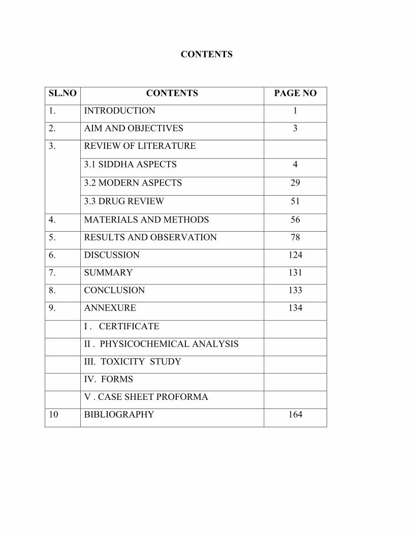
CONTENTS
SL.NO CONTENTS PAGE NO
1. INTRODUCTION 1
2. AIM AND OBJECTIVES 3
REVIEW OF LITERATURE
3.1 SIDDHA ASPECTS 4
3.2 MODERN ASPECTS 29
3.
3.3 DRUG REVIEW 51
4. MATERIALS AND METHODS 56
5. RESULTS AND OBSERVATION 78
6. DISCUSSION 124
7. SUMMARY 131
8. CONCLUSION 133
9. ANNEXURE 134
I . CERTIFICATE
II . PHYSICOCHEMICAL ANALYSIS
III. TOXICITY STUDY
IV. FORMS
V . CASE SHEET PROFORMA
10 BIBLIOGRAPHY 164

1
INTRODUCTION
Siddha system of medicine is the traditional medical system of India which
has flourished well before many centuries. Siddha is practiced till now rather than
other traditional medical systems. In the wake of changing mode of life and modern
medicine, Siddha continues to sustain its influence because of its incomparable
intrinsic merits. This system was originated by siddhars with their holistic spiritual
powers.
Agathiyar, Thirumoolar, Bogar, Theraiyar, Pulippani are the most remarkable
siddhars. Agathiyar was the top most siddhar who is regarded as the originator of the
Siddha medicine and also the Tamil Language. Siddhars are specialized in
Vaatham,Vaithiyam, Yogam, Gnanam which helps to attain spirituality through
prevent the body from disease, increase the lifespan by rejuvenation.
In India, five types of landscapes such as Kurinji, Mullai, Marutham, Neithal,
Paalai contains many herbs helps to cure the disease of the people living there.
Innumerable varieties of herbs, metals and minerals are mentioned in siddha
literatures.
Siddha medicines are classified into Internal medicine thirty two types and
External medicine thirty two types. The internal medicine starts as a simple
preparations like saaru, kudineer, chooranam and ends in higher order medicines like
kattu, kalangu, chunnam, Theeneer, chendhuram etc,.
Siddhars are alchemist who used metals and minerals in drugs for therapeutic
effect. Mercury and other minerals are generally a poison for uninitiated but for the
siddhars who knows how to detoxify it and pulverize it to nano ions, it is the nector of
immortality. Chendhuram is one type of these internal medicine in siddha which is
metallic substances or arsenical compounds are made into red coloured powders by
the process of burning, frying or insulating or keeping them in specialized pudams by
grinding them with decoctions, ceyaneer juices etc.

2
The size of particles present in higher order medicine are in nano level at the
end of the process. So nano technology is the prime tool for siddha herbo-mineral
drugs in which the medicine targets the affected spot and cures the disease. Nano
medicines like chendhuram will scale down the number of doses and increases the bio
availability. Siddha nano medicines will cure many chronic diseases with minimal
side effects and with minimal drug dose which baffles and eludes even the modern
sophisticated medicine.
Vitiligo is an acquired, idiopathic, depigmentory condition of skin and hair
varying sizes and shapes. It affecting people of all ages and both sexes equally.
Patients lose their skin color mostly in patchy and progressive manner.
The cause is still under debate. Although there are rarely physical symptoms
involved many patients experience stigmatization, unwanted attention, negative
comments, rejection or bullying. The prevalence of vitiligo is often said to range from
0.09 to 8% especially in India.
There are lot of drugs in our siddha system for Vitiligo. For the study, I
selected the Rasa Chendhuram as internal medicine and Palagarai Kuzhambu as
external medicine in the management of Vitiligo both in preclinical as well as clinical
aspect.

3
AIM AND OBJECTIVES
AIM:
To evaluate the Siddha Drug Rasa Chendhuram (internal) and Palagarai
Kuzhambu (external) in the management of Vitiligo.
OBJECTIVES:
Primary objective:
To evaluate the therapeutic efficacy of the siddha drug Rasa Chendhuram
(Internal) and Palagarai Kuzhambu (External) in Venpulli (Vitiligo).
Secondary objectives:
To study the safety profile of trial drug Rasa Chendhuram (Internal).
The main objective of this study is to add the scientific evidences to
Siddhars work which in turn create awareness among the public.
To discuss the various literature evidences of Venpulli in Siddha
Medicine and Vitiligo in modern science.
To correlate the clinical features of Venpulli in Siddha medicine with
Vitiligo in modern science.
To correlate the modern purification method and siddha purification
method of raw drugs.
To get the authentication of the raw drug.
To standardize the standard operating procedure.
To study the Physico - Chemical analysis of the selected trial drug.
To study the acute & sub acute toxicity of the trial drug Rasa
Chendhuram according to OECD guidelines.

4
REVIEW OF LITERATURE
SIDDHA ASPECT
Synonyms:
Venkuttam
Swetha Kuttam
Venthittu
Venpadai
Definition :
Venpulli is defined as the discoloration of the skin characterized by the appearance of the
white patches in irregular shape of the epidermis of the skin and sometimes hair also involved.
In siddha system venpulli is considered as one of the eighteen kinds of kuttam. Siddhar
Yugimuni mentioned this condition as Suvetha Kuttam in “Yugimuni Vaithiya Chinthamani –
800”.
தடிப்பாக தவள நிறம் பபால் வவளுத்துச் சர்வாங்கமும் வவளுத்தாற் றான்றிரும்பும்
மடிப்பாக மயிர்வவளுத்தா லசாத்ய மாகும் வரிவுதடு வுள்ளங்கக குதங்குய்யந்தான்
வநடிப்பாக வநருப்பு பட்டது பபால புண்ணாய் நிறமிருந்தா லசாத்தியவமன்பற யுகைக்கலாகும்
வவடிப்பாக பமனிவயல்லாம் வவளுத்து வஙீ்கி வவண்சுபவத குட்டவமன்பற விளம்பலாபம.
Hypopigmented patches all over the body.
Hypopigmentation on hair, lips, palm, sole, anus and pubic region –incurable.
If the lesion is like burnt scar- incurable.

5
பநாய் வரும் வழி: (Aetiology)
According to siddha system, the predisposing causes for this diseases have been described
as hereditary factor, stress and strain, malnutrition and venereal exposure and no other specific
causes have been mentioned for venpulli.
1) According to Agathiyar Kanma Kandam
“பசர்ந்தகுட்ட பமாடுகுகற பநாய்கள் வந்த பசதிகள் மலைாதவரும்பு வகாய்தல்
தாரிந்த சவீவசந்து வகதகள் வசய்தல் தாய் தந்கத மனதுவநாந்தது பைாகந்தாபன“
தாவனன்ற வதய்வவுருத் தகனயழித்தல் சார்வான வபரிபயார்கள் தகமப் பழித்தல்
காவனன்ற நந்தவனம் பூஞ்வசடிகள் வவட்டல் கருமமடா சரீைத்திற் காசு பபாபல
யூவனன்ற வுடம்வபல்லாம் வமாட்டுப் வபாட்டா யுடன் வவளுத்து குகறபநாயா யுதிைஞ்சிந்தும்
வாவனன்ற கருமங்கள் தரீ்ப்பதற்கு வககவயான்று வசால்பவன் பகள் நந்தவனம்கவபய
அகத்தியர் கன்ம காண்டம் வகௌமதி நூல் பக்கம் – 27
Agathiyar kanmakandam says that picking flowers before blossoming, killing living
creature, giving sorrow to the parents, destroying temples, abusing noble person, cutting plants
cause Venpulli noi.
2) According to Thirumoolar Karukkidai Vaithya Nool
“வியாதியுண் மூவாறு விளங்கிய குட்டங்பகள் சுயாதிக் கிைந்தி சுழன் பமகத்தாலாறும் பயாதி மண்ணுளப் பலவண்டினா வலட்டும் நியாதிப் புழுநாலாய் நின்றதிக் குட்டபம.”
Six types of kuttam i.e, skin diseases are caused by kiranthi and megam, eight types
are caused by insects in the soil, and four types are caused by worms.

6
3) According to Dhanvanthri vaidhyam
“அறிவின்றி விபரிதஞ் பசைாகாைம் புசிக்கலாலும் துகறயன்றி வதாடாத வதான்கற வதாட்டகவப் புசிக்கலாலும் . . . . . . . . . . . . . . . . . . . . . . . . . . . . . . . . . . . . . . . வந்திந்துப் பூருவா வசன் மாந்திை பாவத்தாலுஞ் சந்திக்கக் கற்புமாதர் தங்ககளக் கருதலாலும் வதாந்தித்த குட்டபைாகந் வதாடுக்குவமன்றுகைத்தார் முன்பனார்.”
4) According to Dhanvanthiri Vaidhyam
Intake of unhygienic food
Abusing the elderly people like Siddhars and saints.
Blaming the worshipping ladies.
Sins committed in the previous birth.
Thinking of seducing chaste women. These are the causes mentioned by
Dhanvanthiri.
5) According to “Guru Naadi Nool’’
“கிருமியால் வந்தபதாடம் வபருக வுண்டு
பகட்கவதின் பிரிவதகனக் கிைம மாக
வபாருமிவரும் வாயுவவல்லாம் கிருமி யாபல
புழுக்கடி பபால் காணுமது கிருமியாபல
வசருமிவரும் பவுத்திைங்கள் கிருமியாபல
பதகமதில் பசாககக்குட்டங் கிருமியாபல
துருமிவருஞ் சுபைாணிதங் கிருமியாபல
சூட்சமுடன் கிரிகசப்பால் வதாழில்வசய் வபீை”.
கிருமியால் உண்டாகும் குட்டம் வைலாறு8
“குட்டமது விடகைப்பான் விடநீர் சூகல
சுபைாணிதத்தால் தாதுவகட்டுத் தடிப்புண்டாகும்
மட்டறபம கிருமிவசன்று மருவும் பபாது வககயாய்க் கிருமியுட விடநீர் வசன்று
குட்டமுடன் பதகவமல்லாம் பறக்கும் பபாது
குழிகுழியாய்க் கிருமியினகீ் வகாள்ளும் புள்ளி தட்டறபவ கிருமியுட நீைால்வந்த
சகலகுட்டம் விடகைப்பான் சாற்ற லாபம.”
7
5) According to “Siddha Maruthuvam Sirappu”
The etiology and the characters of Venpulli are clearly explained in the text “Siddha
Maruthuvam Sirappu” as follows:
In the affected area, reduction or total loss of skin pigment melanin on the epidermis is
observed. As the distinct aetiology is not known, there exist certain beliefs and hypothesis about
the disease. They are
Constant irritation to the skin owing to clothes, rubber, plastics or other chemical
substances.
Some essential metal or mineral deficiency in the food
6. According to “Eighteen Siddhars Naadi Nool”
Excessive intake of acidic food stuffs leads to pallor and discolouration of skin are the
cause for vitiligo said by Pathinen Siddhar Naadi Nool.
7. According to “Agathiyar Vaithyam”
குயல்வாய் குஷ்டம் சயங்குன்ம நீரிழிவு சுைக்கிைாணி நீைகடப்பு பாண்டு மூல வாய்வு
கயல் வாயு வருங்கண்ணில் குத்தாய் கடிந்த தசவாய்வு
காணவாக முன் வசய்த உயிர்களும் விகனதாபன
Kuttam may be hereditary, apart from all other etiological factors Kuttam is also
considered to be followed by Sins committed in the previous birth (Kanma vinai).
CLASSIFICATION:
1) According to yugi vaithiya chinthamani:
In “Yugimuni Vaithiya Chinthamani – 800”, Kuttam is classified into eighteen
types. Swetha Kuttam (Venpulli) is one among them. It is mentioned as below:
“முத்தாகும் குட்டந்தான் பதிவனட்டுக்கும் முனியான யூகினான் வசால்லக் பகளாய் புத்தாகும் புண்டரீக குஷ்டத்வதாடு வபருகின்ற விற்பபாடகக் குட்டமாகும் பத்தாகும் பாமா குஷ்ட ஏகசர்ம குஷ்டம் பரிவான கர்னகுஷ்டம் சர்மகுஷ்டம் கித்தாகுங் கிருஷ்ண குட்டம் அவுதும்பை குட்டம்

8
பகடியான மண்டல குஷ்டமாகு வமன்பன குட்டமா மபரிச குஷ்ட பமாடு மருவலாங் கிடீப குஷ்டந் சர்மதல குஷ்டந் திட்டமாற் தத்துரு குஷ்ட பமாடு தக்கான சித்துமா குஷ்டஞ் சதாரு குஷ்டந் துட்டமாஞ் சுபவத குஷ்டதன் பனாவடாக்கச் சுயம்பான பதிவனட்டு குட்டமாச்பச.
(Page No:191)
1.Pundareeka Kuttam
2.Virpodaka Kuttam
3.Baama Kuttam
4.Gaja Saruma Kuttam
5.Karna Kuttam
6.Siguram
7.Krishna Kuttam
8.Avudhumbaram
9.Mandala Kuttam
10.Abarisa Kuttam
11.Visarchika Kuttam
12.Vibaathika Kuttam
13.Kideeba Kuttam
14.Sarmathala Kuttam
15.Thethru Kuttam
16.Sithumaa Kuttam
17.Sathaaru Kuttam
18.Swetha Kuttam
2) According to “siddha maruthuvam sirappu”
Venpadai has been classified into four types :
1.vaatha venpadai
2.piththa venpadai
3.kabha venpadai

9
3) According to athma rakshamirtha vaidhya sarasankiraham
Kuttam is classified into 4 types
1. Venkuttam
2. Senkuttam
3. Karunkuttam
4. Peru viyathi
4) According to “Pararasa Sekaram”
Kuttam is classified into 5 types:
1. Venkuttam
2. Senkuttam
3. Karunkuttam
4. Vishakuttam
5. Azhukannikuttam
5) A classical work “Madhava Nithanam” classifies Venpulli noi as
1. Savithram
2. Kilesam
3. Varunam
6) According to siddhar aruvai maruthuvam
Venpadai has been classified into 3 types on the basis of mukkutram, they are,
1. Vaatha venpadai
2. Piththa venpadai
CLINICAL FEATURES :
1) According to “Yugimuni Vaithiya Chinthamani – 800”
Yugimuni attributed the Venpulli under the headline of Swetha Kuttam which is one
of the eighteen kuttams and he mentioned the clinical features of swetha kuttam as below:

10
தடிப்பாக தவள நிறம் பபால் வவளுத்துச் சர்வாங்கமும் வவளுத்தாற் றான்றிரும்பும்
மடிப்பாக மயிர்வவளுத்தா லசாத்ய மாகும் வரிவுதடு வுள்ளங்கக குதங்குய்யந்தான்
வநடிப்பாக வநருப்பு பட்டது பபால புண்ணாய் நிறமிருந்தா லசாத்தியவமன்பற யுகைக்கலாகும்
வவடிப்பாக பமனிவயல்லாம் வவளுத்து வஙீ்கி வவண்சுபவத குட்டவமன்பற விளம்பலாபம.
Yugimuni gives a clear definition of Venpulli and he mentioned the conditions
which will not responded to treatment (Asathiyam) as said below:
1. Whitish discoloration of the part of the body or entire body. Sometimes hair also
turns white.
2. When white patches occur on the palms or muco-cutaneous junctions like lips,
anus and genitals, it is said to be rarely curable.
3. If the hair becomes white, prognosis will be very bad.
4. Fissured body becomes oedematous.
2) According to thanvanthiri vaithiyam
மீக்வகௌத் பதாறூவமலுபமார் முகம் வவளுக்குமாகில்
பநாக்கியல் மரிக்குஞ் வசான்ன வவண்குட்டமாபம.
When the colour of the face becomes white, it is called Venkuttam.
3) According to Vaithya Sarasangiragam
Sole, hands, lips, scalp, fingers and wrist joint all these organs are found with white
coloured patches which are circumscribed along with thickened border and gradually spread
which is known as venpulli . blood, muscle and adipose tissue are affected by disease.
Discolouration of hairs, absence of normal skin texture when compared with the
adjoining normal skin area and appearance of burn scar is incurable.

11
Premonitory symptoms:
1. The skin appears glittering and rough
2. There is an excessive perspiration or no perspiration
3. Discoloration
4. Heat and itching of the skin
5. Numbness in some parts of the body.
4) According to Pararasa Sekaram
1. Watery discharge
2. Grey colour
3. Foul smelling
4. Dryness of the scalp
5) A classical work “Madhava Nithanam” classifies Venpadai as
Savithram – venpulli affecting muscular tissue.
Kilesam - venpulli affecting the skin.
Varunam - venpulli affecting the adipose tissue.
These types are not having any pathological discharge.
Kilesam is classified on the basis of mukkutram and their features are as follows:
Vaatha kilesam - Reddish white in colour.
Piththa kilesam - Red coloured patches resembling the petal colour of lotus.
Kaba kilesam - milky thickened white patches with itching.
6) According to anubhava vaithiya deva ragasiyam
இந்பநாகய குஷ்டவமன கூறினும் இது குஷ்ட வகககளின்று பவறுபட்டது என்பகதயும் குஷ்டத்கதப் பபால் அவ்வளவு வகாடுகமயான வியாதி அல்லவவன்றும் உணைபவண்டும். இந்பநாயில் திட்டு திட்டாக வவண்கம நிறமான பகடகள் உண்டாகி பிறகு பதகம் முழுவதும் பைவி உடகல விகாைப்படுத்துதல் முதலிய குணங்ககள உகடயது.
12
Three types
1. Vatha venpadai
2. Piththa venpadai
3. Kaba venpadai
CLINICAL FEATURES
1. The skin appears glittering and rough
2. There is an excessive perspiration
3. Discolouration
4. Heat and itching of the skin
5. Numbness in some parts of the body
According to Sirappu Maruthuvam
1. Vaatha Venpadai
2. Piththa Venpadai
3. Kaba Venpadai
4. Mega Venpadai
1. VAATHA VENPADAI
It is characterized by the presence depigmented patches ,which are dry ,rough, reddish or
pale black in colour.
2. PITHA VENPADAI
It is characterized by the presence of depigmented patches which are red in colour like
lotus flower, spreading with burning sensation and loss of hairs on that area.
3. KABA VENPADAI
It is characterized by the depigmented patches which are white in colour like leucas
flower spreads with rashes and itching .

13
4. MEGA VENPADAI
It is due to the veneral disease and it occurs after 4 or 6 months after the onset of disease,
syphilis within four or six months of the attack. This venpadai develops initially along the nape
and the adjoining spaces. Also gradually it affects the shoulder joints, back of trunk .
Clinical features of this type
Depigmented patches are small in number. Pale in colour, turmeric colour or dark colour margin
marked with hyperpigmented signs. These lesion are circumscribed with 2mm to 3mm diameter
or above. This correct picture of hypopigmented and hyper pigmented skin seems to be more or
less a multi eyed filter (sieve–like) Females are more prone to this mega venpadai, therefore anti
–syphilitic therapy is mandatory in the early period of the treatment.
CHARACTER OF VENPADAI
Skin color will change to reddish black or reddish white or white colour with spreading
nature. The imbalance of the three thathu produces certain lesions in skin known as kuttam.
Absence of perspiration and thickening of skin may produce the colour changes in skin.
தரீும், தைீாதகவ
1) ACCORDING TO DHANVANTHIRI VAITHYAM
சாத்தியம்-11 பூண்டந் நுைவிபனாடு சதாரிகம் புண்டரீ கந்த தாண்டு விற்பபாடம் பாமாவுடன்கமதலம் வவண்குட்டம் கூண்டிடு காகறந்தி சிறுகம யசல குட்டம் பவண்டியவியாதிபயாடும் பதிவனான்றும் விரித்துக் காபன. (Page No : 325)
அசாத்தியம் வசால்லுகுட்டம் ஏழுவககபபர் வசால்லிக் கபால சர்மீகம் வவல்லு முதும்பா பமகிடிபம் விசர்ச்சிமண்டலக் கிைமும் மல்லல் தருமீசி யகுகவ யாகும் வபயபைா ழாகும் வல்லகியாதிக் குணமதகன வகுத்துப் பாரிலுகைகைப்பபபன”

14
CURABLE-11
1. Thethru Kuttam
2. Sadhaaru Kuttam
3. Pundareega Kuttam
4. Virpodaga Kuttam
5. Sarmathala Kuttam
6. Baama Kuttam
7. Kaha Nandhi
8. Venkuttam
9. Sithuma Kuttam
10. Alasa Kuttam
11. Vibaathiga Kuttam
INCURABLE -7
1. Kabaala kuttam
2. Sarumamega kuttam
3. Kideeba kuttam
4. Avudhumbara kuttam
5. Visarchika kuttam
6. Aguvai kuttam
7. Mandala kuttam
ACCORDING TO YUGI CHINTHAMANI-800
குட்டந்தான் பதிவனட்டில் சாத்தியந்தான் கூறக்பகள் விற்பபாடக பாமா குட்டம் திட்டந்தான் வகசசர்ம குட்டவமாடு கிருட்டிண குட்டமவுதும்பை குட்டந்தானும் திட்டமாந் பதத்துருக் குட்டவமாடு வசய்சித்துமா குட்டங் கிடிப குட்டம் தட்டந்தான் மிகுந்தச தாரு குட்டம் சமகிருட்டிண குட்டம்சாத் தியமா வமன்பன” (Page No:200)
15
CURABLE
1. Virpodagam kuttam
2. Baama kuttam
3. Gaja Saruma kuttam
4. Krishna kuttam
5. Avuthumbara kuttam
6. Thethuru kuttam
7. Sithuma kuttam
8. Kideepa kuttam
9. Sathaaru kuttam
10. Sarmathala kuttam
INCURABLE
1. Pundareeka kuttam
2. Karna kuttam
3. Sikura kuttam
4. Mandala kuttam
5. Abarisa kuttam
6. Visarchika kuttam
7. Swetha kuttam
8. Vibathika kuttam .
2) According to the text “Siddha Maruthuvam Sirappu”
Curable conditions in Venpulli are:
Lesions without any change in hair colour.
Lesions without coarse texture.
Lesions that are not appearing like white burnt scar.
Uncurable conditions in Venpulli are:
Lesions with whitened hair.
Lesions feeling rough.

16
Lesion appearing like white burnt scar.
If the lesion first appears on genitalia, anus palms and lips.
Lesions of fast spreading nature.
SIDDHA PATHOLOGY:
Disease occurs due to the derangement in
muththathukkal
seven Udal thathukkal
Kaala marupadukal (seasonal changes)
Thinai (living lands) and
Udal vanmai.
Muththathukkal
The function of the three uyir thathus:
a) Vali – Kattru + Veli
b) Azhal – Thee
c) Iyyam – Neer + Mann
The alteration of three thathu in their reaction to extrinsic or intrinsic factors results in
disharmony. This altered harmony and balance variation of the three thathus results in disease.
Their natural ratio (1:1/2:1/4) to each other is discerned by the physician at the wrist and each
nadi is individually assessed for its strength, speed and regularity.
VATHAM
The term vatham denotes vayu, dryness, pain and flatulence , sensitiveness, lightness and
also air. Based on functions and locations it is classified into ten types.
1. Piranan
(Uyirkkaal)
Responsible for respiration and it is necessary for proper digestion.
In venpulli noi pranan is normal.

17
Abanan (Keel nokkukkaal)
Responsible for all the downward forces such as voiding of urine, stools, semen, menstrual flow.
In venpulli noi abanan is normal.
Viyanan(Paravukaal)
Dwells in the skin and is concerned with the sense of touch, extension and flexion of the parts of
the body and distribution of the nutrients to various parts of the body.
In venpulli noi viyanan affected. (skin color changed into white).
Uthanan (Melnokkukaal)
Responsible for all kinds of upward motion such as nausea, vomiting etc.,
In venpulli noi uthanan is normal.
Samanan(Nadukkaal)
Considered essential for proper digestion, assimilation and carries the digested nutrients to each
and every organ.
In venpulli noi samanan is normal.
Nagan
Helps in opening and closing of eyelids.
In venpulli noi nagan is normal.
Koorman
Responsible for vision, lacrimation and yawning.
In venpulli noi koorman is normal.
Kirugaran
Induces appetite, salivation, all secretions in the body including nasal secretion and sneezing.
In venpulli noi kirugaran is normal.

18
Thevathaththan
Induces and stimulates a person to become alert, get anger, to quarrel, to sleep etc.,
In venpulli noi thevathathan is normal.
Dhananjeyan
Resides in the cranium and produces bloating of the body after death. This leaves from the body
after 3 days of death, forming a way through the skull.
In venpulli noi dhananjeyan is normal.
PITHAM
It is the thermal life force of the body. It is sub divided into five types. They are
Anarpitham
Peps up the appetite and aids in digestion.
In venpulli noi anarpitham is normal.
Ranjagapitham
Responsible for the colour and contents of blood.
Ranjaga pitham affected in venpulli noi.
Saathagapitham
Controls the whole body and is held responsible for fulfilling a purpose.
In venpulli noi saathaga pitham is normal.
Pirasagapitham
Dwells in the skin and concerned with the shine, glow, texture and its complexion.
In venpulli noi prasaga pitham is affected.
Alosagapitham
Responsible for the perception of vision. In venpulli noi alosaga pitham is normal.

19
KABAM
It is responsible for the stream line functions of the body and maintains body’s defence
mechanism intact. It is again classified into 5 types.
Avalambagam
Lies in the respiratory organs, exercises authority over other kabhas and control the heart and
circulatory system.
In venpulli noi avalambagam is normal.
Kilethagam
Found in stomach as it seat, moistens the food, softens and helps to be digested.
In venpulli noi kilethagam is normal.
Pothagam
Responsible for the perception of taste.
In venpulli noi pothagam is normal.
Tharpagam
Presents in the head and is responsible for the coolness of the eyes, sometimes may be referred to
as cerebrospinal fluid.
In venpulli noi tharpagam is normal.
Santhigam
Necessary for the lubrication and the free movements of joints .
In venpulli noi santhigam is normal.
PARUVAKALAM MAARUBADUKAL :
With reference to the position of the sun in the orbit, the years divided into six seasons .
They are,
Perum pozhuthugal
1) Kaar kaalam

20
(Aavani & Purattasi)
Mid August to Mid October
Mukkutra marupaadugal
VATHAM - Vaetrunilai valarchi
PITHAM – Thannilai valarchi
Perum pozhuthugal
2) Koothir kaalam
(Iypasi & Karthigai)
Mid October to Mid December
VATHAM – Thannilai adaidhal
PITHAM - Vaetrunilai valarchi
Perum pozhuthugal
3)Munpani kaalam
(Margazhi & Thai)
Mid December to Mid February
PITHAM – Thannilai adaidhal
Perum pozhuthugal
4)Pinpani kaalam
(Masi & Panguni)
Mid February to Mid June
KABHAM – Thannilai valarchi
Perum pozhuthugal
5)Elavenir kaalam
(Chithirai & Vaikaasi)
Mid April to Mid June
KABHAM – Vaetrunilai valarchi
21
Perum pozhuthugal
6) Mudhuvenir kaalam
(Aani & Aadi)
Mid June to Mid August
VATHAM – Thannilai valarchi
KABHAM – Thannilai adaidhal
In every season there will be changes in the land, water, plants, animals and human
beings , which will modify the physiology and making (rendering) them susceptible to certain
specific disease which are common in these seasons. The siddhars have been anticipated those
changes and advised certain measures in the form of diet, purgative, etc
THINAI (LAND)
Siddhars classified the lands into five types. They are
1. Kurunji – Mountain range
2. Mullai – Pastoral area of the forest
3. Marudham – The fertile river bed
4. Neidhal – The coastal region
5. Paalai – Arid desert
Kabha diseases will occur in Kurinji land. Pitha diseases occur in Mullai land. Vadha
diseases occur in Neidhal land. Staying in Paalai land is not good to health. Marudham
land is the fertile area where no disease occurs. So, Marudham land is the best one to stay
in.
The winter season gives good health to the man, early summer and later rainy gives
moderate health. Whereas early rainy and later summer are more prone to diseases, that’s why
siddhars called it as Aanadha kaalam.

22
RELATION BETWEEN MUKKUTRAM, KAALANGAL AND THINAIGAL
Mukkutram
Paruvakaalam (Seasons)
Thinai
Thannilai
valarchi
(Accumulation)
Vaetrunilai
valarchi
(Aggravation)
Thannilai
adaidhal
(Alleviation)
VATHAM Mudhuvenil
kaalam
Kaar kaalam Koothir kaalam Vatha disease is
more prevalent in
Neidhal land
PITHAM Kaar kaalam Koothir kaalam Munpani kaalam Pitha disease is
more prevalent in
Mullai land
KABHAM Pinpani kaalam Elavenil kaalam Mudhuvenil
kaalam
Kabha disease is
more prevalent in
Kurunji land
UDAL VANMAI (IMMUNITY)
Siddhars classify udal vanmai into three types. They are
1. Iyarkai vanmai
2. Kala vanmai
3. Seyarkai vanmai
UDAL KATTUGAL
S.No Udal kattugal General Features Changes in
venpulli
1 Saaram
(Digestive essence)
Responsible for the growth and
development. It keeps the individual in
good temperament and it enriches the body.
affected
2 Senneer (Blood) Responsible for the color of the blood and
for the intellect, nourishment, strength of
the body.
Affected

23
3 Oon (Muscle) Gives lookable contour to the body as
needed for the physical activity. It feed the
fat next day and gives a sort of plumpness
to the body.
Normal
4 Kozhuppu (Fat) Lubricates the organs to facilitate
frictionless functions.
Normal
5 Enbu (Bones) Supports and protects the vital organs,
gives the definite structure of the body and
responsible for the posture and movements
of the body.
Normal
6 Moolai (Bone marrow) Nourishes the bone marrow and brain
which is the centre that controls other
system of body.
Normal
7 Sukkilam/Suronitham
(Sperm/Ova)
Responsible for reproduction30. Normal
PINIYARI MURAIMAI (DIAGNOSIS)
Four steps are followed in diagnosing the disease. They are
1. Poriyaal aridhal
2. Pulanal therdhal
3. Vinaadhal
4. Envagai thervugal
PORIYAAL ARIDHAL:
In this, the physician should carefully observe the changes that occur in the five sensory
organs (porigal) of the patient.
PULANAL THERDHAL:
The physician carefully applies his five senses of perception, smell, taste, vision, touch
and sound to understand the condition of the patient.

24
VINAADHAL:
The physician should interrogate about the patients name, age, occupation, socio-
economic status, food habits, history of past illness, history of present illness, family history,
marital status, menstrual history and frequency of pain.
ENVAGAI THERVUGAL:
நாடிப்பரிசம் நாநிறம் வமாழிவிழி மலம் மூத்திைமிகவ மருத்துவைாயுதம்
noi nadal noi mudhal nadal thirattu, part-i (pg no-270)
Nowadays advanced diagnostic tools have been developed by modern bio medical
scientists. But siddhars have given eight diagnostic methodological tools. They are called as
Envagai thervu.
Eight fold system of clinical assessments:
Siddhars have given eight diagnostic methodological tools. They are
1. Naadi
2. Sparisam
3. Naa
4. Niram
5. Mozhi
6. Vizhi
7. Malam
8. Moothiram
GENERAL FINDINGS:
NAADI:
Naadi is responsible for the existence of life, can be felt one inch below the wrist on the
radial side by means of palpation with tips of index, middle and ring finger, corresponding to
vatham, pitham, kabham.
Three humours Vatham, Pitham, and Kabham are in the ratio 1:1/2:1/4 normally.
Derangement in these ratio leads to various disease conditions.
25
Naadi in venpulli
Vathapitham or pitha kabam
SPARISAM:
By sparisam, the temperature of skin (thatpam- cold or veppam – heat), smoothness,
roughness, sweating, dryness, hard patches, swelling, abnormal growth of organs and tenderness
can be felt.
In venpulli – hypopigmented patches present.
NAA:
Signs and symptoms in the tongue are noted here. Colour, salivary secretion, ulcers,
coating, inflammation, taste changes, deviation and its nature are generally noted.
In venpulli in anaemic conditioned tongue may be pallor.
NIRAM:
The colour of the skin is noted here.
In venpulli – the natural skin color become white.
MOZHI:
Character of the speech is noted, mainly uraththa oli (high pitched), thazhndha oli (low
pitched), or resembles the sound of any instrument.
In venpulli no changes in voice.
VIZHI:
Character of the eye is noted. Colour, warm, burning sensation, irritation, visual
perception are generally noted.
In venpulli no changes in vizhi.
MALAM:
The stools are examined for quantity, hardening (malakattu), loose motion (bedhi), colour
and smell.
In venpulli no changes.

26
MOOTHIRAM:
a) NEERKURI (Urine examination)
Urine examination is good diagnostic method compare to naadi and other Envagai
thervugal. Theraiyar mention it as.
“அருந்துமாறிைதமும் அவிபைாதமதாய் அஃகல் அலர்தல் அகாலவூண் தவிர்ந்தழற் குற்றளவருந்தி உறங்கி கவகககற ஆடிக்கலசத் தாவிபய காது வபய் வதாருமுகூர்த்தக் ககலக்குட்படு நீரின் நிறக்குறி வநய்க்குறி நிருமித்தல் கடபன”
noi nadal noi mudhal nadal thirattu, part-i (pg no-282)
The early morning urine sample is collected and sample should be examined within one
and hour hours.
SIRUNEERIN POTHU GUNAM:
“வந்த நீர்க்கரி எகட மணம் நுகை எஞ்சவலன கறந்தியலுளகவ யகறகுது முகறபய” noi nadal noi mudhal nadal thirattu, part-i (pg no-282)
The urine is examined for its Niram (colour), Eadai (Specific gravity), Nurai (Froth),
Natram (Smell), Enjal (Deposits).
NIRAM (COLOUR)
NIRA THOGAI
“பீதம் வசம்கமகபங் கருகம வவண்கமவயன்று பறாகதங்வகாழுகமகய வயாத்துகு நீபை”
noi nadal noi mudhal nadal thirattu, part-i (pg no-283)
1. Yellow
2. Red
3. Green

27
4. Black
5. White
Urine may be any colour as mentioned above.
EADAI (SPECIFIC GRAVITY)
Urine, not thick is considerably healthy. This is mentioned as
“மிகத் தடிப்பும் மிகத் பதறலும் இன்வறனில் சுகத்கத தரும் வமய்ச் சுபாவ நீர் நன்பற”
noi nadal noi mudhal nadal thirattu, part-i (pg no-294)
NURAI (FROTH)
Urine may be frothy in nature, if it is reduced in vali, azhal and ayyam are said to be
deranged. This is mentioned as
நுகைபதய்த் வதாழுகு நீர் நுவலுரும் மும்மலம் ககைய இளகிடும் காலத் வதன்பன”
noi nadal noi mudhal nadal thirattu, part-i (pg no-296)
NAATRAM (SMELL)
Foul odour with pyuria is observed in patients with urinary lithiasis associated with
urinary tract infection and ulcer. This is mentioned as
“வவய்ய துர்க்கந்தம் வசீுநீர் மூத்திைப் கபநாளமிவற்கறப் பற்று புண்குறிபய அம்வமாழியின் வறனினனிலபம முதலிய மும்மலச் சுதபம மூலவமன் றுணபை.”
noi nadal noi mudhal nadal thirattu, part-i (pg no-294)
ENJAL (DEPOSITS)
If urine excretion look like curd water white colour and sand like deposits in urine
indicate stones in kidney. This is mentioned as

28
“நார்த்ததி நீகைபபால நகவயுற்றங் கிழியுமானால் மாைற்ப முற்றநீரி லடிமண்டிக் கிடந்த தானால் பாரிந்த வமழுகுமாங்காய் பற்றிய கல்லினாபல சரீுற்ற வசய்ககவயன்று வதறிவுறச் வசப்பலாபம” noi nadal noi mudhal nadal thirattu, part-i (pg no-321)
NEIKURI
The early morning urine of the patient is analyzed by dropping a drop of gingely oil on
the surface of the urine sample. The accumulation, formations, changes, and dispersal under the
sunlight without any external disturbances of the urine sample can be noted.
The urine kept on the kidney tray in sun light, on non wind condition, should be
examined by dropping a drop of gingili oil gently with rod. If oil spread like snake, it indicates
vali neer; a ring indicates azhal neer and float like a pearl indicates iyya neer and sinks in urine
indicates mukkutram.
“அைவவன நீண்டினஃபக வாதம் ஆழி பபாற்பைவின் அஃபத பித்தம் முத்வதாத்து நிற்கின் வமாழிவவதன் கபபம”
noi nadal noi mudhal nadal thirattu, part-i (pg no-298 - 299)
Vatha neer – The oil spreads like snake
Pitha neer – The oil spreads like ring
Kabha neer – The oil spreads like pearl
If the oil spreads gradually, it indicates good prognosis
If the oil spreads fast or gets mixed completely with urine or sinks in urine, it suggests
bad prognosis.

29
MODERN ASPECT
Dermatology is the branch of medicine dealing with both normal and
abnormal skin and associated structures such as hair, nails, and oral and genital
mucous membranes.
IMPORTANCE OF DERMATOLOGY
Skin diseases are very common, affecting up to a one third of the population at
any one time.
Skin disease is often easily noticeable than other disease so this is a cause of
great social concern to the patient.
Skin diseases have serious impacts on life. They can cause physical damage,
embarrassment, and social and occupational restrictions. Chronic skin diseases
may cause financial constraints with repeated sick leave. Some skin conditions
can be life-threatening.
SKIN ANATOMY
The skin is the largest organ of the body, with a total area of about 20 square feet.
The skin protects us from microbes and the elements, helps regulate body temperature
and permits the sensations of touch, heat and cold.
Skin has three layers:
The epidermis, the outer most layer of skin, provides a waterproof barrier and
creates our skin tone.
The dermis, beneath the epidermis, contains tough connective tissue, hair
follicles, and sweat glands.
The deeper subcutaneous tissue (hypodermis) is made of fat and connective
tissue.
Epidermis develop from the ectoderm and dermis from the mesoderm. The
general ectoderm of the early human embryo consists of single layer of cuboidal cells.
By fifth week of intra-uterine life, this become double layered. The most superficial
layer of flattened cells is called Periderm or epitrichium because the hair later grow
from the deeper layer

30
3.2.1. Structure of the Skin
About the fifth week of fetal development the epidermis and its appendages
are developed from the ectoderm. End of second month of intra-uterine life - the
derma consists of closely-packed, spindle-shaped mesenchymal cells.
During third month of foetal life –three layers of cells are recognizable, the
periderm, the intermediate and the basal layer which is close to the dermis. Fine
reticulum fibres are demonstrable, which later increase in number and thickness, form
the collagenous fibres.
The subcutaneous fat is apparent by the end of the third month of intra-uterine
life, but it becomes abundant only during the later months of foetal life. The nail starts
as an epidermal specialization on the dorsum of the tips of the digits.
During fourth month of foetal life - all the sweat glands and the subcutaneous
fat are being to develop. During fifth month of foetal life the basal cells multiply
rapidly.
During the sixth month of the foetal life -The elastic fibres appear. Most of the
sebaceous glands in the body develop in connection with hair
EPIDERMIS
The epidermis is the outer layer of skin. The top layer of epidermis composed
of dead cells containing keratin, the horny protein that makes up hair and nail.The
epidermis has no blood supply and is nourished almost exclusively by diffused

31
oxygen from the surrounding air. The thickness of the epidermis varies in different
types of skin. It is the thinnest on the eyelids and thickest in palm and sole.
It has 95% keratinocytes which produces a tough protein called keratin but
also contains melanocytes, Langerhans cells, Merkel cells and inflammatory cells.
Rete ridges are epidermal thickenings that extend downward between dermal papillae.
Blood capillaries are found beneath the epidermis and are linked to an arteriole and
a venule.
The skin’s colour is created by special cells called melanocytes, which produce
the pigment melanin. Melanocytes are located in the epidermis.
Layers
The epidermis is composed of 5 layers depending on the region of skin being
considered. Those layers in descending order are,
1. Stratum corneum / Horny layer
The most superficial layer of the epidermis from which dead skin sheds and is
the thickest of epidermal layers having 15 – 30 layerrs of keratinized cells.
The Stratum corneum is also normally devoid of nuclelic and consists of
eosinophilic layers of keratin.
Most of the barrier functions of the epidermis localize to this layer.
2. Stratum lucidum
It is a clear or translucent layer.
It is present only in palms and soles.
It is composed of flattened and hardened skin cells madeof keratin.
3. Stratum granulosum ( Granular layer)
The stratum granulosm is one to four cellular layers thick consistitng of
flattened rows of cells
This layer produces large amount of the protein keratin.
Keratin is extremely durable and water resistant.It is also the protein that
forms the basic structure of hair and nails.
4. Stratum spinosum / Spinous layer/ Pricky cell layer
This layer is composed of several layers of polygonal prickle cells (or)
squamous cells. The layers become flat as they are near the surface, so that their long

32
axis appears parallel to the skin surface. These cells possess intercellular bridges (or)
Tonofilaments.
Composed mainly of proliferating and non-proliferating keratinocytes,
attached to the basement membrane by hemidesmosomes.
Melanocytes are present, connected to numerous keratinocytes in this and
other strata through dendrites.
Merkel cells are also found in the stratum basale with large numbers in touch-
sensitive sites such as the fingertips and lips. They are closely associated with
cutaneous nerves and seem to be involved in light touch sensation.[9]
The epidermis is separated from the dermis, its underlying tissue, by a basement
membrane.
Melanocytes
They are melanin producing neural crest derived cells located in the bottom
layer of the skin's epidermis, the middle layer of the eye , the inner ear, meninges,
bones and heart. It is formed from tyrosine by oxidation metabolism and
polymarization.
They are spidery black cells that produce the brown to black pigment known
as melanin.Melanin is the pigment primarily responsible for skin colour. Once
synthesised, melanin is contained in a special organelle called a melanosome and
moved along arm-like structures called dendrites, so as to reach the keratinocytes.
Layers of the skin
5. Stratum Basal/germinal layer

33
DERMIS
The dermis is a thick layer of skin between the epidermis and subcutaneous
tissues, that primarily consists of dense irregular connective tissue and cushions the
body from stress and strain. The dermis also varies in thickness depending on the
location of the skin. The dermis is a tough layer of skin.
The dermis is tightly connected to the epidermis through a basement
membrane. Structural components of the dermis
Collagen
Elastic fibers
Extrafibrillar matrix.
The hair follicles, sweat glands, sebaceous glands, apocrine glands, lymphatic
vessels and blood vessels are present in the dermis.
The dermis is composed of three major types of cells
Melanocytes

34
Fibroblasts
Macrophages
Adipocytes
Dermis is divided into two layers, the superficial area adjacent to the epidermis called
the papillary region and a deep thicker area known as the reticular dermis.
Papillary dermis ( stratum papillarosum)
The papillary dermis is the uppermost layer of the dermis. It accounts for 1/5
th of the dermis. It intertwines with the rete ridges of the epidermis and is composed
of fine and loosely arranged collagen fibers.
The papillary region is composed of loose areolar connective tissue. This is
named for its finger like projections called papillae, that extend toward the epidermis
and contain either terminal networks of blood capillaries or tactile Meissner's
corpuscles
The papillary layer provides the layer above it, the epidermis, with nutrients to
produce skin cells called keratinocytes. It also helps regulate the temperature of our
skin and thus the body as a whole.
Reticular dermis (stratum reticularosum)
The reticular dermis is the lower layer of the dermis, found under the
papillary dermis, composed of dense irregular connective tissue featuring densely
packed collagen fibers.
The reticular layer serves to strengthen the skin and also provides our skin
with elasticity.
The reticular region is usually much thicker than the overlying papillary
dermis. It receives its name from the dense concentration of collagenous, elastic,
and reticular fibers that weave throughout it.
The reticular layer also contains hair follicles, sweat glands, and sebaceous
glands.
The sweat gland can either be apocrine, such as those found in the armpits and
the groin area, or the eccrine glands, which are found all over the body. The former
help contribute to body odor and the latter help regulate our body temperature through
the process of evaporation.
35
The sebaceous glands found in the dermis secrete a substance
called sebum that helps to lubricate and protect our skin from drying out.
HAIR
The hair grows from the bottom of the follicle. It has, therefore, an
intracutaneous portion present in the hair follicle and the shaft. The hair follicle
consists of epithelial and connective tissue components. Hair is composed primarily
of keratin. The dead keratinocytes fuse together to form the hair.
ARRECTORES PILORUM
These are small bundles of smooth muscle attached to each hair follicle that
contracts in response to cold, fright and other emotions. When the muscle contracts,
the hair becomes more erect, the follicle is dragged upwards so as to become
prominent on the surface of the skin producing what is known as ‘goose skin’.
NAILS
The nails are thickening of the deeper part of the stratum corneum that
develops as specially modified portion of the skin called nail bed. The nails is
composed of clear horny cells, resembling stratum lucidum but are much more
keratinized.
The dermis also contains:
Nerve endings that transmit various stimuli such as pain, itch, pressure, and
temperature. Specialized Nerve Cells
Pacinian corpuscles –pressure receptor
Meissner corpuscles– touch receptor
Ruffini corpuscles– hot receptor
End bulbs of Krause –cold receptor
Free nerve ending– pain receptor
Lymphatic vessels that transport immune system cells, the cells that help
destroy infectious organisms that may have found their way into our body via
a scratch on the skin.
Collagen, a protein that helps strengthen our skin, and elastin, a protein that
helps keep our skin flexible.

36
The dermis is well supplied with blood vessels, both arterioles and capilleries
that originate from arteries and veins in the subcutaneous layer. Blood vessels
within the dermis supply nutrients to the stratum basale as well as to the
cellular structures of the dermis such as glands and hair follicles. Those blood
vessels provide nourishment and waste removal for both dermal and epidermal
cells.
HYPODERMIS / SUBCUTANEA
Beneath the dermis is the deepest layer of our skin.
It contains many collagen cells as well as fat. Fat, in particular, helps insulate
our body from the cold and act as a cushion for our internal structures.
PHYSIOLOGY
FUNCTIONS OF THE SKIN:
1. PROTECTIVE FUNCTION
It protects underlying tissues and organs from chemicals, microbes and shock
impacts. The cornified layer of the epidermis possess properties of physical
toughness, strength, flexibility, elasticity and also retards proliferation of
microorganism and their penetration. The skin protects the body from from ultra
violet damage from the sun by producing melanin.
2. THERMOREGULATION
The skin contains several types of receptors which are involved in
thermoregulation. Skin regulates body temperature by constricting blood vessels and
driving blood inward in cold temperatures to preserve body heat and produce sweat in
warm temperatures to cool the body by water evaporation.
3. SENSE ORGAN
Sensation is a very important function of the skin. Skin preserves a number of
sensations like touch, pressure, warmth, cold and pain
4. STORAGE FUNCTION
The skin acts as two way barrier to prevent the inward or outward passage of
water and electrolyte. The dermis and subcutaneous fatty tissue acts as storage organ
of energy and others. It synthesize and stores vitamin D.
37
5. ABSORPTION
The skin surface also performs absorptive function and is the basis of topical
therapy in dermatology. The skin is capable of absorbing fat soluble nutrients such as
vitamins A, D, E and K.
6. EXCRETION
Some of the toxins may be excreted through the skin.
7. IMMUNE SURVEILLANCE
This immunological function is performed by langerhans’ cells, dendritic cell,
and keratinocytes. Thus the skin forms the front line defense of the body against
invasion by foreign agents.
8. MECHANICAL FUNCTION
The mechanical properties of the skin depend mainly on the dermis, although,
the epidermis and subcutaneous fats also plays some role.
9. COSMETIC FUNCTION
Colour of the skin, hair and nail are important for their decorative value. Hair
does not perform a vital physiological function but it does provide a sexually
attractive ornament.
Melanin Formation
Melanin synthesis is initially catalysed by a copper containing enzyme known
as tyrosinase. The broad of melanin synthesis from the oxidation of phenylalanine
or tyrosine are as follows.

38
Melanin produced in the melanocytes is donated via their dendrites to
neighbouring keratinocytes. Melanin formation in both human and amphibian skin is
augmented by the hormone known as intermedin or melanocyte – stimulating
hormone (MSH) secreted by the pars intermedia of the pituitary gland.
Adrenocortico tropic hormone (ACTH) secreted by Anterior Pituitary has
melanocyte – stimulating activity similar to MSH although to a much lower degree.
MSH causes the serum copper to rise and this is accompanied by inner case in
the melanin formation.
Diminished formation of melanin is seen in albinism and leucoderma. In
melanotic sarcoma, melanin may be found in the urine.
Melanin Formation
39
VITILIGO
White skin is the literal meaning of leucoderma, derma being derived from the
Greek words, leucas and dermis. Leucas means white and dermis means skin.
Vitiligo is a non contagious acquired pigmentation disorder characterized by
sharply defined white patches of variable shape and dimensions, increasing in size
and number with time.
The disorder affects all races and both sexes equally, however, it is more
noticeable in people with dark skin
DEFINITION
Vitiligo is defined as acquired idiopathic , circumscribed, progressive
hypopigmentation of skin and hair. A type of leucoderma often familial characterized
microscopically by an absence of melanocytes.
This disease is characterized by the presence of white macules or patches on
the skin caused by the loss of functioning epidermal melanocytes Vitiligo can also
affect the mucous membranes and the eye.
It can be examined by naked eye and can furnish a lot of information about the
person and the disease.
EPIDEMIOLOGY
Vitiligo is an hypopigmentation disorder that affects approximately 1-2 % of
the population worldwide.
In India 0.25-2.5% were affected. 30% hereditary condition.
Stable type of vitiligo was common which accounted for 65.21%. Lower lip
was involved in 75% of mucosal vitiligo.
Lower limbs were the most common site of onset of vitiligo. Family history
exists in 6.25%-38% of patients with vitiligo
HISTOPATHOLOGIC CHANGES IN VITILIGO
Absence of melanocytes
Negative silver stain for melanin
Negative dopa reaction
Lymphocytic inflammation may be seen
Melanophages may be seen.
40
In the affected area the basal cells and the keratinzing cells of the other layers of
epidermis do not contain melanin pigment granules in them.
At the border of the patches of vitiligo the melanocytes often appear large and
posses long dendritic process filled with melanin granules.
Electron microscopic studies confirm the absence of melanocytes in areas of long
standing vitiligo.
There are collections of mononuclear cells at dermo epidermal junction at the
border between vitilliginous and normal skin. These cells are predominately small
lymphocytes. In the long standing cases where the skin has become thick and
scaly, varying amount of keratosis is seen.
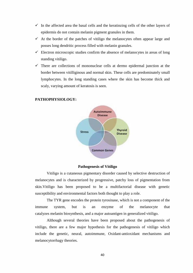
PATHOPHYSIOLOGY:
Vitiligo is a cutaneous pigmentary disorder caused by selective destruction of
melanocytes and is characterized by progressive, patchy loss of pigmentation from
skin.Vitiligo has been proposed to be a multifactorial disease with genetic
susceptibility and environmental factors both thought to play a role.
The TYR gene encodes the protein tyrosinase, which is not a component of the
immune system, but is an enzyme of the melanocyte that
catalyzes melanin biosynthesis, and a major autoantigen in generalized vitiligo.
Although several theories have been proposed about the pathogenesis of
vitiligo, there are a few major hypothesis for the pathogenesis of vitiligo which
include the genetic, neural, autoimmune, Oxidant-antioxidant mechanisms and
melanocytorrhagy theories.
Pathogenesis of Vitiligo

41
Autoimmune and cytotoxic hypotheses
Aberration of immune surveillance results in melanocyte dysfunction or
destruction. Autoimmune diseases such as thyroid diseases and diabetes mellitus
are often associated with vitiligo. These diseases cause defects in the immune system,
which can cause destruction of melanocytes and the loss of pigmentation . The
autoimmune theory proposes alteration in humoral and cellular immunity in the
destruction of melanocytes of vitiligo.
In addition, antibodies against melanocytes were found in serum of patient,
and these can engage the apoptosis of melanocytes when they are present . T cells
were also found in perilesional vitiligo plaque biopsies and they are enriched with
cytotoxicity against melanocyte antigens. Destruction of melanocytes may be directly
mediated by autoreactive CD8+ T cells. Activated CD8+ T cells have been
demonstrated in perilesional vitiligo skin.
Vitiligo is sometimes associated with autoimmune and inflammatory
diseases such as Hashimoto's thyroiditis, scleroderma, rheumatoid arthritis, type 1
diabetes mellitus, psoriasis, Addison's disease, pernicious anemia, alopecia
areata, systemic lupus erythematosus, and celiac disease.
Neural hypothesis
A neurochemical mediator destroys melanocytes or inhibits melanin
production. The neural hypothesis is based on the contact of the melanocytes with
nerve endings in depigmented skin . Neuropeptides and nerve growth factors such as
tumor necrosis factor-α, intercellular adhesion molecule-1 and interferon-γ were
found in perilesional skin, which suggest that nerves can have a role in destruction of
melanocytes . The toxic hypothesis suggests that the mechanism of natural protection
of melanocytes is defective. The melanocytes are unable to eliminate toxic molecules,
and these are accumulated in the cells
Oxidant-antioxidant mechanisms
An intermediate or metabolic product of melanin synthesis causes melanocyte
destruction. Studies suggest that accumulation of free radicals toxic to melanocytes
leads to their destruction. Because patients with Vitiligo exhibit a characteristic

42
yellow/green or bluish fluorescence in clinically affected skin, this led to the
discovery that the fluorescence is due to accumulation of 2 different oxidized
pteridines. The overproduction of pteridines led to the discovery of a metabolic defect
in tetrahydrobiopterin homeostasis in patients with Vitiligo, which results in the
accumulation of melanocytotoxic hydrogen peroxide.
Melanocytorrhagy hypothesis
It emphasizes that depigmentation is because of chronic detachment of
melanocytes. Trauma or repeated friction can contribute toward the detatchment of
melanocytes over time. Melanocytes in unstable vitiligo have been found to lose the
ability to adhere to key surrounding structures. Tenascin, an extracellular matrix
molecule that inhibits adhesion of melanocytes to fibronectin, has been detected in the
basal membrane in the papillary dermis and can contribute toward chronic
detatchment and epidermal loss of melanocytes.
Genetics of vitiligo
The inheritance of vitiligo may involve genes associated with the biosynthesis
of melanin, a response to oxidative stress, and regulation of autoimmunity. Human
leukocyte antigens (HLAs) may be associated, but not in a consistent manner.
For example, HLA-DR4 is increased in blacks, HLA-B13 is increased in
Moroccan Jews, and HLA-B35 is increased in Yemenite Jews. An association with
HLAB13 is described in the presence of antithyroid antibodies. Only one gene,
SMOC2, is in the region of association, within which SNP rs13208776 attained
genome-wide significance for association with other autoimmune diseases and vitiligo
Other Common factors are,
Nutritional - defects in copper, proteins and vitamins in diet, digestive upsets
like amoebiasis, helminthics, chronic diarrhoea, dysentery etc.,
Endocrines – Association with thyrotoxicosis and diabetes.
Trophoneurosis and autonomic imbalance – emotional stress and strain.
Infections and toxic products, Enteric fever ill health, focal sepsis.
Drugs and chemicals
Triggers also include inflammatory skin conditions, burns, intralesional steroid
injections and abrasions.

43
Clinical features
White patches on the skin are the main sign of vitiligo. These patches are more
common in areas where the skin is exposed to the sun.
The patches may be on the hands, feet, arms, face, and lips. Other common
areas for white patches are:
The armpits and groin (where the leg meets the body), around the mouth, eyes,
nostrils, navel, genitals, rectal areas, Loss of colour in the tissues that line the inside
of your mouth and nose (mucous membranes), Loss of or change in colour of the
inner layer of the eyeball (retina)
People with vitiligo often have hair that turns gray early. Those with dark skin
may notice a loss of colour inside their mouths.
Initially, the vitiligo starts as a simple spot, a little paler than the rest of the
skin. But gradually, as time passes, this spot will become much paler until it becomes
white.
The shape of these patches are completely irregular, and, at times, the edges
can become a little inflamed with a slight red tone, sometimes resulting in itchiness.
Other than the appearance of the spots and occasional itchiness, vitiligo does
not cause any discomfort, irritation, soreness or dryness in the skin.
Predicting whether vitiligo will spread, and by how much, is particularly
difficult. The spread of white patches might occur in a matter of weeks for some, and
for others, they might stabilize, not growing for months or even years.
If the first symptoms of the white patches are symmetrical (non-segmental
vitiligo), the development is much slower than if the patches are in only one area of
the body (segmental vitiligo).
Depending on the type of vitiligo, the discolored patches may cover:
Many parts of the body
With this most common type, called generalized vitiligo, the discolored
patches often progress similarly on corresponding body parts (symmetrically).
Only one side or part of the body
This type, called segmental vitiligo, tends to occur at a younger age, progress
for a year or two, then stop.
44
One or only a few areas of the body
This type is called localized (focal) vitiligo. Sometimes the patches stop
forming without treatment. In most cases, pigment loss spreads and eventually
involves most of your skin. Rarely, the skin gets its color back.
Types of vitiligo
Scientists separate vitiligo into two types
Non-segmental vitiligo
Segmental vitiligo.
Non-segmental vitiligo
Non-segmental vitiligo is the most common type of vitiligo and occurs in up
to 90% of people who have the disorder. In non-segmental vitiligo, the patches often
appear equally on both sides of the body, with some measure of symmetry.
New patches also appear over time and can be generalized over large portions
of the body or localized to a particular area.
NSV can come about at any age .The symmetrical patches most commonly
appear on skin that is exposed daily to the sun, such as the face, neck and hands, but
can also appear in other areas:
Backs of the hands
Arms
Eyes
Knees
Elbows
Feet
Mouth.
Non-segmental vitiligo is further broken down into sub-categories:
Generalized vitiligo
No specific area or size of patches, this is the most common type
Acrofacial vitiligo
Mostly on the fingers or toes
45
Mucosal vitiligo
Depigmentation generally appears around the mucous membranes and lips
Universal vitiligo
Depigmentation covers most of the body, this is very rare
Focal vitiligo
One or few, scattered white patches in a discrete area. Most often occurs in
young children.
Segmental vitiligo
Segmental vitiligo has a different form; this condition spreads more rapidly
but is considered more constant and stable than non-segmental. It is much less
common and affects only about 10% of people with vitiligo.
Segmental vitiligo is more noticeable in early age groups, affecting about 30%
of children diagnosed with vitiligo.
It is non-symmetrical and usually tends to affect areas of skin attached to
nerves arising in the dorsal roots of the spine. It is more stable, less erratic and
responds well to topical treatments.
Complications
People with vitiligo may be at increased risk of:
Social or psychological distress
Sunburn and skin cancer
Eye problems, such as inflammation of the iris (iritis)
Hearing loss
PATIENT EVALUATION
Assessment of severity — The evaluation of the patient with vitiligo involves a
detailed history and a complete skin examination to assess disease severity and
individual prognostic factors. Factors that may influence the approach to treatment
include:
●Age at onset of lesions
●Type of vitiligo (segmental, nonsegmental)
●Mucosal involvement, Koebner phenomenon

46
●Rate of progression or spread of lesions
●Previous episodes of repigmentation
●Type and response to previous treatments
●Family history of vitiligo and/or autoimmune diseases
●Presence of concomitant diseases
●Current medications and supplements
●Occupation, exposure to chemicals
●Effects of disease on the quality of life
A full-body skin examination should be performed to assess the extent of the
disease, with particular attention to sites of vitiligo predilection, such as the lips and
perioral area, periocular areas, dorsal surface of the hands, fingers, flexor surface of
the wrists, elbows, axillae, nipples, umbilicus, sacrum, groin, inguinal /
anogenital regions, and knees.
The percentage of the body area involved can be estimated by the so-called 1
percent rule or "palm method." In both children and adults, the palm of the hand,
including the fingers, is approximately 1 percent of the total body surface area
(TBSA), while the palm excluding the fingers is approximately 0.5 percent of the
TBSA. An alternative method is the "rule of nines":
●Each leg represents 18 percent of the TBSA.
●Each arm represents 9 percent of the TBSA.
●The anterior and posterior trunk each represent 18 percent of the TBSA.
●The head represents 9 percent of the TBSA.
Goals of treatment
The goals of treatment for vitiligo should be set with the individual patient or
parents in the case of children, based upon the patient's age and skin type, the extent,
location, and degree of disease activity, and the impact of the disease on the patient's
quality of life. An open discussion with the patient about the limitations of treatment
may be helpful to create realistic expectations.

47
Psychosocial aspects
The patient's psychologic profile and ability to cope with a lifelong disease
should be carefully evaluated at the time of treatment planning. Psychologic support
should be offered to patients if needed
The main symptom of vitiligo is the appearance of lesions, with an often
distressing sense of disfigurement and associated stigma.
Social isolation, reduced sense of worth, adverse effects on education,
occupation, and personal relationships and depressive illness can be consequences
Inferiority complex immediately following the start of disease, the patient
thinks himself inferior to those with whom he was at par or excelled for so long.
When the patient feels his disease is incurable he becomes gradually
depressed.
Diagnosis:
1. The distribution, the age of onset and the hyper pigmented border will suggest
the diagnosis.
2. Vitiligo areas are milky white while other lack this milky white colouration.
3. It is usually apparent. In doubtful and early case, Wood’s lamp is great help in
diagnosis.
4. Careful examination of the texture of the unpigmented skin should exclue
lichen sclerosus and scleroderma.
5. Post-inflammatory leucoderma, which is frequent in the darker races, shows an
irregular mottling of hyper pigmented and hypopigmented blotches.
6. Stationary patches are well-defined and have hyperpigmented borders.
7. Sensations are normal, so is texture unless the patches have been irritated with
treatment.
8. Absence of scaling, crusting and itching help to eliminate seborrhoeids and
pityriasis versicolor.
9. These areas often fluorescence a golden yellow when examined under a
Wood’s lamp. The hypomelanotic macules in leprosy are anaesthetic.
10. Examination of the skin in long wave UVR helps distinguish whether there is
total depigmenation (as in Vitiligo) or not. It may also detect areas of depigmenation
not easily seen in ordinary daylight, as well as detecting a lemon-yellow fluorescence
seen in some cases of pityriasis versicolor.
48
Differential Diagnoses
Pityriasis alba
It appears as white patches on the upper arms and sometimes the thighs. Close
examination shows an indistinct border and fine surface scale.
Tinea versicolor
It is casused by yeast. It affects the chest and back. Patches have a fine scale
on the surface. The colour of the patches varies from pale to orange brown.
Halo nevi
It develop a white border. One or several of these nevi may be present on the back of
children.
Leprosy
One or several paler macules on trunk or limbs that are hypoaesthetic.
White macules of affecting tuberous sclerosis
Uncomming development of anomaly of CNS, connective tissue and skin;
several “maple leaf” shaped hypopigmented macules.
Post inflammatory hypopigmentation
After inflammatory skin disease (after eczema or trauma to the skin; irregular
in shape and in depth of pallor).
Chemical toxicity
May look very much like vitiligo; seen in workers in rubber industry exposed
to parateriary benzyltoluence.
Outlook (Prognosis)
The course of vitiligo varies and is unpredictable. Some areas may regain
normal pigment , but other new areas of pigment loss may appear. Skin that is
repigmented may be slightly lighter or darker than the surrounding skin. Pigment loss
may get worse over time.
Treatment
Vitiligo is difficult to treat. Early treatment options include the following:
Phototherapy, a medical procedure in which your skin is carefully exposed to
ultraviolet light. Phototherapy may be given alone, or after you take a drug
that makes your skin sensitive to light.
Certain lasers may help the skin repigment.

49
Medicines applied to the skin, such as corticosteroid creams or ointments,
immunosuppressant creams or ointments such as pimecrolimus (Elidel) and
tacrolimus (Protopic), or topical drugs such as methoxsalen (Oxsoralen) may
also help.
Skin may be moved (grafted) from normally pigmented areas and placed onto
areas where there is pigment loss.
Several cover-up makeups or skin dyes can mask vitiligo.
In extreme cases when most of the body is affected, the remaining skin that
still has pigment may be depigmented. This is a permanent change that is used
as a last option.
It is important to remember that skin without pigment is at greater risk for sun
damage. Be sure to apply a broad-spectrum (UVA and UVB), high-SPF
sunscreen or sunblock.
Sunscreen can also be helpful for making the condition less noticeable,
because unaffected skin may not darken in the sun.
Use other safeguards against sun exposure, such as wearing a hat with a broad
rim and long sleeve shirt and pants.
Surgical therapies — Surgical therapies have been used for vitiligo for the past
25 years and remain viable options for patients with localized depigmented
areas that have been unresponsive to medical intervention.
They include:
Autologous suction blister grafts
Minigrafts or punch grafts
Split-thickness grafts
Autologous melanocyte cultures
Cultured epidermal suspensions
Autologous noncultured epidermal cell suspension
Hair follicle transplantation
DIET AND RESTRICTIONS
Occupation
Cosmetic things
Diet
50
During bathing – the powder of Bengal gram and green gram or any other
herbal products can be used.
Vinegar, cooking soda, food enriched with alcohol must be avoided. These
items may promote bleaching of skin pigment.
Using soaps and detergents also promote bleaching of skin.
Copper and zinc content vegetable such as cooked green gram or Bengal gram
at least one time a day.
Highly nutritious food like spinach, pomegranate, cheese, butter, milk,
almond, germinating grams and foods rich in tyrosinase to be added.

51
DRUG REVIEW
INTERNAL MEDICINE: RASA CHENDHURAM
INGREDIENTS:
Rasam (Hydragyram)
Gandhagam (Sulphur)
Aridharam (Arsenic trisulphide)
Egg shell
RASAM (HYDRAGYRUM)
Rasam comes under the classification of pancha sootham. It is one of the
important siddha raw drugs .
இரசத்தின் ப ொதுக்குணம் விழிந ொய் கிரந்தி குன்மம் பமய்ச்சூலை புண்குட் டழிகொைில் விந்துவினொல் அத்லத – வழியொய் புரியு விதி யொது புரியிநனொ பயல்ைொம் இரியுவிதி யொது மில்லை
Uses : It cures venereal diseases , skin diseases , eight types of ulcers.
Taste : six tastes present, sweet is dominant.
Potency : hot and cool
Action : alterative, antiseptic, purgative, diuretic, tonic

52
GENERAL PROPERTIES :
Atomic number : 80
Phase : liquid
Melting point : 234.3210
Heat of fusion : 2.29 kj /mol
Heat of vaporization : 59.11 kj / mol
GANDHAGAM (SULPHUR)
Gandhagam comes under the division of paadanangal. It is one of the most
useful raw drug in siddha.
ப ல்ைிக்கொய்க் கந்தகத்தின் ப ொதுகுணம்
(GOOSEBERRY SULPHUR)
“ப ல்ைிக்கொய்க் கந்திக்கு ீள் திபனண் குட்டமந்தம் வல்லை கவிலசகுன்ம வொயுகண்நணொய் – ப ொல்ைொ விடக்கடிவன் நமகந ொய் வறீுசுரம் ந தி திடக்கிரசு ணகீ ம்ந ொனந் நதர்.” Uses: It cures eighteen types of skin diseases, leprosy, veneral diseases, ulcer, etc,.
Taste: kaippu, thuvarppu
Action : astringent, laxative, alterative, insecticide
General properties :
Atomic number : 16
Phase : solid
Melting point : 388.36 k
Heat of fusion : 1.727 kj /mol
Heat of vaporization : 45 kj / mol

53
THALAGAM ( YELLOW ARSENIC TRISULPHIDE)
Thalagam is come under the division of padanangal. It is one of the important
raw drug in siddha system.
தொளகம் ப ொதுக்குணம்
“தொளகத்தின் ந ருலரக்கத் தொலுகவுள் ந ொய்குஷ்டம் ீளக் குளிர்கொய்ச்சல் ீடுக ம் – ொளகங்பகொள் துஷ்டப் ரங்கிப்புண் சூழழுகண் மண்லடந ொய் கிட்டப் டு மொ கிளத்து.”
uses: it is effective in treatment of skin diseases, urinary tract infections, incurable
ulcers, etc.
Action : antipyretic , expectorant , emetic , convalescent , tonic.
EXTERNAL MEDICINE: PALAGARAI KUZHAMBU
INGREDIENTS:
Palagarai (Marine shell)
Lemon juice
Gingelly oil

54
PALAGARAI (CYPREA MONETA)
Palagarai is considered as one of the five wealth of the sea. The white marine
shell is considered as superior for medicinal purposes
PALAGARAI
ைகலை ப ொதுக்குணம்
மந்தந்தொ கங்கிரகணி மொவிடச் சுரங்கண்நணொய் பதொந்தம் ரி ொமச் சூலைகய – மிந்த வுைகலைலயக் கொபைொடிலவ நயொடு லரத்த ைகலைலய கொணினியம் ொர்.
Uses: it cures skin diseases, jaundice, liver enlargement, spleen enlargement, eye
diseases etc.
suvai: kaippu
action: rubifacient, Stimulant, Expectorant, Demulcent.
LEMON
Botanical name : citrus limon
Family : rutaceae
எலுமிச்லச ப ொதுக்குணம் தபீதலு மிச்சங்கொய் நடர்முத்நதொ டத்லதயுமுள் வொதக சூலைலயயும் மொபகொடிய – சொதிபயனுஞ் சர்த்திகுன் மத்லதயுமுள் தங்கமருந் திட்டலதயும் ித்தபவப்ல யுந்தணிக்கும் ந சு.

55
suvai: pulippu
action: anti bacterial , anti fungal , antimicrobial, anti oxidant
GINGELLY OIL
Botanical name: Sesamamum indicum
Family: meliaceae
Parts used: seed
ல்பைண்பணயின் ப ொதுக்குணம்
“புத்தி யனக்குளிர்ச்சி பூரிப்பு பமய்ப்புளகஞ் சத்துவங் கந்தி தனியிளலம – பமத்தவுண்டொங் கண்நணொய் பசவிந ொய் க ொைவழல் கொசந ொய் புண்நணொய் ந ொ பமண்பணய்யொற் ந ொற்று. “ Uses: oil used for cooling effect , external application for wounds and scabies.
Suvai : inippu
Actions : Antifungal, Anticancer, Anti Aging, Anti coagulant, Anti Inflammatory

56
MATERIALS AND METHODS:
SELECTION OF THE TRIAL MEDICINES:
I have selected the trial drug “ Rasa Chendhuram” (Int) for this study
from Classical Siddha literature Pulipanni Vaithiyam -500
and “palagarai
kuzhambu” (Ext) from Gunapadam-thaadhu seevavaguppu.
SOURCE OF RAW DRUGS:
The required raw drugs are produced from as well reputed indigenous drug
shop. The raw drugs will be authenticated by the concerned pharmacognosist,
Government siddha medical college, ,arumbakkam, Chennai. As i have registered my
trial in clinical registry of India (CTRI). My CTRI no is (CTRI/2017/07/008969) .
STANDARD OPERATING PROCEDURE:
MEDICINE – 1
RASA CHENDHURAM
INGREDIENTS:
Rasam (Hydragyram) - 35 gm
Gandhagam (Sulphur) - 35 gm
Aridharam (Arsenic trisulphide) - 35 gm
Egg shell - Q.S
METHOD OF PURIFICATION:
PURIFICATION OF RASAM (MERCURY):
Materials used :
Mercury , brick powder , turmeric powder , acalypha indica leaf juice.
Equipments used:
Stove, mudplate.
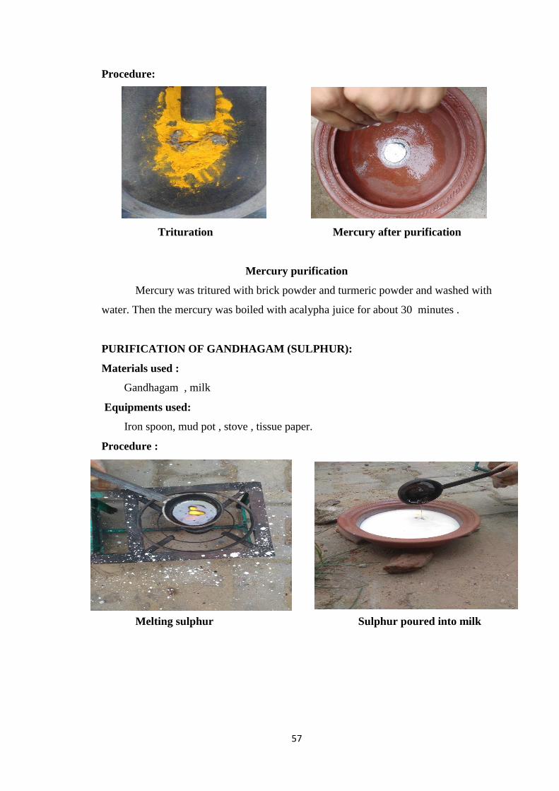
57
Procedure:
Mercury purification
Mercury was tritured with brick powder and turmeric powder and washed with
water. Then the mercury was boiled with acalypha juice for about 30 minutes .
PURIFICATION OF GANDHAGAM (SULPHUR):
Materials used :
Gandhagam , milk
Equipments used:
Iron spoon, mud pot , stove , tissue paper.
Procedure :
Trituration Mercury after purification
Melting sulphur Sulphur poured into milk

58
Gandhagam is placed in an iron spoon , the spoon is heated till the gandhagam
melts. Then it is poured in milk. This is repeated for 2 times . After that gandhagam is
taken out and washed with cool water and allowed it to dry.
PURIFICATION OF ARSENIC TRISULPHIDIUM (THALAGAM) :
Materials used :
Thaalagam , limestone , small cloth
Equipments used:
Stove , mud pot , small cloth .
Procedure :
Thalagam is placed in small cloth and tied with a small rope. Then it was buried
in limestone . water was poured till the limestone immersed and it is heated till water
evaporated completely . the thalagam is taken out and washed in cool water and allowed
it dry.
Sulphur after purification
Thalagam buried into limestone Thalagam after purification

59
METHOD OF PREPARATION:
(Rasa chendhuram)
The above mentioned purified raw drugs is to be added in the stone mortar and
made as a fine powder. Then the fine powder is to be fried well. Next the egg shell is
powdered. Place the half the quantity of powdered egg shell in a mud plate as a layer.
Then spread the powdered raw drug as a second layer then put the remaining quantity
of egg shell spreaded on it, then it is heated with kukkuda pudam (10 cow dung cakes)
to make as a chendhuram. Then the end product is preserved in an air tight container.
DOSAGE : 65 mg
ADJUVANT : Palm jaggery.
INDICATION : Parangisogai, Kiranthi, Kuttarogam, Thaadhunattam,
Lingaputtru, Vippurudhi.
Heating Trituration
Final product

60
MEDICINE – 2
EXTERNAL MEDICINE
PALAGARAI KUZHAMBU
INGREDIENTS:
Palagarai (Marine shell)
Pazhacharu (Citrus limon)
Gingelly oil
METHOD OF PREPARATION:
Palagarai is powdered and kept in mud pot. Lemon juice is added to it, kept for
6 days and allowed to dried. The dried palagarai is mixed well with gingelly oil and
applied over the skin.
INDICATIONS: Kuttam, Kurainoi, pungal.
STANDARDIZATION PARAMETERS :
Traditional way of testing Chendhuram :
1. Colour:
Red in colour without any shiny appearance
2. Taste and odour:
Tasteless and odourless
3. Luster
Did not regain luster on heating again at same temperature
4. Floating on water
Sample floats on water. Did not immediately immersed in water
5. Finger furrows test.
Impinged in the papillary ridges when the sample rubbed in
between Index finger and thumb

61
PHYSICO- CHEMICAL ANALYSIS
Determination of pH
1% solution of Rasa Chendhuram was prepared in distilled water and pH was
determined by using pH meter Systronics digital pH meter.
Determination of moisture content
Moisture content was determined by LOD (Loss on Drying) method. 3gm
VPM was taken and kept in oven at 1050c till a constant weight was obtained.
Amount of moisture present in the sample was calculated as referred to the air dried
drug.
Total Ash
A weighed amount of the powder was taken in a silica crucible previously
ignited, cooled and weighed. It was incinerated using incinerator by gradually
increasing the heat not exceeding dull red heat (450°C) until free from carbon, cooled
and weighed. The percentage of ash was calculated with reference to air-dried drug.
The procedure was repeated to get constant weight.
Water soluble ash
The total ash was boiled with 25 ml water and filtered through ash less filter
paper (Whatmann 4.1). It was followed by washing with hot water. The filter paper
was dried and ignited in a silica crucible, cooled and the water insoluble ash was
weighed. The water-soluble ash was calculated by subtracting the water insoluble ash
from the total ash.
Acid insoluble ash
The total ash obtained was boiled for 5 minutes with 25 ml of dilute hydrochloric
acid (10% w/v) and filter through ash less filter paper (Whatmann No.1). The filter
paper was ignited in a silica crucible, cooled and weighed.
Determination of Alcohol Soluble Extractive
The air dried drug was finely grounded, added with 100 ml of ethanol of
specified strength in a closed flask for twenty-four hours, shaken frequently during
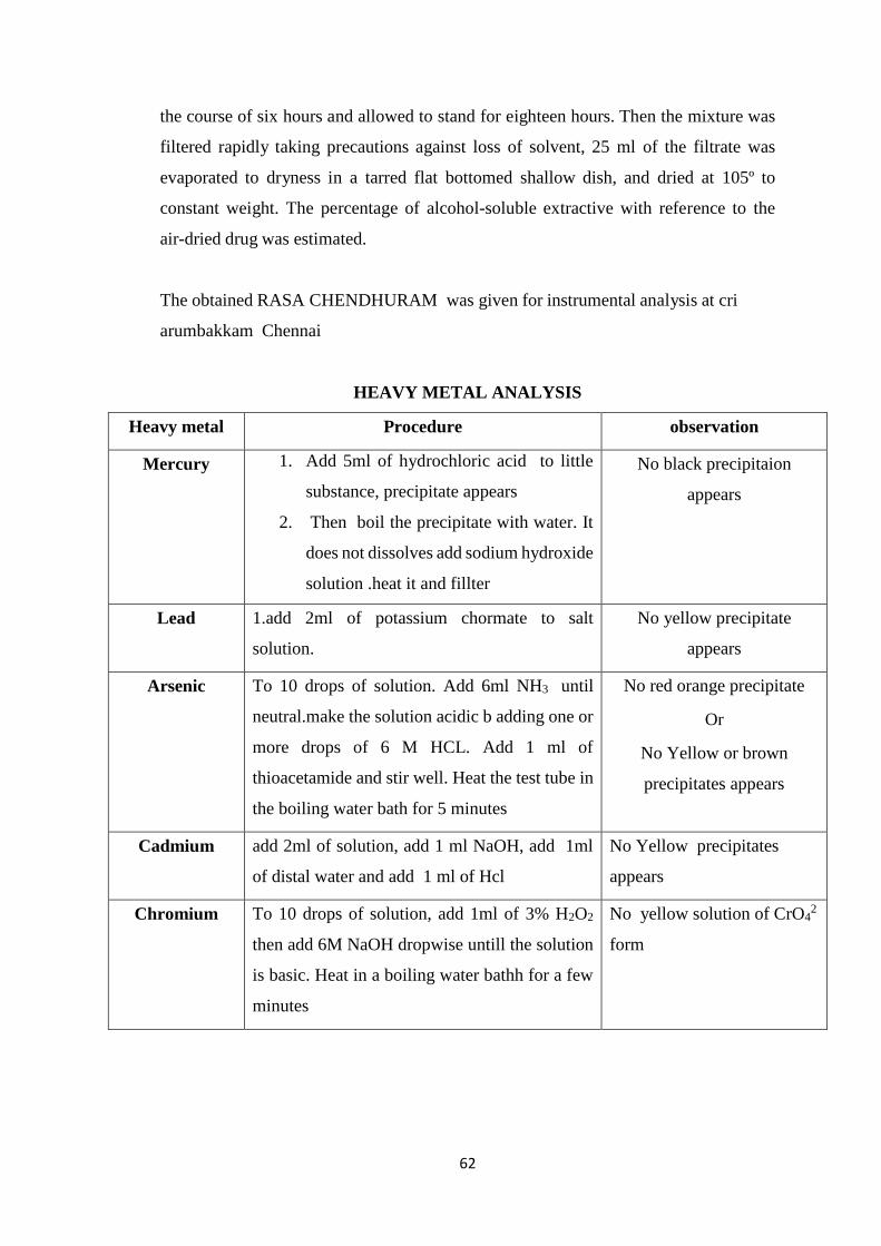
62
the course of six hours and allowed to stand for eighteen hours. Then the mixture was
filtered rapidly taking precautions against loss of solvent, 25 ml of the filtrate was
evaporated to dryness in a tarred flat bottomed shallow dish, and dried at 105º to
constant weight. The percentage of alcohol-soluble extractive with reference to the
air-dried drug was estimated.
The obtained RASA CHENDHURAM was given for instrumental analysis at cri
arumbakkam Chennai
HEAVY METAL ANALYSIS
Heavy metal Procedure observation
Mercury 1. Add 5ml of hydrochloric acid to little
substance, precipitate appears
2. Then boil the precipitate with water. It
does not dissolves add sodium hydroxide
solution .heat it and fillter
No black precipitaion
appears
Lead 1.add 2ml of potassium chormate to salt
solution.
No yellow precipitate
appears
Arsenic To 10 drops of solution. Add 6ml NH3 until
neutral.make the solution acidic b adding one or
more drops of 6 M HCL. Add 1 ml of
thioacetamide and stir well. Heat the test tube in
the boiling water bath for 5 minutes
No red orange precipitate
Or
No Yellow or brown
precipitates appears
Cadmium add 2ml of solution, add 1 ml NaOH, add 1ml
of distal water and add 1 ml of Hcl
No Yellow precipitates
appears
Chromium To 10 drops of solution, add 1ml of 3% H2O2
then add 6M NaOH dropwise untill the solution
is basic. Heat in a boiling water bathh for a few
minutes
No yellow solution of CrO42
form
63
ACUTE ORAL TOXICITY STUDY OF RASA CHENDHURAM (PULIPANI)
(OECD GUIDELINE – 423)
Introduction:
The acute toxic class method is a stepwise procedure with the use of 3 animals
of a single sex per step.
Depending on the mortality and/or the moribund status of the animals, on
average 2-4 steps may be necessary to allow judgement on the acute toxicity of
the test substance.
This procedure is reproducible, uses very few animals and is able to rank
substances in a similar manner to the other acute toxicity testing methods.
The acute toxic class method is based on biometric evaluations with fixed doses,
adequately separated to enable a substance to be ranked for classification
purposes and hazard assessment.
In principle, the method is not intended to allow the calculation of a precise
LD50, but does allow for the determination of defined exposure ranges where
lethality is expected since death of a proportion of the animals is still the major
endpoint of this test.
The method allows for the determination of an LD50 value only when at least
two doses result in mortality higher than 0% and lower than 100%.
The use of a selection of pre-defined doses, regardless of test substance, with
classification explicitly tied to number of animals observed in different states
improves the opportunity for laboratory to laboratory reporting consistency and
repeatability.
Principle of the Test:
It is the principle of the test that based on a stepwise procedure with the use of
a minimum number of animals per step, sufficient information is obtained on the acute
toxicity of the test substance to enable its classification. The substance is administered
orally to a group of experimental animals at one of the defined doses. The substance is
tested using a stepwise procedure, each step using three animals of a single sex.
Absence or presence of compound-related mortality of the animals dosed at one step
will determine the next step, i.e.
− no further testing is needed
− dosing of three additional animals, with the same dose

64
− dosing of three additional animals at the next higher or the next lower dose
level. The method will enable a judgment with respect to classifying the test substance
to one of a series of toxicity classes.
Methodology:
Selection of Animal Species
The preferred rodent species is the wistar albino rat, although other rodent
species may be used. Healthy young adult animals are commonly used laboratory
strains should be employed. Females should be nulliparous and non-pregnant. Each
animal, at the commencement of its dosing, should be between 6 to 8 weeks old and the
weight (150-200gm) should fall in an interval within±20 % of the mean weight of any
previously dosed animals.
Housing and Feeding Conditions
The temperature in the experimental animal room should be 22ºC + 3ºC.
Although the relative humidity should be at least 30% and preferably not exceed 70%
other than during room cleaning the aim should be 50-60%. Lighting should be
artificial, the sequence being 12 hours light, 12 hours dark. For feeding, conventional
laboratory diets may be used with an unlimited supply of drinking water. Animals may
be group-caged by dose, but the number of animals per cage must not interfere with
clear observations of each animal.
Preparation of animals:
The animals are randomly selected, marked to permit individual identification,
and kept in their cages for at least 7 days prior to dosing to allow for acclimatization to
the laboratory conditions.
Test Animals and Test Conditions:
Sexually mature Female Wistar albino rats (150-200gm) were obtained from
TANUVAS, Madhavaram, Chennai. All the animals were kept under standard
environmental condition (22±3°C). The animals had free access to water and standard
pellet diet (Sai meera foods, Bangalore).
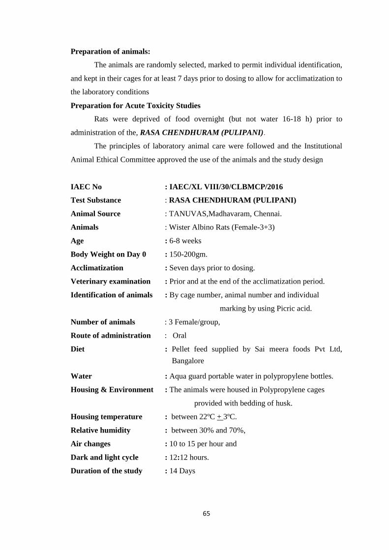
65
Preparation of animals:
The animals are randomly selected, marked to permit individual identification,
and kept in their cages for at least 7 days prior to dosing to allow for acclimatization to
the laboratory conditions
Preparation for Acute Toxicity Studies
Rats were deprived of food overnight (but not water 16-18 h) prior to
administration of the, RASA CHENDHURAM (PULIPANI).
The principles of laboratory animal care were followed and the Institutional
Animal Ethical Committee approved the use of the animals and the study design
IAEC No : IAEC/XL VIII/30/CLBMCP/2016
Test Substance : RASA CHENDHURAM (PULIPANI)
Animal Source : TANUVAS,Madhavaram, Chennai.
Animals : Wister Albino Rats (Female-3+3)
Age : 6-8 weeks
Body Weight on Day 0 : 150-200gm.
Acclimatization : Seven days prior to dosing.
Veterinary examination : Prior and at the end of the acclimatization period.
Identification of animals : By cage number, animal number and individual
marking by using Picric acid.
Number of animals : 3 Female/group,
Route of administration : Oral
Diet : Pellet feed supplied by Sai meera foods Pvt Ltd,
Bangalore
Water : Aqua guard portable water in polypropylene bottles.
Housing & Environment : The animals were housed in Polypropylene cages
provided with bedding of husk.
Housing temperature : between 22ºC + 3ºC.
Relative humidity : between 30% and 70%,
Air changes : 10 to 15 per hour and
Dark and light cycle : 12:12 hours.
Duration of the study : 14 Days
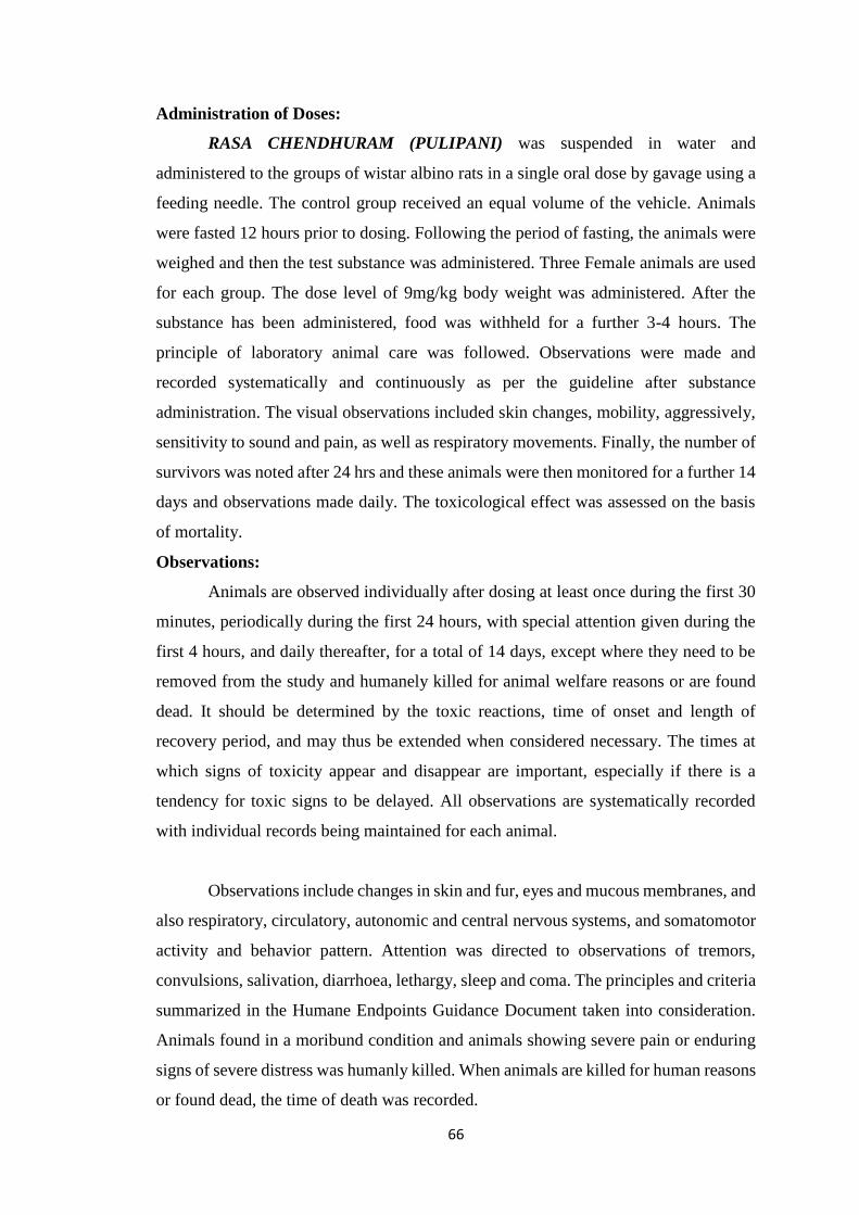
66
Administration of Doses:
RASA CHENDHURAM (PULIPANI) was suspended in water and
administered to the groups of wistar albino rats in a single oral dose by gavage using a
feeding needle. The control group received an equal volume of the vehicle. Animals
were fasted 12 hours prior to dosing. Following the period of fasting, the animals were
weighed and then the test substance was administered. Three Female animals are used
for each group. The dose level of 9mg/kg body weight was administered. After the
substance has been administered, food was withheld for a further 3-4 hours. The
principle of laboratory animal care was followed. Observations were made and
recorded systematically and continuously as per the guideline after substance
administration. The visual observations included skin changes, mobility, aggressively,
sensitivity to sound and pain, as well as respiratory movements. Finally, the number of
survivors was noted after 24 hrs and these animals were then monitored for a further 14
days and observations made daily. The toxicological effect was assessed on the basis
of mortality.
Observations:
Animals are observed individually after dosing at least once during the first 30
minutes, periodically during the first 24 hours, with special attention given during the
first 4 hours, and daily thereafter, for a total of 14 days, except where they need to be
removed from the study and humanely killed for animal welfare reasons or are found
dead. It should be determined by the toxic reactions, time of onset and length of
recovery period, and may thus be extended when considered necessary. The times at
which signs of toxicity appear and disappear are important, especially if there is a
tendency for toxic signs to be delayed. All observations are systematically recorded
with individual records being maintained for each animal.
Observations include changes in skin and fur, eyes and mucous membranes, and
also respiratory, circulatory, autonomic and central nervous systems, and somatomotor
activity and behavior pattern. Attention was directed to observations of tremors,
convulsions, salivation, diarrhoea, lethargy, sleep and coma. The principles and criteria
summarized in the Humane Endpoints Guidance Document taken into consideration.
Animals found in a moribund condition and animals showing severe pain or enduring
signs of severe distress was humanly killed. When animals are killed for human reasons
or found dead, the time of death was recorded.

67
Acute oral toxicity study of RASA CHENDHURAM (PULIPANI)
Table 1: Dose finding experiment and its behavioral Signs of acute oral Toxicity
Observation done:
SL Group
CONTROL
Observation SL Group
TEST GROUP
Observation
1 Body weight Normal 1 Body weight Normally increased
2 Assessments of posture Normal 2 Assessments of
posture
Normal
3 Signs of Convulsion
Limb paralysis
Normal 3 Signs of
Convulsion
Limb paralysis
Absence of sign (-)
4 Body tone Normal 4 Body tone Normal
5 Lacrimation Normal 5 Lacrimation Absence
6 Salivation Normal 6 Salivation Absence
7 Change in skin color No significant
color change
7 Change in skin
color
No significant color
change
8 Piloerection Normal 8 Piloerection Normal
9 Defecation Normal
9 Defecation Normal
10 Sensitivity response Normal 10 Sensitivity
response
Normal
11 Locomotion Normal 11 Locomotion Normal
12 Muscle gripness Normal 12 Muscle
gripness
Normal
13 Rearing Mild 13 Rearing Mild
14 Urination Normal 14 Urination Normal

68
Behaviour:
The animals will be observed closely for behaviour in the first four hours which
includes abnormal gait, aggressiveness, exophthalmos, ptosis, akinesia, catalepsy,
convolusion, excitation, head twitches, lacrimation, loss of corneal reflex, loss of
traction, piloerection reactivity of touch, salivation, scratching, sedation, chewing,
head movements, sniffing, straub, tremor and writhes, diarrhea, leathery, sleep and
coma.
Body Weight:
Individual weight of animals was determined before the test substance was
administered and weights will be recorded at day 1, 7, and 14 of the study. Weight
changes were calculated and recorded. At the end of the test, surviving animals were
weighed and humanly killed.
Food and water Consumption:
Food and water consumed per animal was calculated for control and the treated dose
groups.
Mortality:
Animals were observed for mortality throughout the entire period.
REPEATED DOSE 28-DAY ORAL TOXICITY (407) STUDY OF
RASA CHENDHURAM (PULIPANI)
Test Substance : RASA CHENDHURAM (PULIPANI)
Animal Source : TANUVAS,Madhavaram, Chennai.
Animals : Wister Albino Rats (Male -24, and Female-24)
Age : 6-8 weeks
Body Weight : 150-200gm.
Acclimatization : Seven days prior to dose.
Veterinary examination : Prior and at the end of the acclimatization period.
Identification of animals : By cage number, animal number and individual
marking by using Picric acid
Diet : Pellet feed supplied by Sai meera foods Pvt Ltd,
Bangalore
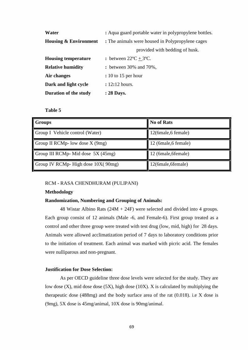
69
Water : Aqua guard portable water in polypropylene bottles.
Housing & Environment : The animals were housed in Polypropylene cages
provided with bedding of husk.
Housing temperature : between 22ºC + 3ºC.
Relative humidity : between 30% and 70%,
Air changes : 10 to 15 per hour
Dark and light cycle : 12:12 hours.
Duration of the study : 28 Days.
Table 5
RCM - RASA CHENDHURAM (PULIPANI)
Methodology
Randomization, Numbering and Grouping of Animals:
48 Wistar Albino Rats (24M + 24F) were selected and divided into 4 groups.
Each group consist of 12 animals (Male -6, and Female-6). First group treated as a
control and other three group were treated with test drug (low, mid, high) for 28 days.
Animals were allowed acclimatization period of 7 days to laboratory conditions prior
to the initiation of treatment. Each animal was marked with picric acid. The females
were nulliparous and non-pregnant.
Justification for Dose Selection:
As per OECD guideline three dose levels were selected for the study. They are
low dose (X), mid dose dose (5X), high dose (10X). X is calculated by multiplying the
therapeutic dose (488mg) and the body surface area of the rat (0.018). i.e X dose is
(9mg), 5X dose is 45mg/animal, 10X dose is 90mg/animal.
Groups No of Rats
Group I Vehicle control (Water) 12(6male,6 female)
Group II RCMp- low dose X (9mg) 12 (6male,6 female)
Group III RCMp- Mid dose 5X (45mg) 12 (6male,6female)
Group IV RCMp- High dose 10X( 90mg) 12(6male,6female)

70
RASA CHENDHURAM (PULIPANI) suspended in prescribed solvent, It
was administered to animals at the dose levels of X, 5X, 10X. The test substance
suspensions were freshly prepared every two days once for 28 days. The control
animals were administered vehicle only. The drug was administered orally by using
oral gavage once daily for 28 consecutive days.
Observations:
Experimental animals were kept under observation throughout the course of
study for the following:
Body Weight:
Weight of each rat was recorded on day 0, at weekly intervals throughout the course
of study.
Food and water Consumption:
Food and water consumed per animal was calculated for control and the treated
dose groups.
Clinical signs:
All animals were observed daily for clinical signs. The time of onset, intensity
and duration of these symptoms, if any, were recorded.
Mortality:
All animals were observed twice daily for mortality during entire course of
study.
Necropsy:
All the animals were sacrificed by excessive anaesthesia on day 29. Necropsy
of all animals was carried out.
Laboratory Investigations:
Following laboratory investigations were carried out on day 29 in animals fasted
over-night. Blood samples were collected from orbital sinus using sodium heparin
(200IU/ml) for Bio chemistry and potassium EDTA (1.5 mg/ml) for Hematology as
anticoagulant. Blood samples were centrifuged at 3000 r.p.m. for 10 minutes.
Haematological Investigations:
Haematological parameters were determined using Haematology analyzer.
Preparation and Administration of Dose:

71
Biochemical Investigations:
Biochemical parameters were determined using auto-analyzer.
Histopathology:
Control and highest dose group animals will be initially subjected to
histopathological investigations. If any abnormality found in the highest dose group
than the low, then the mid dose group will also be examined. Organs will be collected
from all animals and preserved in 10% buffered neutral formalin for 24 h and washed
in running water for 24 h. The organ sliced 5 or 6µm sections and were dehydrated in
an auto technicon and then cleared in benzene to remove absolute alcohol. Embedding
was done by passing the cleared samples through three cups containing molten paraffin
at 50°C and then in a cubical block of paraffin made by the “L” moulds. It was followed
by microtome and the slides were stained with Haematoxylin-eosin red.
Statistical analysis:
Findings such as body weight changes, water and food consumption,
hematology and blood chemistry were subjected to One-way ANOVA followed by
dunnet t test using a computer software programme – Graph pad version 7.All data
were summarized in tabular form, (Table-6 to 12)
STUDY DESIGN:
An Open Comparative Clinical Trial
STUDY PLACE :
OPD section of post graduate, sirappu maruthuvam department attached
to Arignar Anna Government Hospital, Government Siddha Medical College,
Arumbakkam, Chennai.
DATA COLLECTION:
Literary evidence collected from various
Siddha literatures
Books on modern medicine
TRIAL DRUGS:
INTERNAL DRUG: RASA CHENDHURAM , 65 mg , twice daily.
EXTERNAL DRUG: PALAGARAI KUZHAMBU

72
SAMPLE SIZE:
40 cases OPD
20 patients treated with internal drug alone
20 patients treated with internal and external drugs
INCLUSION CRITERIA
Age: Between 18 - 60 years.
Sex: Both male and female
Hypo-pigmented patches with hyper pigmented border without any structural
changes in any part of the body
Patient Willing to sign consent form.
Hypopigmentation due to Worm infestation
EXCLUSION CRITERIA
Albinism
Leprosy
STD
HIV
Burns
Pregnancy and Lactation
Cardiac diseases
History of long term use of steroids.
WITHDRAWAL CRITERIA
Intolerance to the drug and development of adverse reactions during the period
of drug trial.
Poor patient compliance & defaulters resulting in adverse events.
ADR REPORTING
If ADR is reported patients will be referred to SCRI (Peripheral Pharmaco vigilance
centre)
EVALUATION OF CLINICAL PARAMETERS:
Patients are clinically evaluated by the following parameters.

73
HISTORY TAKING:
Age , sex, occupation , socio economic status, complaints and its duration ,
previous illness , family history , personal habits were recorded in the case shet for
every patient at the time of first visit to OP.
MODERN INVESTIGATIONS:
Blood:
Hb, TC, DC, ESR, Blood sugar (R)
Kidney Function Tests:
Blood Urea, Serum Creatinine
Liver Function Tests:
Direct Bilirubin,
Serum total bilirubin,
In Direct bilirubin,
SGOT, SGPT
Urine:
Albumin,
Sugar
Deposits
ASSESSMENT TOOLS
A. Clinical assessment:
VITILIGO AREA SEVERITY INDEX (VASI):
Its name is an adoption from PASI score in psoriasis. The percentage of vitiligo
involvement is calculated in terms of hand units. One hand unit is approximately
equivalent to 1% of the total body surface area. The degree of pigmentation is
estimated to the nearest of one of the following percentages: 100% - complete
depigmentation, no pigment is depigmentation]present; 90% - specks of pigment
present; 75% - depigmented area exceeds the pigmented area; 50% - pigmented and
depigmented areas are equal;
25% - pigmented area exceeds depigmented area; and 10% - only specks of
depigmentation present . The VASI for each body region is determined by

74
the product of the area of vitiligo in hand units and the extent of depigmentation within
each hand unit measured patch.
Total
The VASI for each body region is determined by the product of the area of
vitiligo in hand units and the extent of depigmentation within each hand unit measured
patch.
Total body VASI = S All body sites [Hand Units] ´ [Residual depigmentation] [1].
VASI score ~−50 Very much worse
VASI score -50 -25 Much worse
VASI score -26 -10 Worse
VASI score -10 0 Minimally worse
VASI score 0 10 Minimally improved
VASI score 10 25 Improved
VASI score 25 50 Much improved
VASI score +50~ Very much improved
CALCULATION:
The body is divided into four sections (head (H) (10% of a person's skin); arms
(A) (20%); trunk (T) (30%); legs (L) (40%)). Each of these areas is scored by itself, and
then the four scores are combined into the final PASI. For each section, the percent of
area of skin involved, is estimated and then transformed into a grade from 0 to 6:
0% of involved area, grade: 0
< 10% of involved area, grade: 1
10–29% of involved area, grade: 2
30–49% of involved area, grade: 3
50–69% of involved area, grade: 4
70–89% of involved area, grade: 5
90–100% of involved area, grade: 6
Within each area, the severity is estimated by three clinical signs: erythema
(redness), induration (thickness) and desquamation (scaling). Severity parameters are
measured on a scale of 0 to 4, from none to maximum.
body VASI = S All body sites [Hand Units] ´ [Residual depigmentation] [1].
75
The sum of all three severity parameters is then calculated for each section of
skin, multiplied by the area score for that area and multiplied by weight of respective
section (0.1 for head, 0.2 for arms, 0.3 for body and 0.4 for legs).
B. Photo assessment:
Photos of the patient before and after treatment for clinical improvement evidence.
DRUG STORAGE:
The trial drug is stored in clean air tight glass container (borosil container) and
it is dispensed to the patients.
DATA COLLECTION FORMS:
Required information will be collected from each patients by using following
forms.
Form I : Screening and selection proforma
Form II : History taking proforma
Form III : Clinical assessment proforma
Form IV : Clinical assessment during and after trial
Form V : Laboratory investigation proforma
Form VI : Informed consent form
Form VII : Withdrawal form
Form VIII : Patients information sheet
STUDY ENROLLMENT
In this study Patients reporting at the OPD with the clinical symptoms of Hypo
pigmentation, itching, burning sensation etc will be examined clinically for enrolling
in the study based on the inclusion and exclusion criteria.
The patients who are to be enrolled would be informed (Form IV) about the
study, trial drug, possible outcomes and the objectives of the study in the language and
terms understandable to them.
After ascertaining the patient’s willingness, informed consent would be
obtained in writing from them in the consent form (Form IV). All these patients will be
Address, Phone number and Doctors phone number etc. will be given, so as to report
easily should any complications arise.
given unique registration card in which patients Registration number of the study,

76
Complete clinical history, complaints and duration, examination findings-- all
would be recorded in the prescribed Proforma in the history and clinical assessment
forms separately. Screening Form- I will be filled up: Form –II and Form –III will be
and laboratory investigations respectively. Patients would be advised to take the trial
drug and appropriate dietary advice (Form IV-D) would be given according to the
CONDUCT OF THE STUDY:
Patients satisfying the inclusion and exclusion criteria will be included in the
trial.
treatment. Photos will be taken before and after the treatment.
At the end of the study the trial patients are advised to report when there is
reoccurrence.
DATA ANALYSIS:
After enrolling the patients in the study, a separate file for each patient will be
maintained and all forms will be kept in the file. Whenever the patients visits OPD
during the study period necessary entries will be made in the assessment forms. The
data entries and adverse events if any will be monitored by the head of the department.
Primary Outcome
The therapeutic efficacy of trial drugs will be assessed by repigmentation and
also by reduction in the size.
VASI Score (Vitiligo area scoring index)
To evaluate the prevention of recurrence by follow-up after two months from
the start of intervention.
To evaluate the days of outcome with in the treatment of 48 days.
Secondary Outcome:
Secondary outcome is assessed by comparing the safety parameters before and after
treatment.
used for recording the patients history, clinical examination of symptoms and signs
patients perfect understanding.
Modern investigations will be carried out before treatment and at the end of the

77
ADR REPORTING
If ADR is reported patients will be referred to SCRI (Peripheral Pharmaco vigilance
centre)
ETHICAL ISSUES:
• Informed consent will be obtained from the patients after explaining about the
clinical trial in regional tongue.
• After the consent of the patient (through consent form) if they are in the
inclusion criteria they will be enrolled in the study.
• Treatment will be provided free of cost.
• Concomitant medications will be given when required.
• Rescue medications will be given when needed.
• The patients who are excluded (as per exclusion criteria) are given proper
treatment with full care at OPD.

RESULTS AND OBSERVATION
RASA CHENDHURAM ORGANOLEPTIC CHARACTORS
COLOR OF THE INGREDIENTS
RASA CHENDHURAM - PHYSICOCHEMICAL ANALYSIS RESULT
CHARACTERS RESULTS
Colour Dark red
Odour Oderless
Taste Tasteless
Appearance Fine powder
Solubility Sparingly soluble in both water and
alcohol
S.NO RAW DRUGS BEFORE PURIFICATION AFTER
PURIFICATION
1 Gandhagam Yellow solid Yellow granules
2 Rasam Colourless, dust floated Colourless
3. Thalagam Bright yellow Yellow
S.NO NAME OF EXPERIMENT VALUE
1 Loss on drying 0 %
2. Total ash 91.5 %
3. Water soluable ash 29 %
4. Acid insoluable ash 48 %
5. pH value (10%) 1.47
078

TRADITIONAL TESTING METHODS FOR CHENDHURAM
S.NO TESTS INFERENCE
1. Floating on water +
2. Finger furrows test +
3. Lusterless +
4. Tasteless +
5. Colour Dark red
QUALITATIVE ANALYSIS OF HEAVY METALS
S.NO HEAVY METAL RESULT
1 LEAD ND
2 MERCURY ND
3 ARSENIC ND
4 CADMIUM ND
079
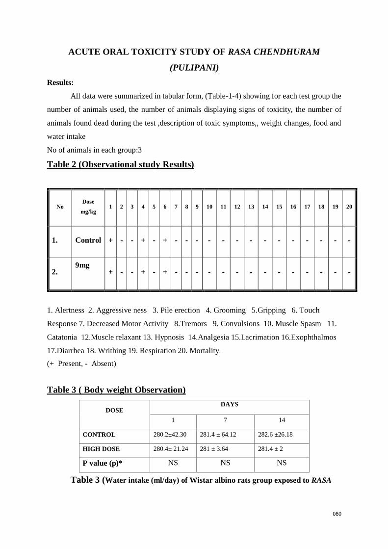
ACUTE ORAL TOXICITY STUDY OF RASA CHENDHURAM
(PULIPANI)
Results:
All data were summarized in tabular form, (Table-1-4) showing for each test group the
number of animals used, the number of animals displaying signs of toxicity, the number of
animals found dead during the test ,description of toxic symptoms,, weight changes, food and
water intake
No of animals in each group:3
Table 2 (Observational study Results)
No Dose
mg/kg 1 2 3 4 5 6 7 8 9 10 11 12 13 14 15 16 17 18 19 20
1. Control + - - + - + - - - - - - - - - - - - - -
2. 9mg
+ - - + - + - - - - - - - - - - - - - -
1. Alertness 2. Aggressive ness 3. Pile erection 4. Grooming 5.Gripping 6. Touch
Response 7. Decreased Motor Activity 8.Tremors 9. Convulsions 10. Muscle Spasm 11.
Catatonia 12.Muscle relaxant 13. Hypnosis 14.Analgesia 15.Lacrimation 16.Exophthalmos
17.Diarrhea 18. Writhing 19. Respiration 20. Mortality.
(+ Present, - Absent)
Table 3 ( Body weight Observation)
Table 3 (Water intake (ml/day) of Wistar albino rats group exposed to RASA
DOSE DAYS
1 7 14
CONTROL 280.2±42.30 281.4 ± 64.12 282.6 ±26.18
HIGH DOSE 280.4± 21.24 281 ± 3.64 281.4 ± 2
P value (p)* NS NS NS
080

CHENDHURAM (PULIPANI):
DOSE DAYS
1 6 14
CONTROL 61 ± 1.12 62±2.22 63.9±1.14
HIGH DOSE 62.2±1.1 63±1.14 64.20±24
P value (p)* NS NS NS
N.S- Not Significant, **(p > 0.01), *(p >0.05), n = 10 values are mean ± S.D (One
way ANOVA followed by Dunnett’s test)
Table 4: Food intake (gm/day) of Wistar albino rats group exposed to RASA
CHENDHURAM (PULIPANI)
DOSE DAYS
1 7 14
CONTROL 56.24±2.22 56.2±7.42 58.4±3.46
High DOSE 60.6±1.63 60.6±2.62 64.1±5.38
RESULTS
Repeated Dose 28- day oral toxic study of RASA CHENDHURAM
(PULIPANI)
Table 6: Body weight of wistar albino rats group exposed to RASA CHENDHURAM
(PULIPANI)
DOSE DAYS
1 7 14 21 28
CONTROL 230.2±15.45 231.5 ± 25.15 231.5 ± 15.50 232.5± 15.16 232.4 ± 15.15
LOW DOSE 235.2 ± 15.15 235.7 ± 32.22 236.6± 46.14 236 ± 62.18 236.41± 15.24
MID DOSE 200.4± 06.64 200.3 ± 16.24 201.2 ± 18.12 201.2 ±
11.26 202.4 ± 24.10
HIGH DOSE 210.6± 24.24 210.6 ± 10.42 211.4 ± 12.24 211 ± 14.38 212 ± 54.61
P value (p)* NS NS NS NS NS
081

NS- Not Significant, **(p > 0.01),*(p >0.05), n = 10 values are mean ± S.D (One way
ANOVA followed by Dunnett’s test)
Table 7: Water intake (ml/day) of Wistar albino rats group exposed to RASA CHENDHURAM
(PULIPANI)
DOSE DAYS
1 6 14 21 28
CONTROL 50.1 ± 4.32 50±4.12 50.2±1.10 50±1.12 50.4±1.12
LOW DOSE 52.1±1.11 52.8±2.22 52.6±1.42 53.2±2.26 52.4±1.21
MID DOSE 53.1±1.12 53.3±1.11 53.1±2.21 53.4±1.12 53.4±1.42
HIGH DOSE 54.1±1.41 54.2±1.42 54.4±1.44 54.6±1.52 55.8±2.82
P value (p)* NS NS NS NS NS
N.S- Not Significant, **(p > 0.01), *(p >0.05), n = 10 values are mean ± S.D (One way
ANOVA followed by Dunnett’s test)
Table 8: Food intake (gm/day) of Wistar albino rats group exposed to RASA
CHENDHURAM (PULIPANI)
DOSE DAYS
2 7 23 22 28
CONTROL 30±5.14 31.2±2.12 31.3±2.18 31.2±1.14 32±2.12
LOW DOSE 30.2±1.14 31.3±1.31 31.1±1.21 31.5±1.32 31.5±1.62
MID DOSE 32.1±2.22 32.2±3.40 32.2±2.24 32.2±2.16 33.2±1.24
HIGH DOSE 33.1±1.12 33.1±1.14 33.6±2.26 34.2±1.10 34.6±3.42
P value (p)* NS NS NS NS NS
N.S- Not Significant, **(p > 0.01), *(p >0.05), n = 10 values are mean ± S.D (One
way ANOVA followed by Dunnett’s test
082

Table 9: Haematological parameters of Wistar albino rats group exposed to RASA
CHENDHURAM (PULIPANI)
Category Control Low dose Mid dose High dose P value
(p)*
Haemoglobin(g/dl) 16.7±0.71 16.60±0.24 16.5±0.23 16.82±0.16 N.S
Total WBC (×103
l)
10.81±0.32 10.64±0.21 10.54±0.42 9.60±1.12 N.S
Neutrophils (%) 31.12±0.01 31.02±0.12 32.11±1.22 33.02±6.21 N.S
lymphocyte (%) 72.12±1.24 72.12±1.32 73.10±2.34 73.20±2.44 N.S
Monocyte (%) 0.9±0.02 0.9±0.01 0.9±0.04 0.9±0.03 N.S
Eosinophil (%) 0.5±0.03 0.5±0.04 0.5±0.05 0.5±0.08 N.S
Platelets cells103/µl 680.17±3.13 682.41±4.12 682.13±2.02 684.10±2.34 N.S
Total RBC 106/µl 8.42±0.12 8.46±0.53 8.49±0.44 8.74±0.46 N.S
PCV% 42.12±0.2 42.62±1.02 43±1.20 44.40±2.10 N.S
MCHC g/dL 34.5±1.20 34.2±1.10 34.8±1.70 34.33±1.30 N.S
MCV fL(µm3) 58.2±4.02 59.2±1.10 58.9±1.40 58.8±1.20 N.S
N.S- Not Significant, **(p > 0.01), *(p >0.05), n = 10 values are mean ± S.D (One way
ANOVA followed by Dunnett’s test)
Table 10 : Biochemical Parameters of of Wistar albino rats group exposed to RASA
CHENDHURAM (PULIPANI)
BIOCHEMICAL
PARAMETERS CONTROL LOW DOSE MID DOSE HIGH DOSE
P Value
(p)*
GLUCOSE (R) (mg/dl) 125.11±3.2 125.12±2.10 126.10±13.0
8 128.12±4.2 N.S
T.CHOLESTEROL(mg/dl) 120.16±1.20 120.25±1.30 122.60±1.18 123.24±1.30 N.S
083

TRIGLY(mg/dl) 54.16±1.52 54.12±1.42 56.15±1.23 56.16±1.23 N.S
LDL 72.4±2.14 72.12±2.54 73.10±1.32 73.24±10.20 NS
VLDL 11.2±1.30 11.20±2.21 11.22±1.24 11.14±12.14 NS
HDL 27.14±6.12 27.42±2.30 28.16±2.60 28.17±2.14 NS
Ratio
1(T.CHO/HDL) 3.41±1.16 3.42±1.40 3.74±1.04 3.64±2.03 NS
Ratio 2(LDL/HDL) 1.92±1.14 1.91±1.12 1.71±2.20 1.96±08.02 NS
Albumin (g/dL) 5.43±0.16 5.50±0.52 5.04±9.30
5.42±9.48
NS
NS- Not Significant, **(p > 0.01), * (p >0.05), n = 10 values are mean ± S.D (One way
ANOVA followed by Dunnett’s test
Table 11: Renal function test of of Wistar albino rats group exposed to RASA
CHENDHURAM (PULIPANI)
PARAMETERS CONTROL LOW DOSE MID DOSE HIGH DOSE
P Value
(p)*
UREA (mg/dl) 26.70±0.19 26.50±0.26 27.16±1.28 27.68±1.24 N.S
CREATININE(mg/dl) 0.22±0.02 0.21±0.04 0.22±0.05 0.24±0.07 N.S
BUN(mg/dL) 17.1±0.01 17.10±0.64 17.6±0.52 17.86±1.02 NS
URIC ACID(mg/dl) 6.04±0.34 6.06±0.51 6.6±0.15 6.42±0.20 N.S
NS- Not Significant, **(p > 0.01), * (p >0.05) , n = 10 values are mean ± S.D (One way
ANOVA followed by Dunnett’s test)
Table 12: Liver Function Test of of Wistar albino rats group exposed to Rasa
Chendhuram (Pulipani)
PARAMETERS CONTROL LOW DOSE MID DOSE HIGH DOSE P Value
(p)*
T BILIRUBIN(mg/dl). 0.07±0.01 0.07±0.02 0.07±0.01 0.07±0.03 N.S
SGOT/AST(U/L) 81.14±1.63 81.31±0.02 82.01±1.24 82.64±1.63 N.S
084

SGPT/ALT(U/L) 78.12±1.08 78.21±1.24 78.14±1.26 77.68±0.01 N.S
ALP(U/L) 119.21±3.16 119±32.10 119±12.14 120.03±8.32 N.S
T.PROTEIN(g/dL) 6.2.10±0.04 6.2±0.11 6.2±0.10 6.4±0.46 N.S
NS- Not Significant, **(p > 0.01), * (p >0.05), n = 10 values are mean ± S.D (One way
ANOVA followed by Dunnett’s test.
HISTO PATHOLOGY
CONTROL GROUP
High dose
085
Kidney Liver Spleen
Kidney Liver Spleen

1. AGE
S. No
AGE( In years)
NO. OF CASES (Out of 40)
PERCENTAGE (%)
1.
18-20 Years
4
10%
2.
21-30 Years
9
22.5%
3.
31-40Years
8
20%
4.
41-50years
12
30%
5.
51-60years
7
17.5%
Inference:
Out of 40 cases, 4(10%) of cases belonged to the age group of 18-20 years, 9
(22.5%) of cases belonged to the age group of 21-30 years, 12 (30%) of cases belonged
to the age group of 31-40 years, 11(27.5%) of cases belonged to the age group of 41-
50 years, and 7(17.5%) of cases belonged to the age group of 51-60 years.
10%
22.5%
20%
30%
17.5%
0
5
10
15
20
25
30
35
18-20 yrs 21-30 yrs 31-40 yrs 41-50 yrs 51-60 yrs
No o
f p
ati
en
ts i
n p
erc
en
tage
Age
Age
086

2. GENDER DISTRIBUTION
S. No GENDER NUMBER OF CASES PERCENTAGE
(%)
1 Male 18 45%
2 Female 22 55%
Inference:
Out of 40 cases, 18(45%) were Male, 22(55%) cases were Female.
45%55%
Gender distribution
Male
Female
087
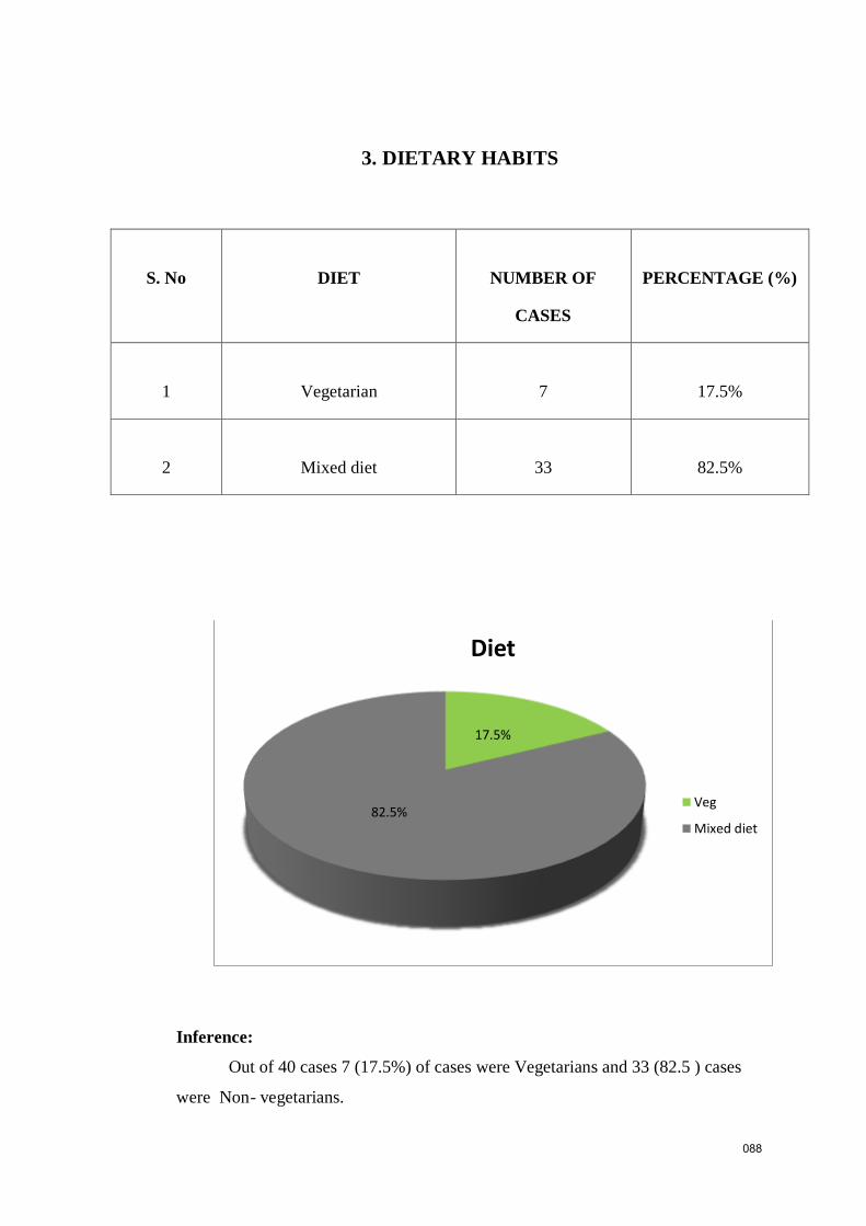
3. DIETARY HABITS
S. No
DIET
NUMBER OF
CASES
PERCENTAGE (%)
1
Vegetarian
7
17.5%
2
Mixed diet
33
82.5%
Inference:
Out of 40 cases 7 (17.5%) of cases were Vegetarians and 33 (82.5 ) cases
were Non- vegetarians.
17.5%
82.5%
Diet
Veg
Mixed diet
088

4. OCCUPATION
S.No OCCUPATION NUMBER OF
CASES
PERCENTAGE (%)
1 Office worker 9 22.5%
2 Home maker 15 37.5%
3 Business 4 10%
4 Student 8 20%
5 Farmer 0 0%
6 Others 4 10%
Inference:
Out of 40 cases 8 (20 %) were Student, 15 (37.5%) were Homemaker, 9
(22.5%) were Office Workers, 4 (10%)were businessmen, 0 (0 %) were farmer, 4
(10%) were others
22.5%
37.5%
10%
20%
0%
10%
0
5
10
15
20
25
30
35
40
Officeworker
Homemaker
Business Student Farmer Others
No o
f p
ati
en
ts i
n p
erc
en
tage
Occupation
Occupation
089

5. SOCIO – ECONOMIC STATUS
S.No SOCIO – ECONOMIC
STATUS
NUMBER OF
CASES
PERCENTAGE (%)
1 Low Income
27 67.5%
2 Moderate Income 9 22.5%
3 High Income 4 10%
Inference:
Among 40 cases 67.5% comes under low economic status, 22.5% of them under
moderate status and 10% of them under high income status.
67.50%
22.50%
10%
socio economic status
low income
moderate income
high income
090
6. FAMILY HISTORY:
FAMILY HISTORY NUMBER OF CASES PERCENTAGE
PRESENT 4 10%
ABSENT 36 90%
Inference:
Out of 40 cases 4 (10 %) of cases have family history and 36 (90%)of
cases have no family history.
10 %
90 %
Family History
Present
Absent
091

7 .DISTRIBUTION OF THINAI
S.No THINAI NUMBER OF CASES PERCENTAGE (%)
1 Kurinji 0 0%
2 Mullai 0 0%
3 Marutham 4 10%
4 Neithal 36 90%
5 Paalai 0 0%
Inference:
Out the 40 patients, no patients were recorded from kurinchi ,mullai and
Paalai thinai, 4(10%) of cases from Marutham thinai, 36(90%) from Neithal
thinai.
0 % 0 %
10 %
90 %
0 %0
10
20
30
40
50
60
70
80
90
100
Kurinji Mullai Marutham Neithal Paalai
No
of
pa
tien
ts i
n p
erce
nta
ge
Thinai
Thinai
092

8. PARUVAKAALAMDISTRIBUTION:
S.No KAALAM (Season) NUMBER OF CASES PERCENTAGE (%)
1 Kaarkaalam
( Mid Aug – Mid Oct)
5 12.5%
2 koothirKaalam
(Mid Oct – Mid Dec)
8 20%
3 Munpanikaalam
(Mid Dec – Mid Feb)
11 27.5%
4 Pinpanikaalam
(Mid Feb – Mid Apr)
6 15%
5 Elavenirkaalam
(Mid Apr – Mid Jun)
3 7.5%
6 Muthuvenirkaalam
(Mid Jun – Mid Aug)
7 17.5%
Inference:
Out of 40 cases, 5 patients (12.5 %) were recorded in Karkaalam, 8 patients
(20%) were recorded in Koothir Kaalam, 11 patients (27.5%) were recorded in Munpani
12.5 %
20 %
27.5%
15%
7.5 %
17.5 %
0
5
10
15
20
25
30
No
of
pa
tien
ts i
n p
erce
nta
ge
Kaalam
Kaalam
093
Kaalam, 6 patients (15%) were recorded in Pinpani Kaalam, 3 patients (7.5%) were
recorded in Elavenil Kaalam and 7 patients (17.5%) were recorded in Mudhuvenil
Kaalam.

9. EZHU UDALKATTUGAL
S.NO UDAL KATTUGAL No. ofCases Percentage
1. Saaram 40 100%
2. Senneer 40 100%
3. Oon 15 37.5 %
4. Kozhuppu 15 37.5 %
5. Enbu - -
6. Moolai - -
7. SukkilamSuronitham - -
100 % 100 %
37.5 % 37.5 %
0 % 0 % 0 %0
20
40
60
80
100
120
No
of
pa
tien
ts i
n p
erce
nta
ge
Udal kattugal
Udal kattugal
094
Inference:
Out of 40 cases Saaram was affected in all the 40 cases (100%), Senneer in 40
cases (75%) and oon in 15 cases , kozhuppu in 15 cases (37.5) and no other changes
seen in other Udal Kattugal.

10. DISTRIBUTION OF MUKKUTRAM:
A. Derangement in the types of Vatham
S.No Classification of vatham No. of Cases Percentage
1. Piranan - -
2. Abanan 18 45 %
3. Viyanan 15 37.5%
4. Uthanan - -
5. Samanan 40 100
6. Nagan - -
7. Koorman
8. Kirukaran - -
9. Devathathan - -
0 %
45 %37.5 %
0 %
100 %
0 % 0 % 0 % 0 % 00
20
40
60
80
100
120
No
of
pa
tien
ts i
n p
erce
nta
ge
Vatham
Vatham
095
Inference:
A. Vatham:
Out of 40 cases Abanan was affected in 18 (45%) of cases. Viyanan was affected in
15 (37.5%) of cases and Samanan was affected in 40 (100%) of cases

B.Derangement in types of Pitham
S.No Types of pitham No. of Cases Percentage
1. Analapitham - -
2. Ranjagam 20 50
3. Saathagam - -
4. Prasagam 40 100
5. Alosagam - -
0 %
50 %
0 %
100 %
0 %0
20
40
60
80
100
120
Analapitham Ranjagam Saathagam Prasagam Alosagam
No
of
pa
tien
ts i
n p
erce
nta
ge
Pitham
Pitham
096
B.Inference:
Pitham: Ranjagam was affected in 20 of cases (50%) and Prasagam was
affected in all the 40 (100%) of cases.

3. DERANGEMENT IN TYPES OF KABAM :
S.No KABAM NUMBER OF CASES PERCENTAGE (%)
1 Avalambagam 15 37.5%
2 Klethagam 0 0%
3 Pothagam 0 0%
4 Tharpagam 0 0%
5 Santhigam 15 37.5%
0
5
10
15
20
25
30
35
4037.5%
0 % 0 % 0 %
37.5 %
No
of
pa
tien
ts i
n p
erce
nta
ge
Kabam
Kabam
097
Inference:
Out of 40 patients, Avalambagam was affected in 15 patients (37.5%),
Santhigam was affected in 15 patients (37.5%).
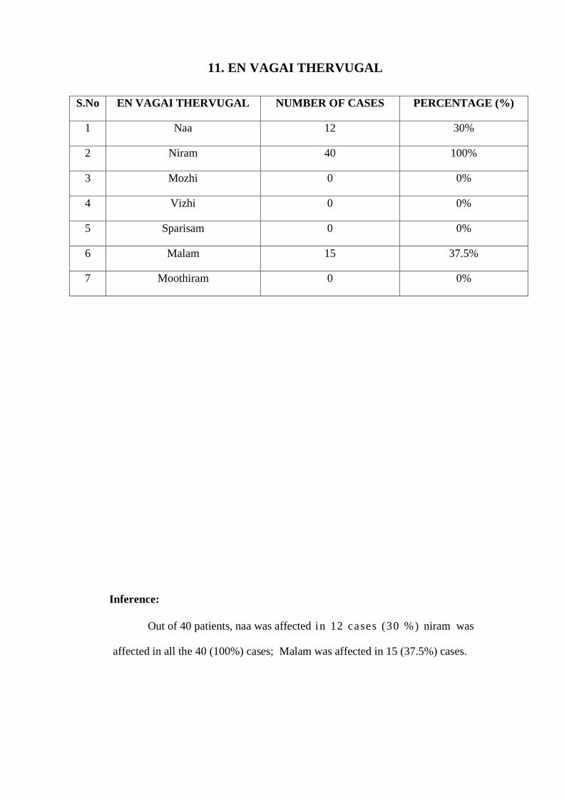
11. EN VAGAI THERVUGAL
S.No EN VAGAI THERVUGAL NUMBER OF CASES PERCENTAGE (%)
1 Naa 12 30%
2 Niram 40 100%
3 Mozhi 0 0%
4 Vizhi 0 0%
5 Sparisam 0 0%
6 Malam 15 37.5%
7 Moothiram 0 0%
Inference:
Out of 40 patients, naa was affected in 12 cases (30 %) niram was
affected in all the 40 (100%) cases; Malam was affected in 15 (37.5%) cases.
0
20
40
60
80
100
30 %
100 %
0 % 0 % 0 %
37.5 %
0 %No
of
pa
tien
ts i
n p
erce
nta
ge
En vagai Thervugal
En vagaiThervugal
098

12 .SITE OF LESIONS :
SITE OF LESIONS NO OF CASES
OUT OF 40
PERCENTAGE(%)
Scalp 0 0%
Face 12 30%
Lips 4 10%
Abdomen 3 7.5%
Upper limb 9 22.5%
Lower limb 5 12.5%
Multiple 7 17.5%
0
5
10
15
20
25
30
0 %
30 %
10 %7.5 %
22.5 %
12.5 %
17.5 %
No o
f p
ati
en
ts i
n p
erc
en
tage
Site of lesions
Site oflesions
Inference :
Out of 40 patients 12 patients (30 %) were affected in the face , 3 patients
(7.5%) were affected in the abdomen , 13 patients (7.5%) were affected in the upper
limb , patients (12.5 %) were affected in the lower limb, 7 patients (17.5%) were
affected in the multiple
099

13 .DURATION OF DISEASE
DURATION OF DISEASE NO OF CASES PERCENTAGE
10 Years 3 7.5%
6 Years 1 2.5%
5 Years 5 12.5%
4 Years 3 7.5%
3 Years 6 15%
2 Years 7 17.5%
1 Year 6 15%
6 Month 8 20%
0
5
10
15
20
10 yrs 6 yrs 5 yrs 4 yrs 3 yrs 2 yrs 1 yr 6month
7.5 %
2.5 %
12.5 %
7.5 %
15 %
17.5 %
15 %
20%
Duration of disease
Duration of disease
100
Inference:
The higher incidence of illness is 6 month of duration.

14 .NAADI
S.No NAADI NUMBER OF CASES PERCENTAGE (%)
1 ValiAzhal 18 45%
2 ValiIyam 4 10%
3 AzhalVali 12 30%
4 AzhalIyam 2 5%
5 IyaVatham 2 5%
6 IyaVali 2 5%
0
5
10
15
20
25
30
35
40
45
ValiAzhal vali Iyam Azhal Vali Azha Iiyam Iya Vatham Iya Vali
45 %
10 %
30 %
5 % 5 % 5 %
No
of
pa
tien
ts i
n p
erce
nta
ge
Naadi
Naadi
101
Inference:
Out of 40 patients 18 patients (45%) had Valiazhal naadi, 4 patients (10%) had
ValiIyam naadi, 12 patients (30%) had Azhalvali naadi, 2 Patient (5%) had Azhaliyam
naadi, 2 Patienst (5%) had Iyavatham naadi, 2 patients (5%) had Iyavali naadi.

.
13. NEERKKURI:
S.No
Niram
No. ofCases
Percentage
1.
Straw colour 10 25
2.
Yellow colour 27 67.5
3. Pale yellow colour 3 7.5
0
10
20
30
40
50
60
70
Straw colourYellow colour
Pale yellowcolour
No o
f p
ati
en
ts i
n p
erc
en
tage
Niram
102
Inference:
Out of 40 cases, 10 (25%) of cases showed straw coloured urine, 27
(67.5%) of cases showed yellow coloured urine, 3 (7.5%) of cases showed Pale
yellow coloured urine.
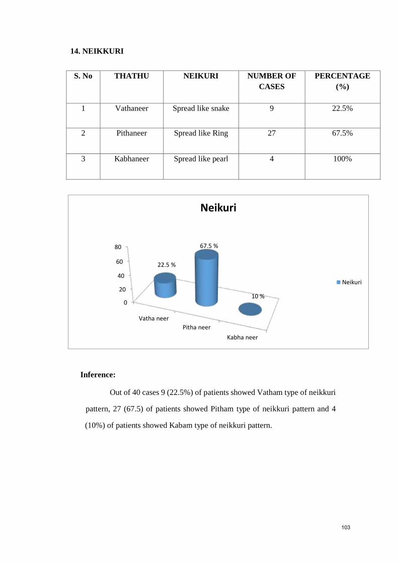
14. NEIKKURI
S. No THATHU NEIKURI NUMBER OF
CASES
PERCENTAGE
(%)
1 Vathaneer Spread like snake 9 22.5%
2 Pithaneer Spread like Ring 27 67.5%
3 Kabhaneer Spread like pearl 4 100%
Inference:
Out of 40 cases 9 (22.5%) of patients showed Vatham type of neikkuri
pattern, 27 (67.5) of patients showed Pitham type of neikkuri pattern and 4
(10%) of patients showed Kabam type of neikkuri pattern.
0
20
40
60
80
Vatha neer
Pitha neer
Kabha neer
22.5 %
67.5 %
10 %
Neikuri
Neikuri
103
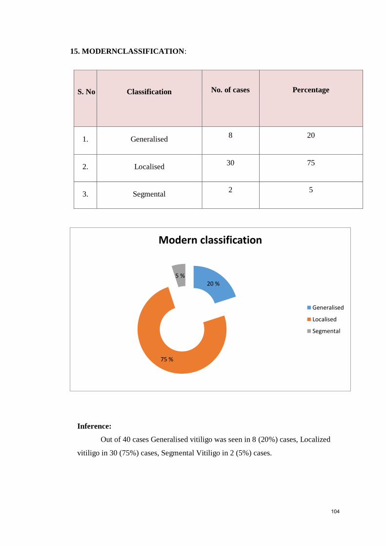
15. MODERNCLASSIFICATION:
S. No Classification No. of cases Percentage
1. Generalised 8 20
2. Localised 30 75
3. Segmental 2 5
Inference:
Out of 40 cases Generalised vitiligo was seen in 8 (20%) cases, Localized
vitiligo in 30 (75%) cases, Segmental Vitiligo in 2 (5%) cases.
20 %
75 %
5 %
Modern classification
Generalised
Localised
Segmental
104

16. PRECIPITATING FACTORS
S. No PRECIPITATING
FACTORS No. of Cases Percentage
1. Stress 26 65
2. Anemia 10 25
3. Injury 4 10
Inference:
Out of 40 cases, Stress precipitated Vitiligoin 26 (65%) cases, Anemia
precipitated Vitiligo in 10 (25%) cases and Injury precipitated Vitiligo in 2 (10%)
cases.
65%
25 %
10 %
Precipitating factors
Stress
Anemia
Injury
105

17. CLINICAL FEATURES
SIGN AND
SYMPTOMS
BEFORE TREATMENT AFTER TREATMENT
NO.OF
CASES
PERCENTAGE
(%)
NO.OF CASES PERCENTAGES
(%)
Hypopigmentation
patches in skin
40 100% 30 75%
Depigmentation of
hair
7 17.5 % 4 10%
Itching 0 0% 0 0%
Erythema 0 0% 0 0%
0%10%20%30%40%50%60%70%80%90%
100%
100%
17.50%
0% 0%
75%
10%
0% 0% BT
AT
106
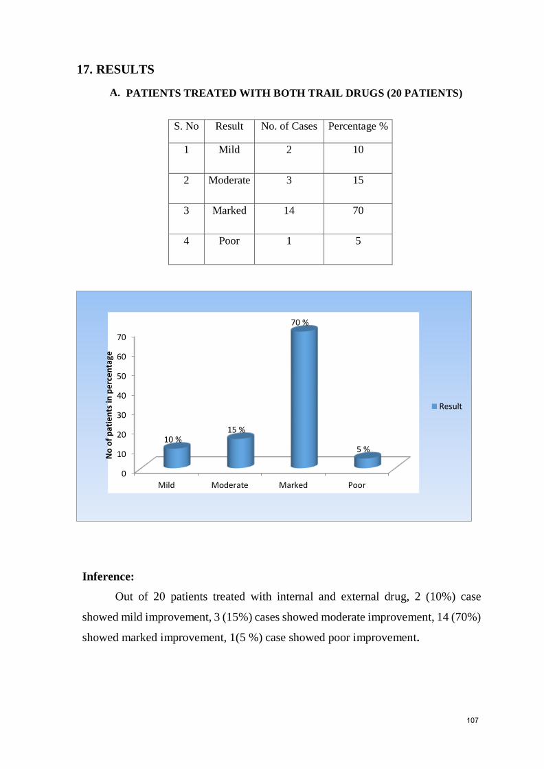
17. RESULTS
A. PATIENTS TREATED WITH BOTH TRAIL DRUGS (20 PATIENTS)
S. No Result No. of Cases Percentage %
1 Mild 2 10
2 Moderate 3 15
3 Marked 14 70
4 Poor 1 5
Inference:
Out of 20 patients treated with internal and external drug, 2 (10%) case
showed mild improvement, 3 (15%) cases showed moderate improvement, 14 (70%)
showed marked improvement, 1(5 %) case showed poor improvement.
0
10
20
30
40
50
60
70
Mild Moderate Marked Poor
10 %15 %
70 %
5 %
No
of
pat
ien
ts in
per
cen
tage
Result
107

B. PATIENTS TREATED WITH INTERNAL TRAIL DRUG (20
PATIENTS)
S. No Result No. of Cases Percentage%
1 Mild 2 10
2 Moderate 4 20
3 Marked 12 60
4 poor 2 10
Inference:
20 patients treated with internal drug, which yield Mild Improvement in 2 [ 10%) cases,
Moderate Improvement in 4 (20%) cases, Marked Improvement in 12 (60%) cases and poor
improvement in 2(10%) cases were noted.
10%
20%
60%
10%
Mild
Moderate
Marked
Poor
108

C.COMPARISION OF RESULTS:
PATIENTS TREATED WITH BOTH DRUGS (20 Pts) AND INTERNAL
DRUG ALONE(20 Pts)
S.No Results
Both Drug Internal Drug only
No. ofCases Percentage% No. ofCases Percentage%
1 Mild 2 10 2 10
2 Moderate 3 15 4 20
3 Marked 14 70 12 60
4 poor 1 5 2 10
Inference:
The higher incidence is Marked improvement occurs in administration of both
drugs (70%) than internal drug alone (60%).
INT & EXT
INT0%
10%
20%
30%
40%
50%
60%
70%
MILDMODERATE
MARKEDPOOR
10% 15%
70%
5%
10%
20%
60%
10%
INT & EXT
INT
109

D. RESULTS AFTER TREATMENT
S. No Results No. of Cases Percentage
1. Mild 4 10
2. Moderate 7 17.5
3. Marked 28 70
4. Poor 1 2.5
Inference:
Out of 40 cases, 4 (10 %) of cases showed Mild improvement,7 (17.5%) of cases
showed Moderate improvement, 28 (70 %)of cases showed Marked improvement and
1 (2.5%) of case showed Poor improvement.
10%17.5 %
70%
2.5 %
Mild
Modderate
Marked
Poor
110

OP NO : 5739 AGE /SEX: 24/ F
BEFORE TREATMENT DURING TREATMENT
AFTER TREATMENT
111

OP.NO - 6020 AGE/SEX – 58/ M
BEFORE TREATMENT DURING TREATMENT
AFTER TREATMENT
112

OP.NO – 2068 AGE/SEX – 23/M
113

BL
OO
D A
NA
LY
SIS
FO
R O
PD
PA
TIE
NT
S T
RE
AT
ED
WIT
H IN
TE
RN
AL
ME
DIC
INE
RA
SA
CH
EN
DH
UR
AM
(INT
) PA
LA
GA
RA
I KU
ZH
AM
BU
(EX
T)
SI.
No
OP
.
NO
A
GE
/
SE
X
Hb
(gm
) T
C (c
u.m
m)
DC
E
SR
B
l sug
ar
Urea
crea
tinin
e
urea
crea
tinin
e
N
L
E
½
hr
1/2
hr
1h
r
1h
r
R
BT
B
T
AT
A
T
BT
A
T
BT
A
T
BT
AT
BT
AT
BT
AT
BT
AT
B
T
AT
B
T
AT
1
50
03
25
/F
12
.2
13
82
00
87
00
69
71
25
27
6
2
10
4
18
6
10
5
11
0
18
0.8
2
0
0.7
2
51
17
52
/M
14
.4
14
98
00
96
00
57
59
38
38
5
3
18
8
40
12
10
6
98
22
0.4
1
8
0.6
3
50
78
44
/ M
13
14
95
00
99
00
60
64
34
33
6
3
24
4
50
10
11
0
12
0
30
0.6
2
0
0.7
4
52
22
41
/ F
13
.2
13
.5
80
00
85
00
58
61
35
34
7
6
10
4
18
6
10
0
96
20
0.5
1
4
0.5
5
57
39
24
/ F
13
.2
13
.4
84
00
97
00
52
57
40
38
8
5
5
2
12
4
80
90
18
0.4
2
4
0.6
6
67
58
37
/ F
11
.2
12
10
00
0
10
40
0
46
49
4
8
48
6
3
22
4
38
6
11
2
11
6
22
0.4
2
7
0.6
7
67
98
56
/ F
12
.8
13
76
00
89
00
49
55
41
39
10
6
14
2
20
4
10
2
94
17
0.6
2
4
0.8
8
64
88
52
/ M
12
.5
13
.5
92
00
93
00
56
62
41
36
3
2
16
4
34
6
11
8
10
0
19
0.7
1
9
0.7
9
20
68
23
/ M
13
14
87
00
94
00
62
68
32
29
4
3
12
4
32
10
8
5
11
2
14
0.6
2
9
0.6
10
37
11
37
/ M
12
.6
13
85
00
97
00
55
61
40
36
5
3
8
2
16
4
94
88
30
0.6
2
2
0.8
11
43
74
47
/ M
13
13
.5
93
00
10
00
0
65
69
31
29
4
2
16
8
32
6
10
3
10
6
34
0.6
2
4
0.7
12
20
64
30
/ F
10
.8
12
78
00
89
00
62
68
33
30
5
2
6
2
12
6
11
3
12
0
16
1.1
2
3
0.8
13
38
61
33
/ F
9.6
1
1
10
50
0
10
70
0
53
60
40
37
7
3
4
2
10
4
87
98
21
0.5
2
0
0.9
14
66
58
37
/ F
11
11
.5
97
00
10
00
0
64
70
33
27
3
2
20
12
40
22
97
11
4
23
0.6
1
9
0.6
15
73
04
27
/M
13
.5
14
82
00
85
00
54
58
39
37
7
5
10
2
34
4
11
1
86
14
0.5
1
4
0.7
16
42
83
59
/ F
12
.8
13
83
00
90
00
59
66
34
30
7
4
30
4
62
10
98
11
2
28
0.3
1
6
0.5
17
48
83
30
/ F
10
11
99
00
10
30
0
63
70
31
27
6
3
32
2
52
6
11
0
90
22
0.5
2
8
0.5
18
60
20
60
/ M
14
.1
14
.5
5,5
00
75
00
64
70
28
26
8
4
16
4
22
8
60
80
16
0.7
2
6
0.6
19
51
88
50
/ M
13
13
.6
79
00
86
00
49
57
45
37
6
6
10
2
20
6
11
5
93
20
0.5
2
0
0.7
20
67
84
34
/ F
12
.1
13
14
40
0
14
60
0
66
70
28
26
6
4
5
2
15
4
94
10
5
18
0.8
2
2
0.9
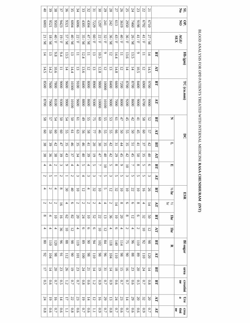
BL
OO
D A
NA
LY
SIS
FO
R O
PD
PA
TIE
NT
S T
RE
AT
ED
WIT
H IN
TE
RN
AL
ME
DIC
INE
RA
SA
CH
EN
DH
UR
AM
(INT
)
SI.
No
OP
.
NO
A
GE
/
SE
X
Hb
(gm
) T
C (c
u.m
m)
DC
E
SR
B
l sug
ar
urea
crea
tini
ne
Ure
a
crea
tini
ne
N
L
E
½ h
r
½
hr
1h
r
1h
r
R
BT
A
T
BT
A
T
BT
A
T
BT
A
T
BT
A
T
BT
A
T
BT
A
T
BT
A
T
BT
B
T
AT
A
T
21
67
10
27
/ M
14
14
.5
97
00
99
00
52
57
42
40
6
3
26
14
50
12
98
12
0
14
0.8
2
0
0.7
22
67
92
41
/ F
11
12
69
00
67
00
58
60
37
35
5
5
16
4
32
10
92
11
0
24
0.7
3
2
0.9
23
91
98
41
/ F
9
10
.5
84
00
90
00
41
45
41
50
8
5
10
8
6
2
11
0
89
20
0.5
2
0
0.6
24
74
60
26
/ M
13
.5
14
87
00
86
00
42
50
51
45
7
5
22
6
12
4
96
88
23
0.6
2
9
0.6
25
29
50
36
/ F
13
.6
14
87
00
92
00
45
55
35
42
10
3
10
6
8
2
75
90
16
0.7
1
4
0.7
26
36
19
40
/ F
11
12
72
00
80
00
47
50
44
45
9
5
10
8
20
4
11
4
98
15
0.7
2
3
0.6
27
61
3
43
/ F
14
.4
14
.8
66
00
70
00
61
66
31
29
8
5
32
14
10
6
14
0
11
0
18
0.6
2
4
0.7
28
24
2
36
/ M
12
13
84
00
90
00
53
55
42
42
5
3
12
2
36
4
11
8
10
4
14
0.9
1
3
0.8
29
24
1
32
/ F
9
12
10
00
0
10
10
0
55
55
35
40
10
5
5
4
13
12
84
96
31
0.7
2
9
0.7
30
12
97
22
/ F
10
.5
12
.8
98
00
10
00
0
60
46
30
47
7
7
10
4
22
10
10
2
98
19
0.9
1
6
0.9
31
72
29
60
/ F
13
13
89
00
93
00
75
77
20
19
5
3
32
2
52
6
94
11
0
14
1.2
1
2
1.1
32
43
04
47
/ M
11
12
80
00
86
00
55
58
41
39
4
3
4
2
10
6
90
98
21
0.6
1
4
0.6
33
43
05
35
/ M
12
.4
13
.6
84
00
88
00
49
56
46
40
5
4
12
2
24
6
89
10
0
24
0.8
1
9
0.7
34
60
06
22
/ F
11
13
91
00
96
00
61
63
35
34
4
3
10
2
20
4
11
9
10
1
23
0.7
2
3
0.6
35
60
04
40
/ M
13
14
.4
10
30
0
10
10
0
50
61
46
37
4
2
40
6
82
12
98
90
19
0.7
2
2
0.8
36
93
21
57
/ M
13
.5
14
96
00
99
00
54
55
35
43
9
1
30
4
62
10
88
11
2
26
1.2
1
7
1.1
37
94
23
19
/ F
9.4
1
1
90
00
93
00
5
0
60
47
39
3
2
8
16
16
36
96
91
23
0.5
1
4
0.6
38
95
03
40
/ F
9.8
1
0.6
7
800
81
00
54
60
42
38
2
1
4
2
10
4
12
0
98
20
0.7
1
6
0.7
39
95
21
18
/ M
13
14
.2
76
00
79
00
57
59
39
36
4
5
2
2
4
4
11
0
10
4
14
0.6
1
9
0.6
40
60
03
21
/ M
14
14
.5
85
00
87
00
58
60
38
38
4
2
4
2
8
4
80
92
17
0.5
2
4
0.8
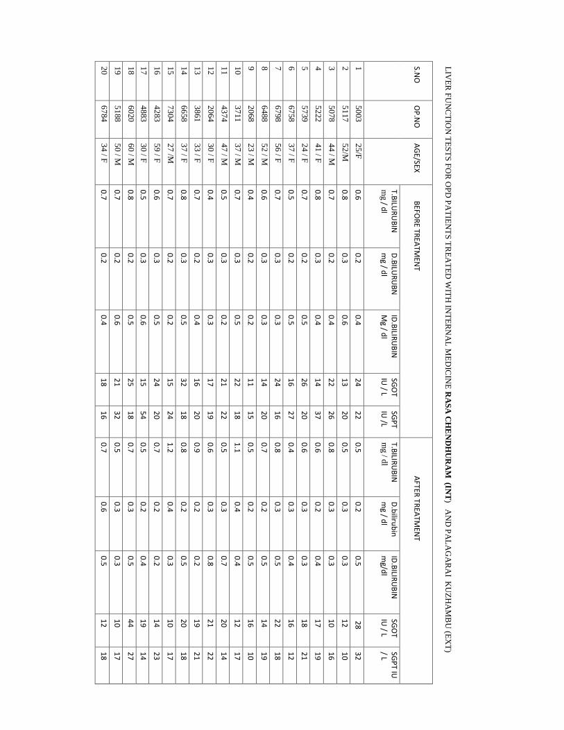
LIV
ER
FU
NC
TIO
N T
ES
TS
FO
R O
PD
PA
TIE
NT
S T
RE
AT
ED
WIT
H IN
TE
RN
AL
ME
DIC
INE
RA
SA
CH
EN
DH
UR
AM
(INT
) AN
D P
AL
AG
AR
AI K
UZ
HA
MB
U (E
XT
)
S.NO
O
P.N
O
AG
E/SEX
BEFO
RE TR
EATM
ENT
A
FTER T
REA
TMEN
T
T.BILU
RU
BIN
m
g / dl
D.B
ILUR
UB
N
mg / d
l ID
.BILIR
UB
IN
Mg / d
l SG
OT
IU / L
SGP
T IU
/L T.B
ILIRU
BIN
m
g / d
l
D.b
ilirub
in
mg / d
l ID
.BILIR
UB
IN
mg/d
l SG
OT
IU / L
SGP
T IU
/ L
1
50
03
25
/F
0.6
0
.2
0.4
2
4
22
0
.5
0.2
0
.5
28
3
2
2
51
17
52
/M
0.8
0
.3
0.6
1
3
20
0
.5
0.3
0
.3
12
1
0
3
50
78
44
/ M
0.7
0
.2
0.4
2
2
26
0
.8
0.3
0
.3
10
1
6
4
52
22
41
/ F
0.8
0
.3
0.4
1
4
37
0
.6
0.2
0
.4
17
1
9
5
57
39
24
/ F
0.7
0
.2
0.5
2
6
20
0
.6
0.3
0
.3
18
2
1
6
67
58
37
/ F
0.5
0
.2
0.5
1
6
27
0
.4
0.3
0
.4
16
1
2
7
67
98
56
/ F
0.7
0
.3
0.3
2
4
16
0
.8
0.3
0
.5
22
1
8
8
64
88
52
/ M
0.6
0
.3
0.3
1
4
20
0
.7
0.2
0
.5
14
1
9
9
20
68
23
/ M
0.4
0
.2
0.2
1
1
15
0
.5
0.2
0
.5
16
1
0
10
37
11
37
/ M
0.7
0
.3
0.5
2
2
18
1
.1
0.4
0
.4
12
1
7
11
43
74
47
/ M
0.5
0
.3
0.2
2
1
22
0
.5
0.3
0
.7
20
1
4
12
20
64
30
/ F
0.4
0
.3
0.3
1
7
19
0
.6
0.3
0
.8
21
2
2
13
38
61
33
/ F
0.7
0
.2
0.4
1
6
20
0
.9
0.2
0
.2
19
2
1
14
66
58
37
/ F
0.8
0
.3
0.5
3
2
18
0
.8
0.2
0
.5
20
1
8
15
73
04
27
/M
0.7
0
.2
0.2
1
5
24
1
.2
0.4
0
.3
10
1
7
16
42
83
59
/ F
0.6
0
.3
0.5
2
4
20
0
.7
0.2
0
.2
14
2
3
17
48
83
30
/ F
0.5
0
.3
0.6
1
5
54
0
.5
0.2
0
.4
19
1
4
18
60
20
60
/ M
0.8
0
.2
0.5
2
5
18
0
.7
0.3
0
.5
44
2
7
19
51
88
50
/ M
0.7
0
.2
0.6
2
1
32
0
.5
0.3
0
.3
10
1
7
20
67
84
34
/ F
0.7
0
.2
0.4
1
8
16
0
.7
0.6
0
.5
12
1
8

LIV
ER
FU
NC
TIO
N T
ES
TS
FO
R O
PD
PA
TIE
NT
S T
RE
AT
ED
WIT
H IN
TE
RN
AL
ME
DIC
INE
RA
SA
CH
EN
DH
UR
AM
(INT
)
S.NO
O
P.N
O
AG
E/SEX
BEFO
RE TR
EATM
ENT
A
FTER T
REA
TMEN
T
T.BILU
RU
BIN
m
g / dl
D.B
ILUR
UB
N
mg / d
l ID
.BILIR
UB
IN
Mg / d
l SG
OT
IU / L
SGP
T IU
/L T.B
ILIRU
BIN
m
g / d
l
D.b
ilirub
in
mg / d
l ID
.BILIR
UB
IN
mg/d
l SG
OT
IU / L
SGP
T IU
/ L
21
67
10
27
/ M
0.8
0
.3
0.5
1
2
13
0
.5
0.2
0
.5
26
2
4
22
67
92
41
/ F
1.2
0
.4
0.5
1
3
24
0
.6
0.3
0
.6
17
1
2
23
91
98
41
/ F
0.9
0
.3
0.4
2
6
21
0
.8
0.2
0
.3
32
3
4
24
74
60
26
/ M
0.8
0
.2
0.6
1
9
15
0
.7
0.2
0
.4
10
1
2
25
29
50
36
/ F
1.5
0
0.3
1
.29
1
8.5
1
8
0.4
0
.2
0.3
1
9
12
26
36
19
40
/ F
0.6
0
.3
0.2
1
3
19
1
.3
0.3
0
.4
21
2
4
27
61
3
43
/ F
0.7
0
.3
0.3
1
6
25
0
.8
0.3
0
.6
10
1
7
28
24
2
36
/ M
0.8
0
.2
0.3
2
3
63
0
.6
0.2
0
.5
32
4
0
29
24
1
32
/ F
0.4
0
.3
0.4
3
7
23
0
.9
0.3
0
.5
18
2
0
30
12
97
22
/ F
0.7
0
.3
0.4
1
9
24
0
.6
0.3
0
.5
16
2
4
31
72
29
60
/ F
0.8
0
.2
0.3
1
5
19
0
.8
0.2
0
.4
16
2
0
32
43
04
47
/ M
0.8
0
.2
0.5
2
2
18
0
.4
0.4
0
.5
28
2
4
33
43
05
35
/ M
0.6
0
.2
0.3
1
7
16
0
.7
0.3
0
.4
17
2
2
34
60
06
22
/ F
0.4
0
.3
0.2
1
3
15
0
.9
0.2
0
.4
23
2
8
35
60
04
40
/ M
0.9
0
.3
0.5
2
7
33
0
.6
0.3
0
.2
15
1
1
36
93
21
57
/ M
1.0
0
.3
0.5
1
7
13
0
.5
0.2
0
.3
27
1
9
37
94
23
19
/ F
0.5
0
.2
0.3
3
2
31
0
.8
0.4
0
.2
18
2
2
38
95
03
40
/ F
0.4
0
.2
0.3
1
4
17
0
.8
0.4
0
.5
28
2
4
39
95
21
18
/ M
0.8
0
.3
0.6
2
3
26
0
.9
0.3
0
.3
16
2
8
40
60
03
21
/ M
0.6
0
.3
0.5
2
2
17
0
.7
0.2
0
.4
12
1
9

VA
SI S
CO
RE
FO
R O
PD
PA
TIE
NT
S T
RE
AT
ED
WIT
H IN
TE
RN
AL
ME
DIC
INE
RA
SA
CH
EN
DH
UR
AM
(INT
) PA
LA
GA
RA
I KU
ZH
AM
BU
(EX
T)
S.
NO
O
P.
NO
H
EA
D
UP
PE
R L
IMB
T
RU
NK
L
OW
ER
LIM
B
TO
TA
L
SC
OR
E
DH
A
H
DH
A
H
DT
AT
DL
AL
B
T
A
T
B
T
AT
B
T
A
T
B
T
A
T
B
T
A
T
B
T
A
T
B
T
A
T
B
T
A
T
B
T
AT
1
50
03
1
1
1
1
0
0
0
0
0
0
0
0
0
0
0
0
0.1
0
.1
2
51
17
2
1
2
1
0
0
0
0
0
0
0
0
0
0
0
0
0.4
0
.1
3
50
78
2
0
3
0
0
0
0
0
0
0
0
0
0
0
0
0
0.6
0
4
52
22
3
1
2
1
0
0
0
0
0
0
0
0
0
0
0
0
0.6
0
.1
5
57
39
0
0
0
0
3
1
5
3
0
0
0
0
0
0
0
0
3
0.6
6
67
58
0
0
0
0
2
1
4
2
0
0
0
0
0
0
0
0
1.6
0
.4
7
67
98
0
0
0
0
2
1
2
1
0
0
0
0
0
0
0
0
0.8
0
.2
8
64
88
0
0
0
0
2
1
2
1
0
0
0
0
0
0
0
0
0.8
0
.2
9
20
68
0
0
0
0
2
1
2
1
0
0
0
0
0
0
0
0
0.8
0
.2
10
37
11
0
0
0
0
2
1
2
1
0
0
0
0
0
0
0
0
0.8
0
.1
11
43
74
0
0
0
0
3
2
2
1
0
0
0
0
0
0
0
0
1.2
0
.4
12
20
64
0
0
0
0
0
0
0
0
2
0
4
0
0
0
0
0
2.4
0
13
38
61
0
0
0
0
0
0
0
0
2
1
3
1
0
0
0
0
1.8
0
.3
14
66
58
0
0
0
0
0
0
0
0
1
1
1
1
0
0
0
0
0.3
0
.3
15
73
04
0
0
0
0
0
0
0
0
0
0
0
0
3
0
1
0
1.2
0
16
42
83
0
0
0
0
0
0
0
0
0
0
0
0
3
1
3
2
3.6
0
.8
17
48
83
0
0
0
0
0
0
0
0
0
0
0
0
2
1
2
1
1.6
0
.4
18
60
20
0
0
0
0
0
0
0
0
0
0
0
0
2
1
2
1
1.6
0
.4
19
51
88
0
0
0
0
0
0
0
0
0
0
0
0
3
2
2
1
2.4
0
.8
20
67
84
0
0
0
0
0
0
0
0
0
0
0
0
2
1
3
2
2.4
0
.8

VA
SI S
CO
RE
FO
R O
PD
PA
TIE
NT
S T
RE
AT
ED
WIT
H IN
TE
RN
AL
ME
DIC
INE
RA
SA
CH
EN
DH
UR
AM
(INT
)
S.
NO
O
P.
NO
H
EA
D
UP
PE
R L
IMB
T
RU
NK
L
OW
ER
LIM
B
TO
TA
L
SC
OR
E
DH
A
H
DH
A
H
DT
AT
DL
AL
B
T
A
T
B
T
AT
B
T
A
T
B
T
A
T
B
T
A
T
B
T
A
T
B
T
A
T
B
T
A
T
B
T
AT
21
67
10
0
0
0
0
3
1
5
3
0
0
0
0
0
0
0
0
3
0.6
22
67
92
0
0
0
0
2
1
4
2
0
0
0
0
0
0
0
0
1.6
0
.4
23
91
98
0
0
0
0
3
2
2
1
0
0
0
0
0
0
0
0
1.2
0
.4
24
74
60
0
0
0
0
0
0
0
0
2
0
4
0
0
0
0
0
2.4
0
25
29
50
2
1
2
1
0
0
0
0
0
0
0
0
0
0
0
0
0.4
0
.1
26
36
19
2
0
3
0
0
0
0
0
0
0
0
0
0
0
0
0
0.6
0
27
61
3
3
1
2
1
0
0
0
0
0
0
0
0
0
0
0
0
0.6
0
.1
28
24
2
0
0
0
0
0
0
0
0
0
0
0
0
2
1
2
1
1.6
0
.4
29
24
1
0
0
0
0
0
0
0
0
0
0
0
0
3
2
2
1
2.4
0
.8
30
12
97
0
0
0
0
0
0
0
0
2
1
3
1
0
0
0
0
1.8
0
.3
31
72
29
0
0
0
0
0
0
0
0
1
1
1
1
0
0
0
0
0.3
0
.3
32
43
04
0
0
0
0
0
0
0
0
0
0
0
0
3
0
1
0
1.2
0
33
43
05
0
0
0
0
2
1
2
1
0
0
0
0
0
0
0
0
0.8
0
.2
34
60
06
0
0
0
0
2
1
2
1
0
0
0
0
0
0
0
0
0.8
0
.1
35
60
04
0
0
0
0
0
0
0
0
0
0
0
0
3
1
3
2
3.6
0
.8
36
93
21
0
0
0
0
0
0
0
0
0
0
0
0
2
1
2
1
1.6
0
.4
37
94
23
2
1
2
1
0
0
0
0
0
0
0
0
0
0
0
0
0.4
0
.1
38
95
03
2
0
3
0
0
0
0
0
0
0
0
0
0
0
0
0
0.6
0
39
95
21
0
0
0
0
0
0
0
0
1
1
1
1
0
0
0
0
0.3
0
.3
40
60
03
0
0
0
0
0
0
0
0
0
0
0
0
3
0
1
0
1.2
0

UR
INE A
NA
LYSIS FOR
OP
D P
ATIEN
TS TR
EATED
WITH
INTER
NA
L MED
ICIN
E R
ASA
CH
END
HU
RA
M (IN
T) AN
D P
ALA
GA
RA
I KU
ZHA
MB
U (EX
T)
S.NO
O
P.N
O
AG
E/SEX
BEFO
RE TR
EATM
ENT
A
FTER TR
EATM
ENT
ALB
UM
IN
SUG
AR
D
EPO
SITS A
LBU
MIN
SU
GA
R
DEP
OSITS
EPITH
ELIAL
CELLS
PU
S CELLS
EPITH
ELIAL
CELLS
PU
S CELLS
1
50
03
25
/F
NIL
NIL
1-2
2-4
NIL
NIL
1-2
1-2
2
51
17
52
/M
NIL
NIL
3 -4
1
-4 N
IL N
IL 2
-4 1
-2
3
50
78
44
/ M
NIL
NIL
2-5
2-4
NIL
NIL
1-2
1-2
4
52
22
41
/ F
NIL
NIL
2-4
1-2
NIL
NIL
1-2
2-3
5
57
39
24
/ F
NIL
NIL
1-5
NIL
NIL
NIL
2-3
1-2
6
67
58
37
/ F
NIL
NIL
2-4
2-4
NIL
NIL
1-2
1-2
7
67
98
56
/ F
NIL
NIL
2-4
2-3
NIL
NIL
2-3
2-3
8
64
88
52
/ M
NIL
NIL
2-3
3-6
NIL
NIL
1-2
4-5
9
20
68
23
/ M
NIL
NIL
2-4
2-4
NIL
NIL
2-3
1-2
10
37
11
37
/ M
NIL
NIL
2-4
1-2
NIL
NIL
1-2
1-2
11
43
74
47
/ M
NIL
NIL
1-3
2-4
NIL
NIL
1-2
1-2
12
20
64
30
/ F
NIL
NIL
3-5
2-3
NIL
NIL
2-4
4-5
13
38
61
33
/ F
NIL
NIL
4-6
6-8
NIL
NIL
2-4
1-2
14
66
58
37
/ F
NIL
NIL
2-4
2-4
NIL
NIL
1-2
2-3
15
73
04
27
/M
NIL
NIL
1-2
1-2
NIL
NIL
2-4
1-2
16
42
83
59
/ F
NIL
NIL
2-4
1-3
NIL
NIL
2-4
1-2
17
48
83
30
/ F
NIL
NIL
2-3
1-3
NIL
NIL
1-2
1-2
18
60
20
60
/ M
NIL
NIL
3-5
2-4
NIL
NIL
2-4
3-5
19
51
88
50
/ M
NIL
NIL
2-4
1-3
NIL
NIL
1-2
1-3
20
67
84
34
/ F
NIL
NIL
2-4
3-5
NIL
NIL
1-2
1-3

UR
INE
AN
AL
YS
IS F
OR
OP
D P
AT
IEN
TS
TR
EA
TE
D W
ITH
INT
ER
NA
L M
ED
ICIN
E R
AS
A C
HE
ND
HU
RA
M (IN
T)
S.NO
O
P.N
O
AG
E/SEX
BEFO
RE TR
EATM
ENT
A
FTER TR
EATM
ENT
ALB
UM
IN
SUG
AR
D
EPO
SITS A
LBU
MIN
SU
GA
R
DEP
OSITS
EPITH
ELIAL
CELLS
PU
S CELLS
EPITH
ELIAL
CELLS
PU
S CELLS
21
67
10
27
/ M
NIL
NIL
2-5
2-4
NIL
NIL
2-4
1-2
22
67
92
41
/ F
NIL
NIL
4-6
2-4
NIL
NIL
2-3
1-3
23
91
98
41
/ F
NIL
NIL
6-8
3-6
NIL
NIL
2-3
1-2
24
74
60
26
/ M
NIL
NIL
1-2
2-4
NIL
NIL
1-2
1-3
25
29
50
36
/ F
NIL
NIL
1 – 3
2
-3 N
IL N
IL 1
-3 1
-3
26
36
19
40
/ F
NIL
NIL
1-2
2-4
NIL
NIL
2-4
1-4
27
61
3
43
/ F
NIL
NIL
1-2
1-2
NIL
NIL
1-2
1-2
28
24
2
36
/ M
NIL
NIL
2-4
2-4
NIL
NIL
1-2
1-3
29
24
1
32
/ F
NIL
NIL
2-4
2-3
NIL
NIL
2-4
1-2
30
12
97
22
/ F
NIL
NIL
2-6
2-4
NIL
NIL
1-4
1-4
31
72
29
60
/ F
NIL
NIL
1-2
1-3
NIL
NIL
1-2
1-4
32
43
04
47
/ M
NIL
NIL
4-6
2-4
NIL
NIL
2-4
1-3
33
43
05
35
/ M
NIL
NIL
6-8
2-3
NIL
NIL
2-4
1-5
34
60
06
22
/ F
NIL
NIL
3-5
1-2
NIL
NIL
1-2
3-5
35
60
04
40
/ M
NIL
NIL
1-2
1-2
NIL
NIL
1-2
1-3
36
93
21
57
/ M
NIL
NIL
2-4
3-6
NIL
NIL
2-4
2-3
37
94
23
19
/ F
NIL
NIL
2-4
2-4
NIL
NIL
1-2
3-5
38
95
03
40
/ F
NIL
NIL
2-3
2-3
NIL
NIL
1-2
1-2
39
95
21
18
/ M
NIL
NIL
1-3
2-4
NIL
NIL
1-3
1-2
40
60
03
21
/ M
NIL
NIL
1-2
1-2
NIL
NIL
2-4
1-2
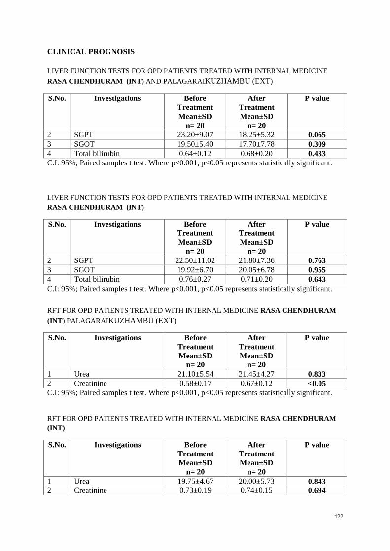
CLINICAL PROGNOSIS
LIVER FUNCTION TESTS FOR OPD PATIENTS TREATED WITH INTERNAL MEDICINE
RASA CHENDHURAM (INT) AND PALAGARAIKUZHAMBU (EXT)
S.No. Investigations Before
Treatment
Mean±SD
n= 20
After
Treatment
Mean±SD
n= 20
P value
2 SGPT 23.20±9.07 18.25±5.32 0.065
3 SGOT 19.50±5.40 17.70±7.78 0.309
4 Total bilirubin 0.64±0.12 0.68±0.20 0.433
C.I: 95%; Paired samples t test. Where p<0.001, p<0.05 represents statistically significant.
LIVER FUNCTION TESTS FOR OPD PATIENTS TREATED WITH INTERNAL MEDICINE
RASA CHENDHURAM (INT)
S.No. Investigations Before
Treatment
Mean±SD
n= 20
After
Treatment
Mean±SD
n= 20
P value
2 SGPT 22.50±11.02 21.80±7.36 0.763
3 SGOT 19.92±6.70 20.05±6.78 0.955
4 Total bilirubin 0.76±0.27 0.71±0.20 0.643
C.I: 95%; Paired samples t test. Where p<0.001, p<0.05 represents statistically significant.
RFT FOR OPD PATIENTS TREATED WITH INTERNAL MEDICINE RASA CHENDHURAM
(INT) PALAGARAIKUZHAMBU (EXT)
S.No. Investigations Before
Treatment
Mean±SD
n= 20
After
Treatment
Mean±SD
n= 20
P value
1 Urea 21.10±5.54 21.45±4.27 0.833
2 Creatinine 0.58±0.17 0.67±0.12 <0.05
C.I: 95%; Paired samples t test. Where p<0.001, p<0.05 represents statistically significant.
RFT FOR OPD PATIENTS TREATED WITH INTERNAL MEDICINE RASA CHENDHURAM
(INT)
S.No. Investigations Before
Treatment
Mean±SD
n= 20
After
Treatment
Mean±SD
n= 20
P value
1 Urea 19.75±4.67 20.00±5.73 0.843
2 Creatinine 0.73±0.19 0.74±0.15 0.694
122

C.I: 95%; Paired samples t test. Where p<0.001, p<0.05 represents statistically significant.
BLOOD ANALYSIS FOR OPD PATIENTS TREATED WITH INTERNAL MEDICINE RASA
CHENDHURAM (INT) PALAGARAIKUZHAMBU (EXT)
S.No. Investigations Before
Treatment
Mean±SD
n= 20
After
Treatment
Mean±SD
n= 20
P value
1 Hb 12.49±1.27 13.02±1.01 <0.001
2 ESR1hr 28.85±14.87 7.20±4.22 <0.001
C.I: 95%; Paired samples t test. Where p<0.001, p<0.05 represents statistically significant.
BLOOD ANALYSIS FOR OPD PATIENTS TREATED WITH INTERNAL MEDICINE RASA
CHENDHURAM (INT)
S.No. Investigations Before
Treatment
Mean±SD
n= 20
After
Treatment
Mean±SD
n= 20
P value
1 Hb 11.90±1.77 12.99±1.34 <0.001
2 ESR1 hr 24.85±21.33 7.90±7.41 <0.05
C.I: 95%; Paired samples t test. Where p<0.001, p<0.05 represents statistically significant.
VASI SCORE FOR OPD PATIENTS TREATED WITH INTERNAL MEDICINE RASA
CHENDHURAM (INT) PALAGARAI KUZHAMBU (EXT)
S.No. BT
VASI
Score
AT
VASI
Score
1 0.1 0.1
2 0.4 0.1
3 0.6 0
4 0.6 0.1
5 3 0.6
6 1.6 0.4
7 0.8 0.2
8 0.8 0.2
9 0.8 0.2
10 0.8 0.1
11 1.2 0.4
12 2.4 0
13 1.8 0.3
123
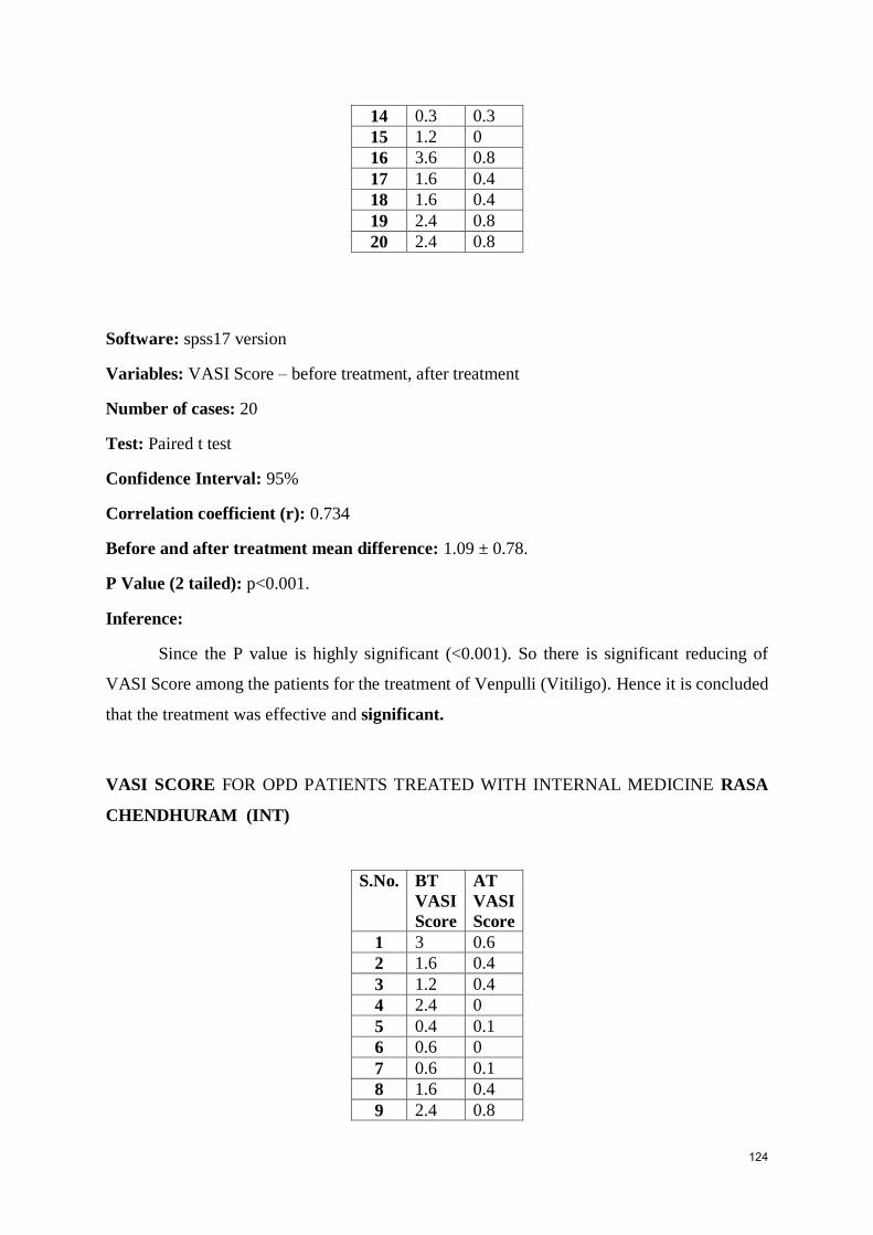
14 0.3 0.3
15 1.2 0
16 3.6 0.8
17 1.6 0.4
18 1.6 0.4
19 2.4 0.8
20 2.4 0.8
Software: spss17 version
Variables: VASI Score – before treatment, after treatment
Number of cases: 20
Test: Paired t test
Confidence Interval: 95%
Correlation coefficient (r): 0.734
Before and after treatment mean difference: 1.09 ± 0.78.
P Value (2 tailed): p<0.001.
Inference:
Since the P value is highly significant (<0.001). So there is significant reducing of
VASI Score among the patients for the treatment of Venpulli (Vitiligo). Hence it is concluded
that the treatment was effective and significant.
VASI SCORE FOR OPD PATIENTS TREATED WITH INTERNAL MEDICINE RASA
CHENDHURAM (INT)
S.No. BT
VASI
Score
AT
VASI
Score
1 3 0.6
2 1.6 0.4
3 1.2 0.4
4 2.4 0
5 0.4 0.1
6 0.6 0
7 0.6 0.1
8 1.6 0.4
9 2.4 0.8
124
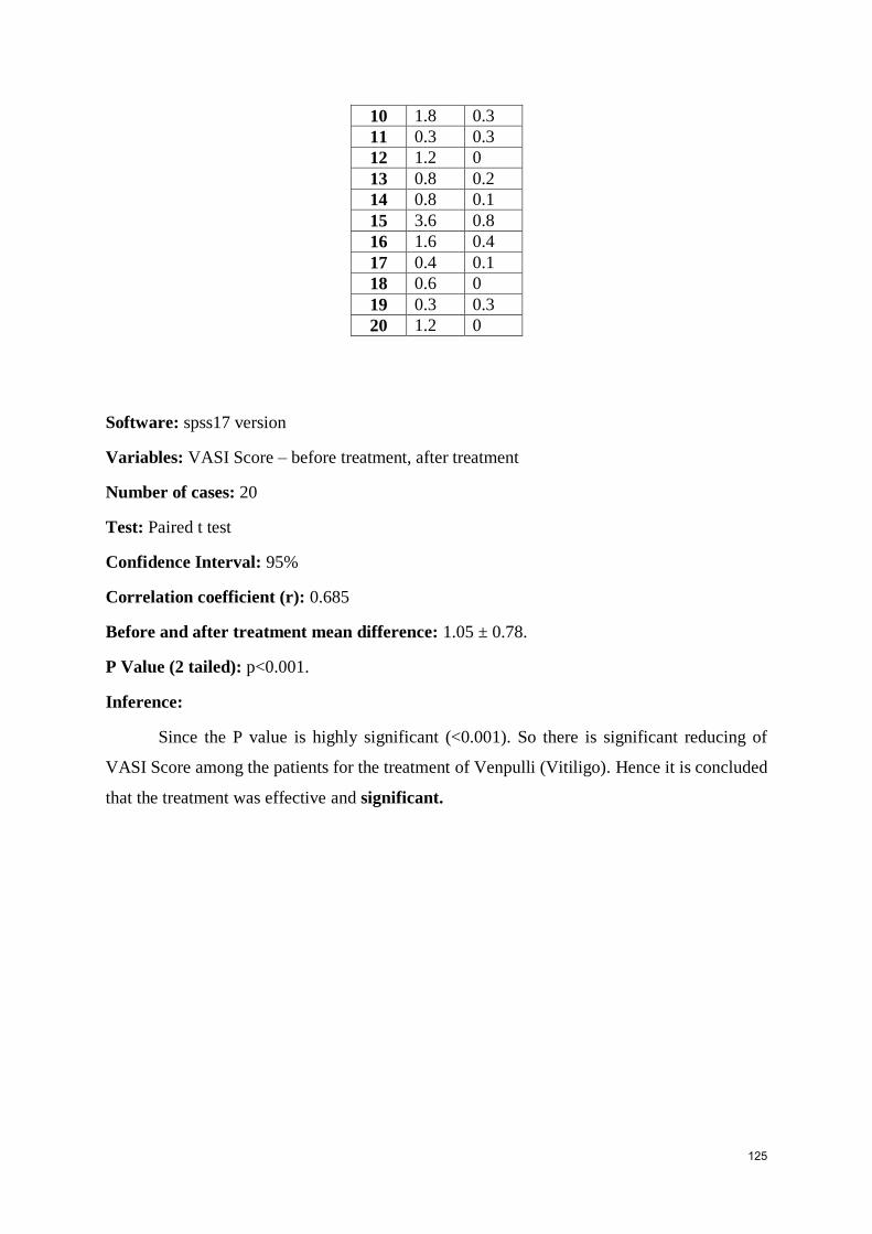
10 1.8 0.3
11 0.3 0.3
12 1.2 0
13 0.8 0.2
14 0.8 0.1
15 3.6 0.8
16 1.6 0.4
17 0.4 0.1
18 0.6 0
19 0.3 0.3
20 1.2 0
Software: spss17 version
Variables: VASI Score – before treatment, after treatment
Number of cases: 20
Test: Paired t test
Confidence Interval: 95%
Correlation coefficient (r): 0.685
Before and after treatment mean difference: 1.05 ± 0.78.
P Value (2 tailed): p<0.001.
Inference:
Since the P value is highly significant (<0.001). So there is significant reducing of
VASI Score among the patients for the treatment of Venpulli (Vitiligo). Hence it is concluded
that the treatment was effective and significant.
125

DISCUSSION
Skin disease is often easily noticeable than other disease. Appearance
of the skin can determine the status of each individual incarnation of himself .Due to
changes in a person’s appearance, venpulli (vitiligo) patients often experience
psychological stress and suffer from social stigma.
Yugi vaithiya chinthamani mention venpulli noi as swetha kuttam which
comes under the eighteen types of kuttam. As the term kuttam bears social stigma,
the disease is called as venpulli.
The clinical entity of venpulli noi is more or less similar to that of
vitiligo in modern medicine. Vitiligo is an acquired, idiopathic, depigmentory
condition of skin and hair varying sizes and shapes. It is varying sizes and shapes.
Besides loss of colour there is no other structural change.
The author has collected information largely from various siddha and
modern textbook. 40 patients were selected from Govt Siddha Medical College
OPD on both siddha and modern diagnostic method under the investigation of my
guide.
The trial drugs for this study were prepared by the Author in the
Gunapadam practical lab, Govt Siddha Medical College, under the supervision of
the members of the teaching faculty and guided by the Head of the Department of
Sirappu Maruthuvam, Govt Siddha Medical College, Arumbakkam, Chennai -106.
The trial drugs Rasa Chenduram (Internal) and palagarai kuzhambu (External) were
given for 48 days. Out-Patients were asked to visit the hospital once in 7 days. For
Out-Patients the drugs were given for 48 days and the clinical assessment was done
on 0th
day, 8th
day, 15th
day, 22th
day 29th
day, 36th day, 43rdday, 48thday.
Patients were instructed to take the medicines regularly and apply the
external medicine twice a day and to expose the affected parts to sunlight in the
morning. It was ensured that the diet restrictions such as non citric acid foods.
imposed were followed properly by the patients. Before and after the course of
treatment, the patients were subjected to laboratory investigations and photographs
were taken.
Based on various criterias, the datas were collected and tabulate
126

DISTRIBUTION ACCORDING TO AGE :
Out of 40 cases, 4 (10%) of cases belonged to the age group of 18-20
years.
9 (22.5%) of cases belonged to the age group of 21-30 years.
12 (30%) of cases belonged to the age group of 31-40 years
11 (27.5%) of cases belonged to the age group of 41-50 years
7 (17.5%) of cases belonged to the age group of 51-60 years.
DISTRIBUTION ACCORDING TO GENDER:
18(45%) were Male,
22 (55%) cases were Female.
DISTRIBUTION ACCORDING TO DIETARY HABITS :
Out of 40 cases 7(17.5%) of cases were Vegetarians
33 (82.5 %) cases were Non- vegetarians.
DISTRIBUTION ACCORDING TO OCCUPATION :
8 (20 %) were Student
15 (37.5%) were Home maker,
9 (22.5%) were Office Workers
4 (10%) were businessmen
4 ( 10%) were others
DISTRIBUTION ACCORDING TO SOCIO – ECONOMIC STATUS
67.5% comes under low economic status
22.5% of them under moderate status
10 % of them under high income status.
DISTRIBUTION ACCORDING TO FAMILY HISTORY :
36 (90%) of cases have no family history.
4 (10 %) of cases have family history
127
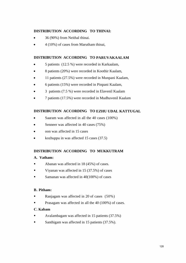
DISTRIBUTION ACCORDING TO THINAI:
36 (90%) from Neithal thinai.
4 (10%) of cases from Marutham thinai,
DISTRIBUTION ACCORDING TO PARUVAKAALAM
5 patients (12.5 %) were recorded in Karkaalam,
8 patients (20%) were recorded in Koothir Kaalam,
11 patients (27.5%) were recorded in Munpani Kaalam,
6 patients (15%) were recorded in Pinpani Kaalam,
3 patients (7.5 %) were recorded in Elavenil Kaalam
7 patients (17.5%) were recorded in Mudhuvenil Kaalam
DISTRIBUTION ACCORDING TO EZHU UDAL KATTUGAL
Saaram was affected in all the 40 cases (100%)
Senneer was affected in 40 cases (75%)
oon was affected in 15 cases
kozhuppu in was affected 15 cases (37.5)
DISTRIBUTION ACCORDING TO MUKKUTRAM
A. Vatham:
Abanan was affected in 18 (45%) of cases.
Viyanan was affected in 15 (37.5%) of cases
Samanan was affected in 40(100%) of cases
Ranjagam was affected in 20 of cases (50%)
Prasagam was affected in all the 40 (100%) of cases.
Avalambagam was affected in 15 patients (37.5%)
Santhigam was affected in 15 patients (37.5%).
128
C. Kabam
B. Pitham:

DISTRIBUTION ACCORDING TO EN VAGAI THERVUGAL
naa was affected in 12 cases (30 %)
niram was affected in all the 40 (100 %) cases
Malam was affected in 15 (37.5%) cases.
DISTRIBUTION ACCORDING TO NAADI
18 patients (45%) had Valiazhal naadi
4 patients (10%) had ValiIyam naadi,
12 patients (30%) had Azhalvali naadi
2 Patient (5%) had Azhaliyam naadi,
2 Patienst (5%) had Iyavathamnaadi
2 patients (5%) had Iyavalinaadi .
DISTRIBUTION ACCORDING TO NEERKKURI:
10 (25%) of cases showed straw coloured urine
27 (67.5%) of cases showed yellow coloured urine
3 (7.5%) of cases showed Pale yellow coloured urine.
DISTRIBUTION ACCORDING TO NEIKKURI :
9 (22.5%) of patients showed Vatham type of neikkuri pattern
27 (67.5) of patients showed Pitham type of neikkuri pattern
4 (10%) of patients showed Kabam type of neikkuri pattern. .
DISTRIBUTION ACCORDING TO PRECIPITATING FACTORS :
Stress precipitated Vitiligo in 26 (65%) cases
Anemia precipitated Vitiligo in 10 (25%) cases
Injury precipitated Vitiligo in 2 (10%) cases.
129

RESULTS
A.PATIENTS TREATED WITH BOTH TRAIL DRUGS (20 PATIENTS)
2 (10%) case showed mild improvement
3 (15%) cases showed moderate improvement
14 (70%) showed marked improvement
1 (5 %) case showed poor improvement
B. PATIENTS TREATED WITH INTERNAL TRAIL DRUG ( 20
PATIENTS)
Mild Improvement in 2 (10%) cases
Moderate Improvement in 4 (20%) cases
Marked Improvement in 12 (60%) cases
Poor improvement in 2(10%) cases
C.COMPARISION OF RESULTS
PATIENTS TREATED WITH BOTH DRUG (20 Pts) AND INTERNAL DRUG
ALONE (20 Pts)
Marked improvement occurs in administration of both drugs (70%) than internal
drug alone (60%).
Statistics Report :
Since the P value is highly significant (<0.001). So there is
significant reducing of VASI Score among the patients for the treatment of
Venpulli (Vitiligo). Hence it is concluded that the treatment was effective and
significant.
P value for LFT & RFT is significant value. Hence the drug proves
to be effective and significant.
130

SUMMARY
AN OPEN COMPARATIVE CLINICAL EVALUATION ON VENPULLI
(VITILIGO) WITH SIDDHA TRIAL DRUGS “RASA CHENDHURAM”
(INTERNAL) AND “PALAGARAI KUZHAMBU” (EXTERNAL) has been
choosen for the dissertation work by the author
The aim of the study was to evaluate the safety and and efficacy of siddha
drug rasa chendhuram (internal) and palagarai kuzhambu (external) in the
management of venpulli (vitiligo).
Various Literature evidences have been collected from siddha and modern
text books and also the drug review also said.
Standared operative procedure for both trial drugs was standardized.
Pre - clinical toxicity study was done for the trial drug rasa chendhuram in
using of Wister albino rats. Toxicity study was carried out after getting
proper permission in Institutional Animal Ethical Committee (IAEC).
The study is conducted after the drug being screened by the screening
committee and the trial is also approved by the Institutional Ethical
Committee (IEC). The clinical trial also registered in Clinical Trial
Registry Of India(CTRI)
Qualitative and Quantitative study on the trial drug such as Physico-
chemical analysis had been done, result sare normal in range.
40 Patients of both gender and in age group between 18-60 years were
selected for this clinical trial.
All the details about this study and the trial Drug rasa chendhuram consent
forms duly signed by them, separate proforma was maintained for each
patients.
Before starting the treatment, the blood samples of the selected patients
were subjected to investigation and photographs of the lesions were taken.
From the first day onwards RASA CHENDHURAM , 65 mg twice daily
was given internally with honey and PALAGARAI KUZHAMBU for
external application given to the patients.
Diet restrictions were strictly imposed during the treatment period
especially foods rich in Vit-c should be avoided.
131

The patients were assessed for clinical improvement and adverse
effects during every visit.
Before starting the treatment and at the end of the treatment the
laboratory investigations were repeated.
The photographs of the lesions were taken whenever necessary.
Photographs were also taken before and after the trial. The improvement
was assessed by change in colour of the lesion, repigmentation and
reduction in size of the lesion.
132

CONCLUSION The clinical study reveals the trial drugs showed marked improvement in
65% of cases, Moderate improvement in 17.5% of cases, Mild improvement in 10 %
of cases, and Poor improvement in 7.5% of case.
A toxicological study in animal models reveals the safety and efficacy of
the trial drug Rasa chenduram.
Clinically, no adverse effects were reported during the trial and the
laboratory investigations were also within normal limit. So, the drug is assumed to be
safe for humans.
Hereby I conclude my study by reveals the effectiveness of the trial drugs
“Rasa Chendhuram - internal” and “Palagarai kuzhambu - external” are effective
in producing repigmentation and reducing the size of the hypopigmented patches in
133
the skin and hair “Venpulli noi” (Vitiligo ).

134

135

136

137

138

139
140

141

142
GOVERNMENT SIDDHA MEDICAL COLLEGEARIGNAR ANNA GOVERNMENT HOSPITAL OF INDIAN MEDICINE
CHENNAI – 600 106.
POST - GRADUATE- DEPARTMENT OF SIRAPPU MARUTHUVAM
AN OPEN COMPARATIVE CLINICAL TRIAL ON VENPULLI
(VITILIGO) WITH THE SIDDHA TRIAL DRUGS “RASA CHENDHURAM”
(INTERNAL) AND “PALAGARAI KUZHAMBU” (EXTERNAL)
FORM l - SCREENING & SELECTION PROFORMA
1.OP NO: ....................... 2. NAME: ………………………………………….
3. AGE: ……… 4.GENDER: …… 5. OCCUPATION: ……………
6.INCOME:
7. ADDRESS:
……………………………………
……………………………………
……………………………………
8. CONTACT NO: ……………………………………
INCLUSION CRITERIA
Age :18-60 Yrs Yes/ No
Sex : Both male and female Yes/ No
Patients having symptoms of Depigmented patches without any structural
changes Yes / No
Patients are willing to give blood and urine for laboratory investigations
Yes / No
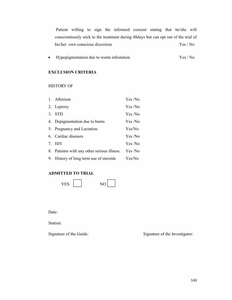
143
Patient willing to sign the informed consent stating that he/she will
conscientiously stick to the treatment during 48days but can opt out of the trial of
his/her own conscious discretion Yes / No
Hypopigmentation due to worm infestation. Yes / No
EXCLUSION CRITERIA
HISTORY OF
1. Albinism Yes /No
2. Leprosy Yes /No
3. STD Yes /No
4. Depigmentation due to burns Yes /No
5. Pregnancy and Lactation Yes/No
6. Cardiac diseases Yes /No
7. HIV Yes /No
8. Patients with any other serious illness. Yes /No
9. History of long term use of steroids Yes/No
ADMITTED TO TRIAL
YES NO
Date:
Station:
Signature of the Guide: Signature of the Investigator:

144
GOVERNMENT SIDDHA MEDICAL COLLEGEARIGNAR ANNA GOVERNMENT HOSPITAL OF INDIAN MEDICINE
CHENNAI – 600 106
POST - GRADUATE- DEPARTMENT OF SIRAPPU MARUTHUVAM
AN OPEN COMPARATIVE CLINICAL TRIAL ON VENPULLI
(VITILIGO) WITH THE EVALUATION OF SIDDHA TRIAL DRUG “RASA
CHENDHURAM” (INT) AND “PALAGARAI KUZHAMBU” (EXT)
FORM II – HISTORY TAKING PROFORMA
1. SERIAL NO OF THE CASE: ………………… 2.OP NO: ..........................
3. NAME: …………………………… 4. AGE: ……… 5.GENDER: .................
6. OCCUPATION: ……………………………… 7. INCOME: …………………..
8. COMPLAINTS & DURATION:
7. HABITS OF
SMOKING, YES / NO If yes, specify duration ---------- yrs
TOBACCO, YES / NO If yes, specify duration ---------- yrs
ALCOHOL, YES / NO If yes, specify duration ---------- yrs
8. DRUG HISTORY:
9. FAMILY HISTORY: Whether this problem runs in family? 1. Yes 2.No
If yes, mention the relationship of affected person(s)
1._________________ 2._________________
10.DIETARY HABIT: 1.Vegetarian 2.Non-vegetarian
11. MENSTRUAL HISTORY:
Date:
Station:
Signature of the Investigator: Signature of the Guide:

145
GOVERNMENT SIDDHA MEDICAL COLLEGEARIGNAR ANNA GOVERNMENT HOSPITAL OF INDIAN MEDICINE
CHENNAI – 600 106.
POST - GRADUATE- DEPARTMENT OF SIRAPPU MARUTHUVAM
AN OPEN COMPARATIVE CLINICAL TRIAL ON VENPULLI(VITILIGO) WITH THE EVALUATION OF SIDDHA TRIAL DRUG “RASA
CHENDHURAM” (INT) AND“PALAGARAI KUZHAMBU”(EXT)
FORM III – CLINICAL ASSESSMENT PROFORMA
1.OP NO: ----------- 2.SERIAL NO: ------
3. NAME: ---------------------- 4. AGE: --------- 5.GENDER: ------
6. DATE OF INITIAL ASSESSMENT: ----------------------
7. GENERAL EXAMINATION:
1. Body weight [Kg] :
2. Height [cm] :
3. Body Temperature [F] :
4. Blood Pressure (mmHg) :
5. Pulse Rate /min. :
6. Heart Rate / min. :
7. Respiratory Rate /min. :
Yes No
8. Pallor :
9. Jaundice :
10. Clubbing :
11. Cyanosis :
12. Pedal Oedema :
13. Lymphadenopathy :
14. Jugular venous pulsation :
8. SYSTEMIC EXAMINATION:
Nervous system -----------------------
cardiovascular system ----------------------
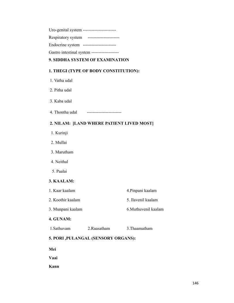
146
Uro-genital system -----------------------
Respiratory system ----------------------
Endocrine system -----------------------
Gastro intestinal system -------------------
9. SIDDHA SYSTEM OF EXAMINATION
1. THEGI (TYPE OF BODY CONSTITUTION):
1. Vatha udal
2. Pitha udal
3. Kaba udal
4. Thontha udal ------------------------
2. NILAM: [LAND WHERE PATIENT LIVED MOST]
1. Kurinji
2. Mullai
3. Marutham
4. Neithal
5. Paalai
3. KAALAM:
1. Kaar kaalam 4.Pinpani kaalam
2. Koothir kaalam 5. Ilavenil kaalam
3. Munpani kaalam 6.Muthuvenil kaalam
4. GUNAM:
1.Sathuvam 2.Raasatham 3.Thaamatham
5. PORI ,PULANGAL (SENSORY ORGANS):
Mei
Vaai
Kann

147
Mooku
Sevi
6. KANMENDHIRIYAM (MOTOR ORGANS):
Kai
Kaal
Vaai
Eruvaai
Karuvaai
7. KOSANGAL (SHEATH):
Annamaya kosam
Pranamaya kosam
Manomaya kosam
Vignana maya kosam
Anandamaya kosam
8. UYIR THAATHUKKAL: [THREE HUMORS] (VALI, AZHAL,IYAM)
A) VALI
Praanan –--------------------
Abaanan --------------------
Samaanan ---------------------
Uthaanan ------------------------
Vyanan -------------------------

148
Naagan ---------------------------
Koorman -----------------------------
Kirukaran ----------------------------
Devathathan ----------------------------
Dhananjayan ----------------------------
B) AZHAL
Aamalakam --------------------
Ranjakam ---------------------
Saathakam -----------------------
Prasakam ------------------------
Aalosakam --------------------------
C) IYAM
Avalambagam ------------------------
Kilethagam ---------------------------
Pothagam ------------------------------
Tharpagam ----------------------------
Santhigam -----------------------------
9.SEVEN UDAL DHATHUS: (7 SOMATIC COMPONENTS)
Saaram
Sennee
Oon
Kozhuppu
Enbu
Moolai
Sukkilam/Suronitham

149
SIDDHA SYSTEM OF EXAMINATION
1. ENVAGAI THERVU: [EIGHT TYPES OF EXAMINATION]
I. NAADI: [PULSE PERCEPTION]
II .SPARISAM:
III. NAA: [TONGUE]
IV.NIRAM: [COMPLEXION]
1. Vadham 2. Pitham 3. Kabam
V.MOZHI: [VOICE]
1. High Pitched
2. Low Pitched
3. Medium Pitched
VI.VIZHI: [EYES]
VII. MALAM: [BOWEL HABITS / STOOLS]
Niram
Irugal
Ilagal
Others
VIII. MOOTHIRAM [URINE EXAMINATION]
NEERKKURI:
Niram

150
Manam
Edai
Nurai
Enjal
NEIKKURI
Date:
Station:
Signature of the Guide: Signature of the Investigator:
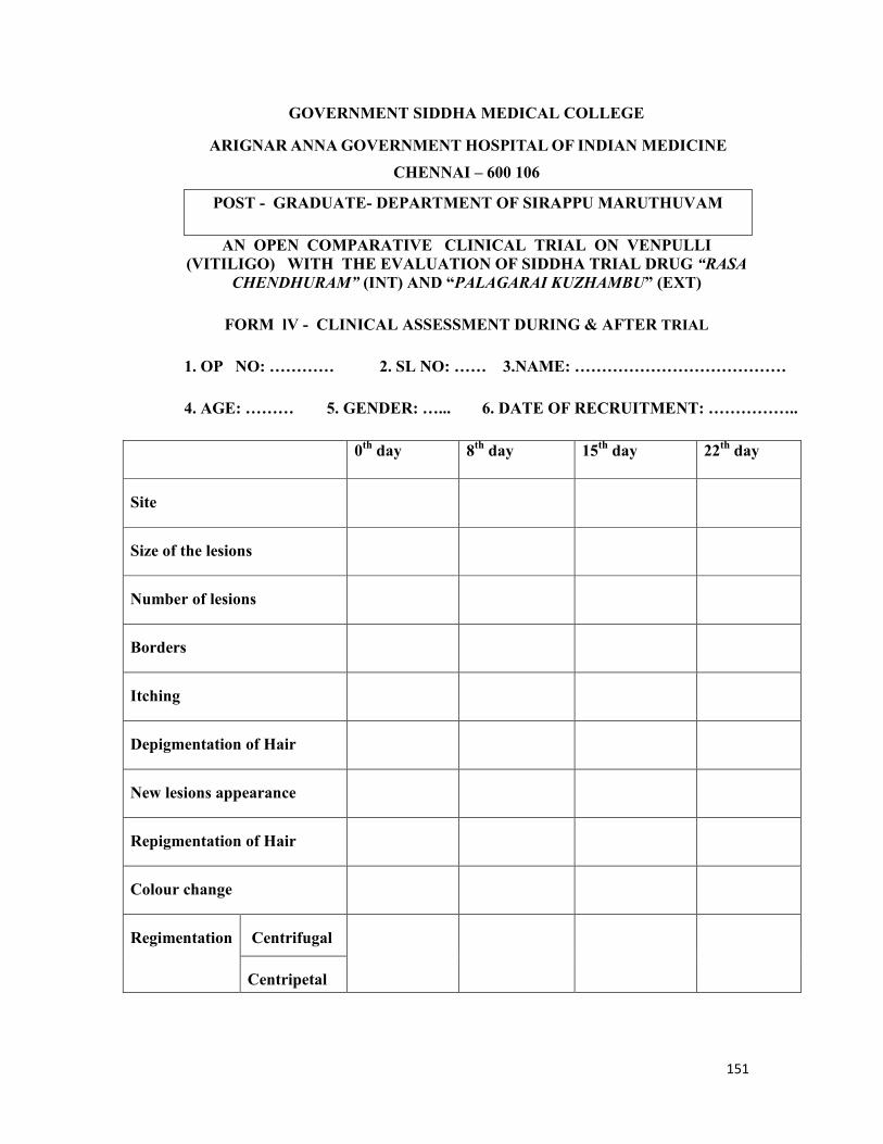
151
GOVERNMENT SIDDHA MEDICAL COLLEGE
ARIGNAR ANNA GOVERNMENT HOSPITAL OF INDIAN MEDICINECHENNAI – 600 106
POST - GRADUATE- DEPARTMENT OF SIRAPPU MARUTHUVAM
AN OPEN COMPARATIVE CLINICAL TRIAL ON VENPULLI(VITILIGO) WITH THE EVALUATION OF SIDDHA TRIAL DRUG “RASA
CHENDHURAM” (INT) AND “PALAGARAI KUZHAMBU” (EXT)
FORM lV - CLINICAL ASSESSMENT DURING & AFTER TRIAL
1. OP NO: ………… 2. SL NO: …… 3.NAME: …………………………………
4. AGE: ……… 5. GENDER: …... 6. DATE OF RECRUITMENT: ……………..
0th day 8th day 15th day 22th day
Site
Size of the lesions
Number of lesions
Borders
Itching
Depigmentation of Hair
New lesions appearance
Repigmentation of Hair
Colour change
CentrifugalRegimentation
Centripetal

152
Date:
Station:
Signature of the Guide Signature of the Investigator
29th day 36th day 43th day 49th day
Site
Size of the lesions
Number of lesions
Borders
Itching
Depigmentation of Hair
New lesions appearance
Repigmentation of Hair
Colour change
CentrifugalRepigmentation
Centripetal

153
GOVERNMENT SIDDHA MEDICAL COLLEGEARIGNAR ANNA GOVERNMENT HOSPITAL OF INDIAN MEDICINE
CHENNAI – 600 106.
POST - GRADUATE- DEPARTMENT OF SIRAPPU MARUTHUVAM
AN OPEN COMPARATIVE CLINICAL TRIAL ON VENPULLI(VITILIGO) WITH THE EVALUATION OF SIDDHA TRIAL DRUG “RASA
CHENDHURAM” (INT) AND “PALAGARAI KUZHAMBU” (EXT)
FORM-V - LABORATORY INVESTIGATIONS
1. SERIAL NO OF THE CASE: ………………… 2.OP NO: ................................
3. NAME: …………………………… 4.AGE: …… 5.GENDER: .................
A) Blood Investigation
Blood Investigations Normal Values Before trmt(Date)
After trmt(Date)
Hb (gm/dl) M:1215/W:11.5-14
T.WBC (cells/Cu.mm)4000-11000
Polymorphs 40-75
Lymphocytes 20-40
Monocytes 2-10
Eosinophils 1-6
DIFFERENTIALCOUNT (%)
Basophils 0-1
T.RBC(million cells/Cu.mm) M:4.0-5.5
W:3.5-4.5
ESR(mm/hour) ½ hr.
1 hr.
M:6-12
W:7-18

154
Fasting 70-110Blood glucose(mg/dl) PP 80-140
Blood urea 16-50
RFT (mg/dl) Serumcreatinine 0.6-1.2
Total bilirubin 0.2-1.2
Direct bilirubin 0.1-1.2
Indirectbilirubin 0.2-0.7
SGOT 0-40
SGPT 0-35
LFT (mg/dl)
Alkalinephosphatase 80-290
URINE INVESTIGATION
Urine investigation Before treatment After treatment
Albumin
Fasting sugar
PP sugar
Date:
Station:
Signature of the Guide: Signature of the Investigator

155
GOVERNMENT SIDDHA MEDICAL COLLEGE ARIGNAR ANNA GOVERNMENT HOSPITAL OF INDIAN MEDICINE
CHENNAI – 600 106POST - GRADUATE- DEPARTMENT OF SIRAPPU MARUTHUVAM
AN OPEN COMPARATIVE CLINICAL TRIAL ON VENPULLI(VITILIGO) WITH THE EVALUATION OF SIDDHA TRIAL DRUG“RASA CHENDHURAM” (INT) AND “PALAGARAI KUZHAMBU”(EXT)
FORM VI-INFORMED CONSENT FORM
“I have read the foregoing information, or it has been read to me. I have
had the opportunity to ask questions about it and any questions I have asked have
been answered to my satisfaction.
I consent voluntarily to participate in this study and understand that I have
the right to withdraw from the study at any time without in any way it affecting my
further medical care”.
"I have received a copy of the information sheet/consent form".
Date:
Signature of the participant
In case of illiterate participant
“I have witnessed the accurate reading of the consent form to the potential
participant, and the individual has had the opportunity to ask questions. I confirm
that the individual has given consent freely.”
Date:
Signature of a witness Left thumb Impression of the Participant
(Selected by the participant bearing no connection with the survey team)
Date:Station:
Signature of participant:
Signature of the Guide: Signature of the Investigator

156
, -106.
(
)
.
அ .
ேததி :
:
: :
157
.
.
.
( ) ( )
.
ேததி : :
: :
ேததி : :
: :
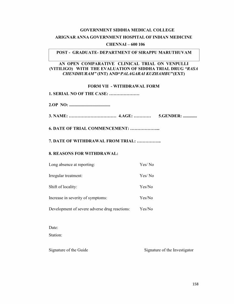
158
GOVERNMENT SIDDHA MEDICAL COLLEGEARIGNAR ANNA GOVERNMENT HOSPITAL OF INDIAN MEDICINE
CHENNAI – 600 106
POST - GRADUATE- DEPARTMENT OF SIRAPPU MARUTHUVAM
AN OPEN COMPARATIVE CLINICAL TRIAL ON VENPULLI(VITILIGO) WITH THE EVALUATION OF SIDDHA TRIAL DRUG “RASA
CHENDHURAM” (INT) AND“PALAGARAI KUZHAMBU”(EXT)
FORM VII - WITHDRAWAL FORM
1. SERIAL NO OF THE CASE: …………………
2.OP NO: ......................................
3. NAME: …………………………… 4.AGE: ………… 5.GENDER: .............
6. DATE OF TRIAL COMMENCEMENT: ………………...
7. DATE OF WITHDRAWAL FROM TRIAL: ……………..
8. REASONS FOR WITHDRAWAL:
Long absence at reporting: Yes/ No
Irregular treatment: Yes/ No
Shift of locality: Yes/No
Increase in severity of symptoms: Yes/No
Development of severe adverse drug reactions: Yes/No
Date:
Station:
Signature of the Guide Signature of the Investigator

159
GOVERNMENT SIDDHA MEDICAL COLLEGEARIGNAR ANNA GOVERNMENT HOSPITAL OF INDIAN MEDICINE
CHENNAI – 600 106.
POST - GRADUATE- DEPARTMENT OF SIRAPPU MARUTHUVAM
AN OPEN COMPARATIVE CLINICAL TRIAL ON VENPULLI
(VITILIGO) WITH THE EVALUATION OF SIDDHA TRIAL DRUG
“RASA CHENDHURAM” (INT) AND “PALAGARAI KUZHAMBU” (EXT)
FORM VIII– PATIENT INFORMATION SHEET
Name of the principal Investigator: Dr.R.Kalaimani
Name of the institute: Government siddha medical college,Arumbakkam
Chennai - 106INFORMATION SHEET FOR PATIENTS PARTICIPATING IN THE OPEN
CLINICAL TRIAL.
I ,Dr . R.Kalaimani. studying MD (Siddha) at Government Siddha Medical College,
Chennai, is doing a trial on the study VENPULLI (VITILIGO). Vitiligo is a common
persistent skin disease occurring through the world.
In this regard, I am in a need to ask you a few questions. I will maintain
confidentiality of your comments and data obtained. There will be no risk of
disclosing your identity and no physical, psychological or professional risk is
involved by taking part in this study.
Taking part in this study is voluntary. No compensation will be paid to you for taking
part in this study.
You can choose not to take part. You can choose not to answer a specific question.
There is no specific benefit for you if you take part in the study. However, taking part
in the study may be of benefit to the community, as it may help us to understand the
problem of defaulters and potential solutions.
If you agree to be a participant in this study, you will be included in the study
primarily by signing the consent form and then you will be given the internal
medicine Rasa Chendhuram (Internal Medicine- 65 mg BD with palm jaggery for 48

160
days) and Palagarai Kuzhambu (External Medicine), assuring that you will not be
definitely hurt during the course of treatment.
The information I am collecting in this study will remain confidential. I will
ask you few questions through a questionnaire. I will not write your name on this
form. I will use a code instead.
The questionnaire will take approximately 20 minutes of your time.
If you wish to find out more about this study before taking part, you can ask
me all the questions you want or contact Dr.R.KALAI MANI , PG Scholar cum
principal investigator of this study, attached to Government siddha medical college,
Arumbakkam, Chennai – 106. You can also contact the Member-secretary of Ethics
committee, , Govt. Siddha Medical College, Chennai,

161
, -106.
(
).
: . இரா.: ,
-106.
.இரா.
.
., , ,
, .
..
,.
.
65மி.கி , பைன , 2 ேவைள (காைல, மாைல)
(48 ). 48

162
.7
.
.
,,
: இரா. ( ,) . 7845949200.
IEC .
சி .
.
,,
.

163
GOVERNMENT SIDDHA MEDICAL COLLEGE ARIGNAR ANNA GOVERNMENT HOSPITAL OF INDIAN MEDICINE
CHENNAI – 600 106
POST - GRADUATE- DEPARTMENT OF SIRAPPU MARUTHUVAM
AN OPEN COMPARATIVE CLINICAL TRIAL ON VENPULLI(VITILIGO) WITH THE EVALUATION OF SIDDHA TRIAL DRUG“RASA CHENDHURAM” (INT) AND “PALAGARAI KUZHAMBU”(EXT)
FORM IX - ADVERSE REACTION FORM
SERIAL NO:
OP NO:
NAME:
AGE:
GENDER:
DATE OF TRIAL COMMENCEMENT:
DATE OF OCCURRENCE OF THE ADVERSE REACTION:
TIME:
DESCRIPTION OF ADVERSEREACTION:
Date:
Station:
Signature of the Guide: Signature of the Investigator:

164
BIBLIOGRAPY
1.R.C. Mohan, AgathyarVaithyaVallathi 600, First Edition, April 2001,
2., MuthuKarupuPillai , Sri Vasi Villa Publication , ThanvanthiriVaithyam .
3.Ramachandran, AgathyarKanmaKaandamKowmathiNool, ThamaraiNoolagam,Second Edition – 2009.
4.R.C.Mohan, ThirumoolarKarukkidaiVaithyam- 600, thamarainoolagam 3 rdedition.,2012.
5.Dr. S.Venkatraman , ThanvanthiriVaithyaNool, 2 NdVolume,ThamaraiNoolagam.1990.
6., S.B. Ramachandran ,YugiVaithyaChinthamani Second Edition ,ThamaraiNoolagam, 2005.
7.PathinenSiddharNaadiNool
8.Sababathy, Anbuselvi,Publisher, AathmaRatchamirthaVaithyaSaaraSangiragam,First Edition.
9. I.Ponnaiyan ,Pararasasekaram, Thirugnanasambandhar Printers ,1935 , 4 Thpart,
10.MaathavaNithaanam
11.PulippaniVaithyam 700
12. Dr.R. Thyagarajan L.I.M , GunapadamThaathuSeevaVaguppu, Indian MedicineAnd Homeopathy Department, 2004 .
13.SiddhaMaruthuvamSirappu , Dr.R. Thyagarajan L.I.M , Indian Medicine AndHomeopathy Department.
14. Dr. K.S. Uthamarayan , H.B.I.M. SiddharAruvaiMaruthuvam , Indian MedicineAnd Homeopathy Department, Sixth Edition ,2013.
15. Dr. M .ShanmugaVelu, H.B.I.M, NoiNadalNoiMudhalNadalThirattu, Part -1,Indian Medicine And Homeopathy,Fourth Edition.2006.
15. K.S. MurugesaMudhaliar ,GunapadamMooligai , Indian Medicine AndHomeopathy Department.
16. Dr. K. Radhakrishnan L.I.M., AnubavaVaidhya Deva Ragasiyam,RathnaNaayakar And Sons.
17. Kandasamy Pillai, History Of Siddha Medicine, Indian Medicine And
Homeopathy, Second Edition, 2008.

165
18.Neenakhanna, Illustrated synopsis of Dermatology and sexually transmitted
diseases, Fifth edition, 2016
19.paul k buxton, ABC of dermatology,fourth edition.,2003.
20. article from east west school for herbal and aromatic studies, anatomy and
physiology of skin
21.article from researchgate, vitiligo,2015
22. Dr. K. M. Nadkarni, Indian material medica, Volume -1, Indian medicine and
homeopathy, 1968.
Copyright © 2022 FDOKUMEN


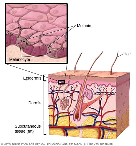