An fMRI study of long-term everyday memory using SenseCam
13
PLEASE SCROLL DOWN FOR ARTICLE This article was downloaded by: [University College London] On: 9 May 2011 Access details: Access Details: [subscription number 936074359] Publisher Psychology Press Informa Ltd Registered in England and Wales Registered Number: 1072954 Registered office: Mortimer House, 37- 41 Mortimer Street, London W1T 3JH, UK Memory Publication details, including instructions for authors and subscription information: http://www.informaworld.com/smpp/title~content=t713683358 An fMRI study of long-term everyday memory using SenseCam F. Milton a ; N. Muhlert b ; C. R. Butler c ; A. Smith b ; A. Benattayallah d ; A. Z. Zeman b a Department of Psychology, University of Exeter, Exeter, UK b Department of Neurology, Peninsula Medical School, University of Exeter, Exeter, UK c Department of Clinical Neurology, University of Oxford, Oxford, UK d Peninsula MR Research Centre, Peninsula Medical School, Exeter, UK First published on: 14 March 2011 To cite this Article Milton, F. , Muhlert, N. , Butler, C. R. , Smith, A. , Benattayallah, A. and Zeman, A. Z.(2011) 'An fMRI study of long-term everyday memory using SenseCam', Memory,, First published on: 14 March 2011 (iFirst) To link to this Article: DOI: 10.1080/09658211.2011.552185 URL: http://dx.doi.org/10.1080/09658211.2011.552185 Full terms and conditions of use: http://www.informaworld.com/terms-and-conditions-of-access.pdf This article may be used for research, teaching and private study purposes. Any substantial or systematic reproduction, re-distribution, re-selling, loan or sub-licensing, systematic supply or distribution in any form to anyone is expressly forbidden. The publisher does not give any warranty express or implied or make any representation that the contents will be complete or accurate or up to date. The accuracy of any instructions, formulae and drug doses should be independently verified with primary sources. The publisher shall not be liable for any loss, actions, claims, proceedings, demand or costs or damages whatsoever or howsoever caused arising directly or indirectly in connection with or arising out of the use of this material.
-
Upload
manchester -
Category
Documents
-
view
3 -
download
0
Transcript of An fMRI study of long-term everyday memory using SenseCam

PLEASE SCROLL DOWN FOR ARTICLE
This article was downloaded by: [University College London]On: 9 May 2011Access details: Access Details: [subscription number 936074359]Publisher Psychology PressInforma Ltd Registered in England and Wales Registered Number: 1072954 Registered office: Mortimer House, 37-41 Mortimer Street, London W1T 3JH, UK
MemoryPublication details, including instructions for authors and subscription information:http://www.informaworld.com/smpp/title~content=t713683358
An fMRI study of long-term everyday memory using SenseCamF. Miltona; N. Muhlertb; C. R. Butlerc; A. Smithb; A. Benattayallahd; A. Z. Zemanb
a Department of Psychology, University of Exeter, Exeter, UK b Department of Neurology, PeninsulaMedical School, University of Exeter, Exeter, UK c Department of Clinical Neurology, University ofOxford, Oxford, UK d Peninsula MR Research Centre, Peninsula Medical School, Exeter, UK
First published on: 14 March 2011
To cite this Article Milton, F. , Muhlert, N. , Butler, C. R. , Smith, A. , Benattayallah, A. and Zeman, A. Z.(2011) 'An fMRIstudy of long-term everyday memory using SenseCam', Memory,, First published on: 14 March 2011 (iFirst)To link to this Article: DOI: 10.1080/09658211.2011.552185URL: http://dx.doi.org/10.1080/09658211.2011.552185
Full terms and conditions of use: http://www.informaworld.com/terms-and-conditions-of-access.pdf
This article may be used for research, teaching and private study purposes. Any substantial orsystematic reproduction, re-distribution, re-selling, loan or sub-licensing, systematic supply ordistribution in any form to anyone is expressly forbidden.
The publisher does not give any warranty express or implied or make any representation that the contentswill be complete or accurate or up to date. The accuracy of any instructions, formulae and drug dosesshould be independently verified with primary sources. The publisher shall not be liable for any loss,actions, claims, proceedings, demand or costs or damages whatsoever or howsoever caused arising directlyor indirectly in connection with or arising out of the use of this material.

An fMRI study of long-term everyday memoryusing SenseCam
F. Milton1, N. Muhlert2, C. R. Butler3, A. Smith2, A. Benattayallah4, and A. Z. Zeman2
1Department of Psychology, University of Exeter, Exeter, UK2Department of Neurology, Peninsula Medical School, University of Exeter, Exeter, UK3Department of Clinical Neurology, University of Oxford, Oxford, UK4Peninsula MR Research Centre, Peninsula Medical School, Exeter, UK
We used a novel automatic camera, SenseCam, to investigate recognition memory for real-life events at a5-month retention interval. Using fMRI we assessed recollection and familiarity memory using theremember/know procedure. Recollection evoked no medial temporal lobe (MTL) activation comparedto familiarity and new responses. Instead, recollection activated diverse regions in neocortex includingmedial prefrontal cortex. We observed decreased activation in anterior hippocampus/ anteriorparahippocampal gyrus (aPHG) at 5 months compared to a 36-hour retention interval. Familiarity wasassociated with greater activation in aPHG and posterior parahippocampal gyrus (pPHG) thanrecollection and new responses. Familiarity activation decreased over time in anterior hippocampus/aPHG and posterior hippocampus/pPHG. The engagement of neocortical regions such as medialprefrontal cortex at a 5-month delay, together with the reduced MTL activation at 5 months relative to at36 hours is in line with the assumptions of Consolidation theory. SenseCam provides a valuable techniquefor assessing the processes that underlie remote everyday recognition memory.
Keywords: fMRI; Recognition memory; SenseCam; Recollection; Familiarity; Consolidation.
Recognition memory is often believed to be the
result of two distinct processes: recollection, which
supports the ability to recognise an item based on
contextual information; and familiarity, the ability
to recognise an item based on memory strength in
the absence of contextual details (Mandler, 1980).
An alternative view proposes that, rather than
reflecting distinct underlying processes, familiar-
ity and recollection differ in the strength of a
common memory signal (Dunn, 2004; Kirwan,
Wixted, & Squire, 2008).There has recently been intense interest in the
neural correlates of recognition memory. These
studies have typically used laboratory stimuli
such as words (Eldridge, Knowlton, Furmanski,
Bookheimer, & Engel, 2000), objects (Vilberg &
Rugg, 2007), and visual scenes (Montaldi, Spencer,
Roberts, & Mayes, 2006). Several approaches have
been taken to distinguish the neural correlates of
recollection and familiarity including confidence
ratings, associative recognition, source memory,
and the remember-know paradigm (for a review,
see Yonelinas, 2002).Recollection memory has been associated with
increased hippocampal and posterior parahippo-
campal gyrus (pPHG) activity compared to famil-
iarity (e.g., Eldridge et al., 2000; Wheeler &
Buckner, 2004). Meanwhile, familiarity is typi-
cally linked to perirhinal and anterior parahippo-
campal regions (aPHG; e.g., Henson, Cansino,
Address correspondence to: F. Milton, Washington Singer Laboratories, Perry Road, Exeter, EX4 4QG UK. E-mail:
This research was supported by grants from the Great Western Research Initiative and Microsoft Research.
MEMORY, 0000, 00 (00), 1�12
# 2011 Psychology Press, an imprint of the Taylor & Francis Group, an Informa businesshttp://www.psypress.com/memory DOI:10.1080/09658211.2011.552185
Downloaded By: [University College London] At: 14:38 9 May 2011
Herron, Robb, & Rugg, 2003). These regionalactivations broadly support dual-process accountsof recognition memory (e.g., Aggleton & Brown,1999; Diana, Yonelinas, & Ranganath, 2007),which propose that recollection is supported bythe hippocampus, whereas familiarity is supportedby aPHG, including perirhinal cortex.
Most recognition studies have used shortretrieval delays (i.e., less than 1 day). However,in our everyday environment we are often re-quired to recognise items that have not been seenfor a considerable time. It is therefore importantto understand the neural substrates of recognitionmemory for items encountered in the remotepast. The extent to which the hippocampus andother medial temporal lobe (MTL) structures areinvolved in declarative memory processes such asrecognition memory has been the subject ofconsiderable debate. One theory, known as thestandard consolidation model (Squire, Stark, &Clark, 2004) posits that the hippocampus has atime dependent role in declarative memory.Initially, memory retrieval is believed to bedependent on the hippocampus, but over timememory traces are reorganised to become lessdependent on the hippocampus, allowing retrievalfrom distributed neocortical structures alone. Incontrast, according to multiple trace theory(MTT; Nadel & Moscovitch, 1997), the hippo-campus is involved in retrieval of episodic mem-ories throughout the lifetime. With time,activation in the hippocampus should remain asgreat or even increase due to the laying down ofseparate (multiple) traces each time a memory isretrieved.
Several recent fMRI studies have used aprospective recognition memory paradigm toinvestigate long-term recognition memory pro-cesses. For instance, Viskontas, Carr, Engel, andKnowlton (2009) used high-resolution scanning ofthe MTL to reveal that 1 week after initialencoding the subiculum was involved in recollec-tion memory. Similarly, Bosshardt et al. (2005)found greater activation in bilateral hippocampuswhen viewing stimuli learned 1 month beforescanning compared to stimuli learned 1 daybefore. Relatedly, Suchan, Gayk, Schmid, Koster,and Daum (2008) used the remember/knowprocedure to measure recognition memory overa period of 6 weeks. For remember responses theyfound consistent hippocampal activity over time.Familiarity responses resulted in decreased peri-rhinal cortex activation over time. Nevertheless,Suchan et al. (2008) argued that familiarity
activation remained relatively stable over time,suggesting it lasts at least up to 6 weeks afterencoding. Both Bosshardt et al. (2005) andSuchan et al. (2008) argued that their findingsprovide support for MTT, although it is possiblethat their time delays were insufficient for con-solidation effects in MTL to be observable.
Dolcos, LaBar, and Cabeza (2005) used alonger retrieval delay, testing recognition memoryafter a 1-year interval. While their interest wasprimarily on the effect emotion has on long-termrecognition memory, some of their findings arerelevant to the current issue. In particular, theyfound that, after a 1-year delay, there was greateractivation in the head of right hippocampus andbilaterally in the hippocampus tail for remembercompared to know responses.
While the majority of prospective recognitionmemory studies have observed hippocampal ac-tivity at long retrieval delays, Takashima et al.(2006) found somewhat discrepant results. Theytested recognition memory at four time periodsranging from hours to 3 months after encoding.They found that hippocampal activation de-creased over time and linked this to memoryconsolidation.
Evidence from retrospective neuroimagingstudies of remote autobiographical memory isrelated to the current issue (for a review seeSvoboda, McKinnon, & Levine, 2006) and alsoshows discrepancies between studies. A numberof these studies have found no effect of the age ofmemories on hippocampus activity (Ryan et al.,2001; Steinvorth, Corkin, & Halgren, 2006), whileothers have found increased hippocampal activityfor remote compared to recent memories (e.g.,Rekkas & Constable, 2005). However, severalstudies have observed a decrease in hippocampusactivity over time suggesting disengagement ofthis region from long-term memory processes(Niki & Luo, 2002; Piefke et al., 2003).
Outside the MTL several regions have beenlinked with long-term recognition memory. Inparticular, the ventrolateral (Bosshardt et al.,2005) and medial prefrontal cortices (mPFC;Dolcos et al., 2005; Takashima et al., 2006)consistently activate at long time intervals. Onesuggestion is that mPFC, rather than the hippo-campus, may integrate information from multipleneocortical regions during remote memory retrie-val (Takashima et al., 2006). Additionally, long-term recognition memory often engages lateraltemporal lobes (e.g., Bosshardt et al., 2005; Dolcoset al., 2005). Retrospective autobiographical
2 MILTON ET AL.
Downloaded By: [University College London] At: 14:38 9 May 2011
memory studies also consistently activate prefron-tal cortex (e.g., Addis, Moscovitch, & McAndrews,2004; Maguire & Frith, 2003) and lateral temporalcortex (e.g., Gilboa, Winocur, Grady, Hevenor, &Moscovitch, 2004; Ryan et al., 2001).
The prospective recognition memory studiesdiscussed, while varying in the time delay, all usedlaboratory-based stimuli. Indeed there has been adearth of studies investigating everyday recogni-tion processes (although see Cabeza et al., 2004).In this study we used a novel wearable camera,SenseCam, to acquire images of everyday eventsand gave participants a recognition test after a 5-month interval. SenseCam, which is activated by arange of environmental sensors taking an averageof one image every 30 seconds without userinvolvement, appears to be an ideal techniqueto investigate recognition memory. First, theautomatic capture of images is methodologicallyadvantageous because it reduces the intentionalencoding of the items that will be tested later.This paradigm therefore allows more direct in-sight into the neural substrates of everydayrecognition memory than previous work. Second,the complexity of the stimuli and their personalnature make them ideally suited to investigaterecognition over lengthy delays because they arelikely to minimise the risk of accuracy flooreffects. In addition, the level of contextual detailavailable is likely to vary between images, permit-ting exploration of remote recollection and famil-iarity memory.
In summary this is, as far as we are aware, thefirst study to examine the neural correlates ofrecollection and familiarity memory for remoteeveryday events. In particular, our study assessedMTL involvement in recollection and familiarityprocesses over time. Additionally, we examinedwhether neocortical regions such as prefrontalcortex and lateral temporal lobes are involved inremote everyday recognition memory.
METHOD
Participants
A total of 10 right-handed participants from theUniversity of Exeter (6 male, 4 female), rangingin age from 18 to 25 years, took part in the study.Participants were paid £15 for their participation.Participants gave informed consent according toprocedures approved by the Psychology EthicsCommittee, University of Exeter.
fMRI data acquisition
Structural and functional imaging was performedwith a 1.5-T Phillips Gyroscan magnet equippedwith a Sense coil. A T2*-weighted echo planarsequence was used (TR �3000ms, Te �45ms, flipangle �908, 32 transverse slices, 3.5�2.5�2.5mm ascending acquisition). A total of 280 vo-lumes were acquired in each of the two runs perparticipant. An additional 5 ‘‘dummy’’ scans wereperformed before each run prior to the stimulussequence. A T1-weighted structural MRI scan wasalso acquired (Tr �25ms, Te �4.1ms, flip an-gle �308, 160 axial slices, 1.6�0.9�0.9 mm).
Equipment
The SenseCam (sized 6.5cm wide�7cm high�1.5 cm long) is built around a PIC 18F8722 6MIPS microcontroller with 128KB of flash mem-ory (Hodges et al., 2006). SenseCam is wornround the neck, with pictures captured using afish eye lens. This maximises the field of view andensures that objects at head height are photo-graphed. Images were captured automaticallyapproximately every 30 seconds.
Procedure
Participants wore a SenseCam for two days whileundertaking their everyday activities. Participantsperformed an initial recognition memory taskinside the scanner approximately 36 hours andthen again approximately 5 months after acquisi-tion of the images. We chose an interval of5 months in order to extend the results of previousstudies of remote recognition memory, which havetypically used shorter delays than this (e.g.,Bosshardt et al., 2005; Suchan et al., 2008; Taka-shima et al., 2006; Viskontas et al., 2009; althoughsee Dolcos et al., 2005) while keeping the eventssufficiently recent for participants to be able torecognise them with a good degree of accuracy.The procedure for the two recognition memorysessions was identical and the images used werethe same for both retrieval sessions. This paperfocuses on the long delay condition (5 months) butuses the short delay condition (36 hours) toinvestigate how the neural basis of everydaymemory changes over time. Results from the shortdelay condition will be presented in a forthcoming
LONG-TERM EVERYDAY RECOGNITION 3
Downloaded By: [University College London] At: 14:38 9 May 2011
paper. Participants were not informed of the longdelay session until 4 months after the first session.
Before each scanning session participants weretrained to distinguish between different types ofrecognition memory. Participants saw a series ofimages, some of which were theirs (old), and therest were images acquired by other participants(new), which had the same general characteristics(e.g., similar types of event, brightness). Partici-pants were asked to report whether images were‘‘remembered’’ or ‘‘known’’ (Tulving, 1985). Theinstructions outlining this distinction were mod-elled on those of Rotello, Macmillan, and Reeder(2004; see also Rajaram, 1993). Specifically, ‘‘re-membering’’ was defined as the ability to beconsciously aware of some aspect or aspects ofwhat happened or what was experienced at thetime the photo was taken (e.g., what one wasthinking or doing at the time). ‘‘Knowing’’ wasdescribed as the feeling that the event was familiarwhile being unable to recollect anything about itsoccurrence or what was experienced at the time ofits occurrence. Participants were all able toverbalise this distinction. Participants completeda training session to further familiarise them withthese judgements. A total of 15 trials (10 ‘‘old’’,5 ‘‘new’’ images) were randomly presented.
Inside the scanner, images were presented on aback-projection screen positioned at the foot endof the scanner and viewed via a mirror mountedon a head coil. Recognition judgements weremeasured using two fibreoptic button boxes heldin the participants’ right and left hands. E-Prime(Psychology Software Tools, Inc., Pittsburgh.) wasused for the presentation and timing of stimuliand collection of response data.
There were 120 trials in the scanning session; 80‘‘old’’ and 40 ‘‘new’’1 images. The events recordedby participants were all characteristic of everydayexperiences and were evenly distributed through-out the acquisition period. Images were selected sothat unique events were presented. In other words,images were excluded if they displayed the same
activity as one of the previously selected images(e.g., looking at the same shelf in a shop, lookingout of a window, or waiting in a queue). There weresix blocks of 20 images and the presentation orderof the stimuli was randomised. The session wasdivided into two runs, each lasting 14 minutes, withthree blocks presented in each run. At the end ofeach of the six blocks a message appeared for 12seconds saying, ‘‘That is the end of the block.Please take a short break.’’ This was followed by amessage lasting 1500 ms instructing participants to‘‘Get ready to start the next block.’’ Participantswere also given the opportunity to rest for as longas they required between the two scanning runs.
Trials began with a blank screen lasting 250�3750 ms, followed by a fixation cross for 150 ms.An image was then presented for 4000 ms duringwhich time participants were asked not to re-spond. This was followed by instructions, whichremained on the screen for 3000 ms, askingparticipants to classify the image as ‘‘Remem-ber’’, ‘‘Know’’, or ‘‘New’’. If no response wasmade during this interval, a message appearedsaying, ‘‘Time out!!’’ ‘‘Remember’’ and ‘‘Know’’responses were made using the response box inone hand, ‘‘New’’ responses using the responsebox in the other hand. The hand corresponding tothese judgements was counterbalanced.
A further message then appeared which variedaccording to the judgement participants made.For ‘‘Remember’’ responses, participants had todistinguish between high and low level of detail.‘‘Know’’ images were to be divided into high andlow familiarity. These sub-categories were col-lapsed at analysis to improve statistical power.For ‘‘New’’ images, participants had to press anybutton. Participants had 3000 ms to respond; if noresponse was made in this time, the ‘‘Time out!!’’message appeared.
Immediately after the recognition task a post-scan interview was administered in which partici-pants were presented with four ‘‘remember’’ andfour ‘‘know’’ images. Participants described theirmemory for these events, which was scored by theexperimenter on a scale from 0 (no details) to 3(many details). Participants also rated events formemory strength on a scale of 1 (very vague) to 6(very clear).
Analysis of fMRI data
Image preprocessing and analyses were per-formed using SPM5 (www.fil.ion.ucl.ac.uk/spm).
1The ‘‘new’’ images are not in the strictest sense new but
were also encountered (for 4000 ms) in the 36-hour retrieval
session. We used the same stimuli in both sessions to ensure
that differences between old and new stimuli were unlikely to
be the result of exposure in the initial session rather than the
personal experiencing of the event itself. The crucial differ-
ence between old and new stimuli is therefore the personal
experience with the old stimuli but not with the new stimuli.
We used the term ‘‘new’’ to make instructions to participants
consistent across sessions; however, we accept that ‘‘lure’’ may
be a more adequate description for the foils.
4 MILTON ET AL.
Downloaded By: [University College London] At: 14:38 9 May 2011

Functional images were corrected for acquisitionorder, and realigned to correct for motion arte-facts. A mean image created from the realignedimages was coregistered with the structural T1volume and the structural volumes spatiallynormalised. The spatial transformation was ap-plied to the realigned T2* volumes which werespatially smoothed using a Gaussian kernel of 8-mm full-width half maximum. A high pass filter of128 s was applied. The BOLD response wasmodelled by a canonical haemodynamic responsefunction together with its temporal and dispersionderivatives. The six head movement parameterswere included as confounds. Onset of the imagewas considered the beginning of the trial. First-level linear contrasts of parameter estimates foreach voxel were generated and a random effectsanalysis performed.
Pairwise contrasts for memory types compris-ing at least eight events were conducted usingrepeated-measures t-tests with a significancethreshold of pB.001 (uncorrected) and a clusterthreshold of five contiguous voxels. Region ofinterest (ROI) analyses were conducted in bilat-eral MTL based on a priori hypothesis that thisregion is active during recognition memory. TheROI mask was applied using the WFU Pickatlas(Maldjian, Laurienti, Burdette, & Kraft, 2003).Coordinates were transformed from normalisedMNI space to Talairach space (http://imaging.mrc-cbu.cam.ac.uk/imaging/MniTalairach).
RESULTS
Behavioural analyses
Table 1 displays the mean proportions for remem-ber, know, misses, correct rejections (i.e., newstimuli that were correctly classified as new), andfalse alarms (i.e., new stimuli that were incorrectlyclassified as old) responses. A total of 2% of trialswere excluded from analyses because participants
failed to respond in time. All participants madesufficient responses of each type (at least eightevents) to be included in all the contrasts. Therelatively low number of incorrectly rejected olditems (18%) and incorrectly accepted new items(11%) precluded further analyses of these classi-fication types.
The post-scan ratings revealed that participantsrecalled more details for images classified asRemember (M�1.86, SD�.47) than those classi-fied as Know (M�.78, SD�.47), t(8) �6.5,pB.001. This occurred even though the post-scan test was self-paced, providing participantsmore opportunity to retrieve specific details forfamiliarity responses than in the scanner. Per-ceived memory strength was greater for Remem-ber (M�3.52, SD�1.08) than Know responses(M�1.72, SD�.61), t(8) �5.5, p �.001. Thesefindings suggest that participants could distinguishbetween recollection and familiarity processes.
Imaging analyses
MTL region of interest (ROI) analyses at 5months. The following analyses used a signifi-cance threshold of pB.01 (uncorrected) and acluster threshold of five contiguous voxels. Therewas no significant activation for Rememberrelative to New responses (i.e., correct rejections),while Know compared to New stimuli elicitedgreater activation in regions of right aPHG andbilateral pPHG (Figure 1a). A contrast comparingRemember to Know responses revealed no re-gions of significant activation, while comparingKnow with Remember yielded activation in twoclusters in right pPHG and one cluster in leftpPHG (Figure 1b; Table 2). New responsesproduced greater activation than Rememberresponses in right aPHG and bilateral pPHG(Figure 1c). The New � Know contrast (Figure1d) produced activation in two clusters in rightaPHG and right pPHG.
Extra-MTL analyses at 5 months. These ana-lyses used a significance threshold of pB.001 anda cluster threshold of five contiguous voxels. Therewas extensive extra-MTL activation in the Re-member�Know contrast (Table 2) comprisingactivation in left superior frontal gyrus, leftdorsolateral prefrontal cortex (DLPFC), rightmPFC, right thalamus, right precuneus, and leftinferior temporal cortex. The Know�Remembercontrast (Table 2) revealed activity in right insula,
TABLE 1
Proportion of recognition responses for old and new items
Old items New items
Remember Know Miss CR FA
Mean .27 .52 .18 .89 .11
SD .13 .14 .17 .09 .09
SD�Standard Deviation; CR�Correct rejection;
FA�False alarm.
LONG-TERM EVERYDAY RECOGNITION 5
Downloaded By: [University College London] At: 14:38 9 May 2011

right posterior cingulate, and right cerebellum.
Remember compared to New (Figure 2a) induced
greater activation in myriad regions including left
ventrolateral frontal cortex (VLFC), anterior
cingulate, cingulate gyrus, left inferior parietal
lobe, left superior temporal gyrus, and right
cerebellum. The Know�New contrast (Figure 2b)
evoked activation in bilateral mPFC, right VLFC,
bilateral insula, left inferior and left superior
temporal lobe together with middle occipital
gyrus. The New�Remember contrast (Figure 2c)included activation in mPFC, right middle frontalgyrus, bilateral insula, left cingulate gyrus andright precuneus. New compared to Know (Figure2d) elicited activation in left VLFC, left anteriorcingulate, right superior temporal gyrus, rightthalamus, right precuneus, and right cuneus.
Short versus long delay for MTL ROI analyses.These analyses used thresholds of pB.01 (uncor-rected) and five contiguous voxels. A contrast
Figure 1. Regions in MTL where activation was greater for (a) Know than New responses; (b) Know compared to Remember
responses; (c) New than Remember responses; (d) New than Know responses.
6 MILTON ET AL.
Downloaded By: [University College London] At: 14:38 9 May 2011
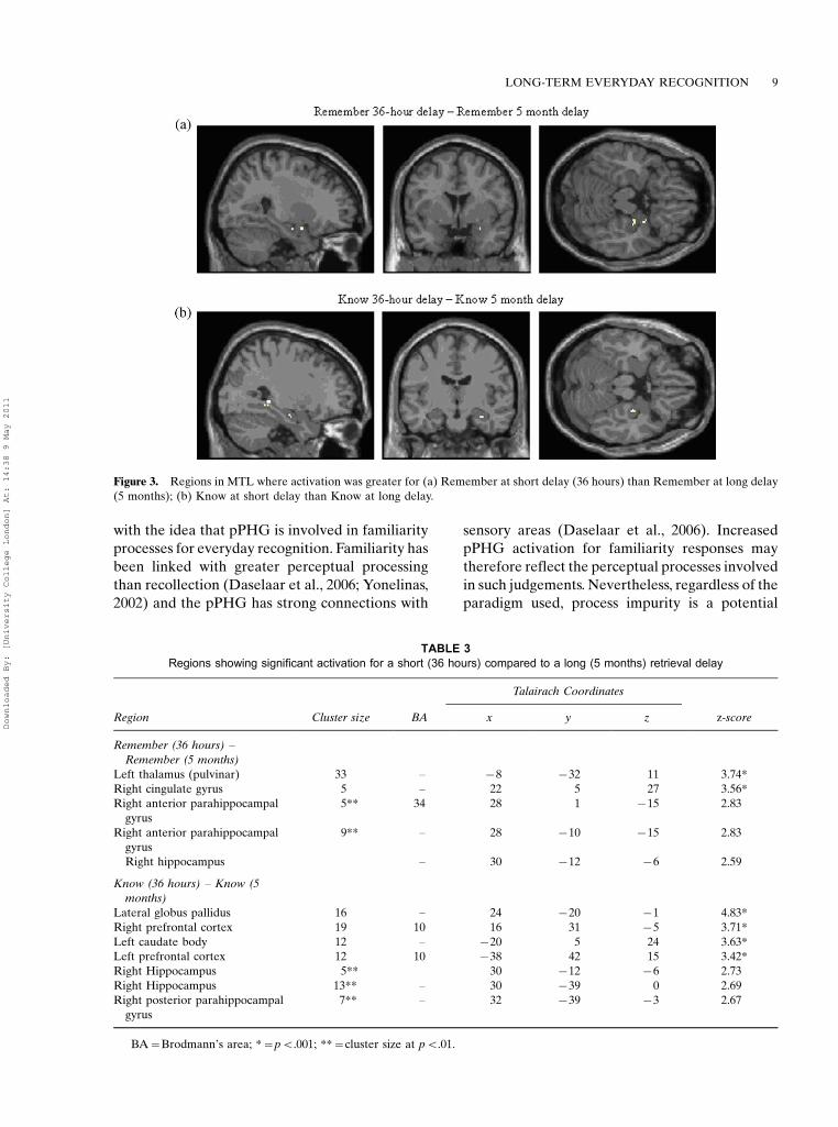
comparing Remember activity at the long delay (5
months) to the short delay (36 hours) found no
significant activation. Contrasting Remember
activity for the short delay to the long delay
(Figure 3a) yielded two clusters in right hippocam-
pus/aPHG.For familiarity, no regions produced more
activation over a long than a short delay. There
was greater activation in right anterior hippo-
campus/aPHG and right posterior hippocampus/
pPHG for Know responses at the short delay
relative to at the long delay (Figure 3b).
Short versus long delay for extra-MTLanalyses. These analyses used thresholds ofpB.001 and five contiguous voxels. No signifi-
cant recollection activation was observed at the
long relative to the short delay. Recollection at
the short delay compared to the long delay
induced activation in right cingulate gyrus and
left thalamus (Table 3).There was no significant familiarity-based
activation at the long compared to the short
delay. Meanwhile, there was greater familiarity
activation at the short relative to the long delay in
bilateral mPFC, the lateral globus pallidus and
left caudate body (Table 3).
DISCUSSION
This study combined the use of SenseCam withfMRI to identify the neural substrates associatedwith everyday recognition memory over a 5-monthretrieval delay. This condition was compared to anidentical recognition memory test conducted ap-proximately 36 hours after the event. We discussthe main findings of our experiment in turn.
Recollection elicits no activation in theMTL at a 5-month delay; there isfamiliarity activation in the MTL butthis is reduced over time
No MTL regions evoked greater activation forRemember compared to either Know or Newresponses. The lack of MTL activation for long-term recollection responses is also illustrated bycontrasting the 5-month to the 36-hours retrievalcondition. No regions were more active at 5months than at 36 hours but two regions in rightanterior hippocampus/aPHG activated more at36 hours than at 5 months.
There was greater MTL activation for Knowcompared to Remember responses in bilateral
TABLE 2
Regions showing significant activation for Remember compared with Know responses and for Know compared with Remember
responses
Talairach coordinates
Region Cluster size BA x y z z-score
Remember � Know
Right anterior cingulate 44 25 4 19 �4 4.19*
Right thalamus 26 � 12 �3 9 4.05*
Left superior frontal cortex 9 6 �18 28 54 3.91*
Right precuneus 42 31 2 �47 34 3.75*
Left cingulate gyrus 31 �8 �41 32 3.62*
Left dorsalateral frontal cortex 5 46 �48 28 24 3.68*
Right occipital lobe 7 19 48 �72 �3 3.56*
Right medial prefrontal cortex 6 10 2 52 1 3.19*
Know � Remember
Right posterior cingulate 46 29 18 �46 12 4.26*
Right insula 14 13 30 8 12 3.92*
Right cerebellum 26 � 16 �48 �18 3.68*
Right posterior parahippocampal
gyrus
46** 19 26 �53 �6 3.17*
Left posterior parahippocampal gyrus 47** 19 �22 �47 �6 2.99
Right posterior parahippocampal
gyrus
31** � 32 �26 �12 2.98
BA�Brodmann’s area; *�pB.001; **�cluster size at pB.01.
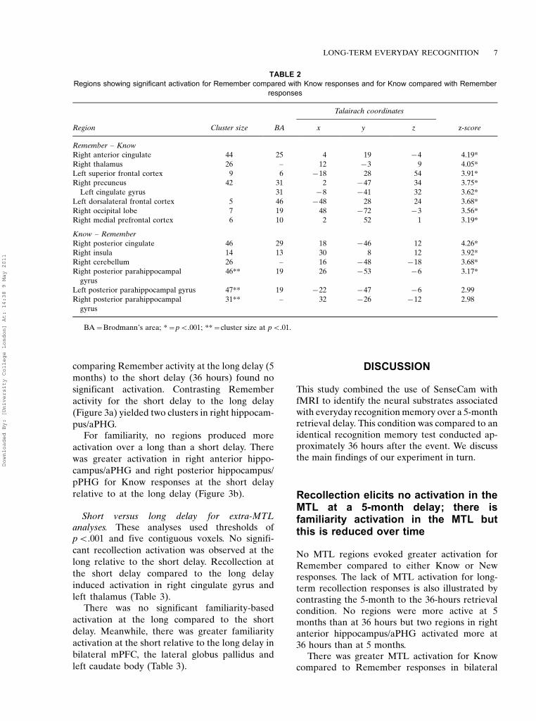
LONG-TERM EVERYDAY RECOGNITION 7
Downloaded By: [University College London] At: 14:38 9 May 2011

pPHG. Additionally, familiarity recruited clusters
in aPHG and pPHG relative to New stimuli. No
region of MTL was more active at the long than
the short delay for familiarity memory, while a
region in anterior hippocampus/aPHG and two
clusters in posterior hippocampus/pPHG acti-
vated more at the short than the long delay.These results suggest that recognition pro-
cesses become disengaged from the MTL over
time and are consistent with Takashima et al.
(2006) who also showed a decrement of MTL
activation across time. Nevertheless, the greater
MTL activation for familiarity than recollection
at a 5-month retrieval delay is noteworthy and
broadly supportive of Suchan et al. (2008) who
argued that the neural signal of familiarity-based
activation is relatively durable. Furthermore, an
explanation for this finding does not directly
emerge from single-process memory strength
accounts (e.g., Dunn, 2004).Familiarity involved both aPHG and pPHG.
Greater aPHG activation has frequently been
observed for familiarity compared to recollection
responses (e.g., Daselaar, Fleck, & Cabeza, 2006;
Henson, Rugg, Shallice, Josephs, & Dolan, 1999).
The greater aPHG activation for familiarity than
New stimuli may reflect retrieval processes. How-
ever, the pPHG activation is more surprising.
While this region has been implicated in familiar-
ity processes (Daselaar et al., 2006), it is incon-
sistent with many previous studies (e.g., Davachi,
Mitchell, & Wagner, 2003; Ranganath, Yonelinas,
Cohen, Dy, Tom, & D’Esposito, 2003) and dual-
process models (e.g., Aggleton & Brown, 1999)
that link this region to recollection. One potential
explanation for the pPHG activation for familiar-
ity responses compared to New stimuli is that
memory processes are seldom ‘‘process-pure’’
(Diana et al., 2007; Wais, Mickes, & Wixted,
2008) and recollection processes may be partially
contributing to this effect. This, however, fails to
explain why pPHG activation was also greater for
familiarity compared to recollection, because a
process contamination explanation would also
predict greater recollection activity in this region.
Our findings therefore appear more consistent
Figure 2. Whole brain analyses showing significant activation for (a) Remember compared to New responses; (b) Know compared
to New responses; (c) New compared to Remember; (d) New compared to Know responses.
8 MILTON ET AL.
Downloaded By: [University College London] At: 14:38 9 May 2011
with the idea that pPHG is involved in familiarity
processes for everyday recognition. Familiarity has
been linked with greater perceptual processing
than recollection (Daselaar et al., 2006; Yonelinas,
2002) and the pPHG has strong connections with
sensory areas (Daselaar et al., 2006). Increased
pPHG activation for familiarity responses may
therefore reflect the perceptual processes involved
in such judgements. Nevertheless, regardless of the
paradigm used, process impurity is a potential
Figure 3. Regions in MTL where activation was greater for (a) Remember at short delay (36 hours) than Remember at long delay
(5 months); (b) Know at short delay than Know at long delay.
TABLE 3
Regions showing significant activation for a short (36 hours) compared to a long (5 months) retrieval delay
Talairach Coordinates
Region Cluster size BA x y z z-score
Remember (36 hours) �Remember (5 months)
Left thalamus (pulvinar) 33 � �8 �32 11 3.74*
Right cingulate gyrus 5 � 22 5 27 3.56*
Right anterior parahippocampal
gyrus
5** 34 28 1 �15 2.83
Right anterior parahippocampal
gyrus
9** � 28 �10 �15 2.83
Right hippocampus � 30 �12 �6 2.59
Know (36 hours) � Know (5
months)
Lateral globus pallidus 16 � 24 �20 �1 4.83*
Right prefrontal cortex 19 10 16 31 �5 3.71*
Left caudate body 12 � �20 5 24 3.63*
Left prefrontal cortex 12 10 �38 42 15 3.42*
Right Hippocampus 5** 30 �12 �6 2.73
Right Hippocampus 13** � 30 �39 0 2.69
Right posterior parahippocampal
gyrus
7** � 32 �39 �3 2.67
BA�Brodmann’s area; *�pB.001; **�cluster size at pB.01.
LONG-TERM EVERYDAY RECOGNITION 9
Downloaded By: [University College London] At: 14:38 9 May 2011
issue for all studies attempting to distinguish bet-ween separate memory processes. Future workusing SenseCam should therefore use alternativemeasurements such as confidence ratings to furtherassess whether pPHG involvement is a robustcharacteristic of everyday familiarity memory.
There is greater activation in regions ofthe MTL for New compared to oldstimuli
Regions of aPHG and pPHG also yielded greateractivation for New stimuli than Remember andKnow responses. Novel stimuli have been linkedwith greater aPHG activation than old stimuli(e.g., Montaldi et al., 2006), which may reflect anovelty signal (Daselaar et al., 2006) or less-efficient item representation (Diana et al., 2007).Meanwhile, several studies have found regions ofpPHG that activate more for new than old stimuli,possibly reflecting incidental encoding processes(e.g., Johnson, Muftuler, & Rugg, 2008; Stark &Okado, 2003; Vilberg & Rugg, 2009).
Remote recollection recruits a numberof extra-MTL regions
In contrast to the lack of recollection basedactivation in MTL, diverse extra-MTL regionswere recruited by recollection memory. Regionsactivated by recollection compared to familiarityincluded left superior frontal gyrus, left DLPFC,right mPFC, and left anterior temporal cortex.Among the regions that recollection memoryengaged to a greater extent than New stimuliwere left VLFC, left inferior parietal lobe, andleft superior temporal gyrus. Nevertheless, noregions induced greater activation at 5-monthsthan at 36-hours retrieval delay. This may reflectgeneral decay of the memory trace over time;however, given the limited increased activation atthe short compared to the long delay (rightcingulate gyrus and left thalamus), it appearsthat extra-MTL traces remain relatively stable,at least over a 5-month period.
For the regions linked to recollection, mPFC hasbeen implicated in recollection processes (Yoneli-nas, Otten, Shaw, & Rugg, 2005), and may supportself-referential mental activity (Gusnard, Akbu-dak, Schulman, & Raichle, 2001). mPFC has alsobeen linked to long-term recognition processes
(e.g., Dolcos et al., 2005; Takashima et al., 2006).Daselaar et al. (2006) and Henson et al. (1999) bothobserved greater left VLFC activation for recollec-tion processes, possibly reflecting source retrieval(Nolde, Johnson, & Raye, 1998). The left superiorfrontal gyrus is involved with recollection (Vilberg& Rugg, 2007), while left DLPFC has beenimplicated in recollection based temporal-orderjudgements (St. Jacques, Rubin, LaBar, & Cabeza,2008). The left inferior parietal lobe consistentlyactivates more for old than new items (e.g., Hensonet al., 1999; Wheeler & Buckner, 2004), whileregions of left temporal cortex are engaged in long-term recognition memory (e.g., Bosshardt et al.,2005; Dolcos et al., 2005) and in autobiographicalmemory (e.g., Gilboa et al., 2004).
Diverse extra-MTL regions are recruitedby remote familiarity memory
Familiarity also resulted in extra-MTL activationscompared to recollection and New stimuli. Famil-iarity activity was elevated relative to recollectionactivation in right insula, right posterior cingulate,and right cerebellum. Relative to New stimuli,familiarity involved greater activity in bilateralmPFC, right VLFC, bilateral insula, and lefttemporal lobe. As for recollection, the bilateralmedial frontal gyrus activation may reflect long-term memory retrieval, while right VLFC has beenlinked with active retrieval processes (e.g., Hen-son, Shallice, & Dolan, 1998), and with increasingactivity as familiarity rises (Montaldi et al., 2006).The left insula has been shown to increase withfamiliarity (Yonelinas et al., 2005), while posteriorcingulate (Daselaar et al., 2006) and left temporallobe (Montaldi et al., 2006) are engaged infamiliarity processes. There was no increasedactivity for familiarity over time in extra-MTLregions but there was reduced activation at thelong compared to the short delay in two smallregions of mPFC. Although this appears somewhatinconsistent with the idea that mPFC is involved inlong-term memory, this may reflect reduced self-referential processing (Gusnard et al., 2001).
Methodological considerations
One methodological feature of our study is that weused the same images in both the short and longdelay retrieval sessions (for a related technique seeViskontas et al., 2009). One reason for this was that
10 MILTON ET AL.
Downloaded By: [University College London] At: 14:38 9 May 2011

participants did not generate a sufficient number
of unique events to use different stimuli in each
session. However, this approach had the advantage
of equating perceptual qualities, event saliency,
and personal significance across the sessions.
Nevertheless, it is possible that this repetition of
stimuli may have influenced the pattern of results
to some degree. Specifically, participants may have
recognised the image from the previous scanning
session rather than through recognition of the
event itself. We believe that this is unlikely due
to the fact that the initial study provided only
limited extra exposure to the stimuli (4000 ms) but
future work could guard again this possibility by
using different sets of target and novel images at
each test interval. Our recent experience using
SenseCam indicates that a ‘‘structured day’’*for
example a day spent at the zoo*provides a
sufficiently rich source of photographs to allow
the creation of several separate sets of material for
subsequent memory testing (Muhlert, Milton,
Butler, Kapur, & Zeman, 2010).
Conclusions
In summary, our results showed different patterns
of activation in recollection and familiarity mem-
ory. In particular, after a 5-month retrieval delay
we observed greater pPHG activation for famil-
iarity than recollection memory. Furthermore, the
reduced activation in the MTL at 5 months
relative to at 36 hours for both recollection and
familiarity memory is consistent with consolida-
tion theory, which predicts disengagement of the
MTL in recognition memory over time. The
continued engagement of extra-MTL regions
such as mPFC and lateral temporal cortex by
both recollection and familiarity memory is also
consistent with consolidation theory, which posits
that remote memory continues to be supported by
neocortical regions. Our findings demonstrate the
value of SenseCam as a technique to investigate
long-term everyday recognition memory.
REFERENCES
Addis, D. R., Moscovitch, M., & McAndrews, M. P.(2004). Consequences of hippocampal damageacross the autobiographical network in left temporallobe epilepsy. Brain, 130, 2327�2342.
Aggleton, J. P., & Brown, M. (1999). Episodic memory,amnesia, and the hippocampal-anterior thalamicaxis. Behavioral & Brain Sciences, 22, 425�489.
Bosshardt, S., Degonda, N., Schmidt, C. F., Boesiger, P.,Nitsch, R. M., Hock, C., et al. (2005). One month ofhuman memory consolidation enhances retrieval-related hippocampal activity. Hippocampus, 15,1026�1040.
Cabeza, R., Prince, S. E., Daselaar, S. M., Greenberg,D. L., Budde, M., Dolcos, F., et al. (2004). Brainactivity during episodic retrieval of autobiographicaland laboratory events: An fMRI study using a novelphoto paradigm. Journal of Cognitive Neuroscience,16, 1583�1594.
Daselaar, S. M., Fleck, M. S., & Cabeza, R. (2006).Triple dissociation in the medial temporal lobes:Recollection, familiarity, and novelty. Journal ofNeurophysiology, 96, 1902�1911.
Davachi, L., Mitchell, J. P., & Wagner, A. D. (2003).Multiple routes to memory: Distinct medial tempor-al lobe processes build item and source memories.Proceedings of the National Academy of Sciences,100, 2157�2162.
Diana, R. A., Yonelinas, A. P., & Ranganath, C. (2007).Imaging recollection and familiarity in the medialtemporal lobe: A three-component model. Trends inCognitive Sciences, 11, 379�386.
Dolcos, F., LaBar, K. S., & Cabeza, R. (2005).Remembering one year later: Role of the amygdalaand the media temporal lobe memory system inretrieving emotional memories. Proceedings of theNational Academy of Sciences, 102, 2626�2631.
Dunn, J. C. (2004). Remember-know: A matter ofconfidence. Psychological Review, 111, 524�542.
Eldridge, L. L., Knowlton, B. J., Furmanski, C. S.,Bookheimer, S. Y., & Engel, S. A. (2000). Remem-bering episodes: A selective role for the hippocampusduring retrieval. Nature Neuroscience, 3, 1149�1152.
Gilboa, A., Winocur, G., Grady, C. L., Hevenor, S. J., &Moscovitch, M. (2004). Remembering our past:Functional neuroanatomy of recollection of recentand very remote personal events. Cerebral Cortex,14, 1214�1225.
Gusnard, D. A., Akbudak, E., Schulman, G. L., &Raichle, M. E. (2001). Medial prefrontal cortex andself-referential mental activity: Relation to a defaultmode of brain function. Proceedings of the NationalAcademy of Sciences, 98, 4259�4264.
Henson, R. N. A., Cansino, S., Herron, J. E., Robb,W. G. D., & Rugg, M. D. (2003). A familiarity signalin human anterior cortex? Hippocampus, 13, 259�262.
Henson, R. N. A., Rugg, M. D., Shallice, T., Josephs, O.,& Dolan, R. J. (1999). Recollection and familiarityin recognition memory: An event related fMRIstudy. The Journal of Neuroscience, 19, 3962�3972.
Henson, R. N. A., Shallice, T., & Dolan, R. J. (1998).Right prefrontal cortex and episodic memory re-trieval: A functional MRI test of the monitoringhypothesis. Brain, 122, 137�1381.
Hodges, S., Williams, L., Berry, E., Izadi, S., Srinivasan,J., Butler, A., et al. (2006). SenseCam: A retrospectivememory aid. In P. Dourish & A. Friday (Eds.),Ubicomp, LNCS 4206 (pp. 177�193). Berlin,Germany: Springer-Verlag.
LONG-TERM EVERYDAY RECOGNITION 11
Downloaded By: [University College London] At: 14:38 9 May 2011
Johnson, J. D., Muftuler, L. T., & Rugg, M. D. (2008).Multiple repetitions reveal functionally and anato-mically distinct patterns of hippocampal activityduring continuous recognition memory. Hippocam-pus, 18, 975�980.
Kirwan, C. B., Wixted, J. T., & Squire, L. R. (2008).Activity in the medial temporal lobe predictsmemory strength, whereas activity in the prefrontalcortex predicts recollection. The Journal of Neu-roscience, 28, 10541�10548.
Maguire, E. A., & Frith, C. D. (2003). Lateral asym-metry in the hippocampal response to the remote-ness of autobiographical memories. The Journal ofNeuroscience, 23, 5302�5307.
Maldjian, J. A., Laurienti, P. J., Burdette, J. B., & Kraft,R. A. (2003). An automated method for neuroana-tomic and cytoarchitectonic atlas-based interroga-tion of fMRI data sets. Neuroimage, 19, 1233�1239.
Mandler, G. (1980). Recognising: The judgement ofprevious occurrence. Psychological Review, 87,252�271.
Montaldi, D., Spencer, T. J., Roberts, N., & Mayes, A.(2006). The neural system that mediates familiaritymemory. Hippocampus, 16, 504�520.
Muhlert, N., Milton, F., Butler, C. R., Kapur, N., &Zeman, A. Z. (2010). Accelerated forgetting of real-life events in transient epileptic amnesia. Neuropsy-chologia, 48, 3235�3244.
Nadel, L., & Moscovitch, M. (1997). Memory consoli-dation, retrograde amnesia and the hippocampalcomplex. Current Opinion in Neurobiology, 7, 217�227.
Niki, K., & Luo, J. (2002). An fMRI study on the time-limited role of the medial temporal lobe in long-term topographical autobiographic memory. Journalof Cognitive Neuroscience, 14, 500�507.
Nolde, S. F., Johnson, M. K., & Raye, C. L. (1998). Therole of prefrontal cortex during tests of episodicmemory. Trends in Cognitive Sciences, 2, 399�406.
Piefke, M., Weiss, P. H., Zilles, K., Markowitcsh, H. J.,& Fink, G. R. (2003). Differential remoteness andemotional tone modulate the neural correlates ofautobiographical memory. Brain, 126, 650�668.
Rajaram, S. (1993). Remembering and knowing: Twomeans of access to the personal past. Memory &Cognition, 21, 89�102.
Ranganath, C., Yonelinas, A. P., Cohen, M. X., Dy,C. J., Tom, S. M., & D’Esposito, M. (2003). Dissoci-able correlates of recollection and familiarity withinthe medial temporal lobes. Neuropsychologia, 42,2�13.
Rekkas, P. V., & Constable, R. T. (2005). Evidence thatautobiographic memory retrieval does not becomeindependent of the hippocampus: An fMRI studycontrasting very recent with remote events. Journalof Cognitive Neuroscience, 17, 1950�1961.
Rotello, C. M., Macmillan, N. A., & Reeder, J. A.(2004). Sum-difference theory of remembering andknowing: A two-dimensional signal-detection model.Psychological Review, 111, 588�616.
Ryan, L., Nadel, L., Keil, K., Putnam, K., Schnyer, D.,Trouard, T., et al. (2001). Hippocampal complex andretrieval of recent and very remote autobiographicalmemories: Evidence from functional magnetic
resonance imaging in neurologically intact people.Hippocampus, 11, 707�714.
Squire, L. R., Stark, C. E., & Clark, R. E. (2004). Themedial temporal lobe. Annual Review of Neu-roscience, 27, 279�306.
Stark, C. E. L., & Okado, Y. (2003). Making memorieswithout trying: Medial temporal lobe activity asso-ciated with incidental memory formation duringrecognition. The Journal of Neuroscience, 23,6748�6753.
St. Jacques, P., Rubin, D. C., LaBar, K. S., & Cabeza, R.(2008). The short and long of it: Neural correlates oftemporal-order memory for autobiographicalevents. Journal of Cognitive Neuroscience, 20,1327�1341.
Steinvorth, S., Corkin, S., & Halgren, E. (2006).Ecphory of autobiographical memories: An fMRIstudy of recent and remote memory retrieval.Neuroimage, 30, 285�298.
Suchan, B., Gayk, A. E., Schmid, G., Koster, O., &Daum, I. (2008). Hippocampal involvement inrecollection but not familiarity across time: Aprospective study. Hippocampus, 18, 92�98.
Svoboda, E., McKinnon, M. C., & Levine, B. (2006).The functional neuroanatomy of autobiographicalmemory: A meta-analysis. Neuropsychologia, 44,2189�2208.
Takashima, A., Petersson, K. M., Rutters, F., Tendolkar,I., Jensen, O., Zwarts, M. J., et al. (2006). Declarativememory consolidation in humans: A prospectivefunctional magnetic resonance imaging study. Pro-ceedings of the National Academy of Sciences, 103,756�761.
Tulving, E. (1985). Memory and consciousness. Cana-dian Psychology, 26, 1�12.
Vilberg, K. L., & Rugg, M. D. (2007). Dissociation ofthe neural correlates of recognition memory accord-ing to familiarity, recollection and amount ofrecollected information. Neuropsychologia, 45,2216�2225.
Vilberg, K. L., & Rugg, M. D. (2009). An investigationof the effects of relative probability of old and newtest items on the neural correlates of successfuland unsuccessful source memory. Neuroimage, 45,562�571.
Viskontas, I. V., Carr, V. A., Engel, S. A., & Knowlton,B. J. (2009). The neural correlates of recollection:Hippocampal activation declines as episodic mem-ory fades. Hippocampus, 19, 265�272.
Wais, P. E., Mickes, L., & Wixted, J. T. (2008).Remember/know judgements probe degrees of re-collection. Journal of Cognitive Neuroscience, 20,400�405.
Wheeler, M. E., & Buckner, R. L. (2004). Functional-anatomic correlates of remembering and knowing.Neuroimage, 21, 1337�1349.
Yonelinas, A. P. (2002). The nature of recollection andfamiliarity: A review of 30 years of research. Journalof Memory & Language, 46, 441�517.
Yonelinas, A. P., Otten, L. J., Shaw, K. N., & Rugg,M. D. (2005). Separating the brain regions involvedin recollection and familiarity in recognition mem-ory. The Journal of Neuroscience, 25, 3002�3008.
12 MILTON ET AL.
Downloaded By: [University College London] At: 14:38 9 May 2011