Common and unique components of inhibition and working memory: An fMRI, within-subjects...
15
Neuropsychologia 46 (2008) 2668–2682 Contents lists available at ScienceDirect Neuropsychologia journal homepage: www.elsevier.com/locate/neuropsychologia Common and unique components of inhibition and working memory: An fMRI, within-subjects investigation Fiona McNab a , Ga ¨ elle Leroux a , Fredrik Strand a , Lisa Thorell a,b , Sissela Bergman a , Torkel Klingberg a,∗ a Developmental Cognitive Neuroscience Group, Stockholm Brain Institute, Karolinska Institutet, Sweden b Department of Psychology, Uppsala University, Sweden article info Article history: Received 16 August 2007 Received in revised form 28 April 2008 Accepted 29 April 2008 Available online 16 May 2008 Keywords: Working memory Inhibition abstract Behavioural findings indicate that the core executive functions of inhibition and working memory are closely linked, and neuroimaging studies indicate overlap between their neural correlates. There has not, however, been a comprehensive study, including several inhibition tasks and several working memory tasks, performed by the same subjects. In the present study, 11 healthy adult subjects completed separate blocks of 3 inhibition tasks (a stop task, a go/no-go task and a flanker task), and 2 working memory tasks (one spatial and one verbal). Activation common to all 5 tasks was identified in the right inferior frontal gyrus, and, at a lower threshold, also the right middle frontal gyrus and right parietal regions (BA 40 and BA 7). Left inferior frontal regions of interest (ROIs) showed a significant conjunction between all tasks except the flanker task. The present study could not pinpoint the specific function of each common region, but the parietal region identified here has previously been consistently related to working memory storage and the right inferior frontal gyrus has been associated with inhibition in both lesion and imaging studies. These results support the notion that inhibitory and working memory tasks involve common neural components, which may provide a neural basis for the interrelationship between the two systems. © 2008 Elsevier Ltd. All rights reserved. 1. Introduction Working memory and inhibition, described as core executive function domains (Goldman-Rakic, 1987; Hasher & Zacks, 1988), are closely related. Both have been linked to IQ (Horn, Dolan, Elliott, Deakin, & Woodruff, 2003), and performance is associated in both normal (e.g. Conway, Cowan, & Bunting, 2001; Kane & Engle, 2000, 2003; Unsworth, Schrock, & Engle, 2004), and clinical pop- ulations such as attention deficit hyperactivity disorder (ADHD) (Hervey, Epstein, & Curry, 2004; ert ´ e, Geurts, Roeyers, Oosterlaan, & Sergeant, 2006; Willcutt, Doyle, Nigg, Faraone, & Pennington, 2005), high-functioning autism (HFA) and Tourette syndrome (TS) (Vert ´ e et al., 2006). It has also been suggested that they may rely on common neural resources (De Fockert, Rees, Frith, & Lavie, 2001; Kane & Engle, 2003). The present study represents an investigation into the extent to which the neural correlates of different forms of inhibition and working memory overlap, and where such common- alities occur. ∗ Corresponding author at: Karolinska Institutet, MR Centrum, Karolinska Sjukhuset N8:00, 17176 Stockholm, Sweden. Tel.: +46 8 5177 6118; fax: +46 8 5177 3266. E-mail address: [email protected] (T. Klingberg). The right inferior frontal cortex (BA 45/47) has been described as showing the most robust common activation across inhibition tasks (Bunge, Dudukovic, Thomason, Vaidya, & Gabrieli, 2002) and iden- tified as being central to inhibitory control (e.g. Aron, Robbins, & Poldrack, 2004; Chambers et al., 2006; Kelly, Hester, Foxe, Shpaner, & Garavan, 2006). It has been reported to show significantly greater activation for no-go trials in which participants inhibit a preponent response compared to go trials in which participants make a pre- ponent response (Booth et al., 2003; Konishi, Nakajima, Uchida, Sekihara, & Miyashita, 1998; Langenecker & Nielson, 2003), has been strongly implicated in the inhibition of an already initiated manual response, the so-called stop task (Aron, Fletcher, Bullmore, Sahakian, & Robbins, 2003; Aron & Poldrack, 2006; Chambers et al., 2006), and has been described as part of a “shared inhibitory neurocognitive network” involved with both go/no-go (GNG) and stop tasks (Rubia et al., 2001). In a flanker task activity in this region has been found to accompany increases in reaction time associated with incongruent trials (Hazeltine, Poldrack, & Gabrieli, 2000), with the authors arguing that the strong correspondence between this activity and that reported in GNG and set shifting studies suggests that response inhibition may most appropriately characterize the function of this region. Inferior parietal activation has also been observed in a number of inhibition tasks (Garavan, Ross, Murphy, Roche, & Stein, 2002; 0028-3932/$ – see front matter © 2008 Elsevier Ltd. All rights reserved. doi:10.1016/j.neuropsychologia.2008.04.023
-
Upload
independent -
Category
Documents
-
view
4 -
download
0
Transcript of Common and unique components of inhibition and working memory: An fMRI, within-subjects...

Neuropsychologia 46 (2008) 2668–2682
Contents lists available at ScienceDirect
Neuropsychologia
journa l homepage: www.e lsev ier .com/ locate /neuropsychologia
Common and unique components of inhibition and working memory:An fMRI, within-subjects investigation
Fiona McNaba, Gaelle Lerouxa, Fredrik Stranda, Lisa Thorell a,b,
Sissela Bergmana, Torkel Klingberga,∗stitut
cate tmaginensivme ss (a sal). Aceshoontalsk. Thion idior frportmay
a Developmental Cognitive Neuroscience Group, Stockholm Brain Institute, Karolinska Inb Department of Psychology, Uppsala University, Sweden
a r t i c l e i n f o
Article history:Received 16 August 2007Received in revised form 28 April 2008Accepted 29 April 2008Available online 16 May 2008
Keywords:Working memoryInhibition
a b s t r a c t
Behavioural findings indiclosely linked, and neuroihowever, been a comprehtasks, performed by the sablocks of 3 inhibition task(one spatial and one verbgyrus, and, at a lower thrand BA 7). Left inferior frtasks except the flanker taregion, but the parietal regstorage and the right inferstudies. These results supneural components, which
1. Introduction
Working memory and inhibition, described as core executivefunction domains (Goldman-Rakic, 1987; Hasher & Zacks, 1988),are closely related. Both have been linked to IQ (Horn, Dolan,Elliott, Deakin, & Woodruff, 2003), and performance is associatedin both normal (e.g. Conway, Cowan, & Bunting, 2001; Kane & Engle,2000, 2003; Unsworth, Schrock, & Engle, 2004), and clinical pop-ulations such as attention deficit hyperactivity disorder (ADHD)(Hervey, Epstein, & Curry, 2004; erte, Geurts, Roeyers, Oosterlaan,& Sergeant, 2006; Willcutt, Doyle, Nigg, Faraone, & Pennington,2005), high-functioning autism (HFA) and Tourette syndrome (TS)(Verte et al., 2006). It has also been suggested that they may relyon common neural resources (De Fockert, Rees, Frith, & Lavie, 2001;Kane & Engle, 2003). The present study represents an investigationinto the extent to which the neural correlates of different forms ofinhibition and working memory overlap, and where such common-alities occur.
∗ Corresponding author at: Karolinska Institutet, MR Centrum, KarolinskaSjukhuset N8:00, 17176 Stockholm, Sweden. Tel.: +46 8 5177 6118;fax: +46 8 5177 3266.
E-mail address: [email protected] (T. Klingberg).
0028-3932/$ – see front matter © 2008 Elsevier Ltd. All rights reserved.doi:10.1016/j.neuropsychologia.2008.04.023
et, Sweden
hat the core executive functions of inhibition and working memory areg studies indicate overlap between their neural correlates. There has not,e study, including several inhibition tasks and several working memory
ubjects. In the present study, 11 healthy adult subjects completed separatetop task, a go/no-go task and a flanker task), and 2 working memory taskstivation common to all 5 tasks was identified in the right inferior frontal
ld, also the right middle frontal gyrus and right parietal regions (BA 40regions of interest (ROIs) showed a significant conjunction between alle present study could not pinpoint the specific function of each commonentified here has previously been consistently related to working memory
ontal gyrus has been associated with inhibition in both lesion and imagingthe notion that inhibitory and working memory tasks involve commonprovide a neural basis for the interrelationship between the two systems.
© 2008 Elsevier Ltd. All rights reserved.
The right inferior frontal cortex (BA 45/47) has been described asshowing the most robust common activation across inhibition tasks(Bunge, Dudukovic, Thomason, Vaidya, & Gabrieli, 2002) and iden-
tified as being central to inhibitory control (e.g. Aron, Robbins, &Poldrack, 2004; Chambers et al., 2006; Kelly, Hester, Foxe, Shpaner,& Garavan, 2006). It has been reported to show significantly greateractivation for no-go trials in which participants inhibit a preponentresponse compared to go trials in which participants make a pre-ponent response (Booth et al., 2003; Konishi, Nakajima, Uchida,Sekihara, & Miyashita, 1998; Langenecker & Nielson, 2003), hasbeen strongly implicated in the inhibition of an already initiatedmanual response, the so-called stop task (Aron, Fletcher, Bullmore,Sahakian, & Robbins, 2003; Aron & Poldrack, 2006; Chambers etal., 2006), and has been described as part of a “shared inhibitoryneurocognitive network” involved with both go/no-go (GNG) andstop tasks (Rubia et al., 2001). In a flanker task activity in this regionhas been found to accompany increases in reaction time associatedwith incongruent trials (Hazeltine, Poldrack, & Gabrieli, 2000), withthe authors arguing that the strong correspondence between thisactivity and that reported in GNG and set shifting studies suggeststhat response inhibition may most appropriately characterize thefunction of this region.Inferior parietal activation has also been observed in a numberof inhibition tasks (Garavan, Ross, Murphy, Roche, & Stein, 2002;

cholog
F. McNab et al. / NeuropsyGaravan, Ross, & Stein, 1999; Langenecker & Nielson, 2003; Liddle,Kiehl, & Smith, 2001; Peterson et al., 2002; Rubia et al., 2001;Sylvester et al., 2003), although this may be related to the storage ofstimulus-response representations rather than inhibition (Hester,Murphy, & Garavan, 2004). Similarly, the dorsolateral prefrontalcortex (DLPFC) activation that has been associated with a range ofinhibition tasks (Bellgrove, Hester, & Garavan, 2004; Garavan et al.,2002; Langenecker & Nielson, 2003; Liddle et al., 2001; Sylvesteret al., 2003; Van Veen, Cohen, Botvinick, Stenger, & Carter, 2001)might be attributed to working memory demands as significantright DLPFC activation was observed for GNG trials during a count-ing GNG task, but not during a simple GNG (Mostofsky et al., 2003).
Furthermore, although a relatively consistent set of regions havebeen implicated in response inhibition paradigms (Wager et al.,2005), different forms of inhibition may be involved in differenttasks (Wager et al., 2005). Within this study we adopt the approachtaken by Barkley (1997) and consider three inhibition processes(rather than mechanisms); (1) inhibition of an initial prepotentresponse, (2) stopping an ongoing response or delayed respond-ing, and (3) limiting interference or distractibility. These processesare employed to a different extent in different tasks (Rubia et al.,2001), for example whilst the Stroop and Erikson flanker tasksmay rely heavily on limiting interference, GNG and stop tasks arelikely to involve a greater reliance on inhibition of a preponentor previously initiated response (although it should be noted thatsuch conflict resolution tasks may well involve other mechanismsbesides inhibition, such as facilitation). Correlations between per-formance on different inhibitory tasks have generally been low(Wager et al., 2005), but sometimes significant (Fan, Flombaum,McCandliss, Thomas, & Posner, 2003; Miyake, Friedman, Emerson,Witzki, & Howerter, 2000). Although it has been suggested thatidiosyncrasies between tasks may obscure the results and accountfor the low behavioral correlations (Friedman & Miyake, 2004), it isalso possible that different mechanisms may be involved in differ-ent forms of response inhibition. Therefore, when investigating thecommonalities between the neural correlates of working memoryand inhibition within the present study, three inhibition tasks wereused, corresponding to the three inhibitory processes described byBarkley (1997).
Reviews of the working memory literature have implicatedthe left inferior frontal cortex, DLPFC, premotor cortex, supe-rior frontal cortex, supplementary motor area, and the parietalcortex (for example, Cabeza & Nyberg, 2000; D’Esposito et al.,1998; D’Esposito, Postle, & Rypma, 2000; Smith & Jonides, 1998,
1999). Working memory tasks that use stimuli from differentsensory modalities have been shown to activate overlapping cor-tical regions, in both prefrontal and parietal cortex (Klingberg,1998; Klingberg, Kawashima, & Roland, 1996), suggested to besupramodal working memory regions. However, a dissociation hasalso been reported whereby verbal working memory is primar-ily associated with left hemisphere regions (in particular the leftprefrontal cortex (Gabrieli, Brewer, & Poldrack, 1998)) and spatialworking memory is linked to right hemisphere regions, even whenthe same letter stimuli are used in verbal and spatial working mem-ory task conditions (Smith, Jonides, & Koeppe, 1996). Furthermore,Leung and Zhang (2004) suggested that different subsets of theworking memory system may be associated with interference res-olution in spatial and verbal domains. It is claimed that whereasthe left inferior frontal gyrus is involved in overcoming interferingverbal stimuli, the right precentral sulcus and superior parietal lobeare involved in overcoming interfering spatial stimuli. It is also pos-sible that the cortical representation of interference resolution inworking memory may be material specific, which could mean thatthe relationship between inhibition and working memory differsdepending upon whether the working memory task (and/or stim-ia 46 (2008) 2668–2682 2669
ulus) is verbal or spatial. For this reason both verbal and spatialworking memory tasks were included within this study.
Evidence from patients suggests that the same right inferiorfrontal region may be involved in both spatial working memoryand response inhibition in a stop task, with correlations reportedbetween damage to this region and both stop signal reaction time(SSRT) and spatial working memory (SWM) performance) (Clark etal., 2007). In order to identify commonalities between inhibitionand working memory, in healthy participants, one approach hasbeen to draw upon the results of different studies. However, groupdifferences may confound such comparisons. Another approach hasbeen to combine working memory and inhibition demands withinthe same task, for example Bunge, Ochsner, Desmond, Glover, andGabrieli (2001) looked at the effects of manipulating the level ofproactive interference in a Sternberg-type working memory task,and Kelly et al. (2006) used a task that required inhibition of prepo-tent responses based on the contents of working memory. However,Hester et al. (2004) reported that maintaining successful inhibitorycontrol under increasing working memory demands tended notto increase activation in overlapping regions, but predominantlyin unique inhibition-specific regions. In the present study, work-ing memory and inhibition processes were isolated using differenttask blocks to avoid confounds associated with the manipulationof inhibition and working memory demands within the same task.
Valid conjunction analysis (Nichols, Brett, Andersson, Wager, &Poline, 2004) was used to localize overlapping activation associ-ated with different forms of inhibition and working memory inthe same subjects (to reduce the effects of individual differences).For each task, event related analysis was used to avoid possibleconfounds associated with using a block design (as described byAron & Poldrack, 2005), for example unbalanced task maintenanceload and switching between blocks which do not include trials thatrequire inhibition and mixed blocks which do include such trials.
Another way in which the present study represents an extensionof the approach taken previously is in the design of the GNG andstop tasks. Trials involving inhibition (no-go or stop trials) havetypically been compared to go trials in which a preponent stim-ulus is presented and a preponent response is required. Such anapproach may introduce confounds associated with differences inthe frequency of presentation of certain stimuli (Aron & Poldrack,2005), with the stimuli associated with inhibition being presentedless frequently than the stimuli that do not involve inhibition. Toovercome such confounds this study made use of oddball trials,which were control trials that did not require response inhibi-
tion, but were matched to no-go trials in terms of frequency ofpresentation. By comparing no-go or stop trials to these oddballtrials we controlled for differences in familiarity associated withthe oddball effect, whereby novel stimuli elicit additional cerebralactivity (Halgren, Marinkovic, & Chauvel, 1998). Although such odd-ball trials may require other forms of inhibition, they do not requireinhibition of a response, enabling us to isolate this form of inhibitionin contrasts between no-go or stop trails and oddball trials.Five contrasts were generated: three from inhibition tasks (cor-responding to the three different inhibitory processes describedby Barkley, 1997) and two from working memory tasks (verbal andspatial), and valid conjunction analysis was used to identify regionsof common activation within and between working memory andinhibition domains. The results of the within domain conjunctionswere also used to generate ROIs which were used to perform smallvolume corrected conjunction analyses between domains. In thisway we were able to test the hypothesis that there are commonregions associated with response inhibition and working memory,observe the extent to which each ROI contributes to each task, anddetermine whether the choice of inhibition and working memorytask influences such commonality. This study represents an exten-

cholog
2670 F. McNab et al. / Neuropsysion of the approach by investigating commonalities between threedifferent forms of inhibition and two different forms of workingmemory and the relative contribution of each ROI to each task,in the same participants, isolating working memory and inhibi-tion within separate task blocks, and using oddball trials in orderto control for differences in familiarity within GNG and stop taskcontrasts.
2. Methods
2.1. Participants
Fourteen right-handed healthy university students were scanned. The data from11 participants were included in the fMRI analysis (mean age 24 years, � = 4 years,range 22–34, 4 males). All participants gave informed consent, and the study wasapproved by the ethical committee at the Karolinska Institute.
2.2. Procedure
Each participant completed three inhibition tasks (a stop task, a GNG task anda flanker task), and two working memory tasks (spatial and verbal). The data fromthree participants were excluded due to movement of >4 mm during fMRI recording.One run of each task (of approximately 10 min) was completed prior to entering thescan room, and two runs of each task were completed during scanning. One run ofeach inhibition task consisted of 152 trials and one run of each working memorytask consisted of 40 trials. Responses were made with the right hand and the orderof task presentation was counterbalanced across subjects.
2.2.1. Inhibition tasksWithin the GNG task (Fig. 1a) 50% of trials involved the presentation of a yellow
square for 1300 ms, followed by a blank screen for 400 ms, and a fixation cross for300 ms (go trials). In response to presentation of the yellow square participants wererequired to press a button. For 25% of the trials the stimulus consisted of a yellowtriangle which indicated that participants should not make a response (no-go trials).For the remaining 25% of trials the same presentation sequence was used, but thestimulus consisted of a blue square, which also required a button press (oddballtrials).
Fig. 1. The inhibition tasks ((a) the GNG task, (b) the stop task and (c) the flanker task)spatial working memory task).
ia 46 (2008) 2668–2682
In the stop task (Fig. 1b) 50% of trials (control trials) involved the presen-tation of a yellow horizontal arrow, displayed for 1500 ms, followed by a blankscreen for 400 ms and a fixation cross for 300 ms. The presentation of the yellowarrow required participants to press a button. In 25% of trials the yellow hori-zontal arrow was followed by a vertical yellow arrow, which signalled that theparticipant should inhibit their response (stop trials). For the first trial within thisinhibition condition the horizontal arrow was displayed for 250 ms and the verticalarrow for 12,500 ms. Following this, if the participant had been successful duringthe previous inhibition trial, 50 ms was added to the duration of the first arrow.If the participant had failed to inhibit their response, the duration was reducedby 50 ms. Limits were imposed so that the minimum duration was 50 ms and themaximum was 1000 ms. The duration of the second arrow was adapted so thatthe total duration for the two arrows was always 1500 ms, in keeping with thecontrol condition. This adaptive duration procedure was used to achieve approx-
imately 50% accuracy, and to reduce the likelihood of participants anticipating theonset time of the second arrow, and delaying their response to the first arrowaccordingly. In the remaining 25% of trials (oddball trials) the initial horizontalyellow arrow was followed by a second horizontal arrow, which was blue, anda button press was still required. In such oddball trials the duration of exposureof the first arrow was determined by the accuracy and exposure duration in theprevious stop trial, the same as for stop trials. However, unlike stop trials, theaccuracy of the oddball trials did not influence any subsequent exposure dura-tion.As discussed previously, in both the GNG and stop tasks, to control for differencesin familiarity associated with the oddball effect, we compared no-go and stop trialsto the frequency-matched oddball trials. However, it was not possible to controlfor the additional motor component associated with oddball trials but not withno-go or stop trials. The trials were distributed across blocks containing differentrelative no-go and stop trial densities in order to improve detection power (Liu,2004).
Within the flanker task (Fig. 1c) a horizontal array of 5 arrows was presented for1300 ms, and followed by a blank screen for 400 ms and a fixation cross for 300 ms.A near equal number of congruent trials (75 trials in which all the arrows pointedin the same direction) and incongruent trials (77 trials in which the central arrowpointed in the opposite direction to that of the surrounding arrows) were used, anda near equal number of trials included left pointing surrounding arrows as rightpointing surrounding arrows within each condition. Participants were required topress a button on the right if the central arrow pointed to the right and on the left ifthe central arrow pointed to the left.
and the working memory tasks ((d) the verbal working memory task and (e) the

cholog
F. McNab et al. / Neuropsy2.2.2. Working memory tasksWithin the verbal working memory task (Fig. 1d) each trial consisted of the serial
presentation of 5 centrally positioned letters. Each letter was displayed for 500 ms.Between each letter the blank screen was shown for 500 ms. A cue was presented1000 ms after the last letter disappeared. The cue stimulus consisted of a numberbetween 1 and 5 (which referred to the serial position in the stimulus sequence), anda letter (but not the letter “a”). Participants were asked to make a yes/no response toindicate whether the number matched the letter, for example “3:W” would requireparticipants to indicate whether the third stimulus in the trial had been “w”. Withinthe control condition every stimulus was the lower-case letter “a”, the cue was anupper case letter “A”, and a “yes” response was always required.
Within the spatial working memory task (Fig. 1e) participants were presentedwith a 4 by 4 grid of white lines on a black background. Within the working memorycondition a series of 5 yellow circles appeared within different spaces on the grid,but not in the corner squares. Each circle was displayed for 500 ms. Between eachcircle presentation the blank grid was shown for 500 ms. 1000 ms after the last circledisappeared a cue was presented within one of the grid spaces. This took the form ofa number between 1 and 5, referring to the serial position in the previous stimulussequence, and a question mark. The participant was asked to indicate with a yes/noresponse whether the number and the grid position matched, for example a 2 in acertain grid position would prompt the participant to indicate whether the secondcircle had appeared in this particular grid position. In the control condition redcircles were presented in the same sequence of corner grid positions, starting in thetop left of the grid and progressing clockwise. The cue in the control condition alwaysconsisted of the number 8 presented in a non-corner space, and always required a“yes” response.
In an attempt to reduce the risk of implicit working memory processes beingemployed within the working memory control conditions, the same stimuli wereused within every trial of these working memory control conditions, which mayhave introduced a confound associated with differences in stimulus novelty to thebetween domain conjunction analyses had oddball trials not been included to con-trol for differences in stimulus novelty within the inhibition task contrasts.
2.3. fMRI scanning
Imaging data were collected using a 1.5 T GE Signa scanner. T2*-weighted, gradi-ent echo, spiral echo-planar images were acquired with TR = 2100 ms, TE = 40 ms, flipangle = 76◦ , 22 axial slices, 5.0 mm slice thickness, 220 mm × 220 mm FOV, 64 × 64grid, resulting in voxels that were 3.4 mm × 3.4 mm × 5.0 mm. Each scan included10 sessions, 2 for each task. Each flanker task and GNG session lasted 308.2 s and
included acquisition of 148 volumes. Each stop task session lasted 338.6 s andincluded acquisition of 162 volumes. Each verbal working memory task sessionlasted 404.2 s and included acquisition of 194 volumes. Each spatial working mem-ory task session lasted 364.2 s and included acquisition of 174 volumes. T1-weightedspin echo images (FOV = 220 mm × 220 mm, 256 × 256 grid) were acquired in thesame position as the functional images.2.4. Data analysis
There was one behavioural measure associated with each inhibition task. In theGNG task this was the number of commission errors. In the flanker task the differ-ence in reaction time between incongruent and congruent trials was calculated. Inthe stop task an estimate of the stop signal reaction time was made by first calcu-lating the average stop signal duration for each participant (the average durationof the first arrow in the stop trials after stabilization, i.e. in stop trials 20–38). Thestop signal reaction time (SSRT) was then determined by subtracting the mean stopsignal duration from the median reaction time for the oddball trials (the medianwas used because of standard positive skewing of RT data) (Band, van der Molen,& Logan, 2003; Clark et al., 2007). In the spatial and verbal working memory tasks,accuracy and reaction times were recorded. Correlations between these behaviouralmeasures were identified.
The fMRI data were analysed with SPM2 (for the preprocessing and generationof individual contrast images) and SPM5 (for the second level analysis) (WellcomeDepartment of Imaging Neuroscience, London, UK). Motion during scanning wasestimated by 6 parameters (3 translations, 3 rotations), which were used to realignthe functional images to the first image in the series. The T1-weighted images
Table 1Correlation coefficients for the correlations between the behavioural measures associated
1 2
1. Flanker RT difference2. GNG commission errors −0.293. Stop signal reaction time 0.16 0.60*
4. Spatial WM accuracy 0.58* −0.275. Spatial WM RT 0.15 0.506. Verbal WM accuracy 0.25 −0.57*
7. Verbal WM RT 0.36 0.36
ia 46 (2008) 2668–2682 2671
were normalised to MNI305 space. The parameters from this normalization werethen used to normalize the functional images, which were sampled to a voxel sizeof 3 mm × 3 mm × 3 mm and then smoothed with an isotropic Gaussian kernel of6.0 mm. For each task, events were modeled with the haemodynamic response func-tion and it’s temporal and spatial derivatives, and contrast images were produced.Event related analysis was used to avoid the confounds associated with using a blockdesign to study inhibition processes (Aron & Poldrack, 2005).
The flanker task was modeled with separate regressors for incongruent and con-gruent trials (event analysis at stimulus onset), the GNG task was modeled withseparate regressors for go, no-go and oddball trials (event analysis at stimulus onset),and the stop task was modeled with separate regressors for control, stop and odd-ball trials (event analysis at the onset of the second stimulus in stop and oddballtrials, and at the onset of the black screen following the horizontal arrow in controltrials). The two working memory tasks were modeled with separate regressors forworking memory and control trials (epoch analysis beginning at the onset of thefirst stimulus, with a duration of 8.5 s, corresponding to the trial duration).
No-go trials were compared to oddball trials in the GNG task, and stop trialswere compared to oddball trials in the stop task. No-go trials were also comparedto go trials in both of these tasks for validation. In the case of the flanker task,incongruent and congruent conditions were compared. Similarly, verbal and spa-tial working memory tasks were compared to their respective control conditions.This gave rise to 5 contrast images for each participant, which were analysed atthe group level using random effects (rfx) analysis. These images were enteredinto a within-subjects ANOVA. “Valid conjunction inference” (Nichols et al., 2004)(implemented in SPM5) was used to identify regions that were significantly activefor each combination of task contrasts, both within and between task domains,with the requirement that each contrast must be individually significant. Suchstatistical maps were thresholded at p < 0.001 uncorrected for multiple compar-isons, and effects were considered to be significant if they fulfilled a correctedcluster level requirement of p < 0.05 (standard family-wise error (FWE) correctionin SPM5).
2.4.1. Region of interest analysisTo more precisely characterise the contribution of these regions to the differ-
ent tasks, and determine whether there were conjunction effects that were belowthreshold in the whole brain analysis, an ROI approach was also taken. Twelve ROIswere generated from the within-domain conjunction analyses (and the flanker taskcontrast image, as this task did not show significant conjunction effects with eitherof the other two inhibition tasks in the whole brain analysis). For each ROI, themean relative signal change associated with each task contrast was calculated and
correlations were performed between the values of mean relative signal change andthe behavioural measure, or measures, associated with that task. The whole brainconjunction analyses were then repeated, and a small-volume correction applied,using the standard small-volume procedure in SPM5, corresponding to each of theseROIs. This enabled us to establish whether any of these regions showed commonbetween-domain activation that had not reached significance in the whole brainanalysis.3. Results
3.1. Behavioral results
In the GNG task significantly longer reaction times wereobserved for oddball trials (mean = 342 ms, � = 38 ms) than go trials(mean = 329 ms, � = 41 ms) (t = 4.141, d.f. = 13, p < 0.005). The meanpercentage of commission errors was 9.4% (� = 7.5%), which wassignificantly greater than zero (t = 4.849, d.f. = 13, p < 0.0005). In thestop task, as in the GNG task, significantly longer reaction timeswere associated with the oddball trials (mean = 507 ms, � = 144 ms)compared to control trials (mean = 468 ms, � = 124 ms) (t = 5.65,d.f. = 13, p < 0.0005). The mean duration of the first arrow in the twofMRI sessions of the stop task (which was dependent upon the num-
with each of the tasks, *p < 0.05, **p < 0.01 (not corrected for multiple comparisons)
3 4 5 6
−0.150.49 −0.11
−0.62* 0.62* −0.290.45 0.09 0.94** −0.14

2672 F. McNab et al. / Neuropsychologia 46 (2008) 2668–2682
Table 2Rfx results for each inhibition and working memory task (p < 0.05 at the cluster level, with a voxel level threshold of p < 0.001)
Voxel-level T value Cluster extent Cluster level correctedp value
MNI coordinates
Flanker task*Incongruent > Congruent 4.74 49 0.017 Right inferior/superior parietal
BA 4030, −54, 45
4.33 39, −45, 51
4.40 48 0.019 Left anterior cingulate/corpuscallosum
−3, 27, 18
4.21 −24, 27, 93.71 −15, 27, 9
Go/no-go taskNo-go > Oddball 6.78 54 0.011 Left insula, BA 13/inferior
frontal gyrus BA 47−42, 12, −3
6.59 318 0.000 Right inferior frontal gyrus BA47
45, 15, −3
5.83 33, 24, −65.17 27, 15, −15
5.88 49 0.017 Right parietal precuneus 12, −75, 48
5.82 82 0.001 Left superior/middle frontalgyrus BA 9
−42, 36, 36
5.77 261 0.000 Right superior frontal gyrus BA9/middle frontal gyrus BA 46
33, 51, 33
5.63 24, 48, 335.22 45, 42, 30
5.38 185 0.000 Right parietal supramarginalgyrus/inferior parietal BA 40
63, −45, 33
4.70 51, −42, 364.62 54, −36, 42
5.18 42 0.033 Left inferior parietal lobule −45, −45, 394.14 −42, −51, 48
4.94 38 0.048 Left inferior occipital gyrus −33, −90, −6
4.14 39 0.044 Right cingulated gyrus BA 23 6, −36, 273.85 0, −30, 30
No-go > Go 7.07 150 0.000 Right inferior frontal gyrus BA47/insula
33, 24, −6
4.78 48, 15, −94.04 33, 12, 3
5.06 98 0.000 Right superior frontal gyrus BA10/middle frontal gyrus BA 46
27, 51, 30
4.81 36, 42, 274.31 48, 39, 27
Stop taskStop > Oddball 8.28 387 0.000 Right inferior frontal gyrus BA
47/insula45, 15, −6
6.92 36, 15, 36.01 36, 18, −18
7.91 309 0.000 Left inferior frontal gyrus BA47/insula BA 13
−45, 15, −9
6.47 −36, 21, 66.31 −36, 12, 12
7.44 290 0.000 Right superior/middle frontalgyrus
30, 45, 36
6.28 30, 42, 245.20 42, 42, 27
7.24 129 0.000 Right superior frontal gyrus BA6
15, 9, 69
4.63 15, −9, 723.85 12, 15, 60
7.02 411 0.000 Right cingulated BA 32/medialfrontal gyrus
9, 18, 45
6.51 9, 6, 486.49 6, 24, 30
5.73 98 0.000 Right parietal supramarginalgyrus BA 40
54, −45, 36
4.02 63, −54, 30
F. McNab et al. / Neuropsychologia 46 (2008) 2668–2682 2673
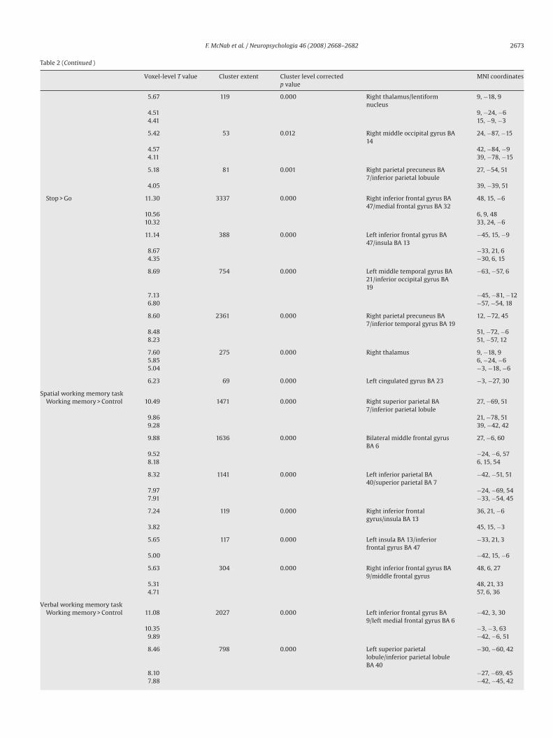
Table 2 (Continued )
Voxel-level T value Cluster extent Cluster level correctedp value
MNI coordinates
5.67 119 0.000 Right thalamus/lentiformnucleus
9, −18, 9
4.51 9, −24, −64.41 15, −9, −3
5.42 53 0.012 Right middle occipital gyrus BA14
24, −87, −15
4.57 42, −84, −94.11 39, −78, −15
5.18 81 0.001 Right parietal precuneus BA7/inferior parietal lobuule
27, −54, 51
4.05 39, −39, 51
Stop > Go 11.30 3337 0.000 Right inferior frontal gyrus BA47/medial frontal gyrus BA 32
48, 15, −6
10.56 6, 9, 4810.32 33, 24, −6
11.14 388 0.000 Left inferior frontal gyrus BA47/insula BA 13
−45, 15, −9
8.67 −33, 21, 64.35 −30, 6, 15
8.69 754 0.000 Left middle temporal gyrus BA21/inferior occipital gyrus BA19
−63, −57, 6
7.13 −45, −81, −126.80 −57, −54, 18
8.60 2361 0.000 Right parietal precuneus BA7/inferior temporal gyrus BA 19
12, −72, 45
8.48 51, −72, −68.23 51, −57, 12
7.60 275 0.000 Right thalamus 9, −18, 95.85 6, −24, −65.04 −3, −18, −6
6.23 69 0.000 Left cingulated gyrus BA 23 −3, −27, 30
Spatial working memory taskWorking memory > Control 10.49 1471 0.000 Right superior parietal BA
7/inferior parietal lobule27, −69, 51
9.86 21, −78, 519.28 39, −42, 42
9.88 1636 0.000 Bilateral middle frontal gyrusBA 6
27, −6, 60
9.52 −24, −6, 578.18 6, 15, 54
8.32 1141 0.000 Left inferior parietal BA40/superior parietal BA 7
−42, −51, 51
7.97 −24, −69, 547.91 −33, −54, 45
7.24 119 0.000 Right inferior frontalgyrus/insula BA 13
36, 21, −6
3.82 45, 15, −3
5.65 117 0.000 Left insula BA 13/inferiorfrontal gyrus BA 47
−33, 21, 3
5.00 −42, 15, −6
5.63 304 0.000 Right inferior frontal gyrus BA9/middle frontal gyrus
48, 6, 27
5.31 48, 21, 334.71 57, 6, 36
Verbal working memory taskWorking memory > Control 11.08 2027 0.000 Left inferior frontal gyrus BA
9/left medial frontal gyrus BA 6−42, 3, 30
10.35 −3, −3, 639.89 −42, −6, 51
8.46 798 0.000 Left superior parietallobule/inferior parietal lobuleBA 40
−30, −60, 42
8.10 −27, −69, 457.88 −42, −45, 42

2674 F. McNab et al. / Neuropsychologia 46 (2008) 2668–2682
Table 2 (Continued )
vel co
nerate
Voxel-level T value Cluster extent Cluster lep value
7.65 128 0.000
7.60 504 0.000
6.896.47
5.66 283 0.000
5.294.58
5.26 123 0.0004.97
5.00 62 0.005
4.65 132 0.0004.224.22
The significant clusters from the contrasts labeled with an asterisk were used to ge
ber of commission errors made by each participant) was 339 ms(� = 155 ms) for stop trials and 346ms (� = 162 ms) for oddball trials.The mean SSRT was 166 ms (� = 66 ms).
In the flanker task, in keeping with incongruent trials involvinga greater reliance on the inhibition of interference from surround-ing arrows, significantly more errors were made in the incongruentcondition (mean = 2.5%, � = 2.6%) compared to the congruent con-dition (mean = 0.9%, � = 0.8%) (t = 3.33, d.f. = 13, p < 0.005), and themean reaction time was significantly longer for the incongru-ent trials (mean = 449 ms, � = 43 ms) compared to congruent trials(mean = 419 ms, � = 43 ms) (t = 8.49, d.f. = 13, p < 0.001).
The mean accuracy for the spatial working memory taskwas 88.4% (� = 6.7%), and the mean reaction time was 1158 ms(� = 219 ms). The mean accuracy for the verbal working memorytask was 91.3% (� = 6.5%), and the mean reaction time was 1460 ms(� = 147 ms).
The results of the correlations between the behavioural mea-sures are presented in Table 1. Significant correlations were seenbetween accuracy on the two working memory tasks and betweenreaction times on the two working memory tasks. Accuracy on thespatial working memory task positively correlated with the reac-tion time difference between incongruent and congruent trials in
the flanker task. Accuracy on the verbal working memory task nega-tively correlated with the number of commission errors made in theGNG task and the SSRT. The SSRT also correlated with the numberof commission errors in the GNG task.3.2. fMRI results
3.2.1. Whole brain analysisThe results of the group analysis in which the experimental
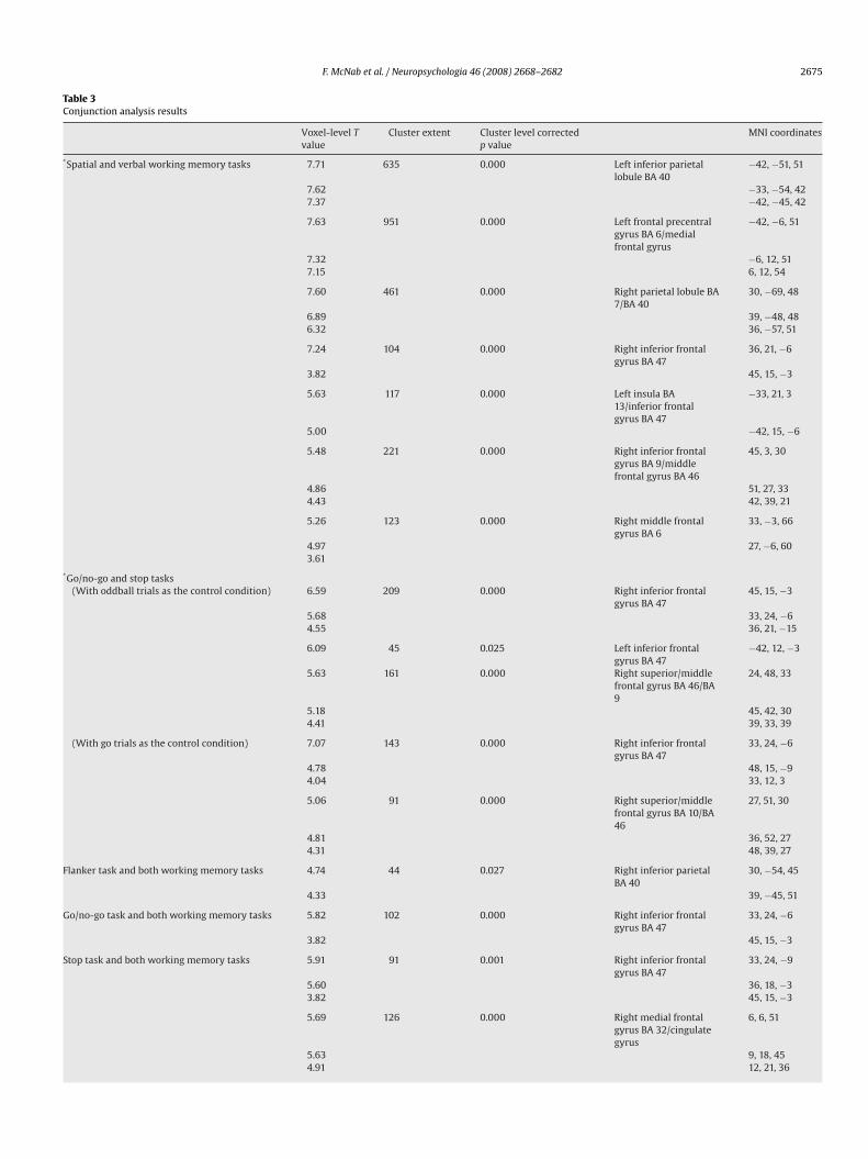
conditions were compared to the respective control condition, foreach task, are shown in Table 2. All combinations of tasks weretested using conjunction analysis, the results of such conjunc-tion analysis within and between task domains are presented inTable 3 and Fig. 2. These conjunction effects reached significanceat p < 0.05 at the corrected cluster level, with a voxel level thresh-old of p < 0.001. The conjunctions that are not reported did not givesignificant effects at the p < 0.05 corrected cluster level previouslydescribed.
Although there was no significant conjunction between all threeinhibition tasks in the whole brain analysis, significant conjunc-
rrected MNI coordinates
Right inferior frontal gyrus 36, 21, −6
Right superior parietal lobuleBA 7/inferior parietal lobule BA40
30, −69, 48
39, −48, 4833, −66, 36
Right inferior frontal gyrus BA9/middle frontal gyrus BA 46
45, 3, 30
51, 24, 3045, 42, 30
Right middle frontal gyrus BA 6 33, −3, 6627, −6, 60
Left middle temporal gyrus −48, −51, 6
Left caudate/putamen −15, −3, 15−15, 9, 3−3, −18, 15
regions of interest for further analysis.
tions were seen between the GNG task and the stop task withinbilateral inferior frontal gyrus (BA 47) and right superior/middlefrontal gyrus (BA 46/9). As anticipated, significant conjunctionswere also seen between the two working memory tasks withinbilateral frontal and parietal regions.
A significant conjunction was seen between both working mem-ory tasks and the GNG and stop tasks within the right inferiorfrontal gyrus (90 voxels, maximum/minimum: x = 45/30, y = 30/15,z = 9/−12, a cluster which mostly contained voxels within right infe-rior frontal cortex BA 47, but which also extended to the insula).The mean relative signal change in this right inferior frontal clus-ter, for each task contrast, is shown in Fig. 2. Fig. 3 shows that thiscluster extends across the border between the right inferior frontalcortex and the insula. Within this cluster we also investigated cor-relations between the mean relative signal change from each taskcontrast included in the significant conjunction and the behaviouralmeasure, or measures, associated with that task, but no significantcorrelations were seen.
For validation, additional analysis was conducted using thego trials as the control condition (with the contrast stop vs.go in the stop task and the contrast no-go vs. go in the GNGtask) and the conjunction analyses were repeated using these
contrast images. Although the right inferior frontal and right mid-dle/superior frontal conjunctions between the GNG/stop tasksremained significant, the left inferior conjunction was no longersignificant. The right inferior frontal conjunction between theGNG task, the stop task and the two WM tasks remained signifi-cant. No additional effects were observed from these conjunctionanalyses.3.2.2. Region of interest analysisROIs were generated from the results of the whole brain con-
junction analyses. The ROIs were the three clusters that showeda significant conjunction between the stop and GNG tasks, the 7clusters that showed a significant conjunction between the twoWM tasks, and the two clusters that showed a significant effect inthe incongruent vs. congruent contrast from the flanker task. Fig. 4shows the mean relative signal change associated with each taskcontrast, for each ROI.
For each ROI we performed small volume corrected conjunc-tion analyses to identify between domain commonalities that hadnot reached statistical significance in the whole brain analysis. Six
F. McNab et al. / Neuropsychologia 46 (2008) 2668–2682 2675
Table 3Conjunction analysis results
Voxel-level Tvalue
Cluster extent Cluster level correctedp value
MNI coordinates
*Spatial and verbal working memory tasks 7.71 635 0.000 Left inferior parietallobule BA 40
−42, −51, 51
7.62 −33, −54, 427.37 −42, −45, 42
7.63 951 0.000 Left frontal precentralgyrus BA 6/medialfrontal gyrus
−42, −6, 51
7.32 −6, 12, 517.15 6, 12, 54
7.60 461 0.000 Right parietal lobule BA7/BA 40
30, −69, 48
6.89 39, −48, 486.32 36, −57, 51
7.24 104 0.000 Right inferior frontalgyrus BA 47
36, 21, −6
3.82 45, 15, −3
5.63 117 0.000 Left insula BA13/inferior frontalgyrus BA 47
−33, 21, 3
5.00 −42, 15, −6
5.48 221 0.000 Right inferior frontalgyrus BA 9/middlefrontal gyrus BA 46
45, 3, 30
4.86 51, 27, 334.43 42, 39, 21
5.26 123 0.000 Right middle frontalgyrus BA 6
33, −3, 66
4.97 27, −6, 603.61
*Go/no-go and stop tasks(With oddball trials as the control condition) 6.59 209 0.000 Right inferior frontal
gyrus BA 4745, 15, −3
5.68 33, 24, −64.55 36, 21, −15
6.09 45 0.025 Left inferior frontalgyrus BA 47
−42, 12, −3
5.63 161 0.000 Right superior/middlefrontal gyrus BA 46/BA9
24, 48, 33
5.18 45, 42, 304.41 39, 33, 39
(With go trials as the control condition) 7.07 143 0.000 Right inferior frontalgyrus BA 47
33, 24, −6
4.78 48, 15, −94.04 33, 12, 3
5.06 91 0.000 Right superior/middlefrontal gyrus BA 10/BA46
27, 51, 30
4.81 36, 52, 274.31 48, 39, 27
Flanker task and both working memory tasks 4.74 44 0.027 Right inferior parietalBA 40
30, −54, 45
4.33 39, −45, 51
Go/no-go task and both working memory tasks 5.82 102 0.000 Right inferior frontalgyrus BA 47
33, 24, −6
3.82 45, 15, −3
Stop task and both working memory tasks 5.91 91 0.001 Right inferior frontalgyrus BA 47
33, 24, −9
5.60 36, 18, −33.82 45, 15, −3
5.69 126 0.000 Right medial frontalgyrus BA 32/cingulategyrus
6, 6, 51
5.63 9, 18, 454.91 12, 21, 36

2676 F. McNab et al. / Neuropsychologia 46 (2008) 2668–2682
tent
Table 3 (Continued )
Voxel-level Tvalue
Cluster ex
5.14 102
5.004.97
4.85 46
4.62
4.01Go/no-go task, Stop task and both working memory tasks(With oddball trials as the control condition) 5.68 90
3.82
(With go trials as the control condition) 7.07 93
4.39
The significant clusters from the contrasts labeled with an asterisk were used to generate
ROIs showed a significant conjunction between all 5 task contrasts.These were the right inferior frontal and right middle frontal ROIsgenerated from the GNG/stop task whole brain conjunction (Fig. 4aand b) and the WM task whole brain conjunction (Fig. 4d and e),the right parietal ROI generated from the WM task whole brain con-junction (Fig. 4h) and the right parietal ROI generated from flankertask group analysis (Fig. 4k). The more superior middle frontal ROI(BA 6) generated from the WM whole brain conjunction showed asignificant small volume corrected conjunction between all tasksexcept the GNG task (Fig. 4i).
Fig. 2. Significant whole brain conjunction effects within and between the working memsignificance of p < 0.05, corrected). The graph shows the mean relative signal change in theconjunction between both working memory tasks, the GNG task and the stop task, in eac
Cluster level correctedp value
MNI coordinates
0.000 Left insula BA13/inferior frontalgyrus BA 47
−33, 24, 3
−42, 15, −6−36, 21, −6
0.022 Right superior parietalBA 7/inferior parietal
27, −54, 48
33, −57, 54
39, −42, 480.001 Right inferior frontalgyrus BA 47
33, 24, −6
45, 15, −3
0.000 Right inferior frontalgyrus BA 47
33, 24, −6
45, 15, −3
regions of interest for further analysis.
A significant small volume corrected conjunction was seenbetween all tasks except the flanker task in the left inferior frontalROI generated from the GNG/stop task whole brain conjunction(Fig. 4c), and the left insula/inferior frontal ROI generated fromthe WM whole brain conjunction (Fig. 4f). A significant small vol-ume corrected conjunction was observed between the stop taskand both WM tasks in the left inferior parietal (Fig. 4g) and theleft precentral/medial frontal (Fig. 4j) ROIs that were both gener-ated from the WM whole brain conjunction. The anterior cingulateROI generated from the flanker task contrast showed a significant
ory tasks and two of the inhibition tasks (the GNG and stop tasks) (cluster levelright inferior frontal cluster in which the whole brain analysis showed a significant
h task contrast.

F. McNab et al. / Neuropsychologia 46 (2008) 2668–2682 2677
Fig. 3. (a) A template brain with the black lines indicating the localisation of the border bNaidich et al. (2004). (b) The cluster identified from the whole brain conjunction betweenas indicated by the black line at y = 20 (MNI).
small volume corrected conjunction between the flanker task andthe stop task.
All of these small volume corrected conjunction resultsremained significant when the no-go vs. oddball contrast was
Fig. 4. The 12 ROIs used for the small volume corrected conjunction analyses, generatedfrom the whole brain conjunction analysis between the two working memory tasks (d–jsignal change associated with each task contrast, for each ROI. Within each ROI, the combnumber of tasks, was identified, and is indicated within this figure.
etween the insula and inferior frontal lobe, at y = 20 (MNI), identified according toall tasks except the flanker task extended across the anterior border of the insula,
replaced with the no-go vs. go contrast and the stop vs. oddballcontrast was replaced with the stop vs. go contrast, except for theconjunction between the flanker task and the stop task in the ante-rior cingulate ROI generated from the flanker task group analysis.
from the whole brain conjunction analysis between the GNG and stop tasks (a–c),), and from the flanker task group analysis (k and l). The graphs show the relativeination of tasks that gave a significant conjunction, while including the maximum

2678 F. McNab et al. / Neuropsycholog
Fig. 5. The correlation between the flanker task activity (from the incongruent vs.congruent contrast) in the right inferior frontal cluster (identified by the conjunctionbetween the two working memory tasks, the GNG task and the stop task), and thedifference in reaction time between the two conditions (r = −0.75, p < 0.01).
Within each ROI, the mean relative signal change associ-ated with each task contrast contributing to the generation ofthat ROI was correlated with the respective behavioural mea-sure. The flanker task was the only task that showed such acorrelation. The mean relative signal change associated with theincongruent vs. congruent contrast significantly correlated withthe difference in reaction time between the two conditions intwo ROIs. These were the right middle frontal ROI generatedfrom the conjunction between the GNG and stop tasks (Fig. 4b)(r = −0.68, p = 0.020), and the right middle frontal ROI generatedfrom the conjunction between the two WM tasks (Fig. 4e) (r = −0.74,p = 0.009). As the right inferior frontal cluster identified by thewhole brain conjunction between all tasks except the Flanker task(Fig. 2) showed a significant conjunction between all five taskswhen the small volume correction was used (now including theflanker task), correlation analyses was performed between theFlanker task activity within this ROI and the difference in reactiontime between the two Flanker task conditions. This was signif-icant (r = −0.75, p < 0.01, Fig. 5), indicating that greater activity
in this region predicted more effective inhibition in incongruenttrials.4. Discussion
Conjunction analysis was used to examine commonalitiesbetween the neural correlates of different forms of inhibition andworking memory, within the same subjects, and using separatetask blocks to avoid the effects of interactions that may occurwhen inhibition and working memory demands are manipulatedwithin the same task (e.g. Hester et al., 2004). From the wholebrain analysis, one area (right inferior frontal gyrus) was identifiedas showing common activation between the two working memorytasks, the GNG task and the stop task. ROIs were identified fromthe whole brain within-domain conjunctions, and significantsmall-volume corrected between-domain conjunctions were iden-tified which did not reach significance in the whole brain analysis.As a result of this additional analysis, significant conjunctionswere seen between all tasks (the two working memory tasks, theGNG task, the stop task and the flanker task) within right inferiorfrontal, right middle frontal and right parietal regions. A significant
ia 46 (2008) 2668–2682
conjunction between all tasks except the flanker task was seen inthe left inferior frontal gyrus.
In terms of the behavioural measures, there was a significantcorrelation between SSRT and the number of commission errorsmade in the GNG task, but not between these measures and thedifference in RT between incongruent and congruent trials in theflanker task. These results support the notion that the flanker taskmay rely more heavily on a different type of inhibition, as indicatedby the results of the whole brain conjunction analysis and the smallvolume corrected conjunction in the left inferior frontal ROI.
4.1. Conjunction analysis—Inhibition tasks
Although the conjunction between all three inhibition tasks wasnot significant in the whole brain analysis, this analysis did showcommon activation between the GNG and stop tasks, and signif-icant small volume corrected conjunctions between all inhibitiontasks in ROIs within the right inferior frontal gyrus, right middlefrontal gyrus and right parietal regions (BA 7/40), and between theGNG task and the stop task in left inferior frontal cortex. TheseROI results show some similarity with the common activationreported by Rubia et al. (2001), who also observed common acti-vation between GNG and stop tasks within bilateral inferior frontal(BA 47/44), right middle frontal (BA 9/6) and right inferior parietal(BA 40) regions.
The right inferior frontal gyrus is the region that has mostconsistently been linked to inhibition by both lesion and neu-roimaging studies (reviewed by Aron et al., 2004). The reducedright inferior frontal gyrus activation associated with the flankertask (Fig. 4), which was below threshold in the whole brain analysis,may therefore suggest a reduced reliance on such response inhibi-tion processes in the flanker task, and perhaps a greater relianceon alternative mechanisms. Flanker tasks are believed to involveinhibition of the perceptual processing of competing stimuli (VanVeen et al., 2001), so the flanker task may differ from the othertwo tasks in that irrelevant information may be inhibited at theperceptual level, rather than at a response-selection stage of pro-cessing (Kornblum, Hasbroucq, & Osman, 1990). Similarly, Wilsonand Kipp (1998) distinguish between an active suppression process(GNG and stop task) and resistance to interference (flanker task),described as a gating mechanism. There are, however, a number ofother task differences which could account for the reduced inferiorfrontal activation seen in the flanker task, for example, the extent towhich the task involves response selection (Rubia et al., 2001) and
withholding a preponent response versus producing an alterna-tive response (Wager et al., 2005). Finally, the three inhibition tasksdiffer with regard to the relative number of trials that require inhi-bition, which has been reported to affect the magnitude of Stroopand Stroop-like interference (e.g. Logan & Zbrodoff, 1998). Whereasthe GNG and stop tasks within this study involved a smaller per-centage of trials requiring inhibition, the numbers of congruentand incongruent trials within the flanker task were approximatelyequal.A reliance on a resistance to perceptual interference in theflanker task contrast might also account for the finding of significantACC activation in this task, which was also seen for the stop task,but not for the GNG task (although differences between the inhi-bition tasks were not specifically investigated here). ACC has beendescribed as part of a network of cortical regions involved in spa-tial selective attention (Mesulam, 1990), and is activated by variouscognitive/attentional inhibition tasks (for example, a flanker task,a stroop task and a spatial conflict task) (Fan et al., 2003; Petersonet al., 2002; Wager et al., 2005), although not consistently linkedto the flanker task (Hazeltine et al., 2000). The control of inhibitoryprocesses required for ignoring the distracting arrows within incon-

cholog
F. McNab et al. / Neuropsygruent trials of the flanker task, and for monitoring the area in thecenter of the screen for the appearance of a vertical arrow in thestop task, may account for the activation of ACC, and the conjunc-tion between the flanker task and the stop task in the ACC ROI,observed here.
The initial conjunction analysis failed to detect right inferiorfrontal commonality between the flanker task and the other inhi-bition tasks, because of the relatively low signal change associatedwith the flanker task (Figs. 2 and 4c and f). However, the corre-lation between the right inferior frontal flanker task activity andflanker task performance (Fig. 5) is strong evidence that the infe-rior frontal gyrus is important for performance in the flanker task.One possibility is that the involvement of the inferior frontal gyrusin the flanker task is different, for example that it is activated forshorter periods of time in flanker trials, but that this activation isnevertheless crucial for correct performance.
A conjunction between all inhibition tasks was also seen withinROIs in the right middle frontal gyrus (BA 9/6), an area previouslyfound to be activated by three different types of inhibition task(Wager et al., 2005), and described as part of a “core set of com-monly activated regions”. Similarly, right inferior parietal ROIs alsoshowed significant commonality between all three inhibition tasks,in keeping with the results of the study by Wager et al. (2005),which also associated this region with three different inhibitiontasks. Such findings will be discussed in more detail later.
4.2. Conjunction analysis – Working memory tasks
Significant conjunctions were seen between spatial and ver-bal working memory task contrasts within bilateral middle frontalgyrus/precentral gyrus (BA 6), bilateral inferior frontal gyrus (BA47), and bilateral parietal cortex (BA 40). Although a number ofstudies have focussed on differences between working memoryfor different types of stimuli, for example spatial versus nonspa-tial (Courtney, Petit, Maisog, Ungerleider, & Haxby, 1998; Courtney,Ungerleider, Keil, & Haxby, 1996), others have argued againstdomain specificity (Owen, 1997a, 1997b). Overlapping activationpatterns have been observed which suggest that any divisionbetween different types of working memory is not absolute (Sala,Rama, & Courtney, 2003). A common prefrontal involvement hasbeen reported in working memory tasks that use verbal, spatial,object and shape stimuli (Baker, Frith, Frackowiak, & Dolan, 1996;Hautzel et al., 2002; Nystrom et al., 2000; Postle, Sten, Rosen, &Corkin, 2000). Klingberg et al. (1996), Klingberg and Roland (1997),
and Klingberg (1998) identified parietal and prefrontal regions acti-vated by both visual and auditory working memory tasks.4.3. Conjunction between inhibition and working memory tasks
Studies that have combined inhibition and working memorydemands within the same task have reported overlapping acti-vation in a variety of frontal and parietal regions. For example,common activation has been identified in left middle frontal (BA9/46) and bilateral inferior frontal regions (BA 47), left inferior pari-etal lobe (BA 40), the left precuneus and right putamen (Kelly et al.,2006) and left prefrontal cortex (BA 45) (Smith & Jonides, 1998). Bymanipulating working memory demands within an inhibition task,regions of spatial overlap were identified within right (BA 9) and left(BA 6) middle frontal gyrus, bilateral inferior parietal lobule, ACC,right insula and left putamen (Hester et al., 2004). Working memoryand inhibition manipulations have shown overlapping activationin the lateral prefrontal cortex (ventral and dorsal), insula, ACC andparietal cortex (Bunge et al., 2001). Perlstein, Dixit, Carter, Noll, andCohen (2003) used separate working memory and inhibition tasksand identified the right DLPFC as forming part of a cognitive control
ia 46 (2008) 2668–2682 2679
mechanism that appeared to be involved in both working memoryand inhibition of a preponent response.
Besides potential problems associated with interactionsbetween inhibition and working memory manipulations within thesame task, discussed in the introduction, another possible sourceof the disparity between the regions of commonality identifiedwithin these studies might be the wide variety of tasks employed.Such tasks may involve different forms of inhibition and workingmemory processing. In the present study three different inhibitiontasks were used (designed to correspond to three different types ofinhibition) and two working memory tasks were used (spatial andverbal) in an attempt to identify between domain commonality thatwas not influenced by the choice of task.
With this approach small volume corrected between-domainconjunction analysis revealed significant commonality between all5 tasks within right inferior frontal (Fig. 4a and b), right middlefrontal (Fig. 4b, e and i) and right parietal ROIs (Fig. 4h and k).Three other ROIs showed small volume corrected between domainconjunctions for a subset of these tasks; the left inferior parietal(Fig. 4g) and left precentral/medial frontal (Fig. 4j), which bothshowed commonality between the stop task and the two WM tasks,and the anterior cingulate (Fig. 4l), which showed commonalitybetween the flanker task and the stop task.
Such between domain commonality may reflect an involvementof response inhibition processes within the working memory tasks(Barkley, 1998; Borgo et al., 2003; Goldman-Rakic, 1995; Roberts &Pennington, 1996), for example, such inhibition processes may playa role in resistance to distraction, which has been closely linked toworking memory (McNab & Klingberg, 2008; Oberauer & Kliegl,2001; Vogel, McCollough, & Machizawa, 2005). Alternatively, com-monality may reflect common mechanisms within the two taskdomains (Braver & Barch, 2002; Casey, Giedd, & Thomas, 2000) or aninvolvement of working memory processes in the inhibition tasks(Aron & Poldrack, 2005; Roberts, Hager, & Heron, 1994), for examplein the retention of task rules.
The observation of between domain commonality within theright inferior frontal gyrus is consistent with correlations betweendamage to this region and both stop signal reaction time and spa-tial working memory performance (Clark et al., 2007). Although thisregion extended to the insula, as was also the case in other studies ofinhibition (Bunge et al., 2002; Kelly et al., 2006; Rubia et al., 2001),it seems unlikely that such commonality reflects general arousal.The use of the oddball condition was designed to reduce confound-ing effects associated with the appearance of infrequent stimuli,
including differences in arousal. Furthermore, this is the regionthat has most consistently been linked to inhibition by the resultsof both lesion and neuroimaging studies (reviewed by Aron et al.,2004) and activation in this region has been specifically related toresponse inhibition (Kawashima et al., 1996; Rubia et al., 2001). Theobservation of enhanced right inferior frontal activation associatedwith the GNG and stop inhibition tasks, and to a lesser extent theflanker task and working memory tasks, further supports the notionthat this region is essentially linked to response inhibition.In a previous study, DLPFC activity was observed for GNG tri-als during a counting GNG task, but not during a simple GNG task(Mostofsky et al., 2003), suggesting that the DLPFC may be linkedto working memory rather than inhibitory demands. However, inworking memory tasks, a requirement for stimuli to be manip-ulated has been shown to involve the additional recruitment ofDLPFC (Owen, Evans, & Petrides, 1996; Owen et al., 1999; Postle,Berger, & D’Esposito, 1999), suggesting an association betweenDLPFC and executive control. A common executive component maytherefore account for the DLPFC conjunction between the WM andinhibition tasks studied here. In line with this suggestion, follow-ing a systematic comparison of 5 cognitive demands as diverse

cholog
2680 F. McNab et al. / Neuropsyas response selection, working memory maintenance and stimu-lus recognition, Duncan and Owen (2000) reported a high degreeof similarity in terms of mid-dorsolateral, mid-ventrolateral anddorsal anterior cingulate recruitment. One of the interpretationsoffered for such a finding is that these frontal regions are associ-ated with functions that are sufficiently general to contribute to abroad range of cognitive problems.
Whereas, in the whole brain analysis, a more anterior right mid-dle frontal region was associated with the conjunction between theGNG and stop inhibition tasks, a more posterior right middle frontalregion showed a significant conjunction between the two workingmemory tasks. As comparisons between activity in the differenttask conditions, within these ROIs, would be confounded by theprocedure in which the ROIs were selected, a domain-specific disso-ciation in the localization of right middle frontal inhibition/workingmemory effects may exist, but further studies would be required toconfirm this.
A right parietal ROI was identified from the working memorytask conjunction. This region closely corresponds to the load sen-sitive region identified by Todd and Marois (2004) and McNab andKlingberg (2008). The cluster extent was x = 12/48, y = −36/−77 andz = 25/56 within this study, and in McNab and Klingberg (2008)the cluster extent was x = 24/59, y = −50/−77 and z = 36/51. Further-more, with the whole brain analysis, this region was associated withthe conjunction between the two working memory tasks, and notwith conjunctions between inhibition tasks. It is therefore possi-ble to speculate that such activity may represent working memorystorage particularly within the working memory tasks, but also, to alesser degree, within the inhibition tasks, perhaps associated withthe storage of task rules and stimulus-response representation, aspreviously suggested (Hester et al., 2004).
Within the two left inferior frontal ROIs (identified from the con-junction between the inhibition tasks and from the conjunctionbetween the GNG and stop tasks) a significant small volume cor-rected conjunction was seen between all tasks except the flankertask. Although studies of inhibition have tended to show right-sidedprefrontal activation (Garavan et al., 1999; Hazeltine et al., 2000;Kawashima et al., 1996; Konishi et al., 1998), the left inferior frontalgyrus has been associated with the inhibition of conflicting ver-bal information (D’Esposito, Postle, Jonides, & Smith, 1999; Jonides,Smith, Marshuetz, Koeppe, & Reuter-Lorenz, 1998). It is thereforepossible to speculate that the reduced left inferior frontal activitymay be linked to a reduced demand upon verbal processes in theflanker task.
Although it is only possible to speculate about the nature of theunderlying mechanisms accounting for such observations of com-monality, the between domain conjunctions observed here providea neural basis for the interrelationship between working memoryand inhibition, and particularly implicate the right inferior frontalgyrus, right middle frontal gyrus and right parietal region. Com-monality in the left inferior frontal gyrus and a more superiorregion of the right middle frontal gyrus seems to be dependenton the nature of the inhibition task, possibly due to different tasksinvolving different forms of inhibition.
The use of oddball trials enabled us to control for stimulus fre-quency (and familiarity) differences within the GNG and stop taskcontrasts, so it seems unlikely that differences in stimulus nov-elty could account for the significant conjunction between taskdomains. Significantly longer reaction times for oddball trials com-pared to go trials are in keeping with the oddball effect and justifythe use of such oddball trials as a control condition. Additional anal-yses were performed using contrasts in which go trials rather thanoddball trials acted as the control condition. Although this resultedin small changes to the results of the within-domain conjunctions(left inferior frontal gyrus showed a significant whole brain con-
ia 46 (2008) 2668–2682
junction between the GNG task and the stop task, and the ACC ROIshowed a significant small volume corrected conjunction betweenthe flanker task and the stop task, only when oddball trials wereused), the choice of control condition did not influence the identi-fication of between domain conjunctions in either the whole brainor the ROI analysis.
It should be noted that the hypothesis of inhibitory and workingmemory demands both placing strain on a common neural net-work assumes that cognitive processes that share brain loci alsoshare underlying cognitive components. This suggestion may notnecessarily be true, for example there is evidence from single cellresearch that cells in the same region can have different functions(Hanes, Patterson, & Schall, 1998). However, by using conjunctionanalysis to identify commonalities between inhibition and work-ing memory processing in separate task blocks, with a repeatedmeasures design, this study extended the approach beyond thecomparison of results from different inhibition and working mem-ory studies involving different subjects, and beyond the use of asingle task in which working memory and inhibition manipulationsare combined. In this way we aimed to reduce confounds associatedwith individual differences, and reduce the effects of interactionsbetween inhibitory and working memory demands. By using differ-ent inhibition and working memory tasks we were able to identifyregions of overlap that were unaffected by the choice of task, appar-ently indicative of commonality between domain general effects.
Acknowledgements
We would like to thank Julian Macoveanu for programming thestimulus presentation software. The study was supported by RoyalAcademy of Science (KVA) and Knut and Alice Wallenberg Founda-tion and the Foundation for Strategic Research (SSF).
References
Aron, A. R., Fletcher, P. C., Bullmore, E. T., Sahakian, B. J., & Robbins, T. W. (2003). Stop-signal inhibition disrupted by damage to right inferior frontal gyrus in humans.Nature Neuroscience, 6, 115–116.
Aron, A. R., & Poldrack, R. A. (2005). The cognitive neuroscience of response inhibi-tion: Relevance for genetic research in attention-deficit/hyperactivity disorder.Biological Psychiatry, 57, 1285–1292.
Aron, A. R., & Poldrack, R. A. (2006). Cortical and subcortical contributions to stopsignal response inhibition: Role of the subthalamic nucleus. The Journal of Neu-roscience, 26(9), 2424–2433.
Aron, A. R., Robbins, T. W., & Poldrack, R. A. (2004). Inhibition and the right inferior
frontal cortex. Trends in Cognitive Science, 8(4), 170–177.Baker, S. C., Frith, C. D., Frackowiak, R. S., & Dolan, R. J. (1996). Active representationof shape and spatial location in man. Cerebral Cortex, 6, 612–619.
Band, G. P., van der Molen, M. W., & Logan, G. D. (2003). Horse-race model simulationsof the stop-signal procedure. Acta Psychologia, 112, 105–142.
Barkley, R. A. (1997). Behavioral inhibition, sustained attention, and executive func-tions: Constructing a unifying theory of ADHD. Psychological Bulletin, 121, 65–94.
Barkley, R. A. (1998). A theory of ADHD: Inhibition, executive functions, self-control,and time. In Attention deficit hyperactivity disorder: A handbook for diagnosis andtreatment (2nd edition). Guilford. [Chapter 7].
Bellgrove, M. A., Hester, R., & Garavan, H. (2004). The functional neuroanatomi-cal correlates of response variability: evidence from a response inhibition task.Neuropsychologia, 42, 1910–1916.
Booth, J. R., Burman, D. D., Meyer, J. R., Lei, Z., Trommer, B. L., Davenport, N. D., etal. (2003). Neural development of selective attention and response inhibition.NeuroImage, 20, 737–751.
Borgo, F., Giovannini, L., Moro, R., Semenza, C., Arcicasa, M., & Zaramella, M. (2003).Updating and inhibition processes in working memory: A comparison betweenAlzheimer’s type dementia and frontal lobe damage. Brain and Cognition, 53(2),197–201.
Braver, T. S., & Barch, D. M. (2002). A theory of cognitive control, aging cognition, andneuromodulation. Neuroscience and Biobehavioral Reviews, 26, 809–817.
Bunge, S. A., Ochsner, K. N., Desmond, J. E., Glover, G. H., & Gabrieli, J. D. (2001).Prefrontal regions involved in keeping information in and out of mind. Brain,124, 2074–2086.
Bunge, S. A., Dudukovic, N. M., Thomason, M. E., Vaidya, C. J., & Gabrieli, J. D. E. (2002).Immature frontal lobe contributions to cognitive control in children: Evidencefrom fMRI. Neuron, 33, 301–311.
cholog
F. McNab et al. / NeuropsyCabeza, R., & Nyberg, L. (2000). Imaging cognition II: An empirical review of 275 PETand fMRI studies. Journal of Cognitive Neuroscience, 12, 1–47.
Casey, B. J., Giedd, J. N., & Thomas, K. M. (2000). Structural and functional braindevelopment and its relation to cognitive development. Biological Psychology,54, 241–257.
Chambers, C. D., Bellgrove, M. A., Stokes, M. G., Henderson, T. R., Garavan, H.,Robertson, I. H., Morris, A. P., & Mattingley, J.B. (2006). Executive “brake failure”following deactivation of human frontal lobe. Journal of Cogntive Neuroscience,18, 444–455.
Clark, L., Blackwell, A. D., Aron, A. R., Turner, D. C., Dowson, J., Robbins, T. W., etal. (2007). Association between response inhibition and working memory inadult ADHD: A link to right frontal cortex pathology? Biological Psychiatry, 61,1395–1401.
Conway, A. R. A., Cowan, N., & Bunting, M. F. (2001). The cocktail party phenomenonrevisited: The importance of working memory capacity. Psychonomic Bulletin &Review, 8(2), 331–335.
Courtney, S. M., Petit, L., Maisog, J. M., Ungerleider, L. G., & Haxby, J. V. (1998). Anarea specialized for spatial working memory in human frontal cortex. Science,279, 1347–1351.
Courtney, S. M., Ungerleider, L. M., Keil, K., & Haxby, J. V. (1996). Object and spa-tial visual working memory activate separate neural systems in human cortex.Cerebral Cortex, 6, 39–49.
De Fockert, J. W., Rees, G., Frith, C. D., & Lavie, N. (2001). The role of working memoryin visual selective attention. Science, 291, 1803–1806.
D’Esposito, M., Aguirre, G. K., Zarahn, E., Ballard, D., Shin, R. K., & Lease, J. (1998).Functional MRI studies of spatial and nonspatial working memory. CognitiveBrain Research, 7, 1–13.
D’Esposito, M., Postle, B. R., Jonides, J., & Smith, E. E. (1999). The neural substrateand temporal dynamics of interference effects in working memory as revealedby event-related functional MRI. Proceedings of the National Academy of Sciences,96, 7514–7519.
D’Esposito, M., Postle, B. R., & Rypma, B. (2000). Prefrontal cortical contributions toworking memory: Evidence from event-related fMRI studies. Experimental BrainResearch, 133, 3–11.
Duncan, J., & Owen, A. M. (2000). Common regions of the human frontallobe recruited by diverse cognitive demands. Trends in Neurosciences, 23(10),475–482.
Fan, J., Flombaum, J. I., McCandliss, B. D., Thomas, K. M., & Posner, M. I. (2003).Cognitive and brain consequences of conflict. NeuroImage, 18, 42–57.
Friedman, N. P., & Miyake, A. (2004). The relations among inhibition and interferencecontrol functions: A latent-variable analysis. Journal of Experimental Psychology:General, 133(1), 101–135.
Gabrieli, J. D. E., Brewer, J. V., & Poldrack, R. A. (1998). Images of medial temporal lobefunctions in human learning and memory. Neurobiology of Learning and Memory,70(1–2), 275–283.
Garavan, H., Ross, T. J., Murphy, K., Roche, P. A. P., & Stein, E. A. (2002). Dissocia-ble executive functions in the dynamic control of behavior: Inhibition, errordetection, and correction. NeuroImage, 17, 1820–1829.
Garavan, H., Ross, T. J., & Stein, E. A. (1999). Right hemispheric dominanceof inhibitory control: An event-related functional MRI study. Proceedingsof the National Academy of Sciences of the United States of America, 96,8301–8306.
Goldman-Rakic, P. S. (1987). Development of cortical circuitry and cognitive func-tion. Child Development, 58, 601–622.
Goldman-Rakic, P. S. (1995). Cellular basis of working memory. Neuron, 14(3),477–485.
Hanes, D. P., Patterson, W. F. I., & Schall, J. D. (1998). Role of frontal eye fields in
countermanding saccades: visual, movement, and fixation activity. Journal ofNeurophysiology, 79, 817–834.Halgren, E., Marinkovic, K., & Chauvel, P. (1998). Generators of the late cognitivepotentials in auditory and visual oddball tasks. Electroencephalography and Clin-ical Neurophysiology, 106(2), 156–164.
Hasher, L., & Zacks, R. T. (1988). Working memory, comprehension, and aging:a review and a new view. Psychology of Learning and Motivation, 22,193–225.
Hautzel, H., Mottaghy, F. M., Schmidt, D., Zemb, M., Shah, N. J., Mvller-Gartner, H. W.,et al. (2002). Topographic segregation and convergence of verbal, object, shapeand spatial working memory in humans. Neuroscience Letters, 323, 156–160.
Hazeltine, E., Poldrack, R., & Gabrieli, J. D. E. (2000). Neural activation during responsecompetition. Journal of Cognitive Neuroscience, 12(S2), 118–129.
Hervey, A. S., Epstein, J. N., & Curry, J. F. (2004). Neuropsychology of adults withattention-deficit/hyperactivity disorder: A meta-analytic review. Neuropsychol-ogy, 18(3), 485–503.
Hester, R., Murphy, K., & Garavan, H. (2004). Beyond common resources: The corticalbasis for resolving task interference. NeuroImage, 23, 202–212.
Horn, N. R., Dolan, M., Elliott, R., Deakin, J. F. W., & Woodruff, P. W. R. (2003). Responseinhibition and impulsivity: An fMRI study. Neuropsychologia, 41, 1959–1966.
Jonides, J., Smith, E. E., Marshuetz, C., Koeppe, R. A., & Reuter-Lorenz, P. A. (1998). Inhi-bition in verbal working memory revealed by brain activation. Proceedings of theNational Academy of Sciences of the United States of America, 95(14), 8410–8413.
Kane, M. J., & Engle, R. W. (2000). Working-memory capacity, proactive interfer-ence, and divided attention: Limits on long-term memory retrieval. Journal ofExperimental Psychology: Learning, Memory and Cognition, 26(2), 336–358.
Kane, M. J., & Engle, R. W. (2003). Working-memory capacity and the control ofattention: The contributions of goal neglect, response competition, and task set
ia 46 (2008) 2668–2682 2681
to stroop interference. Journal of Experimental Psychology: General, 132(1), 47–70.
Kawashima, R., Satoh, R., Itoh, H., Ono, S., Furumoto, S., Gotoh, R., et al. (1996).Functional anatomy of GO/NO-GO discrimination and response selection—A PETstudy in man. Brain Research, 728, 79–89.
Kelly, A. M. C., Hester, R., Foxe, J. J., Shpaner, M., & Garavan, H. (2006). Flexible cog-nitive control: Effects of individual differences and brief practice on a complexcognitive task. NeuroImage, 31, 866–886.
Klingberg, T. (1998). Concurrent performance of two working memory tasks: poten-tial mechanisms of interference. Cerebral Cortex, 8(7), 593–601.
Klingberg, T., Kawashima, R., & Roland, P. E. (1996). Activation of multi-modal cor-tical areas underlies short-term memory. European Journal of Neuroscience, 8,1965–1971.
Klingberg, T., & Roland, P. E. (1997). Interference between two concurrent tasks isassociated with activation of overlapping fields in the cortex. Cognitive BrainResearch, 6, 1–8.
Konishi, S., Nakajima, K., Uchida, I., Sekihara, K., & Miyashita, Y. (1998). No-godominant brain activity in human inferior prefrontal cortex revealed by func-tional magnetic resonance imaging. European Journal of Neuroscience, 10, 1209–1213.
Kornblum, S., Hasbroucq, T., & Osman, A. (1990). Dimensional overlap. Cognitivebasis for stimulus-response compatibility: A model and taxonomy. PsychologicalReview, 97(2), 253–270.
Langenecker, S. A., & Nielson, K. A. (2003). Frontal recruitment during responseinhibition in older adults replicated with fMRI. NeuroImage, 20, 1384–1392.
Leung, H. C., & Zhang, J. X. (2004). Interference resolution in spatial working memory.NeuroImage, 23(3), 1013–1019.
Liddle, P. F., Kiehl, K. A., & Smith, A. M. (2001). Event-related fMRI study of responseinhibition. Human Brain Mapping, 12, 100–109.
Liu, T. T. (2004). Efficiency, power, and entropy in event-related fMRI with multipletrial types. Part II: Designs of experiments. NeuroImage, 21, 401–413.
Logan, G. D., & Zbrodoff, N. J. (1998). Stroop-type interference: Congruity effects incolor naming with typewritten responses. Journal of Experimental Psychology:Human Perception and Performance, 24, 978–992.
McNab, F., & Klingberg, T. (2008). Prefrontal cortex and basal ganglia control accessto working memory. Nature Neuroscience, 11(1), 103–107.
Mesulam, M. M. (1990). Large-scale neurocognitive networks and distributed pro-cessing for attention, memory, and language. Annals of Neurology, 28, 597–613.
Miyake, A., Friedman, N. P., Emerson, M. J., Witzki, A. H., & Howerter, A. (2000). Theunity and diversity of executive functions and their contributions to complex“frontal lobe” tasks: A latent variable analysis. Cognitive Psychology, 41, 49–100.
Mostofsky, S. H., Schafer, J. G. B., Abrams, M. T., Goldberg, M. C., Flower, A. A., Boyce,A., et al. (2003). fMRI evidence that the neural basis of response inhibition istask-dependent. Cognitive Brain Research, 17, 419–430.
Naidich, T. P., Kang, E., Fatterpekar, G. M., Delman, B. N., Gultekin, S. H., Wolfe, D., etal. (2004). The insula: Anatomic study and MR imaging display at 1.5 T. AmericanJournal of Neuroradiology, 25, 222–232.
Nichols, T., Brett, M., Andersson, J., Wager, T., & Poline, J. B. (2004). Valid conjunctioninference with the minimum statistic. NeuroImage, 25, 653–660.
Nystrom, L. E., Braver, T. S., Sabb, F. W., Delgado, M. R., Noll, D. C., & Cohen, J.D. (2000). Working memory for letters, shapes and locations: fMRI evidenceagainst stimulus-based regional organization in human prefrontal cortex. Neu-roImage, 11, 424–446.
Oberauer, K., & Kliegl, R. (2001). Beyond resources: Formal models of complexityeffects and age differences in working memory. European Journal of CognitivePsychology, 13(12), 187–215.
Owen, A. M. (1997a). Tuning into the temporal dynamics of brain activation using
functional magnetic resonance imaging (fMRI). Trends in Cognitive Sciences, 1(4),123–125.Owen, A. M. (1997b). The functional organization of working memory processeswithin human lateral frontal cortex: The contribution of functional neuroimag-ing. European Journal of Neuroscience, 9, 1329–1339.
Owen, A. M., Evans, A. C., & Petrides, M. (1996). Evidence for a two-stage model ofspatial working memory processing within the lateral frontal cortex: A positronemission tomography study. Cerebral Cortex, 6, 31–38.
Owen, A. M., Herrod, N. J., Menon, D. K., Clark, J. C., Downey, S. P., Carpenter, T. A.,et al. (1999). Redefining the functional organization of working memory pro-cesses within human lateral prefrontal cortex. European Journal of Neuroscience,11, 567–574.
Perlstein, W. M., Dixit, N. K., Carter, C. S., Noll, D. C., & Cohen, J. D. (2003). Pre-frontal cortex dysfunction mediates deficits in working memory and prepotentresponding in schizophrenia. Biological Psychiatry, 53(1), 25–38.
Peterson, B. S., Kane, M. J., Alexander, G. M., Lacadie, C., Skudlarski, P., Leung, H., et al.(2002). An event-related functional MRI study comparing interference effectsin the Simon and Stroop tasks. Cognitive Brain Research, 13, 427–440.
Postle, B. R., Berger, J. S., & D’Esposito, M. (1999). Functional neuroanatomical dou-ble dissociation of mnemonic and executive control processes contributing toworking memory performance. Proceedings of the National Academy of Sciencesof the United States of America, 96(22), 12959–12964.
Postle, B. R., Sten, C. E., Rosen, B. R., & Corkin, S. (2000). An fMRI investigation of corti-cal contributions to spatial and nonspatial visual working memory. NeuroImage,11, 409–423.
Roberts, R. J., Hager, L. D., & Heron, C. (1994). Prefrontal cognitive processes: Workingmemory and inhibition in the antisaccade task. Journal of Experimental Psychol-ogy, General, 123, 374–393.

2682 F. McNab et al. / Neuropsycholog
Roberts, R. J., & Pennington, B. F. (1996). An interactive framework for examin-ing prefrontal cognitive processes. Developmental Neuropsychology, 12, 105–126.
Rubia, K., Russell, T., Overmeyer, S., Brammer, M. J., Bullmore, E. T., Sharma, T., et al.(2001). Mapping motor inhibition: Conjunction brain activations across differ-ent versions of go/no-go and stop tasks. NeuroImage, 13, 250–261.
Sala, J. B., Rama, P., & Courtney, S. M. (2003). Functional topography of a distributedneural system for spatial and nonspatial information maintenance in workingmemory. Neuropsychologia, 41, 341–356.
Smith, E. E., & Jonides, J. (1998). Neuroimaging analysis of human working memory.Proceedings of the National Academy of Sciences of the United States of America, 95,12061–12068.
Smith, E. E., & Jonides, J. (1999). Storage and executive processes in the frontal lobes.Science, 283, 1657–1661.
Smith, E. E., Jonides, J., & Koeppe, R. A. (1996). Dissociating verbal and spatial workingmemory using PET. Cerebral Cortex, 6, 11–20.
Sylvester, C. C., Wager, T. D., Lacey, S. C., Hernandez, L., Nichols, T. E., Smith, E. E.,et al. (2003). Switching attention and resolving interference: fMRI measures ofexecutive functions. Neuropsychologia, 41, 357–370.
Todd, J. J., & Marois, R. (2004). Capacity limit of visual short-term memory in humanposterior parietal cortex. Nature, 428, 751–754.
ia 46 (2008) 2668–2682
Unsworth, N., Schrock, J. C., & Engle, R. W. (2004). Working memory capacityand the actisaccade task: Individual differences in voluntary saccade con-trol. Journal of Experimental Psychology: Learning, Memory, and Cognition, 30(6),1302–1321.
Van Veen, V., Cohen, J. D., Botvinick, M. M., Stenger, V. A., & Carter, C. S. (2001). Ante-rior cingulate cortex, conflict monitoring, and levels of processing. NeuroImage,14, 1302–1308.
Verte, S., Geurts, H. M., Roeyers, H., Oosterlaan, J., & Sergeant, J.A. (2006). Therelationship of working memory, inhibition, and response variability in childpsychopathology. Journal of Neuroscience Methods, 151, 5–14.
Vogel, E. K., McCollough, A. W., & Machizawa, M. G. (2005). Neural measures revealindividual differences in controlling access to working memory. Nature, 438,500–503.
Wager, T. D., Sylvester, C. C., Lacey, S. C., Nee, D. E., Franklin, M., & Jonides, J. (2005).Common and unique components of response inhibition revealed by fMRI. Neu-roImage, 27, 323–340.
Willcutt, E. G., Doyle, A. E., Nigg, J. T., Faraone, S. V., & Pennington, B. F. (2005). Validityof the executive function theory of Attention Deficit/Hyperactivity Disorder: Ameta-analytic review. Biological Psychiatry, 57(11), 1336–1346.
Wilson, S. P., & Kipp, K. (1998). The development of efficient inhibition: Evidencefrom directed-forgetting tasks. Developmental Review, 18, 86–123.