Donepezil impairs memory in healthy older subjects: Behavioural, EEG and simultaneous EEG/fMRI...
12
Donepezil Impairs Memory in Healthy Older Subjects: Behavioural, EEG and Simultaneous EEG/fMRI Biomarkers Joshua H. Balsters 1 *, Redmond G. O’Connell 1 , Mary P. Martin 3 , Alessandra Galli 1 , Sarah M. Cassidy 1 , Sophia M. Kilcullen 1 , Sonja Delmonte 1 , Sabina Brennan 1 , Jim F. Meaney 4 , Andrew J. Fagan 4 , Arun L. W. Bokde 2 , Neil Upton 5 , Robert Lai 5,6 , Marc Laruelle 5 , Brian Lawlor 3 , Ian H. Robertson 1 1 Trinity College Institute of Neuroscience and School of Psychology, Trinity College Dublin, Dublin, Ireland, 2 Trinity College Institute of Neuroscience and School of Medicine, Trinity College Dublin, Dublin, Ireland, 3 Mercer’s Institute for Research on Ageing, St. James’s Hospital, Dublin, Ireland, 4 Centre for Advanced Medical Imaging (CAMI), St. James’s Hospital, Trinity College Dublin, Dublin, Ireland, 5 Neurosciences Centre of Excellence for Drug Discovery, GlaxoSmithKline, Harlow, United Kingdom, 6 Neurosciences Discovery Medicine, GlaxoSmithKline, Harlow, United Kingdom Abstract Rising life expectancies coupled with an increasing awareness of age-related cognitive decline have led to the unwarranted use of psychopharmaceuticals, including acetylcholinesterase inhibitors (AChEIs), by significant numbers of healthy older individuals. This trend has developed despite very limited data regarding the effectiveness of such drugs on non-clinical groups and recent work indicates that AChEIs can have negative cognitive effects in healthy populations. For the first time, we use a combination of EEG and simultaneous EEG/fMRI to examine the effects of a commonly prescribed AChEI (donepezil) on cognition in healthy older participants. The short- and long-term impact of donepezil was assessed using two double-blind, placebo-controlled trials. In both cases, we utilised cognitive (paired associates learning (CPAL)) and electrophysiological measures (resting EEG power) that have demonstrated high-sensitivity to age-related cognitive decline. Experiment 1 tested the effects of 5 mg/per day dosage on cognitive and EEG markers at 6-hour, 2-week and 4-week follow- ups. In experiment 2, the same markers were further scrutinised using simultaneous EEG/fMRI after a single 5 mg dose. Experiment 1 found significant negative effects of donepezil on CPAL and resting Alpha and Beta band power. Experiment 2 replicated these results and found additional drug-related increases in the Delta band. EEG/fMRI analyses revealed that these oscillatory differences were associated with activity differences in the left hippocampus (Delta), right frontal-parietal network (Alpha), and default-mode network (Beta). We demonstrate the utility of simple cognitive and EEG measures in evaluating drug responses after acute and chronic donepezil administration. The presentation of previously established markers of age-related cognitive decline indicates that AChEIs can impair cognitive function in healthy older individuals. To our knowledge this is the first study to identify the precise neuroanatomical origins of EEG drug markers using simultaneous EEG/fMRI. The results of this study may be useful for evaluating novel drugs for cognitive enhancement. Citation: Balsters JH, O’Connell RG, Martin MP, Galli A, Cassidy SM, et al. (2011) Donepezil Impairs Memory in Healthy Older Subjects: Behavioural, EEG and Simultaneous EEG/fMRI Biomarkers. PLoS ONE 6(9): e24126. doi:10.1371/journal.pone.0024126 Editor: Alexander J. Annala, City of Hope, United States of America Received February 3, 2011; Accepted August 4, 2011; Published September 8, 2011 Copyright: ß 2011 Balsters et al. This is an open-access article distributed under the terms of the Creative Commons Attribution License, which permits unrestricted use, distribution, and reproduction in any medium, provided the original author and source are credited. Funding: J.H.B., R.O., M.M., S.M.C., S.M.K., S.D., S.B., B.L., and I.H.R. received funding from the GlaxoSmithKline/Trinity College Institute of Neuroscience Research Consortium on Neurodegeneration. J.H.B and R.O. are additionally supported by fellowships from the Irish Research Council for Science Engineering and Technology (IRCSET). N.U., R.L. and M.L. are employees of GlaxoSmithKline and assisted in study design and the preparation of this manuscript. A.G. was supported by funding from Technology Research for Independent Living (TRIL). The funders had no role in data collection, analysis or the decision to publish. Competing Interests: The authors have read the journal’s policy and have the following conflicts: The authors’ commercial funders (GlaxoSmithKline) played a role in designing the studies presented in this manuscript, and the preparation of this manuscript for publication. They were not involved in data collection or analysis. Regarding employment, J.H.B., R.O., M.M., S.M.C., S.M.K., S.D., S.B., B.L., and I.H.R. received funding from the GlaxoSmithKline/Trinity College Institute of Neuroscience Research Consortium on Neurodegeneration. N.U., R.L. and M.L. are employees of GlaxoSmithKline. This does not alter the authors’ adherence to all the PLoS ONE policies on sharing data and materials. * E-mail: [email protected] Introduction The incidence of cognitive impairment rises with age, with 5% of 71–79 year olds showing dementia, rising to 37.4% of 90 year olds and above [1]. The proportion of people over 70 is projected to rise dramatically in the coming years. In the United Kingdom, for instance, the life expectancy at birth for those born in 2009 is projected to be around 90 years (88.7 years for males and 92.3 years for females) [2]. Currently, the life expectancy for those aged 65 is projected to be around 85 years (86.1 years for males and 88.8 years for females) [2]. This demographic change is likely to be accompanied by a mushrooming of the number of people with dementia and age-related cognitive deficits. The health, social and economic burden that this will present to society will be formidable unless methods can be identified to delay cognitive decline among people in their 609s, 709s and even 809s. Perhaps reflecting a growing awareness of the impact of age- related cognitive decline amongst the general public, a recent poll in the journal Nature [3] found that a large number of elderly people (55–65 year olds) are seeking psychopharmaceuticals as a means of improving their cognitive function with acetylcholines- terase inhibitors (AChEIs) such as donepezil, rivastigmine and galantamine being the most commonly prescribed [4,5]. Donepe- zil is the most prescribed pharmaceutical for the treatment of Alzheimer’s Disease (AD) and whilst it has proven effective in treating mild to moderate AD, there are limited data on either the PLoS ONE | www.plosone.org 1 September 2011 | Volume 6 | Issue 9 | e24126
-
Upload
independent -
Category
Documents
-
view
2 -
download
0
Transcript of Donepezil impairs memory in healthy older subjects: Behavioural, EEG and simultaneous EEG/fMRI...

Donepezil Impairs Memory in Healthy Older Subjects:Behavioural, EEG and Simultaneous EEG/fMRI BiomarkersJoshua H. Balsters1*, Redmond G. O’Connell1, Mary P. Martin3, Alessandra Galli1, Sarah M. Cassidy1,
Sophia M. Kilcullen1, Sonja Delmonte1, Sabina Brennan1, Jim F. Meaney4, Andrew J. Fagan4, Arun L. W.
Bokde2, Neil Upton5, Robert Lai5,6, Marc Laruelle5, Brian Lawlor3, Ian H. Robertson1
1 Trinity College Institute of Neuroscience and School of Psychology, Trinity College Dublin, Dublin, Ireland, 2 Trinity College Institute of Neuroscience and School of
Medicine, Trinity College Dublin, Dublin, Ireland, 3 Mercer’s Institute for Research on Ageing, St. James’s Hospital, Dublin, Ireland, 4 Centre for Advanced Medical Imaging
(CAMI), St. James’s Hospital, Trinity College Dublin, Dublin, Ireland, 5 Neurosciences Centre of Excellence for Drug Discovery, GlaxoSmithKline, Harlow, United Kingdom,
6 Neurosciences Discovery Medicine, GlaxoSmithKline, Harlow, United Kingdom
Abstract
Rising life expectancies coupled with an increasing awareness of age-related cognitive decline have led to the unwarranteduse of psychopharmaceuticals, including acetylcholinesterase inhibitors (AChEIs), by significant numbers of healthy olderindividuals. This trend has developed despite very limited data regarding the effectiveness of such drugs on non-clinicalgroups and recent work indicates that AChEIs can have negative cognitive effects in healthy populations. For the first time,we use a combination of EEG and simultaneous EEG/fMRI to examine the effects of a commonly prescribed AChEI(donepezil) on cognition in healthy older participants. The short- and long-term impact of donepezil was assessed using twodouble-blind, placebo-controlled trials. In both cases, we utilised cognitive (paired associates learning (CPAL)) andelectrophysiological measures (resting EEG power) that have demonstrated high-sensitivity to age-related cognitive decline.Experiment 1 tested the effects of 5 mg/per day dosage on cognitive and EEG markers at 6-hour, 2-week and 4-week follow-ups. In experiment 2, the same markers were further scrutinised using simultaneous EEG/fMRI after a single 5 mg dose.Experiment 1 found significant negative effects of donepezil on CPAL and resting Alpha and Beta band power. Experiment 2replicated these results and found additional drug-related increases in the Delta band. EEG/fMRI analyses revealed thatthese oscillatory differences were associated with activity differences in the left hippocampus (Delta), right frontal-parietalnetwork (Alpha), and default-mode network (Beta). We demonstrate the utility of simple cognitive and EEG measures inevaluating drug responses after acute and chronic donepezil administration. The presentation of previously establishedmarkers of age-related cognitive decline indicates that AChEIs can impair cognitive function in healthy older individuals. Toour knowledge this is the first study to identify the precise neuroanatomical origins of EEG drug markers using simultaneousEEG/fMRI. The results of this study may be useful for evaluating novel drugs for cognitive enhancement.
Citation: Balsters JH, O’Connell RG, Martin MP, Galli A, Cassidy SM, et al. (2011) Donepezil Impairs Memory in Healthy Older Subjects: Behavioural, EEG andSimultaneous EEG/fMRI Biomarkers. PLoS ONE 6(9): e24126. doi:10.1371/journal.pone.0024126
Editor: Alexander J. Annala, City of Hope, United States of America
Received February 3, 2011; Accepted August 4, 2011; Published September 8, 2011
Copyright: � 2011 Balsters et al. This is an open-access article distributed under the terms of the Creative Commons Attribution License, which permitsunrestricted use, distribution, and reproduction in any medium, provided the original author and source are credited.
Funding: J.H.B., R.O., M.M., S.M.C., S.M.K., S.D., S.B., B.L., and I.H.R. received funding from the GlaxoSmithKline/Trinity College Institute of Neuroscience ResearchConsortium on Neurodegeneration. J.H.B and R.O. are additionally supported by fellowships from the Irish Research Council for Science Engineering andTechnology (IRCSET). N.U., R.L. and M.L. are employees of GlaxoSmithKline and assisted in study design and the preparation of this manuscript. A.G. wassupported by funding from Technology Research for Independent Living (TRIL). The funders had no role in data collection, analysis or the decision to publish.
Competing Interests: The authors have read the journal’s policy and have the following conflicts: The authors’ commercial funders (GlaxoSmithKline) played arole in designing the studies presented in this manuscript, and the preparation of this manuscript for publication. They were not involved in data collection oranalysis. Regarding employment, J.H.B., R.O., M.M., S.M.C., S.M.K., S.D., S.B., B.L., and I.H.R. received funding from the GlaxoSmithKline/Trinity College Institute ofNeuroscience Research Consortium on Neurodegeneration. N.U., R.L. and M.L. are employees of GlaxoSmithKline. This does not alter the authors’ adherence to allthe PLoS ONE policies on sharing data and materials.
* E-mail: [email protected]
Introduction
The incidence of cognitive impairment rises with age, with 5%
of 71–79 year olds showing dementia, rising to 37.4% of 90 year
olds and above [1]. The proportion of people over 70 is projected
to rise dramatically in the coming years. In the United Kingdom,
for instance, the life expectancy at birth for those born in 2009 is
projected to be around 90 years (88.7 years for males and 92.3
years for females) [2]. Currently, the life expectancy for those aged
65 is projected to be around 85 years (86.1 years for males and
88.8 years for females) [2]. This demographic change is likely to be
accompanied by a mushrooming of the number of people with
dementia and age-related cognitive deficits. The health, social and
economic burden that this will present to society will be formidable
unless methods can be identified to delay cognitive decline among
people in their 609s, 709s and even 809s.
Perhaps reflecting a growing awareness of the impact of age-
related cognitive decline amongst the general public, a recent poll
in the journal Nature [3] found that a large number of elderly
people (55–65 year olds) are seeking psychopharmaceuticals as a
means of improving their cognitive function with acetylcholines-
terase inhibitors (AChEIs) such as donepezil, rivastigmine and
galantamine being the most commonly prescribed [4,5]. Donepe-
zil is the most prescribed pharmaceutical for the treatment of
Alzheimer’s Disease (AD) and whilst it has proven effective in
treating mild to moderate AD, there are limited data on either the
PLoS ONE | www.plosone.org 1 September 2011 | Volume 6 | Issue 9 | e24126
cognitive or neural impact when administered to healthy older
individuals. A recent review of AChEI administration in healthy
older participants [6] found only 13 relevant studies of which 12
were on the effects of donepezil. The findings of these studies were
inconsistent, but generally suggested that AChEI administration
had either no effect or negative effects on healthy individuals.
When positive effects of donepezil were found it was either in the
oldest populations (over 70) [7] or under strenuous circumstances
such as sleep deprivation [8,9]. To date only three AChEI studies
have provided neuroimaging data on healthy young individuals
[8,9,10], none have yet been conducted on healthy older
individuals.
One possible reason there are no imaging studies of AChEI’s in
healthy older individuals is the difficulty in interpreting pharma-
cological functional Magnetic Resonance Imaging (fMRI) results.
In most fMRI studies it is likely that drugs not only modulate
neural activity but also modulate the intervening stages between
neural activity and the BOLD response such as synaptic/
metabolic signalling or vascular responsiveness which can lead to
false positives or false negatives [11,12,13]. Whilst these confounds
can be addressed to some degree (good control conditions,
additional physiological recordings, and perfusion imaging) the
combination of fMRI with a more direct measure of neural
activity, such as electroencephalography (EEG), is highly advan-
tageous. EEG is a more direct measure of neural activity in com-
parison to fMRI and has the additional advantage of unparalleled
temporal resolution. FMRI offers a significant increase in spatial
resolution compared to EEG, including the ability to interrogate
neural changes in subcortical structures like the basal ganglia and
the thalamus. Fusing these two complementary methods has the
potential to greatly improve the quality of neuroimaging research
in more challenging situations such as studies of drugs and disease
[11]. Whilst this multi-modal approach is still in its infancy, the
majority of technical challenges (gradient and balistocardiogram
removal) have been successfully addressed [14,15,16,17]. To our
knowledge this is the first simultaneous EEG/fMRI study
investigating the effects of a pharmacological agent.
Here, we present two double-blind, randomised, placebo-
controlled trials investigating the effects of donepezil on healthy
older subjects. In both cases, we utilised cognitive (continuous
paired associates learning (CPAL)) and electrophysiological
measures (resting EEG power) that have previously demonstrated
high-sensitivity to age-related cognitive decline [18,19]. Experi-
ment 1 tested the effects of 5 mg/per day donepezil dosage on
cognitive and EEG markers at 6-hours after the first dose, 2-weeks
and 4-weeks of treatment. In experiment 2, the same markers were
further scrutinised using simultaneous EEG/fMRI 6-hours after a
single 5 mg dose. As experiment 2 was a single dose study, a cross-
over design was employed for further sensitivity. An extensive
EEG literature has demonstrated that the progression of age-
related cognitive decline is reliably traced by changes in spectral
profile, characterised by increasing delta-band power and
decreasing alpha band power, and this same pattern is exaggerated
in patients with dementia compared to healthy controls
[18,19,20]. Given the inconsistent results reported by previous
studies of donepezil treatment in healthy elderly participants we
proposed a two-tailed hypothesis whereby positive donepezil
outcomes would be accompanied by decreases in Delta power,
increases in posterior Alpha power, and improved memory per-
formance, that is, a reversal of the negative cognitive and EEG
trends reported in previous studies of cognitive decline. Negative
donepezil outcomes would be accompanied by the opposite trends,
increased Delta power, diminished posterior Alpha power and
poorer memory performance.
Results
Experiment 1: Double blind, parallel group, placebo-controlled trial of donepezil administration (5 mg perday) over 4 weeks using cognitive and EEG assessments
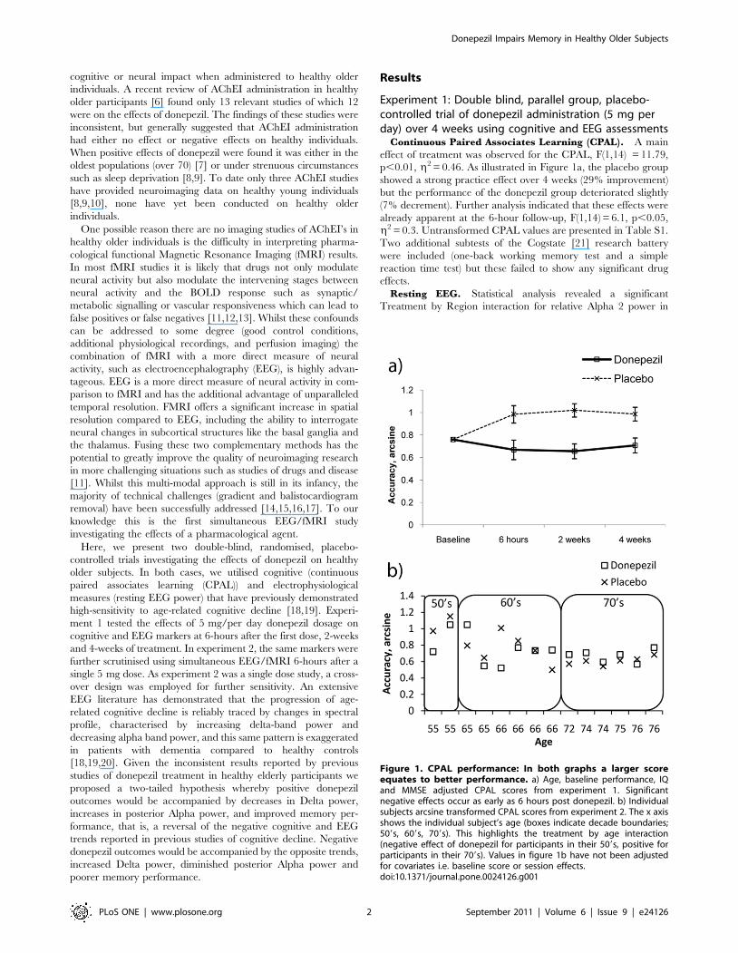
Continuous Paired Associates Learning (CPAL). A main
effect of treatment was observed for the CPAL, F(1,14) = 11.79,
p,0.01, g2 = 0.46. As illustrated in Figure 1a, the placebo group
showed a strong practice effect over 4 weeks (29% improvement)
but the performance of the donepezil group deteriorated slightly
(7% decrement). Further analysis indicated that these effects were
already apparent at the 6-hour follow-up, F(1,14) = 6.1, p,0.05,
g2 = 0.3. Untransformed CPAL values are presented in Table S1.
Two additional subtests of the Cogstate [21] research battery
were included (one-back working memory test and a simple
reaction time test) but these failed to show any significant drug
effects.
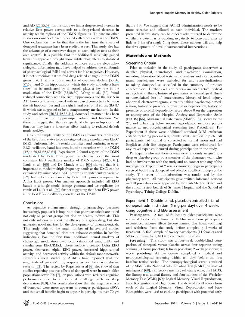
Resting EEG. Statistical analysis revealed a significant
Treatment by Region interaction for relative Alpha 2 power in
Figure 1. CPAL performance: In both graphs a larger scoreequates to better performance. a) Age, baseline performance, IQand MMSE adjusted CPAL scores from experiment 1. Significantnegative effects occur as early as 6 hours post donepezil. b) Individualsubjects arcsine transformed CPAL scores from experiment 2. The x axisshows the individual subject’s age (boxes indicate decade boundaries;509s, 609s, 709s). This highlights the treatment by age interaction(negative effect of donepezil for participants in their 509s, positive forparticipants in their 709s). Values in figure 1b have not been adjustedfor covariates i.e. baseline score or session effects.doi:10.1371/journal.pone.0024126.g001
Donepezil Impairs Memory in Healthy Older Subjects
PLoS ONE | www.plosone.org 2 September 2011 | Volume 6 | Issue 9 | e24126

the eyes closed condition, F(2,24) = 3.9, p,0.05, g2 = 0.25 and a
non-significant trend toward a main effect of Treatment
F(1,12) = 3.5, p = 0.08, g2 = 0.2. Post-hoc contrasts indicated that
the interaction was driven by a significant main effect of Treatment
at parietal electrodes, F(1,14) = 10.1, p,0.01, g2 = 0.42, reflecting a
decrease in Alpha power in the donepezil group across the one-
month follow-up period (see Figure 2a). Further analysis showed a
significant positive correlation with CPAL score and relative alpha
power at the 2week session (r = 0.5, p,0.05) whereby better
performance on the CPAL was associated with larger relative alpha
power. The same positive trend was also apparent at 6 hr (r = 0.3,
p = 0.18) and 4 week (r = 0.1, p = 0.6) follow-ups but did not reach
significance.
In the eyes open resting condition we observed a significant
main effect of Treatment for relative Beta power F(1,11) = 3.9,
p,0.05, g2 = 0.35 driven by an increase in Beta power across
frontal, central and parietal electrodes in the donepezil group
versus placebo.
Experiment 2: Double blind, crossover design, placebo-controlled trial of single dose (5 mg) donepezil usingcognitive and simultaneous EEG/fMRI assessment
Pharmacodynamic markers of donepezil which showed signif-
icant effects from experiment 1 were reassessed in experiment 2
using different subjects.Continuous Paired Associates Learning (CPAL). As in
experiment 1, participants performed significantly worse on the
CPAL 6 hrs after receiving donepezil compared to placebo
F(1,10) = 6.14, p,0.05, g2 = 0.38. Experiment 2 also showed a
significant treatment by age interaction, F(1,10) = 5.55, p,0.05,
g2 = 0.36, illustrating that the negative effects of donepezil
decreased with age (see figure 1b). Untransformed CPAL values
are presented in Table S1.Resting EEG. The EEG power trends (increasing delta,
decreasing alpha) of experiment 1 were replicated in experiment 2
(plots for significant results shown in figure S1). Significant drug-
related increases in Delta EEG power, F(1,11) = 9.68, p,0.05,
Figure 2. Tonic Alpha 2 (11-14 Hz) Drug effects: a) Resting EEG power from experiment 1 and experiment 2 both show significant decreases inrelative Alpha power on donepezil. b) Changes in Alpha 2 EEG power were mapped to right frontal-parietal regions. c) Graphs show the Alpharesponse functions from highlighted regions. In all graphs red corresponds to donepezil, blue to placebo.doi:10.1371/journal.pone.0024126.g002
Donepezil Impairs Memory in Healthy Older Subjects
PLoS ONE | www.plosone.org 3 September 2011 | Volume 6 | Issue 9 | e24126

g2 = 0.47, and decreases in Alpha 1 EEG power, F(1,11) = 10.38,
p,0.05, g2 = 0.49, were observed during the eyes closed con-
ditions. Significant interactions included a drug by session inter-
action in Alpha 1, F(1,11) = 12.53, p,0.05, g2 = 0.53, showing
that the drug effect was eliminated if donepezil was administered
first, and a drug by age interaction for Delta, F(1,11) = 6.99,
p,0.05, g2 = 0.39, that reflected a less negative drug effect for
older participants.
A significant drug by region interaction was observed during the
eyes open condition for Alpha 2 EEG power, F(5,55) = 3.88,
p,0.05, g2 = 0.26, reflecting decreased activity on donepezil over
parietal (T(1,13) = 22.56, p,0.05), occipital (T(1,13) = 22.363,
p,0.05)), and right temporal regions (T(1,13) = 23.63, p,0.005).
There was also a significant drug by region by session interaction
showing the drug effect (decreased Alpha 2 EEG power on
donepezil) was eliminated from the left temporal region if
donepezil was taken first, F(5,55) = 2.67, p,0.05, g2 = 0.2.
Finally there was a drug by region by age interaction showing
that donepezil elicited greater Alpha2 EEG power in the right
temporal region in participants under 70 and greater Alpha 2
EEG power in the central region in participants over 70, F(5,55)
= 4.67, p,0.05, g2 = 0.3. All these EEG drug effects for both eyes
open and eyes closed conditions are illustrated in figure S1.
It is also worth noting a near significant drug effect on Beta
EEG power, whereby donepezil administration lead to greater
relative Beta power compared to placebo (p = 0.06). Participants
also showed near significant drug by age interactions in both eyes
open (p = 0.051, decreased Beta activity on donepezil for
participants under 60) and eyes closed conditions (p = 0.054,
increased Beta activity on donepezil for participants over 70).
Resting EEG/fMRI. Experiment 2 used a different resting
task to experiment 1 where the participant alternated between
eyes-open and eyes-closed conditions every 30 s. This allowed us
to model two types of EEG oscillatory markers within each
frequency band; EEG reactivity (EEG power that changed
between eyes-open and eyes-closed conditions) and tonic EEG
power (EEG power unaffected by the transition between eyes-open
and eyes-closed conditions). This design gave us the opportunity to
further scrutinise EEG oscillatory markers, particularly Alpha [22]
which is a consistent marker of cognitive function, age-related
cognitive decline, and AD [23,24]. Further details of drug effects
on tonic EEG power and EEG reactivity are included in the
supplemental results (Results S1). The simultaneous EEG/fMRI
results are reported in Table 1.
Both tonic Alpha 2 and Alpha 2 reactivity showed significant,
but opposite, treatment effects in our combined EEG/fMRI
analysis. Decreases in Alpha 2 reactivity on donepezil correspond-
ed to increased BOLD activity on donepezil in the left anterior
cingulate (area 24) and the right rolandic operculum (OP1 (84%))
and decreased BOLD activity in the right superior medial gyrus
(area 24) and right anterior cingulate (area 24). Decreases in tonic
Alpha 2 power on donepezil corresponded to decreased BOLD
activity on donepezil in the left superior medial gyrus (areas 9 and
10), left middle frontal gyrus (area 44 (10%)), right superior frontal
Table 1. Table of activations for drug-related differences in EEG/fMRI.
Cluster F Co-ordinate Cytoarchitectonic BA BOLD
Alpha 2 Reactivity Size (x y z) (Probability if available) Response
Right Superior Medial Gyrus 13 7.35 12 38 52 Area 9 Placebo +ve
Right Anterior Cingulate 12 7.41 8 36 12 Area 24 Placebo +ve
Left Anterior Cingulate 14 6.98 22 26 24 Area 24 Drug +ve
Right Rolandic Operculum 16 6.67 52 222 16 OP1 (84%) Drug +ve
Delta Tonic
Right Superior Medial Gyrus 39 9.94 10 58 24 Area 10 Drug +ve
Right Middle Cingulate 49 7.69 10 0 32 Area 24 Drug +ve
Left Superior temporal Gyrus 32 7.99 254 214 24 TE 1.2 (10%) Drug +ve
Left Parahippocampal Gyrus 13 7.02 218 224 220 Hipp (sub)(90%) Drug +ve
Alpha 2 Tonic
Left Superior Medial Gyrus 48 7.45 212 56 0 Area 10 Drug –ve
Left Superior Medial Gyrus 20 7.74 28 40 46 Area 9 Drug –ve
Right Superior Frontal Gyrus 15 7.73 18 40 28 Area 9 Drug –ve
Left Middle Frontal Gyrus 27 8.26 236 18 40 Area 44 (10%) Drug –ve
Right Inferior Parietal Lobule 95 9.04 40 256 46 hIP1 (30%), hIP3 (30%), IPC (Pga)(20%) Drug –ve
Beta Tonic
Right Superior Medial Gyrus 70 8.3 10 52 2 Area 10 Placebo –ve
Right Anterior Cingulate 14 6.97 2 50 14 Area 32 Drug +ve
Right Superior Medial Gyrus 34 8.31 4 38 52 Area 6 (10%) Drug +ve
Right Cerebellum 17 7.23 38 256 230 Crus I (71%), Lobule HVI (29%) Drug –ve
Right Precuneus 18 7.31 6 262 34 SPL(7A)(10%) Placebo –ve
Right Angular Gyrus 24 7.34 46 270 32 IPC(PGp)(90%) Drug D –ve
Cluster size indicates the number voxels active in each cluster. The BOLD response column indicates the direction of the BOLD effect: ‘Drug’/’Placebo’ refers to whetherthe difference was driven by a change in activity in drug or placebo, ‘+ve’/‘-ve’ refers whether the change in activity was driven by positive or negative BOLD.doi:10.1371/journal.pone.0024126.t001
Donepezil Impairs Memory in Healthy Older Subjects
PLoS ONE | www.plosone.org 4 September 2011 | Volume 6 | Issue 9 | e24126

gyrus (area 9), and the right inferior parietal lobule (hIP1 (30%);
hIP3(30%)). The right frontal-parietal network is highlighted in
figure 2 given it is a previously established marker of age-related
cognitive decline [25,26].
Treatment effects were also present in tonic Delta EEG/fMRI
(increase in Delta EEG power on donepezil) showing increased
BOLD activity on donepezil in the superior medial gyrus (area 10),
middle cingulate (area 24), left superior temporal gyrus (T.E. 1.2
(10%)) and the left hippocampus (subiculum (90%)). Figure 3
shows the hippocampal activation along with its haemodynamic
response function and relative EEG power differences. Given that
both the hippocampus and the CPAL are strong predictors of
cognitive decline and dementia a linear regression was run with
CPAL score as a dependent variable and independent variables of
age, session, and hippocampal beta values (taken from the Delta
EEG/fMRI regressors). The results indicated a near significant
relationship between hippocampal activity and CPAL perfor-
mance (r = 20.441, p = 0.057 for drug and r = 20.445, p = 0.055
for placebo). In both cases reduced hippocampal activity was
correlated with better CPAL performance.
Finally, increased tonic Beta EEG power on donepezil was
mapped to decreased BOLD activity on donepezil within medial
territories (right superior medial gyrus (area 10), right anterior
cingulate (area 32) and right precuneus (area 7A (10%))). These
regions have been consistently found to be active during rest and
labelled the default mode network [27]. Figure 4 shows these
results overlayed on a reference image of the default mode
network shown in blue. Donepezil induced significant decreases in
Figure 3. Hippocampal Delta (1.5-3 Hz) drug effects: a) Delta response function taken from the left hippocampus (subiculum 90%). b)Hippopcampal activation driven by differences in Delta EEG power. c) Relative Delta band EEG power differences from experiment 2. In all graphs redcorresponds to donepezil, blue to placebo.doi:10.1371/journal.pone.0024126.g003
Figure 4. Beta (14-30 Hz) default mode drug effects: a) Resting EEG power from experiment 1 and experiment 2 both show significantincreases in relative Beta power on donepezil. b) Beta EEG/fMRI drug differences overlayed on a reference image of the default mode network (blue).c) Graphs show Beta response functions to posterior (left graph) and anterior (right graph) activation clusters. In all graphs red corresponds todonepezil, blue to placebo.doi:10.1371/journal.pone.0024126.g004
Donepezil Impairs Memory in Healthy Older Subjects
PLoS ONE | www.plosone.org 5 September 2011 | Volume 6 | Issue 9 | e24126

activity in the right angular gyrus (PGp (90%)), right superior
medial gyrus (BA 6 (10%)) and prefrontal projecting regions of the
right cerebellar hemisphere (Crus I (71%)).
Discussion
The cholinergic system is critical in supporting cognitive
function, and memory processes in particular, and has been
heavily implicated in AD [28,29,30]. As a result, many
pharmacological therapies for AD have been targeted toward
potentiating cholinergic function. Donepezil increases cholinergic
tone by delaying the breakdown of acetylcholine (ACh) in the
synaptic cleft and has proven efficacy in treating mild to moderate
AD [31]. Recently there has been interest in the potential for these
same drugs to strengthen cognitive function in normal ageing
[3,4,5]. Our study provides a detailed exploration of the
neurophysiological impact of AChEI administration in healthy
older individuals and in so doing provides the first ever combined
EEG/fMRI exploration of a pharmacological intervention. Our
data show that two separate groups of healthy elderly participants
exhibited consistently negative cognitive and neurophysiological
responses to donepezil treatment including a disruption of resting
EEG oscillations, reduced default mode activation, and worsening
memory performance on the CPAL. Furthermore, all of these
drug signals were detectable within six hours of the first 5 mg dose.
Although steady-state equilibrium only occurs after 14 days of
donepezil treatment [32] experiment 1 showed that the effects of
donepezil on cognitive function were still present at 2-week and 4-
week follow-ups.
The literature on the effects of donepezil administration to
healthy elderly individuals has been inconsistent with individual
studies reporting negative, positive, or no effects [6]. One reason
for this lack of consistency may be that donepezil’s effectiveness is
dependent on specific factors including age and mental state. For
example, of the studies on healthy subjects that have reported
positive donepezil effects, two included participants who were in a
state of sleep deprivation [8,9] and another included older age
elderly participants (70 s +) [7]. This tallies with the observations
that AChEI treatment benefits correlate with disease severity
in patients with AD [33]. Our results also highlight an age-
interaction with older participants showing slight improvements
under medication on both behavioural and neurophysiological
markers. Taken together, these findings suggest that future studies
are warranted that would seek to characterise normal elderly sub-
groups who may benefit significantly from cholinergic treatment.
Neural Markers of donepezil administrationResting recordings acquired from EEG or fMRI are an
increasingly common measure of functional connectivity and are
often used as markers of disease or pharmacological intervention
[34,35]. A large number of ageing studies have consistently
highlighted diminished activity within the Alpha band (8–13 Hz)
accompanied by an increase in the power of slower Delta (1–4 Hz)
and Theta (4–8 Hz) frequency ranges. This pattern appears to be
exaggerated in patients with AD who exhibit a further increase in
Delta and Theta power, and decreased Alpha power relative to
age-matched controls [20,36,37]. These pathophysiological trends
are significantly reduced amongst patients who respond to long-
term AChEI treatment [38,39,40]. In both experiment 1 and
experiment 2 we observed the opposite effect in healthy older
subjects, such that acute (6 hr) and chronic (up to 4-week)
administration of donepezil led to increased Delta EEG power and
decreased Alpha EEG power. These effects were accompanied by
significantly reduced memory performance.
Effects of Donepezil on Alpha and the Right Frontal-
Parietal Network. Tonic activity in the Alpha range is thought
to reflect the cognitive and attentional resources available to an
individual [23,24]. Babiloni et al., [38] showed using EEG source
analysis that posterior Alpha activity was the best EEG predictor
of a patient’s response to AChEI treatment. In both experiment 1
and 2 we found that donepezil administration led to a decreases in
posterior relative Alpha power. Alpha EEG power has also been
consistently mapped to the right frontal-parietal regions [41,42]
and in this study we show that donepezil targeted this same
network. Activation of the right frontal-parietal network (but not
the left) has been shown to decrease linearly when comparing
healthy elderly individuals to patients with Mild Cognitive
Impairment (MCI) and when comparing patients with MCI to
patients with AD [25]. Other studies have also found that right
parietal activity is related to donepezil induced changes in
cognitive performance. Chuah et al., [8,9] showed that decreased
activity within the right intraparietal sulcus (IPS) was linked to worse
performance on a visual short term memory task whereas
participants that responded positively to donepezil showed
increased activity within the right IPS. Our combined EEG/
fMRI results confirm that the right frontal-parietal Alpha network
provides a useful index of the efficacy of cholinergic interventions.
Effects of Donepezil on Delta and the Hippocampus. As
with Alpha, changes in Delta EEG power are consistently found to
be markers of cognitive decline in ageing [18,43] and AD [20].
Using simultaneous EEG/fMRI, Dang-Vu et al., [44] showed that
Delta EEG power correlated with activity in both medial and
lateral structures of the prefrontal cortex. Consistent with this work
our results indicate that drug-related differences in tonic Delta
EEG power correlated to activity changes in medial prefrontal
regions along with differences in the left superior temporal gyrus,
and the left hippocampus (subiculum). Hippocampal volume is a
strong predictor of cognitive decline and of MCI conversion to AD
[45,46]. The paired associates learning task is also a strong
predictor of MCI conversion [47] and performance on this task
has been repeatedly linked to hippocampal integrity [48,49].
Although the MRI literature on donepezil treatment is relatively
small, a large number of clinical studies have found that extended
donepezil treatment (from 10 weeks to 2 years) typically leads to
increases in hippocampal volume or resistance to hippocampal
atrophy [50,51,52,53,54]. Hippocampal volume can also be used
to predict patient response to donepezil treatment [51]. To date
however, no studies of donepezil have looked at hippocampal
activity as a marker of cognitive decline using fMRI or PET. There
was a near significant relationship between hippocampal activity
and CPAL performance showing that reduced hippocampal
activity was correlated with better CPAL performance (p = 0.057
for drug; p = 0.055 for placebo). We believe these results suggest
that donepezil had a negative impact on CPAL performance due
to disruption of hippocampal function. Through the use of
simultaneous EEG/fMRI we found that this disruption in
hippocampal function was correlated with changes in relative
Delta EEG power.
Effects of Donepezil on Beta and the Default Mode
Network. The default mode network (DMN) is a term used to
describe a network of regions including medial prefrontal cortex
(BA 10 and anterior cingulate cortex), posterior cingulate/
retrosplenial cortex, and bilateral inferior parietal lobules that
are consistently found to be active during periods of rest [27]. The
DMN is also proving to be a powerful marker of cognitive decline
and disease [27,35]. Activity within the DMN decreases with
poorer performance on working memory and attention tasks
[55,56,57,58] and decreases linearly during healthy ageing, MCI
Donepezil Impairs Memory in Healthy Older Subjects
PLoS ONE | www.plosone.org 6 September 2011 | Volume 6 | Issue 9 | e24126
and AD [25,55,57]. In this study we find a drug-related increase in
relative Beta power corresponds to a drug-related decrease in
activity within regions of the DMN (figure 4). To date no other
studies on donepezil have reported differences within the DMN.
One explanation may be that this is the first time the effects of
donepezil treatment have been studied at rest. This study also has
the advantage of a crossover design so each subject acts as their
own control. It is possible that the additional sensitivity gained
from this approach brought more subtle drug effects to statistical
significance. Finally, the addition of more accurate electrophy-
siological information may have helped to address the confounds
of pharmacological fMRI and correct for false negatives. However,
it is not surprising that we find drug-related changes in the DMN
given that; 1) it is a robust marker of cognitive decline [55,56,
57,58], and 2) the hippocampus (which this study and others have
shown to be modulated by donepezil) plays a key role in the
modulation of the DMN [55,58,59]. Wang et al., [58] found
reduced connectivity with the right hippocampus and the DMN in
AD, however, this was paired with increased connectivity between
the left hippocampus and the right lateral prefrontal cortex (BA 8/
9) which was suggested to be a compensatory mechanism. In this
study and others [50,51,52,53,54], donepezil treatment has been
shown to impact on hippocampal volume and function. We
therefore suggest that these drug-related changes in hippocampal
function may have a knock-on effect leading to reduced default
mode activity.
Given the simple utility of the DMN as a biomarker, it was one
of the first brain states to be investigated using simultaneous EEG/
fMRI. Unfortunately, the results are mixed and confusing as every
EEG oscillatory band has been found to correlate with the DMN
[42,44,60,61,62,63,64]. Experiment 2 found regions of the DMN
modulated by Beta EEG power which has been the most
consistent EEG oscillatory marker of DMN activity [42,60,61].
Laufs et al., [60] and De Munck et al., [65] showed that it is
important to model multiple frequency bands as the DMN can be
explained by using Alpha EEG power as an independent variable
[62] but is better explained by Beta EEG power compared to
Alpha EEG power. Our analysis approach included all EEG
bands in a single model (except gamma) and we replicate the
results of Laufs et al., [60] further suggesting that Beta EEG power
is the best EEG oscillatory correlate of the DMN.
ConclusionsAs cognitive enhancement through pharmacology becomes
increasingly popular it is important that pharmaceuticals are tested
not only on patient groups but also on healthy individuals. This
not only informs us about the efficacy of a given drug, but also
generates novel markers for the development of pharmaceuticals.
This study adds to the small number of behavioural studies
suggesting that donepezil does not enhance cognition in healthy
individuals. For the first time, additional neural markers of
cholinergic modulation have been established using EEG and
simultaneous EEG/fMRI. These include increased Delta EEG
power, decreased Alpha EEG power, increased hippocampal
activity, and decreased activity within the default mode network.
Previous clinical studies of AChEIs have reported that the
magnitude of patients’ drug response is correlated with disease
severity [33]. The review by Repenatis et al [6] also showed that
studies reporting positive effects of donepezil were in much older
populations (over 70) [7], or populations with reduced cognitive
performance due to an external challenge such as sleep
deprivation [8,9]. Our results also show that the negative effects
of donepezil were more apparent in younger participants (509s),
and that small benefits begin to appear in participants over 70 yrs
(figure 1b). We suggest that AChEI administration needs to be
more selective and tailored to each individual. The markers
presented in this study can be quickly administered to determine
whether a patient is responding negatively to donepezil after as
little as 6 hrs of a single 5 mg dose. These markers will also help
the development of novel pharmaceutical interventions.
Materials and Methods
Screening CriteriaPrior to inclusion in the study all participants underwent a
detailed physical, neurological and psychiatric examination,
including laboratory blood tests, urine analysis and electrocardio-
gram. Participants were excluded for any contraindications
to taking donepezil as specified in the summary of product
characteristics. Further exclusion criteria included active medical
or psychiatric illness, history of psychiatric or neurological illness
or unexplained loss of consciousness, history of head injury,
abnormal electrocardiogram, currently taking psychotropic med-
ication, history or presence of drug use or dependency, history or
presence of alcohol dependency, score above 8 on the depression
or anxiety axes of the Hospital Anxiety and Depression Scale
(HADS) [66], Mini-mental state exam (MMSE) [67] scores below
27, and exhibiting below normal age-adjusted memory perfor-
mance at neuropsychological screening (see Procedures). For
Experiment 2 there were additional standard MRI exclusion
criteria including pacemakers, shunts, stents, artificial hip etc. All
participants had normal or corrected to normal vision and had
English as their first language. Participants were reimbursed for
any travel expenses incurred during participation in the study.
Participants who met these criteria were randomly assigned to a
drug or placebo group by a member of the pharmacy team who
had no involvement with the study and no contact with any of the
investigators. Experiment 2 was a crossover design so participants
received both 5 mg donepezil and placebo at different stages of the
study. The order of administration was randomised by the
pharmacy team. All participants gave written informed consent
and all procedures were approved by the Irish Medical Board and
the ethical review boards of St James Hospital and the School of
Psychology, Trinity College Dublin.
Experiment 1: Double blind, placebo-controlled trial ofdonepezil administration (5 mg per day) over 4 weeksusing cognitive and EEG assessment
Participants. A total of 24 healthy older participants were
recruited to the study from the Dublin area. Four participants
experienced adverse effects (severe nausea, diarrhoea, vomiting)
and withdrew from the study before completing 2-weeks of
treatment. A final sample of twenty participants (14 female) aged
59 to 77 (mean 67.5, SD = 5) completed the study.
Screening. This study was a four-week double-blind com-
parison of donepezil versus placebo across four separate testing
sessions (24 hours pre-drug, 6 hours post-drug, 2 weeks post-drug, 4
weeks post-drug). All participants completed a medical and
neuropsychological screening within ten days before the first
baseline testing session. The neuropsychological screen consisted
of the MMSE, the National Adult Reading Test (NART, estimate of
intelligence) [68], a subjective memory self-rating scale, the HADS,
the Stroop test, animal fluency and four subtests of the Wechsler
Memory Test (WMS) [69]: Logical Memory, Visual Reproduction,
Face Recognition and Digit Span. The delayed recall scores from
each of the Logical Memory, Visual Reproduction and Face
Recognition were used to exclude participants with potential early
Donepezil Impairs Memory in Healthy Older Subjects
PLoS ONE | www.plosone.org 7 September 2011 | Volume 6 | Issue 9 | e24126

dementia (defined as a z-score greater than 1.5 standard deviations
below the norm).
On completion of screening, a randomisation procedure was
used by the hospital pharmacy staff to allocate participants to
either the donepezil or placebo group. The randomisation was
stratified to ensure a balanced male-female ratio in each group.
Participants received either 5 mg of donepezil or placebo once a
day for four weeks. The drug and placebo capsules were over-
encapsulated in order to make them indistinguishable. Adminis-
tration of the first dose took place in the hospital. Compliance was
assessed at each of the post-drug testing sessions by checking the
pill count of dispensed medication. Subjects, test administrators
and all medical staff were blinded to the treatment subjects
received. On completion of the study a full medical screening was
repeated.
EEG and Cognitive TestingEach testing session was identical and consisted of a number of
EEG-tailored cognitive tests and three subtests from the Cogstate
Research battery [21]. Each session lasted approximately 90
minutes including frequent rest breaks. The EEG tests included a
three minute eyes-closed resting recording, a three minute eyes-
open resting recording, a two-stimulus auditory oddball task in
which participants were presented with a series of tones and were
required to make a mouse button press when they heard a low-
probability (0.2) target tone (‘passive oddball’), a two-stimulus
auditory oddball task in which participants were required to make
a mouse button press following all tones except for the low-
probability target tones (‘inhibitory oddball’). The Cogstate
subtests that were administered were the continuous paired
associates learning task (CPAL), the one-back task and the
detection task. The CPAL is designed to assess working memory
and proceeds in two stages. In the first stage, the subject must learn
the locations of different pictures that are presented on screen. As
each picture to be learned is revealed, the subject must tap each
location and remember where the picture was located. In the
second part of the task the same pictures are presented in the
centre of the screen, and the subject must tap on the peripheral
location where that picture previously appeared. The participant
receives feedback on the accuracy of each response allowing them
to update their selection until the correct location has been
identified (the one-back and detection tasks have been described
elsewhere, see Fredrickson et al) [21]. All tests were computerised
and were administered by a research nurse or research psychologist
in a sound-attenuated, dimly lit room. Participants were tested while
seated in an armchair ,65 cm from the computer screen. Time-of-
day was held constant across all testing sessions for each subject. The
order of test presentation within each session was also held constant.
The Cogstate battery was always administered half way through the
EEG battery. Participants were asked to abstain from caffeine and
tobacco during the one-hour period prior to testing.
EEG acquisition. Continuous EEG was acquired through
the ActiveTwo Biosemi electrode system from 64 scalp electrodes,
digitized at 512 Hz. Vertical eye movements were recorded with
two vertical electrooculogram (EOG) electrodes placed below the
left and right eye, while electrodes at the outer canthus of each eye
recorded horizontal movements. EEG data pre-processing and
analysis were conducted using BESA Version 5.2 (Brain Electric
Source Analysis) software. Blinks and eye movements were
corrected using an algorithm developed by Berg and Scherg
[70]. All electrode channels were average referenced offline and
any segments with an amplitude deflection of greater than 90 mV
were rejected in order to exclude excessive EOG or other noise
transients.
Data analysis. For analysis and display purposes, data were
average referenced and filtered with a low-pass 0-phase shift 48-
dB, 40-Hz filter and a high-pass 0-phase shift 6-dB, 0.3-Hz. filter.
Eyes-closed and eyes-open resting EEG band power was
calculated using the discrete Fourier transform. Absolute power
values are sensitive to confounds of head volume conduction and
so to counteract this problem the use of relative power measures
has been recommended [71,72,73,74]. Relative power measures
were also calculated by dividing the absolute power within the
band by absolute power for the range of interest (1–30 Hz).
Relative power was calculated separately for the Delta (1.0–
3.5 Hz), Theta (4–7.5 Hz), Alpha 1 (8–10.5 Hz), Alpha 2 (10.5–
13 Hz) and Beta (14–30 Hz) bands. Electrodes were grouped into
separate frontal (F1, Fz, F2), central (C1, Cz, C2) and parietal (P1,
Pz, P2) clusters.Statistical analysis. Any missing values were replaced using
the last observation carried forward method. Missing values
occurred for two participants: in one case because the participant
withdrew from the study before the final follow-up session and in
the other because of excessively noisy data (,50% data yield) at
the two week follow-up during the eyes open resting EEG tasks.
Treatment effects on each of the cognitive and EEG variables were
assessed using a repeated-measures ANCOVA. Factors included
for the cognitive measures were Treatment (Donepezil vs Placebo)
and Time (6-hours, 2 weeks, 4 weeks). Factors of Treatment, Time
and Region (Frontal, Central, Parietal) were included for the EEG
analyses. Baseline performance/band power was included as a
covariate in each case as were age, MMSE score and estimated
IQ. In cases where the Treatment effect reached significance a
separate ANCOVA was conducted to explore acute drug effects at
the 6-hour time point, with the same covariates as above. A
Pearson correlation was run to assess the relationship between
CPAL performance and resting EEG power measures which
showed significant treatment effects. Individual performance/
power measures were z transformed prior to the correlation
analysis.
Experiment 2: Double blind, crossover design, placebo-controlled trial of single dose (5 mg) donepeziladministration using cognitive and simultaneous EEG/fMRI assessment
Participants. A total of twenty six healthy older participants
were recruited to the study from the Dublin area. Eleven failed the
initial screening and did not participate further in the experiment
(same criteria as above – see Experiment 1, participants). One
participant had corrupted EEG data and was excluded from the
analysis. A final sample of fourteen participants (10 female) aged
55 to 76 (mean 67.93, SD = 7) were used for this study.Screening. This study was a double-blind comparison of
donepezil versus placebo within the same subjects across three
separate testing sessions (Baseline (no dosage), Session 1 (donepezil
or placebo), and Session 2 (alternative to session 1)). All
participants completed the same medical and neuropsychological
screening as in experiment 1.
On completion of screening, a randomisation procedure was
used by the hospital pharmacy staff to allocate participants to
receive either donepezil first or placebo first. All participants
received both donepezil and placebo after completing both
sessions. The drug and placebo capsules were over-encapsulated
in order to make them indistinguishable. Subjects, test adminis-
trators and all medical staff were blinded to the treatment subjects
received. Administration of the first dose took place in the hospital
between 9:30 and 10 am. Subjects then returned on the same day
at 3 pm for cognitive testing and EEG-fMRI (entered the scanner
Donepezil Impairs Memory in Healthy Older Subjects
PLoS ONE | www.plosone.org 8 September 2011 | Volume 6 | Issue 9 | e24126

at 4 pm). Keeping scan time consistent helps to ensure a more
morphologically stable BOLD response across sessions [75]. On
completion of the study a full medical screening was repeated.
EEG and Cognitive Testing. Each testing session was
identical and lasted 2 hours with approximately 1 hour of testing
in the MRI scanner. Cognitive tests were administered outside the
MRI scanner before EEG-fMRI tests. Only the CPAL was
administered as this was the only cognitive task to show sig-
nificant treatment effects in experiment 1. Participants then waited
whilst the EEG cap was applied and electrode impedances reduced
(below 10 kV, details in EEG-fMRI data acquisition below). The
subject then entered the MRI scanner and performed an alternating
eyes-open eyes-closed resting state, and three stimulus oddball task.
The results of the three stimulus oddball will not be reported in this
manuscript. The resting state task, inspired by Ben-simon et al.,
[22], had participants keep their eyes open for 30 s (during this time
the screen said ‘‘OPEN’’ in large font). After 30 s the screen said
‘‘CLOSED’’ and participants closed their eyes. Participants then
received a tap on the leg 30 s later to open their eyes again (once
again the screen said ‘‘OPEN’’). Visual instructions were used
because piloting found that older participants often could not hear
an auditory tone over the noise of the MRI scanner. This approach
ensured that participants opened and closed their eyes as instructed.
This resting state paradigm has the additional advantage of
investigating two types of Alpha; Alpha reactivity and tonic
Alpha, which have been shown to correspond to separate neural
networks [22].
EEG/fMRI Apparatus. Subjects lay supine in an MRI
scanner with the thumb of the right hand positioned on a two-
button MRI-compatible response box. Stimuli were projected onto
a screen behind the subject and viewed in a mirror positioned
above the subjects face. Presentation software (Neurobehavioral
Systems, Inc., USA) was used for stimulus presentation both in-
side and outside the scanner. A separate laptop running Brain
Recorder v1.04 (BrainProducts, Munich, Germany) was used to
record the EEG data at a frequency of 5 KHz (band-pass filtering
from 0.016 to 250 Hz) along with event timings, response timings,
and TTL pulses from the MRI scanner at the onset of each
volume acquisition. These TTL pulses were also used to drive the
visual stimuli in Presentation. The EEG clock was synchronised to
the MRI scanner clock using the Brain products Sync-box [76].
Event timings and reaction times were calculated off-line using
event timings acquired by Brain Recorder at this higher sampling
frequency.
EEG/fMRI data acquisition. EEG recordings were
acquired with a 32-channel MR-compatible BrainAmp system
(Brainproducts, Munich, Germany). Thirty-one EEG electrodes
were placed on the scalp not including the reference electrode
positioned in correspondence to the FCz electrode (between
electrodes Fz and Cz) and the ground electrode placed in
correspondence to AFz (directly in front of Fz). One hangdown
electrode was used to acquire the electrocardiogram (ECG). The
impedance of all electrodes was maintained below 10 kV.
On the baseline sessions additional structural images were
acquired. These included high-resolution T1-weighted anatomic
MPRAGE image (FOV = 256*256*160 mm, thickness = 1 mm,
voxel size = 1 mm*1 mm*1 mm), T2-weighted FLAIR (FOV =
230*143*184 mm, thickness = 4 mm (slice gap 1 mm), voxel
= 0.45*0.45*6 mm), and T2-weighted spin echo (FOV = 230*
143*201 mm, thickness = 4 mm (slice gap 1 mm), voxel = 0.45*
0.45*6 mm). On every MRI session field maps were first acquired
(two phase and magnitude images (TE1 = 1.46 ms, TE2 = 7 ms)).
This was directly followed by the resting state EEG/MRI. The field
of view covered the whole brain, 224*224 mm (64*64 voxels), 34
axial slices were acquired (0.05 mm slice gap) with a voxel size of
3.5 mm*3.5 mm*4 mm; TR = 2 s, TE = 32 ms, flip angle = 78u.This was a sparse-sampling sequence with the slices compressed to
the first 1700 ms of the TR, leaving 300 ms uncontaminated by the
MR gradient artefact. This was done to improve EEG data cleanup
[77]. Each EPI session lasted 7 minutes (210 volumes). All MRI data
was collected on a Philips 3T Achieva MRI Scanner (Centre for
Advanced Medical Imaging (CAMI), St James’s Hospital, Dublin).
EEG/fMRI pre-processing. EEG data was pre-processed
using Analyser (BrainProducts, Munich, Germany) on an Intel
Quad Core PC with 4GB of RAM, running Windows Vista SP2.
Before pre-processing the data, the first TTL pulse of each EPI
session was removed as it occurred at a different point in the
gradient artefact template. After this, gradient correction was
applied using the Average Artefact Subtraction (AAS) method
[14], and a moving average of 25 windows. EEG data was then
downsampled to 500 Hz and filtered between 0.53–30 Hz using
IIR filters with an additional 50 Hz Notch filter. R-peaks were
then detected in the ECG channel and removed using the AAS
method [15], and a moving average of 11 artefacts. For both the
gradient correction and balistocardiogram (BCG) correction
multiple average window lengths were tried and these two were
found to be the best based on decreased power at contaminated
frequencies. Independent Components Analysis (ICA) was then
run on the whole data set to remove artefacts including; blinks, eye
movements, EMG and residual BCG artefacts [78]. The
continuous data was then epoched based on TR markers (0–
2000 ms) and artifactual epochs marked but not removed. An
epoch was considered artifactual if the amplitude was 680 mV, the
voltage step was greater than 26 mV/ms, or activity lower than
0.5 mV. A discrete Fast Fourier Transform (FFT) was then run on
each epoch, the results of which were then averaged.
Scans were pre-processed using SPM8 (www.fil.ion.ucl.ac.uk/
spm) on an Intel Quad Core PC with 4GB of RAM, running
Fedora 12 and Matlab 2009a (MathWorks Inc). Before pre-
processing EPI data quality tests were run using the criterion
defined in Iannetti et al., [79]. These include assessing the mean
and standard deviation of the signal timecourse, image signal to
noise, standard deviation of the single voxel signal timecourse, and
visual inspection for ghost artefact and signal dropout. All EPI
data collected passed these tests. Images were then realigned and
unwarped using field maps to correct for motion artefacts,
susceptibility artefacts and motion-by-susceptibility interactions
[80,81]. Images were subsequently normalized to the ICBM EPI
template using the unified segmentation approach [82]. Lastly, a
Gaussian kernel of 8 mm was applied to spatially smooth the
image in order to conform to the Gaussian assumptions of a GLM
as implemented in SPM8 [83,84].
Statistical analysis. For analysis of both the CPAL and
EEG frequency bands, a repeated measures ANOVA was
conducted covarying for session (drug first or placebo first) and
age. In the CPAL analysis baseline scores were also used as an
additional covariate. For EEG frequency band differences a three
way ANOVA was first run using factors of Treatment (Drug,
Placebo), Frequency (Delta, Theta, Alpha 1, Alpha 2, Beta), and
Location(Frontal (F3, F4, Fz), Central (C3, C4, Cz), Parietal (P3,
P4, Pz), Occipital (O1, O2, Oz), Left Temporal (TP9, T7, P7),
Right Temporal (TP10, T8, P8)). These were subsequently broken
down into two way ANOVAs looking at each frequency band
individually.
EEG/fMRI analysis. EEG power time courses were
extracted using the same frequency boundaries as experiment 1
(Delta: 1.5–3.5 Hz; Theta: 4–7.5 Hz; Alpha 1: 8–11.5 Hz; Alpha
2: 12–13.5 Hz; Beta: 14–30 Hz). These timecourses were then
Donepezil Impairs Memory in Healthy Older Subjects
PLoS ONE | www.plosone.org 9 September 2011 | Volume 6 | Issue 9 | e24126

downsampled to the TR sampling rate (0.5 Hz) and convolved
with the informed basis set [85,86]. Epochs previously marked as
bad were replaced by the average of the two preceding and two
proceeding data points. A general linear model (GLM) was
constructed for each EEG-fMRI session consisting of the
powertime course of each frequency band, convolved with three
functions of the informed basis set (canonical hrf, temporal
derivative, and dispersion derivative). Head movement parameters
were also included. In order to assess drug related changes in tonic
EEG (unaffected by eyes open vs eyes closed) a separate GLM was
run which included all of the above regressors, plus an additional
condition describing the changes between eyes open and eyes
closed (this condition was modelled as three regressors due to the
convolution with the informed basis set).
A second level random effects ANOVA was run using three
factors; Treatment (donepezil or placebo), Time (session 1 or
session 2); and Basis function (Canonical hrf, Temporal, and
Dispersion derivative). Age was included as a co-variate given the
drug*age interactions seen in the CPAL and the separate EEG
analyses. Consistent with the literature [22,62,87,88,89,90,91,92],
results were thresholded at p,0.001 uncorrected, with an extent
threshold of 10 unless otherwise specified. Anatomical localisation
was performed using the anatomy toolbox [93]. Frequency
response functions shown in figures 2, 3, 4 were calculated by
multiplying the informed basis set with its respective Beta value
from the peak of a cluster of interest.
Supporting Information
Figure S1 EEG drug effects for eyes open and eyesclosed conditions. a) Delta eyes closed drug effect. b) Alpha1
eyes closed drug effect. c) Delta eyes closed drug by age
interaction. d) Alpha1 eyes closed drug by session interaction. e)
Alpha2 eyes open drug by region interaction. An asterisk next to
the region name indicate a significant difference (p,0.05). f)
Alpha2 eyes open drug by region by session interaction. Circles
highlight the driving force of the interaction. g) Alpha2 eyes open
drug by region by session interaction. Drug placebo differences are
plotted. Central and right temporal differences are highlighted as
they drive the drug by region by session interaction. In plots a–e
red represents relative EEG power on donepezil, blue represents
placebo.
(DOCX)
Table S1 CPAL performance for experiment 1 andexperiment 2. Values are percent accuracy with values in
brackets showing the standard error.
(DOCX)
Results S1 Experiment 2 EEG results for drug effects ontonic EEG power and EEG reactivity.
(DOCX)
Acknowledgments
First, the authors wish to warmly thank everyone who participated in this
study. The authors would also like to thank Lisa Crosby, Meredith Hodder,
Orlaith Keane, and Aliz Takacs who helped organise the participants and
collect data.
Author Contributions
Conceived and designed the experiments: JHB RO MPM SB ALWB NU
RL BL IHR. Performed the experiments: JHB RO MPM AG SMC SMK
SD AJF. Analyzed the data: JHB RO AG SMC SMK. Wrote the paper:
JHB RO MPM AG SB JFM AJF ALWB NU RL ML BL IHR.
References
1. Plassman BL, Langa KM, Fisher GG, Heeringa SG, Weir DR, et al. (2007)
Prevalence of dementia in the United States: the aging, demographics, and
memory study. Neuroepidemiology 29: 125–132.
2. UK Office for National Statistics website. Available: http://www.statistics.gov.
uk/cci/nugget.asp?id = 168. Accessed 2010 Dec. 22.
3. Maher B (2008) Poll results: look who’s doping. Nature 452: 674–675.
4. Farah MJ, Illes J, Cook-Deegan R, Gardner H, Kandel E, et al. (2004)
Neurocognitive enhancement: what can we do and what should we do? Nat Rev
Neurosci 5: 421–425.
5. Larriviere D, Williams MA, Rizzo M, Bonnie RJ (2009) Responding to requests
from adult patients for neuroenhancements: guidance of the Ethics, Law and
Humanities Committee. Neurology 73: 1406–1412.
6. Repantis D, Laisney O, Heuser I (2010) Acetylcholinesterase inhibitors and
memantine for neuroenhancement in healthy individuals: a systematic review.
Pharmacol Res 61: 473–481.
7. FitzGerald DB, Crucian GP, Mielke JB, Shenal BV, Burks D, et al. (2008)
Effects of donepezil on verbal memory after semantic processing in healthy older
adults. Cogn Behav Neurol 21: 57–64.
8. Chuah LY, Chee MW (2008) Cholinergic augmentation modulates visual task
performance in sleep-deprived young adults. J Neurosci 28: 11369–11377.
9. Chuah LY, Chong DL, Chen AK, Rekshan WR, 3rd, Tan JC, et al. (2009)
Donepezil improves episodic memory in young individuals vulnerable to the
effects of sleep deprivation. Sleep 32: 999–1010.
10. Silver MA, Shenhav A, D’Esposito M (2008) Cholinergic enhancement reduces
spatial spread of visual responses in human early visual cortex. Neuron 60:
904–914.
11. Iannetti GD, Wise RG (2007) BOLD functional MRI in disease and pharmaco-
logical studies: room for improvement? Magn Reson Imaging 25: 978–988.
12. Wise RG, Tracey I (2006) The role of fMRI in drug discovery. J Magn Reson
Imaging 23: 862–876.
13. Matthews PM, Honey GD, Bullmore ET (2006) Applications of fMRI in
translational medicine and clinical practice. Nat Rev Neurosci 7: 732–744.
14. Allen PJ, Josephs O, Turner R (2000) A method for removing imaging artifact
from continuous EEG recorded during functional MRI. Neuroimage 12:
230–239.
15. Allen PJ, Polizzi G, Krakow K, Fish DR, Lemieux L (1998) Identification of
EEG events in the MR scanner: the problem of pulse artifact and a method for
its subtraction. Neuroimage 8: 229–239.
16. Niazy RK, Beckmann CF, Iannetti GD, Brady JM, Smith SM (2005) Removal
of FMRI environment artifacts from EEG data using optimal basis sets.
Neuroimage 28: 720–737.
17. Laufs H, Daunizeau J, Carmichael DW, Kleinschmidt A (2008) Recent
advances in recording electrophysiological data simultaneously with magnetic
resonance imaging. Neuroimage 40: 515–528.
18. Babiloni C, Binetti G, Cassarino A, Dal Forno G, Del Percio C, et al. (2006)
Sources of cortical rhythms in adults during physiological aging: a multicentric
EEG study. Hum Brain Mapp 27: 162–172.
19. Babiloni C, Binetti G, Cassetta E, Dal Forno G, Del Percio C, et al. (2006)
Sources of cortical rhythms change as a function of cognitive impairment in
pathological aging: a multicenter study. Clin Neurophysiol 117: 252–268.
20. Babiloni C, Binetti G, Cassetta E, Cerboneschi D, Dal Forno G, et al. (2004)
Mapping distributed sources of cortical rhythms in mild Alzheimer’s disease. A
multicentric EEG study. Neuroimage 22: 57–67.
21. Fredrickson J, Maruff P, Woodward M, Moore L, Fredrickson A, et al. (2010)
Evaluation of the usability of a brief computerized cognitive screening test in
older people for epidemiological studies. Neuroepidemiology 34: 65–75.
22. Ben-Simon E, Podlipsky I, Arieli A, Zhdanov A, Hendler T (2008) Never resting
brain: simultaneous representation of two alpha related processes in humans.
PLoS One 3: e3984.
23. Klimesch W (1999) EEG alpha and theta oscillations reflect cognitive and
memory performance: a review and analysis. Brain Res Rev 29: 169–195.
24. Dockree PM, Kelly SP, Foxe JJ, Reilly RB, Robertson IH (2007) Optimal
sustained attention is linked to the spectral content of background EEG activity:
greater ongoing tonic alpha (approximately 10 Hz) power supports successful
phasic goal activation. Eur J Neurosci 25: 900–907.
25. Rombouts SA, Damoiseaux JS, Goekoop R, Barkhof F, Scheltens P, et al. (2009)
Model-free group analysis shows altered BOLD FMRI networks in dementia.
Hum Brain Mapp 30: 256–266.
26. Damoiseaux JS, Beckmann CF, Arigita EJ, Barkhof F, Scheltens P, et al. (2008)
Reduced resting-state brain activity in the ‘‘default network’’ in normal aging.
Cereb Cortex 18: 1856–1864.
27. Buckner RL, Andrews-Hanna JR, Schacter DL (2008) The brain’s default
network: anatomy, function, and relevance to disease. Ann N Y Acad Sci 1124:
1–38.
28. Drachman DA, Leavitt J (1974) Human memory and the cholinergic system. A
relationship to aging? Arch Neurol 30: 113–121.
Donepezil Impairs Memory in Healthy Older Subjects
PLoS ONE | www.plosone.org 10 September 2011 | Volume 6 | Issue 9 | e24126

29. Francis PT, Palmer AM, Snape M, Wilcock GK (1999) The cholinergichypothesis of Alzeimer’s disease: a review of progress. J Neurol Neurosurg
Psychiatry 66: 137–147.
30. Bartus RT, Dean III RL, Beer B, Lippa AS (1982) The cholinergic hypothesis ofgeriatric memory dysfunction. Science 217: 408–414.
31. Birks J (2006) Cholinesterase inhibitors for Alzheimer’s disease. CochraneDatabase Syst Rev 25: CD005593.
32. Beglinger LJ, Tangphao-Daniels O, Kareken DA, Zhang L, Mohs R, et al.
(2005) Neuropsychological test performance in healthy elderly volunteers beforeand after donepezil administration: a randomized, controlled study. J Clin
Psychopharmacol 25: 159–165.
33. Rogers SL, Farlow MR, Doody RS, Mohs R, Friedhoff LT (1998) A 24-week,
double-blind, placebo-controlled trial of donepezil in patients with Alzheimer’s
disease. Donepezil Study Group. Neurology 50: 136–145.
34. Fox MD, Raichle ME (2007) Spontaneous fluctuations in brain activity observed
with functional magnetic resonance imaging. Nat Rev Neurosci 8: 700–711.
35. Broyd SJ, Demanuele C, Debener S, Helps SK, James CJ, et al. (2009) Default-mode brain dysfunction in mental disorders: a systematic review. Neurosci
Biobehav Rev 33: 279–296.
36. Jeong J (2004) EEG dynamics in patients with Alzheimer’s disease. Clin
Neurophysiol 115: 1490–1505.
37. Huang C, Wahlund L, Dierks T, Julin P, Winblad B, et al. (2000) Discriminationof Alzheimer’s disease and mild cognitive impairment by equivalent EEG
sources: a cross-sectional and longitudinal study. Clin Neurophysiol 111:1961–1967.
38. Babiloni C, Cassetta E, Dal Forno G, Del Percio C, Ferreri F, et al. (2006)
Donepezil effects on sources of cortical rhythms in mild Alzheimer’s disease:Responders vs. Non-Responders. Neuroimage 31: 1650–1665.
39. Reeves RR, Struve FA, Patrick G (2002) The effects of donepezil on quantitativeEEG in patients with Alzheimer’s disease. Clin Electroencephalogr 33: 93–96.
40. Rodriguez G, Vitali P, De Leo C, De Carli F, Girtler N, et al. (2002)
Quantitative EEG changes in Alzheimer patients during long-term donepeziltherapy. Neuropsychobiology 46: 49–56.
41. Laufs H, Holt JL, Elfont R, Krams M, Paul JS, et al. (2006) Where the BOLD
signal goes when alpha EEG leaves. Neuroimage 31: 1408–1418.
42. Mantini D, Perrucci MG, Del Gratta C, Romani GL, Corbetta M (2007)
Electrophysiological signatures of resting state networks in the human brain.Proc Natl Acad Sci U S A 104: 13170–13175.
43. Hartikainen P, Soininen H, Partanen J, Helkala EL, Riekkinen P (1992) Aging
and spectral analysis of EEG in normal subjects: a link to memory and CSFAChE. Acta Neurol Scand 86: 148–155.
44. Dang-Vu TT, Schabus M, Desseilles M, Albouy G, Boly M, et al. (2008)Spontaneous neural activity during human slow wave sleep. Proc Natl Acad
Sci U S A 105: 15160–15165.
45. Teipel SJ, Born C, Ewers M, Bokde AL, Reiser MF, et al. (2007) Multivariatedeformation-based analysis of brain atrophy to predict Alzheimer’s disease in
mild cognitive impairment. Neuroimage 38: 13–24.
46. Risacher SL, Saykin AJ, West JD, Shen L, Firpi HA, et al. (2009) Baseline MRI
predictors of conversion from MCI to probable AD in the ADNI cohort. Curr
Alzheimer Res 6: 347–361.
47. Blackwell AD, Sahakian BJ, Vesey R, Semple JM, Robbins TW, et al. (2004)
Detecting dementia: novel neuropsychological markers of preclinical Alzhei-mer’s disease. Dement Geriatr Cogn Disord 17: 42–48.
48. Gilbert PE, Kesner RP (2002) Role of the rodent hippocampus in paired-
associate learning involving associations between a stimulus and a spatiallocation. Behav Neurosci 116: 63–71.
49. Wise SP, Murray EA (1999) Role of the hippocampal system in conditional
motor learning: mapping antecedents to action. Hippocampus 9: 101–117.
50. Krishnan KR, Charles HC, Doraiswamy PM, Mintzer J, Weisler R, et al. (2003)
Randomized, placebo-controlled trial of the effects of donepezil on neuronalmarkers and hippocampal volumes in Alzheimer’s disease. Am J Psychiatry 160:
2003–2011.
51. Csernansky JG, Wang L, Miller JP, Galvin JE, Morris JC (2005) Neuroana-tomical predictors of response to donepezil therapy in patients with dementia.
Arch Neurol 62: 1718–1722.
52. Hashimoto M, Kazui H, Matsumoto K, Nakano Y, Yasuda M, et al. (2005)
Does donepezil treatment slow the progression of hippocampal atrophy in
patients with Alzheimer’s disease? Am J Psychiatry 162: 676–682.
53. Suda S, Sugihara G, Suyama R, Mori N, Takei N (2010) Donepezil and
concurrent sertraline treatment is associated with increased hippocampal volumein a patient with depression. J Clin Psychiatry 71: 806–808.
54. Wang L, Harms MP, Staggs JM, Xiong C, Morris JC, et al. (2010) Donepezil
treatment and changes in hippocampal structure in very mild Alzheimer disease.Arch Neurol 67: 99–106.
55. Buckner RL, Snyder AZ, Shannon BJ, LaRossa G, Sachs R, et al. (2005)Molecular, structural, and functional characterization of Alzheimer’s disease:
evidence for a relationship between default activity, amyloid, and memory.
J Neurosci 25: 7709–7717.
56. Greicius MD, Krasnow B, Reiss AL, Menon V (2003) Functional connectivity in
the resting brain: a network analysis of the default mode hypothesis. Proc NatlAcad Sci U S A 100: 253–258.
57. Greicius MD, Srivastava G, Reiss AL, Menon V (2004) Default-mode network
activity distinguishes Alzheimer’s disease from healthy aging: evidence fromfunctional MRI. Proc Natl Acad Sci U S A 101: 4637–4642.
58. Wang L, Zang Y, He Y, Liang M, Zhang X, et al. (2006) Changes inhippocampal connectivity in the early stages of Alzheimer’s disease: evidence
from resting state fMRI. Neuroimage 31: 496–504.
59. Vincent JL, Snyder AZ, Fox MD, Shannon BJ, Andrews JR, et al. (2006)
Coherent spontaneous activity identifies a hippocampal-parietal memorynetwork. J Neurophysiol 96: 3517–3531.
60. Laufs H, Krakow K, Sterzer P, Eger E, Beyerle A, et al. (2003)
Electroencephalographic signatures of attentional and cognitive default modesin spontaneous brain activity fluctuations at rest. Proc Natl Acad Sci U S A 100:
11053–11058.
61. Jann K, Kottlow M, Dierks T, Boesch C, Koenig T (2010) Topographic
electrophysiological signatures of FMRI Resting State Networks. PLoS One 5:e12945.
62. Laufs H, Kleinschmidt A, Beyerle A, Eger E, Salek-Haddadi A, et al. (2003)EEG-correlated fMRI of human alpha activity. Neuroimage 19: 1463–1476.
63. Jann K, Dierks T, Boesch C, Kottlow M, Strik W, et al. (2009) BOLD correlatesof EEG alpha phase-locking and the fMRI default mode network. Neuroimage
45: 903–916.
64. Scheeringa R, Bastiaansen MC, Petersson KM, Oostenveld R, Norris DG, et al.
(2008) Frontal theta EEG activity correlates negatively with the default mode
network in resting state. Int J Psychophysiol 67: 242–251.
65. de Munck JC, Goncalves SI, Mammoliti R, Heethaar RM, Lopes da Silva FH
(2009) Interactions between different EEG frequency bands and their effect onalpha-fMRI correlations. Neuroimage 47: 69–76.
66. Zigmond AS, Sims AC (1983) The effect of the use of the internationalclassification of diseases 9th revision: upon hospital in-patient diagnosis.
Br J Psychiatry 142.
67. Folstein MF, Folstein SE, McHugh PR (1975) ‘‘Mini-mental state’’. A practical
method for grading the cognitive state of patients for the clinician. J PsychiatrRes 12: 189–198.
68. Nelson HE (1982) National Adult Reading Test. Windsor UK: NFER-Nelson.
69. Weschler D (1998) WMS-IIIuk Administration and Scoring Manual (Third ed.):
The Psychological Corporation Limited.
70. Berg P, Scherg M (1994) A multiple source approach to the correction of eye
artifacts. Electroencephalography & Clinical Neurophysiology 90: 229–241.
71. Clarke AR, Barry RJ, Dupuy FE, Heckel LD, McCarthy R, et al. (2011)
Behavioural differences between EEG-defined subgroups of children with
Attention-Deficit/Hyperactivity Disorder. Clin Neurophysiol 122: 1333–1341.
72. Klimesch W (1999) EEG alpha and theta oscillations reflect cognitive and
memory performance: a review and analysis. Brain Res Brain Res Rev 29:169–195.
73. Nuwer M (1988) Quantitative EEG I: technique and problems of frequencyanalysis and topographic mapping. J Clin Neurophysiol 5: 1–43.
74. Moretti DV, Babiloni C, Binetti G, Cassetta E, Dal Forno G, et al. (2004)Individual analysis of EEG frequency and band power in mild Alzheimer’s
disease. Clin Neurophysiol 115: 299–308.
75. Aguirre GK, Zarahn E, D’Esposito M (1998) The variability of human, BOLD
hemodynamic responses. Neuroimage 8: 360–369.
76. Mullinger KJ, Morgan PS, Bowtell RW (2008) Improved artifact correction for
combined electroencephalography/functional MRI by means of synchroniza-
tion and use of vectorcardiogram recordings. J Magn Reson Imaging 27:607–616.
77. Debener S, Ullsperger M, Siegel M, Fiehler K, von Cramon DY, et al. (2005)Trial-by-trial coupling of concurrent electroencephalogram and functional
magnetic resonance imaging identifies the dynamics of performance monitoring.J Neurosci 25: 11730–11737.
78. Debener S, Mullinger KJ, Niazy RK, Bowtell RW (2008) Properties of theballistocardiogram artefact as revealed by EEG recordings at 1.5, 3 and 7 T
static magnetic field strength. Int J Psychophysiol 67: 189–199.
79. Iannetti GD, Niazy RK, Wise RG, Jezzard P, Brooks JC, et al. (2005)
Simultaneous recording of laser-evoked brain potentials and continuous, high-
field functional magnetic resonance imaging in humans. Neuroimage 28:708–719.
80. Andersson JL, Hutton C, Ashburner J, Turner R, Friston K (2001) Modelinggeometric deformations in EPI time series. Neuroimage 13: 903–919.
81. Hutton C, Bork A, Josephs O, Deichmann R, Ashburner J, et al. (2002) Imagedistortion correction in fMRI: A quantitative evaluation. Neuroimage 16:
217–240.
82. Ashburner J, Friston KJ (2005) Unified segmentation. Neuroimage 26: 839–851.
83. Friston KJ, Frith CD, Frackowiak RS, Turner R (1995) Characterizing dynamicbrain responses with fMRI: a multivariate approach. Neuroimage 2: 166–172.
84. Friston KJ, Frith CD, Turner R, Frackowiak RS (1995) Characterizing evokedhemodynamics with fMRI. Neuroimage 2: 157–165.
85. Friston KJ, Fletcher P, Josephs O, Holmes A, Rugg MD, et al. (1998) Event-related fMRI: characterizing differential responses. Neuroimage 7: 30–40.
86. Rosa MJ, Kilner J, Blankenburg F, Josephs O, Penny W (2010) Estimating the
transfer function from neuronal activity to BOLD using simultaneous EEG-fMRI. Neuroimage 49: 1496–1509.
87. Debener S, Makeig S, Delorme A, Engel AK (2005) What is novel in the noveltyoddball paradigm? Functional significance of the novelty P3 event-related
potential as revealed by independent component analysis. Brain Res Cogn BrainRes 22: 309–321.
88. Goldman RI, Wei CY, Philiastides MG, Gerson AD, Friedman D, et al. (2009)Single-trial discrimination for integrating simultaneous EEG and fMRI:
Donepezil Impairs Memory in Healthy Older Subjects
PLoS ONE | www.plosone.org 11 September 2011 | Volume 6 | Issue 9 | e24126

identifying cortical areas contributing to trial-to-trial variability in the auditory
oddball task. Neuroimage 47: 136–147.
89. Mulert C, Seifert C, Leicht G, Kirsch V, Ertl M, et al. (2008) Single-trial
coupling of EEG and fMRI reveals the involvement of early anterior cingulate
cortex activation in effortful decision making. Neuroimage 42: 158–168.
90. Scheibe C, Ullsperger M, Sommer W, Heekeren HR (2010) Effects of
parametrical and trial-to-trial variation in prior probability processing revealed
by simultaneous electroencephalogram/functional magnetic resonance imaging.
J Neurosci 30: 16709–16717.
91. Warbrick T, Mobascher A, Brinkmeyer J, Musso F, Richter N, et al. (2009)
Single-trial P3 amplitude and latency informed event-related fMRI models yielddifferent BOLD response patterns to a target detection task. Neuroimage 47:
1532–1544.
92. Musso F, Brinkmeyer J, Mobascher A, Warbrick T, Winterer G (2010)Spontaneous brain activity and EEG microstates. A novel EEG/fMRI analysis
approach to explore resting-state networks. Neuroimage 52: 1149–1161.93. Eickhoff SB, Stephan KE, Mohlberg H, Grefkes C, Fink GR, et al. (2005) A new
SPM toolbox for combining probabilistic cytoarchitectonic maps and functional
imaging data. Neuroimage 25: 1325–1335.
Donepezil Impairs Memory in Healthy Older Subjects
PLoS ONE | www.plosone.org 12 September 2011 | Volume 6 | Issue 9 | e24126