Nanotechnology Skills Foresight -original English langauge text.
Upload
khangminh22Category
view
0download
0
REVIEW ARTICLE
An emerging interface between life science andnanotechnology: present status and prospects ofreproductive healthcare aided by nano-biotechnology
Rakhi K. Jha1*, Pradeep K. Jha1, Koel Chaudhury1, Suresh V.S. Rana2
and Sujoy K. Guha1
1School of Medical Science and Technology, Indian Institute of Technology Kharagpur, India;2Toxicology Laboratory, CCS University, Meerut, India
Received: 5 September 2013; Revised: 14 January 2014; Accepted: 19 January 2014; Published: 26 February 2014
AbstractAmong the various applications of nano-biotechnology,
healthcare is considered one of the most significant domains.
For that possibility to synthesize various kind of nanopar-
ticles (NPs) and the ever-increasing ability to control their
size as well as structure, to improve surface characteristics
and binding NPs with other desired curing agents has played
an important role. In this paper, a brief sketch of various
kinds of nanomaterials and their biomedical applications is
given. Despite claims of bio-nanotechnology about to touch
all areas of medical science, information pertaining to the
role of nanotechnology for the betterment of reproductive
healthcare is indeed limited. Therefore, the various achieve-
ments of nano-biotechnology for healthcare in general have
been illustrated while giving special insight into the role of
nano-biotechnology for the future of reproductive healthcare
betterment as well as current achievements of nanoscience
and nanotechnology in this arena.
Keywords: nanotechnology; nanomaterials; reproductive
healthcare; reproductive organ cancer; fertility control; infertility
It has been witnessed in recent years that nanotech-
nology has immense potential impact on healthcare
and pervades many aspects of a new era aptly labeled
‘nanomedicine’ (1). Equally, it has generated safety con-
cerns both among the scientific community and the pub-
lic at large. The EU Technology defines nanomedicine
as ‘the application of nanotechnology to achieve break-
throughs in healthcare’ (2). It exploits the improved and
often physical, chemical, and biological properties of
materials at the nanometer scale (3).
Novel properties that differentiate nanomaterials from
bulk materials generally develop at a length scale of
B100 nm. However, the size at which materials display
Dr. Rakhi K. Jha is working as a
Research Scientist in the School
of Medical Science & Technol-
ogy, IIT Kharagpur, India since
a decade with multidisciplinary
expertise in areas like bioengi-
neering, reproductive healthcare,
nanomedicine development, mag-
netic field mediated drug deliv-
ery, biodistribution, imaging, biomedical application of
nanoparticles etc. Has extensively worked on nanotech-
nology aided reproductive biomedicine development
like, Cuproferrogel nanomedicine ‘Smart RISUG’; and
injectable male contraceptive RISUG. Dr. Jha published
ground breaking research in top healthcare journals
and presented in national and international conferences.
She is an active member of scientific professional bodies
and reviewer for nanotechnology, toxicology, woman
healthcare journals. Mrs Jha contributed significantly to
various healthcare projects of national interest under
Government of India. Dr. Jha a gold medalist has per-
sistently proved academic brilliance with numerous
honors to her credit and with entrenched determination
she honestly aspires scientific betterment of society.
Dr. Pradeep K. Jha is Senior
Research Scientist in the School
of Medical Science; and Tech-
nology Entrepreneurship Park
at IIT Kharagpur, India since a
decade. Also a visiting faculty in
J.P. School of Business Meerut
and consultant of several start-
ups for small business incuba-
tion. Dr. Jha with M.Sc, M.S.TQM, Ph.D (Health Care
Management) has been practicing interdisciplinary man-
agement research that led to setting up of junction for
OR/MS in reproductive health care. He has developed
Organizing Nanoscience and Nanotechnology for the Future�
Nano Reviews 2014. # 2014 Rakhi K. Jha et al. This is an Open Access article distributed under the terms of the Creative Commons Attribution-Noncommercial 3.0 Unported License (http://creativecommons.org/licenses/by-nc/3.0/), permitting all non-commercial use, distribution, and reproduction inany medium, provided the original work is properly cited. Citation: Nano Reviews 2014, 5: 22762 - http://dx.doi.org/10.3402/nano.v5.22762
1
(page number not for citation purpose)

different properties to the bulk material is material depen-
dent (4) and can certainly be claimed for many materials
at size �100 nm as per Malvern guidelines of 2010. From
the biological point of view, nanomaterials match the
typical size of naturally occurring functional units or
components of living organisms and, for this reason,
enable more effective interaction with biological systems.
The application of nanomaterials in medicine and en-
hancing quality of life can be understood from state of
the art knowledge on nanoscale features of biological
systems in order to learn how to design nanodevices for
biomedical uses (5). While trying to create something in a
nanoscale range, one must notice the well-known biolo-
gical things in various nano-ranges or micro-ranges (see
Table 1). Nanomaterials have a relatively larger surface
area and, therefore, are more chemically reactive. In
addition, the nano-scale has a marked effect on the
strength and electrical properties as the quantum effects
dominate the behavior of materials with respect to their
optical, electrical, and magnetic properties (6).
Basically, nanomaterials fall into three categories: one-,
two-, and three-dimensional. Three-dimensional nano-
materials like carbon nanotubes (CNTs) have generated
considerable interest, and a significant amount of re-
search was done during the past decade on their potential
biomedical applications (7, 8). Boron nitride nanotubes
(BNNT) also generated immense curiosity in view of their
piezo-electric properties through which they are able to
acquire an electric charge on exposure to ultrasound and
polarized light (9). Superparamagnetic iron oxide parti-
cles (SPIONs) have been the standard contrast agent for
magnetic resonance imaging of tumors since the early
1990s. SPIONs coated with dextran are already in esta-
blished clinical use (10).
Sexual and reproductive health has been defined by the
international community as a state of complete physical,
mental, and social wellbeing, and not just merely the
absence of disease or infirmity, in all matters relating to
the reproductive system and to its functions and processes
(11). It is an essential component of young people’s ability
to become well-adjusted, responsible, and productive
members of society as well as quality of life of elders (12).
Our group at IIT Kharagpur has been working on
several reproductive healthcare applications of nano-
technology. For example, a novel fertility control poly-
meric nanocomposite iron oxide�copper�styrene maleic
anhydride�dimethyl sulfoxide (Fe3O4�Cu�SMA�DMSO)
tentatively named ‘Smart RISUG’ (Reversible Inhibition
of Sperm Under Guidance) in the presence of external
pulsed electromagnetic field, can be transported into
reproductive tube, monitored externally, and its biodis-
tribution can be controlled and, finally it can be reversed
non-invasively for restoration of fertility (13�15) when
desired. None of these require surgical intervention due
to the presence of magnetic and electric nanoparticles
(NPs), and the contraceptive property is imparted due
to antimicrobial, one-time injectable, long-term effective
molecule SMA (16, 17).
innovative quality tools and techniques for pharmaceu-
tical quality management. He is heading the male con-
traceptive RISUG development for advanced phase III
clinical trials in India and participating in the technology
transfer of the same with international collaborators. PKJ
has done extensive work on GMP for drug development,
radiation technology for biopolymer and nanomedicine
synthesis. Member of several professional societies like
ISSRF, SAI, WABT France, and EUROMA; he has 10
publications in peer reviewed international journals,
62 conference proceedings and is also a referee of several
reputed journals.
Dr. Koel Chaudhury is an Associ-
ate Professor in the School of
Medical Science and Technol-
ogy at Indian Institute of Tech-
nology, Kharagpur, India. She
teaches Infertility and Assisted
Reproductive Technologies, Med-
ical Imaging and Biomedical
Instrumentation to medical grad-
uates. Dr. Chaudhury has more than 25 publications in
International journals and is reviewer for reputed journals.
She is a member of several professional bodies including
ISSRF, SFRR-India, ASRM and ESHRE etc.
Prof. Suresh V.S. Rana a former
Vice-chancellor of Bundelkhand
University, Jhansi is back as a
professor of Zoology and Envir-
onmental Science, CCS Univer-
sity Meerut. Prof. Rana an eminent
environmental toxicologist has 150
research papers, written 4 text
books, edited 6 books and sev-
eral book chapters and articles to his credit. Recipient of
fellowships like INSA-KOSEF, JSPS etc. he has been
visiting faculty to several international and national
universities and member of several professional bodies.
Prof. Sujoy K. Guha, BTech,
MTech from IIT Kharagpur fol-
lowed by MS and PhD from
Illinois USA and MBBS has
given a foundation for Biomedi-
cal Engineering in India. He has
introduced to the international sci-
entific community the areas of
Bioengineering in Reproductive
Medicine and simple low cost Technology for Rural Health
Care. Prof Guha, inventor of male injectable contraceptive
molecule RISUG† has about 50 scientific innovation pat-
ents, more than 100 research papers in journal of repute,
several books, articles etc.
Rakhi K. Jha et al.
2(page number not for citation purpose)
Citation: Nano Reviews 2014, 5: 22762 - http://dx.doi.org/10.3402/nano.v5.22762

Although current literature claims that nanotech-
nology is going to play a big role in various arenas of
healthcare, information especially pertaining to repro-
ductive healthcare is lacking. Therefore, in this paper a
review status of achievements of nanoscience and nano-
biotechnology in the area of healthcare (Fig. 1) practi-
cally available today is presented with special emphasis
placed on the potential role of nanotechnology for
various aspects of reproductive healthcare, and addi-
tional future possibilities are put forth.
1. Biocompatible nanomaterials promising forhealthcare applicationsIn recent times, the focus of nanoscience and nanotechnol-
ogy research has gradually shifted from the development
of high-quality nanomaterials and investigation of their
properties to application side. Although biomedical
science has been recognized as a field that can greatly
benefit from nanotechnology, not all of the nanomaterials
are suitable for all healthcare applications. Some of the
nanomaterial-based drugs, devices have already entered
the market and others are on the verge of doing so.
A description of nanomaterials found biocompatible for
biomedical application is given in Table 2 (6, 13�15, 18�35).
Furthermore, a general classification of tools and technol-
ogies of nano-biotechnology in healthcare is discussed in
the following sections.
1.1. LiposomesLiposomes are the hollow balls of lipids � the molecules
that form the cell walls of almost every living organism
(Fig. 2A) � and were discovered in 1961 by Alec D.
Table 1. Biological nanoscales with respective natural as well as manmade things in that range
Size range Natural things Synthetic things in that range
5,000,000 nm Ants Head of a pin (2 mm), grain of salt
2,00,000 nm Duet mite Sand grains
10,000�1,00,000 nm Human hair, pollen, cancer cell Sheet of paper
10,000�20,000 nm Fly ash, kidney excretions Polymeric nanoparticles
1,000�10,000 nm Cell MEMS devices
2,000�2,500 nm E. coli, red blood cells Nanomedicine
100�200 nm Virus (T4 bacteriophage) X-ray lens, STM tip
2�20 nm Ribosome QDs, nanopores, nanoshells
10 nm ATP synthatase, nucleic acids (tRNA) Computer chip, single transistor
4�10 nm Proteins (chymotrypsin), antibody, large molecules Dendrimers, plastics
1�2 nm DNA, glucose, small molecules Nanotubes, QDs
0.1 nm Atom, water �
nm, nanometer; mm, millimeter; QDs: quantum dot.
Fig. 1. Trajectory of nanotechnology advancement over the years.
An emerging interface between life science and nanotechnology
Citation: Nano Reviews 2014, 5: 22762 - http://dx.doi.org/10.3402/nano.v5.22762 3(page number not for citation purpose)

Table 2. Examples of biocompatible nanomaterials promising for healthcare application
S. No.
Biocompatible
nanoparticles (NPs) Respective nanomedicine Biomedical applications Key properties References
Metallic
1. Iron oxide Polymer nanocomposite Fe3O4�
Cu�SMA�DMSO
Male and female long-term
contraception
Magnetic field mediated targeted
drug delivery, control of biodistribution,
non-invasive imaging and reversibility
(13�15)
Feridex MRI contrast Targets liver (18)
NanoTherm Cancer therapy Acts against cancerous cells (19)
Iron-Platinum alloy nanoparticles MRI interventional catheter and
guidewire
Diagnostic and therapeutic contrast
agent, Semi active resonant markers
for catheter and passive markers for
guidewires
(20)
2. Gold Verigene In vitro diagnostics Genetic (21)
Nanogold or colloidal gold Drug delivery, Biomedical imaging
and diagnostics tests
Tunable optical and electronic
properties
(22)
Aurimmune Cancer therapy Acts against cancerous cells (23)
3. AIE-active fluorogen-loaded
BSA NPs
Fluorogen, 2-(2,6-bis((E)-4-
(phenyl(4?-(1,2,2-triphenylvinyl)-
[1,1?-biphenyl]-4-yl)amino)styryl)-
4H-pyran-4-ylidene)malononitrile
(TPE-TPA-DCM)
In vivo and in vitro imaging,
excellent cancer cell uptake
Enhanced permeability and retention
effect
(24)
4. Nanoshell Auroshell Auroshell Targets head, neck (25)
Semiconductor
5. Quantum dots Qdots, EviTags In vitro diagnostics Targets tumor cells (26)
6. Semiconductor Nanoco, CrystalPlex, cytodiagnostics Fluorescence contrast Acts at molecular level on tissues (27)
7. Nanocrystals Sensors Contrast agent Sensing structures (6)
Organic
8. Cyanine dyes Quantum dots-protein-dye
conjugates
In vivo NIR fluorophores and
FRET imaging could have far-reaching
application in optical imaging
Tuning the degree of spectral overlap
between donor and acceptor provides
unique configuration
(28)
9. Self-assembled chitosan (CHI) and
modified lecithin (ML)
Biocompatible stable
nanoparticles
Numerous application like reversible
hemostatic action in wounds, drug
delivery carriers
Stable over an extended pH and
ionic strength range 8.7�67.2%
encapsulation efficiency, ability to be
converted to lyophilized powder or
concentrated suspension
(29)
10. Targeted polymer nanoparticles
loaded with (�)-epigallocatechin
3-gallate (EGCG)
Chemotherapeutic agent Powerful potential to prevent prostate
cancer (PCa)
Target prostate-specific membrane
antigen (PSMA)
(30)
Rakh
iK
.Jh
aet
al.
4(pag
en
um
ber
no
tfo
rcita
tion
pu
rpo
se)
Cita
tion:
Nano
Revie
ws
2014,
5:
22762
-http
://dx.d
oi.o
rg/1
0.3
402/n
ano.v5
.22762

Bangham who was studying phospholipids and blood
clotting (36, 37). The main component of liposome mem-
branes is dipalmitoyl phosphatidyl choline (DPPC). In
principle, liposomes can be prepared using PC only (38).
However, some other compounds are added in order
to improve stability or other structural properties. Two
compounds are generally added: dipalmitoyl phosphati-
dyl glycerol (DPPG) and cholesterol. Apparently, choles-
terol has the effect of making the membrane less
permeable by filling up holes or disruptions.
Liposomal doxorubicin (DaunoXome) was first used
as a treatment for Kaposi’s sarcoma, a cancer often
associated with AIDS (39). Doxorubicin had been around
as a cancer drug since the 1960s but its encapsulation in a
liposome carrier was new. PEGylated liposomal doxor-
ubicin (Doxil) has shown substantial efficacy in breast
cancer treatment both as monotherapy and in combina-
tion with other chemotherapeutics. The liposome ball
protects the doxorubicin from attack by the body’s im-
mune system ensuring targeted release and prolonged
action of the compound at the tumor site. Major chal-
lenge with liposome-based drug delivery is the complex
biological environment because it involves the interaction
of plasma proteins (for example, opsonins) and cells
with vesicle surfaces; vesicle size and surface-dependent
interception of liposomes by the fixed macrophages of
the reticuloendothelial system (RES); penetration of
small vesicles through the fenestration of the organ to
reach the parenchymal cells; the distribution of these
small vesicles into the bone marrow; and to a limited
extent extravasation (40).
Liposomal encapsulation technologies (LET) is a
particular method for sustained release of additional
health supplements that can also solve the problem of
bioavailability. Because the system is microscopic and
efficient, considerably smaller dosages are needed, thereby
conserving medical resources. This way, LET has the real
potential to improve healthcare status in a developing
country like India because it is efficient, effective, and
economical for both the consumer and the producer (41).
Magnetic liposomal nanoplatforms for theranostics com-
bine multiple functionalities, including imaging magnetic
guidance to the disease site and delivery of drug payload
through sustained as well as triggered drug release. In-vivo
multimodal imagings using MRI, SPECT, and FMT using
these nanoplatforms have already been demonstrated (42).
1.2. Carbon nanotubesCNTs are usually produced by catalytic chemical vapor
deposition and contain metals, chiefly Fe at their closed
ends. Therefore, CNTs are paramagnetic, which is a useful
property for certain biomedical applications. They have
variable diameters (a few nm to 100 nm) and length (up to
several mm). Their molecular structure (8) accounts for
their unique properties like high tensile strength, highTab
le2
(Co
ntin
ued
)
S.
No
.
Bio
co
mp
atib
le
nano
part
icle
s(N
Ps)
Resp
ective
nano
med
icin
eB
iom
ed
icalap
plic
atio
ns
Key
pro
pert
ies
Refe
rences
11.
Org
anic
ally
mo
difie
dsili
ca
nano
part
icle
s
Bio
co
mp
atib
lenano
part
icle
sIn
vivo
neuro
nta
rgeting
witho
ut
harm
ing
who
leo
rganis
mo
rcausin
g
neuro
nald
eath
Penetr
ate
into
livin
gb
rain
s,
neuro
nal
cell
bo
die
sand
axo
nalp
roje
ctio
ns
witho
ut
aff
ecting
via
bili
ty
(31)
12.
Po
lyd
op
am
ine
fluo
rescent
org
anic
nano
part
icle
s
Bio
co
mp
atib
lenano
part
icle
sC
ell
imag
ing
Tunab
lep
ho
tolu
min
esc
ence
(25)
13.
5-F
luo
roura
cil
(5-F
U)
load
ed
bio
co
mp
atib
lefluo
rescent
zein
nano
part
icle
s
So
lidlip
idnano
part
icle
sF
or
sim
ultaneo
us
bio
imag
ing
and
dru
gd
eliv
ery
ap
plic
atio
n
Bett
er
co
ntr
olle
dre
lease
kin
etics,
imp
roved
sta
bili
ty,
enhanced
dru
g
entr
ap
ment
(32)
14.
No
n-s
tero
idalanti-i
nflam
mato
ry
(NS
AID
s)-
load
ed
nano
part
icle
s
Bio
co
mp
atib
led
rug
load
ed
nano
part
icle
s
Mo
dels
tob
efu
rther
inte
gra
ted
ina
pro
sth
esis
surf
ace
functio
naliz
atio
n
Co
ntr
olle
dd
rug
rele
ase
(33)
15.
Po
lym
eric
nano
part
icle
s(N
Ps)
Bio
co
mp
atib
leN
Ps
with
thera
peutic
eff
ect
Po
tentialco
-deliv
ery
of
thera
peutic
ag
ents
Co
ntr
olle
dd
rug
deliv
ery
,acid
deg
rad
ab
le
(34)
16.
Po
lym
eric
NP
sre
leasin
gcarg
oT
hera
peutic
multifunctio
nal
nano
part
icle
s
Dru
gta
rgeting
,co
ntr
olle
dre
lease
of
thera
peutic
and
dia
gno
stic
ag
ents
Deg
rad
eand
rele
ase
carg
oin
resp
onse
tob
iolo
gic
ally
rele
vant
levels
of
hyd
rog
en
pero
xid
e
(35)
An emerging interface between life science and nanotechnology
Citation: Nano Reviews 2014, 5: 22762 - http://dx.doi.org/10.3402/nano.v5.22762 5(page number not for citation purpose)

electrical conductivity, heat resistance, efficient thermal
conduction, and relative chemical inactivity (Fig. 2B). The
exact structure of CNT, especially their n�m, chirality
determines their electric properties (43). By virtue of their
nano-scale, electron transport in CNTs occurs through
quantum effects and thus only propagates uni-dimensionally
along the axis of the tube.
CNTs are very prevalent in today’s world of medical
research and are being highly researched in the fields of
efficient drug delivery and biosensing methods for disease
treatment and health monitoring (44). One significant
problem that impeded the use of CNTs for biomedical
applications, which has since been resolved, is their
insolubility in aqueous solution, essential for biological
interactions, and biocompatibility. The problem has been
resolved by studies on protocols for non-covalent poly-
mer coating, which has enabled in-vitro cell viability
assays and in vivo studies on biocompatibility (45, 46).
The other development necessary for biomedical use
has been the functionalization of CNTs for carrying
drugs, genes, and other biomolecules to target cells and
tissues. In Europe, a CNT vector has been developed for
gene therapy of certain disorders of the CNS, including
stroke. The NINIVE (Non-Invasive Nanotransducer for
In Vivo gene thErapy) vector offloads its pay load of
genes at the disease site on exposure to static electric
fields and simultaneously enhances cell permeabilization
by a process of CNT-mediated electroporation (47). Resolu-
tion of CNT-mediated complement activation that may
be related to pro-inflammatory reactions following en-
vironmental exposure is largely hindered by the poorly
defined surfaces of nanotubes and lack of their repro-
ducible production (48). However, a clear understanding
of molecular mechanisms that orchestrate complement
activation by both native and surface-modified CNTs will
have an impact in the nanotoxicology field.
Fig. 2. (A) Unilamellar liposomes (Courtesy en.wikipedia.org) (37). (B) Schematic illustrations of carbon nanotube structures
of various kind: i. armchair, ii. zigzag, and iii. chiral SWNTs (8). (C) SEM image of gold nanoparticles (AuNPs) with an average
size of 189 nm. Reproduced with permission from Zhang et al. 2014 (51).
Rakhi K. Jha et al.
6(page number not for citation purpose)
Citation: Nano Reviews 2014, 5: 22762 - http://dx.doi.org/10.3402/nano.v5.22762
1.3. Metal NPsNPs can be synthesized through a variety of chemical and
physical methods. The choice of preparation procedure
depends on the chemical and physical characteristics
required in the final product such as size, dispersion,
chemical miscibility, optical properties, and so on (49).
The range of procedures to prepare metal NPs and films
include chemical reduction method, electrochemical,
hydrothermal, photochemical, sonochemical, chemical
vapor deposition, physical vapor deposition, and so on.
The strong optical absorption and scattering of noble
metal NPs is due to an effect called localized surface
plasmon resonance (50), which enables the development
of novel biomedical applications. Noble metal NPs such
as gold, silver, and platinum are particularly of interest
due to their size- and shape-dependent unique optoelec-
tronic properties. These noble metal NPs, particularly of
gold, have elicited lots of interest for important biome-
dical applications because of their ease of synthesis, char-
acterization, and surface functionalization (51). Since the
manufacture and use of NPs are increasing, humans are
more likely to be exposed occupationally or via consumer
products and the environment. However, so far toxicity
data for most manufactured NPs are limited.
However, the unusual toxicities associated with con-
ventional anti-angiogenic agents (as mentioned pre-
viously) may be overcome if these NPs alone can be
efficacious as an anti-angiogenic agent. In a landmark
study, it was shown that ‘naked’ gold nanoparticles
(AuNP) inhibited the activity of heparin-binding proteins,
such as VEGF and bFGF in vitro and VEGF-induced
angiogenesis in vivo (52). B-chronic lymphocytic leukemia
(B-CLL) is the most widespread form of leukemia. In-
deed, B-CLL cells exposed to AuNP exhibited an increase
in apoptosis in a dose-dependent manner (53). Histori-
cally, gold salts have been used to treat a multitude of
inflammatory diseases (Fig. 2C) (54). In a related study,
gold beads were implanted near the hip joints of dogs
with hip dysplasia in a double-blind clinical trial. Recent
innovations in nanotechnology have demonstrated that
metallic NPs hold great promise as photodynamic therapy
(PDT) and hyperthermic agents. For example, upon X-ray
irradiation, AuNP can induce cellular apoptosis through
the generation of radicals. This treatment strategy has
increased the killing of cancer cells without harming the
surrounding healthy tissue (55�57).
1.4. Oxide NPsPreparation methods for metal oxide NPs may be
grouped into two main streams based on liquid�solid
and gas�solid nature of transformations. Most broadly
used methods are liquid�solid transformations that
include the co-precipitation method, sol-gel processing,
microemulsion technique, solvo-thermal methods, and
template/surface derivatized methods. While gas�solid
transformation methods are restricted to chemical vapor
deposition (CVD) and pulsed laser deposition only (58).
A bunch of novel applications within these fields rely
on the size*dependence of the optical, (electronic and/or
ionic) transport, mechanical, and, obviously, surface/
chemical (redox, acid/base) properties of oxide nanoma-
terials. Engineered metal oxide NPs have immense scope
for targeted drug delivery, therapeutics, and imaging.
For example, iron oxide magnetic NPs in combination
with electric particles and a polymer known as ‘Smart
RISUG’ developed at IIT Kharagpur lab has shown
magnetic field-mediated sperm/ovum interaction (Fig. 3),
controlled biodistribution and hence proved as a po-
tential contraceptive as per laboratory studies (13�15).
Toxicity studies on the same are in progress in Indian
laboratories. Zinc oxide NPs have potential drug delivery
applications and are found suitable for the selective
destruction of tumor cells (59). Before administering any
kind of metal oxide NPs toxicity studies are highly
recommended; however, in most studies no measurable
effect on cells was detected until concentration reached
200 mg/ml (60).
1.5. Carbon nanoparticlesCommon routes in making fluorescent carbon nanopar-
ticle (CNP) include the high energy ion beam radiation-
based creation of point defect in diamond particles
followed by annealing (61, 62), laser ablation of graphite
followed by oxidation and functionalization (63), thermal
decomposition of organic compound (64, 65), electroox-
idation of graphite (66), and oxidation of candle soot
with nitric acid (67). Monodispersed core/shell spinel
ferrite/CNPs are formed by thermolysis of metal (Fe3�,
Co2�) oleates followed by carbon coating (68). Highly
fluorescent crystalline CNPs have been synthesized by
one-step microwave irradiation of sucrose with phospho-
ric acid at 100 W for 3 min 40 s (69). Activated carbon
nanopowder can be produced using a selection of high
temperature superfine charcoal via special carbonization,
activation, grinding, and classification methods.
The three naturally occurring allotropes of carbon are
graphite, diamond, and amorphous carbon. The mor-
phology of CNPs is spherical, and they appear as a black
powder. CNPs can be surface functionalized, with or-
ganic molecules or polymers chemically bound to the
particle surface. Pure carbon has a very low level of
toxicity to humans and therefore these nanoparticles can
be used. CNPs are being explored widely for use in cancer
treatment like breast cancer (70). Studies reveal that
cancer treatment using radio waves can heat and destroy
a tumor, lymphoma, or metastasized cancer.
1.6. Polymer NPsThe NPs are prepared by the polymerization of block
copolymers and their self-assembly in solvents into
An emerging interface between life science and nanotechnology
Citation: Nano Reviews 2014, 5: 22762 - http://dx.doi.org/10.3402/nano.v5.22762 7(page number not for citation purpose)

micelles followed by a subsequent stabilization of their
structure by core cross-linking. Depending on the type
and macrostructure of the block copolymers, the solvent,
the concentration, and other process parameters, a
variety of core-shell NPs of different shapes (spheres,
hollow spheres, ellipsoids, linear and branched strings,
etc.) and sizes have been reproducibly synthesized. Most
of the NPs are composed of a solid, highly cross-linked
core and an elastomeric shell structure (71). Basic
spherical or string type NPs can be used as templates
for the design of composite structures comprising the
basic polymeric NPs and smaller organic, inorganic, or
metallic substructures embedded in and attached to the
elastomeric shell molecules.
Based on size, geometry, and chemistry various kinds
of polymer NPs have a range of utility as adhesives,
coating material or impact modifier in medical diagnos-
tics, drug delivery, etc. They can be magnetic particles,
electrically conductive particles, or stimuli responsive
particles. There are also several classes of biopolymers;
for example, nucleic acids*DNA/RNA, fibrous protein,
globular structural materials for animals, unbranched
polysachharides, lipids, and major structural materials
for plants/animals or insects.
Polymer NPs are ideal candidates as drug and gene
carriers (72, 73) for various purposes like acne treat-
ment, targeted drug delivery, contraception (Fig. 3), etc.
Efficient and targeted delivery of immune-modulatory
and immune-stimulatory molecules to appropriate cells
is key to the successful development of next-generation
vaccines. Polymer-based particulate carriers have emerged
as an attractive means for enhancing the delivery efficacy
and potency of vaccines and associated immunomodula-
tory molecules (70, 74, 75).
1.7. Quantum dotsSeveral routes have been used to synthesize quantum dots
(QDs) (76) but, generally, techniques for QD synthesis
use top�down processing methods and a bottom�up
approach. Top�down processing methods include mole-
cular beam epitaxy (MBE), ion implantation, e-beam
lithography, and X-ray lithography. Using the alternative
bottom�up approach, colloidal QDs are prepared by self-
assembly in the solution following a chemical reduction
(77�80).
Due to the small structures of QDs, some physical
properties such as optical and electron transport char-
acteristics are quite different from those of the bulk
materials. QDs, often described as ‘artificial atoms’,
exhibit discrete energy levels, and their band gap can be
precisely modulated by varying the size (81). QDs are
nanometer-scale semiconductor crystals composed of
Fig. 3. (A) EDS-X-ray microanalysis of the Fe3O4�Cu�SMA�DMSO (Smart RISUG) indicating arrangement of drug particles
in the Cuproferrogel compound. (B) HRTEM of 50�150�10�9 m sized Smart RISUG nanoparticles. (C) Schematic
representation of Fe3O4�Cu nanoparticles surrounded by SMA polymer, and its use as a contraceptive.
Rakhi K. Jha et al.
8(page number not for citation purpose)
Citation: Nano Reviews 2014, 5: 22762 - http://dx.doi.org/10.3402/nano.v5.22762

groups II�VI or III�V elements and are defined as parti-
cles with physical dimensions smaller than the exciton
Bohr radius (82). They exhibit unique luminescence
characteristics and electronic properties such as wide and
continuous absorption spectra, narrow emission spectra,
and high light stability (83).
Because QDs have constant and unique optical pro-
perties, they are the best candidates for cell labeling,
as compared with organic dyes. With the application of
QDs, single particle tracking (SPT) has the potential to
enter into a new era of high resolution and long timescale
imaging (84�86). SPT techniques allow scientists to follow
single molecules in real time and visualize the actual
molecular dynamics in their habitant environment. Using
QDs conjugated to anti-M-cadherin antibody, Ishido and
Kasuga (87) attempted the visualization of satellite cells
in both intact and injured skeletal muscles of rat in situ.
They demonstrated in situ real-time imaging of satellite
cells localized within the skeletal muscle. The development
of multifunctional nanomaterials combining diagnostic
and therapeutic purpose has recently attracted intensive
interest (88�93) that includes: 1) biomarker detection in vari-
ous cancers, 2) imaging and sensing of infectious diseases,
and 3) other clinical therapeutic applications. Figure 4
illustrates qualitative FISH detection of HER2 gene-
amplified SK-BR-3 breast cancer cells with streptavidin-
conjugated Qdot605 and FITC, respectively. However,
the lack of an ideal QD with all positive optical properties
and a standard toxicology protocol make it difficult to
address the toxicity issue associated with Qdots that is
mostly related to leakage of constituent elements, the gener-
ation of reactive oxygen species and the environment (94).
1.8. DendrimerOne of the very first dendrimers, the Newkome dendri-
mers also known as arborol, was synthesized in 1985
(95, 96). Dendrimers can be considered to have three
major portions: a core, an inner shell, and an outer
shell. Ideally, a dendrimer can be synthesized to have
different functionality in each of these portions to con-
trol properties such as solubility, thermal stability, and
attachment of compounds for particular applications.
Synthetic processes can also precisely control the size and
number of branches on the dendrimer. There are two
defined methods of dendrimer synthesis, divergent synth-
esis and convergent synthesis. However, the need to
protect the active site makes dendrimer synthesis very
difficult.
Dendritic molecules are characterized by structural
perfection. Dendrimers and dendrons are monodisperse
and usually highly symmetric, spherical compounds.
The properties of dendrimers are dominated by the fun-
ctional groups on the molecular surface; however, there
are examples of dendrimers with internal functionality
(97�99). Also, it is possible to make dendrimers water
soluble, unlike most polymers, by functionalizing their
outer shell with charged species or other hydrophilic
groups. Other controllable properties of dendrimers in-
clude toxicity, crystallinity, tecto-dendrimer formation,
and chirality (100).
Applications of dendrimers typically involves conjugat-
ing other chemical species to the dendrimer surface that
can function as detecting agents (such as a dye molecule),
affinity ligands, targeting components, radioligands, im-
aging agents, or pharmaceutically active compounds
(Fig. 5). Dendrimers have very strong potential for these
applications because their structure can lead to multi-
valent systems. Although there is widespread concern
as to the safety of dendrimers, preclinical and clinical
experience gained during the development of polymeric
excipients, biomedical polymers and polymer therapeu-
tics shows that judicious development of dendrimer
Fig. 4. Qualitative FISH detection of HER2 gene-amplified SK-BR3 breast cancer cells with (A) Streptavidin conjugated Qdot
605 and (B) fluorescein isothiocyanate (FITC), respectively. Reproduced from Valizadeh et al., 2012 (83).
An emerging interface between life science and nanotechnology
Citation: Nano Reviews 2014, 5: 22762 - http://dx.doi.org/10.3402/nano.v5.22762 9(page number not for citation purpose)

chemistry for each specific application will ensure the
development of safe and important materials for biome-
dical and pharmaceutical use.
Carboxylic acid and phenol terminated water-soluble
dendrimers were synthesized to establish their utility in
drug delivery as well as conducting chemical reactions
in their interiors (102). This might allow researchers to
attach both targeting molecules and drug molecules to
the same dendrimer, which could reduce negative side
effects of medications on healthy cells. Globally, dendri-
mer labs are persistently trying to manipulate dendrimer’s
solubilizing trait, in their way to explore dendrimer
as drug delivery (101, 103, 104), gene delivery and target
specific carrier (105, 106). Scientists have also studied
dendrimers for use in sensor technologies. Dendrimers
are also being investigated for use as blood substitutes.
Their steric bulk surrounding a heme-mimetic center
significantly slows degradation compared to free heme,
and prevents the cytotoxicity exhibited by free heme
(107).
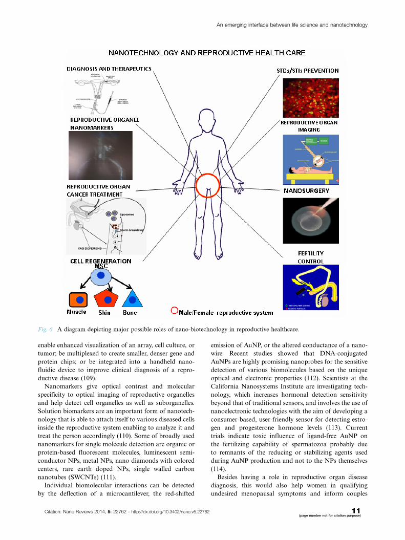
2. Potential role of nano-biotechnology inreproductive healthcareNanoscience and nano-biotechnology is an interdisci-
plinary field having inputs from various fields like bio-
logy, chemistry, physics, mathematics, electronics, etc.
Similarly, it has applications in almost all areas of life.
Very few people know that this new branch of science has
vast potential in the field of reproductive healthcare, one
of the most vital domains of medical science and our
health (Fig. 6).
2.1. Reproductive disease diagnosis andtherapeuticsIn today’s world, many reproductive diseases go undi-
agnosed or misdiagnosed, leading to even more compli-
cations. Nanotechnology may improve the sensitivity,
selectivity, speed, cost, and convenience of diagnosis (108).
Nanoscale labeling agents, such as QDs, have numerous
advantages to intracellular labeling and visualization.
These techniques and others can be further developed to
Fig. 5. (A) Schematic presentation of the encapsulation of anticancer drugs methotraxate (left) and 5-fluorouracil (right) into
PEGylated generation 3 and 4 PAMAM dendrimers and (B) schematic presentation of dendrimers as nano-scaffold for the
attachment of cell-specific ligands, modifiers, and fluorescence tags. Reproduced from Svenson and Tomalia, 2012 (101).
Rakhi K. Jha et al.
10(page number not for citation purpose)
Citation: Nano Reviews 2014, 5: 22762 - http://dx.doi.org/10.3402/nano.v5.22762
enable enhanced visualization of an array, cell culture, or
tumor; be multiplexed to create smaller, denser gene and
protein chips; or be integrated into a handheld nano-
fluidic device to improve clinical diagnosis of a repro-
ductive disease (109).
Nanomarkers give optical contrast and molecular
specificity to optical imaging of reproductive organelles
and help detect cell organelles as well as suborganelles.
Solution biomarkers are an important form of nanotech-
nology that is able to attach itself to various diseased cells
inside the reproductive system enabling to analyze it and
treat the person accordingly (110). Some of broadly used
nanomarkers for single molecule detection are organic or
protein-based fluorescent molecules, luminescent semi-
conductor NPs, metal NPs, nano diamonds with colored
centers, rare earth doped NPs, single walled carbon
nanotubes (SWCNTs) (111).
Individual biomolecular interactions can be detected
by the deflection of a microcantilever, the red-shifted
emission of AuNP, or the altered conductance of a nano-
wire. Recent studies showed that DNA-conjugated
AuNPs are highly promising nanoprobes for the sensitive
detection of various biomolecules based on the unique
optical and electronic properties (112). Scientists at the
California Nanosystems Institute are investigating tech-
nology, which increases hormonal detection sensitivity
beyond that of traditional sensors, and involves the use of
nanoelectronic technologies with the aim of developing a
consumer-based, user-friendly sensor for detecting estro-
gen and progesterone hormone levels (113). Current
trials indicate toxic influence of ligand-free AuNP on
the fertilizing capability of spermatozoa probably due
to remnants of the reducing or stabilizing agents used
during AuNP production and not to the NPs themselves
(114).
Besides having a role in reproductive organ disease
diagnosis, this would also help women in qualifying
undesired menopausal symptoms and inform couples
Fig. 6. A diagram depicting major possible roles of nano-biotechnology in reproductive healthcare.
An emerging interface between life science and nanotechnology
Citation: Nano Reviews 2014, 5: 22762 - http://dx.doi.org/10.3402/nano.v5.22762 11(page number not for citation purpose)

seeking infertility treatments on exact timing of concep-
tion to occur themselves thus providing a cheaper and
faster alternative to current infertility treatments. The
sensor measures hormone concentrations using specially
made hormone tabs (similar to glucose tabs used by
diabetics) made by low cost and precise ink-jet printing
of CNTs.
2.2. Viral protection and increasing immunitySimilarly, nanotechnology can help in solving significant
reproductive healthcare problems (115) like sexually
transmitted diseases (STDs) and sexually transmitted
infections (STIs). For reproductive healthcare mainte-
nance, contraception, infertility treatment, etc., many
people have to undergo surgical intervention like tubect-
omy, vasectomy or laparoscopy at various stages of life.
In order to lessen our pain in future, one will be able to
heal wounds a lot faster with the help of new nanotech-
nological delivery systems that will be sown into ban-
dages and will contain drugs like aluminosilicate, to
promote fast regeneration capabilities and it will allow
wounds to heal faster (116). Nanotechnology will be able
to make this all go a lot faster because, being so tiny,
one can theoretically load thousands of nanomechanical
device called nanites or nanoids or nanorobots with
thousands of different vaccines and inject them into the
host all at once and see if any of them work.
As mentioned earlier, RISUG coated intra uterine
devices (IUDs) are being developed at IIT Kharagpur
that along with giving a contraceptive effect will also have
an antimicrobial effect, as successfully tested on female
goats, thus helping to avoid infections generated after
IUD implantation in women. A new version of RISUG is
also being developed to act against human immunodefi-
ciency virus (HIV) that is ready for clinical trials.
Antimicrobial effects of silver ion or salts are well known,
and although its effect on microorganisms is not clearly
explored (117), Ag NPs appears to be the ideal candidate
to act against STDs.
2.3. Reproductive organ/cell imagingImaging is crucial for in vivo characterization of the
complex behaviors of reproductive disease in time and
space that tells us: where it is, how big it is, how fast it is
developing, how many molecular processes are contri-
buting simultaneously, what to treat it with, how it is
responding to therapy, and how it is changing (118).
Because molecules themselves are obviously too small to
be imaged directly with non-invasive techniques, the
contrast agent should manifest high affinity and avidity
for the target organ like reproductive organ. And, unlike
traditional blood pool contrast agents, a reproductive
site-targeted agent is intended to enhance a selected
biomarker that otherwise might be impossible to distin-
guish from surrounding normal tissue. Molecular ima-
ging actually has been a clinical reality for some time with
the use of targeted radionuclides (119).
However, the explosive growth of biocompatible nano-
technologies now promises to expand the horizon for
molecular imaging and therapy with a host of novel agents.
The desired properties of such targeted contrast agents
are: long circulating half-life (hours), selective binding to
epitopes of interest, low background signal and prominent
contrast-to-noise enhancement, acceptable toxicity pro-
file, ease of production and clinical use, applicability with
standard commercially available imaging modalities, and
promise for adjunctive therapeutic delivery (120). Clinical
availability of these agents is expected to redefine the
practice of imaging by focusing on cellular and molecular
mechanisms of disease, which will create opportunities for
more precise and rational design of conjunctive drug and
gene delivery nanosystems.
The contrast mechanism will depend on the choice
of imaging modality, which itself is determined by the
clinical problem and accessibility for imaging. For exam-
ple, carrier moieties such as NPs (liposomes or emulsions),
dendrimers, viral constructs, buckyballs, or various poly-
mers can be loaded with large payloads of imaging agents
such as paramagnetic or superparamagnetic metals, op-
tically active compounds (e.g. fluorescent molecules), or
radionuclides to enable detection with standard imag-
ing equipment. In the case of ultrasound imaging, the
intrinsic physical properties of the carrier agents them-
selves (density and compressibility) establish the means
for detection (121).
Targeted perfluorocarbon NPs were the first reported
molecular imaging agents for ultrasound applications and
were shown to augment reflectivity from fibrin thrombi in
vivo by two orders of magnitude or more (122). Reflec-
tive liposomes have also been used to specifically target
endothelial integrins that may have huge applications in
female reproductive healthcare. ‘Susceptibility’ or ‘cold
spot’ imaging agents have been produced by combina-
tions of carriers with iron oxides (e.g. ultra-small particles
of iron oxide) or alternative lanthanide species.
QDs NPs have potential use for non-invasive investiga-
tion of mammalian spermatozoa (123). Research has
shown that BRET-QD conjugated with boar spermato-
zoa helped to understand the sperm behavior inside
the uterus followed by their impact on sperm motility,
viability and fertilizing potential (124). QD-based near-
infrared (NIR) fluorescence cancer imaging is a growing
field for both preclinical and clinical application to
the clinical management for cancer patients due to its
advantageous features, including a high spatial resolu-
tion, portability, real-time display and detailed molecular
profiling with the multiplexed use of fluorescent probes
and therefore it can play vital role in imaging reproduc-
tive organ cancer (125).
Rakhi K. Jha et al.
12(page number not for citation purpose)
Citation: Nano Reviews 2014, 5: 22762 - http://dx.doi.org/10.3402/nano.v5.22762

2.4. Reproductive organ cancer treatmentNPs like CNPs and a range of nanodevices like fullerenes
are giving us immense hope against reproductive organ
cancer. For instance, nanoshells work similarly to NPs but
instead of injecting the cancer cells with chemotherapy
(126), they will simply use the heat from infrared light. It
has been discovered that when nanites are irradiated with
X-rays, the nanites produce their own electrons that can
be used to target cancer cells and destroy them without
harming the rest of the body. Nanites can also scatter
through the body to detect cancer cells and tag them so
that doctors know exactly where in the body that cancer
has spread to. That in turn will help physicians to avoid
chemotherapy for example, Qdots the gold nanites are
able to track down cancer cells in the body.
NPs will be able to inject chemotherapy directly into
cancer cells themselves with very minimal damage to the
surrounding cells (127). CNPs have shown immense po-
tential against breast cancer. Metallic nanoparticles like
gold (AuNPs) hold great promise as PDT and hyperther-
mic agents. Upon X-ray irradiation, AuNP can induce
cellular apoptosis through the generation of radicals. This
may have tremendous potential to kill ovarian cancer cells
without harming the surrounding healthy tissue (55�57).
NPs can also act as drug delivery agents, for example,
hypericin-loaded NPs (125) for the photodynamic treat-
ment of cancer. Drug delivery with CNTs for in vivo
treatment of reproductive organ cancer like uterine cancer,
cervical cancer, vaginal and vulvar cancer in females and
prostate cancer or penile cancer in males is another
possibility. RISUGadv invented by Prof. Sujoy K. Guha
prevents prostate cancer, the most prevalent cancer in
elderly man. Also, RISUG-PH studies in small animals
have the potential to act against benign prostate hyper-
plasia (BPH).
2.5. Nano surgeryNanorobots can play a significant role in laparoscopic
reproductive organ surgery and to correct abnormalities.
Surgical nanorobots could be introduced into the body
through the vascular system or at the ends of catheters
into various vessels and other cavities in the human body.
A nanorobot programmed by a surgeon could act as a
semi-autonomous on-site surgeon inside the body. Such
a device could perform various functions like searching
for pathology in reproductive as well as other organs,
and then diagnosing and correcting lesions or cysts
by nanomanipulation, coordinated by a computer thus
maintaining contact with the supervising surgeon via
coded ultrasound signals (128).
The earliest forms of cellular nanosurgery have already
been explored. For example, a rapidly vibrating (100 Hz)
micropipette with a B1 nm tip diameter has been used to
cut dendrites from single neurons without damaging cell
viability. Axotomy of roundworm neuron was performed
by femtosecond laser surgery after which the axons
functionally regenerated. A femtolaser acts like a pair
of nano-scissors by vaporizing tissue locally while leaving
adjacent tissue unharmed. The procedure does not kill
the cell on which nanosurgery was performed (129).
Atomic force microscopes have also been used for the
dissection of a bacterium cell wall in situ in aqueous
solution, with 26 nm thick twisted strands revealed inside
the cell wall after mechanically peeling back large patches
of the cell wall.
2.6. Cell regenerationSince natural tissues or organs are in nanometer dimen-
sion and cells directly interact with (and create) nanos-
tructured extra-cellular matrices (ECM), the biomimetic
features and excellent physiochemical properties of na-
nomaterials play a key role in stimulating cell growth
as well as to guide tissue regeneration. Even though it was
a field in its infancy a decade ago, numerous researchers
are currently fabricating cytocompatible biomimetic
nanomaterial scaffolds encapsulating cells (such as stem
cells, chondrocytes and osteoblasts, etc.) for tissue en-
gineering applications. Nanomaterials exhibit superior
cytocompatible, mechanical, electrical, optical, catalytic
and magnetic properties compared to conventional mi-
crosized materials. These unique properties help to
improve various tissue growth over what is achievable
today (130).
The stem-cell-technology has a new role to play in
reproduction. Firstly, the stem-cell source could be
pooled out of slaughter-house oocytes or from the vast
pool of embryos hatching out in many animal species.
These cells can transform themselves into 200 or more
cell types, which could be used to repair or regenerate
new desired cells. This advanced cell research may help
people suffering from reproductive organ cancer or per-
sons devoid of genital organs (131). Nanobots are 2.5
times smaller than DNA that can enter individual cells
and repair them. With that concept, nanotechnology will
be able to cure just about everything because all problems
start at a cellular level (132).
2.7. Contraception and infertility managementThe use of contraceptives should not only prevent preg-
nancy but also help the individual to maintain good
health. A research team at the Indian Institute of Tech-
nology Kharagpur has been working on several polymer-
based fertility control molecules (13�17) that owe a lot to
nanotechnology. The Cuproferrogel nanomedicine iron
oxide�copper�styrene maleic anhydride�dimethyl sulfox-
ide called Smart RISUG (Figure 3) developed in our
laboratory has proved to be very effective against sperm
as well as ovum, enables controlled delivery to the tar-
get site which in this case is vas deferens/fallopian
tube, controlled distribution with the help of external
An emerging interface between life science and nanotechnology
Citation: Nano Reviews 2014, 5: 22762 - http://dx.doi.org/10.3402/nano.v5.22762 13(page number not for citation purpose)

electromagnetic field and most importantly non-invasive
imaging by X-ray computer tomography (CAT) scan,
MRI, electrical impedance plethysmography, etc. (13�15,
110, 118, 126). Similarly, we are working on an anti-
microbial vaginal contraceptive.
There are so many oral and injectable short-term
contraceptives available over the counter throughout the
world. Oral contraception is preferred in the western
world while IUDs are mostly used in developing coun-
tries due to a lack of reliable alternatives in market beside
permanent sterilization like vasectomy and female ster-
ilization that itself is invasive and associated with several
side-effects and complications. Nanotechnology can im-
prove the dose required, efficacy, and delectability of
many fertility control agents. Electromechanical devices
and radiothermy may also help in proper biodistribution
of the fertility control agent (15) and also reversibility
when desired by the couple. For the pharmaceutical
industries novel drug delivery technologies (133) can
address issues associated with current pharmaceutics
such as extending product life, enhancing their perfor-
mance and acceptability either by increasing efficacy or
improving safety and product compliance.
Many people are not capable of reproducing because
their bodies are not good hosts to a desirable environ-
ment. With the aid of nanotechnology, these little nano-
bots may quickly go to work at reconstructing genitals
and other reproductive features so that one can once
reproduce. However, it is easier said than done to treat
infertility problems with such ease as a major portion of
infertility cases are unexplained. But at least we can
approach the problem in a better way with the help of
advanced nanotechnological tools and monitoring de-
vices. Sensors discussed in section 3.1 or ‘fertility chip’
have huge potential as a treatment for male fertility in the
short-term (134) and female in the long-term. Addition-
ally, as the chip is of nano-proportions, the patient will
have minimum discomfort while the information gener-
ated will be invaluable for prospective patients.
3. Future perspectives of nanotechnology forreproductive healthOn one hand, the exploding population in developing
countries like India and China is a major issue affecting
our socio-economic development; and the other hand
reduced fertility is a sensitive problem emerging globally
due to lifestyle changes and environmental factors (135).
Good reproductive health in turn effects our socio-
economic development directly or indirectly by eradicat-
ing poverty, to achieve primary education when family
size is small, promote gender equality and empower
women, reduce child mortality; improve maternal health,
combat HIV/AIDS/STDs/STIs, etc., ensuring environ-
mental sustainability and global partnerships in a bigger
scenario (136).
The potential of nanotechnology offers some exciting
possibilities in reproductive healthcare. Some techniques
are only imagined, while others are at various stages
of testing, or actually being used today. The use of
nanotechnology in the field of reproductive biomedicine
can revolutionize the way we detect and treat damage to
the human body and disease, and many techniques only
imagined a few years ago are making remarkable progress
towards becoming realities. For instance, this paper has
described a range of antimicrobial, long-term, stable,
single-use male and female injectable contraceptives
being developed like RISUG in advanced phase III clin-
ical trials (137, 138); and its nanotechnological versions
like Smart RISUG (13�17).
Other reproductive biomedicines are being developed
at IIT Kharagpur to prevent cancer; for example, In-
vivgensome (liposome synthesized inside the testis)
invented by Prof. Guha prevents prostate cancer devel-
opment which is one of most prevalent cancers in elderly
males, and RISUG-PH acts against BPH for which trials
are about to begin. Liposomes have great reproductive
healthcare potential both when developed in vitro and
also when self-generated in vivo. Another nanotechnology-
based tool for reproductive organ cancer treatment is
fullerenes described as a nanoscale molecule that is made
up of only carbon. Carbon, as we know, is the basis of
nature’s construction and, therefore, also represents our
very own construction. Fullerenes (139) allow us to build
nanostructures, so that one may integrate our own
programming and machinery that will go on to perform
marvelous tasks in situ.
With respect to STDs or STIs, RISUG has also shown
primary evidence to work against HIV, and RISUG-
coated IUDs do not allow microbes in its vicinity when
placed inside the body after child birth (140). Although
nanotechnology does not appear to play a direct role in
infertility treatment, indirectly it can play an immense
role by helping in early, low-cost and accurate detection
of disease sites with smart sensors, detection of hormone
levels and non-invasive imaging of nanomedicines placed
in the reproductive tube.
The reasons behind most deaths today are either
late diagnosis, inability to diagnose the main reason
or location of disease or misinterpretation of data. We do
hope nanotechnology, for instance nanites, will one day
be able to scurry throughout our bodies via the circula-
tory system (traveling through our blood) and monitor
every single vital sign that exists (141), for example,
whether there are any broken bones, torn muscle tissue,
irregularities, screen metabolism levels, observe choles-
terol levels, monitor hormone levels, make sure that the
organs are functioning properly, and any other require-
ment for a healthy body.
Some companies are developing nanotubes to heal
broken bones by providing bones with a proper structure
Rakhi K. Jha et al.
14(page number not for citation purpose)
Citation: Nano Reviews 2014, 5: 22762 - http://dx.doi.org/10.3402/nano.v5.22762

in order for them to grow back in the way that they are
supposed to (142). CNTs are still a relatively unexplored
area in a rapidly advancing field. Any amount of improve-
ments can be made to CNTs through various techniques
(143) because of their great material properties. For ex-
ample, it was shown that by electrospinning and plasma-
functionalizing SWCNTs, adhesion to surrounding polymer
matrices was greatly improved along with the tensile
properties of the nanotubes. Also, we know that most
nanotubes are cleared from the body very quickly after
being distributed throughout (144). This decreases the
chances of higher toxicity levels in the blood. The good
functionalization of SWCNs allows us to attach a number
of groups to the tubes for different systems. Radioactive
labels could be attached for use in reproductive organ
bioimaging (145). It was shown that CNTs were used
to efficiently deliver drugs to specific cancer cells of the
epithelium (146).
Nanotechnology may also be able to aid and even
perfect the act of regenerating cells/tissues (130). Regen-
eration is the process of bringing a person back to life.
Today, there are many different problems with doing so
but nanotechnology may be able to fix most if not all of
them. One of the biggest problems is due to the crystal-
lization of frozen cells but nanotechnology may be able to
warm those cells and even remake some of them so that
the person doesn’t biologically fall apart when they’re
revived. Nanotechnology may be able to also simply cure
cell damage as soon as we die which means we wouldn’t
even have to be frozen first.
As discussed previously, most of our nanotechnology-
based future healthcare expectations are based on
molecular nanotechnology (MNT). MNT is a technology
based on the ability to build structures to complex,
atomic specifications by means of mechanosynthesis
(147). This is distinct from nanoscale materials. Based
on Richard Feynman’s vision of miniature factories us-
ing nanomachines to build complex products (including
additional nanomachines), this advanced form of nano-
technology (or molecular manufacturing) would make
use of positionally controlled mechanosynthesis guided
by molecular machine systems. MNT would involve com-
bining physical principles demonstrated by chemistry,
other nanotechnologies, and the molecular machinery of
life with the systems engineering principles found in
modern macroscale factories (148).
Nanotechnology in medicine called nanomedicine in-
volves applications of NPs currently under development,
as well as longer ranges research that involves the use of
manufactured nano-robots to make repairs at the cellular
level (149). Future applications of nanomedicine will be
based on the ability to build nanorobots. These nanor-
obots will actually be programmed to repair specific
diseased cells, functioning in a similar way to antibodies
in our natural healing processes. This way, nanomedicine
offers great promise for the future, especially the mixing
of diagnostic and therapeutic capabilities in healthcare.
Nano surgery like laparoscopy is already being used for
reproductive health problem detection, corrective surgery,
tubal sterilization, chronic pelvic pain, etc. since past
many years (150).
Future nanorobots equipped with operating instru-
ments and mobility will be able to perform precise and
refined intracellular surgeries in reproductive organs,
which are beyond the capabilities of direct manipulation
by the human hand. We envision biocompatible surgical
nanorobots that can find and eliminate isolated cancer-
ous reproductive cells, remove microvascular obstructions
and recondition vascular endothelial cells, perform non-
invasive tissue and organ transplant, conduct molecular
repairs on traumatized extracellular and intracellular
structures, and even exchange new complete chromo-
somes for old ones inside individual living human cells
(128).
The future of nanomedicines is undermined by the lack
of financial profitability, consumer distrust, and ineffec-
tive regulation of new and generic products, weak patent
protection and insurance market failure. Its economic
breakthrough is dependent on a series of countervailing
measures and actions. Success requires more investment
induced by cost�effectiveness analyses and business plans
based on clinical data, public education based on nano-
toxicology studies, smart regulatory reform in the areas
of testing, market entry and liability, effective and
strategic patenting, patent dispute prevention and resolu-
tion, and innovative insurance policies.
Acknowledgement
We acknowledge support of Ministry of Health and Family Welfare,
Government of India.
Conflict of interestThere is no conflict of interest between the authors.
References
1. Allhoff F. The coming era of nano medicine. Am J Bioeth
2009; 9: 3�11.
2. EU Technology Platform on Nanomedicine. Available from:
http://www.etp-nanomedicine.eu/public [cited 15 December
2013].
3. Emerich DF, Halberstadt C, Thanos C. Role of nano-
biotechnology in cell-based nanomedicine: a concise review.
J Biomed Nanotechnol 2007; 3: 235�44.
4. Kedziora A, Gorzelanczyk K, Bugla Ploskonska G. Positive
and negative aspects of silver nanoparticles usage. Biology
International 2013; 53: 67�76.
5. Buzea C, Pacheco II, Robbie K. Nanomaterials and nano-
particles: sources and toxicity. Biointerphases 2007; 2(4):
MR17�172.
An emerging interface between life science and nanotechnology
Citation: Nano Reviews 2014, 5: 22762 - http://dx.doi.org/10.3402/nano.v5.22762 15(page number not for citation purpose)

6. Alivisatos P. The use of nanocrystals in biological detection.
Nat Biotechnol 2004; 22: 47�52.
7. Madani SY, Mandel A, Seifalian AM. A concise review of
carbon nanotube’s toxicology. Nano Rev 2013; 4: 21521.
8. Baughman RH, Anvar AZ, Walt AH. Carbon nanotubes � the
route toward applications. Science 2002; 297: 787�92.
9. Ciofani G, Raffa V, Menciassi A, Cuschieri A. Boron nitride
nanotubes: an innovative tool for nanomedicine. Nano Today
2009; 4: 8�10.
10. Bonnemain B. Superparamagnetic agents in magnetic reso-
nance imaging, physicochemical characteristics and clinical
applications a review. J Drug Target 1998; 6: 167�74.
11. United Nations (1995). Population and Development, Vol. 1:
Programme of Action adopted at the International Conference
on Population and Development: Cairo, 5�13 September 1994,
paragraph 7.2. New York: Department of Economic and
Social Information and Policy Analysis, United Nations.
12. United Nations (2002): World Youth Report 2003: Report of
the Secretary- General (E/CN.5/2003/4), para. 16. New York:
Commission for Social Development, United Nations.
13. Jha RK, Jha PK, Guha SK. Smart RISUG: a potential new
contraceptive and its magnetic field mediated sperm interac-
tion. Int J Nanomed 2009a; 4: 55�64.
14. Jha RK, Jha PK, Rana SVS, Guha SK. Spermicidal action of
styrene maleic anhydride polyelectrolyte in combination with
magnetic and electrically conductive particles. Int J Pharmacol
2009b; 5: 1�12.
15. Jha R, Jha PK, Rana SVS, Guha SK. An approach to non-
invasive delivery, biodistribution and fertility control potential
evaluation of Cuproferrogel Fe3O4�Cu�SMA�DMSO in
female. Fertil Steril 2010a; 94: 2850�3.
16. Jha PK, Jha R, Gupta BL, Guha SK. Effect of g-dose rate and
total dose interrelation on the polymeric hydrogel: a novel
injectable male contraceptive. Radiat Phys Chem 2010b; 79:
663�71.
17. Jha PK, Jha R, Datt R, Guha SK. Entropy in good
manufacturing practices: a tool for quality assurance. Eur J
Oper Res 2011; 211: 658�65.
18. Na HB, Song IC, Hyeon T. Inorganic nanoparticles for MRI
contrast agents. Adv Mater 2009; 21: 2133�48.
19. Rivera Gil P, Huhn D, Del Mercato LL, Sasse D, Parak WJ.
Nanopharmacy: inorganic nanoscale devices as vectors and
active compounds. Pharmacol Res 2010; 62: 115�25.
20. Rube MA, Cox BF, Gueorguieva M, Kakchingtabam D,
Andre P, Mezler A. Iron�platinum alloy nanoparticles for
guidewire and resonant markers for catheter localization
during interventional MRI. Biomed Engin/Biomed Tech
2013; 58: 323�86.
21. Radwan SH, Azzazy HME. Gold nanoparticles for molecular
diagnostics. Expert Rev Mol Diagn 2009; 9: 511�24.
22. Spivak MY, Bubnov RV, Yemets IM, Lazarenko LM,
Tymoshok NO, Ulberg ZR. Gold nanoparticles � the ther-
anostic challenge for PPPM: nanocardiology application.
EPMA J 2013; 4: 18.
23. Bawa R. Nanoparticle based therapeutics in human: a survey.
Nanotechnol Law Bus 2008; 5: 135.
24. Qin W, Din D, Liu J, Yuan WZ, Hu Y, Liu B, et al.
Biocompatible nanoparticles with aggregation-induced emis-
sion characteristics as far-red/near-infrared fluorescent bio-
probes for in vitro and in vivo imaging applications. Adv Funct
Mater 2012; 22: 771�9.
25. Schwartz JA, Shetty AM, Price RE, Stafford RJ, Wagon JC,
Uthamanthil RK, et al. Feasibility study of particle-assisted
laser ablation of brain tumors in orthotopic canine model.
Cancer Res 2008; 69: 1659�67.
26. Wang Y, Chen L. Quantum dots lighting up the research
and development of nanomedicine. Nanomedicine 2011; 7:
385�402.
27. Wagh A, Qian SY, Law B. Development of biocompatible
polymeric nanoparticles for in vivo NIR and FRET imaging.
Bioconjugate Chem 2012; 23: 981�92.
28. Tang R, Lee H, Achilefu S. Induction of pH sensitivity on the
fluorescence lifetime of quantum dots by NIR fluorescent dyes.
Am Chem Soc 2012; 134: 4545�8.
29. Chuah AM, Kuroiwa T, Ichikawa S, Kobayashi I, Nakajima
M. Formation of biocompatible nanoparticles via the self-
assembly of Chitosan and Modified Lecithin. J Food Sci 2009;
74: N1�8.
30. Sanna V, Pintus G, Roggio AM, Punzoni S, Posadino AM,
Arca A, et al. Targeted biocompatible nanoparticles for the
delivery of (-)-epigallocatechin 3-gallate to prostate cancer
cells. J Med Chem 2011; 54: 1321�32.
31. Barandeh F, Nguyen PL, Kumar R, Lacobucci GJ, Kuznicki
ML, Kosterman A, et al. Organically modified silica nanopar-
ticles are biocompatible and can be targeted to neurons in vivo.
PLoS One 2012; 7: 1�15.
32. Aswathy RG, Sivakumar B, Brahatheeswaran D, Fukuda T,
Yoshida Y, Maekawa T, et al. Biocompatible fluorescent zein
nanoparticles for simultaneous bioimaging and drug delivery
application. Adv Nat Sci: Nanosci Nanotechnol 2012; 3: 1�7.
33. Roullin VG, Callewaert M, Delavoie F, Molinari M, Seconde
A, Andry MC. Optimised NSAIDs-loaded biocompatible
nanoparticles. Nano-Micro Lett 2010; 2: 247�55.
34. Chan JM, Zhang L, Tong R, Ghosh D, Gao W, Liao G, et al.
Spatiotemporal controlled delivery of nanoparticles to injured
vasculature. Proc Natl Acad Sci U S A 2009; 107: 2213�8.
35. Kamaly N, Xiao Z, Valencia PM, Radovic AF, Farokhzad OC.
Targeted polymeric therapeutic nanoparticles: design, devel-
opment and clinical translation. Chem Soc Rev 2012; 41: 2971�3010.
36. Bangham AD, Horne RW. Action of saponin on biological cell
membranes. Nature 1962; 196: 952�3.
37. Torchilin VP. ‘‘Multifunctional nanocarriers.’’ Adv Drug Deliv
Rev 2012; 64: 302�15.
38. Woodle MC, Papahadjopoulos D. Liposome preparation and
size characterization. Methods Enzymol 1989; 171: 193.
39. Bergin C, O’Leary A, McCreary C. Treatment of Kaposi’s
sarcoma with liposomal doxorubicin. Am J Health Syst Pharm
1995; 52: 2001�4.
40. GreGoriadis G. Engineering liposomes for drug delivery:
progress and problem. Trends Biotechnol 1995; 13: 527�37.
41. Gouin S. Microencapsulation: industrial appraisal of exist-
ing technologies and trends. Trends Food Sci Tech 2004; 15:
330�47.
42. Qian J, Wang W, Li Y, Xu Y, Sun Q. Optical extinction
properties of perforated gold-silica-gold multilayer Nanoshells.
J Phys Chem 2012; 116: 10349�55.
43. Odom TW, Huang JL, Kim P, Lieber CM. Atomic structure
and electronic properties of single-walled carbon nanotubes.
Nature 1998; 391: 62�4.
44. Liu Z, Tabakman S, Welsher K, Dai H. Carbon nanotubes in
biology and medicine: in vitro and in vivo detection, imaging
and drug delivery. Nano Res 2009; 2: 85�120.
45. Martin CR, Kohli P. The emerging field of nanotube biotech-
nology. Nat Rev Drug Discov 2003; 2: 29�37.
46. Klumpp C, Kostarelos K, Prato M, Bianco A. Functionalized
carbon nanotubes as emerging nanovectors for the delivery of
therapeutics. Biochim Biophys Acta 2006; 1758: 404�12.
47. Raffa V, Vittorio O, Riggio C, Cuschieri A. Progress in nano-
technology for healthcare. Minim Invasive Ther Allied Tech
2010; 19: 127�35.
Rakhi K. Jha et al.
16(page number not for citation purpose)
Citation: Nano Reviews 2014, 5: 22762 - http://dx.doi.org/10.3402/nano.v5.22762

48. Hersam MC. Progress towards monodisperse single-walled
carbon nanotubes. Nat Nanotechnol 2008; 3: 387�94.
49. Sun Y, Xia Y. Shape-controlled synthesis of gold and silver
nanoparticles. Science 2002; 298: 2176�9.
50. Liao H, Nehl CL, Hafner JH. Biomedical applications of
plasmon resonant metal nanoparticles. Nanomedicine 2006; 1:
201�8.
51. Zhang Q, Large N, Nordlander P, Wang H. Porous gold
Au with tunable Plasmon resonances and intense field en-
hancements for single particle SERS. J Phys Chem Lett 2014;
5: 370�4.
52. Mukherjee P, Bhattacharya R, Wang P, Wang L, Basu S, Nagy
JA, et al. Antiangiogenic properties of gold nanoparticles.
Clin Cancer Res 2005; 11: 3530�4.
53. Mukherjee P, Bhattacharya R, Bone N, Lee YK, Patra CR,
Wang S, et al. Potential therapeutic application of gold nano-
particles in B-chronic lymphocytic leukemia (BCLL): enhan-
cing apoptosis. J Nanobiotechnol 2007; 5: 1�13.
54. Sigler JW, Bluhm GB, Duncan H, Sharp JT, Ensign DC,
Mccrum WR. Gold salts in the treatment of rheumatoid
arthritisA double-blind study. Ann Intern Med 1974; 80: 21�6.
55. Chatterjee DK, Fong LS, Zhang Y. Nanoparticles in photo-
dynamic therapy: an emerging paradigm. Adv Drug Deliv Rev
2008; 60: 1627�37.
56. Juzenas P, Chen W, Sun Y, Coelho MANC, Generalov R,
Generalova N, et al. Quantum dots and nanoparticles for
photodynamic and radiation therapies of cancer. Adv Drug
Deliv Rev 2008; 60: 1600�14.
57. Paszko E, Ehrhardt C, Senge MO, Kelleher DP, Reynolds JV.
Nanodrug applications in photodynamic therapy. Photodiag-
nosis Photodyn Ther 2011; 8: 14�29.
58. Gupta AK, Gupta M. Synthesis and surface engineering
of iron oxide nanoparticles for biomedical applications.
Biomaterials 2005; 26: 3995�4021.
59. Rasmussen JW, Martinez E, Louka P, Wingett DG. Zinc oxide
nanoparticles for selective destruction of tumor cells and
potential for drug delivery applications. Expert Opin Drug
Deliv 2010; 7: 1063�77.
60. Jeng HA, Swanson J. Toxicity of metal oxide nanoparticles in
mammalian cells. J Environ Sci Health A Tox Hazard Subst
Environ Eng 2006; 41: 2699�711.
61. Krasheninnikov AV, Banhart F. Engineering of nanostructured
carbon materials with electron or ion beams. Nat Mater 2007;
6: 723�33.
62. Ray SC, Saha A, Jana NR, Sarkar R. Fluorescent carbon
nanoparticles: synthesis, characterization, and bioimaging
application. J Phys Chem C 2009; 113: 18546�51.
63. Puretzky AA, Geohegan DB, Fan X, Pennycook SJ. Dynamics
of single-wall carbon nanotube synthesis by laser vaporization.
Appl Phys A 2000; 70: 153�60.
64. Nessim GD, Seita M, O’Brien KP, Hart AJ, Bonaparte RK,
Mitchell RR, et al. Low temperature synthesis of vertically
aligned carbon nanotubes with electrical contact to metallic
substrates enabled by thermal decomposition of the carbon
feedstock. Nano Lett 2009; 9: 3398�405.
65. Jin YH, Seo SD, Shim HW, Park KS, Kim DW. Synthesis of
core/shell spinel ferrite/carbon nanoparticles with enhanced
cycling stability for lithium ion battery anodes. Nanotechnol-
ogy 2012; 23: 125402.
66. Zhao QL, Zhang ZL, Huang BH, Peng J, Zhang M, Pang DW.
Facile preparation of low cytotoxicity fluorescent carbon
nanocrystals by electrooxidation of graphite. Chem Commun
2008; 41: 5116�8.
67. Tian L, Ghosh D, Chen W, Pradhan S, Chang X, Chen S.
Nanosized carbon particles from natural gas soot. Chem
Mater 2009; 21: 2803�9.
68. Chen IH, Wang CC, Chen CY. Fabrication and characteriza-
tion of magnetic cobalt ferrite/polyacrylonitrile and cobalt
ferrite/carbon nanofibers by electrospinning. Carbon 2010; 48:
604�11.
69. Yang Y, Cui J, Zheng M, Hu C, Tan S, Xiao Y, et al. One-step
synthesis of amino-functionalized fluorescent carbon nano-
particles by hydrothermal carbonization of chitosan. Chem
Commun 2012; 48: 380�2.
70. Yezhelyev MV, Gao X, Xing Y, Al-Hajj A, Nie S, O’Regan
RM. Emerging use of nanoparticles in diagnosis and treatment
of breast cancer. Lancet Oncol 2006; 7: 657�67.
71. Bronich TK, Keifer PA, Shlyakhtenko LS, Kabanov AV.
Polymer micelle with cross-linked ionic core. J Am Chem Soc
2005; 127: 8236�7.
72. Li Y, Gao GH, Lee DS. Stimulus-sensitive polymeric nano-
particles and their applications as drug and gene carriers. Adv
Healthc Mater 2013; 2: 388�417.
73. Haley B, Frenkel E. Nanoparticles for drug delivery in cancer
treatment. Urol Oncol 2008; 26: 57�64.
74. Paciotti GF, Myer L, Weinreich D, Goia D, Pavel N,
McLaughlin RE, et al. Colloidal gold: a novel nanoparticle
vector for tumor directed drug delivery. Drug Deliv 2004; 11:
169�83.
75. Leleux J, Roy K. Micro and nanoparticle-based delivery
systems for vaccine immunotherapy: an immunological and
materials perspective. Adv Healthc Mater 2013; 2: 72�94.
76. Bera D, Qian L, Tseng T-K, Holloway PH. Quantum dots and
their multimodal applications: a review. Materials 2010; 3:
2260�345.
77. Mattoussi H, Palui G, Na HB. Luminescent quantum dots as
platforms for probing in vitro and in vivo biological processes.
Adv Drug Deliv Rev 2012; 64: 138�66.
78. Birudavolu S, Nuntawong N, Balakrishnan G, Xin YC, Huang
S, Lee SC, et al. Selective area growth of InAs quantum dots
formed on a patterned GaAs substrate. Appl Phys Lett 2004;
85: 2337�9.
79. Nakata Y, Mukai K, Sugawara M, Ohtsubo K, Ishikawa H,
Yokoyama N. Molecular beam epitaxial growth of InAs self-
assembled quantum dots with light-emission at 1.3 mm. J Cryst
Growth 2000; 208: 93�9.
80. Bertino MF, Gadipalli RR, Martin LA, Rich LE, Yamilov A,
Heckman BR, et al. Quantum dots by ultraviolet and x-ray
lithography. Nanotechnology 2007; 18: 315603.
81. Klimov VI. Spectral and dynamical properties of multiexcitons
in semiconductor nanocrystals. Annu Rev Phys Chem 2007; 58:
635�73.
82. Chan WC, Maxwell DJ, Gao X, Bailey RE, Han M.
Luminescent quantum dots for multiplexed biological detec-
tion and imaging. Curr Opin Biotechnol 2002; 13: 40�6.
83. Valizadeh A, Mikaeili H, Samiei M, Farkhani SM, Zarghami
N, Akbarzadeh A, et al. Quantum dots: synthesis, bioapplica-
tions, and toxicity. Nanoscale Res Lett 2012; 7: 1�14.
84. Chang Y-P, Pinaud F, Antelman J, Weiss S. Tracking bio-
molecules in live cells using quantum dots. J Biophotonics
2008; 1: 287�98.
85. Cherry RJ. Keeping track of cell surface receptor. Trends Cell
Biol 1992; 2: 242�4.
86. Saxton MJ, Jacobson K. Single-particle tracking: applications
to membrane dynamics. Annu Rev Biophys Biomol Struct
1997; 26: 373�99.
87. Ishido M, Kasuga N. In situ real-time imaging of the satellite
cells in rat intact and injured soleus muscles using quantum
dots. Histochem Cell Biol 2011; 135: 21�6.
88. Lim IIS, Njoki PN, Park HY, Wang X, Wang L. Gold and
magnetic oxide/gold core/shell nanoparticles as bio-functional
nanoprobes. Nanotechnology 2008; 19: 305102.
An emerging interface between life science and nanotechnology
Citation: Nano Reviews 2014, 5: 22762 - http://dx.doi.org/10.3402/nano.v5.22762 17(page number not for citation purpose)

89. Duan H, Nie S. Cell-penetrating quantum dots based on
multivalent and endosome-disrupting surface coatings. J Am
Chem Soc 2007; 129: 3333�8.
90. Smith AM, Duan H, Mohs AM, Nie S. Bioconjugated
quantum dots for in vivo molecular and cellular imaging.
Adv Drug Deliv Rev 2008; 60: 1226�40.
91. Wang L, Park HY, Stephanie I, Lim I, Schadt MJ, Mott D,
et al. Core@shell nanomaterials: gold-coated magnetic oxide
nanoparticles. J Mater Chem 2008; 18: 2629�35.
92. Park K, Lee S, Kang E, Kim K, Choi K, Kwon IC. New
generation of multifunctional nanoparticles for cancer imaging
and therapy. Adv Funct Mater 2009; 19: 1553�66.
93. Wu W, Aiello M, Zhou T, Berliner A, Banerjee P, Zhou S.
In-situ immobilization of quantum dots in polysaccharide-based
nanogels for integration of optical pH-sensing, tumor cell
imaging, and drug delivery. Biomaterials 2010; 31: 3023�31.
94. Bottrill M, Green M. Some aspects of quantum dot toxicity.
Chem Commun 2011; 47: 7039�50.
95. Dewald JR, Tomalia DA. Dense star polymers having core,
core branches, terminal groups. U.S. Patent 4,507,466; 1985.
96. Tomalia DA, Baker H, Dewald J, Hall M, Kallos G, Martin S,
et al. A New Class of Polymers: Starburst-Dendritic Macro-
molecules. Polymer Journal 1985; 17: 117.
97. Antoni P, Hed Y, Nordberg A, Nystrom D, von Holst H, Hult
A, et al. Bifunctional dendrimers: from robust synthesis and
accelerated one-pot postfunctionalization strategy to potential
applications. Angew Chem Int Ed Engl 2009; 48: 2126�30.
98. McElhaonon JR, McGrath DV. Toward chiral polyhydroxy-
lated dendrimers. Preparation and chiroptical properties. J Org
Chem 2000; 65: 3525�9.
99. Liang CO, Frechet JMJ. Incorporation of functional guest
molecules into an internally functionalizable dendrimers
through olefin metathesis. Macromolecules 2005; 38: 6276�84.
100. Nanjwade BK, Bechra HM, Derkar GK, Manvi FV, Nanjwade
VK. Dendrimers: emerging polymers for drug-delivery systems.
Eur J Pharm Sci 2009; 38: 185�96.
101. Svenson S, Tomalia DA. Dendrimers in biomedical applica-
tions- reflections on the field. Adv Drug Deliv Rev 2012; 64:
102�15.
102. Kannumalle LS, Ramesh R, Maddipatla MVSN, Nithyanandan
J, Ramamurthy V. Dendrimers as photochemical reaction media.
Photochemical behavior of unimolecular and bimolecular reac-
tions in water-soluble dendrimers. J Org Chem 2005; 70: 5062�9.
103. Khopade AJ, Caruso F, Tripathi P, Nagaich S, Jain NK.
‘‘Cascade’’- and ‘‘Effect of dendrimer on entrapment and
release of bioactive from liposomes.’’ Int J Pharm 2002; 232:
157�62.
104. Prajapati RN, Tekade RK, Gupta U, Gajbhiye V, Jain NK.
Dendimer-mediated solubilization, formulation development
and in vitro�in vivo assessment of piroxicam. Mol Pharm 2009;
6: 940�50.
105. Chauhan AS, Sridevi S, Chalasani KB, Jain AK, Jain SK,
Jain NK, et al. Dendrimer-mediated transdermal delivery:
enhanced bioavailability of indomethacin. J Control Release
2003; 90: 335�43.
106. Kukowska-Latallo JF, Candido KA, Cao Z, Nigavekar SS,
Majoros IJ, Thomas TP, et al. Nanoparticle targeting of
anticancer drug improves therapeutic response in animal model
of human epithelial. Cancer Res 2005; 65: 5317�24.
107. Twyman LJ, Ellis A, Gittins PJ. Pyridine encapsulated hyper-
branched polymers as mimetic models of haeme containing
proteins, that also provide interesting and unusual porphyrin�ligand geometries. Chem Commun 2012; 48: 154�6.
108. Yager P, Edwards T, Fu E, Helton K, Nelson K, Tam MR,
et al. Microfluidic diagnostic technologies for global public
health. Nature 2006; 442: 412�8.
109. Lee WG, Kim YG, Chung BG, Demirci U, Khademhosseini A.
Nano/Microfluidics for diagnosis of infectious diseases in
developing countries. Adv Drug Deliv Rev 2010; 62: 449�57.
110. Horne AW, Duncan WC, Critchley HO. The need for
serum biomarker development for diagnosing and excluding
tubal ectopic pregnancy. Acta Obstet Gynecol Scand 2010; 89:
299�301.
111. Bates M, Huang B, Dempsey GT, Zhuang X. Multicolour
super-resolution imaging with photo switchable fluorescent
probes. Science 2007; 317: 1749�53.
112. Wang L, Song S, Pan D, Li D, Fan C. Gold nanoparticles
based sensing strategies for biomolecular detection. Pure App
Chem 2010; 82: 81�9.
113. Hennequin Y, Allier CP, McLeod E, Mudanyali O, Migliozzi
D, Ozcan A, et al. Optical detection and sizing of single
nanoparticles using continuous wetting films. ACS Nano 2013;
7: 7601�9.
114. Barchanski A, Taylor U, Klein S, Petersen S, Rath D,
Barcikowski S. Golden perspective: application of laser-
generated gold nanoparticle conjugates in reproductive biol-
ogy. Reprod Domest Anim 2011; 46: 42�52.
115. Betty YSK, Rutka JT, Chan WCW. Current concepts. Nano-
medicine. N Eng J Med 2010; 363: 2434�43.
116. Chu Y, Yu D, Wang P, Xu J, Li D, Ding M. Nanotechnology
promotes the full-thickness diabetic wound healing effect of
recombinant human epidermal growth factor in diabetic rats.
Wound Rep Reg 2010; 18: 499�505.
117. Kim JS, Kuk E, Yu KN, Kim JH, Park SJ, Lee HJ, et al.
Antimicrobial effects of silver nanoparticles. Nanomedicine
2007; 3: 95�101.
118. Wickline SA, Lanza GM. Nanotechnology for molecular
imaging and targeted therapy. Circulation 2003; 107: 1092�5.
119. Ersahin D, Doddamane I, Cheng D. Targeted radionuclide
therapy. Cancers 2011; 3: 3838�55.
120. Choi HS, Frangioni JV. Nanoparticles for biomedical imaging:
fundamentals for clinical translation. Mol Imaging 2010; 6:
291�310.
121. Thomson K, Varma D. Safe use of radiographic contrast
media. Australian Prescriber 2010; 33: 19�22.
122. Frangioni JV, Hajjar RJ. In vivo tracking of stem cells for
clinical trials in cardiovascular disease. Circulation 2004; 110:
3378�83.
123. Feugang JM, Youngblood RC, Greene JM, Fahad AS,
Monroe WA, Willard ST, et al. Application of quantum dot
nanoparticles for potential non-invasive bio-imaging of mam-
malian spermatozoa. J Nanobiotechnol 2012; 10: 45.
124. So MK, Loening AM, Gambhir SS, Rao J. Creating self-
illuminating quantum dot conjugates. Nat Protoc 2006; 1:
1160�4.
125. Kosaka N, Ogawa M, Choyke PL, Kobayashi H. Clinical
implications of near-infrared fluorescence imaging in cancer.
Future Oncol 2009; 5: 1501�11.
126. Bardhan R, Lal S, Joshi A, Halas NJ. Theranostic nanoshells:
from probe design to imaging and treatment of cancer.
Accounts Chem Res 2011; 44: 936�46.
127. Liang XJ, Chen C, Zhao Y, Wang PC. Circumventing tumor
resistance to chemotherapy by nanotechnology. Methods Mol
Biol 2010; 596: 467�88.
128. Cavalcanti A, Shirinzadeh B, Murphy D, Smith JA. Nano-
robots for laparoscopic cancer surgery. 6th IEEE/ACIS Inter-
national Conference on Computer and Information Science;
Melbourne, Australia; 11�13 July; 2007.
129. Yanik MF, Cinar H, Cinar HN, Gibby A, Chisholm AD.
Nerve regeneration in Caenorhabditis elegans after femtose-
cond laser axotomy. Selected Topics in Quantum Electronics
IEEE Journal of 2006; 12: 1283�91.
Rakhi K. Jha et al.
18(page number not for citation purpose)
Citation: Nano Reviews 2014, 5: 22762 - http://dx.doi.org/10.3402/nano.v5.22762

130. Zhang L, Webster TJ. Nanotechnology and nanomaterials:
promises for improved tissue regeneration. Nano Today 2009;
4: 66�80.
131. Calin M, Stan D, Simion V. Stem cell regenerative potential
combined with nanotechnology and tissue engineering for
myocardial regeneration. Curr Stem Cell Res Ther 2013; 8:
292�303.
132. Loizidou M, Seifalian AM. Nanotechnology and its applica-
tions in surgery. Br J Surg 2010; 97: 463�5.
133. Sahoo SK, Labhasetwar V. Nanotech approached to drug
delivery and imaging. Drug Discov Today 2003; 8: 1112�20.
134. Rae MT, Price D, Harlow CR, Critchley HOD, Hillier SG.
Glucocorticoid receptor-mediated regulation of MMP9 gene
expression in human ovarian surface epithelial cells. Fertil
Steril 2009; 92: 703�8.
135. Project Report No. 2006ST21:D5. Nanotechnology develop-
ments in India � a status report. Supported by The Interna-
tional Development Research Centre (IDRC), Canada, The
Energy and Resources Institute (TERI); 2009.
136. United Nations. Population and Development, Vol. 1: Pro-
gramme of Action adopted at the International Conference on
Population and Development, Cairo, 5�13 September 1994,
paragraph 7.2. New York: Department of Economic and
Social Information and Policy Analysis, United Nations; 1995.
137. Guha SK. Contraceptive for use by a male. US Patent
5,488,075; 1996.
138. Guha SK. Biophysical mechanism-mediated time-dependent
effect on sperm of human and monkey vas implanted poly-
electrolyte contraceptive. Asian J Androl 2007; 9: 221�7.
139. Stepanow S, Lin N, Vidal F, Landa A, Ruben M, Barth JV,
et al. Programming supramolecular assembly and chirality in
two dimensional dicarboxylate networks on a Cu(100) sur-
face. Nano Lett 2005; 5: 9.
140. Guha SK. RISUGTM (reversible inhibition of sperm under
guidance)�an antimicrobial as male vas deferens implant for
HIV free semen. Med Hypotheses 2005; 65: 61�4.
141. Hauck TS, Giri S, Gao Y, Chan WCW. Nanotechnology
diagnostics for infectious diseases prevalent in developing
countries. Adv Drug Deliv Rev 2010; 62: 438�48.
142. Usui Y, Aoki K, Narita N, Murakami N, Nakamura I,
Nakamura K, et al. Carbon nanotubes with high bone-tissue
compatibility and bone-formation acceleration effects. Small
2008; 4: 240�6.
143. Hilder TA, Hill JM. Modeling the loading and unloading of
drugs into nanotubes. Small 2009; 5: 300�8.
144. Lu F, Gu L, Meziani MJ, Wang X, Luo PG, Veca LM, et al.
Advances in bioapplications of carbon nanotubes. Adv Mater
2009; 21: 139�52.
145. Liu Z, Cai W, He L, Nakayama N, Chen K, Sun X, et al. In
vivo biodistribution and highly efficient tumour targeting of
carbon nanotubes in mice. Nat Nanotechnol 2007; 2: 47�52.
146. Bhirde AA, Patel V, Gavard J, Zhang G, Sousa AA,
Masedunskas A, et al. Targeted killing of cancer cells in Vivo
and in Vitro with EGF-directed carbon nanotube-based drug
delivery. ACS Nano 2009; 3: 307�16.
147. Hersam MC, Guisinger NP, Lyding JW. Silicon-based mole-
cular nanotechnology. Nanotechnology 2000; 11: 70.
148. Lapshin RV. Feature-oriented scanning probe microscopy. In:
Nalwa HS, ed. Encyclopedia of nanoscience and nanotech-
nology, vol. 14. USA: American Scientific Publishers; 2011.
p. 105�15.
149. Cavalcanti A, Freitas RA, Kretly RC. Nanorobotics control
design: a practical approach tutorial. 28th Biennial Mechan-
isms and Robotics Conference, Salt Lake City, UT, 2004;
September 28�October 2.
150. Latthe P, Latthe M, Say L, Gulmezoglu M, Khan KS.
WHO systematic review of prevalence of chronic pelvic pain:
a neglected reproductive health morbidity. BMC Public Health
2006; 6: 117.
*Rakhi K. JhaC/O- Dr Pradeep K. JhaSchool of Medical Science and TechnologyIndian Institute of Technology KharagpurKharagpur-721 302, West Bengal, IndiaEmail: [email protected]; [email protected]
An emerging interface between life science and nanotechnology
Citation: Nano Reviews 2014, 5: 22762 - http://dx.doi.org/10.3402/nano.v5.22762 19(page number not for citation purpose)
Copyright © 2022 FDOKUMEN