Nanotechnology for Diagnosis and Treatment of Infectious Diseases
14
Delivered by Publishing Technology to: Chung-Ang University IP: 165.194.5.66 On: Wed, 07 May 2014 02:35:09 Copyright: American Scientific Publishers Copyright © 2014 American Scientific Publishers All rights reserved Printed in the United States of America Review Journal of Nanoscience and Nanotechnology Vol. 14, 7374–7387, 2014 www.aspbs.com/jnn Nanotechnology for Diagnosis and Treatment of Infectious Diseases Muhammad Qasim 1 , Dong-Jin Lim 2 , Hansoo Park 1 ∗ , and Dokyun Na 1 ∗ 1 School of Integrative Engineering, Chung-Ang University, Seoul, 156-756, Korea 2 Department of Biomedical Engineering, University of Alabama at Birmingham, AL, 35233, USA The emergence of co-infections and the evolution of drug-resistant pathogens limit the utility of current therapies against infections, and developing countries in particular are facing a great chal- lenge in combating infectious disease. Moreover, any failure to control the spread of infectious diseases would also represent a threat to developed countries. Recent developments in nanotech- nology allow us to address this issue at two levels: diagnostics and treatment. Prevention of the spread of infectious pathogens requires rapid and accurate identification of the infectious agents for proper treatment. Recently developed fluorescent nanoparticles are so sensitive that even a single nanoparticle is capable of emitting a strong enough signal to be captured, thus enabling early identification of infections. Proper and effective treatment not only saves the patient, but also prevents the spread of the pathogens. Specific nanoparticle vehicles developed to encapsulate ther- apeutic agents and deliver them to a target site represent a promising strategy to boost immune responses for vaccination and boost the efficacy of drugs for treatment. Here, we describe a variety of nanotechnologies for use in applications such as immune response modulation, drug delivery, diagnostics, and treatment, which are especially needed in developing countries. Keywords: Infectious Diseases, Nanotechnology, Drug Resistance, Therapies, Diagnostics. CONTENTS 1. Introduction ........................................ 7374 1.1. The Immune System ............................. 7376 1.2. Current Treatments and Their Limitations ............. 7376 1.3. Current Diagnostic Techniques and Their Limitations ................................ 7378 2. Nanotechnology and Infectious Disease ................... 7379 2.1. Modulation of Immune Response by Nanoparticles (NPs) for Efficient Vaccination ..................... 7379 2.2. Nano-Based Drug Delivery System for Infectious Diseases ............................... 7380 2.3. Nanoparticles as Therapeutic Drugs .................. 7382 2.4. Nano-Based Diagnosis of Infectious Diseases .......... 7383 3. Conclusions ........................................ 7384 Acknowledgment .................................... 7385 References and Notes ................................ 7385 1. INTRODUCTION Pathogens such as viruses, 1–4 bacteria, 5 6 fungi, 7 or parasites 8 that cause infectious diseases are one of the major causes of deaths, accounting for approximately ∗ Authors to whom correspondence should be addressed. 15 million annual deaths worldwide. 9 10 Of the many infectious diseases, acute respiratory infections including influenza and pneumonia are the top-ranked causes of deaths, accounting for 3.5 million deaths a year, followed by acquired immunodeficiency syndrome (AIDS), which is responsible for 2.5 million deaths. Other infectious dis- eases such as diarrheal diseases, pneumonia, tuberculosis (TB), and malaria are also major causes of deaths, account- ing for 2.2 million, 1.8 million, 1.5 million, and 1.2 million deaths, respectively. 11 12 For example, in 2010, 8.8 million people were infected with TB and 1.5 million of them died of it. Over 95% of the deaths caused by infectious diseases occur in low-income and mid-income countries. 13 Despite great efforts to develop effective pharmaceuti- cals and new technologies to produce drugs at low cost, the remarkable increase in drug resistance of infectious agents prevents efficient treatment of these diseases. The discovery of penicillin was one of the greatest advances in health care in the 20th century and antibiotics have dramat- ically increased the survival rate for infectious diseases. However, overuse or misuse of antibiotics or incomplete therapy may result in the emergence of resistant infectious 7374 J. Nanosci. Nanotechnol. 2014, Vol. 14, No. 10 1533-4880/2014/14/7374/014 doi:10.1166/jnn.2014.9578
Transcript of Nanotechnology for Diagnosis and Treatment of Infectious Diseases

Delivered by Publishing Technology to: Chung-Ang UniversityIP: 165.194.5.66 On: Wed, 07 May 2014 02:35:09
Copyright: American Scientific Publishers
Copyright © 2014 American Scientific PublishersAll rights reservedPrinted in the United States of America
ReviewJournal of
Nanoscience and NanotechnologyVol. 14, 7374–7387, 2014
www.aspbs.com/jnn
Nanotechnology for Diagnosis and Treatment of
Infectious Diseases
Muhammad Qasim1, Dong-Jin Lim2, Hansoo Park1�∗, and Dokyun Na1�∗1School of Integrative Engineering, Chung-Ang University, Seoul, 156-756, Korea
2Department of Biomedical Engineering, University of Alabama at Birmingham, AL, 35233, USA
The emergence of co-infections and the evolution of drug-resistant pathogens limit the utility ofcurrent therapies against infections, and developing countries in particular are facing a great chal-lenge in combating infectious disease. Moreover, any failure to control the spread of infectiousdiseases would also represent a threat to developed countries. Recent developments in nanotech-nology allow us to address this issue at two levels: diagnostics and treatment. Prevention of thespread of infectious pathogens requires rapid and accurate identification of the infectious agentsfor proper treatment. Recently developed fluorescent nanoparticles are so sensitive that even asingle nanoparticle is capable of emitting a strong enough signal to be captured, thus enablingearly identification of infections. Proper and effective treatment not only saves the patient, but alsoprevents the spread of the pathogens. Specific nanoparticle vehicles developed to encapsulate ther-apeutic agents and deliver them to a target site represent a promising strategy to boost immuneresponses for vaccination and boost the efficacy of drugs for treatment. Here, we describe a varietyof nanotechnologies for use in applications such as immune response modulation, drug delivery,diagnostics, and treatment, which are especially needed in developing countries.
Keywords: Infectious Diseases, Nanotechnology, Drug Resistance, Therapies, Diagnostics.
CONTENTS1. Introduction . . . . . . . . . . . . . . . . . . . . . . . . . . . . . . . . . . . . . . . . 7374
1.1. The Immune System . . . . . . . . . . . . . . . . . . . . . . . . . . . . . 7376
1.2. Current Treatments and Their Limitations . . . . . . . . . . . . . 7376
1.3. Current Diagnostic Techniques and
Their Limitations . . . . . . . . . . . . . . . . . . . . . . . . . . . . . . . . 7378
2. Nanotechnology and Infectious Disease . . . . . . . . . . . . . . . . . . . 7379
2.1. Modulation of Immune Response by Nanoparticles
(NPs) for Efficient Vaccination . . . . . . . . . . . . . . . . . . . . . 7379
2.2. Nano-Based Drug Delivery System for
Infectious Diseases . . . . . . . . . . . . . . . . . . . . . . . . . . . . . . . 7380
2.3. Nanoparticles as Therapeutic Drugs . . . . . . . . . . . . . . . . . . 7382
2.4. Nano-Based Diagnosis of Infectious Diseases . . . . . . . . . . 7383
3. Conclusions . . . . . . . . . . . . . . . . . . . . . . . . . . . . . . . . . . . . . . . . 7384
Acknowledgment . . . . . . . . . . . . . . . . . . . . . . . . . . . . . . . . . . . . 7385
References and Notes . . . . . . . . . . . . . . . . . . . . . . . . . . . . . . . . 7385
1. INTRODUCTIONPathogens such as viruses,1–4 bacteria,5�6 fungi,7 or
parasites8 that cause infectious diseases are one of the
major causes of deaths, accounting for approximately
∗Authors to whom correspondence should be addressed.
15 million annual deaths worldwide.9�10 Of the many
infectious diseases, acute respiratory infections including
influenza and pneumonia are the top-ranked causes of
deaths, accounting for 3.5 million deaths a year, followed
by acquired immunodeficiency syndrome (AIDS), which
is responsible for 2.5 million deaths. Other infectious dis-
eases such as diarrheal diseases, pneumonia, tuberculosis
(TB), and malaria are also major causes of deaths, account-
ing for 2.2 million, 1.8 million, 1.5 million, and 1.2 million
deaths, respectively.11�12 For example, in 2010, 8.8 million
people were infected with TB and 1.5 million of them died
of it. Over 95% of the deaths caused by infectious diseases
occur in low-income and mid-income countries.13
Despite great efforts to develop effective pharmaceuti-
cals and new technologies to produce drugs at low cost,
the remarkable increase in drug resistance of infectious
agents prevents efficient treatment of these diseases. The
discovery of penicillin was one of the greatest advances in
health care in the 20th century and antibiotics have dramat-
ically increased the survival rate for infectious diseases.
However, overuse or misuse of antibiotics or incomplete
therapy may result in the emergence of resistant infectious
7374 J. Nanosci. Nanotechnol. 2014, Vol. 14, No. 10 1533-4880/2014/14/7374/014 doi:10.1166/jnn.2014.9578
Delivered by Publishing Technology to: Chung-Ang UniversityIP: 165.194.5.66 On: Wed, 07 May 2014 02:35:09
Copyright: American Scientific Publishers
Qasim et al. Nanotechnology for Diagnosis and Treatment of Infectious Diseases
bacteria. Bacteria can evolve to defend themselves against
antibiotics through genetic mutations or the acquisition
of antibiotic-resistant genes. Methicillin-resistant Staphy-lococcus aureus and vancomycin-resistant Enterococcusare well-known examples of drug-resistant bacteria that
have been reported to prolong the state of illness and
increase the death rate.14
Infections caused by drug-resistant microorganisms
often fail to respond to conventional treatment, result-
ing in prolonged infection and increased risk of death.
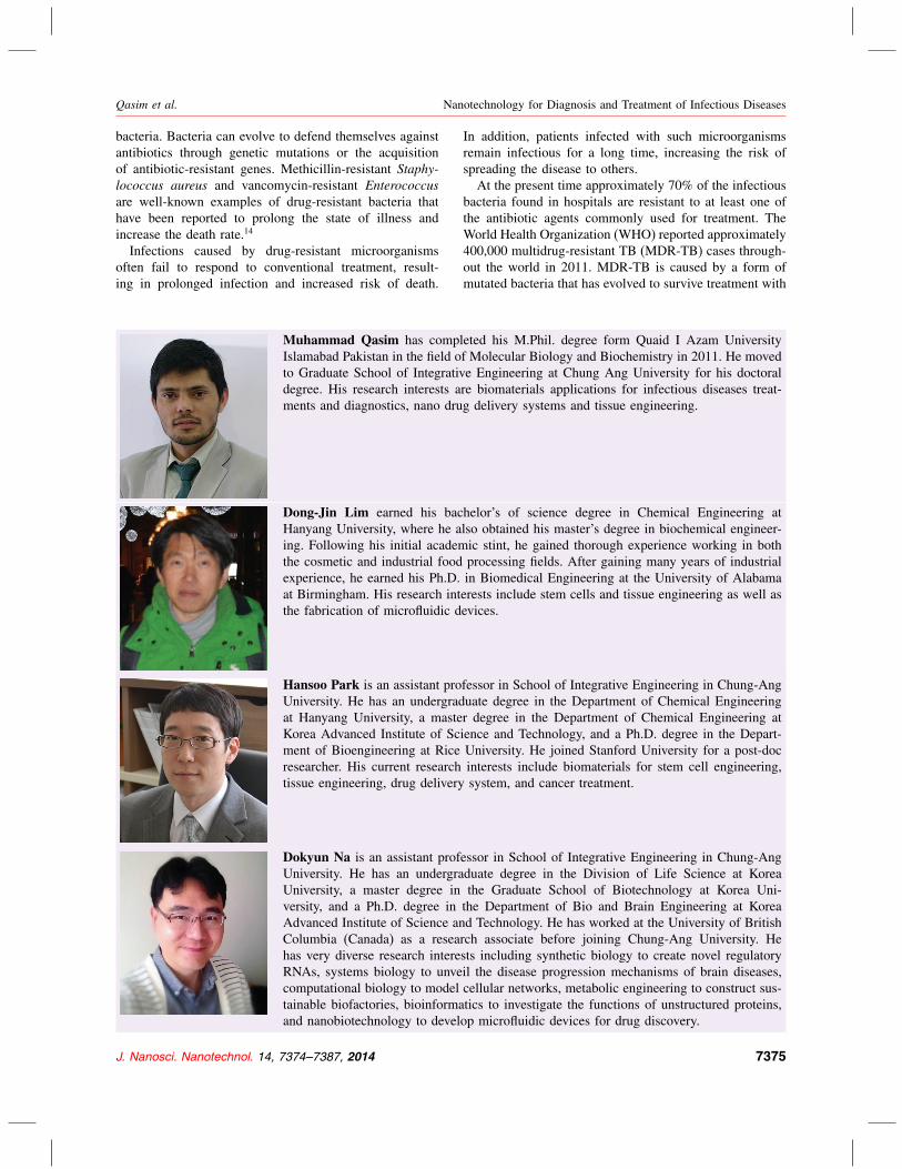
Muhammad Qasim has completed his M.Phil. degree form Quaid I Azam University
Islamabad Pakistan in the field of Molecular Biology and Biochemistry in 2011. He moved
to Graduate School of Integrative Engineering at Chung Ang University for his doctoral
degree. His research interests are biomaterials applications for infectious diseases treat-
ments and diagnostics, nano drug delivery systems and tissue engineering.
Dong-Jin Lim earned his bachelor’s of science degree in Chemical Engineering at
Hanyang University, where he also obtained his master’s degree in biochemical engineer-
ing. Following his initial academic stint, he gained thorough experience working in both
the cosmetic and industrial food processing fields. After gaining many years of industrial
experience, he earned his Ph.D. in Biomedical Engineering at the University of Alabama
at Birmingham. His research interests include stem cells and tissue engineering as well as
the fabrication of microfluidic devices.
Hansoo Park is an assistant professor in School of Integrative Engineering in Chung-Ang
University. He has an undergraduate degree in the Department of Chemical Engineering
at Hanyang University, a master degree in the Department of Chemical Engineering at
Korea Advanced Institute of Science and Technology, and a Ph.D. degree in the Depart-
ment of Bioengineering at Rice University. He joined Stanford University for a post-doc
researcher. His current research interests include biomaterials for stem cell engineering,
tissue engineering, drug delivery system, and cancer treatment.
Dokyun Na is an assistant professor in School of Integrative Engineering in Chung-Ang
University. He has an undergraduate degree in the Division of Life Science at Korea
University, a master degree in the Graduate School of Biotechnology at Korea Uni-
versity, and a Ph.D. degree in the Department of Bio and Brain Engineering at Korea
Advanced Institute of Science and Technology. He has worked at the University of British
Columbia (Canada) as a research associate before joining Chung-Ang University. He
has very diverse research interests including synthetic biology to create novel regulatory
RNAs, systems biology to unveil the disease progression mechanisms of brain diseases,
computational biology to model cellular networks, metabolic engineering to construct sus-
tainable biofactories, bioinformatics to investigate the functions of unstructured proteins,
and nanobiotechnology to develop microfluidic devices for drug discovery.
In addition, patients infected with such microorganisms
remain infectious for a long time, increasing the risk of
spreading the disease to others.
At the present time approximately 70% of the infectious
bacteria found in hospitals are resistant to at least one of
the antibiotic agents commonly used for treatment. The
World Health Organization (WHO) reported approximately
400,000 multidrug-resistant TB (MDR-TB) cases through-
out the world in 2011. MDR-TB is caused by a form of
mutated bacteria that has evolved to survive treatment with
J. Nanosci. Nanotechnol. 14, 7374–7387, 2014 7375

Delivered by Publishing Technology to: Chung-Ang UniversityIP: 165.194.5.66 On: Wed, 07 May 2014 02:35:09
Copyright: American Scientific Publishers
Nanotechnology for Diagnosis and Treatment of Infectious Diseases Qasim et al.
at least two of the primary antibiotic drugs.15�16 The conse-
quences of antimicrobial resistance were higher mortality,
increased cost due to the use of more expensive drugs, and
increased burden on the public health care system.
Consequently, there is an increase in the demand for
new strategies, pharmaceuticals, and devices to diag-
nose and treat diseases accurately, easily, and efficiently.
A number of nanotechnology-based materials have been
studied with the aim of effective control and prevention of
infectious diseases.17 Recently, nanotechnology has been
employed to enhance immune responses against antigens
for effective vaccination, to deliver pharmaceuticals to a
target site and release them at a controlled rate, and to
detect and identify diseases accurately and rapidly at low
cost. This review will discuss how nanotechnology has
improved the treatment, diagnostics, and prevention of
infectious diseases.
1.1. The Immune SystemHumans defend themselves from infection with for-
eign pathogens through the innate and adaptive immune
systems.17�18 The innate immune system acts as the first
line of defense against invading particles and consists
of several cell types including dendritic cells (DCs),
macrophages, and natural killer (NK) cells, whose primary
role is to eliminate foreign materials without specificity.19
The innate immune cells often recognize common fea-
tures of pathogens such as lipopolysaccharides for effi-
cient defense. Antigen-presenting cells (APCs), including
DCs, are able to digest foreign materials into fragments
and present these fragments on their membrane surface
through major histocompatibility complex (MHC) class
II proteins. The presented antigens trigger the adaptive
immune system, which acts as a second line of defense
and is composed of highly specialized processes that
eliminate infection through the antigen-specific responses
of humoral and cell-mediated immunity.20 This adap-
tive immunity displays antigenic specificity, diversity, and
memorization.
The role of humoral immunity is to eliminate foreign
antigens from the body. Once antigens are presented on
APCs, B cells that recognize the antigens are activated
to undergo clonal expansion and differentiate into plasma
cells, specialized factories that produce antibodies. Certain
activated B cells differentiate into memory cells, which are
able to rapidly differentiate into plasma cells. Upon second
infection the antigens are recognized and can be quickly
eliminated by the memory cells.
The role of cell-mediated immunity is to eliminate
virus-infected host cells. Every cell in our body presents
its own protein fragments on MHC class I molecules. Den-
dritic cells ingest viruses and present virus molecules on
their surface. Various subpopulations of cytotoxic T cells
recognize the presented virus antigens and become acti-
vated. When activated cytotoxic T cells recognize corre-
sponding antigens presented by MHC class I molecules of
infected host cells they send signals that trigger apoptosis
of the infected cells.
1.2. Current Treatments and Their LimitationsOver the centuries medical research has improved our
understanding of microorganisms and has accordingly led
to better prevention and treatment of infectious diseases
through the discovery of antibiotics, antitoxins, antivirals,
antifungals, and vaccines.21�22 Of these discoveries, the
invention of vaccines was a great leap forward in the man-
agement of infectious diseases because it prevented the
spread of disease rather than treating the disease after
infection. The success of vaccination is illustrated by the
elimination of small pox.23�24
Recently, several therapeutic strategies have been devel-
oped to treat bacterial and viral infections that were pre-
viously considered difficult to conquer. Nonetheless, it
can be hard to recover completely from infection and
there is a great demand for new technologies to diag-
nose, treat, and possibly prevent infection. Below, we dis-
cuss treatment strategies for three well-known infectious
agents: Mycobacterium tuberculosis, Human Immunodefi-
ciency virus (HIV), and Hepatitis C Virus (HCV).
TB is caused by the mycobacterium M. tuberculosistherefore antibiotics are commonly used for treatment.
There are two phases of antibiotic treatment for TB infec-
tion. The first phase is intensive treatment, which involves
a combination of the antibiotics isoniazid, ehtambutol,
rifampicin, and pyrazinamide. The second phase is a 4-
month treatment course with only rifampicin and isoniazid.
Complete eradication of TB requires both intensive and
continuous treatment for about 6 to 8 months. This is the
most effective treatment to date for TB, but because of the
long treatment period the drugs have side effects such as
jaundice, dyspepsia, exanthema, and arthralgia.25
For the treatment of acquired human deficiency syn-
drome (AIDS) caused by infection with HIV there are
currently five classes of drugs that inhibit the replication
and integration of the viral genome at different stages
in the life cycle of HIV-1 virus: nucleotide reverse tran-
scriptase inhibitors, non-nucleoside reverse transcriptase
inhibitors, protease inhibitors, fusion inhibitors, and inte-
grase inhibitors. At present these drugs are used in com-
bination and this regime is referred to as highly active
antiviral therapy (HAART). Most drugs used in HAART
are costly and have severe side effects, and furthermore
multi-drug resistance to these drugs has been reported.26
HCV is known as a silent killer of infected individu-
als because its symptoms only appear at a late stage of
infection. Chronic HCV infection can lead to liver cancer
and eventually death. Currently there are no available vac-
cines for HCV and therefore no way to prevent infection.
After infection, a combination of antiviral ribavirin and
interferon-� are used for treatment and complete removal
of virus from the bloodstream requires several months of
continuous therapy. However, these compounds are toxic
7376 J. Nanosci. Nanotechnol. 14, 7374–7387, 2014

Delivered by Publishing Technology to: Chung-Ang UniversityIP: 165.194.5.66 On: Wed, 07 May 2014 02:35:09
Copyright: American Scientific Publishers
Qasim et al. Nanotechnology for Diagnosis and Treatment of Infectious Diseases
Table I. Current therapies against selected pathogens and their limitations.
Current treatment
Disease name Causative agent strategies Commonly used drug agents Limitations in treatment Refs
Hepatitis C Hepatitis C (HCV) Combination of
interferon and
broad spectrum
anti-viral
therapy
Pegylated interferon-�, ribavirin,
and protease inhibitor.
—Limited efficacy in patients with
HCV genotype 1.
—Drug resistance is rapidly
emerging.
—Drug administration by injection
over 72 weeks may result in
chronic side effects
[1, 2]
Acquired
immunodeficiency
syndrome (AIDS)
Human
immunodeficiency
virus (HIV)
Highly active
antiretroviral
therapy
(HAART)
—Nucleoside reverse transcriptase
inhibitors (NRTIs): e.g.,
Lamivudine and Zidovudine
—Nonnucleoside reverse
transcriptase inhibitors (NNRTIs):
e.g., Rilpivirine
—Fusion inhibitors: e.g.,
Enfuvirtide
—Protease inhibitors: e.g.,
Tipranavir, Indinavir, and
Amprenavir
—Integrase inhibitors: e.g.,
Raltegravir and Dolutegravir
—Treatment should be continued
throughout life.
—Potential emergence of drug
resistance.
—Complete eradication is not
possible.
—Side effects such as increased
rate of heartbeat, diabetes, liver
diseases, cancer, and premature
aging.
[3]
Cervical cancer Human papilloma
virus (HPV)
Cryosurgery, loop
electrosurgical
excision
procedure
(LEEP), laser
therapy,
hysterectomy,
vaccine
Salicylic acid, aldara, zyclara,
condylox, trichloroacetic acid
—No treatment for existing cervical
cancer except for removal of
cervix.
—Vaccine has side effects and is
effective only before exposure to
the virus.
[35]
Hepatitis B Hepatitis B virus
(HBV)
Interferon therapy IFN-�, Pegylated interferon,
Lamivudine, Adefovir, Dipivoxil,
Entecavir, Telbivudine, and
Tenofovir
—No treatment available for acute
hepatitis B.
—Cold chain issues for vaccine.
—A booster dose of vaccine is
required therefore follow up of
patients is a major issue.
[4]
Malaria Plasmodium Combination
therapy of
antimalarial
drugs
Artemisinin, Prophylactic
Artemisinin
No vaccine available. [8]
Poliomyelitis Polio virus Vaccine No antiviral drugs available —No treatments available.
Immunization for prevention only.
—Vaccine is expensive and
requires a cold chain for
transportation and storage.
—Issue of oral polio vaccine
(OPV) degradation
[40, 41]
Influenza virus Influenza virus
A and B
Vaccine —Antiviral therapy and vaccination
—Adamantanes inhibitors
(Amantadine and Rimamtadine)
—Neuraminidase inhibitors
(Zanamivir and Oseltamivir)
—Vaccine for Influenza Virus A
(HIN1) Monovalent 2009 vaccine
—These drugs have activity only
against Influenza A strain.
—Amantadine carries a risk of
neuropsychological, atropinic and
dopaminergic adverse effects.
—Zanamivir carries a risk of
life-threatening bronchospasm.
—Emergence of drug resistance is
a crucial problem for treatment of
influenza virus.
[39]
Mumps, measles and
rubella
Paramyxo virus,
mumps virus
rubella
Vaccine MMR vaccine. —No treatment available. Vaccine
for prevention only
—Potential side effects of vaccine
J. Nanosci. Nanotechnol. 14, 7374–7387, 2014 7377

Delivered by Publishing Technology to: Chung-Ang UniversityIP: 165.194.5.66 On: Wed, 07 May 2014 02:35:09
Copyright: American Scientific Publishers
Nanotechnology for Diagnosis and Treatment of Infectious Diseases Qasim et al.
Table I. Continued.
Current treatment
Disease name Causative agent strategies Commonly used drug agents Limitations in treatment Refs
Tuberculosis (TB) MycobacteriumTuberculosis
Antibiotics Isoniazide, Pyrazinamide,
Rifampicin, Ethambutol,
Ethionamide, Prothionamide,
Cycloserine, Capreomycin,
Paraaminosalicylic Acid,
Fluroroquinolones
—No vaccine available.
—Drug resistance may emerge due
to breakdown of therapy by
patients therefore TB is
re-emerging.
—Drug administration requires the
presence of a second person.
[37, 38]
Candidiasis Candida species Antifungal agents Fluconazole, Itraconazole,
Posaconazole, Clotrimazole,
Amphotericin B, Deoxycholate,
Econazole, Ciclopirox,
Miconazole, Ketoconazole,
Nystatin, Clotrimazole,
Voriconazole, Candins,
Flucytosine, Ravuconazole,
Posaconazole, Micafungin, and
Anidulafungin
—Most antifungal drugs have low
water solubility due to
hydrophobic nature.
—Drug resistance has emerged
against the two most useful
antifungal drugs, Amphotericin B
and Ketoconazole.
[7]
Leishmaniasis Leishmania species Antifungal and
Antileishmanial
drugs
Liposomal Amphotericin B,
Ketoconazole, Paromomycin
(Humatin) and Miltefosine
(Miltex).
Anti-leishmanial drugs are toxic and
have to be administered
parenterally for long periods,
especially for visceral
leishmaniasis.
—Re-emergence of diseases due to
drug resistance, especially in
tropical areas.
[36]
and have severe side effects, and moreover are not equally
effective for all genotypes of HCV.27
Despite the discovery and development of new thera-
peutic strategies for treatment, complete prevention and
eradication of infectious diseases has not been achieved.
Certain therapeutic compounds have serious side effects
and therefore cannot be widely applied; for example, nerve
damage caused by the DPT vaccine (diphtheria, pertussis,
and tetanus) and long-term pain, numbness, infertility, and
paralysis due to the HPV vaccine in Japan. Moreover, cer-
tain retroviruses change their genomic sequence at a high
rate therefore no effective vaccines have been developed.
Available strategies and therapeutics, as well as their lim-
itations, are listed in Table I.
In addition to the difficulty in developing new therapeu-
tics, emergence of drug resistance of existing pathogens
that escape from current treatment strategies is another
significant problem in infectious diseases. The world-
wide prevalence of drug-resistant strains indicates that
this is a global issue.5�28 The Center for Disease Con-
trol and Prevention reported that the number of annual
infections by multidrug-resistant Staphylococcus aureus(MRSA) increased from 127,000 in 1999 to 278,000 in
2005.16�29 Similarly, the number of annual deaths caused
by MRSA in the United States increased from 11,000 in
2005 to 17,000 in 2007.30 Recently, more than 1,500 peo-
ple in Germany were infected with a new virulent strain
of Escherichia coli (104:H4) that has not been reported in
any prior outbreaks.6
1.3. Current Diagnostic Techniques andTheir Limitations
Effective treatment and prevention of infectious diseases
requires up-to-date diagnostics. In addition, the efficacy of
treatment should be monitored during therapy by detec-
tion of pathogens. Conventional techniques available for
the diagnosis of infectious disease include microscopy,
tissue culture, lateral flow immunoassays (also known
as dipsticks or immune chromatographic test, ICTs),
enzyme-linked immunosorbent assays (ELISAs), and bio-
chemical tests (Table II). More recently, molecular diag-
nostics techniques such as polymerase chain reaction
(PCR) and real-time PCR have been widely used to diag-
nose and monitor infections such as HIV/AIDS and HCV
because they have a higher specificity and sensitivity than
ELISA-based diagnostics. However, because these tech-
niques are costly and time-consuming and require prior
sample preparation, they are commonly used in devel-
oped countries but are often poorly suited for developing
countries, where infectious diseases are leading causes of
morbidity and mortality, because the availability of trained
clinical staff and specific laboratory facilities may be
limited.31 Thus, there is a great demand for new diagnostic
technologies. The ideal diagnostic device for developing
countries would be a cost-effective, portable, and point-
of-source detection system that is also highly reliable,
sensitive, and accurate.32 Furthermore, the ideal diagnostic
technique would be able to detect multiple pathogens in a
single reaction.
7378 J. Nanosci. Nanotechnol. 14, 7374–7387, 2014

Delivered by Publishing Technology to: Chung-Ang UniversityIP: 165.194.5.66 On: Wed, 07 May 2014 02:35:09
Copyright: American Scientific Publishers
Qasim et al. Nanotechnology for Diagnosis and Treatment of Infectious Diseases
Table II. Diagnostics tests for infectious diseases in developing countries and their limitations.42
Disease Diagnostic test Limitations
Acute respiratory
infections (ARIs)
Blood/sputum culture —Culture is time-consuming and costly and results are highly
dependent on stringent transport conditions to maintain specimen.
—Requires constant supply of reagents, well-maintained equipment,
and adequately trained staff.
Acquired
immunodeficiency
syndrome (AIDS)
—Serology (antigen/antibody detection); detection by
PCR amplification of the LTR region of gag gene,env gene, and pol gene
High rate of false negative and positives.
—PCR cannot be performed during the HIV incubation period.
—Nucleic acid amplification technologies (NAAT) such as PCR are
expensive and may produce many false positives due to
contamination
Diarrheal diseases —Microscopy stool culture Microscopy requires well-trained and supervised technologists.
—Pathogen culture is time-consuming and costly.
Malaria Blood film; antigen detection (dipstick); antibody
detection (ELISA and Rapid)
Rapid tests help to control disease spreading but have high false
positive rates.
Tuberculosis (TB) Sputum microscopy; pathogen solid and liquid culture;
Tuber skin test; PCR-based Gene Xpert
Microscopy requires experts and well-trained staff. Antibody tests,
Tuber skin test (tuberculin) may produce false positive result due
to allergic reactions.
Visceral leishmaniasis Direct agglutination test (DAT); serological field test;
microscopy/culture of spleen or bone morrow cells
Bone morrow sampling is painful and invasive.
Conventional molecular diagnostic technologies are
based on the amplification of specific DNA sequences
from extracted nucleic acids (DNA or RNA), for example
target amplification (e.g., PCR, reverse transcriptase
PCR (RT-PCR), and strand displacement amplification),
signal amplification (e.g., branched DNA assays and
hybrid capture), probe amplification (e.g., ligase chain
reaction, cleavage-invader, and cycling probes), or post-
amplification analysis (e.g., sequencing the amplified prod-
ucts or melting curve analysis). However, amplification
methods are so sensitive that false positives may easily
occur due to trace contamination of the specimen or equip-
ment. In addition, because these techniques depend on
enzymatic activity, false negatives can occur when samples
contain contaminants that inhibit the enzymes.33
DNA microarrays or DNA chips are one of the lat-
est methods for rapid infectious disease diagnostics. DNA
microarrays are essentially a high-throughput version of
the Southern blot method.34 Each microarray contains a
number of different DNA oligonucleotides that recognize
specific target genes from a pathogen through complemen-
tary DNA–DNA binding. Diagnosis is made by the detec-
tion of oligonucleotides hybridized to pathogen genes.
Although DNA microarrays have demonstrated poten-
tial in diagnostics their practical use in clinical settings is
hampered by several considerations, especially difficulties
in the identification of pathogen-unique target genes and in
the design of oligonucleotide primers for multiplex PCR.
The first difficulty lies in finding a gene that is unique
to a particular pathogen among a tremendous number
of genes.43 The gene most commonly used to identify
microorganisms is ribosomal 16S rRNA, because every
bacterium has a variant of the gene. However, organisms
belonging to the same species share a large number of
nucleotides in the 16S rDNA gene, which limits the identi-
fication of bacteria below the species level.44 Furthermore,
certain phylogenetically unrelated bacteria have been
reported to have very similar 16S rRNA sequences.
Another challenge is to design oligonucleotides that will
amplify particular pathogen genes in multiplex PCR with-
out non-specific amplifications. Because multiplex PCR is
limited to approximately a dozen reactions, several hundred
rounds of PCR would be required to completely cover all
the potential probes that identify all infectious diseases on
an array. PCR on such a massive scale is not practical in
routine use.
Emerging nanotechnology-based techniques have
recently attracted interest as an approach that may over-
come the problems of current diagnostic techniques
through their specific mode of actions and unique phys-
ical properties (i.e., shape, size, surface charge, and
dimension).45 These techniques may be applied to develop
accurate, reliable, rapid, safe, cost-efficient, sensitive,
specific, and easily accessible techniques for the detec-
tion of pathogens.46 In the following section we discuss
new nanotechnology-based methods for the treatment,
diagnosis, or prevention of infectious diseases.
2. NANOTECHNOLOGY ANDINFECTIOUS DISEASE
2.1. Modulation of Immune Response byNanoparticles (NPs) for Efficient Vaccination
Vaccines are specifically designed antigens that effectively
induce immune responses so that the immune system will
remember the antigens and can rapidly respond upon sub-
sequent infection. Vaccination is a reliable method of pre-
venting infectious diseases, and plays a very important
role in the control of mortalities and morbidities associated
with infectious diseases.47 It is estimated that vaccines pre-
vent almost 6 million deaths/year worldwide48 and a few
diseases such as smallpox have been successfully eradi-
cated through widespread vaccination programs.49
J. Nanosci. Nanotechnol. 14, 7374–7387, 2014 7379
Delivered by Publishing Technology to: Chung-Ang UniversityIP: 165.194.5.66 On: Wed, 07 May 2014 02:35:09
Copyright: American Scientific Publishers
Nanotechnology for Diagnosis and Treatment of Infectious Diseases Qasim et al.
Effective vaccines must induce strong immune
responses. Conventional vaccines display various degrees
of immunogenicity and safety. For example, attenuated
vaccines are highly immunogenic but are also associated
with a risk of infection by the vaccines themselves as a
result of the virus reverting to a virulent form. On the
other hand, antigenic protein vaccines are not infectious
and therefore safer, but are less immunogenic and repeated
vaccination is required. Furthermore, a well-established
cold chain is required to transport vaccines safely and there
are insufficient facilities to store and transport vaccines in
developing countries. Recent advancements in nanotech-
nology have provided new approaches for effective immu-
nization against infectious diseases.
Activation of the cell-mediated immune response is an
important target to improve the efficacy of therapeutic
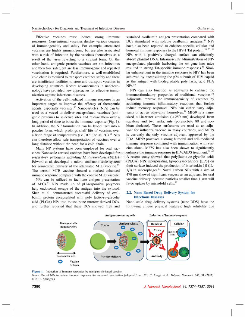
agents, especially vaccines.50 Nanoparticles (NPs) can be
used as a vessel to deliver encapsulated vaccines (anti-
genic proteins) to selective sites and release them over a
long period of time to boost the immune response (Fig. 1).
In addition, the NP formulation can be lyophilized into a
powder form, which prolongs shelf life of vaccines over
a wide range of temperatures (i.e., 0 �C to 40 �C).51 NPs
can therefore allow safe transportation of vaccines over a
long distance without the need for a cold chain.
Many NP systems have been employed for oral vac-
cines. Nanoscale aerosol vaccines have been developed for
respiratory pathogens including M. tuberculosis (MTB).
Edward et al. developed a micro- and nano-scale system
for aerosolized delivery of the attenuated MTB vaccine.22
The aerosol MTB vaccine showed a marked enhanced
immune response compared with the control MTB vaccine.
NPs can be utilized to facilitate antigen presentation
of APCs.53 NPs made up of pH-responsive polymers
help endosomal escape of the antigen into the cytosol.
Shen et al. demonstrated successful delivery of oval-
bumin protein encapsulated with poly lactic-co-glycolicacid (PLGA) NPs into mouse bone marrow-derived DCs,
and further reported that these DCs showed high and
Figure 1. Induction of immune responses by nanoparticle-based vaccine.
Notes: Use of NPs to induce immune responses for enhanced vaccination (adopted from [52], T. Akagi, et al., Polymer Nanomed. 247, 31 (2012).© 2012, Springer.)
sustained ovalbumin antigen presentation compared with
DCs stimulated with soluble ovalbumin antigens.54 NPs
have also been reported to enhance specific cellular and
humoral immune responses to the HIV-1 Tat protein.53�55�56
NPs with a positively charged surface can efficiently
absorb plasmid DNA. Intramuscular administration of NP-
encapsulated plasmids harboring the tat gene into mice
resulted in strong Tat-specific immune responses.56 Simi-
lar enhancement in the immune response to HIV has been
achieved by encapsulating the p24 subunit of HIV capsid
as the antigen with biodegradable poly lactic acid PLA
NPs.57
NPs can also function as adjuvants to enhance the
immunostimulatory properties of traditional vaccines.52
Adjuvants improve the immunogenicity of vaccines by
activating immune inflammatory reactions that further
induce memory responses. NPs can either carry adju-
vants or act as adjuvants themselves.58 MF59 is a nano-
sized oil-in-water emulsion (< 250 nm) developed from
squalene and two surfactants (polysorbate 80 and sor-
bitan tiroleate). These surfactants are used as an adju-
vant for influenza vaccine in many countries, and MF59
is currently the only vaccine adjuvant approved by the
FDA. MF59 provokes a strong humoral and cell-mediated
immune response compared with immunization with vac-
cine alone. MF59 has also been shown to significantly
enhance the immune response in HIV/AIDS treatment.59�60
A recent study showed that poly(lactic-co-glycolic acid)
(PLGA) NPs incorporating lipopolysaccharides (LPS) on
their surface induced the production of interleukin 1� (IL-
1�) in macrophages.61 Novel carbon NPs with a size of
470 nm showed significant success as an adjuvant for oral
vaccine delivery, because particles smaller than 1 �m will
favor uptake by microfold cells.55
2.2. Nano-Based Drug Delivery System forInfectious Diseases
Nano-scale drug delivery systems (nano-DDS) have the
following unique physical features: high solubility due
7380 J. Nanosci. Nanotechnol. 14, 7374–7387, 2014
Delivered by Publishing Technology to: Chung-Ang UniversityIP: 165.194.5.66 On: Wed, 07 May 2014 02:35:09
Copyright: American Scientific Publishers
Qasim et al. Nanotechnology for Diagnosis and Treatment of Infectious Diseases
to their inherent hydrophilicity and solubilizing moieties,
thermo-sensitivity, and capability for controlled release of
encapsulated drugs, easy surface modification, and high
surface area-to-volume ratios. These unique features of
nano-DDS may allow medical scientists to overcome the
problems associated with the increased drug resistance of
infectious agents. By scaling down the size of compounds,
nano-DDS can modulate and improve the performance
of many drugs to an extent not achievable with conven-
tional drugs. Nano-DDS can encapsulate drugs and thereby
enhance their stability, solubility, and absorption. Nano-
DDS can also increase blood circulation time, deliver
drugs to specific cells or tissues, and release them in a
controlled manner in response to a specific stimulus.14
Targeted and efficient drug delivery becomes possible
with the use of NPs.62 NPs have the ability to encapsu-
late therapeutic agents or vaccines at high density. Because
these NPs protect encapsulated drugs from diffusion and
enzymatic degradation, the drugs can be delivered at high
doses to a particular target site.62 In addition, NPs are
able to release drugs at the slow rate required to prolong
immune responses.52 Importantly, certain NPs are able to
deliver drugs across the blood brain barrier (BBB), the
tight junction of endothelial cells that isolates and protects
the inner blood circulation of the brain from the rest of the
blood circulation.63�64 Thus, NPs are efficient drug delivery
vehicles for the treatment of diverse infectious diseases,
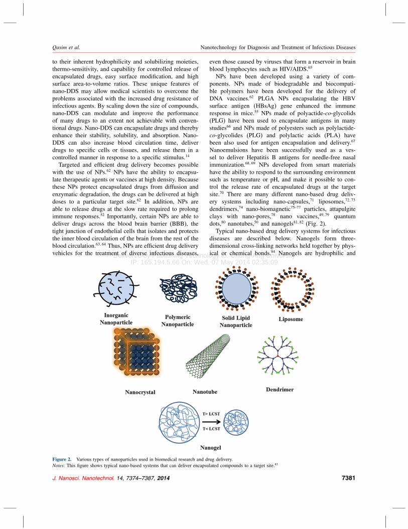
Figure 2. Various types of nanoparticles used in biomedical research and drug delivery.
Notes: This figure shows typical nano-based systems that can deliver encapsulated compounds to a target site.83
even those caused by viruses that form a reservoir in brain
blood lymphocytes such as HIV/AIDS.65
NPs have been developed using a variety of com-
ponents. NPs made of biodegradable and biocompati-
ble polymers have been developed for the delivery of
DNA vaccines.62 PLGA NPs encapsulating the HBV
surface antigen (HBsAg) gene enhanced the immune
response in mice.55 NPs made of polyactide-co-glycolids(PLG) have been used to encapsulate antigens in many
studies66 and NPs made of polyesters such as polylactide-
co-glycolides (PLG) and polylactic acids (PLA) have
been also used for antigen encapsulation and delivery.67
Nanoemulsions have been successfully used as a ves-
sel to deliver Hepatitis B antigens for needle-free nasal
immunization.68�69 NPs developed from smart materials
have the ability to respond to the surrounding environment
such as temperature or pH, and make it possible to con-
trol the release rate of encapsulated drugs at the target
site.70 There are many different nano-based drug deliv-
ery systems including nano-capsules,71 liposomes,72�73
dendrimers,74 nano-biomagnetic75–77 particles, attapulgite
clays with nano-pores,78 nano vaccines,49�79 quantum
dots,80 nanotubes,50 and nanogels81�82 (Fig. 2).
Typical nano-based drug delivery systems for infectious
diseases are described below. Nanogels form three-
dimensional cross-linking networks held together by phys-
ical or chemical bonds.84 Nanogels are hydrophilic and
J. Nanosci. Nanotechnol. 14, 7374–7387, 2014 7381

Delivered by Publishing Technology to: Chung-Ang UniversityIP: 165.194.5.66 On: Wed, 07 May 2014 02:35:09
Copyright: American Scientific Publishers
Nanotechnology for Diagnosis and Treatment of Infectious Diseases Qasim et al.
therefore have the ability to absorb a large amount of water
while maintaining their structure and remaining undis-
solved. Smart nanogels with thermoresponsive properties
have been used to control drug release.82 Nanogels with
surface charge are able to enhance antifungal and antibac-
terial activity.85 Moreover, nanogels loaded with drugs can
be lyophilized into a powder form and can be transported
over long distances without a cold chain.86
Solid lipid NPs are stable and able to incorporate drugs
at high density, which reduces the risk of retention of
a high level of residual organic solvent.73 For example,
in the treatment of TB a single administration of solid
NPs encapsulating rifampicin, isoniazid, and pyrazinamide
was sufficient to elicit recovery from infection within
1 week, whereas non-encapsulated drugs must be admin-
istered every day for 1 week to obtain the same result.87
Liposomes are vesicular carriers of 20 to 30 nm in
size that are composed of phospholipid bilayers around
an aqueous core.88 Encapsulation and delivery of drugs
in liposomes has shown great success in the treatment of
HIV/AIDS. Encapsulation of Indinavir, an antiviral agent
used to treat HIV, in liposomes resulted in more effi-
cient delivery of the drug to lymphoid tissues than treat-
ment with soluble Indinavir and successfully reduced HIV
viral load and potentially recovered CD4 T cells.89 Load-
ing of Indinavir into lipid nano-capsules (LNCs) greatly
improved drug delivery to the brain and testis of mice.90
Similarly, when Zidovudine (AZT) was encapsulated in
liposomes, the accumulated concentration of AZT in lym-
phoid tissues was 27 times higher than when soluble AZT
was administered.91 The AZT plasma concentration was
also significantly higher in patients treated with AZT lipo-
somes compared with patients treated with AZT solution.92
Dendrimers are a hyper-branched and mono-dispersed
class of polymers with a defined molecular weight. They
have a central core and three-dimensional branches with
functionalized surface groups. Dendrimers improve the
solubility of hydrophobic drugs by ionic interactions and
thereby improve their efficacy.84 Targeting of the drug
efavirenz to monocytes/macrophages using a mannose-
targeting poly (propyleneimine) dendrimer increased the
cellular uptake of efavirenz up to 12-fold.93
A carbon nanotube is a tube-shaped material made of
carbon with a diameter in the nanometer scale. Carbon
nanotubes have diverse structural variations in thickness
and the number of layers of graphite. When nanotubes car-
rying siRNA specific for C-X-C chemokine receptor type
4 (CXCR4) were targeted to CD4 cells, the expression of
CXCR4 receptor proteins was reduced by 60–90%.50�74
2.3. Nanoparticles as Therapeutic DrugsNPs that have been developed to have novel immunother-
apeutic properties can themselves be used as drugs.94
Under UV light, metallic NPs and their oxides produce
reactive oxygen species that possess antimicrobial activity
Figure 3. Mechanisms by which nanoparticles kill bacteria cells.
Notes: Certain NPs and their ions produce free radicals that lead to oxida-
tive stress, for example through reactive oxygen species. These reactive
species induce damage in bacterial proteins and DNAs, which eventually
lead to bacterial death.
(Fig. 3).95 Metallic NPs incorporating Ag,96–98 Au,98�99
Cu,100 Ti,96 Mg, Zn,79 Fe, or metal oxides101 have signif-
icant antimicrobial, antifungal, and antiviral activities.102
Ag-NPs effectively kill many bacterial species including
E. coli, S. aureus, B. subtilis, and S. typhai.103 Cu-NPs canalso have a profound toxic effect; in one study no colonies
were formed when S. cerevisiae was incubated on a Cu-
NP-loaded polymer thin film.100
Nanomaterials with inherent antimicrobial activities are
called nanoantibiotics.102 Nitric oxide-releasing NPs (NO-
NPs) act through many simultaneous antimicrobial mecha-
nisms. NO exerts its antimicrobial activity largely through
reactive nitrogen oxide intermediates (RNOS), which are
formed after NO reacts with superoxide (O−2 ). The RNOS
react with amino acid residues of bacterial proteins and
plasma membrane proteins, leading to death of bacterial
cells. RNOS also directly damage bacterial DNA through
strand breaks, formation of basic sites, and deamination
of nucleotides.104 NO-NPs have been shown to inhibit the
growth of antibiotic-resistant strains of P. aeruginosa, E.faecalis, K. pneumoniae, and E. coli. When administered
at a concentration of 1.25-5 mM, NO-NPs successfully
killed MRSA, E. faecalis, and E. coli.105 (Fig. 3).
In addition, nanoantibiotics have advantages over con-
ventional antibiotics because they interact with multi-
ple biological pathways in bacteria and are stable for a
long time in terms of their action and storage. In addi-
tion, antibiotic NP polymers enhance the efficacy of
traditional antimicrobial agents not only through the addi-
tional antimicrobial activity of the NP polymers, but also
by increasing the solubility and efficiency of delivery of
7382 J. Nanosci. Nanotechnol. 14, 7374–7387, 2014

Delivered by Publishing Technology to: Chung-Ang UniversityIP: 165.194.5.66 On: Wed, 07 May 2014 02:35:09
Copyright: American Scientific Publishers
Qasim et al. Nanotechnology for Diagnosis and Treatment of Infectious Diseases
the antimicrobial. Nylon-6 nano-fiber incorporated with
5,5-dimethyl hydantoin (DMH) exhibited strong antimi-
crobial activity compared with DHM alone.106
2.4. Nano-Based Diagnosis of Infectious DiseasesNPs with appropriate surface modifications are able to
interact with biomolecules such as proteins and DNA.45�107
The unique physical and chemical properties of NPs allow
accurate, rapid, sensitive, and cost-efficient diagnostics.
For example, NPs with enhanced fluorescent properties
can improve sensitivity in diagnostic molecular bioimag-
ing. In antibody-based diagnoses, primary antibodies rec-
ognize antigenic proteins of cells or viruses and secondary
antibodies recognize the primary antibodies, specifically
the constant regions. Labeling the secondary antibodies
with fluorescent NPs can remarkably enhance detection
sensitivity.108
Fluorescent silica NPs (FSNPs)98 have been devel-
oped to detect MTB. In this application, FSNPs encap-
sulate two organic and metallic fluorescent dyes, Tris
(2,2′bipyridyl) osmium bis (hexafluorophosphate) (OsBpy)
and Tris (bipyridine) ruthenium(II) dichloride (RuBpy),
that are excited by a single wavelength. These FSNPs
exhibit high signal amplification and photostability. MTB
is detected using anti-MTB primary antibody and a sec-
ondary antibody labeled with FSNPs for enhanced detec-
tion of anti-MTB antibody. Using this system, MTB was
detected in a mixture of bacteria and sputum with high
sensitivity and within only 4 hours.108
A system for the detection of anthrax protective agent
(PA) has been developed using fluorescence ELISA that
incorporated Eu(III) polymeric NPs. This system showed
approximately 100-fold higher sensitivity than conven-
tional ELISA.109�110 A rapid ELISA system to detect Her-
pes Simplex Virus type 2 (HSV-2) has been developed
using Au-NPs bound to anti-human IgG and allows detec-
tion of HSV-2 in 15–20 minutes using a test strip.111 MTB
DNA also can be detected rapidly and accurately using an
Au-NP–based colorimetric assay.112 A protein assay chip
using a signal amplification method involving NPs has
been developed for the detection of HBV and HCV.113
Quantum dots (Qdots) are special nano-crystal semicon-
ductors made of metals such as Si or Ge that range in
size from 1 nm to 10 nm.21 Qdots possess very strong
fluorescence intensity that makes them suitable for sen-
sitive image acquisition and signal amplification in real
time.80 Many Qdots have been developed to detect various
types of viral and bacterial proteins in order to diagnose
pathogenic diseases with enhanced sensitivity and speci-
ficity over conventional organic fluorophores. The Tripe
and Nie groups successfully demonstrated that dual core
Qdots could detect a single copy of virus or viral pro-
tein with high sensitivity and in real time.114 The dual
core Qdots were composed of two fluorescent NPs green
color and red one (40 nm carboxyl-modified fluorescent
NPs: GNPs, 505/51 and RNPs, 488/685) and streptavidin-
coated Qdots (488/605). These dual core Qdots were
used to label secondary antibodies that recognize anti-
bodies against respiratory syncytial virus (RSV).114�115
Qdots that create a barcode for multiplex identification of
biomolecules have been developed to identify multiple tox-
ins in blood.75 Barcodes have also been created using Qdots
(ZnS-capped CdSe Qdots) conjugated with three pathogen
antigens: HBsAg for HCV, HCV nonstructural protein 4
(NSP-4) for HCV, and glycoprotein 41 (gp41) for HIV.
This barcode-based multiplex identification system allowed
detection at a threshold of 10−10 to 10−12 M from 100 �lhuman serum spiked with corresponding viral antigens.80
Certain metallic NPs have a special optical property
known as surface plasmon resonance (SPR) that pro-
duces intense absorption when excited by electromagnetic
radiation.108 Gold NPs have been used for the detection of
DNA since 1996,116 and gold nano-wire arrays (GNWA)
linked with specific antibodies against E.coli have been
developed to detect urinary tract infection.117 Immuno-gold
silver staining with Au-NPs is very a sensitive method
for the detection of single molecules that does not need
advanced instruments and has been applied to the detec-
tion of HCV and HBV.113�118 Gold nano-rods have two
surface plasmon spectra: a strong and long wavelength
band at 600–950 nm and a short wavelength band at
520 nm. Non-linear optical properties (NLO) of gold nano-
rods vary with their shape and size and can be monitored
using the Hyper Rayleigh Scattering (HRS) technique. The
NLO properties of gold nano-rods allow them to be used
in bioconjugation-based diagnostic methods. For example,
HIV-1 DNA (gag gene) has been successfully quantified
by monitoring the light scattering pattern of dye-tagged
ssDNA gold nano-rods using HRS spectroscopy.119 The
detection limit of this method for the HIV-1 gag gene was
100 pM. This technique is highly sequence-specific; even
a single base pair mismatch results in a different light pat-
tern and can therefore be captured.
Polystyrene NPs loaded with Eu(III) ions have unique
photophysical properties such as sharp line-like emission
peaks, longer lifetime, and a large stroke shift. These
properties allow rapid and cheap detection of Anthrax
protective agent (PA) using these NPs. Polystyrene NPs
co-polymerized with acrylic acid have been used to
identify P. falciparum, the causative agent of malaria,
in the outfield.113
Surface Enhanced Raman Scattering (SERS) silver
nano-rod arrays have been used to track the molecular
fingerprints of several infectious pathogens.120 SERS is
a Raman spectroscopic technique that provides greatly
enhanced Raman signals from Raman-active analytic
molecules adsorbed on a rough metal surface or on nano-
structures such as silver nano-rods, silica nanotubes, or
carbon nanotubes. Gold nanowires coated with silica were
used to develop a sandwich DNA hybridization assay to
J. Nanosci. Nanotechnol. 14, 7374–7387, 2014 7383

Delivered by Publishing Technology to: Chung-Ang UniversityIP: 165.194.5.66 On: Wed, 07 May 2014 02:35:09
Copyright: American Scientific Publishers
Nanotechnology for Diagnosis and Treatment of Infectious Diseases Qasim et al.
Figure 4. Test strip assay format.
Notes: Toxins are captured by the gangliosides attached on the liposome surface. The toxin-bound liposomes migrate through the nitrocellulose test
strip. Toxins bound to the liposomes are captured by anti-toxin antibodies in the analytical zone, producing a color change that indicates the presence
of toxins.124
detect multiple viruses.98 Nanowires made of different
metals by electrode position were used to study multiplex
detection of protein and nucleic acid based on the barcod-
ing phenomenon.121
Magnetic particles such as Fe3O4 or Fe2O3 also have
great potential for detecting food-borne pathogens such as
E. coli O157:H7 and Salmonella.122 Magnetic NPs coated
with antibodies are used for the detection of pathogens
by immunogenic separation. Vancomycin-conjugated FePt
magnetic NPs were developed to capture and iden-
tify vancomycin-resistant enterococci (VRE) and other
Gram-positive bacteria present at very low concentra-
tions in samples. Subsequently, these Fept@van biofunc-
tional magnetic NPs were combined with fluorescent
dyes for rapid detection of bacteria from human blood
samples.75�76 Biocompatible super paramagnetic iron oxide
(SPIO) nano-biosensors developed for the detection of
HSV-1 and adenoviruses were able to detect five viral par-
ticles in 10-�l serum samples without any PCR amplifica-
tion steps.77
Liposomes have become very versatile tools in
biomedical applications due to their enormous diversity
in structure and composition. Engineered liposomes are
able to mimic cells and recognize target toxins, and can
therefore be used for toxin detection. When such lipo-
somes were labeled with fluorescent markers (rhodamine
dyes) and used in a sandwich fluoroimmunoassay on
antibody-coated microtiter plates, they could detect the
cholera toxin with a threshold of 1 nM.123 In another
recent study, a sensitive bioassay system for cholera toxin
(CT) was developed using ganglioside GM1-labeled lipo-
somes (∼ 200 nm in diameter). In this sandwich assay,
CT bound to the liposomes was captured by immobilized
antibodies and detected as a colored band on a nitrocel-
lulose membrane strip, as shown in Figure 4. The limit
of detection was 10 fg/ml and the assay could be com-
pleted in 20 minutes.124 A similar assay was developed
to detect botulinum toxin (BT) using trisialoganglioside
GT1b, a receptor for BT. In this immunoassay, BT bound
to GT1b-liposomes was captured by immobilized anti-BT
antibodies and detected as a colored band on a nitrocellu-
lose membrane strip.125
3. CONCLUSIONSThis review presented current limitations of conventional
treatment and diagnostics as well as current advances
facilitated by nanoparticles to address the limitations.
The emerging field of nanotechnology addresses the cur-
rent limitations of conventional treatment and diagnostics.
Modern nano-based vaccines are more reliable than con-
ventional vaccines and now occupy almost 65% of the
worldwide vaccine market. The worldwide market value
of nanotechnology products has reached 11,671 million
US dollars and is expected to increase to 26,000 million
US dollars by 2015 with an annual growth rate of 11.1%.
Although nanotechnology already provides new directions
for the advancement of treatments and diagnostics for
infectious diseases, it should be developed further for
practical use in daily life. Furthermore, nanotechnology-
based therapeutics, vaccines, and diagnostics may foster
easy, cheap, safe, and portable use of end-products, which
will help control infectious diseases, especially in devel-
oping countries, and improve public health. Nanoparticles
7384 J. Nanosci. Nanotechnol. 14, 7374–7387, 2014

Delivered by Publishing Technology to: Chung-Ang UniversityIP: 165.194.5.66 On: Wed, 07 May 2014 02:35:09
Copyright: American Scientific Publishers
Qasim et al. Nanotechnology for Diagnosis and Treatment of Infectious Diseases
make a great leap from traditional diagnosis and treatment
to more advanced and improved ones by allowing con-
trolling many parameters in diagnosis and treatment such
as controlled slow release of encapsulated drugs. For fur-
ther advance in nanoparticle-based technologies, it is very
essential to develop to design nanoparticles with a desired
function: efficient encapsulation of drugs, specific binding
to targets, on-site release, etc. So far, studies have replied
on empirical or intuitive approaches. We believe that in
order to further facilitate the advance of nanoparticle-based
approaches it is necessary to develop systematic and ratio-
nal design principles for nanoparticle design.
Acknowledgment: This work was supported by a grant
from the Basic Science Research Program (2011-0013819)
through the National Research Foundation of Korea
(NRF) funded by the Ministry of Education, Science and
Technology.
References and Notes1. A. L. Armstead and B. Y. Li, Int. J. Nanomed. 6, 3281 (2011).2. S. D. Sharma, Ind. J. Med. Res. 131, 17 (2010).3. D. D. Richman, D. M. Margolis, M. Delaney, W. C. Greene,
D. Hazuda, and R. J. Pomerantz, Science 323, 1304 (2009).4. S. K. Fung and A. S. Lok, Clin. Gastroenterol. Hepatol. 2, 839
(2004).5. A. J. Kallen, Y. Mu, S. Bulens, A. Reingold, S. Petit, K. Gershman,
S. M. Ray, L. H. Harrison, R. Lynfield, G. Dumyati, J. M. Townes,
W. Schaffner, P. R. Patel, S. K. Fridkin, and A. M. I. E. I, JAMA304, 641 (2010).
6. R. Hyde, Lancet 377, 1991 (2011).7. P. G. Pappas, J. H. Rex, J. D. Sobel, S. G. Filler, W. E.
Dismukes, T. J. Walsh, and J. E. Edwards, Clin. Infect Dis 38, 161(2004).
8. P. Olliaro, Trop Med. Int. Heal. 14, 488 (2009).9. F. von Groote-Bidlingmaier and A. H. Diacon, Ther. Umsch 68,
395 (2011).10. Lancet Infect Dis. 11, 253 (2011), http://www.thelancet.com/
journals/laninf/article/PIIS1473-3099(11)70077-9/fulltextdoi:10.1016/
S1473-3099(11)70077-9.
11. D. M. Morens, G. K. Folkers, and A. S. Fauci, Nature 430, 242
(2004).12. D. M. Morens, G. K. Folkers, and A. S. Fauci, Nature 463, 122
(2010).13. J. P. Millet, A. Moreno, L. Fina, L. del Bano, A. Orcau, P. G. de
Olalla, and J. A. Cayla, Eur. Spine J. 22, 539 (2013).14. F. Andrade, D. Rafael, M. Videira, D. Ferreira, A. Sosnik, and
B. Sarmento, Adv. Drug Deliv. Rev 65, 1816 (2013).15. L. Phillips, Nature 493, 14 (2013).16. R. Choudhury, S. Panda, and D. V. Singh, Indian J. Med. Microbiol.
30, 384 (2012).17. K. Blecher, A. Nasir, and A. Friedman, Virulence 2, 395 (2011).18. K. Hoebe, E. Janssen, and B. Beutler, Nat. Immunol. 5, 971
(2004).19. S. Akira, S. Uematsu, and O. Takeuchi, Cell 124, 783 (2006).20. M. S. Diamond, B. Shrestha, E. Mehlhop, E. Sitati, and M. Engle,
Viral Immunol. 16, 259 (2003).21. W. G. Lee, Y. G. Kim, B. G. Chung, U. Demirci, and
A. Khademhosseini, Adv. Drug Deliv. Rev. 62, 449 (2010).22. L. Garcia-Contreras, Y. L. Wong, P. Muttil, D. Padilla, J. Sadoff,
J. DeRousse, W. A. Germishuizen, S. Goonesekera, K. Elbert, B. R.
Bloom, R. Miller, P. B. Fourie, A. Hickey, and D. Edwards, Pro-ceedings of the National Academy of Sciences of the United Statesof America 105, 4656 (2008).
23. H. Ledford, Nat. Biotechnol. 26, 591 (2008).24. K. Uberla, PLoS Pathog 4, e1000114 (2008).25. P. Garner, H. Smith, S. Munro, and J. Volmink, Bull World Health
Organ 85, 404 (2007).26. L. Waters and M. Nelson, Int. J. Clin. Prac 61, 983 (2007).27. C. I. Yu and B. L. Chiang, J. Biomed. Biotechnol. 2010, 548280
(2010).28. E. A. Bancroft, Resistant, Ethicillin Aureus, S Taphylococcus,
JAMA 298, 15 (2007).29. S. B. Levy and B. Marshall, Nat, Med. 10, S122 (2004).30. A. J. Kallen, Y. Mu, S. Bulens, A. Reingold, S. Petit, K. Gershman,
S. M. Ray, L. H. Harrison, R. Lynfield, G. Dumyati, J. M. Townes,
W. Schaffner, P. R. Patel, and S. K. Fridkin, JAMA 304, 641 (2010).31. A. Sosnik and M. Amiji, Adv. Drug Del. Rev. 62, 375 (2010).32. T. S. Hauck, S. Giri, Y. L. Gao, and W. C. W. Chan, Adv. Drug
Del. Rev. 62, 438 (2010).33. C. S. L. J. Hartman and D. A. Norwood, Mol. Cell Prob 19, 51
(2005).34. M. Schena, D. Shalon, R. W. Davis, and P. O. Brown, Science 270,
467 (1995).35. A. C. Linhares and L. L. Villa, J. Pediatr (Rio J.) 82, S25 (2006).36. S. Sundar and M. Rai, Curr. Opin. Infect Dis. 15, 593 (2002).37. J. P. Lynch, Chest 119, 371s (2001).38. D. L. Monnet, C. Suetens, S. Earnshaw, C. Gagliotti,
J. Griskeviciene, O. E. Heuer, E. Khazeeva, N. Kleinkauf,
E. Liljestedt, A. P. Magiorakos, F. Santos, M. J. Struelens, J. T.
Weber, K. Weist, and E. A. R. Heal, Eurosurveillance 15, 2 (2010).39. R. Kandel and K. L. Hartshorn, Biodrugs 15, 303 (2001).40. D. L. Barnard, Curr. Pharm. Des. 12, 1379 (2006).41. J. L. Melnick, Clin. Microbiol. Rev. 9, 293 (1996).42. D. Mabey, R. W. Peeling, A. Ustianowski, and M. D. Perkins, Nat.
Rev. Microbiol. 2, 231 (2004).43. J. M. Janda and S. L. Abbott, J. Clin. Microbiol. 45, 2761 (2007).44. M. Grunstein, Curr. Contents/Life Sciences 72, 3961 (1986).45. N. Daum, C. Tscheka, A. Neumeyer, and M. Schneider, WIREs
Nanomed. Nanobiotechnol. 4, 52 (2012).46. N. L. Rosi and C. A. Mirkin, Chem. Rev. 105, 1547 (2005).47. F. E. Andre, R. Booy, H. L. Bock, J. Clemens, S. K. Datta, T. J.
John, B. W. Lee, S. Lolekha, H. Peltola, T. A. Ruff, M. Santosham,
and H. J. Schmitt, Bull. World Health Organ. 86, 140 (2008).48. J. Ehreth, Vaccine 21, 596 (2003).49. T. D. Nandedkar, J. Biosci. 34, 995 (2009).50. Z. Liu, M. Winters, M. Holodniy, and H. J. Dai, Angewandte
Chemie-Int Edition 46, 2023 (2007).51. M. Holzer, V. Vogel, W. Mantele, D. Schwartz, W. Haase, and
K. Langer, Eur. J. Pharm. Biopharm. 72, 428 (2009).52. T. Akagi, M. Baba, and M. Akashi, Polymer Nanomed. 247, 31
(2012).53. S. T. Clemente-Casares, X. Yang, and Y. S. Pere, J. Mol. Med.
(Berl) 89, 733 (2011).54. H. Shen, A. L. Ackerman, V. Cody, A. Giodini, E. R. Hinson,
P. Cresswell, R. L. Edelson, W. M. Saltzman, and D. J. Hanlon,
Immunol. 117, 78 (2006).55. X. W. He, F. Wang, L. Jiang, J. Li, S. K. Liu, Z. Y. Xiao, X. Q.
Jin, Y. N. Zhang, Y. He, K. Li, Y. J. Guo, and S. H. Sun, J. Gen.Virol. 86, 601 (2005).
56. F. Lori, J. Trocio, N. Bakare, L. M. Kelly, and J. Lisziewicz,
Vaccine 23, 2030 (2005).57. D. Lamalle-Bernard, S. Munier, C. Compagnon, M. H. Charles,
V. S. Kalyanaraman, T. Delair, B. Verrier, and Y. Ataman-Onal,
J. Controlled Release 115, 57 (2006).58. L. J. Peek, C. R. Middaugh, and C. Berkland, Adv. Drug Deliv Rev.
60, 915 (2008).
J. Nanosci. Nanotechnol. 14, 7374–7387, 2014 7385

Delivered by Publishing Technology to: Chung-Ang UniversityIP: 165.194.5.66 On: Wed, 07 May 2014 02:35:09
Copyright: American Scientific Publishers
Nanotechnology for Diagnosis and Treatment of Infectious Diseases Qasim et al.
59. G. Ott, G. L. Barchfeld, D. Chernoff, R. Radhakrishnan, P. van
Hoogevest, and G. Van Nest, Pharm. Biotechnol. 6, 277 (1995).60. D. K. Mitchell, S. J. Holmes, R. L. Burke, A. M. Duliege, and S. P.
Adler, Pediatr Infect Dis J. 21, 133 (2002).61. S. L. Demento, S. C. Eisenbarth, H. G. Foellmer, C. Platt, M. J.
Caplan, W. M. Saltzman, I. Mellman, M. Ledizet, E. Fikrig, R. A.
Flavell, and T. M. Fahmy, Vaccine 27, 3013 (2009).62. M. D. Bhavsar and M. M. Amiji, Expet Opin Drug Deliv 4, 197
(2007).63. A. Nowacek and H. E. Gendelman, Nanomed 4, 557 (2009).64. H. L. Wong, N. Chattopadhyay, X. Y. Wu, and R. Bendayan, Adv.
Drug Deliv. Rev. 62, 503 (2010).65. H. Y. Dou, C. B. Grotepas, J. M. McMillan, C. J. Destache,
M. Chaubal, J. Werling, J. Kipp, B. Rabinow, and H. E. Gendelman,
J. Immunol. 183, 661 (2009).66. S. C. Diesner, X. Y. Wang, E. Jensen-Jarolim, E. Untersmayr, and
F. Gabor, Ther. Deliv. 3, 277 (2012).67. A. C. Stanley, D. Buxton, E. A. Innes, and J. F. Huntley, Vaccine
22, 3929 (2004).68. E. L. Giudice and J. D. Campbell, Adv. Drug Deliv. Rev. 58, 68
(2006).69. P. E. Makidon, A. U. Bielinska, S. S. Nigavekar, K. W. Janczak,
J. Knowlton, A. J. Scott, N. Mank, Z. Cao, S. Rathinavelu, M. R.
Beer, J. E. Wilkinson, L. P. Blanco, J. J. Landers, and J. R. Baker,
Jr, PLoS One 3, e2954 (2008).70. T. Nochi, Y. Yuki, H. Takahashi, S. I. Sawada, M. Mejima,
T. Kohda, N. Harada, I. G. Kong, A. Sato, N. Kataoka, D. Tokuhara,
S. Kurokawa, Y. Takahashi, H. Tsukada, S. Kozaki, K. Akiyoshi,
and H. Kiyono, Nat. Mater. 9, 685 (2010).71. C. E. Mora-Huertas, H. Fessi, and A. Elaissari, Int. J. Pharm. 385,
113 (2010).72. M. J. Copland, T. Rades, N. M. Davies, and M. A. Baird, Immunol.
Cell Biol. 83, 97 (2005).73. S. A. Wissing, O. Kayser, and R. H. Muller, Adv. Drug Deliv. Rev.
56, 1257 (2004).74. N. Weber, P. Ortega, M. I. Clemente, D. Shcharbin, M. Bryszewska,
F. J. de la Mata, R. Gomez, and M. A. Munoz-Fernandez, J. Con-trolled Release 132, 55 (2008).
75. J. H. Gao, L. Li, P. L. Ho, G. C. Mak, H. W. Gu, and B. Xu, Adv.Mat. 18, 3145 (2006).
76. H. W. Gu, P. L. Ho, K. W. T. Tsang, L. Wang, and B. Xu, J. Am.Chem. Soc. 125, 15702 (2003).
77. L. Josephson, J. M. Perez, and R. Weissleder, Angewandte Chem.-Int. Edition 40, 3204 (2001).
78. N. Krikorian and D. F. Martin, J. Environ. Sci. Health a Tox HazardSubst. Environ. Eng. 40, 601 (2005).
79. D. J. Bharali, S. A. Mousa, and Y. Thanavala, Adv. Exp. Med. Biol.601, 415 (2007).
80. J. M. Klostranec, Q. Xiang, G. A. Farcas, J. A. Lee, A. Rhee, E. I.
Lafferty, S. D. Perrault, K. C. Kain, and W. C. Chan, Nano Lett. 7,2812 (2007).
81. W. Cai and X. Chen, Small 3, 1840 (2007).82. S. P. Hudson, R. Langer, G. R. Fink, and D. S. Kohane,
Biomaterials 31, 1444 (2010).83. A. H. Faraji and P. Wipf, Bioorg. Med. Chem. 17, 2950 (2009).84. K. Winnicka, M. Wroblewska, P. Wieczorek, P. T. Sacha, and
E. Tryniszewska, Molecules 17, 4612 (2012).85. A. Zumbuehl, L. Ferreira, D. Kuhn, A. Astashkina, L. Long, Y. Yeo,
T. Iaconis, M. Ghannoum, G. R. Fink, R. Langer, and D. S. Kohane,
Proc Natl. Acad. Sci. USA 104, 12994 (2007).86. T. R. Hoare and D. S. Kohane, Polymer 49, 1993 (2008).87. R. Pandey, S. Sharma, and G. K. Khuller, Tuberculosis (Edinb) 85,
415 (2005).88. R. H. Muller, K. Mader, and S. Gohla, Eur. J. Pharm. Biopharm.
50, 161 (2000).
89. L. Kinman, S. J. Brodie, C. C. Tsai, T. Bui, K. Larsen, A. Schmidt,
D. Anderson, W. R. Morton, S. L. Hu, and R. J. Ho, J. AcquirImmune Defic Syndr. 34, 387 (2003).
90. M. P. De Oliveira, E. Garcion, N. Venisse, J. P. BenoIt, W. Couet,
and J. C. Olivier, Pharm Res. 22, 1898 (2005).91. S. X. Jin, D. Z. Bi, J. Wang, Y. Z. Wang, H. G. Hu, and Y. H.
Deng, Pharmazie 60, 840 (2005).92. C. D. Kaur, M. Nahar, and N. K. Jain, J. Drug Target 16, 798
(2008).93. T. Dutta, H. B. Agashe, M. Garg, P. Balakrishnan, M. Kabra, and
N. K. Jain, J. Drug Target 15, 89 (2007).94. M. Look, A. Bandyopadhyay, J. S. Blum, and T. M. Fahmy, Adv.
Drug Del. Rev. 62, 378 (2010).95. R. P. Allaker and G. Ren, Trans R Soc. Trop Med. Hyg. 102, 1
(2008).96. F. Martinez-Gutierrez, P. L. Olive, A. Banuelos, E. Orrantia,
N. Nino, E. M. Sanchez, F. Ruiz, H. Bach, and Y. Av-Gay,
Nanomed. Nanotechnol. Bio. Med. 6, 681 (2010).97. M. Rai, A. Yadav, and A. Gade, Biotechnol. Adv. 27, 76 (2009).98. J. A. Sioss, R. L. Stoermer, M. Y. Sha, and C. D. Keating, Langmuir
23, 11334 (2007).99. C. Brandenberger, B. Rothen-Rutishauser, C. Muhlfeld, O. Schmid,
G. A. Ferron, K. L. Maier, P. Gehr, and A. G. Lenz, Tox Appl.Pharm. 242, 56 (2010).
100. N. Cioffi, L. Torsi, N. Ditaranto, G. Tantillo, L. Ghibelli,
L. Sabbatini, T. Bleve-Zacheo, M. D’Alessio, P. G. Zambonin, and
E. Traversa, Chem. Mater. 17, 5255 (2005).101. M. R. Mihu, U. Sandkovsky, G. Han, J. M. Friedman, J. D.
Nosanchuk, and L. R. Martinez, Virulence 1, 62 (2010).102. A. J. Huh and Y. J. Kwon, J. Controlled Release 156, 128 (2011).103. F. Martinez-Gutierrez, P. L. Olive, A. Banuelos, E. Orrantia,
N. Nino, E. M. Sanchez, F. Ruiz, H. Bach, and Y. Av-Gay,
Nanomed. 6, 681 (2010).104. R. Y. Pelgrift and A. J. Friedman, Adv. Drug Deliv. Rev (2013).105. M. J. Hajipour, K. M. Fromm, A. A. Ashkarran, D. J. de Aberasturi,
I. R. de Larramendi, T. Rojo, V. Serpooshan, W. J. Parak, and
M. Mahmoudi, Trends Biotechnol. 30, 499 (2012).106. S. S. Al-Deyab, M. H. El-Newehy, E. R. Kenawy, and A. Abdel-
Mageed, Abstr. Pap Am. Chem. S 2011, 1 (2011).107. P. Tallury, A. Malhotra, L. M. Byrne, and S. Santra, Adv. Drug
Deliv. Rev. 62, 424 (2010).108. D. L. Qin, X. X. He, K. M. Wang, X. J. J. Zhao, W. H. Tan, and
J. Y. Chen, J. Biomed. Biotechnol 2007, 1 (2007).109. X. Q. Chi, D. T. Huang, Z. H. Zhao, Z. J. Zhou, Z. Y. Yin, and
J. H. Gao, Biomaterials 33, 189 (2012).110. S. X. Tang, M. Moayeri, Z. C. Chen, H. Harma, J. Q. Zhao, H. J.
Hu, R. H. Purcell, S. H. Leppla, and I. K. Hewlett, Clin VaccineImmunol. 16, 408 (2009).
111. E. I. Laderman, E. Whitworth, E. Dumaual, M. Jones, A. Hudak,
W. Hogrefe, J. Carney, and J. Groen, Clin Vaccine Immunol. 15,159 (2008).
112. P. V. Baptista, M. Koziol-Montewka, J. Paluch-Oles, G. Doria, and
R. Franco, Clinic Chem. 52, 1433 (2006).113. L. L. Duan, Y. F. Wang, S. S. C. Li, Z. X. Wan, and J. X. Zhai,
BMC Infect Dis. 5, 1 (2005).114. A. Agrawal, R. A. Tripp, L. J. Anderson, and S. Nie, J. Virol. 79,
8625 (2005).115. E. L. Bentzen, F. House, T. J. Utley, J. E. Crowe, Jr, and D. W.
Wright, Nano Lett. 5, 591 (2005).116. C. A. Mirkin, R. L. Letsinger, R. C. Mucic, and J. J. Storhoff,
Nature 382, 607 (1996).117. A. de la Escosura-Muniz and A. Merkoci, Small 7, 675 (2011).118. Y. F. Wang, D. W. Pang, Z. L. Zhang, H. Z. Zheng, J. P. Cao, and
J. T. Shen, J. Med. Virol. 70, 205 (2003).119. G. K. Darbha, U. S. Rai, A. K. Singh, and P. C. Ray, Chem. Euro
J. 14, 3896 (2008).
7386 J. Nanosci. Nanotechnol. 14, 7374–7387, 2014

Delivered by Publishing Technology to: Chung-Ang UniversityIP: 165.194.5.66 On: Wed, 07 May 2014 02:35:09
Copyright: American Scientific Publishers
Qasim et al. Nanotechnology for Diagnosis and Treatment of Infectious Diseases
120. J. D. Driskell, S. Shanmukh, Y. J. Liu, S. Hennigan, L. Jones, Y. P.
Zhao, R. A. Dluhy, D. C. Krause, and R. A. Tripp, IEEE Sens J.8, 863 (2008).
121. B. He, T. J. Morrow, and C. D. Keating, Curr. Opin. Chem. Biol.12, 522 (2008).
122. L. D. Qin, O. Vermesh, Q. H. Shi, and J. R. Heath, Lab Chip 9,
2016 (2009).
123. A. K. Singh, S. H. Harrison, and J. S. Schoeniger, Anal. Chem. 72,
6019 (2000).
124. S. Ahn-Yoon, T. R. DeCory, A. J. Baeumner, and R. A. Durst, Anal.
Chem. 75, 2256 (2003).
125. S. Ahn-Yoon, T. R. DeCory, and R. A. Durst, Anal. Bioanal. Chem.
378, 68 (2004).
Received: 5 October 2013. Accepted: 3 November 2013.
J. Nanosci. Nanotechnol. 14, 7374–7387, 2014 7387