Amniotic fluid stem cells improve survival and enhance repair of damaged intestine in necrotising...
11
ORIGINAL ARTICLE Amniotic fluid stem cells improve survival and enhance repair of damaged intestine in necrotising enterocolitis via a COX-2 dependent mechanism Augusto Zani, 1 Mara Cananzi, 1 Francesco Fascetti-Leon, 1 Giuseppe Lauriti, 1 Virpi V Smith, 2 Sveva Bollini, 1 Marco Ghionzoli, 1 Antonello D’Arrigo, 3 Michela Pozzobon, 4 Martina Piccoli, 4 Amy Hicks, 5 Jack Wells, 6 Bernard Siow, 6 Neil J Sebire, 2 Colin Bishop, 5 Alberta Leon, 3 Anthony Atala, 5 Mark F Lythgoe, 6 Agostino Pierro, 1 Simon Eaton, 1 Paolo De Coppi 1 ▸ Additional material is published online only. To view please visit the journal online (http://dx.doi.org/10.1136/ gutjnl-2012-303735). 1 Surgery Unit, University College London Institute of Child Health, London, UK 2 Department of Histopathology, Great Ormond Street Hospital for Children, London, UK 3 Research & Innovation S.p.A., Padova, Italy 4 Laboratory of Oncohematology, Department of Pediatrics, University of Padova, and Fondazione Città della Speranza, Padova, Italy 5 Wake Forest Institute for Regenerative Medicine, Wake Forest University School of Medicine, Medical Center Blvd, Winston-Salem, North Carolina, USA 6 Division of Medicine, UCL Centre for Advanced Biomedical Imaging, Institute of Child Health, University College London, London, UK Correspondence to Dr Paolo De Coppi and Dr Simon Eaton, Surgery Unit, University College London Institute of Child Health, 30 Guilford Street, London WC1N 1EH, UK; [email protected] and [email protected] AZ and MC contributed equally. Received 30 October 2012 Revised 25 January 2013 Accepted 1 March 2013 To cite: Zani A, Cananzi M, Fascetti-Leon F, et al. Gut Published Online First: [ please include Day Month Year] doi:10.1136/gutjnl- 2012-303735 ABSTRACT Objective Necrotising enterocolitis (NEC) remains one of the primary causes of morbidity and mortality in neonates and alternative strategies are needed. Stem cells have become a therapeutic option for other intestinal diseases, which share some features with NEC. We tested the hypothesis that amniotic fluid stem (AFS) cells exerted a beneficial effect in a neonatal rat model of NEC. Design Rats intraperitoneally injected with AFS cells and their controls (bone marrow mesenchymal stem cells, myoblast) were analysed for survival, behaviour, bowel imaging (MRI scan), histology, bowel absorption and motility, immunofluorescence for AFS cell detection, degree of gut inflammation (myeloperoxidase and malondialdehyde), and enterocyte apoptosis and proliferation. Results AFS cells integrated in the bowel wall and improved rat survival and clinical conditions, decreased NEC incidence and macroscopic gut damage, improved intestinal function, decreased bowel inflammation, increased enterocyte proliferation and reduced apoptosis. The beneficial effect was achieved via modulation of stromal cells expressing cyclooxygenase 2 in the lamina propria, as shown by survival studies using selective and non-selective cyclooxygenase 2 inhibitors. Interestingly, AFS cells differentially expressed genes of the Wnt/β- catenin pathway, which regulate intestinal epithelial stem cell function and cell migration and growth factors known to maintain gut epithelial integrity and reduce mucosal injury. Conclusions We demonstrated here for the first time that AFS cells injected in an established model of NEC improve survival, clinical status, gut structure and function. Understanding the mechanism of this effect may help us to develop new cellular or pharmacological therapies for infants with NEC. INTRODUCTION Necrotising enterocolitis (NEC) represents up to 10% of admissions to neonatal intensive care unit, 1 and remains a major cause of neonatal morbidity and mortality despite changes in medical and surgi- cal treatment. 12 Although administration of breast milk, arginine or probiotics may reduce the inci- dence of the disease, there are no specific medical therapies which are of clinical benefit in infants with NEC. 3 Surgical resection of affected segments Significance of this study What is already known on this subject? ▸ Necrotising enterocolitis (NEC) is the most common gastrointestinal surgical emergency occurring in neonates, with high mortality rates ranging from 15% to 30%. ▸ Despite extensive research in this field, there are still no medical therapies that have proven to be of clinical benefit for the cure of affected neonates. Surgery is still the treatment of choice in case of necrotic bowel. ▸ There is a growing body of evidence that stem cells could play a therapeutic role in inflammatory bowel diseases and other intestinal pathologies. What are the new findings? ▸ In a well-established neonatal rat model of NEC, amniotic fluid stem (AFS) cells improve rat survival, decrease morbidity, and reduce NEC incidence. ▸ In this model, AFS cells are able to improve intestinal function, decrease bowel inflammation, increase enterocyte proliferation and reduce apoptosis. ▸ AFS cell beneficial effect is achieved via modulation of stromal cells expressing COX-2 in the lamina propria. How might it impact on clinical practice in the foreseeable future? ▸ Stem cell therapy may represent a new therapeutic option for children with NEC. Moreover, understanding the mechanism of action of AFS cells in the experimental NEC may help us develop new cellular or pharmacological therapies for infants with NEC. Zani A, et al. Gut 2013;0:1–10. doi:10.1136/gutjnl-2012-303735 1 In fl ammatory bowel disease Gut Online First, published on March 24, 2013 as 10.1136/gutjnl-2012-303735 Copyright Article author (or their employer) 2013. Produced by BMJ Publishing Group Ltd (& BSG) under licence. group.bmj.com on April 1, 2013 - Published by gut.bmj.com Downloaded from
-
Upload
independent -
Category
Documents
-
view
0 -
download
0
Transcript of Amniotic fluid stem cells improve survival and enhance repair of damaged intestine in necrotising...
ORIGINAL ARTICLE
Amniotic fluid stem cells improve survivaland enhance repair of damaged intestine innecrotising enterocolitis via a COX-2 dependentmechanismAugusto Zani,1 Mara Cananzi,1 Francesco Fascetti-Leon,1 Giuseppe Lauriti,1
Virpi V Smith,2 Sveva Bollini,1 Marco Ghionzoli,1 Antonello D’Arrigo,3
Michela Pozzobon,4 Martina Piccoli,4 Amy Hicks,5 Jack Wells,6 Bernard Siow,6
Neil J Sebire,2 Colin Bishop,5 Alberta Leon,3 Anthony Atala,5 Mark F Lythgoe,6
Agostino Pierro,1 Simon Eaton,1 Paolo De Coppi1
▸ Additional material ispublished online only. To viewplease visit the journal online(http://dx.doi.org/10.1136/gutjnl-2012-303735).1Surgery Unit, UniversityCollege London Institute ofChild Health, London, UK2Department of Histopathology,Great Ormond Street Hospitalfor Children, London, UK3Research & Innovation S.p.A.,Padova, Italy4Laboratory of Oncohematology,Department of Pediatrics,University of Padova, andFondazione Città dellaSperanza, Padova, Italy5Wake Forest Institute forRegenerative Medicine, WakeForest University School ofMedicine, Medical Center Blvd,Winston-Salem, North Carolina,USA6Division of Medicine, UCLCentre for AdvancedBiomedical Imaging, Instituteof Child Health, UniversityCollege London, London, UK
Correspondence toDr Paolo De Coppi andDr Simon Eaton, Surgery Unit,University College LondonInstitute of Child Health,30 Guilford Street, LondonWC1N 1EH, UK;[email protected] [email protected]
AZ and MC contributedequally.
Received 30 October 2012Revised 25 January 2013Accepted 1 March 2013
To cite: Zani A, Cananzi M,Fascetti-Leon F, et al. GutPublished Online First:[please include Day MonthYear] doi:10.1136/gutjnl-2012-303735
ABSTRACTObjective Necrotising enterocolitis (NEC) remains oneof the primary causes of morbidity and mortality inneonates and alternative strategies are needed. Stemcells have become a therapeutic option for otherintestinal diseases, which share some features with NEC.We tested the hypothesis that amniotic fluid stem (AFS)cells exerted a beneficial effect in a neonatal rat modelof NEC.Design Rats intraperitoneally injected with AFS cellsand their controls (bone marrow mesenchymal stemcells, myoblast) were analysed for survival, behaviour,bowel imaging (MRI scan), histology, bowel absorptionand motility, immunofluorescence for AFS cell detection,degree of gut inflammation (myeloperoxidase andmalondialdehyde), and enterocyte apoptosis andproliferation.Results AFS cells integrated in the bowel wall andimproved rat survival and clinical conditions, decreasedNEC incidence and macroscopic gut damage, improvedintestinal function, decreased bowel inflammation,increased enterocyte proliferation and reduced apoptosis.The beneficial effect was achieved via modulation ofstromal cells expressing cyclooxygenase 2 in the laminapropria, as shown by survival studies using selective andnon-selective cyclooxygenase 2 inhibitors. Interestingly,AFS cells differentially expressed genes of the Wnt/β-catenin pathway, which regulate intestinal epithelial stemcell function and cell migration and growth factorsknown to maintain gut epithelial integrity and reducemucosal injury.Conclusions We demonstrated here for the first timethat AFS cells injected in an established model of NECimprove survival, clinical status, gut structure andfunction. Understanding the mechanism of this effectmay help us to develop new cellular or pharmacologicaltherapies for infants with NEC.
INTRODUCTIONNecrotising enterocolitis (NEC) represents up to10% of admissions to neonatal intensive care unit,1
and remains a major cause of neonatal morbidityand mortality despite changes in medical and surgi-cal treatment.1 2 Although administration of breast
milk, arginine or probiotics may reduce the inci-dence of the disease, there are no specific medicaltherapies which are of clinical benefit in infantswith NEC.3 Surgical resection of affected segments
Significance of this study
What is already known on this subject?▸ Necrotising enterocolitis (NEC) is the most
common gastrointestinal surgical emergencyoccurring in neonates, with high mortality ratesranging from 15% to 30%.
▸ Despite extensive research in this field, thereare still no medical therapies that have provento be of clinical benefit for the cure of affectedneonates. Surgery is still the treatment ofchoice in case of necrotic bowel.
▸ There is a growing body of evidence that stemcells could play a therapeutic role ininflammatory bowel diseases and otherintestinal pathologies.
What are the new findings?▸ In a well-established neonatal rat model of
NEC, amniotic fluid stem (AFS) cells improve ratsurvival, decrease morbidity, and reduce NECincidence.
▸ In this model, AFS cells are able to improveintestinal function, decrease bowelinflammation, increase enterocyte proliferationand reduce apoptosis.
▸ AFS cell beneficial effect is achieved viamodulation of stromal cells expressing COX-2in the lamina propria.
How might it impact on clinical practicein the foreseeable future?▸ Stem cell therapy may represent a new
therapeutic option for children with NEC.Moreover, understanding the mechanismof action of AFS cells in the experimentalNEC may help us develop new cellular orpharmacological therapies for infants with NEC.
Zani A, et al. Gut 2013;0:1–10. doi:10.1136/gutjnl-2012-303735 1
Inflammatory bowel disease Gut Online First, published on March 24, 2013 as 10.1136/gutjnl-2012-303735
Copyright Article author (or their employer) 2013. Produced by BMJ Publishing Group Ltd (& BSG) under licence.
group.bmj.com on April 1, 2013 - Published by gut.bmj.comDownloaded from
leads to intestinal failure and/or short bowel syndrome, withsubsequent long-term dependence on parenteral nutrition orneed for intestinal transplantation.1–3
Stem cells have become a therapeutic option for other intes-tinal diseases, which share some features with NEC, such asinflammatory bowel diseases (IBD).4 Following the first reportin 1993, in which autologous stem cell transplantation used forhaematopoietic malignancy caused regression of Crohn’sdisease,4 stem cell therapy has become available for refractoryIBD.4 It remains however unclear whether bone marrow (BM)cells act by immunoregulatory mechanisms, and/or by intestinalregeneration. BM cells have an anti-inflammatory effect in inter-leukin (IL) 10 knockout mice5 and experimental colitis.6 Theymay also differentiate into epithelial cells of the gastrointestinal(GI) tract, in animals and humans, in which repopulation of theGI tract epithelia by donor cells is related to the degree of epi-thelial damage.7 BM cells also integrate in the mucosa in experi-mental colitis where they are involved in repair and formationof blood vessels, contributing to endothelial cells, vascularsmooth muscle cells and pericytes.8 These two mechanisms,namely anti-inflammatory and regenerative, could also operatetogether, as BM mesenchymal stem cells (BM-MSCs) topicallyimplanted in inflamed areas not only differentiate into colonicinterstitial cells, but can also provide various factors, such asvascular endothelial growth factor (VEGF) and transforminggrowth factor (TGF)-β1 to the injured area, which are respon-sible for fibroblast activation, angiogenesis and tissue repair.9
Given these data and the limited clinical management options inhuman NEC, we investigated the potential use of stem cells inexperimental NEC. A well-established neonatal rat model ofNEC, based on gavage-feeding with hyperosmolar formula,hypoxia and oral administration of lipopolysaccharide (LPS),factors known to be implicated in the pathogenesis of humanNEC, was used.10
In this model we first attempted to use BM-MSCs, but a lackof effect on survival prompted us to focus on cells from amni-otic fluid, which may have higher regeneration potential, due totheir fetal origin. Amniotic fluid stem (AFS) cells are immunose-lected by the stem cell factor receptor c-kit (CD117) and areable to give rise to lineages representing the three germ layers invitro and in vivo.11 12 Moreover, they can also exert a beneficialparacrine action in model of bladder, heart, kidney and lungdisease, but have not been tested before in a model of boweldisease.13 Interestingly, their use could be supported by a recentpaper from Good et al.14 where amniotic fluid was shown toattenuate the severity of intestinal damage in experimental NECvia inhibition of Toll-like receptor 4 signalling.14 Herein wepresent the first demonstration that a similar effect can beachieved using AFS cells in a neonatal rat model of NEC.
METHODSCellsClonal AFS cells lines were generated from green fluorescentprotein (GFP)+ transgenic Sprague-Dawley rats at E14 as previ-ously described.11 Clones E8, E9 and E11 were characterisedand used for the experiments. BM-MSCs were obtained fromthe femurs of adult Sprague-Dawley rats as previouslyreported.15 Adherent cells were characterised by flow cytometricanalysis and were used up to a maximum of nine passages. Ratskeletal muscle myoblasts were used as control. Conditionedmedium collected from supernatant of AFS cells seeded at2×103/cm2, and cultured in α-minimum essential medium(MEM) for 30 h, was filtered using a 0.22 μm hydrophilicDurapore Membrane Filter (Millipore).
AnimalsThis study was approved under the UK Home Office regulationsfor Animals (Scientific Procedures) Act 1986 (LICENCE N6723). NEC was induced using a well-established protocol10
based on gavage-feeding with hyperosmolar formula, hypoxiaand oral administration of LPS. After 24 h of life, NEC ratswere randomised to receive either: cells (2×10⁶ AFScells, BM-MSCs or myoblasts in 50 μl of phosphate bufferedsaline, PBS) 50 μl of PBS alone or 50 μl of conditioned or non-conditioned media at 24 h and 48 h of life via intraperitonealinjection.16 A further control group consisted of breastfed (BF)animals. Survival curves were compared by the logrank test.
Magnetic resonance imagingMRI studies were performed using a Varian 9.4T VNMRS20 cm horizontal-bore system (Varian Inc. Palo Alto, California,USA), using 100 G/cm imaging gradients. A 26 mm quadraturebirdcage coil (RAPID Biomedical GmbH, Wurzburg, Germany)was used for volume transmit and receive. Details are reportedin the online supplementary information.
ImmunohistochemistryThree micrometre thick tissue sections from formalin-fixed par-affin embedded samples were immunostained using the follow-ing antibodies: anti-GFP (either mouse or rabbit; Invitrogen;1:200) and rabbit anti-Cytokeratin wide spectrum 1:100(Dako); mouse anti-smooth muscle actin 1:200 Dako); mouseanti-TujI 1:500 (Covance); anti-cyclooxygenase (COX) antibody(mouse; BD Biosciences); cleaved caspase 3; 488 Alexa Fluor;568 Alexa Fluor (Molecular Probes). Slides were mounted inVectashield with 40, 6-diamidino-2-phenylindole (DAPI) (VectorLaboratories). Sections were viewed with a ZeissAxiophotmicroscope attached to a Leica DC500 digital colour cameraemploying the LeicaFirecam software. Images were compiledusing Adobe Photoshop CS4.
Gut motility and gut permeabilityTo assess motility, 0.1 ml carmine red solution (10 mg/ml inwater) was administered by gavage at 92 h of life and GI transit(stomach to rectum) tested blindly by two independent scorersafter 4 h.17 Permeability was assessed as previously described.18
Molecular biologyRNA extraction. After sacrifice of the animals at 96 h, the entireintestine ( jejunum-cecum) was isolated from six NEC rats, (twoPBS rats and four AFS cell rats). A third group of BF ratswas used as reference (n=5). Samples were snap-frozen in liquidN2 immediately after collection using RNase-free vials withoutother protective solutions. After tissue homogenisation witha rotor-stator homogeniser with disposable probe tips(Ultra-Turrax, Ika), total RNA was extracted with TRIzolReagent (Invitrogen) and quantified with a ND-1000 spectro-photometer (Nanodrop). RNA was extracted with TRIzolReagent and retrotranscribed in cDNA (Invitrogen). Vegfa, Fgf2,Tgfa, Tgfb1 and PDGFβ expression was assessed in duplicatesthrough real-time PCR (Sybr Green method). Transcript levelswere normalised on the geometrical mean of three differenthousekeeping genes (Actb, Gadph, B2m) using Genorm soft-ware. A microarray-based gene expression analysis was per-formed as previously reported.19 Briefly, cDNA microarrayswere employed to interrogate expression of 3734 rodent genes,selected on the basis of their relevance to processes such asinflammation, apoptosis, cell cycle regulation and others.
2 Zani A, et al. Gut 2013;0:1–10. doi:10.1136/gutjnl-2012-303735
Inflammatory bowel disease
group.bmj.com on April 1, 2013 - Published by gut.bmj.comDownloaded from

A GE Healthcare microarray platform was used to deposit DNAprobes onto aminosilane-coated mirrored slides (AmpliSlide,GeneWave). Labelled cDNA was obtained from total RNA byreverse transcription by using a Genisphere Array50 kit.Data analysis was performed with GeneSpring GX Software(Agilent).
Gut inflammationAt sacrifice, the intestine (from the jejunum to the proximalcolon) was removed from NEC rats receiving either PBS or AFScells, and malondialdehyde (MDA; lipid peroxidation marker)and myeloperoxidase (MPO; measure of neutrophil infiltration)were measured as described previously.20 MDA and MPO werenormalised to protein.
Apoptotic indexGroups were compared by two blinded investigators using amodified apoptotic index (0=no apoptosis; 1=scattered apop-totic cells at the villus tip; 2=numerous apoptotic cells at thevillus tip; 3=scattered apoptotic cells in the villus axis;4=numerous apoptotic cells in the villus axis; 5=apoptotic cellsin the crypts).21
Enterocyte migration/proliferationPBS and AFS cell-injected NEC rats received an intraperitonealinjection of 5-ethynyl-20-deoxyuridine (EdU) at 72 h of life(Click-iT EdU Cell Proliferation Assays, Invitrogen, UK; 100 μgin 40 μl of PBS).22
Treatment with COX-2 inhibitors-survival studyAt 24 h of life NEC and BF animals, were randomly divided infour subgroups receiving by gavage: (1) vehicle (1% dimethylsulfoxide (DMSO), tid); (2) COX-1 inhibitor (sc-560, 20 mg/kg,bid); (3) COX-1+2 inhibitor (ibuprofen, 120 mg/kg, tid); (4)COX-2 inhibitor (celecoxib, 60 mg/kg, bid).
Statistical analysisContinuous data (mean±SEM) were compared using t test orMann-Whitney tests as appropriate (where two groups werecompared), parametric or non-parametric analysis of variance(ANOVA), with Tukey or Dunn’s post-test, as appropriate(where more than two groups were compared). Dichotomousdata were compared using Fisher’s exact test. Survival curveswere compared using the logrank test; p<0.05 was consideredstatistically significant.
RESULTSAFS cells improve mortality and morbidity in rats with NECby preserving gut functionWe first observed that NEC rats injected intraperitoneally withBM-MSCs (see online supplementary figure S1) at 24 h and48 h of life did not show improved survival compared withanimals injected with PBS (figure 2SA). Age-matched BF ratsnot subjected to NEC induction had 92% survival (see onlinesupplementary figure S2A). Considering this lack of effect, NECrats were injected with AFS cells, BM-MSCs, myoblasts (as acommitted negative control), PBS and compared with BF rats asnormal controls (figure 1B, see online supplementary figureS2B). NEC rats injected with AFS cells showed significantlyhigher survival at 7 days when compared with all the othergroups (figure 1B, see online supplementary figure S2B), evenwhen tested on a large number of animals (figure 1C, see onlinesupplementary figure S2C). At 96 h, AFS cells improved theclinical status10 of NEC rats (figure 1D). Peritoneal fluid
accumulation (assessed using MRI imaging (figure 1E) as voxelswith T2>160 ms), was significantly greater in the NEC ratsinjected with PBS, than those treated with AFS or the BF rats(see online supplementary figure S3A). In addition, using high-resolution mMRI after gadolinium fixation,23 PBS pups, simi-larly to human infants with NEC, but not AFS or BF animals,displayed dilated bowel loops, with significantly thinned gutwalls (row 4, vii. and viii.; figure 1E, see online supplementaryfigure 3B). The superior clinical status and MRI appearance dueto AFS cells was also reflected in gut function; intestinal motil-ity, measured with carmine red transit, was severely decreased inNEC rats injected with PBS in comparison with BF rats, but itwas preserved in the AFS cell treated rats (figure 1F). Althoughgut weight and length did not differ among the groups (seeonline supplementary table S1), carmine red completed guttransit in 75% of BF rats, 19% of PBS rats and 47% of thosetreated with AFS cells. This was confirmed by organ bathstudies in which only intestine from AFS rats showed spontan-eous contractions, resembling peristalsis (see onlinesupplementary Video S1). Similarly, while intestinal permeability(plasma lactulose/mannitol ratio) was significantly higher in PBSrats compared with BF rats, AFS cell injections in NEC rats par-tially prevented this increase (figure 1G).
AFS cells decrease intestinal damage, localise to thedamaged gut and migrate systemically.At 96 h, macroscopic gut appearance of AFS rats was similar toBF rats, with significantly less damage and necrosis than PBSrats (figure 2A). Gut damage by NEC, evidenced by the histo-logical presence of villus sloughing, core separation and venouscongestion,10 was less in AFS rats than PBS rats (figure 2B).This corroborates previous studies in which stem cells reversedcolonic damage in an IBD model.8 Since AFS rats had improvedsurvival, clinical status, intestinal function and histology, wehypothesised that AFS cells had migrated and integrated in thedamaged intestine. Indeed, AFS cells exhibited various degreesof distribution, sometimes forming a characteristic ring aroundthe intestine (figure 2C). After 48 h, cell bundles were adherentto the mesentery (figure 2D); at 72 h, AFS cells were in theserosa and in the muscularis (figure 2D); whereas at 96 h a fewAFS cells were found in the villi as smooth muscle positive(figure 2D,E) and cytokeratin and antineuron-specific class IIIβ-tubulin negative (data not shown). Newborns with NEC maydevelop multiorgan failure over time. Interestingly, DNAextracted from various organs of 32 NEC rats injected with AFScells showed that the intestine was always positive for GFP,while liver was positive in 32% of animals, kidneys in 21%,spleen in 20%, heart in 17% and lungs in 15% while no GFPsignal was detected in brain and BM. Half of AFS rats wereGFP+ exclusively in the intestine; 23% were positive in theintestine plus one other organ, 17% in intestine and two otherorgans and 10% in intestine and three or more organs (10%).
AFS cells decrease gut inflammation and enterocyteapoptosis and promote enterocyte proliferation/migration inrats with NECAlthough the benefits of AFS cells appeared to be related totheir gut presence, the low degree of engraftment suggested aparacrine action. To test this hypothesis, 155 neonatal NEC ratswere randomised on day 1 of life to intraperitoneal injection ofeither PBS (n=42), AFS cells (n=46), α-MEM (n=23) or condi-tioned medium (CM, n=44). Rats injected with CM had a sig-nificantly longer survival than rats injected with PBS (p<0.01)or with α-MEM (p<0.0001; see online supplementary
Zani A, et al. Gut 2013;0:1–10. doi:10.1136/gutjnl-2012-303735 3
Inflammatory bowel disease
group.bmj.com on April 1, 2013 - Published by gut.bmj.comDownloaded from
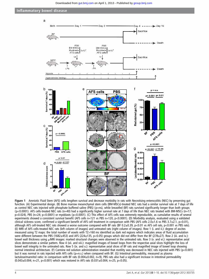
Figure 1 Amniotic Fluid Stem (AFS) cells lengthen survival and decrease morbidity in rats with Necrotising enterocolitis (NEC) by preserving gutfunction. (A) Experimental design. (B) Bone marrow mesenchymal stem cells (BM-MSCs)-treated NEC rats had a similar survival rate at 7 days of lifeas control NEC rats injected with phosphate buffered saline (PBS) (p=ns), while breastfed (BF) rats survived significantly longer than both groups(p<0.0001). AFS cells-treated NEC rats (n=40) had a significantly higher survival rate at 7 days of life than NEC rats treated with BM-MSCs (n=17;p=0.024), PBS (n=24; p<0.0001) or myoblasts (p<0.0001). (C) This effect of AFS cells was extremely reproducible, as cumulative results of severalexperiments showed a consistent survival benefit (AFS cells n=121 vs PBS n=120, p<0.0001). (D) Morbidity analysis, evaluated using a validatedclinical sickness score, confirmed a significant benefit of AFS cell treatment in comparison with PBS (AFS cells 2.0±1.6 vs PBS 3.7±2.1, p<0.01),although AFS cell-treated NEC rats showed a worse outcome compared with BF rats (BF 0.2±0.39, p<0.01 vs AFS cell rats, p<0.001 vs PBS rats).(E) MRI of AFS cells-treated NEC rats (left column of images) and untreated rats (right column of images). Row 1 (i. and ii.): degree of ascitesmeasured using T2 maps: the total number of voxels with T2>160 ms identified as dark red regions which indicates areas of fluid accumulationwere different between the PBS (1682±453) and AFS (224±135, p<0.05) groups which did not differ from the BF (278±27). Row 2 (iii. and iv.):bowel wall thickness using mMRI images: marked structural changes were observed in the untreated rats. Row 3 (v. and vi.): representative axialslices demonstrate a similar pattern. Row 4 (vii. and viii.): magnified images of bowel loops from the respective axial slices highlight the loss ofbowel wall integrity in the untreated rats. Row 5 (ix. and x.): representative axial slices of BF rats and magnified image of bowel loop showingnormal intestinal architecture. (F) Carmine red solution administration revealed that motility was decreased in NEC rats injected with PBS (p<0.001)but it was normal in rats injected with AFS cells (p=n.s.) when compared with BF. (G) Intestinal permeability, measured as plasmalactulose/mannitol ratio: in comparison with BF rats (0.004±0.002, n=9), PBS rats also had a significant increase in intestinal permeability(0.043±0.004; n=21, p<0.001) which was restored in AFS rats (0.031±0.004; n=25, p<0.05).
4 Zani A, et al. Gut 2013;0:1–10. doi:10.1136/gutjnl-2012-303735
Inflammatory bowel disease
group.bmj.com on April 1, 2013 - Published by gut.bmj.comDownloaded from

figure S4). Similarly, rats injected with AFS cells survived signifi-cantly longer than rats which received PBS (p<0.001) orα-MEM (p<0.0001). No differences were noted between AFScell group and CM group (p=n.s.), thus supporting a paracrinemechanism of action (see online supplementary figure S4).
Hierarchical cluster analysis of cDNA arrays identified 37 genes,which distinguished the two groups of animals (figure 3A).Unsurprisingly, genes with the largest expression differences wereinvolved in inflammation and tissue repair (eg, Aoc3, Itgb6), cellcycle regulation (eg, Atf2, Dusp16, Gpx4, Mxd1) and enterocytedifferentiation (eg, Acsl5, Rab8a, Thra).24 In human18 and experi-mental NEC,25 26 therapies have been aimed at the inflammatorycascade. In NEC rats, AFS cells reduced lipid peroxidation (MDAlevel), (figure 3B) and significantly decreased neutrophil infiltration(MPO activity; figure 3C). Villus apoptosis is another key factor ingut barrier failure, in human and experimental NEC.27 As wasrecently shown in myocardial infarction,28 AFS cells reduced apop-tosis (cleaved caspase 3) in NEC rats, particularly in the crypts(positive cells in 45% of PBS rats with NEC vs 12% of AFS rats,p<0.05; figure 3D). These results have parallel findings in IBDmodels, where BM-MSCs decrease apoptosis.29 Finally, in AFSrats, EdU positive enterocytes22 migrated significantly further fromthe villus crypt than in PBS rats (figure 3E), indicating that AFS
cells stimulate proliferation, similar to the reported effects ofHB-epidermal growth factor (EGF).30
AFS cells modulate stromal cells expressing COX-2in the lamina propriaHence, AFS cells diminish apoptosis and inflammation, andpromote enterocyte proliferation. Intriguingly, inducible COX-2,normally at low levels in intestine also decreases enterocyte apop-tosis,31 diminishes inflammation32 and promotes epithelial prolif-eration.33 We therefore questioned whether AFS cells acted via aCOX-2 related mechanism. COX-2+ cells, present in the laminapropria of BF rats and AFS rats, were markedly diminished in PBSrats (figure 4A,B). While the number of COX-2+ cells in the villusaxis was similar in AFS rats or BF rats (figure 4C), cryptal COX-2+ cells were increased in AFS rats compared with BF rats and PBSrats (figure 4D). Moreover, the number of COX-2+ cells per villusunit (figure 4E) and the number of cryptal COX-2+ cells(figure 4F) inversely correlated with the degree of intestinaldamage.
To further investigate whether the beneficial effects of AFScells were COX-2-dependent, we performed a survival studyusing COX-1 and COX-2 inhibitors. BF rats and NEC ratsreceiving PBS or AFS cells were randomised to receive:
Figure 2 Amniotic Fluid Stem (AFS) cells decrease macroscopic and microscopic intestinal damage, localise to the damaged gut and migratesystemically. (A) Examples of the macroscopic gut appearance of breastfed (BF) rats and necrotising enterocolitis (NEC) rats injected with phosphatebuffered saline (PBS) or AFS cells. Gut damage of NEC rats injected with AFS cells (0.4±0.7, n=27) was significantly smaller than in animals injectedwith PBS (1.4±1.1 n=24, p<0.001), but showed no difference with BF rats (0.1±0.3; n=12, p=n.s.). (B) NEC rats treated with AFS cells revealedsignificantly less histological damage (0.23±0.8, n=48, p<0.001) in comparison with NEC rats treated with PBS (1.78±0.7, n=50); no damage wasobserved in BF rats (0.08±0.2, n=12). (C) Macroscopic appearance of bundles of AFS cells injected intraperitoneally on the mesentery and on thegut wall. (D) GFP epifluorescence from AFS cells 24 h after injection was detected in the mesentery and adherent to the serosa of the intestine. At48 h and 72 h rare AFS cells were found within the villus structure in close junction to the epithelial layer. (E) Sections of rat ileum stained withanti-GFP antibodies to localise AFS cells (green) and anti-smooth muscle actin (SMA) antibodies to label smooth muscle cells (red) (scale bar 20μm).AFS cells coexpressing GFP and SMA were found integrated in the mucosal layer.
Zani A, et al. Gut 2013;0:1–10. doi:10.1136/gutjnl-2012-303735 5
Inflammatory bowel disease
group.bmj.com on April 1, 2013 - Published by gut.bmj.comDownloaded from
(1) vehicle; (2) sc-560 (COX-1 inhibitor); (3) ibuprofen(COX-1+2 inhibitor) and (4) celecoxib (COX-2 inhibitor). Asexpected, NEC rats treated with AFS cells+vehicle survivedsignificantly longer (figure 4G) and had a better clinical scorethan NEC rats treated with PBS+vehicle (0.77±0.36 vs 3.09±1.10, p<0.05). The survival effect of AFS cells was abolishedby the selective COX-2 and the non-selective COX-1+2 inhibi-tors, but unaffected by the selective COX-1 inhibitor (figure4G). Similarly, the improved clinical status in AFS rats wasannulled by COX-2 inhibition, reduced by COX-1+2 inhib-ition and unaffected by COX-1 inhibition (figure 4H). None ofthe COX inhibitors modified survival (figure 4G) or clinicalstatus (data not shown) of PBS or BF rats. In comparison withBM cells, AFS cells differentially expressed genes in the wnt-βcatenin pathway which regulate intestinal epithelial stem cellfunction (eg, AXIN, APC and CTNNA1) and cell migration(CXCL12), and growth factors known to maintain gut epithe-lial integrity and reduce mucosal injury in experimental IBD
(eg, insulin-like growth factor (IGF)-1, fibroblast growth factor(FGF)-1, FGF-3 and FGF-4, fibroblast growth receptor1;figure 5A). Moreover, when cultured in the presence of LPS,AFS cells increased expression of VEGFα, FGF-2, TGFβ1,TGFα and platelet-derived growth factor (PDGF)β, comparedwith BM cells and myoblasts which could also justify theirunique therapeutic effect in this model of disease (figure 5B–F).
DISCUSSIONNEC remains a major cause of neonatal morbidity and mor-tality.1 We demonstrated for the first time that AFS cells sig-nificantly improve survival of rats with NEC. The ability ofAFS cells to lengthen survival is particularly important, asintensive care support cannot be given to pup rats and thismodel is not compatible with long-term survival.34 The speci-ficity of this effect to AFS cells is in contrast with otheranimal models of bowel disease, in which BM-MSCs areeffective.7 35 This may be due to differences in pathogenesis;
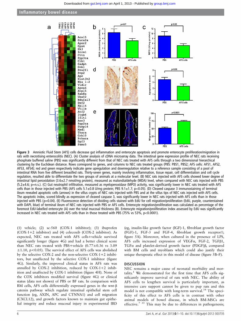
Figure 3 Amniotic Fluid Stem (AFS) cells decrease gut inflammation and enterocyte apoptosis and promote enterocyte proliferation/migration inrats with necrotising enterocolitis (NEC). (A) Cluster analysis of cDNA microarray data. The intestinal gene expression profile of NEC rats receivingphosphate buffered saline (PBS) was significantly different from that of NEC rats treated with AFS cells through a two dimensional hierarchicalclustering by the Euclidean distance. Rows correspond to genes, and columns to NEC rats treated groups (PBS: PBS1, PBS2; AFS cells: AFS1, AFS2,AFS3, AFS4); red and green respectively indicate gene upregulation and downregulation relative to a reference sample consisting of a pool ofintestinal RNA from five different breastfed rats. Thirty-seven genes, mainly involving inflammation, tissue repair, cell differentiation and cell cycleregulation, resulted able to differentiate the two groups of animals at a molecular level. (B) NEC rats injected with AFS cells showed lower degree ofintestinal lipid peroxidation (3.6±2.7 nmol/mg protein), measured as malondialdehyde (MDA) level, when compared with NEC rats injected with PBS(5.2±4.6; p=n.s.). (C) Gut neutrophil infiltration, measured as myeloperoxidase (MPO) activity, was significantly lower in NEC rats treated with AFScells than in those injected with PBS (AFS cells 5.1±0.8 U/mg protein; PBS 9.1±1.7; p<0.05). (D) Cleaved caspase 3 immunostaining of terminalileum revealed apoptotic cells (arrows) in the villus crypts of NEC rats injected with PBS and at the villus tips of NEC rats injected with AFS cells.The apoptotic index, scored blindly as expression of cleaved caspase 3, was significantly lower in NEC rats injected with AFS cells than in thoseinjected with PBS (p=0.04). (E) Fluorescence detection of dividing cells stained with EdU for cell migration/proliferation (EdU, purple, counterstainedwith DAPI, blue) of terminal ileum of NEC rats injected with PBS or AFS cells. Enterocyte migration/proliferation was calculated as percentage of theforemost EdU-labelled enterocyte (A) over the total mucosal thickness (B). Enterocyte migration/proliferation index assessed by EdU was significantlyincreased in NEC rats treated with AFS cells than in those treated with PBS (75% vs 53%, p<0.0001).
6 Zani A, et al. Gut 2013;0:1–10. doi:10.1136/gutjnl-2012-303735
Inflammatory bowel disease
group.bmj.com on April 1, 2013 - Published by gut.bmj.comDownloaded from

NEC is associated with ischaemia and bowel immaturity,whereas in IBD, the pathological changes are primarily relatedto immune dysregulation.7 Hence, while IBD can be rescuedby MSCs, beneficial effects from cell therapy in NEC appearto require a different mechanism. Rat models of IBD areusually obtained using either dextran sodium sulfate6 or intra-mural injection of peptidoglycan-polysaccharide, whereas theNEC model comprises several pathogenic factors that are alsodirectly implicated in the human disease.36
In addition to the pronounced and consistent effect on sur-vival, several clinical indicators also demonstrated the beneficialeffects of AFS cell treatment. First, AFS rats clinically improved,
which is a marker of less severe gut damage (macroscopic andmicroscopic) in this animal model.10 While human NEC can besuspected radiologically, confirmed at surgery and graded histo-logically,2 3 only the latter have been used in experimentalmodels. Herein, for the first time, we were able to define bowelappearance using MRI imaging. Similarly to human infants withNEC, we demonstrated that peritoneal fluid collection anddilated bowel loops are features of rats with NEC, while MRIimages of animals treated with AFS cells were indistinguishablefrom BF rats. Moreover, treatment with AFS cells rescued gutmotility and partially restored intestinal permeability inNEC rats. Villus sloughing, venous congestion and villus core
Figure 4 Amniotic fluid stem (AFS) cells modulate stromal cells expressing COX-2 in the lamina propria. (A) Representative cryosections of theterminal ileum from breastfed (BF) and necrotising enterocolitis (NEC) rats receiving phosphate buffered saline (PBS) and AFS cells stained withanti-COX2 Ig (red) and DAPI (blue). Scale bars: 20 μm. (B) In NEC rats treated with AFS cells, the number of COX-2+ cells per villus unit (4.97±0.46;n=7, p=n.s.) was similar to that of BF rats (4.25±0.52; n=8), but higher compared with NEC rats treated with PBS (2.37±0.29; n=8, p<0.05).(C, D) This difference was not determined by the quantity of COX-2+ cells in the villi, which was similar between the BF and AFS (1.95±0.28 vs1.32±0.16, p=ns) rats, but by their number underlying the cryptae which was higher in NEC rats treated with AFS cells (3.01±0.41) compared withBF rats (1.41±0.23, p<0.01) and NEC rats injected with PBS (1.05±0.18, p<0.001). (E, F) The number of COX-2+ cells per villus unit and in thecryptae inversely correlated with the histological grade of NEC by linear regression. (G) A survival study employing selective and non-selective COXinhibitors showed that COX inhibitors did not modify the high survival rate of BF rats (n=32; BF+DMSO vs BF+sc-560, BF+ibuprofen, BF+celecoxib:p=n.s.) and the low survival rate of PBS-treated NEC rats (n=77; PBS+DMSO vs PBS+sc-560, PBS+ibuprofen, PBS+celecoxib: p=n.s.). However, theimproved survival of NEC rats receiving AFS cells (n=78) was annulled by COX-2 (AFS cells+celecoxib vs AFS cells+DMSO: p<0.0001; AFS cells+celecoxib vs PBS+DMSO: p=n.s.) and COX-1+2 inhibitors (AFS cells+ibuprofen vs AFS cells+DMSO: p<0.01; AFS cells+ibuprofen vs PBS+DMSO:p=n.s.), but conserved in rats receiving COX-1 inhibitor (AFS cells+sc-560 vs AFS cells+DMSO: p=n.s.; AFS cells+sc-560 vs PBS+DMSO: p=0.001).(H) The clinical sickness score improvement observed in NEC rats treated with AFS cells (0.77±0.36) was abolished by COX-2 inhibitor (7.15±0.89;AFS cells+celecoxib vs AFS cells+DMSO: p<0.001), diminished by COX-1+2 inhibitor (2.86±0.91; AFS cells+ibuprofen vs AFS cells+vehicle: p=n.s.)and unaltered by COX-1 inhibitor (0.92±0.39; AFS cells+sc-560 vs AFS cells+vehicle: p=n.s.).
Zani A, et al. Gut 2013;0:1–10. doi:10.1136/gutjnl-2012-303735 7
Inflammatory bowel disease
group.bmj.com on April 1, 2013 - Published by gut.bmj.comDownloaded from
separation are classic histological features of NEC, and weshowed that AFS cell-treated animals had normal intestinalarchitecture with decreased incidence of all of these hallmarks.These findings corroborate a very recent study in which enteraladministration of amniotic fluid per se attenuated the severity ofexperimental NEC through activation of the epidermal growthfactor receptor (EGFR).14
The beneficial role of AFS cells on clinical outcome and sur-vival was closely related to their presence in the gut. While AFScells administered intravenously home primarily in the lung, andsubsequently colonise spleen and liver, with no distribution tothe gut,37 we have shown that AFS cells injected intraperitone-ally colonise the gut in 80% of BF pup rats.16 Remarkably, AFScells injected intraperitoneally, localised in 100% of intestines,homing to the mesentery or the gut. In most of the animals, 48h or 72 h after injection, AFS cells were present, albeit in smallnumbers, in the smooth muscle, submucosal layers and/or in thevilli. As the improvements in morbidity and mortality occurredwithin hours after injection, at which time relatively smallnumbers of AFS cells were found in the bowel, their direct con-tribution to tissue regeneration is unlikely to be the major mech-anism for the beneficial effects. This is further confirmed by theimprovement obtained with conditioned media or amnioticfluid14 administration, thus supporting a paracrine mechanismof action. We hypothesised that in this environment, AFS cells
released specific growth factors that acted on resident progenitorcells. In particular, damage resolution is probably achieved viaactivation of multiple pathways acting on tissue inflammation,cell apoptosis and proliferation.18 Microarray analyses of NECguts receiving AFS cells showed modification of the transcrip-tional profile of genes involved in inflammation and tissuerepair (eg, Aoc3, Itgb6), cell cycle regulation (eg, Atf2, Dusp16,Gpx4, Mxd1) and enterocyte differentiation processes (eg,Acsl5, Rab8a, Thra).
Severe intestinal inflammation leading to intestinal damage isthe main pathological event that characterises NEC.18 24 In theattempt to reduce incidence and severity of NEC, studies haveattempted to directly influence the inflammatory cascade inhuman24 and experimental NEC.25 26 38 We observed that AFScell injection reduced gut lipid peroxidation and neutrophilsequestration in NEC rats. Stem cells are well known to haveanti-inflammatory effects, which are exerted in different wayson different organs.39–41 When injected in a model ofendotoxin-induced lung inflammation, BM-MSCs downregulatethe proinflammatory response while increasing production ofanti-inflammatory IL10.39 Interestingly, in a similar model oflung injury, BM-MSCs decrease the systemic and local inflamma-tory responses induced by endotoxin.41 These effects do notrequire either lung engraftment or differentiation of the stemcells and are due at least in part to the production of stem cell
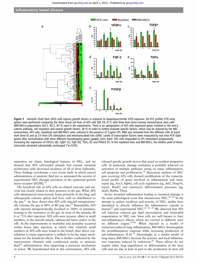
Figure 5 Amniotic Fluid Stem (AFS) cells express growth factors in response to lipopolysaccharide (LPS) exposure. (A) RT2 profiler PCR arraysystem were performed comparing the three clonal cell lines of AFS cells (E8, E9, E11) with three from bone marrow mesenchymal stem cells(BM-MSCs) preparations (b3-2, B5-2, B7-3) used in the experiments. There is an upregulation of AFS cells expressed genes involved in the wnt-βcatenin pathway, cell migration and several growth factors. (B–F) In order to further evaluate specific factors, which may be induced by the NECenvironment, AFS cells, myoblasts and BM-MSCs were cultured in the presence of 1μg/ml LPS. RNA was extracted from the different cells at basallevel (time 0) and at 3 h from LPS stimulation and retrotranscribed into cDNA. Levels of transcription factors were measured by real time PCR (Sybrgreen) after normalisation with three different housekeeping genes (gadph, b2m, bact). AFS cells responded to LPS stimulation progressivelyincreasing the expression of VEGFα (B), Tgfβ1 (C), Fgf2 (D), TGFα (E) and PDGFβ (F). In the myoblast lines and BM-MSCs, the relative yield of thesetranscripts remained substantially unchanged (*p<0.05).
8 Zani A, et al. Gut 2013;0:1–10. doi:10.1136/gutjnl-2012-303735
Inflammatory bowel disease
group.bmj.com on April 1, 2013 - Published by gut.bmj.comDownloaded from
chemoattractants by the lungs and to humoral and physicalinteractions between stem cells and lung cells.41 Adult progeni-tor cells can also improve postischaemic myocardial functionwhen used as a preventive measure, by inducing a 50% reduc-tion in proinflammatory cytokine production,42 or when usedas a therapy after myocardial infarction.40 In the latter scenario,MSCs decreased proinflammatory cytokines, inhibited collagendeposition, decreased expression of matrix metalloproteinase-1and tissue inhibitor of metalloproteinase-1 and attenuated leftventricle cavitary dilation and transmural infarct thinning, thuspreventing myocardial remodelling.40
In addition to inflammation, intestinal apoptosis has alsobeen shown to be a key factor in gut barrier failure, in humanand experimental NEC. Abundant epithelial apoptosis of thevilli is observed in histological specimens collected at thetime of bowel resection in patients with NEC.27 It usually pre-cedes widespread tissue damage43 and its reduction in experi-mental NEC has been achieved using various agents such asepidermal growth factor,44 anti-TNF-α,21 HB-EGF,45 IGF-1,46
Lactobacillus GG,47 Lactobacillus bulgaricus.48 Herein, wedemonstrated for the first time that AFS cells also reduce apop-tosis in NEC rats, particularly in the crypts. These results alsoparallel findings in animal models of IBD, in which administra-tion of BM-MSCs decreases apoptosis.29 Finally, we have shownthat AFS cells are able to influence villus cell proliferation.Impairment of cell proliferation and migration, which extendsbeyond the crypts, is continuous, irregular or spreads into thecovering villi,49 and is commonly observed in human50 andexperimental NEC . Administration of HB-EGF protects theintestine from NEC via preservation of enterocyte migrationand proliferation.30 Similarly, we found that proliferation andmigration of EdU positive enterocytes along the whole villuslength was observed in NEC rats treated with AFS cells,whereas proliferation and migration was markedly decreased inuntreated NEC rats. This is in keeping with results obtained in amodel of radiation-induced intestinal injury, where humanMSCs transplanted into immunotolerant non-obese diabetic/severe combined immunodeficiency mice increase small intes-tinal villus height and increase gut self-renewal.51 52 Similarly,enteral administration of amniotic fluid in neonatal mice withNEC restored enterocyte proliferation to levels similar to thoseof untreated mice.14 Epithelial barrier integrity is paramount forintestinal recovery in NEC, but it is still unclear which factorscould influence its restoration and/or maintenance.
Hence, AFS cells diminish apoptosis and inflammation, andpromote enterocyte proliferation. Intriguingly, COX-2, normallyexpressed at very low levels in intestine52 has been reportedto have similar effects: it decreases enterocyte apoptosis,31 53
diminishes granulocyte infiltration32 54 and promotes epithelialproliferation.55–57 COX-2 has also been suggested to have a dualrole in NEC, with high activities being proinflammatory, andlower levels protective58; systemic COX-2 inhibitors worsen intes-tinal inflammation and increase mortality in an NEC model.59
Moreover, perinatal and postnatal exposure to glucocorticoids andnon-steroidal anti-inflammatory drugs, which inhibit COX-2expression and activity, respectively, are risk factors for NEC devel-opment.60 In previous studies of COX-2 in NEC, no correlationwas found between COX-2 expression and intestinal injury sever-ity.59 This apparent discrepancy can be reconciled when the local-isation of COX-2+ cells is taken into account, as in these reportsexpression was evaluated throughout the whole intestinal wall orthe entire mucosa. Hence COX-2+ cells in the crypts promoteepithelial proliferation and migration while preventing apoptosis;this agrees with the observation that repositioning of COX-2+
cells to the crypts is necessary to maintain proliferation ofcolonic epithelial progenitors after damage.33 Although theiridentity remains to be completely established, COX-2+ cellsmay consist of a population of stromal CD44 /haematopoieticlineage-negative/myofibroblast lineage-negative cells and theiractivation could involved different pathways.33
The mechanism by which AFS cells specifically activateCOX-2+ cells in the lamina propria needs to be further investi-gated. However, our results show that, after LPS stimulation,AFS cells respond increasing the expression of growth factorsable to induce COX-2 directly (ie, VEGFα, FGF-2, TGFβ1) orvia activation of the EGFR (ie, TGFα). Not only, in fact, COX-2products (PGE2) activate EGFR but also EGFR plays an import-ant role in the induction of COX-2 expression in enterocytes.61
Moreover, as well as COX-2, EGFR activation in enterocytesinduces repair mechanisms following GI mucosal injury, pro-motes cell survival, reduces intestinal inflammation and protectsagainst experimental NEC.61
In conclusion, AFS cells injected in a model of NEC,improved survival, clinical status, gut structure and function.These beneficial effects were not due to direct repopulation ofdamaged intestine by AFS cells, but instead were probablyrelated to paracrine effects including decreased inflammationand apoptosis and concomitant increase in enterocyte prolifer-ation and migration, thus aiding epithelial restitution. Theseeffects may be mediated, at least in part, by COX-2+ cells, astheir presence in the crypts was enhanced by AFS cell injection,and beneficial effects were abolished by COX-2 inhibitors (seeonline supplementary figure S5). Future work should focus onthe potential clinical use of AFS cells and further elucidation oftheir mechanism of action in order to develop innovativepharmacological agents suitable for neonates affected by NEC.
Acknowledgements Rat myoblasts were kindly provided by Dr Jennifer Morgan(UCL Institute of Child Health, London, UK). GFP+ Sprague-Dawley rats were kindlyprovided by Prof S Schiaffino, Venetian Institute of Molecular Medicine, Padua, Italy.
Contributors AZ, MC, AA, AP, SE, PDC: study concept, and manuscript drafting.AZ, MC, FFL, GL: animal experiments. VVS, NJS: histology review. SB, MPo, MPi:cell isolation and characterisation. JW, BS, MFL: MRI. ADA, AL: microarray-basedgene expression analysis. MC, MG: Molecular biology. AH, CB: cDNA microarrays.
Funding This work was supported by the Great Ormond Street Hospital CharityPump Prime grant (bone marrow mesenchymal stem cells for the treatment ofnecrotising enterocolitis); Mittal Foundation; Eugenio Litta Foundation, Geneva; Cittàdella Speranza, Vicenza, Italy.
Competing interests ML has been supported by the Biotechnology and BiologicalSciences Research Council, the British Heart Foundation and the Engineering andPhysical Sciences Research Council. SE and PDc are supported by Great OrmondStreet Hospital Children’s Charity.
Provenance and peer review Not commissioned; externally peer reviewed.
REFERENCES1 Henry MC, Moss RL. Neonatal necrotizing enterocolitis. Semin Pediatr Surg
2008;17:98–109.2 Rees CM, Eaton S, Pierro A. Trends in infant mortality from necrotising enterocolitis
in England and Wales and the USA. Arch Dis Child Fetal Neonatal Ed 2008;93:F395–6.
3 Lin PW, Stoll BJ. Necrotising enterocolitis. Lancet 2006;368:1271–83.4 Cassinotti A, Annaloro C, Ardizzone S, et al. Autologous haematopoietic stem cell
transplantation without CD34+ cell selection in refractory Crohn’s disease. Gut2008;57:211–17.
5 Bamba S, Lee CY, Brittan M, et al. Bone marrow transplantation amelioratespathology in interleukin-10 knockout colitic mice. J Pathol 2006;209:265–73.
6 Tanaka F, Tominaga K, Ochi M, et al. Exogenous administration of mesenchymalstem cells ameliorates dextran sulfate sodium-induced colitis via anti-inflammatoryaction in damaged tissue in rats. Life Sci 2008;83:771–9.
7 Okamoto R, Yajima T, Yamazaki M, et al. Damaged epithelia regenerated by bonemarrow-derived cells in the human gastrointestinal tract. Nat Med2002;8:1011–17.
Zani A, et al. Gut 2013;0:1–10. doi:10.1136/gutjnl-2012-303735 9
Inflammatory bowel disease
group.bmj.com on April 1, 2013 - Published by gut.bmj.comDownloaded from
8 Khalil PN, Weiler V, Nelson PJ, et al. Nonmyeloablative stem cell therapy enhancesmicrocirculation and tissue regeneration in murine inflammatory bowel disease.Gastroenterology 2007;132:944–54.
9 Hayashi Y, Tsuji S, Tsujii M, et al. Topical implantation of mesenchymal stem cellshas beneficial effects on healing of experimental colitis in rats. J Pharmacol ExpTher 2008;326:523–31.
10 Zani A, Cordischi L, Cananzi M, et al. Assessment of a neonatal rat model ofnecrotizing enterocolitis. Eur J Pediatr Surg 2008;18:423–6.
11 De Coppi P, Bartsch G Jr, Siddiqui MM, et al. Isolation of amniotic stem cell lineswith potential for therapy. Nat Biotechnol 2007;25:100–6.
12 Ditadi A, de Coppi P, Picone O, et al. Human and murine amniotic fluid c-Kit+Lin-cells display hematopoietic activity. Blood 2009;113:3953–60.
13 Cananzi M, Atala A, De Coppi P. Stem cells derived from amniotic fluid: newpotentials in regenerative medicine. Reprod Biomed Online 2009;18(Suppl 1):17–27.
14 Good M, Siggers RH, Sodhi CP, et al. Amniotic fluid inhibits Toll-like receptor 4signaling in the fetal and neonatal intestinal epithelium. Proc Natl Acad Sci U S A2012;109:11330–5.
15 Bollini S, Pozzobon M, Nobles M, et al. In vitro and in vivo cardiomyogenicdifferentiation of amniotic fluid stem cells. Stem Cell Rev 2011;7:364–80.
16 Ghionzoli M, Cananzi M, Zani A, et al. Amniotic fluid stem cell migration afterintraperitoneal injection in pup rats: implication for therapy. Pediatr Surg Int2010;26:79–84.
17 Mihatsch WA, Franz AR, Kuhnt B, et al. Hydrolysis of casein acceleratesgastrointestinal transit via reduction of opioid receptor agonists released from caseinin rats. Biol Neonate 2005;87:160–3.
18 Ford HR. Mechanism of nitric oxide-mediated intestinal barrier failure: insight intothe pathogenesis of necrotizing enterocolitis. J Pediatr Surg 2006;41:294–9.
19 D’Arrigo A, Belluco C, Ambrosi A, et al. Metastatic transcriptional pattern revealedby gene expression profiling in primary colorectal carcinoma. Int J Cancer2005;115:256–62.
20 Stefanutti G, Pierro A, Parkinson EJ, et al. Moderate hypothermia as a rescuetherapy against intestinal ischemia and reperfusion injury in the rat. Crit Care Med2008;36:1564–72.
21 Halpern MD, Clark JA, Saunders TA, et al. Reduction of experimental necrotizingenterocolitis with anti-TNF-alpha. Am J Physiol Gastrointest Liver Physiol 2006;290:G757–64.
22 Salic A, Mitchison TJ. A chemical method for fast and sensitive detection of DNAsynthesis in vivo. Proc Natl Acad Sci U S A 2008;105:2415–20.
23 Cleary JO, Modat M, Norris FC, et al. Magnetic resonance virtual histology for embryos:3D atlases for automated high-throughput phenotyping. Neuroimage 2011;54:769–78.
24 Halac E, Halac J, Begue EF, et al. Prenatal and postnatal corticosteroid therapy toprevent neonatal necrotizing enterocolitis: a controlled trial. J Pediatr1990;117:132–8.
25 Zuckerbraun BS, Otterbein LE, Boyle P, et al. Carbon monoxide protects against thedevelopment of experimental necrotizing enterocolitis. Am J Physiol GastrointestLiver Physiol 2005;289:G607–13.
26 De Plaen IG, Liu SX, Tian R, et al. Inhibition of nuclear factor-kappaB amelioratesbowel injury and prolongs survival in a neonatal rat model of necrotizingenterocolitis. Pediatr Res 2007;61:716–21.
27 Ford H, Watkins S, Reblock K, et al. The role of inflammatory cytokines and nitricoxide in the pathogenesis of necrotizing enterocolitis. J Pediatr Surg1997;32:275–82.
28 Bollini S, Pozzobon M, Nobles M, et al. In vitro and in vivo cardiomyogenicdifferentiation of amniotic fluid stem cells. Stem Cell Rev 2010;7:364–80.
29 Mizoguchi E, Hachiya Y, Kawada M, et al. TNF receptor type I-dependent activationof innate responses to reduce intestinal damage-associated mortality.Gastroenterology 2008;134:470–80.
30 Feng J, Besner GE. Heparin-binding epidermal growth factor-like growth factorpromotes enterocyte migration and proliferation in neonatal rats with necrotizingenterocolitis. J Pediatr Surg 2007;42:214–20.
31 Tessner TG, Muhale F, Riehl TE, et al. Prostaglandin E2 reduces radiation-inducedepithelial apoptosis through a mechanism involving AKT activation and baxtranslocation. J Clin Invest 2004;114:1676–85.
32 Gilroy DW, Colville-Nash PR, Willis D, et al. Inducible cyclooxygenase may haveanti-inflammatory properties. Nat Med 1999;5:698–701.
33 Brown SL, Riehl TE, Walker MR, et al. Myd88-dependent positioning ofPtgs2-expressing stromal cells maintains colonic epithelial proliferation during injury.J Clin Invest 2007;117:258–69.
34 Sodhi C, Richardson W, Gribar S, et al. The development of animal models for thestudy of necrotizing enterocolitis. Dis Model Mech 2008;1:94–8.
35 Tayman C, Uckan D, Kilic E, et al. Mesenchymal stem cell therapy in necrotizingenterocolitis: a rat study. Pediatr Res 2011;70:489–94.
36 Barlow B, Santulli TV, Heird WC, et al. An experimental study of acuteneonatal enterocolitis—the importance of breast milk. J Pediatr Surg1974;9:587–95.
37 Perin L, Sedrakyan S, Giuliani S, et al. Protective effect of human amniotic fluidstem cells in an immunodeficient mouse model of acute tubular necrosis. PLoS ONE2010;5:e9357.
38 Caplan MS, Hedlund E, Adler L, et al. The platelet-activating factor receptorantagonist WEB 2170 prevents neonatal necrotizing enterocolitis in rats. J PediatrGastroenterol Nutr 1997;24:296–301.
39 Gupta N, Su X, Popov B, et al. Intrapulmonary delivery of bone marrow-derivedmesenchymal stem cells improves survival and attenuates endotoxin-induced acutelung injury in mice. J Immunol 2007;179:1855–63.
40 Guo J, Lin GS, Bao CY, et al. Anti-inflammation role for mesenchymal stem cellstransplantation in myocardial infarction. Inflammation 2007;30:97–104.
41 Xu J, Woods CR, Mora AL, et al. Prevention of endotoxin-induced systemic responseby bone marrow-derived mesenchymal stem cells in mice. Am J Physiol Lung CellMol Physiol 2007;293:L131–41.
42 Wang M, Tsai BM, Crisostomo PR, et al. Pretreatment with adult progenitor cellsimproves recovery and decreases native myocardial proinflammatory signaling afterischemia. Shock 2006;25:454–9.
43 Jilling T, Simon D, Lu J, et al. The roles of bacteria and TLR4 in rat and murinemodels of necrotizing enterocolitis. J Immunol 2006;177:3273–82.
44 Clark JA, Lane RH, Maclennan NK, et al. Epidermal growth factor reduces intestinalapoptosis in an experimental model of necrotizing enterocolitis. Am J PhysiolGastrointest Liver Physiol 2005;288:G755–62.
45 Feng J, El-Assal ON, Besner GE. Heparin-binding epidermal growth factor-likegrowth factor decreases the incidence of necrotizing enterocolitis in neonatal rats.J Pediatr Surg 2006;41:144–9; discussion 144–9.
46 Baregamian N, Song J, Jeschke MG, et al. IGF-1 protects intestinal epithelial cellsfrom oxidative stress-induced apoptosis. J Surg Res 2006;136:31–7.
47 Lin HC, Hsu CH, Chen HL, et al. Oral probiotics prevent necrotizing enterocolitis invery low birth weight preterm infants: a multicenter, randomized, controlled trial.Pediatrics 2008;122:693–700.
48 Hunter CJ, Williams M, Petrosyan M, et al. Lactobacillus bulgaricus preventsintestinal epithelial cell injury caused by Enterobacter sakazakii-induced nitric oxideboth in vitro and in the newborn rat model of necrotizing enterocolitis. InfectImmun 2009;77:1031–43.
49 Schultz A Le Mandat, Bonnard A, Barreau F, et al. Expression of TLR-2, TLR-4,NOD2 and pNF-kappaB in a neonatal rat model of necrotizing enterocolitis. PLoSONE 2007;2:e1102.
50 Vieten D, Corfield A, Carroll D, et al. Impaired mucosal regeneration in neonatalnecrotising enterocolitis. Pediatr Surg Int 2005;21:153–60.
51 Semont A, Francois S, Mouiseddine M, et al. Mesenchymal stem cells increaseself-renewal of small intestinal epithelium and accelerate structural recovery afterradiation injury. Adv Exp Med Biol 2006;585:19–30.
52 Dempke W, Rie C, Grothey A, et al. Cyclooxygenase-2: a novel target for cancerchemotherapy? J Cancer Res Clin Oncol 2001;127:411–17.
53 Joseph RR, Yazer E, Hanakawa Y, et al. Prostaglandins and activation of AC/cAMPprevents anoikis in IEC-18. Apoptosis 2005;10:1221–33.
54 Ajuebor MN, Singh A, Wallace JL. Cyclooxygenase-2-derived prostaglandin D(2) isan early anti-inflammatory signal in experimental colitis. Am J Physiol GastrointestLiver Physiol 2000;279:G238–44.
55 Arber N, Eagle CJ, Spicak J, et al. Celecoxib for the prevention of colorectaladenomatous polyps. N Engl J Med 2006;355:885–95.
56 Bertagnolli MM, Eagle CJ, Zauber AG, et al. Celecoxib for the prevention ofsporadic colorectal adenomas. N Engl J Med 2006;355:873–84.
57 Shao J, Sheng GG, Mifflin RC, et al. Roles of myofibroblasts in prostaglandinE2-stimulated intestinal epithelial proliferation and angiogenesis. Cancer Res2006;66:846–55.
58 Lugo B, Ford HR, Grishin A. Molecular signaling in necrotizing enterocolitis:regulation of intestinal COX-2 expression. J Pediatr Surg 2007;42:1165–71.
59 Grishin AV, Wang J, Potoka DA, et al. Lipopolysaccharide induces cyclooxygenase-2in intestinal epithelium via a noncanonical p38 MAPK pathway. J Immunol2006;176:580–8.
60 Guthrie SO, Gordon PV, Thomas V, et al. Necrotizing enterocolitis among neonatesin the United States. J Perinatol 2003;23:278–85.
61 McElroy SJ, Hobbs S, Kallen M, et al. Transactivation of EGFR by LPS inducesCOX-2 expression in enterocytes. PLoS One 2012;7:e38373.
10 Zani A, et al. Gut 2013;0:1–10. doi:10.1136/gutjnl-2012-303735
Inflammatory bowel disease
group.bmj.com on April 1, 2013 - Published by gut.bmj.comDownloaded from
doi: 10.1136/gutjnl-2012-303735 published online March 24, 2013Gut
Augusto Zani, Mara Cananzi, Francesco Fascetti-Leon, et al. dependent mechanismnecrotising enterocolitis via a COX-2and enhance repair of damaged intestine in Amniotic fluid stem cells improve survival
http://gut.bmj.com/content/early/2013/03/23/gutjnl-2012-303735.full.htmlUpdated information and services can be found at:
These include:
References http://gut.bmj.com/content/early/2013/03/23/gutjnl-2012-303735.full.html#ref-list-1
This article cites 61 articles, 18 of which can be accessed free at:
P<P Published online March 24, 2013 in advance of the print journal.
serviceEmail alerting
the box at the top right corner of the online article.Receive free email alerts when new articles cite this article. Sign up in
Notes
(DOIs) and date of initial publication. publication. Citations to Advance online articles must include the digital object identifier citable and establish publication priority; they are indexed by PubMed from initialtypeset, but have not not yet appeared in the paper journal. Advance online articles are Advance online articles have been peer reviewed, accepted for publication, edited and
http://group.bmj.com/group/rights-licensing/permissionsTo request permissions go to:
http://journals.bmj.com/cgi/reprintformTo order reprints go to:
http://group.bmj.com/subscribe/To subscribe to BMJ go to:
group.bmj.com on April 1, 2013 - Published by gut.bmj.comDownloaded from