AMM 525 101
7
The Effect of pH and Heat Treatment on the Porous TiO2 Nanostructures Derived from Triblock Copolymer Templating-Precipitation Technique of TiOSO4 Solution Akhmad Herman Yuwono 1,a , Hadi Sahal Fadly Daulay 1,b , Latifa Hanum Lalasari 1,2,c , Amalia Sholehah 1 1 Department of Metallurgy and Materials Engineering, Faculty of Engineering, University of Indonesia, Jawa Barat 16424, Indonesia 2 Research Centre for Metallurgy, Indonesian Institute of Sciences (LIPI), Kawasan Puspiptek Serpong, Tangerang Selatan 15314, Indonesia a [email protected], b [email protected], c [email protected], c [email protected] Keywords: TiO2 nanostructure; TiOSO4, ilmenite, band gap energy. Abstract. The synthesis and characterization of TiO2 nanostructure has become intensive nowadays because of its superior properties among other semiconductor materials. In this work, TiO2 nanostructures have been derived from ilmenite mineral by using precipitation technique with various pH and calcination temperature. The resulting nanostructures were characterized to investigate the effects of those variables on the phase, crystallite size, and band gap energy. The characterization was performed by using XRD, FT-IR, UV-Vis DRS, SEM, EDS, and TEM. The results showed that TiO2 sample prepared under low pH value of 0.3 demonstrated porous structures although they are not well-ordered yet, while the sample with a pH adjustment up to 7.0 provided nanotube structure. The biggest crystallite size of 3.43 nm and low band gap energy of 3.07 eV was obtained in the TiO2 samples synthesized without pH adjustment and calcined at a a temperature of 300 o C. This characteristics shows that TiO2 nanostructure in this study is potential for the applications of dye sensitized solar cell (DSSC) and photocatalysist. Introduction Among the semiconducting materials, titanium dioxide (TiO2) is one of the most promising ones due to its high photosensitivity, non-toxicity, easy availability, environmentally friendly, and low cost [1]. Subsequently, research interests in TiO2 have grown significantly and has been a widely used materials because of its strategic application in dye-sensitized solar cells (DSSC) [2, 3], water photocatalytic purification [4, 5], and pigment [6]. In DSSC application, TiO2 should possess high surface area and large porosity to achieve high efficiency. Therefore, TiO2 with very fine particle such as nanostructure is expected to absorb more amount of dye solution so that a higher electrical connection with redox electrolytes can be achieved [3, 7]. Many attempts in TiO2 nanostructure preparation have been performed with commonly used precursors including titanium tetraisopropoxide [8] and titanium butoxide [9]. However, these precursors are expensive to be employed for TiO2 nanostructure synthesis in a large scale. Therefore, the use of lower cost precursors have been applied such as titanium tetrachloride (TiCl4) [10] and titanyl sulfate (TiOSO4) [11], which are obtained by chloride and sulfate process during TiO white-pigment synthesis, respectively. The beneficial potential of TiOSO4 such as low cost and lower volatility than TiCl4 make it becomes an attractive precursor for TiO2 production. Several works have been reported to obtain TiO2 nanostructure by using TiOSO4 as the precursor. For example, Suwanchawalit et. al. [1], used triblock copolymer as the template and solvent extraction as the template removal. Their results showed a combination of amorphous and anatase phase TiO2 nanostructure that posses high surface area and high pore volume, which are 301 m 2 /g and 0.365 cm 3 /g, respectively. On the basis of their work, it can be concluded that this method is easily controlled and environmental friendly. However, this solvent extraction method also had a hindrance because it can only be used for inorganic surfactants. Tian et. al. [12] also reported the TiO2 nanostructure prepared by using the composite Applied Mechanics and Materials Vol. 525 (2014) pp 101-107 © (2014) Trans Tech Publications, Switzerland doi:10.4028/www.scientific.net/AMM.525.101 All rights reserved. No part of contents of this paper may be reproduced or transmitted in any form or by any means without the written permission of TTP, www.ttp.net. (ID: 125.161.211.12, University of Indonesia, Depok, Indonesia-02/02/14,15:03:05)
-
Upload
independent -
Category
Documents
-
view
0 -
download
0
Transcript of AMM 525 101

The Effect of pH and Heat Treatment on the Porous TiO2 Nanostructures Derived from Triblock Copolymer Templating-Precipitation Technique of
TiOSO4 Solution
Akhmad Herman Yuwono1,a, Hadi Sahal Fadly Daulay1,b, Latifa Hanum Lalasari1,2,c, Amalia Sholehah1
1Department of Metallurgy and Materials Engineering, Faculty of Engineering, University of Indonesia, Jawa Barat 16424, Indonesia
2Research Centre for Metallurgy, Indonesian Institute of Sciences (LIPI), Kawasan Puspiptek Serpong, Tangerang Selatan 15314, Indonesia
[email protected], [email protected], [email protected],[email protected]
Keywords: TiO2 nanostructure; TiOSO4, ilmenite, band gap energy.
Abstract. The synthesis and characterization of TiO2 nanostructure has become intensive nowadays because of its superior properties among other semiconductor materials. In this work, TiO2 nanostructures have been derived from ilmenite mineral by using precipitation technique with various pH and calcination temperature. The resulting nanostructures were characterized to investigate the effects of those variables on the phase, crystallite size, and band gap energy. The characterization was performed by using XRD, FT-IR, UV-Vis DRS, SEM, EDS, and TEM. The results showed that TiO2 sample prepared under low pH value of 0.3 demonstrated porous structures although they are not well-ordered yet, while the sample with a pH adjustment up to 7.0 provided nanotube structure. The biggest crystallite size of 3.43 nm and low band gap energy of 3.07 eV was obtained in the TiO2 samples synthesized without pH adjustment and calcined at a a temperature of 300oC. This characteristics shows that TiO2 nanostructure in this study is potential for the applications of dye sensitized solar cell (DSSC) and photocatalysist.
Introduction
Among the semiconducting materials, titanium dioxide (TiO2) is one of the most promising ones due to its high photosensitivity, non-toxicity, easy availability, environmentally friendly, and low cost [1]. Subsequently, research interests in TiO2 have grown significantly and has been a widely used materials because of its strategic application in dye-sensitized solar cells (DSSC) [2, 3], water photocatalytic purification [4, 5], and pigment [6]. In DSSC application, TiO2 should possess high surface area and large porosity to achieve high efficiency. Therefore, TiO2 with very fine particle such as nanostructure is expected to absorb more amount of dye solution so that a higher electrical connection with redox electrolytes can be achieved [3, 7]. Many attempts in TiO2 nanostructure preparation have been performed with commonly used precursors including titanium tetraisopropoxide [8] and titanium butoxide [9]. However, these precursors are expensive to be employed for TiO2 nanostructure synthesis in a large scale. Therefore, the use of lower cost precursors have been applied such as titanium tetrachloride (TiCl4) [10] and titanyl sulfate (TiOSO4) [11], which are obtained by chloride and sulfate process during TiO white-pigment synthesis, respectively.
The beneficial potential of TiOSO4 such as low cost and lower volatility than TiCl4 make it becomes an attractive precursor for TiO2 production. Several works have been reported to obtain TiO2 nanostructure by using TiOSO4 as the precursor. For example, Suwanchawalit et. al. [1], used triblock copolymer as the template and solvent extraction as the template removal. Their results showed a combination of amorphous and anatase phase TiO2 nanostructure that posses high surface area and high pore volume, which are 301 m2/g and 0.365 cm3/g, respectively. On the basis of their work, it can be concluded that this method is easily controlled and environmental friendly. However, this solvent extraction method also had a hindrance because it can only be used for inorganic surfactants. Tian et. al. [12] also reported the TiO2 nanostructure prepared by using the composite
Applied Mechanics and Materials Vol. 525 (2014) pp 101-107© (2014) Trans Tech Publications, Switzerlanddoi:10.4028/www.scientific.net/AMM.525.101
All rights reserved. No part of contents of this paper may be reproduced or transmitted in any form or by any means without the written permission of TTP,www.ttp.net. (ID: 125.161.211.12, University of Indonesia, Depok, Indonesia-02/02/14,15:03:05)

template of cetyltrimethylammonium bromide (CTAB) and tri-block copolymer EO20PO70EO20 (P-123). They obtained a thermally stable TiO2 nanostructure with high BET specific surface area and TiO2 nanostructure was successfully obtained at a lower temperature by adjusting the pH of the system. Nevertheless, it is still found difficult since a small change of pH could already affects the TiOSO4 solutions stability.
Motivated by previous studies, therefore, in this work TiO2 nanostructure was synthesized by using TiOSO4 as the inorganic precursor which is previously derived from the sulfate process of Bangka – Indonesia ilmenite ore. This Bangka Island ilmenite still has various impurities that could impede the synthesize process of TiO2 nanostructure. Hence, controlling process of TiO2 nanostructure was performed by investigating several influencing factors such as variation of pH and calcination temperature.
Experimental Detail
Materials and preparation. TiOSO4 solution was taken from the sulfuric acid route leaching of Bangka Indonesia ilmenite. The composition of TiOSO4 solution is Ti = 55.11 g/l, Fe = 82 g/l as analyzed by using Atomic Absorption Spectroscopy (AAS) [13]. The Pluronic P123 from BASF was used as surfactant. All the chemicals were obtained from commercial sources and used without further purification.The reagent formulation is calculated from the parameter used by Tian et al [12] which the molar ratio of TiOSO4:CTAB:P123 is 1:0.1:0.01, the molar ratio of H2O:Ti is 110:1, and the volume ratio of H2O : ethanol is 4:1.
The procedure of preparing the precursor of TiO2 nanostructure was adopted from the methods developed by Suwanchawalit et al [1]. Triblock copolymer P123 was dissolved in water under vigorous stirring until homogenous solution obtained. Furthremore, 50 ml TiOSO4 solution was added drop wise to the solution and stirred at 90oC for 1 hour. The initial TiOSO4 precursor has a pH value of 0.3 and further pH adjustment was performed by adding NH4OH during hydrolysis. The condensation of the precursors was carried out at room temperature for 24 h. The resulting solution contained white precipitate which was subsequently filtered and washed with distilled water until no sulfate ion was found by BaCl2 solution test. The washed samples were dried and calcined. Samples with pH = 0.3 were calcined at temperature 200oC and 300oC and denoted as sample A and B, while samples with pH = 7.0 were also calcined at the same temperature variations, denoted as sample C and D.
Characterization
The structure and phase identification were performed by XRD patterns examination which obtained by Shimadzu 7000 with Cu Kα irradiation (λ = 1.54056 Å). The average crystallite size was calculated by using Scherrer’s formula [14]. FT-IR spectra was examined through PerkinElmer UATR Two FT-IR Spectrometer, the surface morphologies were examined by scanning electron microscopy (SEM, JEOL JED 2300), while the band gap energy (Eg) of the products was determined by applying Kubelka-Munk equation on UV-Vis spectroscopy absorbance data. The external features and morphology of porous TiO2 were observed using a transmission electron microscope (TEM, JEOL JEM 1400).
Results and Discussion
Effects of pH and calcination temperature. Fig. 1 shows the XRD patterns of the TiO2 nanostructure samples under various pH and calcinations temperature. Of these samples, the TiO2 nanostructure samples with no pH adjustment during hydrolysis (pH = 0.3) are typical anatase TiO2 crystal nanostructure. However, in the samples with pH adjustment to pH = 7.0, there existed ilmenite, hematite, and goethite phases, in addition to anatase TiO2. The resulting other phases aside from anatase TiO2 were similar with that of reported by Bakardjieva [15]. The pH adjustment to pH = 7.0 also gives much more –OH groups to the Ti which can lead to the more amorphous TiO2
102 Development of Industrial Manufacturing
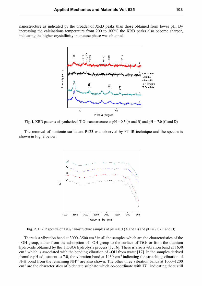
nanostructure as indicated by the broader of XRD peaks than those obtained from lower pH. By increasing the calcinations temperature from 200 to 300oC the XRD peaks also become sharper, indicating the higher crystallinity in anatase phase was obtained.
Fig. 1. XRD patterns of synthesized TiO2 nanostructure at pH = 0.3 (A and B) and pH = 7.0 (C and D)
The removal of nonionic surfactant P123 was observed by FT-IR technique and the spectra is
shown in Fig. 2 below.
Fig. 2. FT-IR spectra of TiO2 nanostructure samples at pH = 0.3 (A and B) and pH = 7.0 (C and D)
There is a vibration band at 3000–3500 cm-1 in all the samples which are the characteristics of the –OH group, either from the adsorption of –OH group to the surface of TiO2 or from the titanium hydroxide obtained by the TiOSO4 hydrolysis process [1, 16]. There is also a vibration band at 1630 cm-1 which is associated with the bending vibration of –OH from water [17]. In the samples derived fromthe pH adjustment to 7.0, the vibration band at 1430 cm-1
indicating the stretching vibration of N-H bond from the remaining NH4+ are also shown. The other three vibration bands at 1000–1200 cm-1 are the characteristics of bidentate sulphate which co-coordinate with Ti4+ indicating there still
Applied Mechanics and Materials Vol. 525 103

remains sulphate in TiO2 which prepared by sol–gel method from TiOSO4 [16-18]. However, in all samples, the vibration bands at 2800–3000 cm-1 and 1150–1400 cm-1 are not shown here, which confirms that the P123 template has been completely removed [1].
Fig. 3 shows the SEM images of the synthesized TiO2 nanostructure samples under various pH and calcination temperatures. The SEM images depicted that the synthesized nanostructures have the particle size which depends on the pH and calcination temperature. The samples with no pH adjustment or low pH value of 0.3 in Fig. 3 (a) and (b) demonstrated a relatively uniform particle size than those with the pH adjustment to 7.0. It is also clearly seen that by increasing the calcinations temperature the particle size became bigger [19].
Fig. 3. SEM images of the synthesized TiO2 nanostructures at pH = 0.3 (a and b) and pH = 7.0 (c and d)
TEM observation was also carried out of these samples and the results are depicted in Fig. 4. It is apparently shown that the samples with higher calcination temperature provide bigger particle size. It can also be observed that the TiO2 nanostructure samples with no pH adjustment or low pH value of 0.3 show pore structures although it is still not well-ordered yet. In samples with pH adjustment to 7.0, surprisingly the obtained structure is nanotube, which is similar to that of obtained by Bakardjieva et al [15]. This porous structures can be considered as an advantage because it can transport guest species sites effectively due to its morphological and electrical advantages [3].
(a) (b)
(c) (d)
104 Development of Industrial Manufacturing

Fig. 4. TEM images of the synthesized TiO2 nanostructures at pH = 0.3 (a and b) and pH = 7.0 (c and d)
Optical properties of TiO2 nanostructure. The optical properties of TiO2 nanostructure are obtained by analyzing the diffuse reflectance spectra (DRS) as shown in Fig. 5. Table 1 summarizes the crystallite size and band gap energy of the TiO2 nanostructure samples. From Table 1, it can be seen that samples with pH adjustment to 7.0 have a lower band gap energy than samples without pH adjustment (pH = 0.3). This can be attributed to iron (Fe) content and other impurities remaining in the samples [13]. The same phenomena also occurred in samples without pH adjustment (pH = 0.3) which has anatase phase but do not possess the band gap energy of bulk anatase TiO2 , which is 3.20 eV. From this result, it can be confirmed that samples with higher lower crystallite size have higher band gap energy.
(a) (b)
(c) (d)
Applied Mechanics and Materials Vol. 525 105
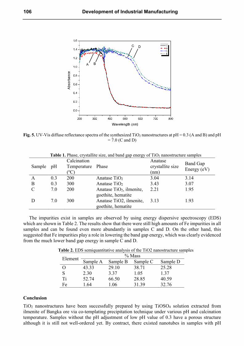
Fig. 5. UV-Vis diffuse reflectance spectra of the synthesized TiO2 nanostructures at pH = 0.3 (A and B) and pH
= 7.0 (C and D)
Table 1. Phase, crystallite size, and band gap energy of TiO2 nanostructure samples
Sample pH Calcination Temperature (oC)
Phase Anatase crystallite size (nm)
Band Gap Energy (eV)
A 0.3 200 Anatase TiO2 3.04 3.14 B 0.3 300 Anatase TiO2 3.43 3.07 C 7.0 200 Anatase TiO2, ilmenite,
goethite, hematite 2.21 1.95
D 7.0 300 Anatase TiO2, ilmenite, goethite, hematite
3.13 1.93
The impurities exist in samples are observed by using energy dispersive spectroscopy (EDS)
which are shown in Table 2. The results show that there were still high amounts of Fe impurities in all samples and can be found even more abundantly in samples C and D. On the other hand, this suggested that Fe impurities play a role in lowering the band gap energy, which was clearly evidenced from the much lower band gap energy in sample C and D.
Table 2. EDS semiquantitative analysis of the TiO2 nanostructure samples
Element % Mass
Sample A Sample B Sample C Sample D O 43.33 29.10 38.71 25.28 S 2.30 3.37 1.05 1.37 Ti 52.74 66.50 28.85 40.59 Fe 1.64 1.06 31.39 32.76
Conclusion
TiO2 nanostructures have been successfully prepared by using TiOSO4 solution extracted from ilmenite of Bangka ore via co-templating precipitation technique under various pH and calcination temperature. Samples without the pH adjustment of low pH value of 0.3 have a porous structure although it is still not well-ordered yet. By contract, there existed nanotubes in samples with pH
106 Development of Industrial Manufacturing

adjustment to 7.0. The biggest crystallite size of 3.43 nm and band gap energy of 3.07 eV is obtained in the samples prepared without the adjustment of pH (pH = 0.3) and calcined at a higher temperature of 300oC. This low band gap energy is highly possible due to the presence of the impurities remaining the sample. The results show that the TiO2 nanostructures in this study are potential for the strategic applications such as dye sensitized solar cell (DSSC) and photocatalysist decomposition of pollutant.
Acknowledgements
The authors are grateful for the main financial support from Competence Research Grant of Directorate of Higher Education (DIKTI)-Ministry of Education Republic of Indonesia 2013 with contract no. 2475/H2.R12/HKP.05.00/2013.
References
[1] C. Suwanchawalit, S. Wongnawa, J Nanopart Res., Vol. 12: p. 2895-2906 (2010).
[2] H.-G. Jung, Y. S. Kang, andY.-K. Sun, Electrochimica Acta, Vol. 2010: p. 4637-4641 (2010).
[3] M. A. Khan, M. S. Akhtar, andO.-B. Yang, Solar Energy, Vol. 84: p. 2195-2201 (2010).
[4] H. U. Lee, et al., Chemical Engineering Journal, Vol. 223 (2013).
[5] T. Ochiai, A. Fujishima, Journal of Photochemistry and Photobiology C: Photochemistry Reviews, Vol. 13: p. 247-262 (2012).
[6] C. Tian, S. Huang, andY. Yang, Dyes and Pigments, Vol. 96: p. 609-613 (2013).
[7] R. Zhang, et al., Nano Today, Vol. 7: p. 344-366 (2012).
[8] H.-s. Yun, et al., Materials Science and Engineering C, Vol. 23: p. 487-494 (2003).
[9] K. Yu, et al., Materials Letters, Vol. 59: p. 2515-2518 (2005).
[10] A. Sun, et al., Powder Technology, Vol. 201: p. 130-137 (2010).
[11] Y. Yang, et al., Surface Science, Vol. 605: p. 1281-1286 (2011).
[12] C. Tian, et al., Materials Letters, Vol. 62: p. 77-80 (2008).
[13] L. H. Lalasari, A. H. Yuwono, and F. Firdiyono, Applied Mechanics and Materials, Vol. 391: p. 34-40 (2013).
[14] B. D. Cullity, Elements of X-Ray Diffraction, Addison-Wesley Reading, Massachussets, 1978.
[15] S. Bakardjieva, et al., Acta Microscopica, Vol. 18: p. 113-114 (2009).
[16] L. Ge, et al., Materials Research Bulletin, Vol. 41: p. 1596-1603 (2006).
[17] L. Afanador, et al., Fuel, Vol. 100: p. 43-47 (2012).
[18] X. Wang, et al., Journal of Photochemistry and Photobiology A: Chemistry, Vol. 179: p. 339-347 (2006).
[19] A. M. Luís, et al., Materials Chemistry and Physics, Vol. 125: p. 20-25 (2011).
Applied Mechanics and Materials Vol. 525 107


![[3937]-101 P553](https://static.fdokumen.com/doc/165x107/63338430a6138719eb0aa5bc/3937-101-p553.jpg)


![[5365]-11 MBA 101](https://static.fdokumen.com/doc/165x107/6322631aae0f5e819105deed/5365-11-mba-101.jpg)