Intranuclear accumulation of plant tubulin in response to low temperature
Upload
independentCategory
view
0download
0
Copyright @ 2006 by the American Association of Neuropathologists, Inc. Unauthorized reproduction of this article is prohibited.
ORIGINAL ARTICLE
Altered Cellular Distribution and Subcellular Sorting of F-Tubulinin Diffuse Astrocytic Gliomas and Human Glioblastoma
Cell Lines
Christos D. Katsetos, MD, PhD, MRCPath, Goutham Reddy, MS, Eduarda Draberova, PhD,
Barbora Smejkalova, PhD, Luis Del Valle, MD, Qazi Ashraf, MD, Ashot Tadevosyan, MD, PhD,
Karina Yelin, MD, Theodoros Maraziotis, MD, Om P. Mishra, PhD, Sverre Mork, MD, PhD,
Agustin Legido, MD, PhD, Jonathan Nissanov, PhD, Peter W. Baas, PhD,
Jean-Pierre de Chadarevian, MD, FRCPC, and Pavel Draber, PhD
AbstractCentrosome amplification is a pivotal mechanism underlying
tumorigenesis but its role in gliomas is underinvestigated. The present
study specifically examines the expression and distribution of the
centrosome-associated cytoskeletal protein F-tubulin in 56 primary
diffuse astrocytic gliomas (grades IIYIV) and in 4 human glioblas-
toma cell lines (U87MG, U118MG, U138MG, and T98G). Mono-
clonal anti-peptide antibodies recognizing epitopes in C-terminal or
N-terminal domains of the F-tubulin molecule were used in
immunohistochemical, immunofluorescence, and immunoblotting
studies. In tumors in adults (n = 46), varying degrees of localization
were detected in all tumor grades, but immunoreactivity was
significantly increased in high-grade anaplastic astrocytomas and
glioblastomas multiforme as compared to low-grade diffuse astrocy-
tomas (p = 0.0001). A similar trend was noted in diffuse gliomas in
children but the sample of cases was too small as to be statistically
meaningful. Two overlapping patterns of ectopic cellular localization
were identified in both primary tumors and glioblastoma cell lines: A
punctate pattern, in which F-tubulin was partially co-distributed with
pericentrin in the pericentriolar region, and a diffuse pattern, indepen-
dent of pericentrin staining, denoting a soluble pool of F-tubulin.
Cellular F-tubulin was detected in both soluble and insoluble (nocoda-
zole-resistant) fractions of glioblastoma cells. Divergent localizations
of F-tubulin and pericentrin suggest a differential distribution of these 2
centrosome-associated proteins in glioblastoma cell lines. Our results
indicate that overexpression and ectopic cellular distribution of F-tubulin
in astrocytic gliomas may be significant in the context of centrosome
protein amplification and may be linked to tumor progression and
anaplastic potential.
Key Words: Anaplastic change, Astrocytoma, Centrosome ampli-
fication, Glioblastoma, Glioma, Pericentrin, F-tubulin.
INTRODUCTIONTumor progression in diffuse gliomas is an intricate
multistep process characterized by accumulation of geneticdefects, including an abnormal number of chromosomes, acondition known as aneuploidy. Genomic stability requiresproper coordination of DNA replication and centrosomeduplication, which is facilitated by factors such as p53, pRb,cyclin E, and cyclin A (1, 2).
The centrosomes are organelles subserving diversecellular functions. First, they are essential for the process ofcytoplasmic division (cytokinesis), ensuring balanced chromo-some segregation (1Y3). Second, they are involved in theinitiation of the S-phase and the regulation of cell cycleprogression (4Y7). Third, centrosomes play an important rolein microtubule nucleation and organization, thus contributingto cell architecture and the acquisition of cell polarity (4).Normal diploid somatic cells contain a single centrosome,which consists of 2 centrioles that are surrounded by anamorphous pericentriolar matrix. The latter comprises a largerepertoire of proteins including pericentrin, the cytoskeletalprotein F-tubulin, and numerous key regulators of cell cycleprogression (2, 5, 8Y12). Pericentrin and F-tubulin form aprotein complex with a lattice-like organization at the
465J Neuropathol Exp Neurol � Volume 65, Number 5, May 2006
J Neuropathol Exp NeurolCopyright � 2006 by the American Association of Neuropathologists, Inc.
Vol. 65, No. 5May 2006
pp. 465Y477
From Department of Pediatrics (CDK, GR, QA, AT, KY, OPM, AL, JPD),Drexel University College of Medicine, St. Christopher’s Hospital forChildren, Philadelphia, Pennsylvania; Department of Pathology andLaboratory Medicine (CDK, JPD), St. Christopher’s Hospital for Children,Philadelphia, Pennsylvania; Laboratory of the Biology of Cytoskeleton(ED, BS, PD), Institute of Molecular Genetics, Academy of Sciences of theCzech Republic, Prague, Czech Republic; Center for Neurovirology (LD),Department of Neuroscience, Temple University School of Medicine,Philadelphia, Pennsylvania; Department of Neurosurgery (TM), Universityof Patras School of Medicine, Patras, Greece; Department of Neurobiologyand Anatomy (JN, PWB), Drexel University College of Medicine,Philadelphia, Pennsylvania; and The Gade Institute (SM), Department ofPathology, University of Bergen, Haukeland Hospital, Bergen, Norway.
Send correspondence and reprint requests to: Christos D. Katsetos, MD,PhD, MRCPath, Section of Neurology, St. Christopher’s Hospital forChildren, Erie Avenue at Front Street, Philadelphia, PA 19134; E-mail:[email protected]
Dedicated to Professor Peter L. Lantos on the occasion of his retirement asHead of the Department of Neuropathology, Institute of Psychiatry,London, England.
Supported by an Established Investigator Award from the St. Christopher’sFoundation for Children (CDK), a Student Research Sponsorship from theDebbie Romano Memorial Foundation (GR), grant S50523015 from theMinistry of Education of the Czech Republic, and by Institutional ResearchSupport AVOZ 50520514 (PD).
Copyright @ 2006 by the American Association of Neuropathologists, Inc. Unauthorized reproduction of this article is prohibited.
centrosome (11, 12). The centrosome-associated F-tubulinplays an important role in the nucleation of microtubuleassembly and regulates tubulin synthesis (13). There isevidence that F-tubulin may also exist in a soluble formand associate with the lateral arms of microtubules (14Y16).F-Tubulin has also been found to bind to membranouscomponents of the cell (17Y19).
The nucleation of microtubules occurs from within thepericentriolar material where F-tubulin ring complexes act asmicrotubule nucleation templates (20, 21). During normalmitosis, 2 centrosomes ensure the assembly of bipolarspindles and proper chromosomal segregation. Extra copiesof centrosomes result in the formation of multipolar spindlesresulting in chromosomal missegregation (2, 22, 23).
The cells of many human and animal cancers containan excess number of abnormal centrosomes (Bsupernumerarycentrosomes^), which contribute to the formation of multi-polar spindles that underlie unbalanced chromosome segre-gation and aneuploidy (1, 2, 22). This process is commonlyknown as centrosome amplification and involves alterationsin diverse centrosome-associated molecules. Although theexact mechanisms of centrosome amplification are unknown,several models for the generation of supernumerary centro-somes and centrosomal dysfunction have been proposed(reviewed in [1, 2, 22, 23]). Overexpression of thecentrosomal protein pericentrin in primary prostate epithelialcell transfectants gives rise to cells with tumor-like pheno-typic features, suggesting that primary centrosome dysfunc-tion may be pivotal to tumorigenesis (24). Whetherabnormalities in centrosomal proteins constitute the primarycause of chromosomal instability or are secondary to cellcycle deregulation, they are, nonetheless, regarded asimportant correlates of tumorigenesis and anaplastic poten-tial (2). Regardless of their origin, Bsupernumerary^ (orotherwise structurally abnormal or dysfunctional) centro-somes promote malignant change and tumor progression byfavoring the development of chromosomal instability and byinfluencing tumor cell motility and architecture (2).
To date, centrosome abnormalities have been reportedin a wide range of predominantly epithelial tumors, includ-ing carcinomas of the breast, bile ducts, pancreas, adrenalcortex, colon, prostate, cervix, lung, and head and neckregion (22, 25Y32). Moreover, centrosome abnormalitieshave been described in conjunction with genomic instabilityin osteosarcoma, a malignant mesenchymal neoplasm (33).
With respect to the central nervous system, centro-some amplification has, until now, only been reported incerebral primitive neuroectodermal tumors exhibiting TP53mutations (34). However, the presence of centrosomeprotein abnormalities among common astrocytic tumors isa hitherto underinvestigated aspect in glioma tumorigenesis.
We have hypothesized that centrosomal abnormalitiesare associated with malignant (anaplastic) transformation indiffuse astrocytic gliomas by favoring the development ofgenomic instability (aneuploidy) and also, by exerting in-fluence on aberrant microtubule nucleation and tumor archi-tecture. The present study specifically examines the cellularexpression and localization of F-tubulin, a key structuralprotein of the pericentriolar material of centrosomes, in the
context of histological tumor grade in primary diffuseastrocytic gliomas and in 4 established human glioblastomacell lines.
MATERIALS AND METHODS
Tissue SamplesThe localization of F-tubulin was evaluated in 56
surgically resected samples of diffuse astrocytic gliomasfrom adults (n = 46) and children (n = 10) representative ofthe World Health Organization (WHO) histological grades IIto IV. The tumor specimens from adult patients includedlow-grade diffuse astrocytomas/WHO grade II (n = 20),anaplastic astrocytomas/WHO grade III (n = 5), andglioblastomas multiforme/WHO grade IV (n = 21). Pediatricgliomas included examples of anaplastic glioma (WHOgrade III) (n = 2), and diffuse gliomas (WHO grade II) of thecerebral hemispheric white matter (n = 2), thalamus (n = 3),brainstem (n = 2), and spinal cord (n = 2). Sections fromformalin-fixed, paraffin-embedded archival tissues wereprocessed for immunohistochemistry. Control tissuesincluded surgical and autopsy tissue samples from casesexhibiting gliosis (n = 8). Paraffin sections of normalcerebral hemispheric white matter from term piglet brains(marked by active gliogenesis) were also included (n = 5).The archival tissue samples used in this study were derivedfrom the following institutions: Department of AnatomicalPathology and Cytology, University of Patras School ofMedicine, Patras, Greece, the Department of Histopathologyand Morbid Anatomy, The Royal London Hospital, London,UK, and the Department of Pathology and LaboratoryMedicine, St. Christopher’s Hospital for Children, Philadel-phia, PA.
Cell LinesFour well-established human glioblastoma cell lines,
T98G, U87MG, U118MG, and U138MG were used in thepresent study. T98G and U87MG were maintained in 10%FBS in DMEM, U118MG was maintained with Dulbecco`smodified Eagle media containing 4 mM L-glutamine, andU138MG was maintained in Minimum Essential Mediacontaining 2 mM L-glutamate. All cell lines were obtainedfrom the American Type Culture Collection (ATCC;Manassas, VA).
AntibodiesFor immunohistochemistry, immunofluorescence,
and immunoblotting, 3 monoclonal anti-peptide antibodiesrecognizing epitopes in C-terminal or N-terminal domains ofF-tubulin molecule were used. These included GTU-88(IgG1) generated against the EEFATEGTDRKDVFFY pep-tide corresponding to the human sequence 38Y53 in theN-terminal region of F-tubulin sequence (Sigma Aldrich, St.Louis, MO; cat. no. T6557) and antibodies TU-31 (IgG2b)and TU-32 (IgG1) generated against EYHAATRP-DYISWGTQ peptide corresponding to the human sequence434Y449 in the C-terminal region of F-tubulin (35). Tubulinwas detected by mouse monoclonal antibody TU-01 (lgG1)
Katsetos et al J Neuropathol Exp Neurol � Volume 65, Number 5, May 2006
� 2006 American Association of Neuropathologists, Inc.466

Copyright @ 2006 by the American Association of Neuropathologists, Inc. Unauthorized reproduction of this article is prohibited.
(36, 37) or by affinity purified rabbit antibody TUB against >/A-tubulin dimer (38). Affinity-purified rabbit antibody M8against pericentrin (39) and a polyclonal antibody againstpericentrin (Covance, Berkeley, CA) were used as genericcentrosome markers.
Indocarbocyanate (Cy3)-conjugated anti-mouse andanti rabbit antibodies as well as fluorescein isothiocyanate(FITC)-conjugated anti-mouse antibody were from JacksonImmunoresearch Laboratories (West Grove, PA). FITC-conjugated anti-mouse antibody was also bought fromVector Laboratories. Anti-mouse antibodies conjugated withhorseradish peroxidase were purchased from Promega(Madison, WI). FITC-conjugated anti-mouse (IgG) andTexas red anti-rabbit (IgG) antibodies were purchased fromVector Laboratories (Burlingame, CA).
Preparation of Cell ExtractsFor preparation of soluble and detergent-resistant
fractions at 37-C, cells on 6-cm Petri dishes were eitherdirectly used for the assay, or they were preincubated 6 hourswith 0.5 Kg nocodazole/ml to before assay to disruptmicrotubule arrays. Attached cells were rinsed twice inmicrotubule-stabilizing (MSB) buffer (100 mM MES adjustedto pH 6.9 with KOH, 2 mM EGTA, 2 mM MgCl2) or in MSBbuffer containing nocodazole (MSB/nocodazole), and thenextracted with 0.5 ml of MSB buffer (37-C) or MSB/nocodazole supplemented with protease inhibitor cocktail(BComplete EDTA-free^ tablets) (Roche Molecular Biochem-icals, Mannheim, Germany), phosphatase inhibitors (1 mMNa3V04, 1 mM NaF) and 0.2% (v/v) Triton Xj100. Afterone-minute incubation at 37-C, the extract was gentlyremoved, spun down at 5,000 g for 1 minute at 25-C, andone fourth volume of 4x SDS/PAGE-sample buffer wasadded to the supernatant. The cytoskeletons remaining on theplate were gently rinsed twice with warm MSB buffercontaining inhibitors and solubilized with 0.625 ml of samplebuffer, prepared by mixing 2x SDS/PAGE-sample buffer with2x extraction buffer (1:1). Pelleted material obtained afterspinning down the extract was combined with the cytoskeletalfraction. Samples were boiled for 5 minutes.
ImmunohistochemistryPrior to immunohistochemistry, tissue sections were
subjected to microwave antigen unmasking in Na+ EDTAbuffer at pH 8.0. Immunohistochemistry was performedaccording to the avidin biotin complex (ABC) peroxidasemethod using the Mouse IgG ABC Elite\ detection kit(Vector Labs) as previously described (40). Anti-F-tubulinmonoclonal antibody GTU-88 was diluted 1:300 and poly-clonal antibody against pericentrin was diluted 1:150.
Histologic and immunostained preparations were eval-uated by a panel of 2 neuropathologists (CDK and SM) andone pediatric pathologist (JPD). Because the tissue specimenswere obtained from 3 different laboratories, we opted toensure that histologic classification and tumor grading weremade according to homogeneous criteria by having speci-mens evaluated by different members of the panel. In case ofdisagreement, histologic typing was assigned by consensus at
conference. Clinicopathological-neuroimaging correlationswere carried out whenever feasible.
The minimal criterion for the identification of apositive cell in the context of putative centrosome dysfunc-tion and possible amplification was the detection of 3 ormore punctate dot-like immunoreactive signals in thecytoplasm of a single tumor cell. The indirect immunoper-oxidase method employed in this study was sufficientlysensitive to detect the gain of one or more punctate signals inexcess of 1 or 2 F-tubulin-positive juxta-nuclear presumptivecentrosomes typifying non-transformed diploid cells (29,31). Tumor cells with a multipunctate staining pattern wereadmixed in any given case with cells exhibiting a nonde-script diffuse pattern of immunoreactivity (see Results).Accordingly, for the purpose of cell counting, immunola-beled tumor cells were considered positive regardless ofwhether they displayed a predominantly multipunctate ordiffuse pattern of F-tubulin localization.
Manual cell counting of labeled tumor cells wasperformed independently by 3 observers (CDK, AT, andKY). Cell counting and statistical analysis were carried outonly in the adult group of astrocytic gliomas (n = 46).Between 298 to 954 tumor cells were evaluated per case in20 non-overlapping high-power (40x) fields and a labelingindex was determined for each case. Labeling index (LI) wasexpressed as the percentage (%) of F-tubulin-labeled cellsout of the total number of tumor cells counted in each case.The median labeling index (MLI) and the interquartile range(IQR) - delimited by the 25th and 75th percentiles - weredetermined for the set of cases in each histological gradeusing the UNIVARIATE procedure of the SAS package(SAS Institute, Cary, NC). The statistical significance ofdifferences in labeling indices between WHO histologicalgrades were examined with non-parametric statistical tech-niques using Kruskal-Wallis analysis of variance tests andWilcoxon rank-sum post-hoc tests. These analyses werecarried out using the NONPAR1WAY procedure of the SASpackage (SAS Institute) as described previously (40).Because of the small number of pediatric gliomas (n = 10)included in this study, only qualitative assessment wasperformed in these cases.
Immunofluorescence MicroscopyFor immunofluorescence microscopy, cells were plated
onto two-chamber glass slides coated with poly-L-lysine(Boehringer-Mannheim) until cultures reached approxi-mately 80% to 85% confluence. After rinsing with PBS, cellswere fixed with cold acetone for 3 minutes and rinsed withPBS. Cells were then blocked with 5% normal horse serumfor 2 hours at room temperature and primary antibodies wereincubated overnight at room temperature. Alternatively, cellsgrown on coverslips were fixed for 10 minutes in methanol atj20-C followed by 6 minutes in acetone at j20-C, and afterrinsing with PBS were incubated with primary antibodies for45 minutes at room temperature. Affinity purified polyclonalantibody TUB against >A-tubulin dimer was diluted 1:10,anti-F-tubulin monoclonal antibody GTU-88 was diluted1:500, and anti-F-tubulin monoclonal antibody TU-31 was
J Neuropathol Exp Neurol � Volume 65, Number 5, May 2006 F-Tubulin in Astrocytic Gliomas
� 2006 American Association of Neuropathologists, Inc. 467

Copyright @ 2006 by the American Association of Neuropathologists, Inc. Unauthorized reproduction of this article is prohibited.
used as undiluted hybridoma supernatant. Polyclonal anti-body against pericentrin was diluted 1:250. After rinsingwith PBS, Cy3-tagged or FITC-tagged secondary antibodieswere incubated for 1 hour at room temperature in the dark.For double label fluorescence the coverslips were incubatedsimultaneously with the primary antibodies and after wash-ing incubated simultaneously with secondary antibodies.Cy3-conjugated anti-mouse or anti-rabbit antibodies werediluted 1:1000, and FITC-conjugated anti-mouse or anti-rabbit antibodies were diluted 1:200. For double labelingimmunofluorescence studies on archival histological sections,we used monoclonal antibody GTU-88 against F-tubulin, andanti-pericentrin antiserum at the same dilutions as in themethanol-fixed cells from the glioblastoma cell lines, as wellas FITC- and Texas red-tagged secondary antibodies bothdiluted at 1:200. 4’,6-diamidino-2-phenylindole (DAPI) wasused to label cell nuclei. Finally, after rinsing with PBS, slideswere coverslipped using an aqueous based mounting medium(Vector Laboratories). Visualization was carried out with aNikon Eclipse TE300 inverted fluorescence microscope andimages were captured and processed using IPLab ScientificImage Processing software, version 3.5.4 (Scanalytics Inc.Fairfax, VA). Alternatively samples were examined withOlympus A70 Provis microscope. Conjugates alone did notgive any detectable staining.
ImmunoblotsWhole-cell extracts for SDS-PAGE were prepared by
solubilizing washed cells in hot SDS-sample buffer andboiling for 5 minutes. Proteins were separated by 7.5% SDS-PAGE and subsequently transferred onto nitrocellulose\membranes. Molecular weight standards were from Sigma.Blocking was achieved by incubating with 3% BSA inTBST (10 mM Tris-HCl adjusted to pH 7.4 with HCl,150 mM NaCl, 0.05% (v/v) Tween 20) and then incubatedovernight with primary antibodies diluted in TBST. Theanti-tubulin antibodies TU-01 and TU-32, in the form ofspent culture supernatants, were diluted 1:10, whereas theantibody GTU-88 was diluted 1:5,000. The membranes werethen washed 3times with TBST and incubated for 1 hour atroom temperature with secondary anti-mouse antibodyconjugated with peroxidase diluted 1:10,000 (Promega).After several washes, the binding was detected withchemiluminescence reagents (Pierce, Rockford, IL) inaccordance with the manufacturer`s directions. Exposedautoradiography films X-Omat AR (Eastman Kodak,Rochester, NY) were evaluated using gel documentationsystem GDS 7500 (UVP).
RESULTS
Primary Brain TumorsIn non-neoplastic glia from human and piglet brains,
including cases of gliosis, discrete. F-tubulin labeling wasdetected in the form of one or 2 paranuclear dotscorresponding to the expected cytoplasmic localization ofcentrosomes (Fig. 1A, B).
In the series of astrocytic gliomas in adults, varyingdegrees of F-tubulin labeling were detected in all histolog-ical grades of diffuse astrocytomas (grades IIYIV). However,staining was significantly increased in the high-grade ana-plastic astrocytomas and glioblastomas (grades III/IV) (MLI:56.4%; IQR: 41.2%Y73.4%) as compared to the low-gradediffuse astrocytomas (grade II) (MLI: 6.4%; IQR:1.6%Y9.1%) (P = 0.0001) (Figs. 1CYF, 2AYF).
As demonstrated by both immunohistochemistry inprimary tumors (from both adults and children) and FITC-immunofluorescence microscopy in tumor cell lines (seebelow), the localization of F-tubulin acquires 2 overlappingstaining patterns, notably punctate and diffuse, which mergeimperceptibly within individual tumor cells. The punctatepattern was characterized by 3 or more either discrete and/orcoalescent, variously sized and abnormally shaped immu-noreactive dots that were present throughout the cytoplasmof tumor cells (Figs. 1CYE, 2C). The diffuse staining patternwas evidenced throughout the cytoplasm of tumor cells,including the proximal segments of glial cell processes(Figs. 1F, 2B, DYF). The latter was frequently encounteredin conjunction with the multipunctate pattern. Immunola-beled cells were focally and unevenly dispersed within tumorsheets (Fig. 2A, B, D). As a rule, F-tubulin-positive tumorcells were morphologically indistinguishable from F-tubulin-negative cells. Staining for F-tubulin was not associated withdistinctive morphology, although there was an overalltendency of immunoreactive tumor cells to be pleomorphicand multinucleated (Figs. 1F, 2BYF). These alterations wereconsiderably less widespread in the low-grade astrocyticgliomas where the localization of aberrant F-tubulin-positivecytoplasmic dots was generally patchy and less frequent ascompared to the high-grade tumors (Fig. 1CYF). Variouslypronounced F-tubulin localization was present in the cytoplasmof vascular endothelial cells in areas of tumor angiogenesis(microvascular proliferation) in glioblastomas (Fig. 3AYD).
Glioblastoma Cell LinesVariably robust fluorescein green (FITC) staining was
present in cell cultures of all 4 glioblastoma lines reproducingoverlapping punctate and diffuse patterns of F-tubulin local-ization (Fig. 4AYF). Cells of the U118MG cell line predom-inantly exhibited a multi-punctate pattern of localizationconsisting of irregular, variously sized F-tubulin aggregatessuggestive of abnormal centrosome protein aggregates (Fig.4A, B), while cells of the other cell lines exhibitedpredominantly diffuse or mixed patterns of immunoreactivity(Fig. 4CYF). Confluent cytoplasmic mass-like conglomerateswere also identified (Fig. 4C, E, F, arrowheads).
By immunoblotting, prominent F-tubulin proteinbands were present in supernatants of glioblastoma cellextracts (Fig. 5). Differences in staining intensity observedwith GTU-88 and TU-32 antibodies could reflect concen-tration and/or affinity of antibodies. No cross-reactivity wasobserved with GTU-88 and TU-32 on immunoblots.
Extraction of T98G and U87MG cells with 0.2% TritonXj100 in microtubule stabilizing buffer at 37-C for 1 minuteshowed differences in relative distribution of the proteinsbetween the soluble and insoluble fractions. Results of a
Katsetos et al J Neuropathol Exp Neurol � Volume 65, Number 5, May 2006
� 2006 American Association of Neuropathologists, Inc.468

Copyright @ 2006 by the American Association of Neuropathologists, Inc. Unauthorized reproduction of this article is prohibited.
typical experiment are shown in Figure 6. F-Tubulin waspresent in soluble and cytoskeletal fractions in similarquantities in resting cells and cells preincubated withnocodazole that efficiently disrupted microtubule arrays,which was confirmed by immunofluorescence microscopy(not shown). The same results were obtained with antibodiesGTU-88 and TU-32 that are directed against different epitopesin the F-tubulin molecule. On the other hand, >-tubulin waspresent in both soluble and cytoskeletal fractions in similarquantities in resting cells only. When microtubules weredisrupted, the substantially lower amount of >-tubulin wasdetected in the insoluble fraction. These data indicate that inthese cell lines a substantial portion of cellular F-tubulin ispresent in the detergent-resistant fraction.
Differential Distribution of F-Tubulinand Pericentrin
Staining for >/A-tubulin dimers yielded 2 patterns oflocalization. The first and most prominent was a robustcentripetal distribution that was mainly detected in thepericentriolar region where it attained in some cells theproportions of confluent paranuclear immunoreactive masses
(Fig. 7A). The second staining pattern corresponded tomicrotubule arrays in the tumor cell bodies and proximalsegments of cell processes (Fig. 7A). Double labelinglocalization revealed that F-tubulin had a punctate or diffuse,centrifugal localization extending to the periphery of thecytoplasm of T98G glioblastoma cells. However, in somecells, multiple bright F-tubulin dots were observed fromwhich microtubules were not nucleated (Fig. 7B, C).
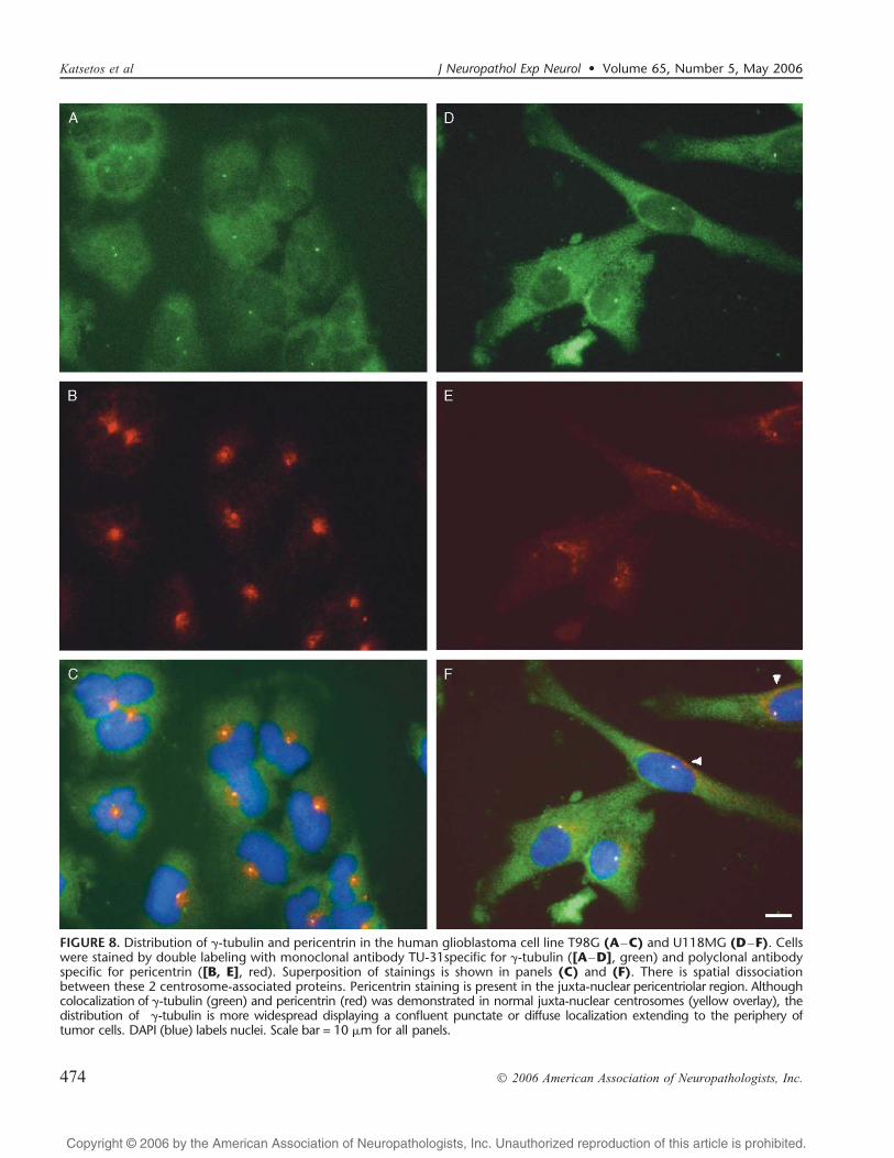
By immunofluorescence microscopy performed on glio-blastoma cell lines and surgically resected glioblastomas therewas a discernible spatial dissociation between these 2 centro-some-associated proteins (Fig. 8AYF). Pericentrin staining wasdetected for the most part in the paranuclear pericentriolarregion (Fig. 8B and E [single pericentrin labeling] and Fig. 8Cand F [double labeling for pericentrin and F-tubulin]).Confluent perinuclear areas of punctate pericentrin stainingconsistent with supernumerary centrosomes were demonstratedin some cells (Fig. 8E [single pericentrin labeling] and Fig. 8F[double labeling for pericentrin and F-tubulin]). Althoughcolocalization of F-tubulin and pericentrin was demonstratedin the pericentriolar region (Fig. 8C, F), the cytoplasmicdistribution of F-tubulin was distinctly more widespread as
FIGURE 1. Cellular distribution ofF -tubulin in non-neoplastic glia fromadult human (A) and newborn piglet(B) brains. Single or double dot-likestaining (arrows) is consistent withcentrosomal localization. (CYF) Panelsdepict the cellular distribution ofF -tubulin in diffuse astrocytoma, WHOgrade II. Staining is present in a smallnumber of tumor cells (arrows) whereit is discrete and variably prominent(CYF). Scattered cells exhibit multiple(>3) punctate, irregular and coalescentcytoplasmic localizations (CYE) merg-ing into a diffuse staining pattern.Avidin biotin complex (ABC) peroxidasewith hematoxylin counterstain. Scalebar = (A, B, DYF) 10 Km; (C) 20 Km.
J Neuropathol Exp Neurol � Volume 65, Number 5, May 2006 F-Tubulin in Astrocytic Gliomas
� 2006 American Association of Neuropathologists, Inc. 469
Copyright @ 2006 by the American Association of Neuropathologists, Inc. Unauthorized reproduction of this article is prohibited.
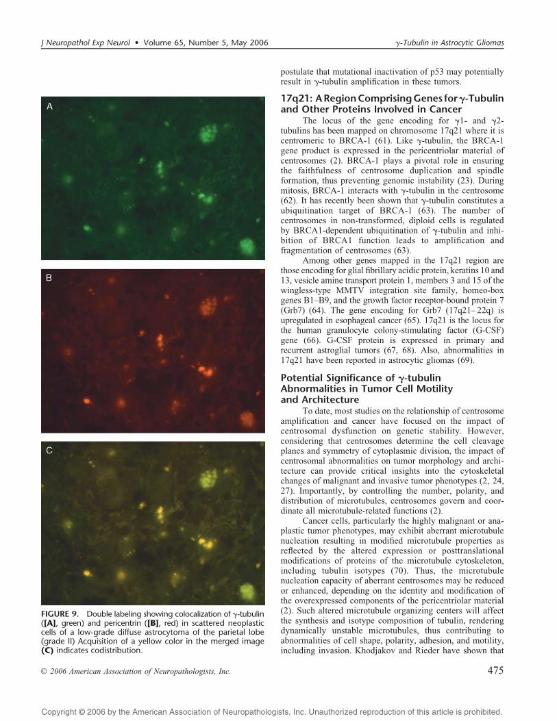
compared to pericentrin (Fig. 8A, C, D, F). The former (F-tubulin) displayed a confluent punctate or diffuse centrifugallocalization extending to the periphery of glioblastoma cells,whereas the latter (pericentrin) was spatially restricted to arelatively narrow, irregular cytoplasmic rim surrounding thetumor cell nuclei (Fig. 8C, F). On the other hand, colocaliza-tion of F-tubulin and pericentrin was demonstrated inscattered neoplastic cells in low-grade diffuse astrocytomas(grade II) (Fig. 9AYC).
DISCUSSIONThe identification of a number of molecular alterations
in transformed glial cells has provided critical insights intothe role of tumor suppressor genes and signaling pathways inthe development of gliomas (41Y45). Genetic alterationsfrequently observed in high-grade adult astrocytic gliomasinvolve either the p53/MDM2/p14ARF or the Rb/CDK4/p16INK4a tumor suppressor pathways (46). However, thereare presently no specific, or clinically useful, cellular ormolecular genetic markers that can reliably predict the onsetof a tumor`s shift to a higher level of malignancy in this
clinico-pathological setting. Although numerical chromosomalabnormalities (present predominantly in high-grade as com-pared to low-grade tumors) contribute to this process, thepatterns of chromosomal instability and involvement of tumorsuppressor pathways are both varying and different betweenthe adult and pediatric groups (46, 47). This disparity ofcytogenetic findings points to underpinning fundamentaldefects of cellular systems governing the physical segregationof chromosomes as opposed to the presence of specificprimary chromosomal abnormalities per se. It is thought thatcentrosomes, which integrate control of cell division andgenomic stability, cell cycle progression, and cytoskeletaldynamics, may be critically important in this regard (2, 23).
Centrosome Amplification in CancerThere is persuasive evidence to date pointing to
abnormalities in the number of chromosomes (aneuploidy) asa cause rather than an effect of anaplastic transformation (48).The chromosome missegregation in malignant tumors couldresult from defects in centrosome function (1Y3, 22, 23).Consequently, centrosome dysfunction, reflected by abnor-
FIGURE 2. Cellular distribution of F -tubulin in glioblastoma multiforme,WHO grade IV. Robust staining ispresent in a large number of tumorcells (AYD). Pleomorphic and multi-nucleated tumor cells exhibit innumer-able, finely punctate and confluentcytoplasmic localizations merging intoa diffuse staining pattern (BYF). ABCperoxidase with hematoxylin counter-stain. Scale bars = (A) 50 Km; (B, D)20 Km; (C, E, F) 10 Km.
Katsetos et al J Neuropathol Exp Neurol � Volume 65, Number 5, May 2006
� 2006 American Association of Neuropathologists, Inc.470

Copyright @ 2006 by the American Association of Neuropathologists, Inc. Unauthorized reproduction of this article is prohibited.
malities in the expression and sorting of centrosomal proteins,may antecede changes of DNA content. We have focused ourattention on F-tubulin, a key cytoskeletal protein of centro-
somes, in the context of malignancy of astrocytic gliomas andin an effort to elucidate new markers of incipient anaplasticphenotypes in low-grade diffuse astrocytomas.
FIGURE 3. Glioblastoma multiforme: Localiza-tion of F-tubulin in endothelial cells of tumorblood vessels exhibiting microvascular prolifer-ation. Note endothelial cell hypertrophy,nuclear atypia, and particulate multipunctatecytoplasmic localization of F-tubulin. (A)Image depicts vascular lumen; panel (B) is ahigher magnification of the frame in (A). Scalebars = (A) 20 Km; (BYD) 10 Km.
FIGURE 4. Localization of F-tubulinwith monoclonal antibodies GTU-88(AYC) and TU-31 (DYF) in humanglioblastoma cell lines. (A, B) Panels(U118MG line) depict multi-punctate,irregularly shaped and variously sizedcoalescent cytoplasmic localizations.Panels (C, D) (T98G line), (E)(U87MG line), and (F) (U138MG line)reveal diffuse and confluent localiza-tions in cells exhibiting also singlebright paranuclear dots consistentwith centrosomes. Note confluentcytoplasmic mass-like aggregates in([C, E, F], arrowheads) in addition todiffuse, less robust cytoplasmic stain-ing. FITC labels cytoplasmic protein.Scale bar = (AYC) 10 Km; (D) 28 Km;(E, F) 19 Km.
J Neuropathol Exp Neurol � Volume 65, Number 5, May 2006 F-Tubulin in Astrocytic Gliomas
� 2006 American Association of Neuropathologists, Inc. 471
Copyright @ 2006 by the American Association of Neuropathologists, Inc. Unauthorized reproduction of this article is prohibited.
Amplified centrosomes are accompanied by increasedor otherwise altered expression of centrosomal proteins,including F-tubulin and pericentrin (24, 25, 28, 29, 31).Also, they exhibit increased protein phosphorylation andaltered functional properties, such as an increased or faultymicrotubule nucleating capacity (reviewed in [2, 3, 22]).
Indirect evidence of centrosome amplification in astro-cytomas may be deduced from the increased human polo-likekinase-1 expression in these tumors (49). Increased polo-likekinase expression is also present in 3 human glioblastomacell lines (U87MG, U118MG, and U138MG) and primaryexplants from patients with glioblastoma (49). In addition,c-Jun N-terminal kinase (JNK), a stress-activated proteinkinase associated with the centrosome (50), is thought toplay a significant role in glioma tumorigenesis (51).
Overexpression of F-Tubulin inAstrocytic Gliomas
To assess the presence of centrosome amplification inglial tumors we evaluated the cellular expression andlocalization of F-tubulin in 56 surgically resected diffuseastrocytic gliomas representing WHO grades II through IV,and in 4 human glioblastoma cell lines (U87MG, U118MG,U138MG, and T98G). We demonstrate that neoplastic cellsof human astrocytic gliomas exhibit increased expression ofF-tubulin associated with altered patterns of cellular distri-bution in malignant tumor phenotypes. These alterationsoccur according to an ascending scale of histological malig-nancy and are particularly prominent in tumor cell subpopu-lations of glioblastomas. Our results indicate that F-tubulinexpression is significantly increased in the high-grade, ana-plastic astrocytomas, and glioblastomas multiforme (grades III/IV) as compared to low-grade diffuse astrocytomas (grade II).In the context of the findings of the present study, the detectionof either multipunctate and/or diffuse F-tubulin localizations in
tumor cells relates statistically to a high-grade histologicallesion if it falls within the interquartile range (IQR) of labelingindex (LI) values (42% to 73% or above). At the same time,even LI values within the IQR for low-grade diffuseastrocytomas (2% to 9% or upwards), may be potentiallysignificant inasmuch as they may portend incipient genomicinstability in a subpopulation of tumor cells with a predis-position for anaplastic change.
Non-neoplastic glial cells contain one or two para-nuclear centrosomes, which are visualized using anti-F-tubulin, anti-pericentrin and anti->-tubulin antibodies; incontrast, tumor cells from primary glial tumors and tumorcell lines exhibit two distinctive but overlapping patterns ofaltered cellular localization of F-tubulin. These includepunctate and diffuse patterns of immunoreactivity. Largeamounts of cytoplasmic F-tubulin exhibiting a predominantlydiffuse staining pattern were demonstrated by bright-fieldimmunohistochemistry and immunofluorescence microscopyin glioblastoma cells from primary surgically resectedspecimens and tumor cell lines. We have confirmed by immu-noblotting that diffuse staining may represent monomeric oroligomeric pools of F-tubulin, which in tumor cells probablyexist in a state of dynamic instability, possibly available forrapid recruitment by tumor cells in the context of centrosome
FIGURE 5. Immunoblots with anti-F-tubulin antibodies GTU-88(A) and TU-32 (B) on panel of human glioblastoma cell lines.Lanes 1 to 4 correspond to total lysates of U118MG (lane 1),U138MG (lane 2), U87MG (lane 3), and T98G cells (lane 4).The same amount of protein (10 Kg) was loaded into eachlane. Positions of molecular mass markers (in kDa) are indicatedon the left.
FIGURE 6. Immunoblot analysis of soluble and insolublefractions from human glioblastoma cell line T98G andU87MG. Samples were prepared from untreated T98G (A)and U87MG (B) cells (lanes 1 and 2) or from cells pre-treatedwith nocodazole to disrupt microtubule arrays (lanes 3 and 4).To compare the relative distribution of immunoblottedproteins, pelleted material was resuspended in a volumeequal to the corresponding supernatant. Immunostainingwith antibody TU-01 against >-tubulin (>-Tb) and antibodyTU-32 against F-tubulin (F-Tb). S, supernatant; P, pellet.
Katsetos et al J Neuropathol Exp Neurol � Volume 65, Number 5, May 2006
� 2006 American Association of Neuropathologists, Inc.472

Copyright @ 2006 by the American Association of Neuropathologists, Inc. Unauthorized reproduction of this article is prohibited.
amplification. In addition, we have noted coalescent punctatelocalizations especially in the U118MG glioblastoma cellline. Although these immunofluorescence localizations do notappear to correspond to intact Bsupernumerary centrosomes^they are likely to represent abnormal protein assemblies eitherin the form of Bacentriolar bodies,^ aberrant accumulations ofectopic pericentriolar material, and/or fragmented centro-somes (2). It is noteworthy that a substantial proportion ofcellular F-tubulin was found in the detergent (nocodazole)-resistant fraction of glioblastoma cell line extracts.
Our findings suggest that centrosome amplificationdoes not necessarily need to be accompanied by structurallyBintact^ supernumerary centrosomes, but rather it may becharacterized by molecular/biochemical alterations in whichthere is increased (or otherwise altered) expression ofproteins of the pericentriolar material, including F-tubulin.
In the developmental context, soluble F-tubulin hasbeen demonstrated in nucleated chicken erythrocytes and pigbrain cells (19, 20). However, to date, the subcellular sortingof F-tubulin in cancer cells in general and glioma cells inparticular is unknown. Draberova and co-workers havedemonstrated that rat basophilic leukemia cells contain largesoluble pools of >/A-tubulin dimers and F-tubulin, whichexist in large complexes with other molecules and arelocalized around centrosomes (52). These investigators haveshown that complexes of soluble F-tubulin released fromactivated rat basophilic leukemia cells contain tyrosine-phosphorylated proteins (52). Similarly, in P19 embryonalcarcinoma cells undergoing neuronal differentiation, F-tubulinis phosphorylated and forms complexes with protein tyrosinekinases of the Src family (53).
F-Tubulin interacts, in addition to >/A-tubulin dimers(16) and pericentrin (11, 12), with a host of other proteins.These include BRCA-1, Ras association domain familyprotein 1, RAC GTPase activating protein 1, paxillin,phosphatidylinositol 3 kinase (PI3K) regulatory >-subunit,RING finger protein 19 (54), and dynamin-2 (55). Geneticalterations of class IA PI3K subunit genes have been shown toplay a role in human glioblastomas (56). In addition,colocalization of F-tubulin with nerve growth factor has beendemonstrated at the centrosomes or the spindle polesthroughout the cell cycle in the human glioblastoma cell lineU251 MG (57).
The presence of numerous key regulators of cell cycleprogression at the centrosome has led to the speculation thatthe centrosome itself provides an important structuralcontext for coordinating cell cycle regulation (2, 5).
Genes involved in different signal transduction pathwayshave been implicated in centrosome amplification. Theseinclude genes of the p53 pathway (p53, WAF-1, Gadd45, andMdm2) and the DNA-repair pathway (ATR, BRCA-1, BRCA-2,and XRCC2/3), as well as genes involved in ubiquitin-relatedprotein degradation (Tsg101, Skp2, and RAD6) and mitosis(Aurora-A) (reviewed in (2), (32), (58), (59)).
Deletion or mutational/functional inactivation of p53leads to centrosome amplification (60). Because TP53 genemutations are genotypic hallmarks of Bsecondary^ glioblas-tomas, which arise as a consequence of malignant change inpre-existing diffuse low-grade astrocytomas (42, 43), we
FIGURE 7. Distribution of F-tubulin and tubulin dimers in thehuman glioblastoma cell line T98G. Cells were stained by doublelabeling with polyclonal antibody specific for tubulin dimers (A)and monoclonal antibody TU-31 for F-tubulin (B). Superpositionof stainings is shown in panel (C). Note robust tubulin dimerstaining (green) delineating a large paranuclear immunoreactivemass. Staining of microtubule arrays (green) in the tumor cellbodies and proximal segments of cell processes is also detected.Double labeling reveals that the localization of F-tubulin (red) isspatially distinct and exhibits a diffuse localization involving (inthis field) the outer cytoplasmic portions of tumor cells. DAPI(blue) labels multiple nuclei in each cell (C). Scale bar = 10 Kmfor all panels.
J Neuropathol Exp Neurol � Volume 65, Number 5, May 2006 F-Tubulin in Astrocytic Gliomas
� 2006 American Association of Neuropathologists, Inc. 473
Copyright @ 2006 by the American Association of Neuropathologists, Inc. Unauthorized reproduction of this article is prohibited.
FIGURE 8. Distribution of F-tubulin and pericentrin in the human glioblastoma cell line T98G (AYC) and U118MG (DYF). Cellswere stained by double labeling with monoclonal antibody TU-31specific for F-tubulin ([AYD], green) and polyclonal antibodyspecific for pericentrin ([B, E], red). Superposition of stainings is shown in panels (C) and (F). There is spatial dissociationbetween these 2 centrosome-associated proteins. Pericentrin staining is present in the juxta-nuclear pericentriolar region. Althoughcolocalization of F-tubulin (green) and pericentrin (red) was demonstrated in normal juxta-nuclear centrosomes (yellow overlay), thedistribution of F-tubulin is more widespread displaying a confluent punctate or diffuse localization extending to the periphery oftumor cells. DAPI (blue) labels nuclei. Scale bar = 10 Km for all panels.
Katsetos et al J Neuropathol Exp Neurol � Volume 65, Number 5, May 2006
� 2006 American Association of Neuropathologists, Inc.474
Copyright @ 2006 by the American Association of Neuropathologists, Inc. Unauthorized reproduction of this article is prohibited.
postulate that mutational inactivation of p53 may potentiallyresult in F-tubulin amplification in these tumors.
17q21: ARegionComprisingGenes forF-Tubulinand Other Proteins Involved in Cancer
The locus of the gene encoding for F1- and F2-tubulins has been mapped on chromosome 17q21 where it iscentromeric to BRCA-1 (61). Like F-tubulin, the BRCA-1gene product is expressed in the pericentriolar material ofcentrosomes (2). BRCA-1 plays a pivotal role in ensuringthe faithfulness of centrosome duplication and spindleformation, thus preventing genomic instability (23). Duringmitosis, BRCA-1 interacts with F-tubulin in the centrosome(62). It has recently been shown that F-tubulin constitutes aubiquitination target of BRCA-1 (63). The number ofcentrosomes in non-transformed, diploid cells is regulatedby BRCA1-dependent ubiquitination of F-tubulin and inhi-bition of BRCA1 function leads to amplification andfragmentation of centrosomes (63).
Among other genes mapped in the 17q21 region arethose encoding for glial fibrillary acidic protein, keratins 10 and13, vesicle amine transport protein 1, members 3 and 15 of thewingless-type MMTV integration site family, homeo-boxgenes B1YB9, and the growth factor receptor-bound protein 7(Grb7) (64). The gene encoding for Grb7 (17q21Y 22q) isupregulated in esophageal cancer (65). 17q21 is the locus forthe human granulocyte colony-stimulating factor (G-CSF)gene (66). G-CSF protein is expressed in primary andrecurrent astroglial tumors (67, 68). Also, abnormalities in17q21 have been reported in astrocytic gliomas (69).
Potential Significance of F-tubulinAbnormalities in Tumor Cell Motilityand Architecture
To date, most studies on the relationship of centrosomeamplification and cancer have focused on the impact ofcentrosomal dysfunction on genetic stability. However,considering that centrosomes determine the cell cleavageplanes and symmetry of cytoplasmic division, the impact ofcentrosomal abnormalities on tumor morphology and archi-tecture can provide critical insights into the cytoskeletalchanges of malignant and invasive tumor phenotypes (2, 24,27). Importantly, by controlling the number, polarity, anddistribution of microtubules, centrosomes govern and coor-dinate all microtubule-related functions (2).
Cancer cells, particularly the highly malignant or ana-plastic tumor phenotypes, may exhibit aberrant microtubulenucleation resulting in modified microtubule properties asreflected by the altered expression or posttranslationalmodifications of proteins of the microtubule cytoskeleton,including tubulin isotypes (70). Thus, the microtubulenucleation capacity of aberrant centrosomes may be reducedor enhanced, depending on the identity and modification ofthe overexpressed components of the pericentriolar material(2). Such altered microtubule organizing centers will affectthe synthesis and isotype composition of tubulin, renderingdynamically unstable microtubules, thus contributing toabnormalities of cell shape, polarity, adhesion, and motility,including invasion. Khodjakov and Rieder have shown that
FIGURE 9. Double labeling showing colocalization of F-tubulin([A], green) and pericentrin ([B], red) in scattered neoplasticcells of a low-grade diffuse astrocytoma of the parietal lobe(grade II) Acquisition of a yellow color in the merged image(C) indicates codistribution.
J Neuropathol Exp Neurol � Volume 65, Number 5, May 2006 F-Tubulin in Astrocytic Gliomas
� 2006 American Association of Neuropathologists, Inc. 475

Copyright @ 2006 by the American Association of Neuropathologists, Inc. Unauthorized reproduction of this article is prohibited.
at the onset of mitosis, the centrosome suddenly gains theability to bind several times the amount of F-tubulin thanduring interphase, which does not require microtubules (71).It follows then that mitotically active tumor cells mayrequire increased amounts of F-tubulin. Moreover, since therecruitment of F-tubulin ring complexes is a prerequisite forincreased microtubule-nucleating activity, in addition to theregulation of tubulin synthesis and cycle progression are(13), the increased expression of F-tubulin may potentiallyculminate in aberrant expression of tubulin isotypes ingliomas (Katsetos et al, unpublished observation). In anoligonucleotide microarray analysis of high- versus low-grade gliomas, Rickman and colleagues observed that genesencoding for a number of cytoskeletal and cytoskeleton-associated proteins, including F-tubulin and AIV-tubulin,were highly expressed in glioblastomas (72).
Interestingly, both diffuse and multipunctate F-tubulinstaining was detected in endothelial cells in areas ofmicrovascular proliferation in glioblastomas. This indicatesthat centrosome abnormalities may underlie neoplastic neo-vascularization in gliomas. It has recently been shown thatendothelial cells in solid tumors are aneuploid and areassociated with multiple centrosomes (73). Studies areunderway in our laboratory to elucidate the role of F-tubulin inglioblastoma angiogenesis.
Our results indicate that ectopic cellular expression ofF-tubulin in diffuse astrocytic gliomas may be significant inthe context of centrosome dysfunction/amplification andmay be linked to tumor progression where it may potentiallyserve as a novel marker of anaplastic change. It remains to bedetermined in a significantly larger cohort of tumor specimenswhether centrosomal defects, including derangements in theexpression of F-tubulin (which may precede alterations ofgenomic stability) may lay the foundation for a new andpromising approach to molecular stratification of gliomas.
ACKNOWLEDGMENTSWe are indebted to Dr. Jennian F. Geddes, formerly,
Reader in Clinical Neuropathology, Department of Histo-pathology and Morbid Anatomy, Queen Mary, University ofLondon, The Royal London Hospital, London, England, forproviding tissue samples used in this study and for helpfuldiscussions. We thank Dr. Chrisoula D. Scopa, Departmentof Anatomical Pathology and Cytology, University Hospital,Patras, Greece for the procurement of archival tissuematerial from brain tumor cases operated by one of the co-authors (T.M.). We thank Dr. Steven J. Doxsey, University ofMassachusetts Medical Center, Worcester, MA for his gift ofthe antibody against pericentrin.
REFERENCES1. Brinkley BR, Goepfert TM. Supernumerary centrosomes and cancer:
Boveri`s hypothesis revisited. Cell Motil Cytoskeleton 1998;41:281Y882. Nigg EA. Centrosome aberrations: cause or consequence of cancer
progression? Nat Rev Cancer 2002;2:815Y253. D’Assoro AB, Lingle WL, Salisbury JL. Centrosome amplification and
the development of cancer. Oncogene 2002;21:6146 Y534. Brinkley BR. Microtubule organizing centers. Annu Rev Cell Biol
1985;1:145Y72
5. Doxsey S. Re-evaluating centrosome function. Nat Rev Mol Cell Biol2001;2:688Y98
6. Hinchcliffe EH, Miller FJ, Cham M, et al. Requirement of acentrosomal activity for cell cycle through G1 into S phase. Science2001;291:1547Y50
7. Khodjakov A, Rieder CL. Centrosomes enhance the fidelity ofcytokinesis in vertebrates and are required for cell cycle progression.J Cell Biol 2001;153:237Y 42
8. Stearns T, Evans L, Kirschner M. F-tubulin is a highly conservedcomponent of the centrosome. Cell 1991;65:825Y36
9. Stearns T, Kirschner M. In vitro reconstitution of centrosome assemblyand function: the central role of F-tubulin. Cell 1994;76:623Y37
10. Zheng Y, Jung K, Oakley BR. F-Tubulin is present in Drosophilamelanogaster and Homo sapiens and is associated with the centrosome.Cell 1991;65:817Y23
11. Dictenberg JB, Zimmerman W, Sparks CA, et al. Pericentrin andF-tubulin form a protein complex and are organized into a novel latticeat the centrosome. J Cell Biol 1998;141:163Y74
12. Young A, Dictenberg JB, Purohit A, et al. Cytoplasmic dynein-mediatedassembly of pericentrin and F-tubulin onto centrosomes. Mol Biol Cell2000;11:2047Y56
13. Zhou J, Shu H-B, Joshi HC. Regulation of tubulin synthesis and cellcycle progression in mammalian cells by F-tubulin-mediated micro-tubule nucleation. J Cell Biochem 2002;84:472Y83
14. Leguy R, Melki R, Pantaloni D, et al. Monomeric F-tubulin nucleatesmicrotubules. J Biol Chem 2000;275:21975 Y 80
15. Linhartova I, Novotna B, Sulimenko V, et al. F-Tubulin in chickenerythrocytes: changes in localization during cell differentiation andcharacterization of cytoplasmic complexes. Dev Dynam 2002;223:229Y 40
16. Sulimenko V, Sulimenko T, Poznanovic S, et al. Association of brainF-tubulins with >A-tubulin dimers. Biochem J 2002;365:889Y95
17. Chabin-Brion K, Marceiller J, Perez F, et al. The Golgi complex is amicrotubule-organizing organelle. Mol Biol Cell 2001;12:2047Y60
18. Drykova D, Sulimenko V, Cenklova V, et al. Plant F-tubulin interactswith >A-tubulin dimers and forms membrane-associated complexes.Plant Cell 2003;15:465Y80
19. Rios RM, Sanchis A, Tassin AM, et al. GMAP-210 recruits F-tubulincomplexes to cis-Golgi membranes and is required for Golgi ribbonformation. Cell 2004;118:323Y35
20. Joshi HC, Palacios MJ, McNamara L, et al. F-Tubulin is a centrosomalprotein required for cell cycle-dependent nucleation. Nature 1992;356:80 Y82
21. Zheng Y, Wong ML, Alberts B, et al. Nucleation of microtubuleassembly by F-tubulin-containing ring complex. Nature 1995;378:578Y83
22. Salisbury JL, D’Assoro AB, Lingle WL. Centrosome amplification andthe origin of chromosomal instability in breast cancer. J MammaryGland Biol Neoplasia 2004;9:275 Y83
23. Emdad L, Sarkar D, Su ZZ, et al. Emerging roles of centrosomalamplification and genomic instability in cancer. Front Biosci 2005;10:728Y 42
24. Pihan GA, Purohit A, Wallace J, et al. Centrosome defects can accountfor cellular and genetic changes that characterize prostate cancerprogression. Cancer Res 2001;61:2212Y19
25. Pihan GA, Purohit A, Wallace J, et al. Centrosome defects and geneticinstability in malignant tumors. Cancer Res 1998;58:3974 Y85
26. Pihan GA, Wallace J, Zhou Y, et al. Centrosome abnormalities andchromosome instability occur together in pre-invasive carcinomas.Cancer Res 2003;63:1398Y1404
27. Lingle WL, Barrett SL, Negron VC, et al. Centrosome amplificationdrives chromosomal instability in breast tumor development. Proc NatlAcad Sci U S A 2002;99:1978Y83
28. Sato N, Mizumoto K, Nakamura M, et al. Centrosome abnormalities inpancreatic ductal carcinoma. Clin Cancer Res 1999:963Y70
29. Kuo K-K, Sato N, Mizumoto K, et al. Centrosome abnormalities inhuman carcinomas of the gallbladder and intrahepatic and extrahepaticbile ducts. Hepatology 2000;31:59Y 64
30. Skyldberg B, Fujioka K, Hellstrom A-C, et al. Human papillomavirusinfection, centrosome aberration, and genetic stability in cervicallesions. Mod Pathol 2001;14:279Y84
31. Setoguchi A, Okuda M, Nishida E, et al. Results of hyperamplification
Katsetos et al J Neuropathol Exp Neurol � Volume 65, Number 5, May 2006
� 2006 American Association of Neuropathologists, Inc.476
Copyright @ 2006 by the American Association of Neuropathologists, Inc. Unauthorized reproduction of this article is prohibited.
of centrosomes in naturally developing tumors of dogs. Am J Vet Res2001;62:1134Y 41
32. Goepfert TM, Adigun YE, Zhong L, et al. Centrosome amplificationand overexpression of Aurora A are early events in rat mammarycarcinogenesis. Cancer Res 2002;62:4115Y22
33. Al-Romaih K, Bayani J, Vorobyova J, et al. Chromosomal instability inosteosarcoma and its association with centrosome abnormalities. CancerGenet Cytogenet 2003;144:91Y99
34. Weber RG, Bridger JM, Benner A, et al. Centrosome amplification as apossible mechanism for numerical chromosome aberrations in cerebralprimitive neuroectodermal tumors with TP53 mutations. Cytogenet CellGenet 1998;83:266 Y 69
35. Novakova M, Draberova E, Schurmann W, et al. F-Tubulin redistri-bution in taxol-treated mitotic cells probed by monoclonal antibodies.Cell Motil Cytoskeleton 1996;33:38Y51
36. Viklicky V, Draber P, Hasek J, et al. Production and characterization ofa monoclonal antitubulin antibody. Cell Biol Int Rep 1982;6:725Y31
37. Draber P, Draberova E, Linhartova I, et al. Differences in the exposureof C- and N-terminal tubulin domains in cytoplasmic microtubulesdetected with domain-specific monoclonal antibodies. J Cell Sci 1989;92:519Y28
38. Draber P, Draberova E, Viklicky V. Immunostaining of humanspermatozoa with tubulin domain-specific monoclonal antibodies.Recognition of a unique A-tubulin epitope in the sperm head.Histochemistry 1991;95:519 Y24
39. Doxsey SJ, Stein P, Evans L, Calarco P, Kirschner M. Pericentrin, ahighly conservedprotein of centrosomes involved in microtubuleorganization. Cell 1994;76:639 Y50
40. Katsetos CD, Del Valle L, Geddes JF, et al. Aberrant localization of theneuronal class III A-tubulin in astrocytomas. Arch Pathol Lab Med2001;125:613Y24
41. Perry A. Pathology of low-grade gliomas: an update of emergingconcepts. Neuro-oncol 2003;5:168Y78
42. Louis DN, Holland EC, Cairncross JG. Glioma classification: amolecular reappraisal. Am J Pathol 2001;159:779Y86
43. Watanabe K, Tachibana O, Sato K, et al. Overexpression of the EGFreceptor and p53 mutations are mutually exclusive in the evolution ofprimary and secondary glioblastomas. Brain Pathol 1996;6:217Y23
44. Hirose Y, Aldape KD, Chang S, et al. Grade II astrocytomas aresubgrouped by chromosome aberrations. Cancer Genet Cytogenet 2003;142:1Y7
45. Rickert CH, Paulus W. Prognosis-related histomorphological andimmunohistochemical markers in central nervous system tumors ofchildhood and adolescence. Acta Neuropathol (Berl) 2005;109:69Y92
46. Sung T, Miller DC, Hayes RL, et al. Preferential inactivation of the p53tumor suppressor pathway and lack of EGFR amplification distinguishde novo high grade pediatric astrocytomas from de novo adultastrocytomas. Brain Pathol 2000;10:249Y59
47. Rickert CH, Strater R, Kaatsch P, et al. Pediatric high-gradeastrocytomas show chromosomal imbalances distinct from adult cases.Am J Pathol 2001;158:1525Y32
48. Sen S. Aneuploidy and cancer. Curr Opin Oncol 2000;12:82Y 8849. Dietzmann K, Kirches E, von Bossanyi, et al. Increased human
polo-like kinase-1 expression in gliomas. J Neurooncol 2001;53:1Y1150. MacCorkle-Chosnek RA, Van Hooser AA, Goodrich DW, et al. Cell
cycle regulation of c-Jun N-terminal kinase (JNK) activity at thecentrosomes. Biochem Biophys Res Commun 2001;289:173 Y80
51. Tsuiki H, Tnani M, Okamoto I, et al. Constitutively active forms ofc-Jun NH2-terminal kinase are expressed in primary glial tumors.Cancer Res 2003;63:250Y55
52. Draberova L, Draberova E, Surviladze Z, et al. Protein tyrosine kinasep53/p56lyn forms complexes with F-tubulin in rat basophilic leukemiacells. Int Immunol 1999;11:1829Y39
53. Kukharskyy V, Sulimenko V, Macurek L, et al. Complexes ofF-tubulin with nonreceptor protein tyrosine kinases Src and Fyn indifferentiating P19 embryonal carcinoma cells. Exp Cell Res 2004;298:218Y28
54. Human Protein Reference Database (Johns Hopkins University, Baltimore,MD) http://www.hprd.org/protein/01853?selectedtab=INTERACTIONS.
55. Thompson HM, Cao H, Chen J, et al. Dynamin-2 binds F-tubulin andparticipates in centrosome cohesion. Nat Cell Biol 2004;6:335 Y 42
56. Mizoguchi M, Nutt CL, Mohapatra G, et al. Genetic alterations ofphosphoinositide 3-kinase subunit genes in human glioblastomas. BrainPathol 2004;14:372Y77
57. Zhang Z, Yang Y, Gong A, et al. Localization of NGF and TrkA atmitotic apparatus in human glioma cell line U251. Biochem BiophysRes Commun 2005;337:68Y74
58. Stenoien DL, Sen S, Mancini MA, et al. Dynamic association of atumor amplified kinase, Aurora-A, with the centrosome and mitoticspindle. Cell Motil Cytoskeleton 2003;55:134 Y 46
59. Zhou H, Kuang J, Zhong L, et al. Tumour amplified kinaseSTK15/BTAK induces centrosome amplification, aneuploidy and trans-formation. Nat Genet 1998;20:189Y93
60. Tarapore P, Fukasawa K. Loss of p53 and centrosome hyperamplifica-tion. Oncogene 2002;21:6234 Y 40
61. Rommens JM, Durocher F, McArthur J, et al. Generation of atranscription map at the HSD17B locus centromeric to BRCA1 at17q21. Genomics 1995;28:530 Y 42
62. Deng CX. Roles of BRCA1 in centrosome duplication. Oncogene 2002;21:6222Y27
63. Starita LM, Machida Y, Sankaran S, et al. BRCA1-dependentubiquitination of F-tubulin regulates centrosome number. Mol CellBiol 2004;24:8457Y 66
64. On-line Mendelian Inheritance in Man (OMIM) Gene map (JohnsHopkins University, Baltimore, MD) http://www.ncbi.nlm.nih.gov/Omim/getmap.cgi?l191135.
65. Walch A, Specht K, Braselmann H, et al. Coamplification andcoexpression of GRB7 and ERBB2 is found in high grade intra-epithelial neoplasia and in invasive Barrett’s carcinoma. Int J Cancer2004;112:747Y53
66. Tweardy DJ, Cannizzaro LA, Palumbo AP, et al. Molecular cloning andcharacterization of a cDNA for human granulocyte colony-stimulatingfactor (G-CSF) from a glioblastoma multiforme cell line and localizationof the G-CSF gene to chromosome band 17q21. Oncogene Res 1987;1:209Y20
67. Mueller MM, Herold-Mende CC, Riede D, et al. Autocrine growthregulation by granulocyte colony-stimulating factor and granulocytemacrophage colony-stimulating factor in human gliomas with tumorprogression. Am J Pathol 1999;155:1557Y 67
68. Stan AC, Walter GF, Welte K, et al. Expression of granulocytecolony-stimulating factor in recurrent glial tumors is inversely corre-lated with tumor progression. J Neuroimmunol 1999;94:66 Y73
69. Nauman P, Czernicki Z . The molecular basis of glia-derived tumoursof the brain. Part I. Neurol Neurochir Pol 2002;36:553 Y 64
70. Katsetos CD, Herman MM, Mork SJ. Class III A-tubulin in humandevelopment and cancer. Cell Motil Cytoskeleton 2003;55:77Y96
71. Khodjakov A, Rieder CL. The sudden recruitment of F-tubulin to thecentrosome at the onset of mitosis and its dynamic exchangethroughout the cell cycle, do not require microtubules. J Cell Biol1999;146:585Y96
72. Rickman DS, Bobek MP, Misek DE, et al. Distinctive molecularprofiles of high-grade and low-grade gliomas based on oligonucleotidemicroarray analysis. Cancer Res 2001;61:6885Y91
73. Hida K, Klagsbrun M. A new perspective on tumor endothelial cells:unexpected chromosome and centrosome abnormalities. Cancer Res2005;65:2507Y10
J Neuropathol Exp Neurol � Volume 65, Number 5, May 2006 F-Tubulin in Astrocytic Gliomas
� 2006 American Association of Neuropathologists, Inc. 477
Copyright © 2022 FDOKUMEN