Altered anxiety-related and abnormal social behaviors in rats exposed to early life seizures
8
ORIGINAL RESEARCH ARTICLE published: 09 May 2013 doi: 10.3389/fnbeh.2013.00036 Altered anxiety-related and abnormal social behaviors in rats exposed to early life seizures Adelisandra Silva Santos Castelhano 1 , Gustavo dos Santos Teada Cassane 1 , Fulvio Alexandre Scorza 2 and Roberta Monterazzo Cysneiros 1 * 1 Laboratory of Neurobiology, Developmental Disabilities Graduate Program, Presbyterian Mackenzie University, São Paulo, Brazil 2 Department of Neurology and Neurosurgery, Experimental Neurology, Escola Paulista de Medicina, Federal University of São Paulo, São Paulo, Brazil Edited by: Valérie Doyère, CNRS, France Reviewed by: Guillaume L. Poirier, Ecole Polytechnique Fédérale de Lausanne, Switzerland Alexis Faure, Neurobiologie de la Prise de Décision Centre Neurosciences Paris Sud CNPS CNRS UMR 8195, France *Correspondence: Roberta Monterazzo Cysneiros, Laboratory of Neurobiology, Developmental Disabilities Graduate Program, Presbyterian Mackenzie University, Rua da Consolação, 930. Prédio 28, CEP01302-907, São Paulo, Brazil. e-mail: roberta.cysneiros@ mackenzie.br; [email protected] Neonatal seizures are the most common manifestation of neurological dysfunction in the neonate. The prognosis of neonatal seizures is highly variable, and the controversy remains whether the severity, duration, or frequency of seizures may contribute to brain damage independently of its etiology. Animal data indicates that seizures during development are associated with a high probability of long-term adverse effects such as learning and memory impairment, behavioral changes and even epilepsy, which is strongly age dependent, as well as the severity, duration, and frequency of seizures. In preliminary studies, we demonstrated that adolescent male rats exposed to one-single neonatal status epilepticus (SE) episode showed social behavior impairment, and we proposed the model as relevant for studies of developmental disorders. Based on these facts, the goal of this study was to verify the existence of a persistent deficit and if the anxiety-related behavior could be associated with that impairment. To do so, male Wistar rats at 9 days postnatal were submitted to a single episode of SE by pilocarpine injection (380 mg/kg, i.p.) and control animals received saline (0.9%, 0.1 mL/10 g). It was possible to demonstrate that in adulthood, animals exposed to neonatal SE displayed low preference for social novelty, anxiety-related behavior, and increased stereotyped behavior in anxiogenic environment with no locomotor activity changes. On the balance, these data suggests that neonatal SE in rodents leads to altered anxiety-related and abnormal social behaviors. Keywords: neonatal status epilepticus, pilocarpine, social anxiety, general anxiety, social behavior INTRODUCTION Status epilepticus (SE), an acute condition characterized by repet- itive or prolonged seizures, affects between 120,000 to 200,000 people per year in the United States (Neill et al., 2005; Lowenstein, 2006). It occurs more often in children than in adults, and 40–50% of children under the age of 2 years (Shinnar et al., 1997). The incidence of neonatal seizures varies according to the age, severity of the etiologic factor, and of the population studied. The clear incidence has not yet been established, although it has been estimate that varies between 1.1–8.6/1000 live births (Saliba et al., 1996, 1999; Sheth et al., 1999; Mosley, 2010). Furthermore, the prognosis of neonatal seizures is highly variable (Costa et al., 2001), and persists the controversy whether the severity, duration or frequency of seizures may contribute to brain damage inde- pendently of its etiology (Udani, 2008). In general terms, half of the cases exhibit minimum sequels and the other half evolves into death or serious squeals (Costa et al., 2001). In addition, newborns with transient focal ischemia, metabolic abnormali- ties without a clear etiology seem to evolve in a more satisfac- tory manner, unlike those with hypoxic-ischemic encephalopathy, infection of the nervous system and prenatal brain dysgenesis that generally have a worse prognosis (Udani, 2008). From a clinical standpoint, most studies fail to show that seizures per se regardless the severity of the etiologic factor is able to disrupt brain devel- opment. In this context, animal models allow the observation and analysis of variables under controlled conditions and have been used to explore these issues. Thus, the animal models with neonatal injury that results in permanent impairment, especially of social interaction are considered useful for the study of neu- rodevelopmental disorders, such as schizophrenia and autism (Schneider and Koch, 2005; Tordjman et al., 2007). Following these lines of reasoning, animal data indicates that seizures during development are associated with a high proba- bility of long-term adverse effects such as, learning and memory impairment, behavioral changes and epilepsy that are strongly age dependent, as well as the severity, duration, and frequency of seizures (Holmes, 2004, 2009). The mechanisms that under- lie these changes are not yet completely understood, but neonatal seizures do not lead to cell loss, rather than considerable synaptic reorganization. Studies focused primarily on behavioral changes, rather than epileptogenic activity, are less frequent. Two stud- ies have shown that neonatal seizures produce anxiety-related behavior (Sayin et al., 2004; Shi et al., 2007), and another study demonstrated deficit of cognitive flexibility (Kleen et al., 2011). Recently, we demonstrated that the neonatal SE (Castelhano et al., 2010) leads to impairment of social interaction in adolescent male rats. Our study brings an important contribution to demonstrate that sequels of neonatal seizures extending beyond the learning and memory deficits, increased anxiety and loss in cognitive flex- ibility. Animals studies reveal that early life seizures can disrupt some brain structure such as the hippocampus (Holmes et al., 1998; Lynch et al., 2000; Santos et al., 2000; Villeneuve et al., Frontiers in Behavioral Neuroscience www.frontiersin.org May2013 | Volume 7 | Article 36 | 1 BEHAVIORAL NEUROSCIENCE
-
Upload
independent -
Category
Documents
-
view
3 -
download
0
Transcript of Altered anxiety-related and abnormal social behaviors in rats exposed to early life seizures
ORIGINAL RESEARCH ARTICLEpublished: 09 May 2013
doi: 10.3389/fnbeh.2013.00036
Altered anxiety-related and abnormal social behaviors inrats exposed to early life seizuresAdelisandra Silva Santos Castelhano 1, Gustavo dos Santos Teada Cassane1,
Fulvio Alexandre Scorza2 and Roberta Monterazzo Cysneiros 1*
1 Laboratory of Neurobiology, Developmental Disabilities Graduate Program, Presbyterian Mackenzie University, São Paulo, Brazil2 Department of Neurology and Neurosurgery, Experimental Neurology, Escola Paulista de Medicina, Federal University of São Paulo, São Paulo, Brazil
Edited by:
Valérie Doyère, CNRS, France
Reviewed by:
Guillaume L. Poirier, EcolePolytechnique Fédérale deLausanne, SwitzerlandAlexis Faure, Neurobiologie de laPrise de Décision CentreNeurosciences Paris Sud CNPSCNRS UMR 8195, France
*Correspondence:
Roberta Monterazzo Cysneiros,Laboratory of Neurobiology,Developmental Disabilities GraduateProgram, Presbyterian MackenzieUniversity, Rua da Consolação, 930.Prédio 28, CEP 01302-907, SãoPaulo, Brazil.e-mail: [email protected];[email protected]
Neonatal seizures are the most common manifestation of neurological dysfunction inthe neonate. The prognosis of neonatal seizures is highly variable, and the controversyremains whether the severity, duration, or frequency of seizures may contribute tobrain damage independently of its etiology. Animal data indicates that seizures duringdevelopment are associated with a high probability of long-term adverse effects such aslearning and memory impairment, behavioral changes and even epilepsy, which is stronglyage dependent, as well as the severity, duration, and frequency of seizures. In preliminarystudies, we demonstrated that adolescent male rats exposed to one-single neonatal statusepilepticus (SE) episode showed social behavior impairment, and we proposed the modelas relevant for studies of developmental disorders. Based on these facts, the goal of thisstudy was to verify the existence of a persistent deficit and if the anxiety-related behaviorcould be associated with that impairment. To do so, male Wistar rats at 9 days postnatalwere submitted to a single episode of SE by pilocarpine injection (380 mg/kg, i.p.) andcontrol animals received saline (0.9%, 0.1 mL/10 g). It was possible to demonstrate thatin adulthood, animals exposed to neonatal SE displayed low preference for social novelty,anxiety-related behavior, and increased stereotyped behavior in anxiogenic environmentwith no locomotor activity changes. On the balance, these data suggests that neonatal SEin rodents leads to altered anxiety-related and abnormal social behaviors.
Keywords: neonatal status epilepticus, pilocarpine, social anxiety, general anxiety, social behavior
INTRODUCTIONStatus epilepticus (SE), an acute condition characterized by repet-itive or prolonged seizures, affects between 120,000 to 200,000people per year in the United States (Neill et al., 2005; Lowenstein,2006). It occurs more often in children than in adults, and40–50% of children under the age of 2 years (Shinnar et al., 1997).The incidence of neonatal seizures varies according to the age,severity of the etiologic factor, and of the population studied.The clear incidence has not yet been established, although it hasbeen estimate that varies between 1.1–8.6/1000 live births (Salibaet al., 1996, 1999; Sheth et al., 1999; Mosley, 2010). Furthermore,the prognosis of neonatal seizures is highly variable (Costa et al.,2001), and persists the controversy whether the severity, durationor frequency of seizures may contribute to brain damage inde-pendently of its etiology (Udani, 2008). In general terms, halfof the cases exhibit minimum sequels and the other half evolvesinto death or serious squeals (Costa et al., 2001). In addition,newborns with transient focal ischemia, metabolic abnormali-ties without a clear etiology seem to evolve in a more satisfac-tory manner, unlike those with hypoxic-ischemic encephalopathy,infection of the nervous system and prenatal brain dysgenesis thatgenerally have a worse prognosis (Udani, 2008). From a clinicalstandpoint, most studies fail to show that seizures per se regardlessthe severity of the etiologic factor is able to disrupt brain devel-opment. In this context, animal models allow the observationand analysis of variables under controlled conditions and have
been used to explore these issues. Thus, the animal models withneonatal injury that results in permanent impairment, especiallyof social interaction are considered useful for the study of neu-rodevelopmental disorders, such as schizophrenia and autism(Schneider and Koch, 2005; Tordjman et al., 2007).
Following these lines of reasoning, animal data indicates thatseizures during development are associated with a high proba-bility of long-term adverse effects such as, learning and memoryimpairment, behavioral changes and epilepsy that are stronglyage dependent, as well as the severity, duration, and frequencyof seizures (Holmes, 2004, 2009). The mechanisms that under-lie these changes are not yet completely understood, but neonatalseizures do not lead to cell loss, rather than considerable synapticreorganization. Studies focused primarily on behavioral changes,rather than epileptogenic activity, are less frequent. Two stud-ies have shown that neonatal seizures produce anxiety-relatedbehavior (Sayin et al., 2004; Shi et al., 2007), and another studydemonstrated deficit of cognitive flexibility (Kleen et al., 2011).Recently, we demonstrated that the neonatal SE (Castelhano et al.,2010) leads to impairment of social interaction in adolescent malerats. Our study brings an important contribution to demonstratethat sequels of neonatal seizures extending beyond the learningand memory deficits, increased anxiety and loss in cognitive flex-ibility. Animals studies reveal that early life seizures can disruptsome brain structure such as the hippocampus (Holmes et al.,1998; Lynch et al., 2000; Santos et al., 2000; Villeneuve et al.,
Frontiers in Behavioral Neuroscience www.frontiersin.org May 2013 | Volume 7 | Article 36 | 1
BEHAVIORAL NEUROSCIENCE
Castelhano et al. Social behavior following neonatal seizures
2000; Sogawa et al., 2001; Cornejo et al., 2007; Nishimura et al.,2011) neocortex (da Silva et al., 2005; Isaeva et al., 2010), pre-frontal cortex (PFC) (Kleen et al., 2011) and thalamus (Santoset al., 2000; Kubova et al., 2001) allowing the argument thatthe behavioral implications are broad, impacting the cognitive,and adaptive performance. In human condition, different neona-tal conditions are likely to cause neurological damage e alterbrain development. Atladóttir et al. (2012) using cohort studydesign investigated neonatal conditions and the risk for AutismSpectrum Disorders (ASDs). They found an increased risk of ASDafter exposure to perinatal hypoxia, neonatal seizures, intracranialhemorrhage, neonatal hypoglycemia, neonatal septicemia, andmeningitis.
Based on accumulated evidence, animal models submittedto neonatal seizures may be relevant for studies of neurode-velopmental disorders. In this context, it is necessary a morecomprehensive study on the behavioral repertoire, particularly inthe area of sociability, repetitive behaviors, and anxiety. On thebalance, the aim of the present study was to investigate the exis-tence of a persistent deficit and if the anxiety-related behaviorcould be associated with that impairment.
MATERIALS AND METHODSANIMALSAll procedures were approved by Universidade PresbiterianaMackenzie Ethical Committee (CEUA, 076/02/2011). Newlyborn Wistar rats were maintained under controlled conditions(07:00–19:00 h, light/dark cycle; 22–24◦C) with their mother.Pups’ ages were determined from the day of birth (P0). Colonieswere randomly assigned into different groups. All procedureswere carried out in male rats.
SE INDUCTIONSE is usually defined as continuous seizure activity lastingfor 30 min or longer or intermittent seizures lasting 30 minor more from which the patient does not regain conscious-ness (Commission on Classification and Terminology of theInternational League Against Epilepsy—ILAE, 1989). However,it has been suggested that seizure activity lasting for 20 mincould also be qualified as SE (Lowenstein et al., 1999).Experimental group (n = 40) received pilocarpine 3.8% in saline(380 mg/kg, i.p), on P9 which corresponds to a full-term neonate(Holopainen, 2008), and control group (n = 30) received salinesolution instead pilocarpine (0.1 mL/10 g). SE started within3–4 min following pilocarpine injection being characterized bycontinuous intense body tremor, scratching, clonic movementsof forelimbs and head bobbing. Following cessation of SE (ca 4 h)animals returned to their mothers. The rate of mortality in exper-imental group after SE induction was about 37%, yielding 25animals. At 21 days postnatal, 2–3 male animals of each litter wererandomly chosen, housed together (4–5 animals per cage) anddistributed in the following groups:
(A) Experimental group (EXP), 16 male rats submitted toneonatal SE.
(B) Control group (CTR), 14 male rats that received salineinjection.
The reminiscent animals, being 9 for EXP and 16 for CTR wereperfused and/or decapitated to further studies.
TESTING PROCEDUREThe behavioral tests started from 60 days postnatal and werevideotaped. Only male offspring were investigated. The homecage containing the animals was transferred to the testing room60 min before each day session. All apparatus were cleaned witha 5% alcohol solution after each behavioral procedure. All behav-ioral tests were carried out in the same room with a controlledintensity of light (9 l×). At the end of behavioral tests, for bothgroups, half of the animals were perfused and the others had theirbrains removed and frozen for further studies. This procedurewas adopted to avoid replicate experiments, reducing the use ofanimals and finally to allow the correlation between data.
SociabilityThe social behavior apparatus was adapted by Novaes et al. (2012)from Crawley (2007). The apparatus was an acrylic rectangu-lar box divided into three compartments of equal size (39 cmheight × 26 cm width × 41 cm deep) by retractable doors. Thesociability test, preceded by a habituation period in the apparatus,was divided in three sequential phases of the 10 min each. Duringthe habituation period, the test rat was placed in the middlechamber for 10 min with the retractable doors closed. Each of thetwo sides contained an identical empty wire cage. After the habit-uation period, the retractable doors were opened allowing the testrat to explore the entire apparatus. In this phase, the number ofentries and time spent in each compartment with objects weremeasured. In the social approach phase, an unfamiliar rat wasenclosed in one of the wire cages and a human observer scoredthe number of snout-snout contacts between the test rat and theunfamiliar rat. In the social novelty phase, a new unfamiliar ratwas enclosed into the wire cage in the opposite compartment andall parameters were again investigated. Of important, before theintroduction of a social stimulus, the test rest was trapped in thecentral chamber.
Elevated plus-mazeThe elevated plus-maze is based on a natural tendency of the ani-mals to explore a new environment vs. the aversive properties ofan elevated open runway. The apparatus had two closed arms withwalls 45 cm in height and two open arms 50 cm long (Insight Ltda,Brazil). The maze was elevated 50 cm from the floor. The animalswere placed in the center of the maze with their nose pointingtoward an open arm and allowed to freely explore the maze for10 min. The sections were videotaped, and the number of entriesand the time spent in both arms were recorded. Both parameterswere expressed as percent of entries or time in open arms [(Openarm/Open arm + Closed arm) × 100].
Open fieldThe apparatus consisted of a circular arena (100 cm diameter)enclosed by plain white walls and a floor divided into 12 zones,being 8 peripheral and 4 central (Insight Ltda, Brazil). Each ani-mal was placed into the central area and observed for 10 min.During this time, the locomotor activity was expressed as the
Frontiers in Behavioral Neuroscience www.frontiersin.org May 2013 | Volume 7 | Article 36 | 2
Castelhano et al. Social behavior following neonatal seizures
number of peripheral, central, or total lines crossed. Central loco-motion was also expressed as ratio of central to total locomotion.In addition, the time of immobility and number of groomingepisodes were measured. The test was repeated 7 days later, inorder to investigate habituation after repeated exposure to thesame stimulus. Repeated exposure to the open field apparatusresults in time dependent changes in behavior.
STATISTICAL ANALYSESThe data were expressed as mean ± standard error. The sociabilitytest was analyzed by Mixed ANOVA using condition (object 1 ×object 2, object × unfamiliar rat or familiar rat vs. social novelty)as within-subjects factor and groups (EXP vs. CTR) as between-subjects factor (Kohl et al., 2013). Significant effects were probedwith post-hoc testing (Bonferroni). The Open Field’s parameterswere analyzed by Mixed ANOVA, using Bonferroni for post-hoctesting. The elevated plus maze’s parameters were analyzed byStudent’s T-test for independent samples. p values of 0.05 or lesswere considered significant. The analyses were effectuated usingcommercial program (Prism 5.03 for windows).
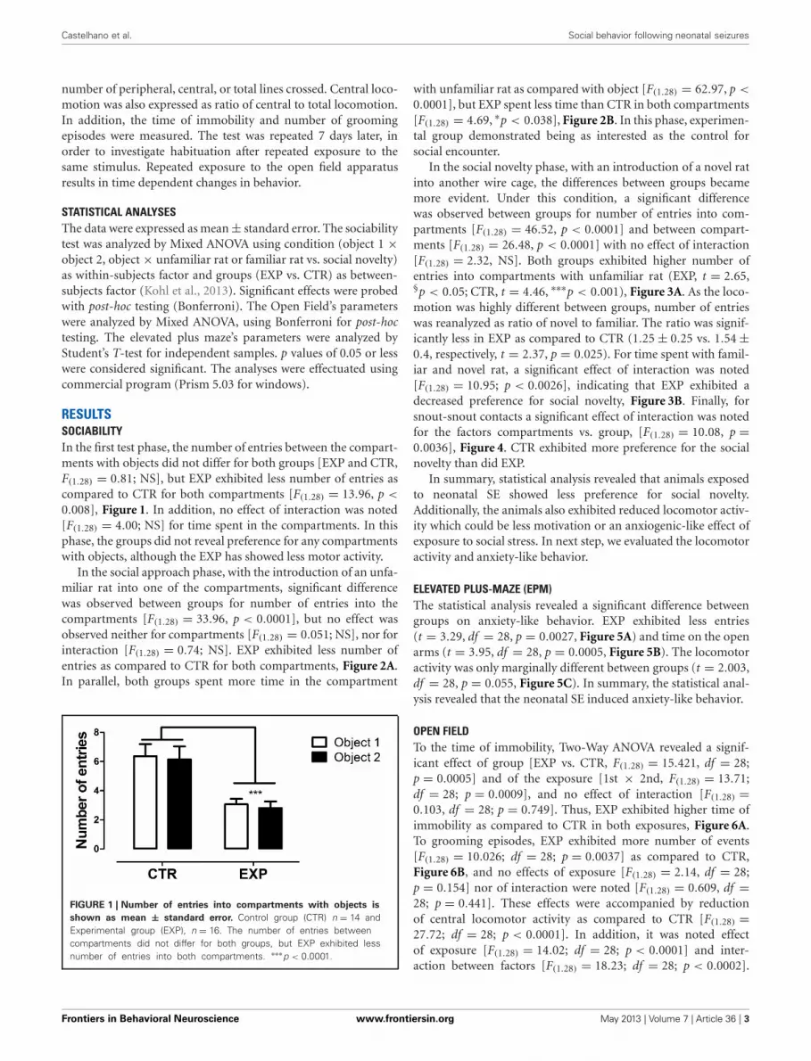
RESULTSSOCIABILITYIn the first test phase, the number of entries between the compart-ments with objects did not differ for both groups [EXP and CTR,F(1.28) = 0.81; NS], but EXP exhibited less number of entries ascompared to CTR for both compartments [F(1.28) = 13.96, p <
0.008], Figure 1. In addition, no effect of interaction was noted[F(1.28) = 4.00; NS] for time spent in the compartments. In thisphase, the groups did not reveal preference for any compartmentswith objects, although the EXP has showed less motor activity.
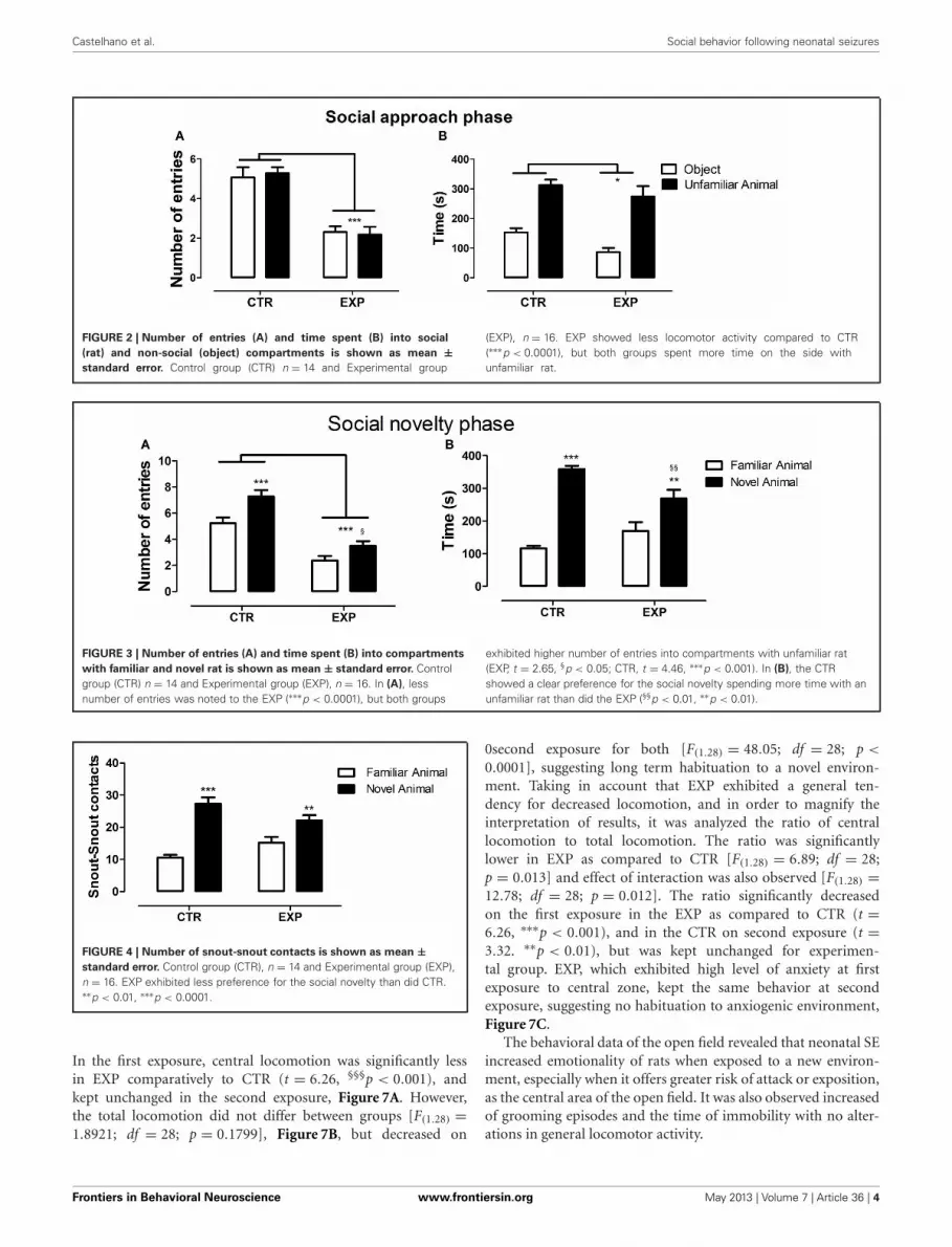
In the social approach phase, with the introduction of an unfa-miliar rat into one of the compartments, significant differencewas observed between groups for number of entries into thecompartments [F(1.28) = 33.96, p < 0.0001], but no effect wasobserved neither for compartments [F(1.28) = 0.051; NS], nor forinteraction [F(1.28) = 0.74; NS]. EXP exhibited less number ofentries as compared to CTR for both compartments, Figure 2A.In parallel, both groups spent more time in the compartment
FIGURE 1 | Number of entries into compartments with objects is
shown as mean ± standard error. Control group (CTR) n = 14 andExperimental group (EXP), n = 16. The number of entries betweencompartments did not differ for both groups, but EXP exhibited lessnumber of entries into both compartments. ∗∗∗p < 0.0001.
with unfamiliar rat as compared with object [F(1.28) = 62.97, p <
0.0001], but EXP spent less time than CTR in both compartments[F(1.28) = 4.69, ∗p < 0.038], Figure 2B. In this phase, experimen-tal group demonstrated being as interested as the control forsocial encounter.
In the social novelty phase, with an introduction of a novel ratinto another wire cage, the differences between groups becamemore evident. Under this condition, a significant differencewas observed between groups for number of entries into com-partments [F(1.28) = 46.52, p < 0.0001] and between compart-ments [F(1.28) = 26.48, p < 0.0001] with no effect of interaction[F(1.28) = 2.32, NS]. Both groups exhibited higher number ofentries into compartments with unfamiliar rat (EXP, t = 2.65,§p < 0.05; CTR, t = 4.46, ∗∗∗p < 0.001), Figure 3A. As the loco-motion was highly different between groups, number of entrieswas reanalyzed as ratio of novel to familiar. The ratio was signif-icantly less in EXP as compared to CTR (1.25 ± 0.25 vs. 1.54 ±0.4, respectively, t = 2.37, p = 0.025). For time spent with famil-iar and novel rat, a significant effect of interaction was noted[F(1.28) = 10.95; p < 0.0026], indicating that EXP exhibited adecreased preference for social novelty, Figure 3B. Finally, forsnout-snout contacts a significant effect of interaction was notedfor the factors compartments vs. group, [F(1.28) = 10.08, p =0.0036], Figure 4. CTR exhibited more preference for the socialnovelty than did EXP.
In summary, statistical analysis revealed that animals exposedto neonatal SE showed less preference for social novelty.Additionally, the animals also exhibited reduced locomotor activ-ity which could be less motivation or an anxiogenic-like effect ofexposure to social stress. In next step, we evaluated the locomotoractivity and anxiety-like behavior.
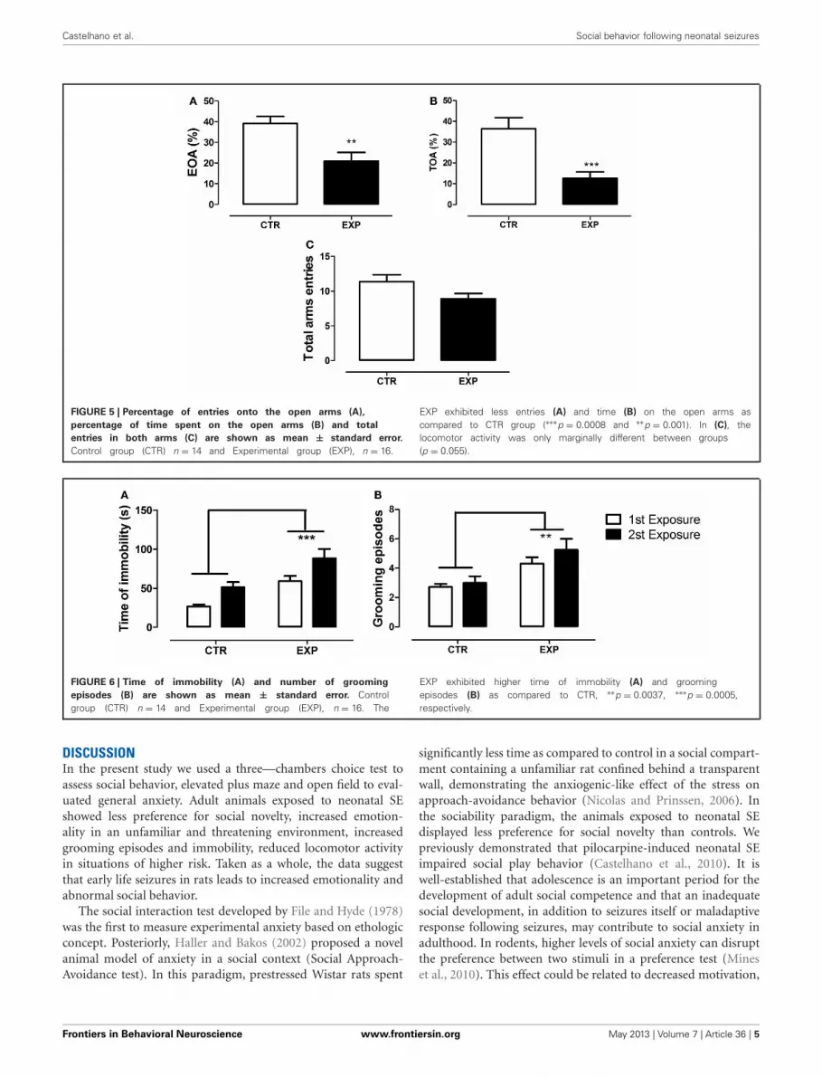
ELEVATED PLUS-MAZE (EPM)The statistical analysis revealed a significant difference betweengroups on anxiety-like behavior. EXP exhibited less entries(t = 3.29, df = 28, p = 0.0027, Figure 5A) and time on the openarms (t = 3.95, df = 28, p = 0.0005, Figure 5B). The locomotoractivity was only marginally different between groups (t = 2.003,df = 28, p = 0.055, Figure 5C). In summary, the statistical anal-ysis revealed that the neonatal SE induced anxiety-like behavior.
OPEN FIELDTo the time of immobility, Two-Way ANOVA revealed a signif-icant effect of group [EXP vs. CTR, F(1.28) = 15.421, df = 28;p = 0.0005] and of the exposure [1st × 2nd, F(1.28) = 13.71;df = 28; p = 0.0009], and no effect of interaction [F(1.28) =0.103, df = 28; p = 0.749]. Thus, EXP exhibited higher time ofimmobility as compared to CTR in both exposures, Figure 6A.To grooming episodes, EXP exhibited more number of events[F(1.28) = 10.026; df = 28; p = 0.0037] as compared to CTR,Figure 6B, and no effects of exposure [F(1.28) = 2.14, df = 28;p = 0.154] nor of interaction were noted [F(1.28) = 0.609, df =28; p = 0.441]. These effects were accompanied by reductionof central locomotor activity as compared to CTR [F(1.28) =27.72; df = 28; p < 0.0001]. In addition, it was noted effectof exposure [F(1.28) = 14.02; df = 28; p < 0.0001] and inter-action between factors [F(1.28) = 18.23; df = 28; p < 0.0002].
Frontiers in Behavioral Neuroscience www.frontiersin.org May 2013 | Volume 7 | Article 36 | 3
Castelhano et al. Social behavior following neonatal seizures
FIGURE 2 | Number of entries (A) and time spent (B) into social
(rat) and non-social (object) compartments is shown as mean ±standard error. Control group (CTR) n = 14 and Experimental group
(EXP), n = 16. EXP showed less locomotor activity compared to CTR(∗∗∗p < 0.0001), but both groups spent more time on the side withunfamiliar rat.
FIGURE 3 | Number of entries (A) and time spent (B) into compartments
with familiar and novel rat is shown as mean ± standard error. Controlgroup (CTR) n = 14 and Experimental group (EXP), n = 16. In (A), lessnumber of entries was noted to the EXP (∗∗∗p < 0.0001), but both groups
exhibited higher number of entries into compartments with unfamiliar rat(EXP, t = 2.65, §p < 0.05; CTR, t = 4.46, ∗∗∗p < 0.001). In (B), the CTRshowed a clear preference for the social novelty spending more time with anunfamiliar rat than did the EXP (§§p < 0.01, ∗∗p < 0.01).
FIGURE 4 | Number of snout-snout contacts is shown as mean ±standard error. Control group (CTR), n = 14 and Experimental group (EXP),n = 16. EXP exhibited less preference for the social novelty than did CTR.∗∗p < 0.01, ∗∗∗p < 0.0001.
In the first exposure, central locomotion was significantly lessin EXP comparatively to CTR (t = 6.26, §§§p < 0.001), andkept unchanged in the second exposure, Figure 7A. However,the total locomotion did not differ between groups [F(1.28) =1.8921; df = 28; p = 0.1799], Figure 7B, but decreased on
0second exposure for both [F(1.28) = 48.05; df = 28; p <
0.0001], suggesting long term habituation to a novel environ-ment. Taking in account that EXP exhibited a general ten-dency for decreased locomotion, and in order to magnify theinterpretation of results, it was analyzed the ratio of centrallocomotion to total locomotion. The ratio was significantlylower in EXP as compared to CTR [F(1.28) = 6.89; df = 28;p = 0.013] and effect of interaction was also observed [F(1.28) =12.78; df = 28; p = 0.012]. The ratio significantly decreasedon the first exposure in the EXP as compared to CTR (t =6.26, ∗∗∗p < 0.001), and in the CTR on second exposure (t =3.32. ∗∗p < 0.01), but was kept unchanged for experimen-tal group. EXP, which exhibited high level of anxiety at firstexposure to central zone, kept the same behavior at secondexposure, suggesting no habituation to anxiogenic environment,Figure 7C.
The behavioral data of the open field revealed that neonatal SEincreased emotionality of rats when exposed to a new environ-ment, especially when it offers greater risk of attack or exposition,as the central area of the open field. It was also observed increasedof grooming episodes and the time of immobility with no alter-ations in general locomotor activity.
Frontiers in Behavioral Neuroscience www.frontiersin.org May 2013 | Volume 7 | Article 36 | 4
Castelhano et al. Social behavior following neonatal seizures
FIGURE 5 | Percentage of entries onto the open arms (A),
percentage of time spent on the open arms (B) and total
entries in both arms (C) are shown as mean ± standard error.
Control group (CTR) n = 14 and Experimental group (EXP), n = 16.
EXP exhibited less entries (A) and time (B) on the open arms ascompared to CTR group (∗∗∗p = 0.0008 and ∗∗p = 0.001). In (C), thelocomotor activity was only marginally different between groups(p = 0.055).
FIGURE 6 | Time of immobility (A) and number of grooming
episodes (B) are shown as mean ± standard error. Controlgroup (CTR) n = 14 and Experimental group (EXP), n = 16. The
EXP exhibited higher time of immobility (A) and groomingepisodes (B) as compared to CTR, ∗∗p = 0.0037, ∗∗∗p = 0.0005,respectively.
DISCUSSIONIn the present study we used a three—chambers choice test toassess social behavior, elevated plus maze and open field to eval-uated general anxiety. Adult animals exposed to neonatal SEshowed less preference for social novelty, increased emotion-ality in an unfamiliar and threatening environment, increasedgrooming episodes and immobility, reduced locomotor activityin situations of higher risk. Taken as a whole, the data suggestthat early life seizures in rats leads to increased emotionality andabnormal social behavior.
The social interaction test developed by File and Hyde (1978)was the first to measure experimental anxiety based on ethologicconcept. Posteriorly, Haller and Bakos (2002) proposed a novelanimal model of anxiety in a social context (Social Approach-Avoidance test). In this paradigm, prestressed Wistar rats spent
significantly less time as compared to control in a social compart-ment containing a unfamiliar rat confined behind a transparentwall, demonstrating the anxiogenic-like effect of the stress onapproach-avoidance behavior (Nicolas and Prinssen, 2006). Inthe sociability paradigm, the animals exposed to neonatal SEdisplayed less preference for social novelty than controls. Wepreviously demonstrated that pilocarpine-induced neonatal SEimpaired social play behavior (Castelhano et al., 2010). It iswell-established that adolescence is an important period for thedevelopment of adult social competence and that an inadequatesocial development, in addition to seizures itself or maladaptiveresponse following seizures, may contribute to social anxiety inadulthood. In rodents, higher levels of social anxiety can disruptthe preference between two stimuli in a preference test (Mineset al., 2010). This effect could be related to decreased motivation,
Frontiers in Behavioral Neuroscience www.frontiersin.org May 2013 | Volume 7 | Article 36 | 5

Castelhano et al. Social behavior following neonatal seizures
FIGURE 7 | Central (A), total locomotion (B) and (C) ratio of central to
total locomotion are shown as mean ± standard error in both
exposures. Control group (CTR) n = 14 and Experimental group (EXP),n = 16. Reduced central locomotion was noted for EXP in both exposures.In CTR, the locomotor activity in central squares decreased significantly
during second exposure (A). The total locomotion did not differ betweengroups, but decreased significantly during 2nd exposure (B). In (C), the ratioof central to total locomotion was significantly less in EXP as compared toCTR, decreased on the 2nd exposure for CTR and was kept unchanged forEXP. ∗∗p < 0.001, ∗∗∗p < 0.0001 and §§§p < 0.0001.
increased emotionality or impaired social memory. Lin et al.(2009) reported in lithium-pilocarpine-induced neonatal seizuresreduced levels of dopamine in the PFC. It is well-establishedthat the dopamine found in the PFC originates from the ven-tral tegmental area, a structure involved in neural network ofsocial behaviors (Goodson and Kabelik, 2009) is also involvedin and emotional responses (Baskerville and Douglas, 2010). Inthis sense, dopamine reduction in PFC could lead to less engage-ment in social interactions. However, taking in account that insocial approach phase experimental group demonstrated beingas interested as the control for a social encounter, the proba-ble mechanisms to explain reduced preference for social noveltyare the increased state of anxiety associate with the deficit insocial discrimination. There is emerging in the literature a dis-cussion about the bi-directional relationship and possibly cyclic,between social interaction impairment and the degree of anxi-ety or emotionality. Anxiety intensifies social impairment, andpoor social function contributes to anxiety (Nicolas and Prinssen,2006; White et al., 2010). In this sense, animals were evalu-ated in the open field and the elevated plus-maze. Experimentalanimals avoided the central area of the open field in both expo-sures, suggesting no habituation to anxiogenic environment, butthey performed similarly to control in total locomotion suggest-ing no locomotor activity changes. In addition, they displayedhigher immobility and stereotyped behavior. In EPM, they spentless time and reduced number of entries on the open arms,suggesting the presence of general anxiety. Reduced locomo-tor activity on sociability test, even after the habituation period
and before social presence, suggest that the new environmentincreased the state of anxiety and that no habituation to anx-iogenic space was reached, as observed in the central zone ofthe open field. Rodents also have emotional behaviors simi-lar to humans when exposed to specific experimental situations(Ramos and Mormède, 1998). In this regard, Nicolas and Prinssen(2006) evaluated the social approach-avoidance behavior in ahigh-anxiety strain, F-344 rats. The animals exhibited sponta-neous avoidance behavior that was sensitive to benzodiazepineagonist and inverse agonist in a bidirectional manner. To thebest of our knowledge, we are the first group to demonstrateabnormal social behavior following early life seizures. However,general anxiety was previously reported. Corroborating with ourresults, Sayin et al. (2004) demonstrated anxiety-related behaviorwithout locomotor activity changes in adult animals submittedto SE by kainic acid injection during early stage of develop-ment. Oliveira et al. (2007) reported anxiety-like behavior in adultrats submitted to lithium-pilocarpine model at 15 days postna-tal. The enhanced emotionality could be associated to changesin hippocampus function. Lugo et al. (2012) and Benedikt et al.(2012) demonstrated that hippocampal damage leads to anxiety-like behavior. Beyond of the hippocampus, others brain regionsexhibit maladaptive response following neonatal seizures. Forexample, Kleen et al. (2011) observed that adults male Wistar ratsaged between 1 and 10 days postnatal that undergo a series toof neonatal seizures by fluorothyl administration exhibited struc-tural changes of the PFC that were positively correlated with theimpairment of cognitive flexibility and insistence to maintain a
Frontiers in Behavioral Neuroscience www.frontiersin.org May 2013 | Volume 7 | Article 36 | 6
Castelhano et al. Social behavior following neonatal seizures
routine previously learned. Individuals with autism often haverepetitive behaviors and strong resistance to the change of rou-tine. Failure to change routine in rodents may be analogous to thecognitive inflexibility. In stressful situations is common the emer-gence or intensification of stereotypes. In our study, we observedincrease in self-grooming when animals were exposed to an anx-iogenic environment. The association of grooming behavior withanxiety or stress is well-established in animal models. Stressfulevents or environments (Langen et al., 2011) as well as appeti-tive situations can cause an animal to develop abnormal repetitivebehavior (Spruijt et al., 1992; Van Erp et al., 1994). Additionally,the enhanced stereotyped behavior could be also due of imbalanceboth within and between the motor, cognitive and limbic cor-ticostriatal circuits. The corticostriatal circuits include striatum,globus pallidus, substancia nigra, thalamus, and cortex. It is well-established that corticostriatal circuits are functionally dividedinto three circuits related to the predominant cerebral corti-cal input to striatum: sensoriomotor circuit functionally relatedto movements (comprising the motor and oculomotor loops),associative circuit functionally related to cognitive functions (dor-solateral prefrontal loop) and the limbic circuit functionallyrelated emotional-motivational behavior (lateral orbitofrontaland anterior cingulate loops) (for an extensive review see; Langenet al., 2011). Abnormal repetition of behavior can result fromdamage to any of the corticostriatal circuits. Experimental datademonstrated that SE can damage brain regions involved withcorticostriatal circuit. Fernandes et al. (1999) observed hyper-metabolism in cortical and forebrain regions plus the substan-tia nigra following lithium–pilocarpine-induced SE in immaturerats. Kubova et al. (2001) demonstrated the presence of silver-positive cells predominantly in the mediodorsal, but also too inthe ventrolateral and ventromedial thalamic nucleus in rats thatexperienced pilocarpine-induced SE at the age of 12 days post-natal. Interestingly, the mediodorsal and ventrolateral nucleus of
thalamus are part of limbic and sensoriomotor circuit, respec-tively. Taking in account that stereotypies can function as copingmechanism to reduce animal’s arousal level when it is exposed tostressful events or environments (Langen et al., 2011), it is licitto suppose that animals with damaged thalamus nuclei could bemore prone to experience stereotypies under these situations.
As previously mentioned, the decreased preference for socialnovelty could be also associated to inability of animals exposedto neonatal SE to distinguish a familiar from an unfamiliar con-specifics. In support to this hypothesis, Sankar et al. (1998)reported in rats submitted to pilocarpine-induced seizures duringsecond week of life, damage in the amygdala and hippocam-pus, brain regions implicated in social recognition memory anemotionality (Ferguson et al., 2002; Yang et al., 2013). Thus, theabnormal social behavior noted in animals exposed to early lifeseizures could be related to high state of anxiety and impairedsocial memory.
Taken as a whole, adult animals exposed to neonatal SE dis-played less preference for social novelty than control, anxiety-related behavior, increased stereotyped behavior in anxiogenicenvironment without locomotor activity changes. The data sug-gest that neonatal SE in rodents leads to altered anxiety-relatedand abnormal social behaviors.
ACKNOWLEDGMENTSThis work was sponsored by grants from MACKPESQUISA,CInAPCe (Cooperação Interinstitucional de Apoio àPesquisa sobre o Cérebro)—FAPESP; CNPq (ConselhoNacional de Desenvolvimento Científico e Tecnológico); andFAPESP/CNPq/MCT (Instituto Nacional de NeurociênciaTranslacional). Adelisandra Silva Santos Castelhano and Gustavodos Santos Teada Cassane are fellows from Coordenação deAperfeiçoamento de Pessoal de Nível Superior (CAPES) andCNPq, respectively.
REFERENCESAtladóttir, H. O., Schendel, D. E.,
Lauritsen, M. B., Henriksen, T. B.,and Parner, E. T. (2012). Patternsof contact with hospital for chil-dren with an autism spectrumdisorder: a Danish register-basedstudy. J. Autism Dev. Disord. 42,1717–1728.
Baskerville, T. A., and Douglas, A.J. (2010). Dopamine and oxytocininteractions underlying behaviors:potential contributions to behav-ioral disorders. CNS Neurosci. Ther.16, 92–23.
Benedikt, J., Inyushin, M.,Kucheryavykh, Y. V., Rivera, Y.,Kucheryavykh, L. Y., Nichols, C.G., et al. (2012). Intracellularpolyamines enhance astrocyticcoupling. Neuroreport 5, 23,1021–1025.
Castelhano, A. S., Scorza, F. A., Teixeira,M. C., Arida, R. M., Cavalheiro, E.A., and Cysneiros, R. M. (2010).
Social play impairment followingstatus epilepticus during earlydevelopment. J. Neural Transmiss.117, 1155–1160.
Cornejo, B. J., Mesches, M. H.,Coultrap, S., Browning, M. D., andBenke, T. A. (2007). A single episodeof neonatal seizures permanentlyalters glutamatergic synapses. Ann.Neurol. 61, 411–426.
Costa, J. C., Nunes, M. L., and Fiori, R.M. (2001). Convulsões no períodoneonatal. J. Pediatr. 77, S1–S115.
Crawley, J. N. (2007). Mouse behavioralassays relevant to the symptoms ofautism. Brain Pathol. 17, 448–459.
da Silva, V. A., Regondi, M. C.,Cavalheiro, E. A., and Spreafico,R. (2005). Disruption of corti-cal development as a consequenceof repetitive pilocarpine-inducedStatus epilepticus in rats. Epilepsia46, 22–30.
Ferguson, J. N., Young, L. J., and Insel,T. R. (2002). The neuroendocrine
basis of social recognition. Front.Neuroendocrinol. 23:229. doi:10.1006/frne.2002.0229
Fernandes, C., González, M. I., Wilson,C. A., and File, S. E. (1999).Factor analysis shows that female ratbehaviour is characterized primar-ily by activity, male rats are drivenby sex and anxiety. Pharmacol.Biochem. Behav. 64, 731–738.
File, S. E., and Hyde, J. R. (1978). Cansocial interaction be used to mea-sure anxiety? Br. J. Pharmacol. 62,19–24.
Goodson, J. L., and Kabelik, D.(2009). Dynamic limbic net-works and social diversity invertebrates: from neural contextto neuromodulatory patterning.Front. Neuroendocrinol. 30:7. doi:10.1016/j.yfrne.2009.05.007
Haller, J., and Bakos, N. (2002). Stress-induced social avoidance: a newmodel of stress-induced anxiety?Physiol. Behav. 77, 327–332.
Holmes, G. L. (2004). Effects ofearly seizures on later behav-ior and epileptogenicity. Ment.Retard. Dev. Disabil. Res. Rev. 10,101–105.
Holmes, G. L. (2009). The long-termeffects of neonatal seizures. Clin.Perinatol. 36, 901–914.
Holmes, G. L., Gairsa, J. L., Chevassus-Au-Louis, N., and Ben-Ari, Y.(1998). Consequences of neonatalseizures in the rat: morphologicaland behavioral effects. Ann. Neurol.44, 845–857.
Holopainen, I. E. (2008). Seizures inthe developing brain: cellular andmolecular mechanisms of neuronaldamage, neurogenesis and cellularreorganization. Neurochem. Int. 52,935–947.
Isaeva, E., Isaev, D., Savrasova, A.,Khazipov, R., and Holmes, G. L.(2010). Recurrent neonatal seizuresresult in long-term increases in neu-ronal network excitability in the
Frontiers in Behavioral Neuroscience www.frontiersin.org May 2013 | Volume 7 | Article 36 | 7
Castelhano et al. Social behavior following neonatal seizures
rat neocortex. Eur. J. Neurosci. 31,1446–1455.
Kleen, J. K., Wu, E. X., Holmes,G. L., Scott, R. C., and Lenck-Santini, P. P. (2011). Enhanced oscil-latory activity in the hippocampal-prefrontal network is related toshort-term memory function afterearly-life seizures. J. Neurosci. 31,15397–15406.
Kohl, C., Riccio, O., Grosse, J.,Zanoletti, O., Fournier, C., Schmidt,M. V., et al. (2013). HippocampalNeuroligin-2 overexpressionleads to reduced aggression andinhibited novelty reactivity inrats. PLoS ONE 8:e56871. doi:10.1371/journal.pone.0056871
Kubová, H., Druga, R., Lukasiuk, K.,Suchomelová, L., Haugvicová, R.,Jirmanová, I., et al. (2001). Statusepilepticus causes necrotic dam-age in the mediodorsal nucleus ofthe thalamus in immature rats.J. Neurosci. 21, 3593–3599.
Langen, M., Kas, M. J., Staal, W. G.,van Engeland, H., and Durston, S.(2011). The neurobiology of repet-itive behavior: of mice. . . Neurosci.Biobehav. Rev. 35, 345–355.
Lin, T. C., Huang, L. T., Huang, Y.N., Chen, G. S., and Wang, J. Y.(2009). Neonatal Status epilepticusalters prefrontal-striatal circuitryand enhances methamphetamine-induced behavioral sensitizationin adolescence. Epilepsy Behav. 14,316–323.
Lowenstein, D. H. (2006). The man-agement of refractory Statusepilepticus: an update. Epilepsia 47,35–40.
Lowenstein, D. H., Bleck, T., andMacdonald, R. L. (1999). It’s time torevise the definition of status epilep-ticus. Epilepsia 40, 120–122.
Lugo, J. N., Brewster, A. L., Spencer,C. M., and Anderson, A. E.(2012). Kv4. 2 knockout mice havehippocampal-dependent learningand memory deficits. Learn. Mem.19, 182–189.
Lynch, M., Sayin, U., Golarai, G.,and Sutula, T. (2000). Long-termconsequences of early postnatalseizures on hippocampal learningand plasticity. Eur. J. Neurosci. 12,2252–2264.
Mines, M. A., Yuskaitis, C. J., King,M. K., Beurel, E., and Jope, R. S.
(2010). GSK3 influences social pref-erence and anxiety-related behav-iors during social interaction in amouse model of fragile X syndromeand autism. PLoS ONE 5:e9706. doi:10.1371/journal.pone.0009706
Mosley, M. (2010). Neonatal seizures.Pediatr. Rev. 31, 127–128.
Neill, J. C., Liu, Z., Mikati, M., andHolmes, G. L. (2005). Pilocarpineseizures cause age-dependentimpairment in auditory locationdiscrimination J. Exp. Anal. Behav.84, 357–350.
Nicolas, L. B., and Prinssen, E. P.(2006). Social approach-avoidancebehavior of a high-anxietystrain of rats: effects of ben-zodiazepine receptor ligands.Psychopharmacology (Berl.) 184,65–74.
Nishimura, M., Gu, X., and Swann, J.W. (2011). Seizures in early life sup-press hippocampal dendrite growthwhile impairing spatial learning.Neurobiol. Dis. 44, 205–214.
Novaes, G. F., Amado, D., Scorza, F.A., and Cysneiros, R. M. (2012).Social behavior impairment in off-spring exposed to maternal seizuresin utero. J. Neural Transm. 119,639–644.
Oliveira, M. R., Silvestrin, R. B., Melloe Souza, T., and Moreira, J. C.(2007). Oxidative stress in thehippocampus, anxiety-like behaviorand decreased locomotory andexploratory activity of adult rats:effects of sub acute vitamin A sup-plementation at therapeutic doses.Neurotoxicology 28, 1191–1199.
Ramos, A., and Mormède, P. (1998).Stress and emotionality: a multidi-mensional and genetic approach.Neurosci. Biobehav. Rev. 22,33–57.
Saliba, R. M., Annegers, J. F., andMizrahi, E. M. (1996). Incidence ofclinical neonatal seizures. Epilepsia37, 13.
Saliba, R. M., Annegers, J. F., Waller,D. K., Tyson, J. E., and Mizrahi,E. M. (1999). Incidence of neona-tal seizures in Harris County, Texas,1992–1994. Am. J. Epidemiol. 150,763–69.
Sankar, R., Shin, D. H., Liu,H., Mazarati, A., Pereira deVasconcelos, A., and Wasterlain,C. G. (1998). Patterns of status
epilepticus-induced neuronal injuryduring development and long-termconsequences. J. Neurosci. 18,8382–8393.
Santos, N. F., Marques, R. H., Correia,L., Sinigaglia-Coimbra, R.,Calderazzo, L., Sanabria, E. R.,et al. (2000). Multiple pilocarpine-induced status epilepticus indeveloping rats: a long-term behav-ioral and electrophysiological study.Epilepsia 41, S57–S63.
Sayin, U., Sutula, T. P., and Stafstrom,C. E. (2004). Seizures in thedeveloping brain causes adverselong-term effects on spatial learn-ing and anxiety. Epilepsia 45,1539–1548.
Schneider, M., and Koch, M. (2005).Deficient social and play behav-ior in juvenile and adult rats afterneonatal cortical lesion: effects ofchronic pubertal cannabinoid treat-ment. Neuropsychopharmacology 30,944–957.
Sheth, R. D., Hobbs, G. R., and Mullett,M. (1999). Neonatal seizures: inci-dence, onset, and etiology by gesta-tional age. J. Perinatol. 19, 40–43.
Shi, X. Y., Wang, J. W., Lei, G. F.,and Sun, R. P. (2007). Long-termeffects of recurrent seizures onlearning, behavior and anxiety: anexperimental study in rats. WorldJ. Pediatr. 3, 61–65.
Shinnar, S., Pellock, J. M., Moshé, S.L., Maytal, J., O’Dell, C., Driscoll,S. M., et al. (1997). In whom doesStatus epilepticus occur: age-relateddifferences in children. Epilepsia 38,907–914.
Sogawa, Y., Monokoshi, M., Silveira, D.C., Cha, B. H., Cilio, M. R., Mccabe,B. K., et al. (2001). Timing ofcognitive deficits following neona-tal seizures: relationship to histo-logical changes in the hippocam-pus. Brain Res. Dev. Brain Res. 131,73–83.
Spruijt, B. M., Van Hooff, J. A., andGispen, W. H. (1992). Ethologyand neurobiology of groom-ing behavior. Physiol. Rev. 72,825–852.
Tordjman, S., Drapier, D., Bonnot,O., Graignic, R., Fortes, S., Cohen,D., et al. (2007). Animal mod-els relevant to schizophrenia andautism: validity and limitations.Behav. Genet. 37, 61–78.
Udani, V. (2008). Long-term prognosisof neonatal seizures – where are we?Indian Pediatr. 45, 739–741.
Van Erp, A. M., Kruk, M. R., Meelis,W., and Willekens-Bramer, D. C.(1994). Effect of environmentalstressors on time course, variabilityand form of self-grooming in therat: handling, social contact, defeat,novelty, restraint and fur moisten-ing. Behav. Brain Res. 65, 47–55.
Villeneuve, N., Ben-Ari, Y., Holmes,G. L., and Gaiarsa, J. L. (2000).Neonatal seizures induced persis-tent changes in intrinsic propertiesof CA1 rat hippocampal cells. Ann.Neurol. 47, 729–738.
White, S. W., Albano, A. M., Johnson,C. R., Kasari, C., Ollendick, T.,Klin, A., et al. (2010). Developmentof a cognitive-behavioral inter-vention program to treat anxietyand social deficits in teens withhigh-functioning autism. Clin.Child Fam. Psychol. Rev. 13, 77–90.
Yang, L., Zou, B., Xiong, X., Pascual,C., Xie, J., Malik, A., et al. (2013).Hypocretin/Orexin neurons con-tribute to hippocampus-dependentsocial memory and synaptic plas-ticity in mice. J. Neurosci. 33,5275–5284.
Conflict of Interest Statement: Theauthors declare that the researchwas conducted in the absence of anycommercial or financial relationshipsthat could be construed as a potentialconflict of interest.
Received: 16 February 2013; accepted: 17April 2013; published online: 09 May2013.Citation: Castelhano ASS, CassaneGdST, Scorza FA and Cysneiros RM(2013) Altered anxiety-related andabnormal social behaviors in ratsexposed to early life seizures. Front.Behav. Neurosci. 7:36. doi: 10.3389/fnbeh.2013.00036Copyright © 2013 Castelhano, Cassane,Scorza and Cysneiros. This is an open-access article distributed under the termsof the Creative Commons AttributionLicense, which permits use, distributionand reproduction in other forums, pro-vided the original authors and sourceare credited and subject to any copy-right notices concerning any third-partygraphics etc.
Frontiers in Behavioral Neuroscience www.frontiersin.org May 2013 | Volume 7 | Article 36 | 8