Alteration in global motor strategy following lateral ankle sprain
12
RESEARCH ARTICLE Open Access Alteration in global motor strategy following lateral ankle sprain Maude Bastien 1,2 , Hélène Moffet 1,2* , Laurent J Bouyer 1,2 , Marc Perron 1,3 , Luc J Hébert 3 and Jean Leblond 2 Abstract Background: Lateral ankle sprain (LAS) has often been considered an injury leading to localized joint impairments affecting the musculoskeletal system. Persistent chronic ankle instability and bilateral alterations in motor control after a first ankle sprain episode suggest that the origin of relapses might be a maladaptive reorganization of central motor commands. The objectives of this study were (1) to compare the quality of motor control through motor strategy variables of two groups (with and without LAS) from a military population (n = 10/group), (2) to evaluate the contribution of the lower limbs and the trunk to global body strategy and (3) to identify which global variable best estimates performance on the Star Excursion Balance Test (SEBT) for each group, reaching direction, and lower limb. Methods: Personal and clinical characteristics of the participants of both groups were collected. Their functional ability was measured using questionnaires and they performed a series of functional tests including the SEBT. During this test, the maximal reach distance (MRD) and biomechanical data were collected to characterize whole body and segmental strategies using a 3D motion capture system. Results: At maximal lower limb reach, participants with LAS had a smaller variation in their vertical velocity in lowering-straightening and lowered the body centre of mass less for all injured limb conditions and some conditions with the uninjured lower limb. The global body centre of mass variables were significantly correlated to SEBT performance (MRD). Conclusion: Modifications in global motor strategies were found in participants with LAS as well as a decreased performance on the SEBT for the injured and uninjured lower limbs. These results support the hypothesis that following LAS, there may be a maladaptive reorganization of the central motor commands. Level of evidence: 3b. Keywords: SEBT, Motor control, Musculoskeletal disorders, Ankle sprain Background Lateral ankle sprain (LAS) is one of the most commonly reported sports injuries among athletes and military personnel [1-5]. The military is the most affected popu- lation with a five to eight times greater prevalence than civilians [5-7]. Up to 33% of the civilians experience chronic ankle instability even in the absence of persist- ent ligament laxity around the ankle joint, as well as recurrence of LAS over the years [8]. Deficits in the limb contralateral to the injury such as postural control change in single-limb stance and altered transverse plane kinematic profiles in jump landing, have also been reported [9-16]. Taken together, these deficits suggest that motor control impairments may be at the origin of the relapses. It is therefore important to better understand the pathophysi- ology and types of motor control deficits during the early recovery phase after LAS to prevent recurrence and prob- lems on the long term from which patients may not fully recovered such as chronic ankle instability [17,18]. The persistence of alterations in motor control for the injured and uninjured limbs strongly supports the hypothesis of a reorganization of central motor com- mands. LAS causes swelling, pain and other peripheral damage. This leads to altered sensory inputs, which trigger a reorganization in sensorimotor processing leading to long-term central modification in movement planning and execution [19-21]. Such a sensorimotor processing * Correspondence: [email protected] 1 Faculty of Medicine, Rehabilitation Department, Laval University, Quebec, QC, Canada 2 Centre for Interdisciplinary Research and Social Integration (CIRRIS), Quebec Rehabilitation Institute, 525 boulevard Hamel, G1M 2S8 Quebec, QC, Canada Full list of author information is available at the end of the article © 2014 Bastien et al.; licensee BioMed Central. This is an Open Access article distributed under the terms of the Creative Commons Attribution License (http://creativecommons.org/licenses/by/2.0), which permits unrestricted use, distribution, and reproduction in any medium, provided the original work is properly credited. The Creative Commons Public Domain Dedication waiver (http://creativecommons.org/publicdomain/zero/1.0/) applies to the data made available in this article, unless otherwise stated. Bastien et al. BMC Musculoskeletal Disorders 2014, 15:436 http://www.biomedcentral.com/1471-2474/15/436
Transcript of Alteration in global motor strategy following lateral ankle sprain

Bastien et al. BMC Musculoskeletal Disorders 2014, 15:436http://www.biomedcentral.com/1471-2474/15/436
RESEARCH ARTICLE Open Access
Alteration in global motor strategy followinglateral ankle sprainMaude Bastien1,2, Hélène Moffet1,2*, Laurent J Bouyer1,2, Marc Perron1,3, Luc J Hébert3 and Jean Leblond2
Abstract
Background: Lateral ankle sprain (LAS) has often been considered an injury leading to localized joint impairmentsaffecting the musculoskeletal system. Persistent chronic ankle instability and bilateral alterations in motor controlafter a first ankle sprain episode suggest that the origin of relapses might be a maladaptive reorganization of centralmotor commands. The objectives of this study were (1) to compare the quality of motor control through motorstrategy variables of two groups (with and without LAS) from a military population (n = 10/group), (2) to evaluate thecontribution of the lower limbs and the trunk to global body strategy and (3) to identify which global variable bestestimates performance on the Star Excursion Balance Test (SEBT) for each group, reaching direction, and lower limb.
Methods: Personal and clinical characteristics of the participants of both groups were collected. Their functional abilitywas measured using questionnaires and they performed a series of functional tests including the SEBT. During this test,the maximal reach distance (MRD) and biomechanical data were collected to characterize whole body and segmentalstrategies using a 3D motion capture system.
Results: At maximal lower limb reach, participants with LAS had a smaller variation in their vertical velocity inlowering-straightening and lowered the body centre of mass less for all injured limb conditions and some conditionswith the uninjured lower limb. The global body centre of mass variables were significantly correlated to SEBTperformance (MRD).
Conclusion: Modifications in global motor strategies were found in participants with LAS as well as a decreasedperformance on the SEBT for the injured and uninjured lower limbs. These results support the hypothesis thatfollowing LAS, there may be a maladaptive reorganization of the central motor commands. Level of evidence: 3b.
Keywords: SEBT, Motor control, Musculoskeletal disorders, Ankle sprain
BackgroundLateral ankle sprain (LAS) is one of the most commonlyreported sports injuries among athletes and militarypersonnel [1-5]. The military is the most affected popu-lation with a five to eight times greater prevalence thancivilians [5-7]. Up to 33% of the civilians experiencechronic ankle instability even in the absence of persist-ent ligament laxity around the ankle joint, as well asrecurrence of LAS over the years [8]. Deficits in thelimb contralateral to the injury such as postural controlchange in single-limb stance and altered transverse plane
* Correspondence: [email protected] of Medicine, Rehabilitation Department, Laval University, Quebec,QC, Canada2Centre for Interdisciplinary Research and Social Integration (CIRRIS), QuebecRehabilitation Institute, 525 boulevard Hamel, G1M 2S8 Quebec, QC, CanadaFull list of author information is available at the end of the article
© 2014 Bastien et al.; licensee BioMed CentralCommons Attribution License (http://creativecreproduction in any medium, provided the orDedication waiver (http://creativecommons.orunless otherwise stated.
kinematic profiles in jump landing, have also been reported[9-16]. Taken together, these deficits suggest that motorcontrol impairments may be at the origin of the relapses. Itis therefore important to better understand the pathophysi-ology and types of motor control deficits during the earlyrecovery phase after LAS to prevent recurrence and prob-lems on the long term from which patients may not fullyrecovered such as chronic ankle instability [17,18].The persistence of alterations in motor control for
the injured and uninjured limbs strongly supports thehypothesis of a reorganization of central motor com-mands. LAS causes swelling, pain and other peripheraldamage. This leads to altered sensory inputs, which triggera reorganization in sensorimotor processing leading tolong-term central modification in movement planningand execution [19-21]. Such a sensorimotor processing
. This is an Open Access article distributed under the terms of the Creativeommons.org/licenses/by/2.0), which permits unrestricted use, distribution, andiginal work is properly credited. The Creative Commons Public Domaing/publicdomain/zero/1.0/) applies to the data made available in this article,

Bastien et al. BMC Musculoskeletal Disorders 2014, 15:436 Page 2 of 12http://www.biomedcentral.com/1471-2474/15/436
deficit could explain the bilateral effects observed inparticipants with chronic ankle instability. Regardingpotential neural control mechanisms that could bealtered after LAS, it is known that individuals withchronic ankle instability have impaired sensorimotorfunction [11,13,22-26], and differences in segmentalmotor strategies during walking, running and jumplanding [26-31].In the present study, we were interested in looking
at the changes in motor control strategies after LASduring a standardized motor task, as such changes giveinsight on the quality of motor control of the targetpopulation. To address this question, healthy participantsand patients with LAS were tested in a challenging, yetstandardized motor task that involves the coordinationof multiple limb segments as well as good motor planning,i.e. that requires an optimal motor control [32-35].The Star Excursion Balance Test (SEBT) was therefore
selected. It has good metrological properties and hasbeen frequently used to study motor control in athletes[9,36-46]. The SEBT is defined by Gribble et al. [46] as“a series of single-limb squats using the nonstance limbto reach maximally to touch a point along 1 of 8 desig-nated lines on the ground. The lines are arranged in agrid that extends from a center point and are 45° fromone another. Each reaching direction offers differentchallenges and requires combinations of sagittal, frontal,and transverse movements” [46]. Maximal reach dis-tance is the variable usually used to characterize per-formance and to identify alterations in motor control.This variable reflects end-point control of the self-
organization of movement. However, no study has usedwhole body kinematics to look at the global organizationof movement contributing to the good performancesobserved on the SEBT. Such measures could provideadditional information on SEBT performances and helpidentify where exactly, in the continuum of global motorstrategies, the bilateral alterations of motor control takeplace. To date, only segmental strategies at the lowerlimb in stance have been described [47-49]. As motorcontrol involves the ability to control the body centre ofmass (CoM) over a base of support through appropriatecoordination between segments [50-52], it seems im-portant to also study its behaviour. The body CoM waschosen instead of body centre of pressure (CoP) sinceCoM takes into account all segmental strategies at thelower limbs, head and trunk and represents their sum-mation in 3D coordinates. On the contrary, CoP com-monly used in previous studies [11,14,53], is a biplanarvariable with some limitation to represent 3D behaviorsduring a complex task. Therefore, in the present study,global body strategy was estimated by quantifying globalbody CoM 3D behaviour to appreciate the quality ofmotor control during the SEBT.
ObjectivesThe objectives of this study were:
(1) to compare the quality of motor control in twogroups of military personnel (with and withoutLAS) on the SEBT, using the selected global andsegmental body strategy variable;
(2) to evaluate the contribution of the lower limbs andtrunk to global body strategy for each group ofparticipants and for each reaching direction; and
(3) to identify which global variable used to quantifyglobal body CoM behaviour best estimatesperformance on the SEBT for each group, reachingdirection, and lower limb.
MethodsParticipant selectionAll volunteers participating in this study were selectedfrom the Canadian Forces (CF) military population. TheLAS group was composed of 10 men with a diagnosis ofacute unilateral LAS [18]. They all received physiother-apy interventions within five days of injury and weredischarged after a maximum of nine weeks. They wereexcluded if they had an ankle fracture (documented byX-ray), a third grade LAS or a high ankle sprain (tibio-fibular sprain) [54-57], or if they reported a historyof neuromuscular or neurodegenerative disease. Thecontrol group consisted of a comparable group of 10men without prior history of LAS or symptoms fromthe lumbar spine and the lower limbs, or a historyof neuromuscular and neurodegenerative disease. Thepresent study was approved by the ethics committees ofthe Quebec Rehabilitation Institute and the CF HealthServices Group. All participants read and signed aninformed consent form.
Study designIn this descriptive study, all participants were evaluatedwithin a single session. Participants in the LAS groupwere selected in acute stage (within two days after in-jury) and were evaluated between eight and ten weeksafter their injury. Personal and clinical characteristics ofthe participants were collected. Their functional abilitywas measured using two questionnaires: the Foot &Ankle Disability Index (FADI) [58-60] and the LowerExtremity Function Scale (LEFS) [61]. Thereafter, they tookpart in a series of complex motor tasks and tests, wherethe testing order was kept the same for all participants.
SEBT procedureDuring the SEBT, the participant had to: (1) touch thefloor as far as possible with the tip of the foot of thereaching limb in three different directions with respectto the stance limb (anteromedial [AM], medial [M] and

Bastien et al. BMC Musculoskeletal Disorders 2014, 15:436 Page 3 of 12http://www.biomedcentral.com/1471-2474/15/436
posteromedial [PM] directions) and (2) return to unipedalstance after each reaching movement while maintainingbalance on the stance limb with the hands resting on theiliac crests. The AM, M and PM directions were the onesproposed by Hertel et al. [62] because these directionswere able to identify significant reach deficits associatedwith chronic ankle instability and provided complemen-tary and non-redundant information. After a practicesession [37,63] (six trials/direction followed by a five-minute rest period), three successful trials, separated bya ten-second rest period, were recorded for each direc-tion (Figure 1). Trials were rejected according to thecriteria used in previous studies [62,64,65] (1) weight-bearing by the reaching limb, (2) displacement of thestance limb, or (3) loss of balance. As shown in Figure 1,the directions to be followed by the reaching limb dur-ing the tests were clearly marked on the floor usinggraduated tape. The order of test conditions (two legsand three directions) was determined as follows: first theleg order was randomly selected, then a test direction wasrandomly determined. All conditions for one leg werecompleted starting with the determined direction followedby the next anticlockwise direction (right leg) or clockwisedirection (left leg). The same condition order was thenused for the second leg.
Data collectionDuring the SEBT, biomechanical data were collected tocharacterize both whole body and segmental strategiesusing a 3D motion capture system (Optotrak 3020;Northern Digital Inc., Waterloo, Ontario, Canada). Forty-five infrared markers placed in non-colinear triads were in-stalled on the following body segments: lower limbs, head,and trunk. For uppers limbs, rings of three markers wereplaced around the wrists. Kinematic data was sampledat 50 Hz and then digitally filtered at 10Hz (low passfilter). Global body CoM (CoMgl) was estimated usingthe equations proposed by Winter [66,67], and adjustedto consider the influence of the upper limb CoMa.
Data analysisAll individual kinematic data analyses were based on amean of three trials per test condition. A mean of twotrials was exceptionally used in a very few cases (5 out of120 conditions). The maximal reach distance (MRD) onthe SEBT was calculated from the horizontal plane coor-dinates (vectorial distance) of the tip of the reaching foot(probed point) at the time it touched the floor.
Key variables used to characterize motor controlThe SEBT task was divided into two main subtasks: thegoing-to- and the return-from-MRD (Figure 1A). The tran-sition between these subtasks has been called the centralphase and was defined as -1 s to +1 s after foot contact
(Figure 1: central grey rectangle). Four variables describingthe behaviour of the CoMgl, called global variables, havebeen chosen as key indicators of global body strategyduring this task. The maximal displacement of theCoMgl vertically; peak-to-peak CoMgl velocities alongthe vertical axis were calculated during the centralphase (Figure 2A); distance between the centre of thefoot and the mean position of the horizontal CoMglwhen the foot contacted the floor and the horizontalexcursion of the CoMgl during the central phase werealso calculated (Figure 2C).In addition, the following variables representing the
limb and trunk strategies during the task were quanti-fied: maximal trunk lowering relative to the pelvis, max-imal pelvis lowering, and magnitude of hip abduction ofthe reaching limb. Stance limb strategy was further ana-lysed using the maximal peak of flexion of three joints(hip, knee and ankle) as well as their angular velocity inthe sagittal plane (peak to peak value). Finally, for thereaching limb, angular velocity in the frontal plane atthe hip was also computed.
Statistical analysesAll statistical analyses (α = 0.05) were conducted usingSPSS for Windows version 12.0. Parametric and de-scriptive analyses were conducted to meet the researchobjectives. Non-parametric tests (Mann–Whitney exacttest) were used for the comparison of personal charac-teristics between groups. Descriptive statistics werecalculated from the global strategy variables (Objective 1).Multivariate analyses were used to measure group andlimb effects on global and segmental strategy variables.No results of univariate ANOVA were reported if themultivariate statistics were not found statistically sig-nificant (Objective 1). The horizontal excursion of theCoMgl was analysed using graphical representations(Objective 1). Mean difference (MD) and 95% confi-dence interval (CI) were calculated for statistically sig-nificant results. Regression analyses were carried out todetermine which combination of two global variablesalong the vertical axis was providing the best estimateof SEBT performance for each test condition (usingstandardized Beta coefficients [β]) (Objective 3).As the majority of the LAS participants (70%) had in-
jured their dominant limb, performance and strategyvariables in all test conditions of the LAS participantswere compared, for the second research objective, to thoseobtained from the dominant limb of the healthy group. Abar graph representing all regression coefficient combina-tions was used to compare each component’s contri-butions to global lowering for each reaching direction (seeFigure 3, Objective 2). Peak-to-peak angular velocities inthe stance limb were compared through multivariate ana-lyses to measure the group and limb effects (Objective 1).

A
B
C
D
Figure 1 (See legend on next page.)
Bastien et al. BMC Musculoskeletal Disorders 2014, 15:436 Page 4 of 12http://www.biomedcentral.com/1471-2474/15/436

(See figure on previous page.)Figure 1 Kinematics variables during the SEBT task, experimental setting and description of subtasks and events. Illustration of the sixconditions at the SEBT (3 reaching directions per limb; AM: anteromedial, M: medial, PM: posteromedial) and profiles (mean ± 1 standard deviation;n = 3 trials) of the vertical position of the reaching foot (A), global body CoM (CoMgl) (B and C) and joint amplitude of motion of the stance limb(D) in a typical healthy subject in the PM reaching direction. The central grey rectangle, called the central phase around foot contact, represents thecritical period used for the analyses. The lowest vertical position of the tip of the reaching foot was used to subdivide the task, which corresponds tothe “foot contact” event (vertical line at 50% of the task). The first subtask is characterized by body lowering (B and D) and alignment of the reachinglimb for foot contact (A). In the second subtask which occurred after foot contact, a rapid straightening up of the entire body (B and C) is performedwhile returning from perturbation in single-limb stance. The transition between subtasks represents a highly challenging period for stability and motorcontrol as a change in movement direction takes place at the perceived limits of stability. This transition period, called the central phase, was definedas -1 s to +1 s after foot contact. Further analysis about the quality of motor control was performed during this phase.
Bastien et al. BMC Musculoskeletal Disorders 2014, 15:436 Page 5 of 12http://www.biomedcentral.com/1471-2474/15/436
The hip abduction velocity of the reaching limb wasanalysed at three times of measure (25, 50 and 75% ofthe central phase duration) to look for potential groupeffects for the injured and the uninjured limbs (repeatedmeasured ANOVA) (Objective 1).
A
C
Figure 2 Global body strategies along the vertical axis (A) and horizolowering (#) and for the peak-to peak velocity of the global CoM (# #) are ifor healthy group and mean for LAS group; n = 10 trials per group) during thedeviation) for global CoM lowering and peak-to peak of CoM vertical velocityduring the different conditions (directions and limbs) in both groups. Horizonwere calculated and used for further statistical analyses. D: mean values (+1 stfigures B and D represent a significant difference between groups (MANOVA;
ResultsParticipants in both healthy and LAS groups were similarin age (means ± 1SD: 26.1 ± 5.1 years and 26.3 ± 6.9 years,respectively), height, weight and lower limbs length. TheLAS group had, however, a lower level of functional ability
B
D
ntal plane (C) in the central phase. A: the profiles for global CoMllustrated for one limb of both groups (mean ± 1 standard deviationmedial reaching direction on the SEBT. B: mean values (+1 standardof both groups. C: CoM displacement in the horizontal plane (# # #)tal resultant lines of global CoM position at foot contact (doted circles)andard deviation) for global CoM in the horizontal plane. Asterisks inp < 0.05; n = 20 limbs per group).
A
C
B
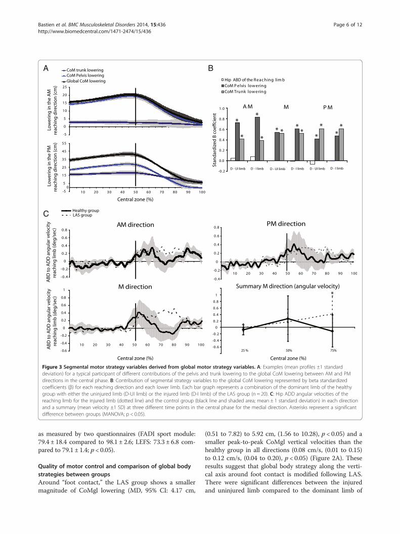
Figure 3 Segmental motor strategy variables derived from global motor strategy variables. A: Examples (mean profiles ±1 standarddeviation) for a typical participant of different contributions of the pelvis and trunk lowering to the global CoM lowering between AM and PMdirections in the central phase. B: Contribution of segmental strategy variables to the global CoM lowering represented by beta standardizedcoefficients (β) for each reaching direction and each lower limb. Each bar graph represents a combination of the dominant limb of the healthygroup with either the uninjured limb (D-UI limb) or the injured limb (D-I limb) of the LAS group (n = 20). C: Hip ADD angular velocities of thereaching limb for the injured limb (dotted line) and the control group (black line and shaded area; mean ± 1 standard deviation) in each directionand a summary (mean velocity ±1 SD) at three different time points in the central phase for the medial direction. Asterisks represent a significantdifference between groups (MANOVA; p < 0.05).
Bastien et al. BMC Musculoskeletal Disorders 2014, 15:436 Page 6 of 12http://www.biomedcentral.com/1471-2474/15/436
as measured by two questionnaires (FADI sport module:79.4 ± 18.4 compared to 98.1 ± 2.6; LEFS: 73.3 ± 6.8 com-pared to 79.1 ± 1.4; p < 0.05).
Quality of motor control and comparison of global bodystrategies between groupsAround “foot contact,” the LAS group shows a smallermagnitude of CoMgl lowering (MD, 95% CI: 4.17 cm,
(0.51 to 7.82) to 5.92 cm, (1.56 to 10.28), p < 0.05) and asmaller peak-to-peak CoMgl vertical velocities than thehealthy group in all directions (0.08 cm/s, (0.01 to 0.15)to 0.12 cm/s, (0.04 to 0.20), p < 0.05) (Figure 2A). Theseresults suggest that global body strategy along the verti-cal axis around foot contact is modified following LAS.There were significant differences between the injuredand uninjured limb compared to the dominant limb of

Bastien et al. BMC Musculoskeletal Disorders 2014, 15:436 Page 7 of 12http://www.biomedcentral.com/1471-2474/15/436
the healthy group for the CoMgl lowering variable in AMand in PM directions (MD, (95% CI): 4.57 cm, (0.85 to8.28) to 7.18 cm (0.88 to 13.48), p value <0.05) (Figure 4B).The healthy group performed better at SEBT in all direc-tions (Figure 4A), as measured by the MRD, except for theuninjured limb in the M direction (MD, (95% CI): 3.13%of height, (0.87 to 5.40) to 4.56% of height, (1.47 to 7.65),p value <0.05). They also significantly lowered their pelvismore and flexed the knee of the stance limb more thanparticipants with LAS except for the uninjured limb in theM direction (see* = p <0.05; Figure 4C-D). The differencein maximal knee flexion between groups for the injuredand dominant limb varied from 11.92° (95% CI: 0.98 to22.85) (M direction) to 20.20° (95% CI: 5.72 to 34.69) (AMdirection; Figure 4D). No difference across groups wasfound for other variables such as the maximal amplitudeof ankle dorsiflexion (except in the AM direction, MD,(95% CI): 6.11°, (0.03 to 12.18)), hip flexion or relativetrunk lowering.A more detailed analysis of the peak-to-peak of CoMgl
vertical velocity showed that only the injured limb wassignificantly different from the healthy group (ranges:healthy group 0.44 to 0.53 cm/s, LAS group 0.36 to0.46 cm/s). Although no significant difference in peak-to-peak angular velocity at the stance limb joints wasdetected except for the ankle of the injured limb in PM
A
B
Figure 4 Comparison of SEBT performance and motor strategies variauninjured) of the LAS group and the dominant limb of the healthy grper direction) and 1 standard deviation. Significant differences between groSEBT performance (maximal reach distance) expressed in percentage of the bmotor strategy: Pelvis lowering and D: Segmental motor strategy: Maximal kn
direction, a tendency for larger values in the healthy groupcompared to the LAS was observed. The hip adductionvelocity for the reaching limb of the LAS group was differ-ent from the healthy group in the central phase(Figure 3C). As shown in Figure 3, the LAS groupbrought back the reaching limb at a more constant vel-ocity, as opposed to the healthy group, which used abell-curve velocity profile before and after foot contact.No significant difference statistically supported this differ-ence although this tendency except in the case of the in-jured limb in M direction (p = 0.04).In the horizontal plane, during the central phase,
CoMgl was progressively moving from a more centredposition according to the stance foot to the perceivedlimit of stability, which is characterized by a U shape(Figure 2C). A descriptive analysis of the profiles of bothgroups highlights similarities in the general pattern ofCoMgl displacement but also differences in the magni-tude of the horizontal CoMgl resultant line. Indeed, forboth groups, the projection of the CoMgl was located inthe anterior and lateral section of the stance foot. Thisdistance was significantly longer in the healthy groupwhen compared to the LAS group (healthy group, 5.91 ±1.38 cm; LAS group, 4.59 ± 2.1 cm; MD, (95% CI):1.31 cm, (0.21 to 2.42), p < 0.05) in the AM reaching dir-ection (Figure 2D). However, not shown in these figures
C
D
bles associated with CoMgl lowering between limbs (injured andoup for each direction. Bar graphs represent the mean (n = 10 limbsups are identified by asterisk (MANOVA test; *p <0.05; # p = 0.052). A:ody height. B: Global motor strategy: Global CoM lowering, C: Segmentalee flexion.

Bastien et al. BMC Musculoskeletal Disorders 2014, 15:436 Page 8 of 12http://www.biomedcentral.com/1471-2474/15/436
were also significant differences between the injured andthe uninjured limb compared to the dominant limb of thehealthy group for the same direction (p value <0.05). Nodifferences were found in the other directions. The mag-nitude of the horizontal CoMgl resultant lines variedaccording to target direction in the healthy group, whileno such variation was observed in the LAS group.
Contribution of the lower limbs and trunk to global bodystrategyThe contribution of the trunk and pelvis to CoMgl lower-ing on the SEBT task varied according to reaching direc-tion (Figure 3A). The trunk contributed less CoMgllowering than the lower limbs (as estimated by the pelvislowering) in the AM direction but the contrary was seen inthe PM direction. Pelvis and trunk lowering both signifi-cantly predicted CoMgl lowering for all directions andlimbs. The β, represented by bars in Figure 3, was higherthan 0.38 in all conditions (Figure 3B). For example, ahigher β was found for the pelvis (0.82) compared to thetrunk (0.38) in the AM direction (combination of thedominant and injured limbs). The opposite finding wasobserved in the PM direction (β: Pelvis, 0.47 and Trunk,0.60). In contrast to trunk and pelvis, the magnitude ofhip abduction of the reaching limb did not seem tocontribute significantly to CoMgl lowering (Figure 3B,white bar). Finally, both segmental components seemedto have a similar contribution to CoMgl lowering in theM reaching direction.
Combination of global body strategy variables that bestpredict the SEBT performanceThe statistical models that best estimated SEBT per-formance were composed of a unique variable or a com-bination of a maximum of two variables (p < 0.05). Ingeneral, performance on the SEBT was best estimated bythe magnitude of CoMgl lowering alone (8 out of the 12conditions: 2 groups*2 limbs*3 directions; standardizedBeta coefficient (β) > 0.54) or in combination with peak-to-peak vertical CoMgl velocity as the second predictor(2 conditions: the injured limb in AM direction [β for thelowering = 0.71 and the velocity = 0.52]; and the dominantlimb in PM direction [β for the lowering = 0.46 and thevelocity = 0.55]). Performance on the SEBT was best esti-mated using only the peak-to-peak vertical CoMgl velocityvariable when the test was performed in AM direction withthe uninjured or the dominant limb (β > 0.65) (Table 1).
DiscussionThis study is the first to identify global motor strategyvariables associated with performance on the SEBT. Fur-thermore, the results of this study demonstrate thatmilitary personnel with LAS do not use the same globalstrategy as healthy controls when performing a goal
oriented task such as the SEBT. Their global motor strat-egy differed in almost all conditions (except for the unin-jured limb in the medial direction), suggesting that thestrategy is not only linked to the conditions where the in-jured limb is in stance, but also when the uninjured limbis used. This difference in global strategy is characterizedby less lowering of the CoMgl and less variation in its ver-tical velocity. This finding highlights the importance ofhaving a sound global motor strategy in order to performwell during the test and it also suggests that every effortshould be made during rehabilitation to help recover sucha strategy.
Global motor strategy differencesAccording to our results, the global strategy variablesalong the vertical axis seem to be very good indicatorsof a subject’s performance on the SEBT. The chosenglobal strategy variables, derived from body CoM dis-placement, helped to characterize performance on theSEBT in terms of motor control abilities. Our findingsalso support the construct validity of the SEBT thatmaximal reach distance, as commonly measured in clin-ical setting, provides a valid estimate of global motorcontrol in the studied population. The body CoM be-haviour improved knowledge of how participants wereorganizing their movements during the critical periodof stability at foot contact (central phase), allowing thetransition between the two subtasks.As supported by a systematic review and original works,
performance on different motor tasks is often altered bilat-erally after LAS [9-13] even in acute LAS stage [14,15].These differences in performance between groups couldindicate a change in motor strategies as observed in thecurrent study. The strategy used by the healthy group,leading to better performance, can be supported by bio-mechanical advantages and constraints. First, a lowervertical position of the CoMgl allows a better control ofCoMgl horizontal displacement by increasing overallbody stability. Indeed, a smaller lever arm in an invertedpendulum decreases muscle force required to maintainbalance. Moreover, a lower body CoMgl position inunipedal stance increases the projection of the reachinglimb by a better orientation of the pelvis, and it alsoincreases the effect of the trunk CoM to counteract theperturbation. Thus, by a greater lowering of the CoMgl,the physical performance is optimized but the motorcontrol requirement is much higher as the person needsto simultaneously control more body segments.Secondly, the group with LAS demonstrated a more
cautious approach near foot contact compared to theirpeers without LAS, as shown by a smaller peak-to-peakvelocity of the body CoM. This finding suggests thatLAS subjects are planning the transition between thesubtasks more cautiously as they try to reduce body

Table 1 Associations between performances on the SEBT and global strategy variables derived from CoMgl in a singlemodel*
Group Reachingdirection
Lower limb Global strategy variables(global CoM in vertical plane)
Modeladjusted R2
p value β Partialcorrelationa
Healthy group
AM Dominant Peak-to-peak velocity (cm/s)b 0.68 0.012 0.71 -
Non dominant Displacement (cm)c 0.46 0.044 0.60 -
M Dominant Displacement (cm) 0.81 0.004 0.80 -
Non dominant 0.63 0.039 0.54 -
PM Dominant Displacement (cm) 0.77 0.003 0.71 0.86
Peak-to-peak velocity (cm/s) 0.014 0.52 0.78
Non dominant Displacement (cm) 0.81 0.002 0.94 -
LAS group
AM Uninjured Peak-to-peak velocity (cm/s) 0.68 0.019 0.66 -
Injured Displacement (cm) 0.85 0.049 0.45 0.67
Peak-to-peak velocity (cm/s) 0.023 0.55 0.74
M Uninjured Displacement (cm) 0.78 0.022 0.59 -
Injured 0.45 0.023 0.72 -
PM Uninjured Displacement (cm) 0.74 0.007 0.71 -
Injured 0.54 0.009 0.81 -
*Only significant combination of variables are shown.aCalculated when two strategy variables were retained.bGlobal CoM vertical velocity (Peak-to-peak value).cGlobal CoM lowering (Maximum value).
Bastien et al. BMC Musculoskeletal Disorders 2014, 15:436 Page 9 of 12http://www.biomedcentral.com/1471-2474/15/436
velocity around this event. After foot contact, a lowervelocity of straightening up may allow more time toadjust for the internal perturbation and therefore en-hance stability. Several studies have shown that a quickdisplacement of a body segment away from the body canefficiently counteract a loss of CoM control [68,69]. Inthe present study, the delay in straightening up after footcontact has been attributed to the maintenance of thelower limb away from the body. Finally, global strat-egies in the horizontal plane also seemed to differ be-tween groups. These variables can show changes inmotor control because they give an idea of how far theparticipant pushed CoMgl to the perceived limits ofstability, which seemed to be further in the healthygroup. Moreover, the horizontal excursion of CoMglseemed to have different characteristics according toreaching directions in the healthy group compared tothe LAS group. Indeed, the LAS group showed a moregrouped horizontal excursion of CoMgl for differentreaching directions. This may be the result of lessadaptability of the central nervous system for the groupwith alterations of motor control (LAS group) as previ-ously observed for static postural control [70].
Segmental motor strategy differencesThe difference in global strategy of the military personnelwith LAS was mainly explained by changes in segmentalmotor strategy such as a decrease in pelvis loweringduring the task and a more cautious bringing back of
the reaching limb. First, the LAS group, which showedthe lowest performance at SEBT, seemed to lower thepelvis less by flexing the knee less, and sometimes alsothe ankle, compared to the healthy group. Other studieshave also reported a decreased range of motion at theknee during poor performance at SEBT [45,49,71]. Inthe LAS group a significant decrease of maximal ankledorsiflexion was found near foot contact for the AMdirection only. Interestingly, it is indeed in the AMdirection that the largest amplitude of ankle dorsiflex-ion is required [72].Secondly, the lower peak-to-peak vertical velocity of
CoMgl for the LAS group seemed to be mostly ex-plained by a more constant velocity of the reaching limbafter the foot contact and a delayed straightening up.Indeed, this smaller variation of vertical velocity in theLAS group could be partially explained by a cautiousreturn of the reaching limb, which may help to prolongthe stability advantages related to a smaller lever arm inthe inverted pendulum. During the return-from-MRDsubtask, the return to baseline velocity for hip abduc-tion was delayed for all conditions of the injured limb ofthe LAS group. A more constant velocity when bringingback the reaching limb towards the body allows time toadjust the movement to preserve stability, diminishesthe magnitude of the internal perturbation after a crit-ical stability period, and also allows the use of the reach-ing limb to distribute mass away from the pivot pointto reduce acceleration of CoMgl. Moreover, a more

Bastien et al. BMC Musculoskeletal Disorders 2014, 15:436 Page 10 of 12http://www.biomedcentral.com/1471-2474/15/436
constant velocity of the reaching limb after foot contactis less demanding in terms of motor control comparedto the bell-curve velocity profile shown in the healthygroup. Indeed, a bell-curve velocity around foot contactrequires excellent muscle coordination and good move-ment planning.
Complementary information from the tested reachingdirectionsThe present study confirms that each tested directionbrings unique information with respect to segmentalmotor strategies. The non-redundancy was shown by theadaptability and differences in segmental motor strategiesas a function of reaching direction along the vertical axisand in the horizontal plane. For example, the AM direc-tion lowering, contrary to the PM direction, is better esti-mated by the pelvis lowering than the relative trunklowering. This can be explained by the fact that the trunkcan counteract the reaching limb less in the anterior direc-tion by a smaller available range of motion at the hip andthe back and by less powerful muscles controlling themovement in AM direction than in PM reaching againstgravity [73]. In fact, a recent study has shown differencesin muscle activation patterns on the SEBT according toreaching directions [47].
Clinical implicationsFrom a clinical point of view, the performance during theSEBT has a significant meaning, as it reflects the motorcontrol abilities during single leg stance, the sub-phaseof walking and running where LAS or giving way usuallyoccurs. Previous studies have shown bilateral decrease inreaching distance when executing the SEBT and bilateralmodifications in the characteristics of CoP displacements,thereby supporting the hypothesis of central nervous sys-tem reorganization [10,12,14]. However, the maximalreach distance and the measures of CoP displacementduring single stance, the latter not being readily accessibleto the clinician, do not provide any information on thenature of the dynamic control impairments. In the presentstudy, we used kinematic data to describe the alterationsin global and segmental strategies of persons with a LAS.Our findings not only give further support to the con-struct validity of the SEBT as a clinical measure of motorcontrol, but they also indicate how alterations in motorcontrol take place. They also provide visual cues that giveinsight on how movement should be retrained. As reach-ing with one limb while standing on the opposite foot mayalso be used as a challenging rehabilitation exercise, itmight be wise for a clinician to ask the patient to reach foran object located on the lateral side of the stance foot toforce a lateral displacement of the CoMgl towards thelimits of the base of support. One could also stimulate thelowering of the CoMgl by asking the patient to further
bend the lower limb while reaching as far as possible withthe free limb.
Study limitationsIn the present study, several variables were measuredusing a fairly small sample size of participants, and thismay have limited the possibility of showing significantdifferences in some global strategy variables, especiallythe ones in the horizontal plane where a high inter-participant variability and differences in foot lengthcould have had an impact. Also, while we ensured thatboth groups had similar personal characteristics, partic-ipants in the two groups were not individually paired.Therefore, it was not possible to control for side ofinjury and limb dominance in our comparative analyses.As previously mentioned, considering the high pro-portion of LAS injuries to the dominant limb, it wasdecided to use the dominant limb of the participantsin the healthy group for intergroup limb comparisons.By doing so, the differences in the uninjured limb of theLAS group may have been slightly overestimated. Finally,it is important to mention that the calculated global bodyCoM is an estimate of true body CoM. Anthropometricsegmental components of the body of each participantwere not incorporated into the calculation of body CoMbut were taken from population averages. This could haveled to a slight over- or under-estimation of CoM position,even if theoretically it should have affected both groupsequally.
ConclusionCompared to healthy subjects, individuals with LAS hada poorer performance and used a global strategy on theSEBT involving different segmental strategies: less ver-tical displacement mainly by less pelvis lowering andlower range of motion in knee flexion than the indi-viduals without LAS. A smaller peak-to-peak of verticalvelocities of CoMgl for the LAS group seemed to bemostly supported by a delayed straightening up and amore constant velocity return of the reaching limb afterfoot contact. The present study is the first to documentbilateral differences in global motor strategy as well asin performance on the SEBT after a very common andhighly prevalent musculoskeletal injury. The presentfindings suggest that rehabilitation after a local injury,namely a LAS, should address global task training anduse the specific variables that are more likely to acceler-ate and enhance motor control recovery.
EndnoteaWinter equation: (2*0.0145foot) + (2*0.0465calf) + (2*0.1
thigh) + 0.497trunk + 0.081head + (2*0.028upper arm) +(2*0.022forearm) = 1.0
Bastien et al. BMC Musculoskeletal Disorders 2014, 15:436 Page 11 of 12http://www.biomedcentral.com/1471-2474/15/436
Competing interestsThe authors declare that they have no competing interests.
Authors’ contributionsMB, HM, LJB, MP and LJH designed the study and were involved in datainterpretation. MB, HM and MP participated in study coordination. MB and MPwere involved in participant recruitment and MB carried out the measurementsand the analyses. JL supervised the statistical analyses and was involved in theinterpretation of data and representation of the results. MB, HM and LJB draftedthe manuscript. All authors read and approved the final manuscript.
AcknowledgementsThe authors acknowledge the support of Guy St-Vincent and Émilie Michaudfor their help with data collection, Chantal Gendron and the team of physio-therapists at the Valcartier military base for the participants’ recruitment andIsabelle Lapointe, Joanie Bédard and Joannie Huot for their assistance duringdata acquisition. Maude Bastien was supported by studentships from theCanadian Institutes of Health Research (CIHR), the CIRRIS and the OPPQ. Theproject was funded by the Ordre professionnel de la physiothérapie duQuébec (OPPQ) and the Quebec Rehabilitation Research network (REPAR).
Author details1Faculty of Medicine, Rehabilitation Department, Laval University, Quebec,QC, Canada. 2Centre for Interdisciplinary Research and Social Integration(CIRRIS), Quebec Rehabilitation Institute, 525 boulevard Hamel, G1M 2S8Quebec, QC, Canada. 3National Defense of Canada- Canadian Forces Health,Quebec, QC, Canada.
Received: 30 September 2013 Accepted: 5 December 2014Published: 16 December 2014
References1. Yeung MS, Chan KM, So CH, Yuan WY: An epidemiological survey on
ankle sprain. Br J Sports Med 1994, 28:112–116.2. Hootman JM, Dick R, Agel J: Epidemiology of collegiate injuries for 15
sports: summary and recommendations for injury prevention initiatives.J Athl Train 2007, 42:311–319.
3. Fong DT, Hong Y, Chan LK, Yung PS, Chan KM: A systematic review onankle injury and ankle sprain in sports. Sports Med 2007, 37:73–94.
4. Davidson PL, Chalmers DJ, Wilson BD, McBride D: Lower limb injuries inNew Zealand defence force personnel: descriptive epidemiology. Aust NZ J Public Health 2008, 32:167–173.
5. Cameron KL, Owens BD, Deberardino TM: Incidence of ankle sprainsamong active-duty members of the United States Armed Services from1998 through 2006. J Athl Train 2010, 45:29–38.
6. Waterman BR, Owens BD, Davey S, Zacchilli MA, Belmont PJ Jr:The epidemiology of ankle sprains in the United States. J Bone Joint SurgAm 2010, 92:2279–2284.
7. Doherty C, Delahunt E, Caulfield B, Hertel J, Ryan J, Bleakley C: Theincidence and prevalence of ankle sprain injury: a systematic review andmeta-analysis of prospective epidemiological studies. Sports Med 2014,44:123–140.
8. van Rijn RM, Willemsen SP, Verhagen AP, Koes BW, Bierma-Zeinstra SM:Explanatory variables for adult patients’ self-reported recovery afteracute lateral ankle sprain. Phys Ther 2011, 91:77–84.
9. Wikstrom EA, Naik S, Lodha N, Cauraugh JH: Balance capabilities afterlateral ankle trauma and intervention: a meta-analysis. Med Sci SportsExerc 2009, 41:1287–1295.
10. Evans T, Hertel J, Sebastianelli W: Bilateral deficits in postural controlfollowing lateral ankle sprain. Foot Ankle Int 2004, 25:833–839.
11. Hass CJ, Bishop MD, Doidge D, Wikstrom EA: Chronic ankle instability alterscentral organization of movement. Am J Sports Med 2010, 38:829–834.
12. Wikstrom EA, Naik S, Lodha N, Cauraugh JH: Bilateral balance impairmentsafter lateral ankle trauma: a systematic review and meta-analysis.Gait Posture 2010, 31:407–414.
13. McKeon PO, Hertel J: Systematic review of postural control and lateralankle instability, part I: can deficits be detected with instrumentedtesting. J Athl Train 2008, 43:293–304.
14. Doherty C, Bleakley C, Hertel J, Caulfield B, Ryan J, Delahunt E: Posturalcontrol strategies during single limb stance following acute lateral anklesprain. Clin Biomech (Bristol, Avon) 2014, 29:643–649.
15. Doherty C, Bleakley C, Hertel J, Caulfield B, Ryan J, Delahunt E: Single-legdrop landing motor control strategies following acute ankle spraininjury. Scand J Med Sci Sports 2014, 2014 Jun 27. doi:10.1111/sms.12282.[Epub ahead of print]
16. Doherty C, Bleakley C, Hertel J, Sweeney K, Caulfield B, Ryan J, Delahunt E:Lower extremity coordination and symmetry patterns during a dropvertical jump task following acute ankle sprain. Hum Mov Sci 2014,38C:34–46.
17. Konradsen L, Bech L, Ehrenbjerg M, Nickelsen T: Seven years follow-upafter ankle inversion trauma. Scand J Med Sci Sports 2002, 12:129–135.
18. Gribble PA, Delahunt E, Bleakley C, Caulfield B, Docherty CL, Fourchet F,Hertel J, Hiller CE, Kaminski TW, McKeon PO, Refshauge KM, van der Wees P,Vicenzino W, Wikstrom EA: Selection criteria for patients with chronic ankleinstability in controlled research: a position statement of the InternationalAnkle Consortium. J Orthop Sports Phys Ther 2013, 43:585–591.
19. Masse-Alarie H, Flamand VH, Moffet H, Schneider C: Corticomotor controlof deep abdominal muscles in chronic low back pain and anticipatorypostural adjustments. Exp Brain Res 2012, 218:99–109.
20. McKeon PO, Booi MJ, Branam B, Johnson DL, Mattacola CG: Lateral ankleligament anesthesia significantly alters single limb postural control.Gait Posture 2010, 32:374–377.
21. Tsao H, Galea MP, Hodges PW: Reorganization of the motor cortex isassociated with postural control deficits in recurrent low back pain.Brain 2008, 131:2161–2171.
22. Hoch MC, McKeon PO, Andreatta RD: Plantar vibrotactile detectiondeficits in adults with chronic ankle instability. Med Sci Sports Exerc 2012,44:666–672.
23. McKeon PO, Hertel J: Spatiotemporal postural control deficits are presentin those with chronic ankle instability. BMC Musculoskelet Disord 2008, 9:76.
24. Wikstrom EA, Fournier KA, McKeon PO: Postural control differs betweenthose with and without chronic ankle instability. Gait Posture 2010,32:82–86.
25. Munn J, Sullivan SJ, Schneiders AG: Evidence of sensorimotor deficits infunctional ankle instability: a systematic review with meta-analysis.J Sci Med Sport 2010, 13:2–12.
26. Hiller CE, Nightingale EJ, Lin CW, Coughlan GF, Caulfield B, Delahunt E:Characteristics of people with recurrent ankle sprains: a systematicreview with meta-analysis. Br J Sports Med 2011, 45:660–672.
27. Delahunt E, Monaghan K, Caulfield B: Ankle function during hopping insubjects with functional instability of the ankle joint. Scand J Med SciSports 2007, 17:641–648.
28. Gutierrez GM, Knight CA, Swanik CB, Royer T, Manal K, Caulfield B, KaminskiTW: Examining neuromuscular control during landings on a supinatingplatform in persons with and without ankle instability. Am J Sports Med2012, 40:193–201.
29. Lin CF, Chen CY, Lin CW: Dynamic ankle control in athletes with ankleinstability during sports maneuvers. Am J Sports Med 2011, 39:2007–2015.
30. Brown C, Bowser B, Simpson KJ: Movement variability during single legjump landings in individuals with and without chronic ankle instability.Clin Biomech (Bristol, Avon) 2012, 27:52–63.
31. Brown C, Padua D, Marshall SW, Guskiewicz K: Individuals with mechanicalankle instability exhibit different motion patterns than those withfunctional ankle instability and ankle sprain copers. Clin Biomech(Bristol, Avon) 2008, 23:822–831.
32. Bouisset S, Do MC: Posture, dynamic stability, and voluntary movement.Neurophysiol Clin 2008, 38:345–362.
33. Kawato M: Internal models for motor control and trajectory planning.Curr Opin Neurobiol 1999, 9:718–727.
34. Kawato M, Kuroda T, Imamizu H, Nakano E, Miyauchi S, Yoshioka T: Internalforward models in the cerebellum: fMRI study on grip force and loadforce coupling. Prog Brain Res 2003, 142:171–188.
35. Kertzman C, Schwarz U, Zeffiro TA, Hallett M: The role of posterior parietalcortex in visually guided reaching movements in humans. Exp Brain Res1997, 114:170–183.
36. Clark RC, Saxion CE, Cameron KL, Gerber JP: Associations between threeclinical assessment tools for postural stability. N Am J Sports Phys Ther2010, 5:122–130.
37. Munro AG, Herrington LC: Between-session reliability of the star excursionbalance test. Phys Ther Sport 2010, 11:128–132.
38. Kinzey SJ, Armstrong CW: The reliability of the star-excursion test inassessing dynamic balance. J Orthop Sports Phys Ther 1998, 27:356–360.
Bastien et al. BMC Musculoskeletal Disorders 2014, 15:436 Page 12 of 12http://www.biomedcentral.com/1471-2474/15/436
39. Arnold BL, De La MS, Linens S, Ross SE: Ankle instability is associatedwith balance impairments: a meta-analysis. Med Sci Sports Exerc 2009,41:1048–1062.
40. McGuine TA, Greene JJ, Best T, Leverson G: Balance as a predictor ofankle injuries in high school basketball players. Clin J Sport Med 2000,10:239–244.
41. Gribble PA, Hertel J: Considerations for normalizing Measures of the StarExcursion Balance Test. Meas Phys Ed Exerc Sc 2003, 7:89–100.
42. Filipa A, Byrnes R, Paterno MV, Myer GD, Hewett TE: Neuromusculartraining improves performance on the star excursion balance test inyoung female athletes. J Orthop Sports Phys Ther 2010, 40:551–558.
43. McLeod TC, Armstrong T, Miller M, Sauers JL: Balance improvements in femalehigh school basketball players after a 6-week neuromuscular-trainingprogram. J Sport Rehabil 2009, 18:465–481.
44. Sefton JM, Yarar C, Hicks-Little CA, Berry JW, Cordova ML: Six weeks ofbalance training improves sensorimotor function in individuals withchronic ankle instability. J Orthop Sports Phys Ther 2011, 41:81–89.
45. Delahunt E, Sweeney L, Chawke M, Kelleher J, Murphy K, Patterson M,Prendiville A: Lower limb kinematic alterations during drop verticaljumps in female athletes who have undergone anterior cruciateligament reconstruction. J Orthop Res 2012, 30:72–78.
46. Gribble PA, Hertel J, Plisky P: Using the Star Excursion Balance Test to assessdynamic postural-control deficits and outcomes in lower extremity injury: aliterature and systematic review. J Athl Train 2012, 47:339–357.
47. Norris B, Trudelle-Jackson E: Hip- and thigh-muscle activation during thestar excursion balance test. J Sport Rehabil 2011, 20:428–441.
48. Gribble PA, Robinson RH: Alterations in knee kinematics and dynamicstability associated with chronic ankle instability. J Athl Train 2009,44:350–355.
49. Robinson R, Gribble P: Kinematic predictors of performance on the StarExcursion Balance Test. J Sport Rehabil 2008, 17:347–357.
50. Iqbal K, Pai Y: Predicted region of stability for balance recovery: motion atthe knee joint can improve termination of forward movement. J Biomech2000, 33:1619–1627.
51. Nashner LM: Organization and programming of motor activity duringposture control. Prog Brain Res 1979, 50:177–184.
52. Pai YC, Patton J: Center of mass velocity-position predictions for balancecontrol. J Biomech 1997, 30:347–354.
53. Ross SE, Guskiewicz KM, Gross MT, Yu B: Balance measures for discriminatingbetween functionally unstable and stable ankles. Med Sci Sports Exerc 2009,41:399–407.
54. Alonso A, Khoury L, Adams R: Clinical tests for ankle syndesmosis injury:reliability and prediction of return to function. J Orthop Sports Phys Ther1998, 27:276–284.
55. Brosky T, Nyland J, Nitz A, Caborn DN: The ankle ligaments: considerationof syndesmotic injury and implications for rehabilitation. J Orthop SportsPhys Ther 1995, 21:197–205.
56. Magee D: Orthopedic Physical Assessment. Philadelphia: Elsevier; 2008.57. Porter DA: Evaluation and treatment of ankle syndesmosis injuries.
Instr Course Lect 2009, 58:575–581.58. Hale SA, Hertel J: Reliability and Sensitivity of the Foot and Ankle
Disability Index in Subjects With Chronic Ankle Instability. J Athl Train2005, 40:35–40.
59. Martin RL, Irrgang JJ: A survey of self-reported outcome instruments forthe foot and ankle. J Orthop Sports Phys Ther 2007, 37:72–84.
60. Eechaute C, Vaes P, Van Aerschot L, Asman S, Duquet W: The clinimetricqualities of patient-assessed instruments for measuring chronic ankleinstability: a systematic review. BMC Musculoskelet Disord 2007, 8:6.
61. Alcock G, Stratford P: Validation of the Lower Extremity Functional Scaleon athletic subjects with ankle sprains. Physiother Can 2002, 54:233–240.
62. Hertel J, Braham RA, Hale SA, Olmsted-Kramer LC: Simplifying the starexcursion balance test: analyses of subjects with and without chronicankle instability. J Orthop Sports Phys Ther 2006, 36:131–137.
63. Robinson RH, Gribble PA: Support for a reduction in the number of trialsneeded for the star excursion balance test. Arch Phys Med Rehabil 2008,89:364–370.
64. Hale SA, Hertel J, Olmsted-Kramer LC: The effect of a 4-week comprehensiverehabilitation program on postural control and lower extremity functionin individuals with chronic ankle instability. J Orthop Sports Phys Ther 2007,37:303–311.
65. Olmsted LC, Carcia CR, Hertel J, Shultz SJ: Efficacy of the Star ExcursionBalance Tests in Detecting Reach Deficits in Subjects With Chronic AnkleInstability. J Athl Train 2002, 37:501–506.
66. Dempster WT, Gaughran GRL: Properties of Body Segments Based on Sizeand Weight. Am J Anat 1967, 120:33–54.
67. Winter DA: Biomechanics and Motor Control of Human Movement, SecondEdition edn. New York: John Wiley & Sons; 1990.
68. Hughey LK, Fung J: Postural responses triggered by multidirectional leglifts and surface tilts. Exp Brain Res 2005, 165:152–166.
69. Kung UM, Horlings CG, Honegger F, Allum JH: The effect of voluntarylateral trunk bending on balance recovery following multi-directionalstance perturbations. Exp Brain Res 2010, 202:851–865.
70. Kluzik J, Peterka RJ, Horak FB: Adaptation of postural orientation tochanges in surface inclination. Exp Brain Res 2007, 178:1–17.
71. Gribble PA, Hertel J, Denegar CR: Chronic ankle instability and fatiguecreate proximal joint alterations during performance of the StarExcursion Balance Test. Int J Sports Med 2007, 28:236–242.
72. Hoch MC, Staton GS, McKeon PO: Dorsiflexion range of motionsignificantly influences dynamic balance. J Sci Med Sport 2011, 14:90–92.
73. Hindle RJ, Pearcy MJ, Cross A: Mechanical function of the human lumbarinterspinous and supraspinous ligaments. J Biomed Eng 1990, 12:340–344.
doi:10.1186/1471-2474-15-436Cite this article as: Bastien et al.: Alteration in global motor strategyfollowing lateral ankle sprain. BMC Musculoskeletal Disorders 2014 15:436.
Submit your next manuscript to BioMed Centraland take full advantage of:
• Convenient online submission
• Thorough peer review
• No space constraints or color figure charges
• Immediate publication on acceptance
• Inclusion in PubMed, CAS, Scopus and Google Scholar
• Research which is freely available for redistribution
Submit your manuscript at www.biomedcentral.com/submit