The Biomechanics of Human Locomotion. - University of Cape ...
Upload
independentCategory
view
2download
0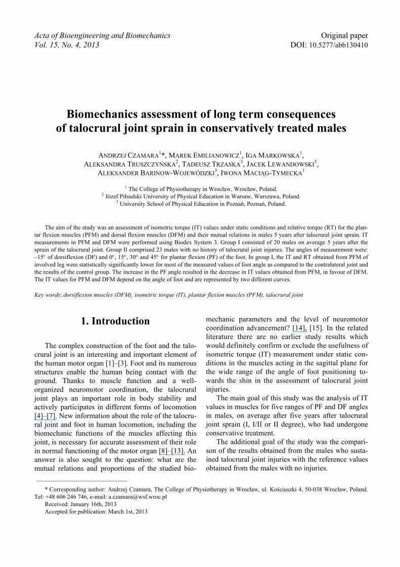
Acta of Bioengineering and Biomechanics Original paperVol. 15, No. 4, 2013 DOI: 10.5277/abb130410
Biomechanics assessment of long term consequencesof talocrural joint sprain in conservatively treated males
ANDRZEJ CZAMARA1*, MAREK EMILIANOWICZ1, IGA MARKOWSKA1,ALEKSANDRA TRUSZCZYŃSKA2, TADEUSZ TRZASKA3, JACEK LEWANDOWSKI3,
ALEKSANDER BARINOW-WOJEWÓDZKI3, IWONA MACIĄG-TYMECKA1
1 The College of Physiotherapy in Wrocław, Wrocław, Poland.2 Józef Piłsudski University of Physical Education in Warsaw, Warszawa, Poland.
3 University School of Physical Education in Poznań, Poznań, Poland.
The aim of the study was an assessment of isometric torque (IT) values under static conditions and relative torque (RT) for the plan-tar flexion muscles (PFM) and dorsal flexion muscles (DFM) and their mutual relations in males 5 years after talocrural joint sprain. ITmeasurements in PFM and DFM were performed using Biodex System 3. Group I consisted of 20 males on average 5 years after thesprain of the talocrural joint. Group II comprised 23 males with no history of talocrural joint injuries. The angles of measurement were:–15° of dorsiflexion (DF) and 0°, 15°, 30° and 45° for plantar flexion (PF) of the foot. In group I, the IT and RT obtained from PFM ofinvolved leg were statistically significantly lower for most of the measured values of foot angle as compared to the contralateral joint andthe results of the control group. The increase in the PF angle resulted in the decrease in IT values obtained from PFM, in favour of DFM.The IT values for PFM and DFM depend on the angle of foot and are represented by two different curves.
Key words: dorsiflexion muscles (DFM), isometric torque (IT), plantar flexion muscles (PFM), talocrural joint
1. Introduction
The complex construction of the foot and the talo-crural joint is an interesting and important element ofthe human motor organ [1]–[3]. Foot and its numerousstructures enable the human being contact with theground. Thanks to muscle function and a well-organized neuromotor coordination, the talocruraljoint plays an important role in body stability andactively participates in different forms of locomotion[4]–[7]. New information about the role of the talocru-ral joint and foot in human locomotion, including thebiomechanic functions of the muscles affecting thisjoint, is necessary for accurate assessment of their rolein normal functioning of the motor organ [8]–[13]. Ananswer is also sought to the question: what are themutual relations and proportions of the studied bio-
mechanic parameters and the level of neuromotorcoordination advancement? [14], [15]. In the relatedliterature there are no earlier study results whichwould definitely confirm or exclude the usefulness ofisometric torque (IT) measurement under static con-ditions in the muscles acting in the sagittal plane forthe wide range of the angle of foot positioning to-wards the shin in the assessment of talocrural jointinjuries.
The main goal of this study was the analysis of ITvalues in muscles for five ranges of PF and DF anglesin males, on average after five years after talocruraljoint sprain (I, I/II or II degree), who had undergoneconservative treatment.
The additional goal of the study was the compari-son of the results obtained from the males who susta-ined talocrural joint injuries with the reference valuesobtained from the males with no injuries.
______________________________
* Corresponding author: Andrzej Czamara, The College of Physiotherapy in Wrocław, ul. Kościuszki 4, 50-038 Wrocław, Poland.Tel: +48 606 246 746, e-mail: [email protected]
Received: January 16th, 2013Accepted for publication: March 1st, 2013

A. CZAMARA et al.74
2. Materials and methods
The study was approved by the College’s Com-mittee of Bioethics and Research. The study was car-ried out and financed at the College of Physiotherapyin Wrocław (Institute of Physiotherapy). Each subjectwas presented the goal of the study and the measure-ment approach to be used. The subjects signed theirinformed consent form to participate in the study.
2.1. Inclusion and exclusion criteria
The randomly selected 300 subjects were askedthe following question: “Have you ever sustainedtalocrural joint injuries?” The inclusion criteria for thestudy were: age between 20 and 30 years, unilateralsprain of the talocrural joint (I, I/II or II degree), con-servative treatment and no additional motor organinjuries. 44 subjects (15%) answered “yes”. 24 sub-jects were excluded from the study as they did notmeet the inclusion criterion (16 females and 4 malesafter bilateral sprain of the talocrural joint, 2 malesafter fibular fracture, 1 male after tibial fracture and 1male after surgical correction of club feet – talipesequinovarus). Eventually, 20 males were selected forgroup I. The subjects sustained a I, I/II or II degreeunilateral talocrural joint sprain five years earlier (mi-nimum 2 years, maximum 8 years) and underwentconservative treatment, which was confirmed in thedocumentation of the orthopaedic examination. 23males aged 20–30 years who volunteered for the studyand did not have any injuries of the talocrural joint, asconfirmed by orthopaedic examination, were selectedfor the study.
Table 1. Basic data of the study groups
Group In = 20
Group IIn = 23Basic data
of the study groups x SD x SDp
Body height (cm) 181.00 5.93 183.52 4.42 0.125Body mass (kg) 79.10 7.89 79.44 10.86 0.981
Age (years) 23.40 2.21 23.17 1.50 0.909
Table 1 presents basic data on the body height(cm), body mass (kg) and the subjects’ age (years).No significant differences in anthropometric parame-ters between groups were noted. The subjects wereoffice employees, entrepreneurs, physiotherapists andstudents. The anamnesis revealed occasional andunsystematic involvement in recreational physicalactivities after working hours.
2.2. Therapeutic procedures
Based on the anamnesis and the medical docu-mentation it was found that in the group I the thera-peutic procedure used by leading physician was asfollows. Firstly, the injured joint was immobilized for2–3 weeks with partial unloading, depending on thedegree of injury. A standard pharmacotherapy wasapplied (antithrombotic, anti-inflammatory and anal-gesic treatment). On removal of the plaster cast (im-mobilization), the patients underwent standard ambu-latory physiotherapy (near their homes) includingcryotherapy, magnetic field and laser treatment forabout 2 weeks. Next, they exercised from 1 to 2 weekswith a physiotherapist and at the end they were in-structed how to perform exercises at home. On com-pletion of this treatment stage, the patients did notsystematically undergo subsequent stages of specialistphysiotherapy [16].
2.3. IT measurement
Isometric torque (IT) measurements under staticconditions of PFM and DFM were performed in bothgroups using Biodex System 3 from 2011 to 2012(Fig. 1). Biodex System 3 was produced in the year2008 and it accommodates measurement and rehabili-tation facilities (Manufacturer: Biodex Medical SystemsSHIRLEY, N.Y. 11967 USA. Model 333-250. Software– Biodex Advantage) [17].
Fig. 1. The measurement of IT using Biodex System 3
Prior to IT measurements, the subjects performeda 12 minute warm-up on a cycle ergometer witha constant speed of 60 revolutions per minute (rpm).The load was 50 watt (W) within the first 6 minutes.Next, without interrupting the warm up, the load was

Biomechanics assessment of long term consequences of talocrural joint sprain in conservatively treated males 75
increased every 2 minutes in 5–10 W increments.The warm-up was followed by a 5 minute rest pe-riod. After the break, the IT values obtained fromPFM and then from DFM were measured (in thesagittal plane) in both lower extremities. In group I,the measurements were started from the uninvolvedleg. In group II, the measurements started from theright leg.
The IT measurements were performed under sta-tic conditions (isometrics) for five angles of footpositioning towards the shin. The measurementsstarted from foot dorsiflexion (DF) for the angle 15°in the talocrural joint, next for the neutral position(NP) involving 0° flexion (foot in 90° flexion to-wards the shin) and next for 15°, 30° and 45° plantarflexion (PF). During the measurements, the patientsassumed a supine position in the measuring chair.The measurements were taken in the sagittal plane.The knee joint was in 30° flexion and the angle ofhip joint flexion was 70°.
Calibration of the measuring system, stabiliza-tion of the patient and positioning of the dynamo-meter as well as measurement length arm were inconformity with the methodology presented by themanufacturer [17]. The measurements comprisedthe range of movements (ROM) in the studied jointsusing Biodex System 3. The foot was placed in thesame line in the sagittal plane as the shin and thethigh. The IT measurement of each position of footflexion angle involved alternate performance ofmaximal isometric contractions of PFM and DFM.On the “start” command, the subject performeda maximal isometric contraction of foot musclesand ended it on the “stop” command. The durationof a single isometric contraction was minimum 6 s.Between subsequent isometric contractions therewere 10 s rest intervals. After performing the me-asurements in one angle, there was a 90 s break andnext the foot positioning angle towards the shinchanged. The change was automatic and the anglesof foot positioning were in accordance with thepreset sequence of foot positioning, introduced intothe computer. The subjects from both groups under-went the measurement in each position for eachmuscle group three times. The highest IT values fora given angle of foot positioning were chosen forthe right and left legs. In order to exclude the effectof possible differences in body mass between thestudied groups on the result of the comparison ofthe values obtained, the relative torque (RT) valuewas calculated for the maximal isometric contrac-tion by dividing the obtained IT value by the sub-ject’s body mass (Nm/kg bm).
2.4. Statistical analysis
The mean value (x) and standard deviation (SD)were calculated for the results obtained from the bio-mechanical tests. The results obtained from the involvedand uninvolved legs of subjects from group I were com-pared with the results obtained from group II. To studythe distribution, the Shapiro–Wilk test was performed.The variables under study had both a normal distribu-tion as well as the distribution showing some ab-normalities. For the comparison of the dependentsamples, the parametric Student-t test or the nonpara-metric Wilcoxon’s test was used. For independentsamples the parametric t-test for independent samplesor the nonparametric Mann–Whitney U test was ap-plied. The significance level was accepted as p < 0.05.The results were next subjected to statistical analysisusing the IBM SPSS Statistics v. 19 program.
3. Results
On average five years after talocrural joint sprain,the IT values for PFM, measured in the involved legsfor 4 angles (–15°, 0° 15° and 45°) of foot positioning,were significantly lower as compared to contralateral,uninvolved legs (Table 2). The highest significancelevel for the differences ( p < 0.001) was noted for the2 extreme ranges of foot positioning angle, namely for45° of PF (deficit 34%) and 15° DF with 19% deficit(Table 2). The IT values produced by the foot musclesin DF in the involved leg were significantly lower at45° PF (deficit at the level of 26 %) as compared tothe contralateral, uninvolved legs (Table 2).
Table 3 shows that the highest IT values for PFM,obtained from group II, were noted for 15° dorsal flexionof the foot, namely x = 163.16 Nm and x = 158.1 Nm forthe right and left leg, respectively. The asymmetry of theparameter studied involved a 3% difference betweenboth legs. Subsequently, for 0° angle the IT values de-creased in the right (x = 122.12 Nm) and left (x =119.66 Nm) leg, respectively, indicating 2% asym-metry. Next, with the increase in the PF angle in bothlegs, IT values further decreased, even eight times ata 45° angle to x = 22.53 Nm and x = 22.1 Nm for theright and left leg respectively, as compared to thevalues obtained from the measurement in the 1st posi-tion of the foot. The asymmetry between both legswas 1%. Generally, in group II, no significant diffe-rences in IT values were found between the right andleft leg in PFM for the same angles of foot positioning

A. CZAMARA et al.76
towards the shin and the highest level of asymmetrydid not exceed 5% (Table 3). In group II, IT characte-ristics for DFM indicated that the highest values were
three times lower than the best values obtained fromPFM, both for the right and left legs. Moreover, thehighest IT values for the studied muscle group were
Table 2. Comparison of IT values produced by PFM and DFMof the involved leg and uninvolved leg in group I
IT of PFM and DFM in group I (Nm)Involved leg Uninvolved legThe angle of foot positioning
towards the shin (°)and measured muscle group – SD x SD
p Deficit(%)
–15° PFM 133.61 42.26 165.22 39.30 0.001 19–15° DFM 36.02 9.95 36.77 9.11 0.741 1
0° PFM 98.07 29.17 116.59 28.96 0.003 160° DFM 46.50 12.49 48.51 8.99 0.492 415° PFM 66.29 20.93 77.17 18.10 0.037 1415° DFM 48.25 12.44 50.51 8.13 0.794 430° PFM 39.49 16.18 45.22 16.95 0.071 1330° DFM 40.78 14.75 42.52 13.50 0.904 445° PFM 14.30 11.90 21.76 12.82 0.001 3445° DFM 26.01 15.35 35.35 12.47 0.046 26
Table 3. Comparison of IT values produced by PFM and DFM of the right leg and left leg in group II
IT of PFM and DFM in group II (Nm)Right leg Left legThe angle of foot positioning
towards the shin (°)and measured muscle group x SD x SD
p Deficit(%)
–15° PFM 163.16 39.10 158.10 41.17 0.343 3–15° DFM 39.22 9.36 39.10 8.10 0.946 0
0° PFM 122.11 26.40 119.65 29.47 0.447 20° DFM 50.30 8.82 49.36 8.37 0.473 215° PFM 83.97 15.38 79.96 19.25 0.273 515° DFM 51.64 9.70 51.19 8.54 0.831 130° PFM 48.74 11.36 49.27 12.05 0.811 130° DFM 47.07 8.17 47.88 8.52 0.496 245° PFM 22.53 11.32 22.09 10.75 0.724 245° DFM 38.85 6.75 39.10 7.95 0.822 1
Table 4. Comparison of IT values of PFM and DFM of the involved leg in group Ito the values obtained from the right and left leg in group II
IT of PFM and DFM in group I and group II (Nm)Group I
Involved legGroup IIRight leg
Group IILeft leg
The angle of foot positioningtowards the shin (°)
and measured muscle group x SD x SDp
x SDp
–15° PFM 133.61 42.26 163.16 39.10 0.022 158.10 41.17 0.062–15° DFM 36.02 9.95 39.22 9.36 0.284 39.10 8.10 0.269
0° PFM 98.07 29.17 122.11 26.40 0.007 119.65 29.47 0.0210° DFM 46.50 12.49 50.30 8.82 0.251 49.36 8.37 0.37615° PFM 66.29 20.93 83.97 15.38 0.003 79.96 19.25 0.03115° DFM 48.25 12.44 51.64 9.70 0.635 51.19 8.54 0.77930° PFM 39.49 16.18 48.74 11.36 0.034 49.27 12.05 0.02930° DFM 40.78 14.75 47.07 8.17 0.091 47.88 8.52 0.06145° PFM 14.30 11.90 22.53 11.33 0.025 22.09 10.75 0.02945° DFM 26.01 15.35 38.85 6.75 0.003 39.10 7.95 0.004
Biomechanics assessment of long term consequences of talocrural joint sprain in conservatively treated males 77
also obtained for other angles of foot positioning, na-mely for 0° and 15° PF. The lowest values were notedfor 15° DF and 45° PF. The level of IT asymmetry be-tween the right and the left leg (maximum 2%) wasstatistically insignificant (Table 3).
The IT values produced by PFM in group I weresignificantly lower for all five measured angles of footpositioning as compared to the values obtained fromthe right leg in group II and the four angle ranges ob-tained from the left leg in group II (Table 4). More-over, the IT values obtained from DFM of theinvolved leg were significantly lower at 45° PF ascompared to group II results.
In group I, the RT values obtained from PFM werelower for the 4 studied angles of foot positioning (0°,15°, 30° and 45°) as compared to the right foot and forthe 3 angle ranges as compared to the left talocrural jointin group II (Table 5). The DFM of the involved talocru-ral joint obtained significantly lower IT values as compa-red to those obtained from the right and left talocruraljoint in group II at 45° PF. Table 5 also presents thepercentage values of RT deficit level for PFM and DFM.In group I, 8% to 36% deficit for PMF and 5% to 33%deficit in DFM were noted in the involved talocruraljoints for different values of foot positioning angle ascompared to the values obtained from the same musclegroups affecting the right and left talocrural joints ingroup II. In the involved legs the greatest deficits werenoted for both muscle groups at 45° PF (Table 5).
4. Discussion
Monitoring of the treatment of talocrural jointinjuries is carried out by an orthopaedist [1], [3]. The
clinical anamnesis and physical examination is mostoften complemented by a functional assessment of thepatients. One of the components of functional assess-ment is the analysis of muscle strength.
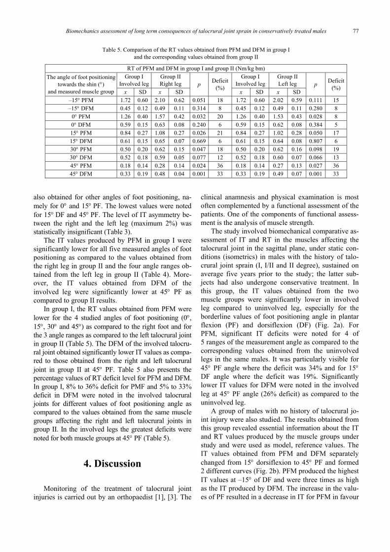
The study involved biomechanical comparative as-sessment of IT and RT in the muscles affecting thetalocrural joint in the sagittal plane, under static con-ditions (isometrics) in males with the history of talo-crural joint sprain (I, I/II and II degree), sustained onaverage five years prior to the study; the latter sub-jects had also undergone conservative treatment. Inthis group, the IT values obtained from the twomuscle groups were significantly lower in involvedleg compared to uninvolved leg, especially for theborderline values of foot positioning angle in plantarflexion (PF) and dorsiflexion (DF) (Fig. 2a). ForPFM, significant IT deficits were noted for 4 of5 ranges of the measurement angle as compared to thecorresponding values obtained from the uninvolvedlegs in the same males. It was particularly visible for45° PF angle where the deficit was 34% and for 15°DF angle where the deficit was 19%. Significantlylower IT values for DFM were noted in the involvedleg at 45° PF angle (26% deficit) as compared to theuninvolved leg.
A group of males with no history of talocrural jo-int injury were also studied. The results obtained fromthis group revealed essential information about the ITand RT values produced by the muscle groups understudy and were used as model, reference values. TheIT values obtained from PFM and DFM separatelychanged from 15° dorsiflexion to 45° PF and formed2 different curves (Fig. 2b). PFM produced the highestIT values at –15° of DF and were three times as highas the IT produced by DFM. The increase in the valu-es of PF resulted in a decrease in IT for PFM in favour
Table 5. Comparison of the RT values obtained from PFM and DFM in group Iand the corresponding values obtained from group II
RT of PFM and DFM in group I and group II (Nm/kg bm)Group I
Involved legGroup IIRight leg
Group IInvolved leg
Group IILeft leg
The angle of foot positioningtowards the shin (°)
and measured muscle group x SD x SDp Deficit
(%)x SD x SD
p Deficit(%)
–15° PFM 1.72 0.60 2.10 0.62 0.051 18 1.72 0.60 2.02 0.59 0.111 15–15° DFM 0.45 0.12 0.49 0.11 0.314 8 0.45 0.12 0.49 0.11 0.280 8
0° PFM 1.26 0.40 1.57 0.42 0.032 20 1.26 0.40 1.53 0.43 0.028 80° DFM 0.59 0.15 0.63 0.08 0.240 6 0.59 0.15 0.62 0.08 0.384 515° PFM 0.84 0.27 1.08 0.27 0.026 21 0.84 0.27 1.02 0.28 0.050 1715° DFM 0.61 0.15 0.65 0.07 0.669 6 0.61 0.15 0.64 0.08 0.807 630° PFM 0.50 0.20 0.62 0.15 0.047 18 0.50 0.20 0.62 0.16 0.098 1930° DFM 0.52 0.18 0.59 0.05 0.077 12 0.52 0.18 0.60 0.07 0.066 1345° PFM 0.18 0.14 0.28 0.14 0.024 36 0.18 0.14 0.27 0.13 0.027 3645° DFM 0.33 0.19 0.48 0.04 0.001 33 0.33 0.19 0.49 0.07 0.001 33

A. CZAMARA et al.78
of IT increase for DFM. IT balance between PFM andDFM was obtained at 30° of PF angle. For 45° PFangle the lowest IT values were found for both musclegroups, however, apparently in favour of the DFM.The level of asymmetry in the IT values obtained se-parately for each muscle group between the right andthe left side did not exceed 5%.
Similarly, the analysis of RT values in group Ishowed the same trend of deficits of IT converted intokg of body mass in the involved talocrural joints forthe muscle groups presented above.
Probably the main reason for such a large deficitin IT and RT in group I was the short and incompletephysiotherapeutic procedure. The anamnesis and theanalysis of patients’ documentation indicated thatmost of the subjects in group I did not undergo the 3rdand 4th stages of physiotherapy program [16]. Most ofthem did not undergo kinesiotherapy based on func-tional training and did not perform systematic pro-prioceptive exercises at different levels of difficulty,joint stabilization exercises and the neighboring kine-matic chains. Most of them did not perform exercisesaimed at direct muscle strength recovery in the musclegroups being studied. Subsequent factors that mighthave affected the results obtained after basic treatmentincluded low physical activity level at work and occa-sional involvement in recreational physical activitywithin the average 5 year period after talocrural jointinjury.
Based on the results obtained we can assume thatthe biomechanical tests conducted are useful in distantevaluation of deficits in IT values obtained from thestudied muscle groups at the side of talocrural jointsprain.
Generally, in the literature there are no papers as-sessing the usefulness of IT value measurement in
shin muscles affecting the foot after talocrural jointsprain under static conditions (isometrics). Peaktorque (PT) values are most often measured underisokinetic conditions in the muscles affecting the talo-crural joint in the sagittal plane and evertor andinvertor muscles of the foot for a given angular velo-city. Isokinetic measurements are carried out undercondition of open and closed kinematic chain duringconcentric, eccentric and concentric-eccentric exerci-ses and the other way round [18]. Isokinetic exercisesresemble natural everyday human activities performedat work and in sports. Isokinetic tests, carried out inconcentric conditions provide us with informationabout the submaximal torque produced depending onthe preset angular velocity. Collado et al. (2010), intheir isokinetic tests, obtained better PT values fromfoot evertor muscles in patients who underwent reha-bilitation with extended eccentric training as compa-red to the patients who underwent concentric training[9]. Kamiński et al. (2003) performed PT measure-ments in evertor (E) and invertor (I) muscles prior toand after different six-week trainings. They did notnote any significant effect or any significant differen-ces in PT and E/I ratio in the studied groups of musc-les between the group involved in strength trainingand the group involved in strength training combinedwith proprioception [19]. Goharpey et al. (2007) inisokinetic tests, during eccentric exercises (angularvelocities of 60°/s and 120°/s) showed that PT measure-ments, normalized for the body mass were moreuseful in assessment of PT deficits in foot invertormuscles in patients with chronic functional instabilityof the talocrural joint as compared with the standardPT measurement under concentric and eccentric con-ditions [11]. Hadzic et al. (2009) showed that too highPT values, produced by PFM in isokinetic conditions
a) b)
Fig. 2. IT values of PFM and DFM depending on the angle of foot positioning towards the shin in group I (a) and group II (b)

Biomechanics assessment of long term consequences of talocrural joint sprain in conservatively treated males 79
and a disturbed ratio to DFM with limited foot dorsi-flexion significantly increase the risk of further talo-crural joint injuries [20]. Möller et al. (2002) compa-red the results of isokinetic measurements for PFMand DFM in patients with the history of Achilles ten-don injuries, who had undergone conservative andsurgical treatment. Concentric and eccentric tests werecarried out at two angular velocities. The studies car-ried out after 6, 12 and 24 months generally did notshow any differences in PT values between the studygroups. However, after 2 years they still noted signifi-cant deficits in PT of the PFM (from over 12% toabout 27%) when they compared the results obtainedfrom the involved and uninvolved legs [21].
Studies carried out in static conditions (maximalisometric contraction) allow essential information tobe obtained about the evoked isometric torque (IT) ofthe studied muscle groups for different angles in thejoint. Adequate organization of such studies, stabili-zation of the patient as well as adequately selectedranges of joint angles depending on the clinical case,qualify this kind of measurements as safe with a smallmeasurement error. Moreover, such studies are mostoften less expensive. They were combined with elec-trophysiological measurements for the comparison ofthe results obtained from younger and older patients[22], [23]. Such tests make it possible to obtain infor-mation on IT behaviour for a defined value of angle inthe joint under condition of an isolated measurement.However, using a proper equipment enables simulta-neous measurement of the parameters studied forseveral large groups of muscles affecting two or morejoints [24], [25]. On the other hand, 2 or 3 attempts ofIT measurement for each of the several ranges of va-lues of the angle in the joint, separately for each mu-scle group in the right and left leg are time consu-ming, which makes the measurements less attractiveunder static conditions. Also errors in EMG recordsare possible during the tests of isometric muscle con-tractions [26]. However, as far as physiological con-ditionings of muscles are concerned, it should be em-phasized that they perform alternate body stabilizingfunctions (isometric contractions) and dynamicfunctions, namely locomotion, thanks to the changesin their contractility and specialized neuromuscularcoordination. Therefore, 2 kinds of tests should beconducted in due time in patients with the history ofmotor organ injuries and 2 kinds of training restoringthe strength in weakened muscles should be appliedin the process of rehabilitation, adequately to theclinical condition, in order to restore reciprocal bio-mechanical characteristics, both quantitative andqualitative ones.
This thesis can be carefully confirmed (due to thestudied different muscle groups and other clinicalcases) by the results of the study of the knee joint afteranterior cruciate ligament (ACLR) reconstruction.Czamara (2008) showed that the patients systemati-cally involved in physiotherapy obtained IT valuesfrom the extensor and flexor muscles of the involvedleg similar to those obtained from the uninvolved legin the 6th month after ACLR [27]. Similarly, Czamaraet al. (2011) showed that similar IT values could beobtained from the involved and uninvolved leg inanother group of patients after ACLR. However, theisokinetic study, carried out in this sample for thesame muscle groups showed at the end of the 6thmonth significant PT deficits in some patients fromthe operated knees for the angular velocity of 60°/s[28]. In a subsequent study, Czamara et al. (2011)compared the IT values responsible for internal andexternal tibial rotation in ACLR patients during the6th month of systematic physiotherapy as compared tothe patients who did not undergo systematic phy-siotherapy after 12 months from the injury. The studyshowed a higher effectiveness of IT measurements(under static conditions) as compared to isokineticmeasurements [29]. Based on the cited reports, we canassume that restoring strength parameters under staticconditions and speed-strength parameters under dy-namic conditions in the muscle groups studied is notsimultaneous and it is not characterized by one levelof the restored IT values for both conditions.
The future measurements should be carried outboth under static and isokinetic conditions. Obtainingnew information about biomechanical parameters inthe studied muscle groups may add new values tocomplex assessment of treatment results in patientswith the history of talocrural joint injuries [3], [9],[30]–[34].
5. Conclusions
1. In males averagely five years from the talocru-ral joint sprain significant deficits in isometric torquevalues obtained from the studied muscle groups werenoted, particularly in the muscles responsible forplantar flexion, as compared to the results obtainedfrom the contralateral joint and those obtained fromthe males with no history of talocrural joint injuries.
2. The highest deficit in isometric torque valuesobtained from both muscle groups under study ascompared to the results obtained from the contra-lateral joint was noted at a 45° plantar flexion.

A. CZAMARA et al.80
3. The highest isometric torque values were obta-ined from plantar flexors in foot dorsiflexion and withthe increase in plantar flexion angle they decreased infavour of dorsal flexors.
4. In the group of males with no history of talocru-ral joint injury the isometric torque values obtainedfrom the studied muscle groups are characterized bya low level of asymmetry in the comparison betweenthe right and the left leg.
References
[1] KADAKIA A.R., Overview of the Ankle in Essentional Ortho-paedics, Saunders Elsevier, 2010.
[2] GOLANO P., VEGA J., DE LEEUW P.A.J., MALAGELADA F.,MANZANARES M.C., GÖTZENS V., VAN DIJK C., Anatomy ofthe ankle ligaments: a pictorial essay, Knee Surg. SportsTraumatol. Arthrosc., 2010, Vol. 18(5), 557–569.
[3] DIGIOVANI CH.W., GREISBERG J., Foot & Ankle CoreKnowledge in Orthopaedics, Elsevier 2007.
[4] RICHARDS J., Chapter 6. Biomechanics in Tidy’s Physiothe-rapy, Elsevier 2008.
[5] LUNDGREN P., NESTER C., LIU A., ARNDT A., JONES R.,STACOFF A., WOLF P., LUNDBERG A., Invasive in vivo measu-rement of rear-, mid- and forefoot motion during walking,Gait Posture, 2008, Vol. 28(1), 93–100.
[6] ARNDT A., WOLF P., LIU A., NESTER C., STACOFF A., JONES R.,LUNDBERG A., Intrinsic foot kinematics measured in vivo du-ring the stance phase of slow running, J. Biomech., 2007,Vol. 40(12), 2672–2678.
[7] LEE S.S.M., PIAZZA S.J., Correlation between plantarflexormoment arm and preferred gait velocity in slower elderlymen, J. Biomech., 2012, Vol. 45(9), 1601–1606.
[8] LENARDI A., BENEDETTI M.G., BERTI L., BETTINELLI D.,NATIVO R., GIANNINI S., Rear-foot, mid-foot and fore-footmotion during the stance phase of gait, Gait Posture, 2007,Vol. 25, 453–462.
[9] COLLADO H., COUDREUSE J.M., GRAZIANI F., BENSOUSSAN L.,VITON J.M., DELARQUE A., Eccentric reinforcement of theankle evertor muscles after lateral ankle sprain, Scand.J. Med. Sci. Sports, 2010, Vol. 20(2), 241–246.
[10] MUNN J., BEARD D.J., REFSHAUGE K.M., LEE R.Y., Eccentricmuscle strength in functional ankle instability, Med. Sci.Sports Exerc., 2003, Vol. 35(2), 245–250.
[11] GOHARPEY S.H., SADEGHI M., MAROUFI N., SHATERZADEH M.J.,Comparison of Invertor and Evertor Muscle Strength in Pa-tients with Chronic Functional Ankle Instability, J. Med. Sci.,2007, Vol. 7(4), 674–677.
[12] SEKIR U., YILDIZ Y., HAZNECI B., ORS F., AYDIN T., Effect ofisokinetic training on strength, functionality and propriocep-tion in athletes with functional ankle instability, Knee Surg.Sports Traumatol. Arthrosc., 2007, Vol. 15(5), 654–664.
[13] TRAPPE S.W., TRAPPE T.A., LEE G.A., COSTILL D.L., Calfmuscle strength in human, Int. J. Sports Med., 2001, Vol. 22(3),186–191.
[14] ARAMPATZIS A., KARAMANIDIS K., STAFILIDIS S., MOREY-KLAPSING G., DEMONTE G., BRÜGGEMANN G.P., Effect ofdifferent ankle- and knee-joint positions on gastrocnemiusmedialis fascicle length and EMG activity during isometricplantar flexion, J. Biomech., 2006, Vol. 39(10), 1891–1902.
[15] SIMONEAU E.M., LONGO S., SEYNNES O.R., NARICI M.V.,Human muscle fascicle behavior in agonist and antagonist iso-metric contractions, Muscle Nerve, 2012, Vol. 45(1), 92–99.
[16] CZAMARA A., Physiotherapeutic procedure after injuries ofsoft tissues of tarsal-crural jont, J. Orthop. Traum. Surg. Rel.Res., 2008, Vol. 4(12), 88–108.
[17] Biodex Advantage Software, Operations Manual (version3.29 and 3.30), Biodex Medical Systems, Inc. New York,2008.
[18] DVIR Z., Isokinetics. Muscle Testing, Interpretation andClinical Applicaction, Elsevier 2004, 167–184.
[19] KAMIŃSKI T.W., BUCKLEY B.D., POWERS M.E., HUBBARD T.J.,ORTIZ C., Effect of strength and proprioception training oneversion to inversion strength ratios in subjects with unila-teral functional ankle instability, Br. J. Sports Med., 2003,Vol. 37(5), 410–415.
[20] HADZIC V., SATTLER T., TOPOLE E., JARNOVIC Z., BURGER H.,DERVISEVIC E., Risk factors for ankle sprain in volleyballplayers: A preliminary analysis, Isokinet. Exerc. Sci., 2009,Vol. 17(3), 155–160. DOI 10.3233/IES-2009-0347.
[21] MÖLLER M., LIND K., MOVIN T., KARLSSON J., Calf musclefunction after Achilles tendon rupture. A prospective, rando-mised study comparing surgical and non-surgical treatment,Scand. J. Med. Sci. Sports, 2002, Vol. 12(1), 9–16.
[22] SIMONEAU E., MARTIN A., VAN HOECKE J., Effects of jointangle and age on ankle dorsi- and plantar-flexor strength,J. Electromyogr. Kinesiol., 2007, Vol. 17(3), 307–316.
[23] SIMONEAU E.M., BILLOT M., MARTIN A., VAN HOECKE J.,Antagonist mechanical contribution to resultant maximaltorque at the talocrural joint in young and older men,J. Electromyogr. Kinesiol., 2009, Vol. 19, 123–131.
[24] HAHN D., OLVERMANN M., RICHTBERG J., SEIBERL W.,SCHWIRTZ A., Knee and talocrural joint torque–angle rela-tionships of multi-joint leg extension, J. Biomech., 2011, Vol. 44,2059–2065.
[25] WYCHOWAŃSKI M., Wybrane metody oceny dynamiki układuruchu człowieka, Studia Monografie, AWF, Warszawa, 2008,73–78.
[26] DE OLIVEIRA L.F., MENEGALDO L.L., Input error analysisof an EMG-driven muscle model of the plantar flexors,Acta Bioeng. Biomech., 2012, Vol. 14(2), 75–81. DOI:10.5277/abb120210.
[27] CZAMARA A., Moments of muscular strength of knee jointextensors and flexors during physiotherapeutic proceduresfollowing anterior cruciate ligament reconstruction in males,Acta Bioeng. Biomech., 2008, Vol. 10(3), 37–44.
[28] CZAMARA A., TOMASZEWSKI W., BOBER T., LUBARSKI B.,The effect of physiotherapy on knee joint extensor and flexormuscle strength after anterior cruciate ligament reconstruc-tion, Med. Sci. Monit., 2011, Vol. 17(1), 35–41.
[29] CZAMARA A., SZUBA Ł., KRZEMIŃSKA A., TOMASZEWSKI W.,WILK-FRAŃCZUK M., Effect of physiotherapy on the strengthof tibial internal rotator muscles in males after anterior cru-ciate ligament reconstruction (ACLR), Med. Sci. Monit.,2011, Vol. 17(9), 523–531.
[30] WATANABE K., KITAOKA H.B., BERGLUND L.J., ZHAO K.D.,KAUFMAN K.R., AN K.N., The role of ankle ligaments andarticular geometry in stabilizing the ankle, Clin. Biomech.(Bristol, Avon), 2012, Vol. 27(2), 189–195. Epub 2011 Oct 13.
[31] BROWN C., BOWSER B., SIMPSON K.J., Movement variabilityduring single leg jump landings in individuals with and wi-thout chronic ankle instability, Clin. Biomech. (Bristol,Avon), 2012, Vol. 27(1), 52–63. Epub 2011 Aug 20.

Biomechanics assessment of long term consequences of talocrural joint sprain in conservatively treated males 81
[32] MENEGALDO L.L., DE OLIVEIRA L.F., Effect of muscle modelparameter scaling for isometric plantar flexion torque pre-diction, J. Biomech., 2009, Vol. 42(15), 2597–2601. Epub2009 Aug 8.
[33] HOPKINS J.T., COGLIANESE M., GLASGOW P., REESE S.,SEELEY M.K., Alterations in evertor/invertor muscle activa-
tion and center of pressure trajectory in participants withfunctional ankle instability, J. Electromyogr. Kinesiol., 2012,Vol. 22(2), 280–285. Epub 2011 Dec 16.
[34] DAVIES G.J., A compendium of isokinetics in clinical usageand rehabilitation techniques, S&S Publishers Wisconsin,1992.
Copyright © 2022 FDOKUMEN