ADVISER'S APPROVAL - ShareOK
53
_/ Date: May 24, 1959 Name: Carl Christian Nelson Institution: Oklahoma State University Location: Stillwater. Oklahoma Title of Study: DISSECTION TECHNIQUES FOR HIGH SCHOOL BIOLOGY TEACHERS Number of Pages in Study: 46 Candidate for Degree of Master of Arts Major Field: Natural Science Nature of Study: Many high school biology teachers fail to offer their students any individual laboratory activities. One phase of activ- ities which can be effectively administered, but is often avoided by high school biology teachers, is the dissection of preserved an- imal specimens. This report deals with various problems which ap- pear while conducting dissection activities. Items such as: obtain- ing preserved specimens; dissection equipment; facilities required for dissection; motivation of students; and, dissection procedures are discussed. Also included are dissection plans. orderly step- by-step procedures, for the earthworm, frog, grasshopper, and the fetal pig. Use of the Study: It is desired that the concepts developed and the dis- section plans included in this report will be of value to secondary biology teachers who do not include dissection activities in their biology classes. Also that some teachers who are not satisified with the methods they currently employ may be assisted by the dis- section techniques discribed. ADVISER'S APPROVAL (
-
Upload
khangminh22 -
Category
Documents
-
view
0 -
download
0
Transcript of ADVISER'S APPROVAL - ShareOK

_/
Date: May 24, 1959
Name: Carl Christian Nelson
Institution: Oklahoma State University
Location: Stillwater. Oklahoma
Title of Study: DISSECTION TECHNIQUES FOR HIGH SCHOOL BIOLOGY TEACHERS
Number of Pages in Study: 46 Candidate for Degree of Master of Arts
Major Field: Natural Science
Nature of Study: Many high school biology teachers fail to offer their students any individual laboratory activities. One phase of activities which can be effectively administered, but is often avoided by high school biology teachers, is the dissection of preserved animal specimens. This report deals with various problems which appear while conducting dissection activities. Items such as: obtaining preserved specimens; dissection equipment; facilities required for dissection; motivation of students; and, dissection procedures are discussed. Also included are dissection plans. orderly stepby-step procedures, for the earthworm, frog, grasshopper, and the fetal pig.
Use of the Study: It is desired that the concepts developed and the dissection plans included in this report will be of value to secondary biology teachers who do not include dissection activities in their biology classes. Also that some teachers who are not satisified with the methods they currently employ may be assisted by the dissection techniques discribed.
ADVISER'S APPROVAL (

DISS.ECTION TECHNIQUES FOR HIGH
SQ!OOL BIOLO~Y TEACHERS
1958-1959
by
CARL C. NELSON
Bachelor of Science
Dana College
Blair, Nebraska
1954
Submitted to the faculty of the Graduate School of the Oklahoma State University in partial fulfillment of the requirements for the degree of
MASTiiR OF SCIENCE May, 1959
ii
DISSECTION TECHNIQUES FOR HIGH
SCHOOL BIOLOGY TEACHERS
Report Approved:
iii

ACKNOWLEDGEMENTS
The writer wishes to express his sincere appreciation and gratitude
for the assistance and many valuable suggestions given him by Drs. Imy
Vincent Holt, Roy Winfield Jones, and Bryan P. Glass, in the planning
and writing of this report.
Indebtedness is acknowledged to my wife, Jane Nelson, who rendered
invaluable aid as typist.
iv

Chapter
I.
II.
III.
TABLE OF CONTENTS
INTRODUCTION ••••••••••••••••••••••••.•••••••••••••••••••••• • 1 Statement of the Problem ••••••••••••••••••••••••••••••• 1 Methods Used in Partial Solution to the Problem •••••••• 2
Use of the Study••••••••••••••••••···········•••••••••• 2
IS DISSECTION IN SECONDARY BIOLOGY A WORTHWHILE ACTIVITY? . .. PROBLEMS INCURED IN PRESENTING DISSECTION ACTIVITIES IN HIGH
SCHOOL BIOLOGY CLASSES .................................... Methods of Presentation ••••••••••••••••••• $ ••••••••••••
Facilities Required for Dissection ••••••••••••••••••••• Motivation of Students •••••••••••••••••••••••••••••••••
4
9 9
11 16
IV. DISSECTION P1--'l0CJ3DURE • • • • • • • • • • • • • • . • • • • • • • • • • • • • • • • • • • • • • • • • 19
V. DISSECfION PLANS • • • • • • • . • • • • • . • • • • • • • • • • • • • • • • • • • • • • • • • • • • • • 23 Earthworm Study ••...•••••.•.......••••••.••••••..•••••• 25 Earthworm Dissection •••••o••••••••••••••••••••••••••••• 27 Grasshopper Study . -•. . . . . • • . . . . • . • . . . . . . . . . • • . • . • . . . . . • • 29 Grasshopper Dissection ••••••••••••••••••••••••••••••••• 31 Frog Study••••••••••••••••••••••••••·•••••••••••••••••• 33 Frog Dissection Fetal Pig Study
......... -.............................. . and Dissection ••••••.•.••••••••••••••••
35 38
B IBLiffiRAPHY • •••• • • • • • • • • • • • • • • • • • • • • • • • • • • • • • • • • • • • • • • • • • • • • • . • • • • • • 45
V

LIST OF TABLES
Table Page
r. Comparative Cost of Biological Specimens from Three Biological Supply Houses ••••••••••••••••••••••••••••••••••••• 15
vi

CHAPTER I
INI'RODUCTION
Statement of~ Problem. There is a tendency among biology teachers
in secondary schools to neglect one of the most important methods of ed
ucation - learning by doing. That is, many teachers fa 1 to off er their
students any individual laboratory activities.
One phase of activities which can effectively be administered but
is often avoided by secondary biology teachers is the dissection of an
imal specimens. Frequently these dissection activities are omitted from
the biology course because the teachers are deficient in training and
their thoughts are vague about how an orderly dissection scheme could be
administered. They also avoid dissection activities because students
traditionally display aversion toward the practice of dissecting pre
served specimens.
This study was made for the purpose of solving some of the problems
associated with the execution of dissection activities. However, some
complications perennially persist. It is the hope of the writer that
ideas discussed in this study may alleviate the efforts of secondary bi
ology teachers in finding their best solution to these problems.
No presumption is made that the techniques discussed in this report
are the best ones for a high school biology teacher to utilize. Neither
is implication intended that preserved animal specimens are superior to
dissect than freshly killed or anaesthetized specimens. The animal types
1

2
which are emphasized were selected with the writers prerogative.
Methods Used in Partial Solution to the Problem. It is believed by ------ --the writer that, if orderly dissection plans were made available to sec-
ondary biology teachers, they would be more likely to include dissection
activities in their biology course.
In searching for a type of dissection procedure deemed desireable
by the writer, several high school biology laboratory manuals and text
books were observed. All sources gave mention of dissection activities,
but none of the sources discribed orderly step-by-step plans which could
be effectively utilized.
The following categories of literature were studied to find addition-
al information pertaining to the topic: text books on science education;
college biology, particularly anatomy, text books and laboratory manuals;
periodic literature; audio-visual reference materials; catalogs and lit-
erature provided by biological supply houses; and dissection plans used
in various high schools. Recall of the writers experience in conducting
dissection activities and opinions received from practicing high school
biology teachers were correlated with the literature in the production of
this reoort and the included dissection plans •
.!:!!! of~ Study. It is the hope of the writer that the concepts
developed and the dissection plans included in this report will be of
value to secondary teachers who do not include dissection activities in
their biology classes. Some teachers who are not satisfied with the dis-
section methods they currently employ may be assisted by ~he techniques
discribed. Through the use of these methods, a high school biology in-

structor may be able to emphasize more effectively the structural and
functional characteristics of animals.
3
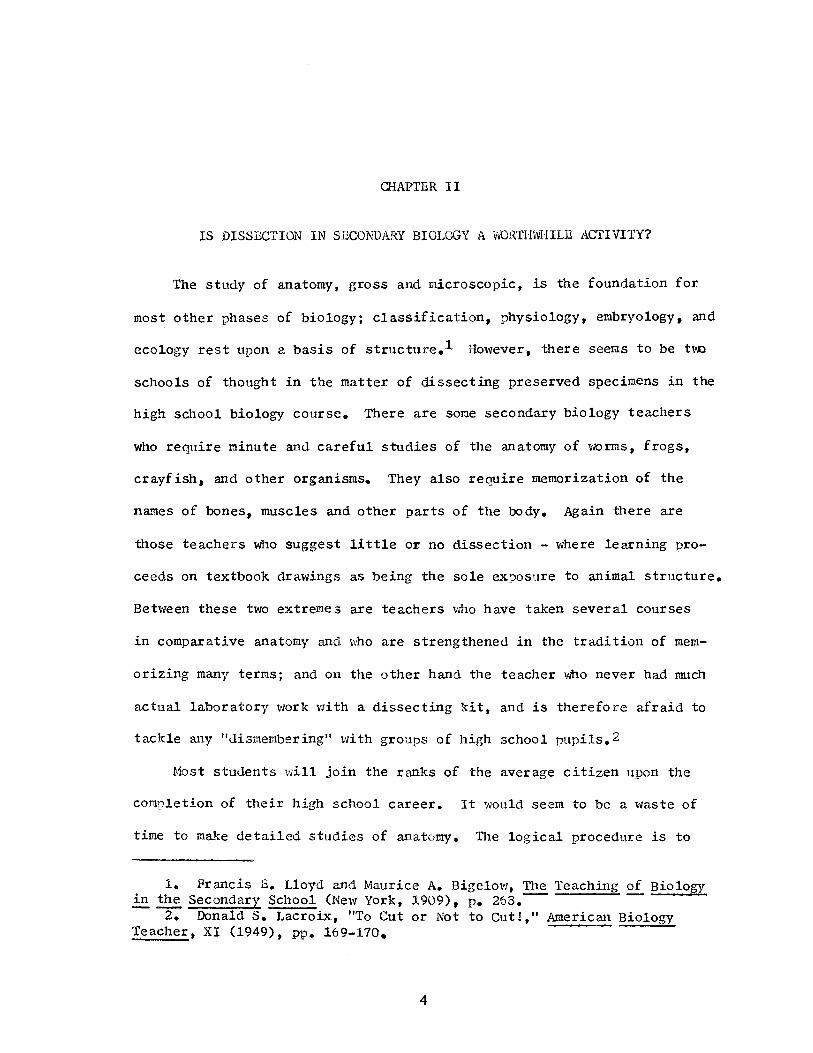
CHAPTER II
IS DISSECTION IN SECONDARY BIOLOGY A WORTH\'VHILE ACTIVITY?
The study of anatomy, gross and microscopic, is the foundation for
most other phases of biology; classification, physiology, embryology, and
ecology rest upon a basis of structure.1 However, there seems to be t\\O
schools of thought in the matter of dissecting preserved specimens in the
high school biology course. There are some secondary biology teachers
who require minute and careful studies of the anatomy of ~,10rms, frogs,
crayfish, and other organisms. They also require memorization of the
names of bones, muscles and other parts of the body. Again there are
those teachers who suggest little or no dissection - where learning pro-
ceeds on textbook drawings as being the sole exposure to animal structure.
Between these two extremes are teachers who have taken several courses
in comparative anatomy and ~ho are strengthened in the tradition of mem-
orizing many terms; and on the other hand the teacher who never had much
actual laboratory work with a dissecting kit, and is therefore afraid to
tackle any ndismembering" with groups of high school pupils. 2
Most students will join the ranks of the average citizen upon the
completion of their high school career. It would seem to be a waste of
time to make detailed studies of anatomy. The logical procedure is to
1. Francis E. Lloyd and Maurice A. Bigelow, The Teaching of Biology ~~Secondary School (New York, 1909), P• 268.~- ~
2. Donald s. Lacroix, "To Cut or Not to Cut!," American Biology Teacher, XI (1949), pp. 169-170.
4

5
give the students an opnortunity to dissect several types of organisms
and alld>w them to observe the development of digestive systems, respi-'
ratory apparatus, excretory and reproductive organs, skeletal struct-
ures and circulatory devices, without having to memorize too many terms.
Care should be taken that the anatomy studies a.re not elevated to
the extent that they would tend to replace similar activities performed
in college zoology. There is little need for the average high school
student to persue the topics in such minute detail. Complex vocabulary
should be kept at a minimum, and when possible, common names should be
substituted for scientific. However, maximum comprehension involves the
development of vocabulary, and in order to be meaningful, it is necessary
that the students memorize certain scientific nane s. Often in anatomy
the scientific name of a structure is also the common name. Most of these
technical names are of Latin or Greek derivation. The use of these names
by the student is facilitated if they are told the roots from which the
names evolved. Through learning the derivation of the scientific names,
the students will have increased their repertory of word roots and in
this way their entire vocabulary will have increased.
Observing and stressing anatomical structures is important when
laying a basis for understanding hody functions. Students should be able
to identify gross organs and relate them to a body activity 0 3 The human
is the organism with which all students are most f amilar. Throughout
their formal education they are taught the structure and function of the
human body, but under no conditions are they actually able to delve in-
3. Lee R. Yothers, "A.n Identification Aid When Dissecting Frogs." School Science and Mathematics, XLVII (1947), p. 421 0

6
side the protective layer of skin in the human. The heart, stomach, pan-
creas, liver, etc., are thoroughly discussed and the major functions of
these familar organs are well understood. The nearest thing available
for the students to use to visualize the relationship of these structures
are the two dimensional diagrams and charts included in textbooks, and
perhaps their past exposure to several well chosen films and filmstrips.
Dissecting animal specimens will allow the high school student to
recognize more vividly the intricate composition of living things, and to
relate the structures and functions of the lower organisms with the human.
The green gland of the crayfish, Malpighian tubules of the grasshopper and
the nephrostome of the earthworm are not similar in structure with the
human kidney, however the activities they perform are comparable. The
kidney of the frog and fetal pig are structurally similar to that of the
human, and the students can get a lasting image of this structure by making
these observations in the lower forms of animals. Personal experience
provides the best basis for learning any subject. One may study alone,
but one usually works more efficiently and progresses more rapidly when
guided by a teacher with previous training and experience in the subject
matter. Many facts, ideas, and conclusions may be learned from lectures
or by reading, but this is "secondhand'' information in the words of some
speaker or writer. By contrast, laboratory study affords opportunity to
obtain knowledge "firsthand11 from personal observations, Laboratory
study constitutes training in scientific observations, 'Which requires
care and precision. 4
Dissection activities also afford an opportunity for the secondary
4. Tracy I. Stoer, Laboratory Manual for General Zoology (York, Pa., 1944), p. s.

7
students to closely scrutinize the external features of the specimens.
Modifications such as the point of attachment of the frog tongue, setae
of the earthworm, horizontal action of the grasshor,per mandibles, tympanic
membranes of the forg and grasshopper, and many more interesting features
would likely never be recognized by the student if he were not given the
ooportunity to dissect these animals.
A specimen gives the pupil actual first hand experience and this
means direct personal contact with the item. Usually the more direct the
experience is the more educative it is. This direct experience can be
direct only if and when the pupil himself sees, feels, handles, operates,
and in other ways actually works with the item.s "Direct, purposeful ex
perience is in reality the basis of all effective learning. u6
Biology is a life science, and no function in the Lmiverse is quite
as complex as life itself. Each living animal is a highly precisioned
mass of protoplasm, admittedly includ.:ing the simplest protozoans and the
apex of advancement, the human. Some students fail to realize that such
things as the insect or earthworm are complex, self-sustaining organisms
which carry out all life functions. They do not realize the close struct-
ural s:imilarity between the frog, pig, and the human. The primary reason
for this oversight is that students have never been exposed to the intri-
cate compostition of living things. If through n~e activity of dis-
section the students can become aware of the Creators genius in construct-
ing living th.:ings, and the simple interrelationship of animals in an evo-
lutionary sequence, the time spent in dissection would not be wasted.
s. Harry c. McKnown and Alvin B. Roberts, Audio-Visual Aids to In-struction (New York, 1940), p. 56 ~~ ~ ~
6. Ibid.

8
' 1Anatomy is absolutely an essential part in any elementary course in the science of biology, and there is no other way of beginning except by giving considerable attention to,structural facts as the basis for determining functions, studying life histories, or interpreting environmental relations."7
It is the writer's opinion that dissection should be included in a
secondary biology course, if for no other reason, to assist the students
in recognizing the complexity, yet regularity of living things, and through
this medium, learn to appreciate nature more fully.
7. Lloyd and Bigelow, P. 268.

CHAPTER III
PROBLEMS INCUR.ED IN PRESENTING DISSECTION
ACTIVITIES IN HIGH SCHOOL BIOLOGY CLASSES
Methods of Presentation. Different methods of topic presentation
are employed by various teachers of secondary biology. Perhaps the most
commonly used form is the textbook-recitation method. This has elicited
a strong following particularly with inexperienced teachers because it is
an easy way to teach, it gives a superficial showing of scholarly attain-
ment, it is possible to cover large amounts of subject matter in a rela-
tively short period of time, it gives the teacher less to do in olanning
and preparing, and it offers a feeling of security by a reliance upon the
supposed authority of the author.8 The general practice in this method
is where the teacher makes assignments of several pages in the textbook
periodically for the pupils to study, and then during the class period,
discusses the important concepts included in the assignment.
Dissection activities can easily fit into this tyne of program. At
sometime during the term• representatives of the animal kingdom are stud-
ied. When reference is made to the physical structure of these animals,
time can be set aside for the purposeful dissection of reDresentative an-
imals to supplement the textual materials. This affords an opportunity
for the students and teacher alike to benefit from the diviation from the
8. David F. Miller and Glenn w. Blaydes, Methods and Materials for Teaching Biological Sciences (New York, 1938), P• 42.
9

methodical textbook-recitation procedure. This break in the routine
greatly stimulates the students to look for knowledge elsewhere tb~n
within their textbook.
10
The lecture-demonstration method is another teaching technique which
is less commonly employed. Periodically the instructor demonstrates var
ious biological concepts to the class which supplement the textual mate
rials. This method has an advantage in high schools because the teacher
has usually had training in performing laboratory experiments and less
time is lost than would be if the less dexterous students performed the
experiments individually. This method is less expensive because it nec
essitates only one set of equipment and supplies. If the students would
do the experiments individually, each student or small group of students
would require separate equipment.
The obvious disadvantage in using this method is that the students
would have difficulty in seeing the demonstrations performed. It would
be nearly impossible for a.large group of students to see and learn from
observing a teacher dissect a specimen. Perhaps the only effective way
this method could be used for teaching a large class would be through
the use of closed-circuit television, and even if this teaching device
were available, the students would miss the important direct contact
with the specimens.
The laboratory method of presentation provides each student or small
group of students with material and equipment for carrying out their own
study. Most teachers look upon the provision of laboratory work as a
great advantage. 9 In some situations, classes are scheduled so that the
9. Miller and Blaydes, P• 43.

11
students have a double class period for laboratory work twice each week,
and the other three single periods each week are spent in discussions of
biological principles. There are distinct advantages inherent in the
laboratory method even when applied to single periods and in rooms not
designed primarily as laboratories. Some of the advantages of this meth
od are: 1. It usually implies learning by doing; 2. The students have
the opportunity of handling the materials and thus have direct experience
with the materials; 3. The students learn to follow directions; 4. The
students perform experiments, record observations and results, and draw
conclusions, ,i1ether written or oral; ands. The student learns to handle
apparatus and does individual thinking, if the course is properly developed.
There are, of course, objectional features pertaining to this method. The
most prominant are: the cost of individual equipment is prohibitive; and
it is more wasteful of time because students are unskilled workers. It
is usually thought that the advantages exceed the disadvantages.lo
Regardless of the method of presentation, dissection activities can
conveniently fit in with the program. The activities would already be
included in the laboratory method. In the lecture-demonstration method,
student performed dissections would be substituted for the teacher demon
stration. This would remove the main criticism of the lecture-demonstra
tion method. The addition of dissection activities in the textbook
recitation method would be progression toward the laboratory method and
away from complete reliance in the text authority.
Facilities Required for Dissection. Unfortunately many secondary
schools do not provide seoarate laboratory space for biology study. How-
10. Miller and Blaydes, pp. 43-44.
12
ever, special classroom facilities are not essential for dissection ac
tivities. A very common arrangement is the combined classroom-laboratory
situation in which any individual work performed by the students is done·
at the same desks or tables they use during the regular class work. Dis
section does not require extensive equipment or individual laboratory area.
This can be done even at the individual desks or tables provided in many
classrooms.
Probably the only essential laboratory equipment required for dis
sections at the secondary school level is the si:.1ecimen, a small scissors,
a razor blade, and some paper towels. Laboratory kits are more perman-
ent fixtures within the school and are fairly inexpensive. The usual
laboratory kit for elementary work consists of a case for the instru
ments, fine pointed scissors, teasing needles, forceps and scalpel. The
scalpel is far more convenient to manipulate than a razor blade. The use
of special dissection tools make the students more "surgically" minded
and consequently they do more careful and accurate work in their dissec
tion. The dissections can be performed on paper towels or news papers,
but dissection pans are helpful in that they support the specimen during
the dissection. These pans include a waxy material into which pins may
be inserted to further support the specimen. Special dissection pins are
available, but ordinary household straight pins are sufficiently effective.
The pins should be inserted obliquely through the specimen and into the
waxy material in the bottom of the pan. There are three reasons for this.
First, when inserted obliquely, the pins are braced against any tension
set up in the specimen and will not easily be pulled out. Second, the
specimen does not have a tendency to "creep" up the pins and change posi
tion. Third, the pins are out of the way of the hand and instruments,

13
thus permitting free access for working on the snecimen.11 In dissecting
larger specimens such as the fetal pig, it is helpful to tie the specimen
in place. This can be done by making a slip knot in the center of a cord
and fastening this knot to one fore leg, and the other cord in the same
way to one hind leg. The cords are then passed under the dissection pan
and secured by a bow knot to the opposite legs. The bow knot can be ad
justed to provide additional spreading if desired.
Dissection pans can be purchased from nearly all science supply
houses at a cost of about $2.00 each. They can be constructed less ex
pensively by obtaining a rectangular cake pan about 1 inch deep from a
five-and-ten-cent store. Melt equal quantities of beeswax and paraffin
over a low fire. Pour this mixture into the cake pan to a depth not to
exceed 3/8 inches. When the wax has hardened the dissection pan is ready
for use 0 12 The writer has found these pans to be effective, but the wax
mixture may fall out of the pan when the pan is inverted. This problem
can be remedied by soldering a piece of metal to form a triangle in to
corners of the pan about 1/4 inch from the bottom.
A primary requisite of good dissection is good materials. Specimens
that are imperfectly preserved, with organs and tissuses in various stages
of disentegration obviously are unfit for dissecting purposes. It is
also desireable that the specimens be properly straightened and extended
for convenience in pinning out or fastening in the position in which they
will be dissected. Specimens of this quality can most conveniently be ob
tained by purchasing them from biological supply houses. Specimens ob
tained from these sources are more uniform in size and are expertly pre-
11. Miller and Blaydes, p. 277.

14
served. The best quality and largest specimens are not essential for
high school, however the difference in cost is nominal. Earth~~rms,
frogs, and large lubber grasshoppers can be purchased at a cost of about
$ .60 for each student, or a cost of$ .30 per pupil if the students dis
sect in pairs. Embalmed fetal pigs are somewhat more expensive. Plain
embalmed fetal pigs, 8 to 11 inches in length, cost about $15.00 per dozen,
or about$ .60 per pupil when they work in pairs. Fetal pigs which are
doubly injected with latex so that the circulatory system can be more
easily studied, cost about twice as much as the plain embalmed fetal pigs.
The technique of dissection is so difficult for beginning students that
it should be limited to simple problems, and dissection of more delicate
structures should be left to advanced classes.12 Since dissection of the
circulatory system requires a great deal of care and precision, this ac
tivity could be eliminated from the dissection exercises, and the plain
embalmed specimens would be just as useful. The cost per nuoil, then,
for the grasshooper, earthworm, frog, and the plain embalmed fetal pig
would be about$ .90.
Fetal pigs are readily available in large quantities from meat pack
ing houses. The fetal pigs are removed from slaughtered sows. They can
be purchased from this source with or without cost depending on the at
titude of the butcher. The pigs can be keot under refrigeration for sev
eral days prior to the time they will be used. If they are kept for a
greater length of time, they should be preserved. They can be easily
preserved by opening up the peritoneum enough to expose the internal or
gans, than by thoroughly washing the animal, and immersing them in a
12. Alfred C. Kinsey, Methods~ Biology (Chicago, 1937), P• 115.

TABLE I
COMPARATIVE COST OF BIOLOGICAL SPECIMENS FROM THREE BIOL(X;ICAL SUPPLY HOUSES
PIG EMBRYOS*
11-13 inches 8-11 inches 5-7 inches
FRffiS**
3-4 inches 2!-3 inches 1!-2! inches
GRASSHOPPERS***
2-3 inches
EARTHWORMS****
9-12 inches 7-9 inches
TOTAL COST FOR MED~ IUM SIZED SPEC-1.M.ENS
COST PER PUPIL: TWO PUPILS PER SPECIMEN
TURTOX
$25.0G/doz. 20.00/doz.
5.50/doz. 4.25/doz. 3.75/doz.
1.90/doz.
2.00/doz. 1.60/doz.
33.15/doz.
1.38
*Sus scrofa, preserved by embalming.
CAROLINA
$24.00/doz. 18.00/doz. 9.00/doz.
5.00/doz. 3.75/doz. 2.00/doz.
1. 75/doz.
1. 75/doz. 1.40/doz.
23.25/doz.
.97
**Rana pipiens, preserved in formaline solution. ***Ror.ialea microntera, preserved in alcohol. ****Lumbricus terrestris, preserved in alcohol.
15
WA...R.D'S
$12.00/doz. 7.50/doz.
3.95/doz. 2.75/doz.
1.40/doz.
1.25/doz.
17 .40/doz.
.73
1. Turtox General Biological Smnily House, Inc., Catalog No. 60, 1955. 8200 South Hoyne Avenue, Chicago, Illinois.
2. Carolina Biological Sunply Co., Catalog No., 39, 1958. Elon College, North Carolina.
3. Ward's Natual Science Establishment, Inc., Catalog No. 478, 1957 •. P.O. Box 24, Beechwood Station Rochester 9,,New York

16
10% formaldehyde solution.
Prolonged work with large specimens preserved in this fashion is
somewhat undesireable for the high school pupils. It is better if the
specimens can be embalmed.13 Further information concerning the preser-
vation of zoological specimens is discussed in the Turtox Service Leaf-
let Number 2.14
Motivation of Students. Every teacher of biology with a class of
students taking the subject for the first time finds certain members of
either sex who have a natural or feigned aversion for handling laboratory
specimens. Such students receive little sympathy from the teacher. The
first day "screams" are just a conventional tribute to feminity with no
real emotion behind them.15 Perhaps the best thing to do about the pre-
liminary complaints is to ignore them. Interest in the specimen and in
the subject soon submerges any aversion on the part of the student.
The student himself should proceed with his dissection with an ex-
ploring spirit - here is something new of which he has a limited knowledge-
and he should be determined to learn for himself the structure, mechanism
and functions of the specimen before him. However, if a strong dislike
toward dissection persists with some of the students, those who are able
may start and soon the others will not be able to restrain because of
their aroused interest. Let natural curiosity take its course.16
13. Miller and Blaydes, p. 187. 14. A complete selection of service leaflets are avilable to biol
ogy teachers, without charge, from the Turtox General Biological Supply House, Inc. Turtox General Biological Supply House, Inc., Catalog No. 57 (Chicago, 1955), p. 715.
15. Doris H. Hawse, "Ooh! Worms!," Science Education, XLI (1957), pp. 436-439.
16. Lacroix, p. 170.
17
Some of the with-drawal can be overcome if the students realize the
nature of the preservatives. The instructor should explain that formalde
hyde, alcohol, and embalming fluids are actually anticeptics necessarily
used to prevent the specimen from decomposing. Students can be made to
realize that the specimen are actually cleaner than they were when alive,
at least as far as harmful parasites are, concerned. Also the characteris
tic odors they detect, and of ten abhor, are not typical of the specimen
but actually. are the vapors of the preservatives. Of ten it is these vapors
that are more distasteful to the student than is the direct handling of
the specimen. The instructor can greatly reduce the odors of alcohol or
formaldehyde by running cold tap water over the specimens for several
hours before the time for dissection. After being rinsed, the specimens
may be _soaked in dilute ammonium hydroxide solution. This should not be
done to embalmed specimens.
If the students previously know what structures they will be search
ing for, they become quite eager to witness the structures with their own
eyes. For this reason a somewhat detailed laboratory procedure outline
could be made available to the student several days before the dissection
activity_.
Conducive factors in good laboratory work are orderliness and clean
liness. The students should have available paper towels upon which they
may wipe their hands during the dissection. Time and facilities should
be provided so that the students can thoroughly wash their hands after the
dissection period. Cleanliness of the dissection instruments and the dis
posal of dissected parts or specimens no longer needed heln keep the stu
dents in the proper frame of mind for good work. Continual insistance on

18
on cleanliness in all dissections will do much to counteract any squeam
ishness among students.17
17. Kinsey, p. 196.

CHAPTER IV
DISSECTION PROCEDURE
In order to derive the maximum benefit from dissections, there are
several activities which may be included. Students should know what to
look for in the animal before they commence to dissect it. An effective
method to accomplish this end is to require the students to make predis
.section drawings of the animal. Usually high school biology text books
include line drawings of the animals. Important structures are labeled
on the diagrams. If the student reproduces these line drawings neatly,
and properly labels the structures, he will develop fundamental concepts
regarding the size, shape, and the relative locations of the structures.
The writer has required the students to perform this activity. To insure
that the drawings would be adequately made, the drawings were graded and
grades recorded with the same magnitude as a daily quiz.
The instructor may choose to require that the students learn more
about the features of the specimens than are discussed in the high school
text book. Dissection olans, orderly step by step procedures, may be
duplicated by the instructor and made available to the students several
days prior to the dissection activity. The students should thoroughly
study these plans and have the procedure well in mind before they attempt
to dissect. The dissection plans may be retained by the students and
made a part of the student's biology notebook.
The formal method of laboratory presentation is perhaps the most
19

20
effective for students inexperienced in laboratory dissection techniques.
In this method, introductory comments are made by the instructor, in
cluding general remarks about the work for the oeriod. The instructor
stresses what the students should look for, adds necessary precautions,
and gives directions. The students nroceed to follow the directions in
the dissection plan, make observations, drawings, and answer questions on
the work as it continues.18
For economy in time, cost of specimens and dissection equipment, it
may be advisable to have the students work in pairs, assisting each other
with the dissection. Since high school students are inexoerienced in lab
oratory practices, they are able to work more effectively by helping each
other with the dissection skills, and thus provide a maximum of oppor
tunity for observations.
It may be more meaningful for the students if the instructor emphas
izes the functional aspects of the structures the students identify. Also,
as the instructor circulates among the students during the dissection
period, he may affirm the structures the students locate to be sure they
do not ma.,~e erroneous identifications.
Students of high school age are occasionally prone to "horse play."
Foolish gestures on the part of one student with the preserved specimen
can destroy all the efforts the instructor invested in motivating the
class. If the students are kept busy continuously during the dissection
period they may not have time for any adverse actions.
The actual dissection should not be the end of the dissection ac
tivities. Several days following the dissection, the students should be
18. Hawse, PP• 436-437.
21
given a laboratory examination on the materials covered. A very eff ec
'tive testing device involves the use of the actual dissected snecimens,
indicating the structures for identification. Several specimens may be
used, each placed in a separate dissection pan and distributed within the
classroom so that the students can move from one station to the next while
t?Jdng the test. A structure can be adequately marked for identification
by attaching a number to it. This number corresponds to a test question
number. The students should number their test papers prior to taking the
examination. When they identify a structure, they can write the name of
the structure on their paper along the side of the number which corre
sponds to the number assigned to the structure. The structures can be
indicated by a thread tied around it or by a nin sticking into or through
it. Body openings can be indicated by a pin sticking through the onen
ing. Attached to the string or pin is a small numbered card. As well as
identifying the various structures by name, the students should also be
responsible for indicating the primary functions of the structures. Sev
eral essay questions could be incorDorated in the laboratory exaxnination.
Line drawings of specimens similar to those given in the text may also
be included in a unit test which summarizes all the dissection activities.
During the dissection it is a valuable exercise for the students to
draw oarts of the snecimen as they see them. The making of original
drawings requires ca.ref ul attention to details, hence this is helnful in
learning. It is also excellent training in the preparation of scientific
records. Dissection requires that the specimen be taken apart in an order
ly fashion, to determine the structure and arrangement of its component
parts. It requires neat and careful work. The dissection should consist
. of separating and exposing the parts of an animal body with as little dam-

22
age to the S'_)ecimen as possible. Cutting and removal of parts should be
done only as directed, and only after the parts are correctly identified.
In some cases a specimen may be used during more than one laboratory exer-
cise, and it is essential that none of the animal parts be molested un-
less so directed. If the students realize that they must draw the dis-
sected specimen, they will take more care during the dissection and be
more prone to follow directions.
In making laboratory drawings, the students should give attention to
the following:
1. Observe and study the laboratory material as carefully as possible, then draw what is seen.
2. Draw directly from the specimen, and use outside references only as guides in identifying structures.
3. Arrange separate figures on a sheet of clean paper in a neat fashion.
4. Make each figure large enough to show all details clearly. 5. Make the drawings simple and clear, mainly in o~tline, and
somewhat diagrammatic. 6. Use a sharp hard pencil (3H or 4H) and avoid sketchy lines. 7. Mark out lightly the extreme length and width to be occupied by
the figure. Draw a faint longitudinal axis as a temporary guide in the placement of parts. Then outline the figure lightly. Estimate the proportionate size of some component parts in relation to the entire specimen, and mark these on the area outlined for the drawing. Draw the major features, fill in the details, and strengthen lines where necessary. Finally erase any unneeded marks.
8. Leave space, preferably at the right of the figure, for the labels. Label every part in each figure unless otherwise directed. Rule a fine line from each part horizontally to its label, and print or write each label neatly. Do not cross the lead lines.
9. Under each drawing give the figure title.19
Guide questions pertaining to the specimen may also be included in
the dissection exercise. These questions should be answered by the stu-
dent on a separate sheet of paper. These answers and the laboratory
drawings may be included in the student's biology note book.
19. Storer, pp. 6-7.
CHAPTER V
DISSECTION PLANS
The following pages include dissection plans and supplementary study
materials on the earthworm, grasshopper, frog, and fetal pig. These plans
are in a form that could be used as guides for individual students, if they
could be reproduced and duplicated. The plans were designed with the in
tent that the subject matter would be challanging for the average high
school student, but not so detailed that their comprehension would be
surpassed.
Some instructors may prefer to use freshly killed or anesthetized
animals for dissection. These dissection plans could be used without
modification for the earthworm and the grasshopper, because the exer
cises are designed to be comnleted in one laboratory period. However,
the dissection of the frog is designed to occupy two laboratory periods,
and the fetal pig dissection five periods. If specimens are retained for
an extended period of time, they should be preserved.
Freshly collected earthworms may be prepared for class dissection by
placing them in pans of water, and with a dropping arrangement allow
ethyl alcohol to drop into the water. They can be anesthetized in about
an hour by adding sufficient alcohol to the water to make a ten per cent
solution. Properly anesthetized, the worms may be laid out in a long pan
and covered with a solution of five per cent formalin, which will both
kill and harden the specimens. They may then be preserved in a fresh
23

24
solution of five per cent formalin.20
Grasshoppers have a heavy chitinous exoskeleton which is difficult
for preservatives to penetrate. If they are to be preserved for dissec-
tion, they must be injected with preservative. If they are to be pre-
served for external study only, it is sufficient to drop the living speci-
mens into the preservative. Enough solution will be swallowed and taken
in throngh the spiracles to give a fairly satisfactory preservation. The
following preparation has been found to be most satisfactory in insect
preservation:
CARL'S SOLUTION
Alcohol, 95% ••.••.••.•.•. 170 c.c. Formalin ••••••••••••••.•• 60 c.c. Glacial acetic acid •••••• 20 c.c. Water ••••..•••••••••••••• 280 c.c.
Omit the acetic acid until just before using.21
Frogs are quickly killed by immersion in 80% alcohol. After death
the body cavity should be injected with 5% formalin, the animal hardened
in the same solution, and then stored in fresh 5% formalin.22
Since fetal pigs are much larger animals they are more difficult to
completely preserve. If freshly obtained, they can be preserved adequately
by opening their body cavity and immersing them in a 10% formaldehyde
solution,,
The selection of these particular snecimens for dissection purposes
was arbitrary. Different representatives may in many instances be more
available and serviceable.
20. Morris M. Wells, The (Chicago, 1932), pp. 28-29-.-
21. Ibid., p. 35. 22. Ibid., p. 52.
Collection and Preservation of Animal Forms

25
EARTHWORM STUDY
1 The earthworm is a renresentative of the Phylum ANNELIDA. Members 'of this phylum are all wor~s, most of which have the body divided into a 'series of similar ring-like segments. The large earthworm (Lumbricus terrestris) of eastern United States is used for dissection. They live in moist loamy soil and burrow extensively. At night they come to or onto the ground to surface feed, mainly on decaying vegetation. They may be collected most easily at night, with the aid of a flashlight, especially after a rain.
The following material will give you further understanding about the physical structure and the body functions of the earthworm.
1. RESPIRATION. The earthworm uses its delicate moist skin as a respiratory organ; the oxygen enters there to reach the blood, and carbon dioxide leaves at the same place. This need for thin wet skin makes drying fatal to the animal, and determines when it can travel, and where ,it occures. It has a nonliving, very thin cuticle over its live outer :cells. Covering the cuticle is a thin layer of mucous which is secreated :by glands in its epidermis.
2. NUTRITION. The earthworm eats dead plant material. It eats a burrow through the soil, taking in dirt at the mouth and using the bits of organic material that are scattered among the mineral fragments of the soil. · The food is sucked in at the mouth by a very muscular pharynx. It passes through an esophagus to a crop where it is stored, moistened, and softened. It then passes to a muscular gizzard that grinds it, using the bits of sand that are regularly present instead of teeth. The food then passes through a long intestine for absorption, and to increase the absorptive area, lengthwise folds called the typhlosole hangs down into the opening of the intestine from above.
3. EXCRETION. The earthworm is divided into about two hundred segments by partitions called septa. Every segment except the first two"'aiid last one contain its own pair of ciled excretory tubes called nephridia. Each nephridium i110rks like a sweat gland or a kidney tubule, giving off a sort of urine which is expelled to the outside through openings called the nephridioporeso
4. CIRCULATION. The earthworm has a complicated circulatory system, based upon a true blood contained in vessels. There is even haemoglobin present, but in solution, not in corpuscles as in our blood. The basic plan of circulation is a longitudinal dorsal and ventral blood vessel. These two vessels are connected in each segment by a circular ring vessel. The left and right arches of these rings in segments 7,8,9, 10, and 11, are somewhat inlarged and serve as aortic arches and function as hearts. These aortic arches pump the blood from the dorsal to the ventral blood vessel. From the ventral vessel some .blood goes into capillaries around the intestine to pick uD digested food, and some to capillaries in the skin to get oxygen. Then the blood goes into the dorsal vessel and back to the aortic arches. · 5. SKELETON. The earthworm has in each segment eight skeletal structures called setae. They project like little bristles. The setae have muscular attachments which allow them to be extended or withdrawn, and in this way they assist the earthworm in locomotion by providing traction with the soil. The earthworm has no ridged skeleton, however,
26
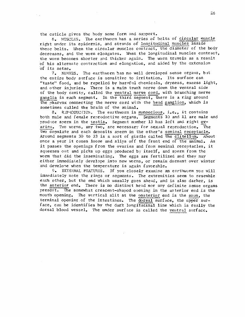
the cuticle gives the body some form and support. 6. MUSCLES. The earthworm has a series of belts of circular muscle
right under its epidermis, and strands of longitudinal muscles inside these belts. When the circular muscles contract, the diameter of the body decreases, and the worm elongates. When the longitudinal muscles contract, the worm becomes shorter and thiclter again.. The worm travels as a result of his alternate contraction and elongation, and aided by the extension of its setae.
7. NERVES. The earthworm has no well developed sense organs, but the entire body surf ace is sensitive to irritation. Its surf ace can "taste" food, and be repelled by harmful chemicals, dryness, excess light, and other injuries. There is a main trunk nerve down the ventral side of the body cavity, called the ventral nerve cord, with branching nerve ganglia in each segment. In the third segment, there is a ring around the pharynx connecting the nerve cord with the head ganglion, which is some.times called the brain of the animal. --
s. REPRODUCTION. The earthworm is monoecious, i.e., it contains both male and female reproductive organs. Segments 10 and 11 are male and prodllce sperm in the testis. Segment number 13 has left and right £Yaries. Two worms, any two, are necessary for sexual reproduction. The two copulate and each deposits sperm in the other's seminal receptacle. Around segments 30 to 33 is a sort of girdle called the clitellum. About once a year it comes loose and slips off the front end of the animal. As it passes the openings from the ovaries and from seminal receptacles, it squeezes out and picks up eggs produced by itself, and sperm from the worm that did the inseminating. The eggs are fertilized and they may either immediately develope into new worms, or remain dormant over winter and develone when the temperature is again favorable.
9. EXTERNAL FEATURES. If you closely examine an earthworm you will innnediately note the rings or segments. The extremities seem to resemble each other, but the end which usually goes ahead, and is also darker, is the anterior end. There is no distinct head nor any definite sense organs present. The somewhat crescent-shaped onening in the anterior end is the mouth opening. The vertical slit at the posterior end is the anus, the terminal opening. of the intestines. The dorsal surf ace, the upper sur-f ace, can be identifies by the dark longitudinal line which is really the dorsal blood vessel. The under surface is called the ventral surface.

27
EARTHWORM DISSECTION
Equipment: Dissection pan, lab kit, 12 pins, hand lens, 2 paper towels, and- the earthworm.
1. EXTERNAL FEATURES. Examine an entire earthworm and identify: Anterior end Mouth (in anterior end) crescent
shaped Lobe over the mouth Posterior end Anus (vertical slit) Dorsal surface (darker, with
median line of dorsal vessel) Ventral surface (slightly flat
tened) Segments (ring-like divisions of
the body) Cli tellum (smooth swelling over
several anterior segments)
Setae (tiny spines on segments) Oviducts (2 small openings,
ventral on segment XIV) Sperm ducts (2 vertical slits
ventral on segment XV) Kidney pores (2 per segment, very
small, latero-ventral)
Count the segments in your specimen (a) anterior to the clitellum, (b) in the clitellum, and (c) behind the clitellum. Do not count the lobe over the mouth.
2. INTERNAL FEATURES. Cut the specimen in half about one inch 1Josterior to the girdle. Keep all parts of the worm until the exercise is completed.
Holding the anterior part of your worm with the dorsal side uppermost (recognized by dark line of dorsal vessel) and using your scissors, cut forward through the dorsal body wall (only), just to the left of the mid-dorsal line to about segment IV. Keep the point of the lower scissors blade from damaging internal organs.
Pin down the specimen by the posterior end, with the worm in the center of the dis:.ecting pan. Beginning posteriorly grasp the cut edges of the body wall with the forceps, and use a dissecting needle to release the thin transverse septa from their attachments to the digestive tract, Keep the blackish dorsal vessel uppermost. Loosen the tissue equally on both sides. As the body wall is spread, fasten it down with nins, inserted obliquely outward, Carefully cut and spread segments IV to I. Your instructor will moisten the worm with water.

Locate the following organs: Pharynx (swollen, with external
muscle fibers, III to V) Esophagus (slender, partially hid
den, VII to XIV) Crop (large, thin walled, spher
ical, XV to XVI) Gizzard (large, thick muscular
walls, XVII to XVIII) Head ganglion. (2 small white
lobes, dorsal in III) Nerve cord (whitish band on ven
tal wall of body cavity)
28
Neohridia (1 pair in each segment, small white coiled tnbes, on the sides of body wall and towards the ventral)
Dorsal vessel (middle, over digestive tract, dark in color)
Intestines (from segment XIX and posterior)
Aortic arches (on the side of the esonhagus, partly hidden under thick tissue, VII to XI)
3. Clean your lab dissection tools. Remove c:,ins from worm and wrap the worm in a paper towel. Discard. Rinse dissection pan in water carefully, and blot dry. Check lab kits to be sure all the tools are in place. Return the equipment to its proper locality.
G.iZ'2arJ
Bra.in
Ven tr.ii +-----:::~-r nerlf~ col"cil
Sem;,ia.l recept~le ._~__. l!i:i~~--=ic-----lr- 'Testis
.Sperm. funnel Semin~l vesic:le

29
GRASSHOPPER STUDY
ThP grasshopper is a representative of the Phylum ARTHROPODA. This major group includes the crustaceans, spiders, centipedes, millipedes,, and the insects, the class in which the grasshopper belongs. Collectively the Arthropods comprise the maioritv of all kno,,m animals. In the insect class alone there are 650,000 known species. Typically the body is segmented and comprised of head, thorax, and abdomen. It bears jointed a9pendages and its body is enclosed in a more or less hardened exoskeleton. The body contains many specialized muscles and has well developed sense organs. These enable many arthropods to be quite active and exhibit quick response to stimuli. All but the crustaceans are predominantly terrestrial.
The following material will give you further understanding about the grasshopper's physical structure and body ftmctions.
1. RESPIRATION. In insects respiration occurs by means of tubes called trachea that take the air from openings through the body wall called spiracles, and release it from other spiracles. Usually each abdominal segment has a pair of spiracles, and so do some thoracic segments. It may be noted that this method of respiration is inefficient because four parts of useless nitrogen are transported for each part of useful oxygen. Insects succeed well only so long as no part of the body is very far from the exterior; no insects are or can possibly be very large for this reason.
2. NUfRITION.. Nearly anything in the world, from wood to blood, that is eaten by anything else is also eaten by one or more kinds of insect. The individual insect sometimes has an amazingly monotonous diet, such as only cabbage or potato plants. The grasshopper is less particular in its diet and will eat nearly anything. .Most of the plants that the grasshopper desires to eat are also directly or indirectly important for our food. As is true with many organisms, their common nutritional desires compete with man. For this reason they are termed pests. The food enters the grasshoppers mouth between two mandibles which chew horizontally. Salivary glands open into the mouth. From the mouth the food goes through a slender esophagus into a large crop. Between the crop and the stomach are six double finger-shaped structu~es called the gastric caeca. These are important in that they provide extra area for absorption. Beyond the stomach is the intestines, rectum, and the terminal opening, the anus.
3. EXCRErION. In insects, this process is carried on by malphighian tubules that empty into the intestines. ·
4. CIRCULATION. The heart of the grasshopper is all ventricle. It lies along the dorsal nart of the body cavity and squirts blood at the head. The blood works its way back to the abdomen where it re-enters the heart through many little openings. Recall the trachea not the blood is responsible for carrying oxygen through out the body.
s. SKELETON. As previously mentioned the insect has an exoskeleton. This is made of a tough, slightly flexible material. Growth occurs only by molting or sheding their skin. A new skeleton develops within a few hours.
6. MUSCLES. The muscles in insects are very efficient. They are attached to the exoskeleton in a way to provide excellent leverage.
30
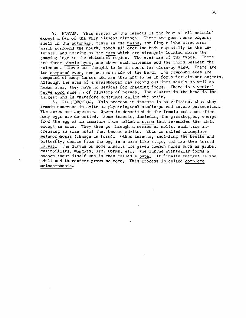
7. NE'lVES. This system in the insects is the best of all animals' excent a few of the very highest classes. There are good sense organs: smell in the antennae; taste in the palps, the finger-like structures which surround the mouth; touch all over the body especially in the antennae; and hearing by the ears which are strangely located above the jumping legs in the abdominal region. The eyes are of two types. There are three simple eyes, one above each antennae and the third between the antennae. These are thought to be in f OCUS for close-up view. There are two compound eyes, one on each side of the head. The compound eyes are composed of many lenses and are thro,,ght to be in focus for distant objects. Al though the eyes of a grasshopper can record outlines nearly as well as human eyes, they have no devices for changing focus. There is a ventral nerve cord made un of clusters of nerves. The cluster in the head is the largest and is therefore sometimes called the brain.
8. REPRODUCTION. This process in insects is so efficient that they remain numerous in sni te of physiological handicaps and severe persecution. The sexes are seperate. Sperm is deposited in the female and soon after mru1y eggs are deposited. Some insects, including the grasshopper, emerge from the egg as an innnature form called a nymph that resembles the adult except in size. They then go through a series of molts, each time increasing in size until they become adults. This is called incomplete metamorphosis (change in form). Other insects, including the beetle and butterfly, emerge from the egg in a worm-like stage, and are then termed larvae. The larvae of some insects are given common names such as grubs, caterpillars, maggots, army worms, etc. The larvae eventually forms a cocoon about itself and is then called a pupa. It finally emerges as the adult and thereafter grows no more. This process is called complete metamorphosis.

31
GRASSHOPPER DISSECTION
Equipment: Dissection pan, lab kit, hand lens, 2 ;:,aper towels, microscope, microscope slide and cover glass, and the grasshopper.
1. EXTE'{NAL FEATURES. Examine an entire grasshopner and identify the parts of the body:
Head Region Abdomen Antennae (2, slender) Segments (number?) Compound eyes (2, large, lateral) Spiracles (small openings on sides Simple eyes (3, small between) of segments; note the two spir-
comoound eyes) acles on the thorax) Mouth parts (ventral). Note the Ear (2, lateral on first abdomenal
way they function. segment) Thorax Ovipositor (on female, of 4 spurs,
Legs (3 pairs) posterior) Wings (2 pairs) Anus (small opening, posterior)
With your forceps, carefully remove the three legs from the left side, using care to bring away the base segment of each. Locate on each: Femur (large, upper leg); Tibia (slender, barbed, middle leg); Tarsus (foot region); and Claws (lateral).
2. INTERNAL FEATURES. · With scissors, and beginning at the tip of the abdomen, make a lengthwise cut in the body covering slightly to the left of the middorsal line and along the entire length of the grasshopper. Cut over the top of the head and down, between the antennae, to the mouth. Make a similar cut ventrally and also up the front of the head. Keep the inner scissors point just inside the body covering to avoid damaging the internal organso
Carefully lift and dissect off the body covering of the left s1cte. Keep asmuch as possible of the internal organs in place in the right side of the body covering. Note the many muscles, especially those in the thorax connecting to the legs and wings. Also note the large tracheal vessels which adhere to the inside of the sniracles. Recall that the body covering that you have removed is the ~xoskeleton of the insect.

32
If the specimen is a mature female, the interior spaces may be filled largely with slender eggs in the ovaries. Remove such of the lateral muscles and tracheae as necessary without injuring other organs. Your instructor will flood the specimen with water to aid in distinguishing the organs.
Locate the following organs: Mouth (between mandibles) Salivary glands (slender, whit-
ish, ventral to crop) .Esophagus (slender, short, in
head) Crop (large, thin-walled in the
thorax) Gizzard (short, thickwalled) Gastric caeca (6, double, finger
like, on sides of stomach)
Stomach Malpighian tubules (many, hair
like, function as excretory organs)
Intestine (tapered, in abdomen) Rectum (swollen, near end of the
abdomen) Anus (terminal opening of the di
gestive system) Heart (region, dorsal in abdomen)
The nervous system in the grasshopper occupies the head and ventral floor of the thorax and abdomen. Identify: Brain (3 lobes, with nerves to Ventral nerve cord (double, length-
the ·three simple ancl two com- wise, with clusters of nerves pound eyes) at various locations)
Stomac~

33
FROG;STUDY
The frog is a representative of the Phylum CHORDATA, Subphylum VERTEBRATA. The Class to \'Illich the frog belongs is the Alllphibia which means dual life. This Class name indicates that most of the species live partly in witer and partly on land. Other common Alllphibians are the toads and salamanders. The larvae of the Amphibians, usually called tadpoles, are always aquatic and breathe by means of gills. Lungs are developed in most species. The adults, even when they spend their entire existence in the water, breathe air. In some salamanders, both gills and lungs are found in the adult. In nearly all cases their eggs are laid in water so that even the toads, which leave the water for a part of their lives, return to it in the breeding season.
The Amphibians are cold blooded animals, that is, their body assumes the temperature of the environment rather than maintaining a constant body temperature. In regions where the temperature in winter goes below freezing the Amphibia burrow into the mud at the bottom of ponds where they hibernate in a dormant condition awaiting the coming of warmer weather.
The following material will give you further understanding about the frogs physical structure and body functions.
l. RESPIRATION. The frog's blood receives oxygen and gives 11p carbon dioxide through its skin, the lining of its mouth, and its lungs. The frog's skin is abundantly supplied with blood vessels and is kept moist with mucus from glands in the skin. Oxygen from the air or w2.ter surrounding the frog passes throngh the cells of the epidermis and of the walls of the capillaries into the blood stream. Carbon dioxide diffuses from the blood through the same structures. The frog lacks a diaphragm muscle, so another device is necessary to force air into its lungs. The frog accomplishes this by gulping a mouth full of air, closing its nostrils with its tongue, and moving unward the floor of its mouth, which forces the air through the glottis, past the larynx or voice box, and into the lungs. When a frog "croaks", air is forced through the larynx between stretched vocal cords. These vibrate and cause the sound.
2. NUTRITION. While the tadpole is strictly a vegetarian, the adult frog feeds on insects and other small animals. It catches insects by flipping out its tongue, which is attached at the front of the mouth. The frog has teeth on its unper jaw, maxillary teeth, and on the roof of its mouth, vomerine teeth. It does not chew its food but swallows it whole, using its teeth only to seize the food. The food ryasses from the mouth, through the tubular esophagus, and into the stomach. The stomach bulges out on the left side and gradually tapers to the pylorus, a valvular structure which can control the passage of food into the intestines. The intestines consist of a long, coiled small intestine, and the much larger large intestine. The solid waste materials pass from the large intestine into a region called the cloaca which is a common receptacle for intestinal wastes, and sperm or eggs produced from the sex organs. Posterior to the cloaca is the terminal opening, the anus. The frog has two imDortant digestive organs; the liver, a dark 'i:'ed=brown structure which is located anterior to the stomach; and the pancreas, a yellow-white organ which is orientated between the stomach and the first coil of the small intestine. The gall bladder, a reservoir for bile produced by the liver, is a green sac-like structure found between the lobes of the liver. The stomach,

34
small intestine, and large intestine are attached to the dorsal body wall along the line of the back bone by a transparent tissue called the~tery. In this mesentery run the blood vessels and nerves to the various parts of the digestive tract.
3. EXCRfil'ION. The kidneys of the frog are two reddish, flat, oval bodies lying against the dorsal body wall about midway between the ends of the.abdominal cavity. They are covered on their ventral side by the peritoneum, a transparent continuous tissue which completely encloses the body cavity. From the side of each kidney runs a tube called the ureter which opens into the cloaca. The urinary bladder, a white sac-like structure, is an outgrowth from the ventral side of the cloaca and is not connected with the ureters.
4. CIRCULATION. The frog has a completely closed circulatory system consisting of arteries connected to veins by capillaries. A muscular heart pumps the blood through these vessels. TI1e heart consists of left and right auricles and a ventricle. The right auricle receives blood from the body, and the left auricle receives blood from the lungs. The ventricle pumps part of theblood to the bngs where carbon dioxide is exchanged for oxygen, and part of the blood to the body. The heart is enclosed in a delicate membrane, the pericardial sac. The spleen in the frog is a small, spherical, dark-colored body in the mesentery not far f rem the stomach. It acts as a reservoir in which blood may be stored to be released into the circulation when needed.
5. STCELL'TON. The internal supporting framework of the frog's body is an endoskeleton consisting largely of cartilage and bone. The main parts of the skeletal system is the cranium, the vertebral colunm, and the bones of the appendages.
6. MUSCLES. The muscles of the hind limhs of the frog are common to many persons who eat them as frogs legs.
7. NERVES. The nervous system of vertebrates is more complex than that of any other animals. It comprises a central nervous system consisting of the brain and spinal cord, and the body nerves. The spinal cord is a thick tube directly connected with the brain. It passes through the bones of the vertebral column. The large eyes of the frog protrude from either· side of the head and look sldewise so that their vision covers entirely different fields. The upper eyelid is capable of only slight movement, but the lower eyelid can be drawn up to cover the remainder of the exposed part of the eye. The lower eyelid is composed of two parts, a lower eyelid proper, which forms the thicker lower portion and a nictitating membrane, a thinner and more transparent upper portion. The external ear of the frog is ap0arent as the eardrum or tympanic membrane. It consists of a tightly stretched membrane on the side of the head 9osterior to the eye.
8. REPRODUCTION. The testes of the frog are two small, white, sausage-shar,ed bodies which lie in the body cavity one on the ventral side of each kidney. Sperm pass through small tubules coruiecting the testes with the ureter, through the ureters to the cloaca and to the exterior. Ovaries of the frog lie in the abdominal cavity ventral to the kidneys, corres':}onding in number and location with the testes. At the beginning of the breeding season they are large irregular masses which almost completely fill the bod, cavity, and the innumerable small black and white eggs vvhich they contain give the organ a speckled anpearance. Eggs pass from the ovaries through oviducts, and into the cloaca. From this point they pass to the exterior.

35
FROG DISSECTION
Day 1: covers outline no .. 1-4. Day 2: covers outline noo 5.
Equipment: Dissection uan, lab kit, hand lens, 12 pins, two paper towels, microscope, microscope slide and cover glass, ru,d the frog.
1. E,'(TERNAL PEAT 1rrns. Examine an entire frog and identify the following structures: Head (to behind eardrum) Trunk (remainder of body) Mouth Anus (opens dorsally at end of
body) Fore limb (unper arm, forearm,
had, wrist, 4 digits) Hind limb (thigh, lower leg,
ankle, foot, 5 digits, webs) Nostrils (2, on snout)
Eye, each with Upper eyelid (fleshy) Lower eyelid (narrow) Nicti tating membrane (large
thin eyelid, inside others) Tympanic membranes (eardrums, be
hind eyes)
The innermost digit of each hand in males is enlarged.
2. MOUTH CAVITY. Open the frog's mouth wide 1 y by bending the lower jaw far back. With your scissors, cut the muscles at the corners of the mouth. Vfash out any mucus ')resent. Maxillary teeth (on upper jaw,
many, like sandpaper) Internal nostrils (2, anterior
O:Jenings, connected to external nostrils)
Vomerine teeth (2 small patches, between internal nostrils)
Eustachian tube openings (2 at corners of mouth;each connects to cavity of middle ear under eardrum)
Tongue Pharynx (posterior part of the
cavity, behind the tongue) Glottis (lengthwise slit in ven
tral wall of pharynx) Esonhag,1s (posterior to "harynx)
Find the Following:
Yornetine -l'.eeili. ~~--- Iiiternal No.shil.s
l-1a>filf ~ty tee.th
f Lt.cf~cla;an. tube openin2 e:;.;;::::a,.;,.~ ...... ....._- f .Sop has us
Gl0Hi1
Tongue
3. MUSCLBS OF THE THIGH. Place the frog on its back with its hind limbs pointing away from you. With your scissors cut through the skin, from the point where the hind limb connects with the tr;mk, to the knee. Cut the skin around the hind limb between it and the trunk. Free the slcin from the underlying muscles. Note that the muscles exist in bundles. With the dull side of the scaple, seperate the muscle bundles and note the white colored thigh bone. Note how the muscles are attached to the bones of the thigh and the knee.
4. SKIN OF THE FROG. Note the skin coloration differs on the uqper and under surface of the frog. With your scissers, cut out a oigmented section of the skin from the leg of the frog. Mount on a slide with

the inner side up. Examine under low ryower. Note the many blood vessels and the pigment cells.
36
5. INTERNAL FEATURES. Lift and continue cutting the skin across the trunk just anterior to the hind legs. Hold ur, the cut edge and slit the skin anteriorly up to the chin. Keep the lower edge of the scissors close uo beneath the skin. Cut the skin free from the entire ventral surf ace of the head and trunk. Note the abdominal muscles beneath the skin. Cut the muscle across just anterior to the hind legs. Cut the muscles of the abdominal wall on each side of the ventral blood vessel, from the hind legs to the head. (Be careful not to injure any of the organs below the muscle.) This leaves a narrow strip of muscle with the abdominal vein inside. Cut this strip anteriorly, then carefully dissect it free from the vein back to the nosterior end. Cut through the shoulder girdle with your scissors. With your thumbs, grasp the cut ends of the bones and gently force them apart. Pin the shoulders to the dissection pan. Free and pin do\m the abdominal muscles. Locate the following struch1res:
*Ovaries and eggs (very prominant Pancreas (irregular flattish tis-in the female only) sue, yellowish white, between
Liver (large, firm, reddish- stomach and first fold of the brown, 3 lobes) small intestine)
Heart (reddish, conical, mus- Mesentery (transparent tissue, cular sup,:orts small intestine) Pericardfr.m (delicate mem- Large intestine (dark, larger
brane enclosing the heart) extension nosterior to the. Auricles (2, upuer parts of small intestine)
the heart) Cloaca (region 9osterior to large Ventricle (lower, largest i;,art intestine, dark)
of the heart) Urinary bladder (attached to cloaca, Gall bladder (thin, S'Jherical sac, ventral)
greenish, between middle and Spleen (small, spherical, dark right lobes of the Ever) reddish, nosterior to stomach)
Stomach (long, whitish, a.long Lungs (2, dorsal to liver, soft, left side, dorsal to liver) thin walled)
Small Intestine (yellowish, Fat bodies (2 soft, finger-like slender, jrregularly coiled) lobes, yellowish, attached to
Peritoneum (transparent tissue, kidneys) encJ.oses body cavity, separ- Kidneys (2, elongated, dark brown ates the kidneys from the di- on dorsal wall of body cavity) jestive organs)
Testes (2, in male, hean-shaned pink or yellow, at anteroventral ends of kidneys)
>'<If you have a female frog, with your fingers, carefrilly grasp the egg masses and remove them from the frog. If you are careful, all of them may be removed in two clusters.

i.
!
!
G.A.11 blaclJc.r
S to twl.tch.. ~i.-a---
P«ne.reiils
:s,,.411 'ini:H!irie.
Mesenia.ry
Aot-tic dYch. ~>.-,:..~..:...+-t-t-
-...:-.J--1-;-- L•Ye v-
~~l-,l~::;;~Y--f- Spleen t/hi~~ Fa.t bocLy
fi=~f4=~HL-+- Testis
~-1--f---,- l(idney
... ~ .. : '-•A
37

Day 1 2 3 4 5
I.
II.
FETAL PIG STUDY AND DISSECTION
Covers Outline No. I - V VI VII IX - X -XI - XII
The fetal pig is removed from the mother in late stages of development. The young pig is attached to the wall of the uterus of the mother by the umbilical cord. After birth the severed cord becomes shriveled and the resulting scar is known as the navel. Preliminary examination of the pig and external features. A. Position of parts of the body.
Anterior (pertaining to head) Posterior (pertaining to tail) Dorsal (pertaining to back) Ventral (pertaining to belly) Lateral (pertaining to sides) Medial (pertaining to midline)
B. Body divisions. The body of fetal pig is divided into three general regions - head, thorax, and abdomen. ~~
Umb;/,ccll Co,-al
/11.:r.le -- ~ -------·
---
F,s- 1
How do the relative sizes of these regions compare with the adult pig?
The thorax, or chest region, extends from the posterior end of the neck to the last rib.
What part of the skeletal system supports the thorax?
38
The abdomen contains the main parts of the digestive tract, and the urogenital (excretory+ reproductive systenil, Notice the umbilical cord arising from the ventral por don of the abdomen. At the posterior end of the body, just under the tail is the anus, the opening through which solid waste products are expelled.~ you have a female pig, the urogenital opening is located just ventral to the anus. If you have a male pig, the urogenital opening is located just posterior to the umbilical cord. The scrotum, the sac which contains the testis, can be identified in the male as to swellings at the posterior end of the body.
The appendages include the fore and hind limbs of the pig. Notice that the first toe of each appendage is missing and the second and fifth toes are reduced in size. The pig walks on the tip of his third and fourth toe - somewhat of a ballet dancer.
The head region is the entire anterior portion of the pig. Located on the head are the ears, the eyes with upper and lower lids and a small mass of tissue in the corner of the eye called the

III.
IV.
nictitating membrane. The pig has a fairly large mouth, and two nostrils rimmed with tough connective tissue.
Brief demonstration showing major incisions and techniques. Major incisions and preliminary dissection. A. With your scissors, make an
incision from the umbilical cord to the hair of the chin. (Fig. 2; cut A)
B. Ma.~e an incision across the abdomen and through the skin and muscle from the umbilical
I
:A I I I I I
D I D. -~~.!:' •• ,: ..
39
cord to the ro,imals left side. (Fig. 2; cut B) Strip the skin and muscle away exposing the peritoneum. The periton-
I '• E f --+--1 ---- £ cJ
eum is a thin, transparent membriane that completely en-closes the organs of the ab-dominal cavity.
c. Cut through the length of the sternum, the breast bone. Do not cut farther. (Fig. 3; cut C)
D. Make an incision across the abdomen and through the skin and muscle exryosing more of the peritoneum toward the an-terior. (Fig. 3; cut D)
E. Note the sex of your pig. 1. If it is a male (c!), make an in
cision i inch to the left of the midline f rem the umbilical cord to the left side of the scrotum. Cut only through the skin and muscle. Since the position of the testes varies, depending on the age of the pig, care must be used in this dissection. (Fig. 2; cut BO) Continue the incision to the anus. Pull down the flap of skin and muscle exposing the peritoneum to the left side. The instructor will cut through the pelvic bone for you.
I '
,:,a. 3
2. If it is a f eraale ( 9 ) , make an incision through the skin and muscle i inch to the left of the midline from the umbilical cord to the urogenital opening, similar to the male dissection. (Fig. 2; cut Ei) Pull dovm the flap of skin and muscle on the left side exposing the tJeri toneum. Do not strip the right side. The instructor will cut through the pelvic bone.

v.
VI.
40
Preparation for preservation: A. Open up the peritoneum enough to expose the organs within, being
C,\reful not to cut the umbilical vein, leading from the umbilical cord to the liver. Through this vein comes food and oxygen from the mother to the embryo pig.
B. Wash the pig thoroughly. c.. Place the pig in formaldehyde for preservation (1 part f ormalde
hyde to four parts water). General anatomy of the thoracic and abdominal cavities. A. Thoracic cavity. Place your finger through the opening in the
thoracic cavity and note the smooth lining. This lining is the pleura. It forms a sac around the lungs. Locate the following organs:
* 1. Right and left lungs. These are reddish, spongy organs and in the pig~ small and collapsed because they have never been inflated.
* 2. Heart. This is a reddish, cone-shaped organ. It is enclosed in transparent membrane, the pericardium. The heart lies between and in front of the lungs.
* 3. Diaphragm. This is a muscular sheet that separates the abdominal cavity from the thoracic cavity. During respiration it moves up and down aiding in filling the lungs. Free the diaphragm on both sides as demonstrated by cutting through it close to its line of attachment to the ribs.
B. Abdominal cavity. This cavi tw lies below the diaphragm and contains other organs. Most of these organs were enclosed in the peritoneum when the dissection was started. Locate the following organs within the abdominal cavity.
* 1. Liver. This is a large, five-lobed red organ just below the diaphragm and to the right in the abdominal cavity. Note that the umbilical vein, carrying food to the embryo from the mother, is attached~the liver. The liver has many functions. Pt.mong the I.TlOst important of these are the storage of glycogen and the secretion of bile which aids in digestion of fats.
* 2. Gall Bladder. A small greenish sac on the ,mcter side of the Tiver. It may be seen by raising the liver. Trace the tiny duct leading from the gall bladder to the duodenum. This is the bile duct. Bile is made in the liver, stored in the gall blad~and is released as needed into the small intestine.
* 3. Stomach. This is a whitish organ to the left in the abdominal cavity and r,artly covered by the liver. The stomach secretes digestive juices, partly digests ryroteins and stores food temryorarily until it is released into the small intestine.
* 4. Spleen. The soleen is a thin red organ wrapped around the left surface of the stomcah. Its function has not been clearly defined. It is part of the circulatory system and can store large quantities of blood. It has been removed in man with few ill effects.
* 5. Pancreas. A gland below the lower margin of the stomach. It is a long, irregular, grayish gland and .quite soft 0 Look c~:i,ref ully and do not destroy it. The pancreas secretes a

41
digestive juice containing three enzymes that aid in the digestion of carbohydrates, proteins and fats. This secretion is carried to the duodenum by a very tiny duct. The pancreas also manufactures the hormone insulin which is secreted directly into the blood stream. Diebetes is an organic disease caused by insufficient secretion of insulin.
* 6. Small intestine. This is a coiled tube that begins at the lower end of the stor.1.ach. Note the extreme length that provides for ade~uate digestion and absor?tion of food. The walls of the small intestine secrete a digestive juice that aids in the digestion of carbohydrates, proteins and fats. It is the organ where most of the food is absorbed into the blood stream.. The first part of the small intestine is the duodenum.
* 7. Large intestine. This is a coiled mass rather tightly bound together in the lower left abdominal cavity and continuing from the small intestine. The large intestine carries undigested food toward the rectum.
*,8. Caecum. This is a part of the large intestine which is a blind pouch at the point where the small intestine joins the large in- Smc1II ita~es~;ne testine. Although the pig has none, this is the approximate location of the appendix in man.
* 9. Mesenteries. Membranes that support the intestines from the back abdominal wall. The blood vessels in the mesenteries carry digested food.
* 10~ Rectum. The extreme end of the digestive tube which Hes at the back of the abdominal cavity under and below the intestinal coils. Here hia.4 wastes are collected and stored until they are excreted through the anus, the lower opening of the digestive tube.
* 11. Kidneys. A pair of bean-shaped, reddish organs at the back of the abdominal cavity. These organs remove urine, liquid wastes, from the blood streamo These organs lie behind the peritoneum.
* 12. Urinary bladder. A single organ attached in the fetal animal to the umbilical cord. UrL,e secreted by the kidneys is carried to the urinary bladder by two small tubes, the ureters. Urine is stored in the urinary bladder until released through a tube, the urethra. Trace the two ureters from the kidneys to the bladder, and the urethra from the bladder to the urogenital opening.
>'< 13. Gonads. The reproductive glands producing sex cells and hormones. a. Testes. These are the male reproductive organs: A pair
of grayish colored organs located below and outside of the abdominal cavity. They are contained in a protective sac, the scrotum, in a fully developed fetus. Sperm, the male reproductive cells, are produced by this organ.

42
b. Ovaries. These are the reproductive organs of the female; Small, paired organs located in the lower part of the abdominal cavity, one on either side below the kidneys. Ova, the female reproductive cells, are produced by this organ.
VII. Sketch the contents of the thoracic and abdominal cavities. A. Make a full page drawing of the dissected pig. B. As directed by the instructor t arrange and spread out the organs
in the dissected animal so that each organ, or at least a part of each organ, marked with an ('') is visible and can be sho1Jm on the drawing. Label each organ marked with an (*) in the above sketch.
c. When the drawing has been completed, remove the liver and gall bladder. Then cut the stomach at the diaohragm and the large intestines at the rectum. Take hold of the entire intestinal tract, lift up, and remove by cutting the supporting membranes. Wrap the removed organs and discard as directed.
VIII. Dissection of the neck region. A. Gently free the tissues from the sternum. Be careful not to cut
any of the large blood vessels leading from the heart. Identify: 1. Thyroid gland. A small, oval, red gland which lies on top of
the trac~r windpi'1e. Like the pancreas it secretes a hormone, thyroxin, into the blood stream. This substance controls the :irowth rate and also the rate at which an animal oxidizes food. Such glands as the thyroid that secrete hormones directly into the blood stream are called endocrine glands.
2. Trachea or windpipe. A long tube in the front of the esophagus. The walls of the trachea are com,,,osed of partial rings of cartilage which keep the air passage opened.
3. Larynx or ''voice box". A rounded, short, hard structure connected to the upoer end of the trachea. Air on its way from the lungs passes through the larynx and in so doing passes over the vocal cords causing them to vibrate and produce sound.
4. Carotid arteries. Extend up the side of the neck, one on each side. They supply blood to the head.
5. The vagus nerves. Large nerves found with the carotid arteries in ac:oiiimoii""sheath. Trace the nerve to the diaphragm.
6. Thymus gland (see IX. A. 1.) IX. Observation of the heart and major blood vessels.
A. Locate the following structures: 1. Thymus gland. An irregular mass in front of and above the
heart. This is another endocrine gland, but its function is not clearly understood. The gland disappears in adulthood. The thymus gland of the calf is sold as a food delicacy known as "sweetbread. 11
2. Pericardium. This is the membrane which surrounds the heart. Dissect avmv the l)eric ardium, the thymus gland, and other loose tissue around the hear.
B. Draw and label the heart, major blood vessels, and the structures identifies in the neck region. (Figs. 5 and 6).

i-----C ,,..,...--JJ
EJ
KEYS TO FIGS. 5 AND 6
A---Aorta C---Carotid artery CC--Common carotid D---Ductus arteriosus EJ--External jugular vein IJ--Internal jugular. vein IVC-Inferior vena cava L---Larynx LA--Lef t auricle
43
L+---if-+-,.-.---~CC SVc ------+___,,.. -#------ s
LV--Left ventricle P---Pulmonary artery RA--Right auricle RV--Right ventricle s---Subclavian artery SVC-Superior vena cava
x.
_......, ____ A --~'-'~---P
---- LA T---Trachea
Jaw incision (See Fig. 7)
Th--Thyroid V---Vagus nerve
Fig. 6 (Showing the ductus arteriosus. This is a short tube between the pulmonary artery and the aorta in the fetal pig. A few days after birth, the ductus arteriosus closes.)
A. Cut through the left jaw bone at the corner of the mouth. (Demonstration.) Bend the lower jaw bone back until the back of the throat is exposed. Locate the following: 1. Epiglottis. A pointed fold of tis
sue that forms a lid over the opening of the larynx. Bend it down to see how it works.
2. Pharynx. This is the throat region, a collllilon passage for both food and air.
3. Esophagus. A tube that lies behind the trachea. It takes food to the stomach.
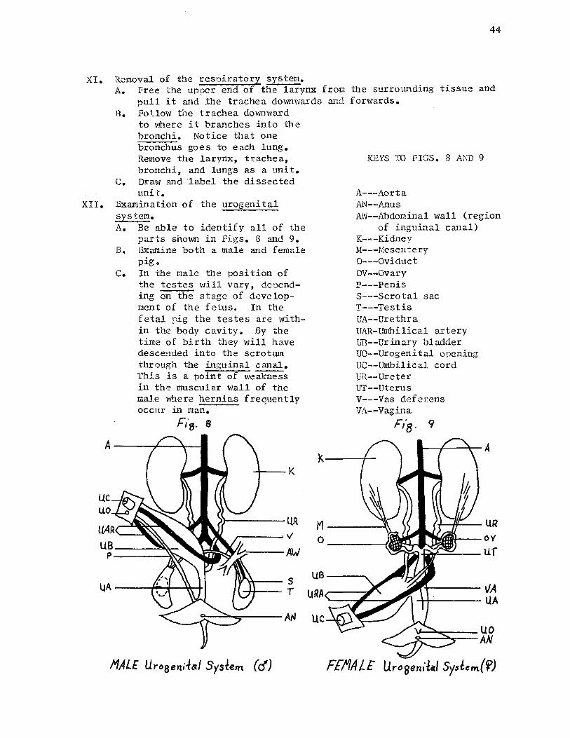
44
XI. Removal of the respiratory system. A. Free the upper end of the larynx from the surrounding tissue and
pull it and the trachea downwards and forwards. B. Follow the trachea downward
to where it branches into the bronchi. Notice that one bronchus goes to each lung. Remove the larynx, trachea, bronchi, and lungs as a unit.
c. Draw and label the dissected unit.
XII. Examination of the urogenital system.
UC uo
A. Be able to identify all of the parts shown in Figs. 8 and 9.
B. Examine both a male and female pig.
c. In the male the position of the testes will vary, depending on the stage of development of the fetus. In the fetal pig the testes are within the body cavity. By the time of birth they will have descended into the scrotum through the inguinal canal. This is a point of weakness in the muscular wall of the male where hernias frequently occur in man.
Fi"8. s
MAL£ Uro5enitaf System (cf)
KEYS TO FIGS. 8 AND 9
A---Aorta l\N--Anus AW--Abdominal wall (region
of inguinal canal) K---Kidney M---Mesentery 0---0viduct OV--Ovary P---Penis S---Scrotal sac T---Testis UA--Urethra UAR-Umbilical artery UB--Urinary bladder UO--Urogenital opening UC--Umbilical cord UR--Ureter UT--Uterus V---Vas deferens VA--Vagina
UB----...
F,·8· 9
~-+----\/A -+-t----UA
FEf/A L£ Urogen,lal Syslem{<f)

BIBLIOGRAPHY
Dale, Edgar. Audio-Visual Methods ~ Teaching. New York: The Dryden Press, 19~
Dawson, James A. and William Etkin. Basic Exercises in College Biology. New York: Thomas Y. Crowell Company, 1951.
de Laubenfels, M. w. Life Science. New York: Prentice-Hall, Inc., 1950.
Evans, w. L., Addison E. Lee, and George Tatum. The Fetal Pig. New York Rinehart and Company, Inc., 1958.
Hawse, Doris H., "Ooh! Worms!" Science Education, XLI (December, 1957), 436-39.
Kinsey, Alfred c. Methods in Biology. Chicago: J. B. Lippencott Company, 1937.
'
Lacroix, Donald s. "To Cut or Not to Cut!" Junerican Biology Teacher. XL (November, 1949), 169-70.
Lloyd, Francis E. and Maurice A. Bigelow. The Teaching of Biology in the Secondary School. New York: Longmans-;-c;reen and co":, 1909. ~~
McKnown, Harry c. and Alvin B. Roberts. Audio-Visual Aids ~ Instruction. New York: McGraw-Hill Book Company, Inc., 1940.
Miller, David F. and Glenn w. Blaydes. Methods and Materials for Teaching Biological Sciences. New York: McGraw-HillBook Company:---inc., 1938.
Morholt, Evelyn, Paul F. Brandwein, and Alexander Joseph. A Source Book for the Biological Sciences. New York: Harcourt, Brace and Company, 1958:--
Odlaug, Theron o. Laboratory Anatomy of~ Fetal Pige Dubuque: Wm. c. Brown Company, 1955.
Rowett, H. Dissection Guides, I. The Frog. New York: Rinehart and Company, Inc., 1957 •
---• Dissection Guides, v. Invertebrates. New York: Rinehart and Company, Inc., 1957.
Storer, Tracy I. General Zoology. New York: McGraw-Hill Book Company, Inc., 1943.
--....,,,.• Laboratory Manual for General ~logy. York, Pa.: Maple Press Company, 1944.
45
46
Wells, Morris Miller. The Collection and P'reservation of Animal Forms. Chicago: General Biological Supply House, Inc., 1932.
Yothers, Lee R. "An Identification Aid \'ben Dissecting Frogs." School Science and Mathematics. XLVII (May, 1947), 421-2.3.

VITA
Carl Christian Nelson
Candidate for the Degree of
Master of Science
Report: DISSECTION TECHNIQUES FOR. HIGH SCHOOL BIOLffiY TEACHERS
Major Field: Natural Science
Biographical:
Personal data: Born at Council Bluffs, Iowa, December 1, 1932, the son of Rev. Carl A. and Caroline Dorothea Nelson.
Education: Attended grade school in Fremont, Nebraska; graduated from Fremont High School, 1950; received the Bachelor of Science in Education degree from Dana College, Blair, Nebraska, with a major in Biology, in May, 1954; was a graduate student in the field of School Administration at the University of Nebraska during the sunnner of 1956; attended Oklahoma State University during the summer of 1957, and the academic year, 1958-1959; completed requirements for Master of Science degree from Oklahoma State University, in May, 1959.
Professional experience: Taught in the science field at Schuyler High School, Schuyler, Nebraska, 1954-1958.