Sulfated nanozirconia: an investigation on acid–base properties and n-butane isomerization activity
Upload
independentCategory
view
1download
0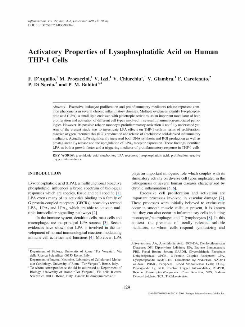
Activatory Properties of Lysophosphatidic Acid on HumanTHP-1 Cells
F. D_Aquilio,1 M. Procaccini,1 V. Izzi,1 V. Chiurchiu_,1 V. Giambra,1 F. Carotenuto,2
P. Di Nardo,2 and P. M. Baldini1,3
Abstract—Excessive leukocyte proliferation and proinflammatory mediators release represent com-
mon phenomena in several chronic inflammatory diseases. Multiple evidences identify lysophospha-
tidic acid (LPA), a small lipid endowed with pleiotropic activities, as an important modulator of both
proliferation and activation of different cell types involved in several inflammation-associated patho-
logies. However, its possible role on monocyte proinflammatory activation is not fully understood yet.
Aim of the present study was to investigate LPA effects on THP-1 cells in terms of proliferation,
reactive oxygen intermediates (ROI) production and release of arachidonic acid-derived inflammatory
mediators. Actually, LPA significantly increased both DNA synthesis and ROI production as well as
prostaglandin E2 release and the upregulation of LPA3 receptor expression. These findings identified
LPA as both a growth factor and a triggering mediator of proinflammatory response in THP-1 cells.
KEY WORDS: arachidonic acid metabolites; LPA receptors; lysophosphatidic acid; proliferation; reactiveoxygen intermediates.
INTRODUCTION
Lysophosphatidic acid (LPA), a multifunctional bioactive
phospholipid, influences a broad spectrum of biological
responses which are species, tissue and cell specific [1].
LPA exerts many of its activities binding to a family of
G protein-coupled receptors (GPCRs), nowadays termed
LPA1, LPA2 and LPA3, which are able to activate mul-
tiple intracellular signalling pathways [2].
In the immune system, dendritic cells, mast cells and
macrophages are the principal LPA sources [3]. Recent
evidences have shown that LPA is involved in the de-
velopment of normal immunological reactions modulating
immune cell activities and functions [4]. Moreover, LPA
plays an important mitogenic role which couples with its
stimulatory activity on diverse cell types implicated in the
pathogenesis of several human diseases characterized by
chronic inflammation [5, 6].
Excessive cell proliferation and activation are
important processes involved in vascular damage [7].
These processes were initially believed to exclusively
occur in smooth muscle cells; at present, it is known
that they can also occur in inflammatory cells including
monocytes/macrophages and T-lymphocytes [8]. In this
context, the presence of locally released soluble
mediators, to whom cells respond synthesizing and
0360-3997/06/0400-0129/0 # 2006 Springer Science+Business Media, Inc.
Inflammation, Vol. 29, Nos. 4Y6, December 2005 (# 2006)
DOI: 10.1007/s10753-006-9008-9
129
1 Department of Biology, University of Rome BTor Vergata’’, Via
della Ricerca Scientifica, 00133 Rome, Italy.2 Department of Internal Medicine, Laboratory of Cellular and Molec-
ular Cardiology, University of Rome BTor Vergata’’, Rome, Italy.3 To whom correspondence should be addressed at Department of
Biology, University of Rome BTor Vergata’’, Via della Ricerca
Scientifica, 00133 Rome, Italy. E-mail: [email protected]
Abbreviations: AA, Arachidonic Acid; DCF-DA, Dichlorofluorescein
Diacetate; DPI, Diphenylene Iodinium; EIA, Enzyme Immunoassay;
FBS, Foetal Bovine Serum; GAPDH, Glyceraldehyde Phosphate
Dehydrogenase; GPCRs, G-Protein Coupled Receptors; LPA,
Lysophosphatidic Acid; LTB4, Leukotriene B4; NADPHox, NADPH
oxidase; PBMC, Peripheral Blood Mononuclear Cells; PGE2,
Prostaglandin E2; ROI, Reactive Oxygen Intermediates; RT-PCR,
Reverse Transcriptase-Polymerase Chain Reaction; SDS, Sodium
Docecyl Sulphate; TCA, TriChloroAcetate.

releasing several biologically active molecules, such as
arachidonic acid metabolites and reactive oxygen
intermediates (ROI), may contribute to lesion initiation
and progression [9, 10]. In particular, ROI produced by
NADPH oxidase (NADPHox), the principal ROI source
in phagocytic cells [11], have been demonstrated to play
an important role in determining the oxidative stress at
inflamed sites, thus worsening the effects of arachidonic
acid metabolites release [12, 13]. Among the latter,
prostaglandins and leukotrienes generated by cycloxy-
genases and 5-lypoxygenase, respectively, are the most
powerful inflammatory lipid mediators [14].
In order to shed light on its capability to activate
THP-1 cells, LPA effects on key events of inflammatory
response, such as proliferation, ROI production and
release of arachidonic acid metabolites, were investigat-
ed. Our results demonstrated that LPA is able to promote
THP-1 cell activation leading to enhanced cell growth
and simultaneous ROI and prostaglandin E2 (PGE2)
release probably by means of a mechanism involving
the stimulation of LPA3 receptor.
MATERIALS AND METHODS
Cell Culture
The human monocytic cell line THP-1, used to
avoid inter- and intraspecific variability of peripheral
blood mononuclear cells (PBMC), was obtained from
American Type Culture Collection (Manassas, VA), cul-
tured in Falcon flasks with RPMI-1640 medium supple-
mented with 10% foetal bovine serum (FBS), L-glutamine
(2 mM), streptomycin (100 mg/ml), penicillin (100 U/ml),
sodium pyruvate (100 mg/ml), and maintained at 37-C in a
humidified atmosphere containing 5% CO2. Cells were
serum starved before each experiment to rule out
possible interferences with cell growth due to serum
components. In all experiments, to determine cell via-
bility, monocyte cultures were stained with Trypan blue
at specific timepoints after treatment with different
reagents. Stained vs living cells were counted under a
microscope using a Neubauer modified chamber.
DNA Synthesis Assay
THP-1 cells were cultured in Falcon flasks 75 cm2
and seeded in 30�15 mm dishes with serum-free
medium (1�106 cells/well). Cells were challenged with
different concentrations of LPA (0.1Y20 mM) for 3, 6 and
9 h. In a series of experiments, cells were pretreated with
Ki16425 (10 mM) for 30 min before LPA addition. [3H]-
thymidine incorporation into DNA was used to measure
the mitogenic response of THP-1 cells. Cells were pre-
treated with Ki16425 for 30 min, pulsed with [3H]-
thymidine (1 mCi/ml) and then challenged with LPA for
the requested times. Cells were then harvested by cen-
trifugation and treated with 5% TCA at 4-C for 15 min.
The TCA-insoluble fraction was resuspended in 0.1%
SDS in NaOH (200 mM) and samples were counted for
radioactivity, after addition of 3.5 ml Optifluor, by a
liquid scintillation counter (Tricarb 2180/TR, Packard
Instruments, Downers Grove, IL).
Reverse Transcriptase-Polymerase Chain Reaction
(RT-PCR) and LightCycler-PCR Analysis
Total RNA isolation and purification were carried
out by SV Total RNA Isolation System (Promega)
according to the manufacturer_s instructions. Briefly, total
RNA was extracted from 3�106 THP-1 cells treated or
not with LPA (1 mM) for 6 h. Total RNA quantity and
quality were assessed by UV absorbance at 260 nm and
electrophoresis on agarose/formaldehyde gel, respective-
ly. For RT-PCR analysis, cDNA was retro-transcribed
from 2 mg of total mRNA for 1 h at 37-C in an incubation
buffer containing deoxynucleotide triphosphate (dNTP,
250 mM each), random primers p(dN)5 (Roche), RNase
inhibitor and RNA murine Moloney leukaemia virus
reverse transcriptase (200 U, Promega). For classic PCR
analysis, an aliquot of each RT product (2 ml) was
amplified in a final volume of 50 ml using PCR buffer
containing dNTP (160 mM), primers designed to amplify
the mRNA of human LPA receptors (15 pmol each,
Invitrogen) and Taq polymerase (1 U Amersham Pharma-
cia Biotech.). All samples were subjected to the following
reaction conditions: 30 cycles at 94-C for 2 min, 94-C for
30 s, (60-C for 30 s for LPA2 and 58-C for 30 s for LPA1,
LPA3 and GAPDH), 72-C for 30 s, followed by a final
extension at 72-C for 3 min. Expression levels of recep-
tor mRNAs in THP-1 cells were quantified by normal-
ising their respective mRNAs levels to the housekeeping
human GAPDH mRNA levels. The amplified fragments
were then separated by electrophoresis on agarose gel
1Y2% in TAE buffer and visualized by ultraviolet
transillumination.
For LightCycler-PCR, an aliquot of each RT
reaction product (2 ml) was amplified using the Light-
Cycler FastStart DNA master SYBR Green I (Roche
130 D_Aquilio, Procaccini, Izzi, Chiurchiu_, Giambra, Carotenuto, Di Nardo, and Baldini

Diagnostic) and primers (10 pmol each). The reactions
for LPA receptors were undergoing to 95-C for 10 min
followed by 45 cycles 94-C for 10 s, (60-C for 5 s for
LPA2, 58-C for 10 s for LPA3, 60-C for 20 s for LPA1),
72-C for 15 s, and one melting curve analysis from
95-C, 65-C for 15 s and to 95-C for 10 min, using the
LightCycler instrument (Roche). The GAPDH reaction
was undergoing to 95-C for 10 min followed by 45
cycles 94-C for 10 s, 58-C for 20 s, 72-C for 15 s, and
one melting curve analysis from 95-C, 65-C for 15 s
and to 95-C, using the LightCycler instrument (Roche).
The expression levels of receptor mRNAs were
quantified by normalizing the mRNA levels to the
housekeeping human GAPDH mRNA by an external
standard curve (LightCycler operator_s Manual). The
external standard curve was created with dilutions of
genomic DNA, amplified with LPA3 primers.
Evaluation of ROI Production by 20,70-Dichlorofluorescein (DCF) Fluorescence
The samples (2�106 cells) were loaded with the
fluorescent indicator DCF-DA (10 mM) for 30 min at
37-C in the dark. DCF-DA diffuses through the cell
membrane and is hydrolyzed by intracellular esterase to
nonfluorescent DCF deacetylated, which is then rapidly
oxidized to highly fluorescent DCF in the presence of
ROI. The DCF fluorescence intensity is proportional to
the amount of intracellular ROI formed [15]. After the
incubation with the fluorescent dye, cells were collected,
centrifuged for 5 min at 1,200 rpm at room temperature,
resuspended in serum-free medium and challenged with
different LPA concentrations (0.1Y20 mM) for 3, 6 and 9 h.
When requested, THP-1 monocytes were pretreated with
diphenylene iodinium (DPI, 10j8 M), a specific inhibitor
of NADPH oxidase, and Ki16425 (10 mM), a LPA1 and
LPA3 receptor antagonist, for 1 h and 30 min, respectively,
before LPA addition. Fluorescence was measured under
continuous magnetic stirring and controlled temperature
(37-C) in a Perkin-Elmer luminescence spectrometer
(Model LS-5) equipped with a chart recorder (Model R
100A), with excitation wavelength at 485 nm and emission
wavelength at 530 nm using 5 and 10 nm slits respectively,
for each light path. Results were expressed as Fluorescence
Intensity, reported as Fluorescence Units (F.U.), respect to
cells loaded with only DCF-DA (C).
Primer Sequence
LPA1 sense 50-CGGCGGGTAGTGGTGGTC-30
LPA1 antisense 50-GTCGCGGTAGGAGTAAATGATG-30
LPA2 sense 50-GTCGAGCCTGCTTGTCTTC-30
LPA2 antisense 50-CCAGGAGCAGTACCACCTG-30
LPA3 sense 50-TCGCGGCAGTGATCAAAAACAGA-30
LPA3 antisense 50-ATGGCCCAGACAAGCAAAATGAGC-30
GAPDH sense 50-CATGGGTGTGAACCATGAGAAG-30
GAPDH antisense 50-GTGGCTGTTGAAGTCAGAGGAG-30
Fig. 1. LPA effects on THP-1 cell growth. THP-1 cells were grown in RPMI-1640 plus 10% FBS and serum-
starved before each experiment to rule out possible interferences with cell growth due to serum components. Cells
were challenged with different LPA concentrations (1Y5 2M) for 3, 6 and 9 h. [3H]-thymidine incorporation into
DNA was assessed as reported in the Materials and Methods section. Data are reported as mean T SD of 4
different experiments. *P < 0.05, as reported from Student_s t test in respect to untreated cells (C).
131Properties of Lysophosphatidic Acid on Human THP-1 Cells
132 D_Aquilio, Procaccini, Izzi, Chiurchiu_, Giambra, Carotenuto, Di Nardo, and Baldini
Roi Generation as Assessed by Microscopy Analysis
ROI generation was carried out on glass chamber
slides. 3�106 cells were labelled with the peroxide-
sensitive fluorescent dye 20,70dichlorofluorescein diace-
tate (DCF-DA, 10 mM) and incubated for 6 h in the
presence or absence of LPA plus or minus DPI (10j8 M)
or Ki16425 (10 mM), as described above. Intracellular
fluorescence was monitored using a fluorescence
microscope (Leica DMRB; objective: �200). Signal-
based averaging was used to quantitate the fluorescence
signal from five randomly selected fields (Delta System
Software).
[3H]-Arachidonic Acid Release Assays
THP-1 cells were seeded in 30�15 mm dishes with
serum-free medium (1�106 cells/well) and labelled for
3 h with 1 mCi [3H]-arachidonic acid (AA) (spec. act.
202.4 Ci/mmol) at 37-C as previously described [16].
To remove non-specific binding of [3H]-AA to cell
surface prior to agonist stimulation, culture medium
was eliminated, cells resuspended in serum-free
medium and challenged with different LPA concen-
trations (1Y20 mM) for 6 h. When requested, cells were
pretreated with Ki16425 (10 mM) for 30 min before LPA
addition. After treatment, supernatants were collected,
centrifuged at 1,200 rpm for 5 min to remove suspended
cells and 100 ml aliquots were added to 3 ml Optifluor
and analyzed by a liquid scintillator counter. Results
were expressed as [3H]-AA cpm/1�106 cells.
Measurement of Leukotriene B4 and Prostaglandin
E2 Release
3�106 cells/well were pre-treated or not with
Ki16425 (10 mM) for 30 min, challenged or not with
LPA (1 mM) for 6 h and, at the end of experimental
time, supernatants were collected and Leukotriene B4
(LTB4) and Prostaglandin E2 (PGE2) levels in the
medium were determined by enzyme immunoassay
(EIA) (Cayman Chemical), following manufacturer_sinstructions. Briefly, 50 ml LTB4 standard or samples
were added to the pre-coated mouse monoclonal anti-
rabbit IgG macrotitre plates. Subsequently, 50 ml LTB4
tracer and 50 ml monoclonal antiserum of LTB4 were
added into each well and the mixture was incubated
overnight at 4-C. After incubation, the content in each
well was removed and the wells were washed 5 times
with PBS buffer containing 0.05% Tween-20. An
aliquot of 200 ml Ellman_s reagent was added into
each well, and the mixture was incubated for 2 h at
room temperature with occasional shaking. The solution
optical density was determined by a multi-well spectro-
photometer (ELISA reader) at 405 nm. A similar proce-
dure was performed for PGE2 release determination,
using pre-coated goat polyclonal anti-mouse IgG macro-
titre plates. LTB4 and PGE2 concentration in each sample
was calculated as the concentration corresponding to
sample_s optic density (O.D.) values plotted on respec-
tive standard built-in curves. Values were reported as
percentile increase in respect to control (C).
Reagents
RPMI 1640, glutamine, penicillin (100 U/ml), and
streptomycin (100 mg/ml) were from Eurobio Labora-
toires. Foetal Bovine Serum (FBS) was from GIBCO
(Grand Island NY, USA). LPA (C18:1, 1-oleoyl-sn-
glycerol-3-phosphate), trypan blue, DPI, Ki16425 and
20,70-dichlorofluorescein diacetate (DCF-DA) were from
Sigma Chemicals. [3H]-thymidine (20 Ci/mmol), [3H]-
arachidonic acid (202.4 Ci/mmol) from Amersham Bio-
sciences. Random primers, murine Molooney leukaemia
virus reverse transcriptase, LPA receptors and GAPDH
primers were purchased from Invitrogen. Taq polymerase
was from Amersham Pharmacia Biotech. SV Total RNA
Isolation System was from Promega. LightCycler Fast-
Start DNA master SYBR Green I kit and all other reagent
for LightCycler-PCR were from Roche Diagnostic.
Leukotriene B4 and Prostaglandin E2 assay kits were
purchased from Cayman Chemical.
Statistical Analysis
Data distribution was preliminarily verified by the
KolmogorovYSmirnov test. Each experiment set was
independently performed and compared with the same
control by Student_s t-test. Quantitative data were
expressed as the meanTSD of at least four replicate
determinations, except where otherwise indicated. Dif-
Fig. 2. LPA effects on ROI production in THP-1 cells. THP-1 cells were
labelled with DCF-DA (10 2M) as reported in the Materials and Methods
section and ROI production was assessed after cell exposure to different
LPA concentrations (1Y5 2M) for 3, 6 and 9 h (a) or after cell exposure
to LPA (1 2M) for 6 h (b). In the experiments with DPI (10j8 M), cells
were pre-treated with the inhibitor for 1 h before LPA addition. Results
are expressed as Fluorescence Intensity, reported as Fluorescence Units
(F.U.), in respect to cells loaded with DCF-DA only (C). Micrographs
were acquired as reported in the Materials and Methods section.
Original magnification: �200. Data are reported as mean T SD of 4
different experiments. *P < 0.05, as reported from Student_s t test in
respect to untreated cells (C).
133Properties of Lysophosphatidic Acid on Human THP-1 Cells
ferences were regarded as significant when P value was
less than 0.05.
RESULTS
Effects of LPA on THP-1 Cell Growth
To evaluate LPA ability to stimulate THP-1 cell
growth, cells were challenged with different lipid concen-
trations (1Y5 mM) and assayed for [3H]-thymidine
incorporation into DNA at 3, 6 and 9 h. As shown in
Fig. 1, only 1 mM LPA induced an increased DNA syn-
thesis that was statistically significant at 3 h and reached
a maximum (+50% in respect to untreated cells) at 6 h.
Longer experimental times and lower (0.1 and 0.5 mM)
or higher (10 and 20 mM) LPA concentrations (data not
shown), did not exert any significant effect.
LPA Effects on ROI Production and NADPH
Oxidase Activation
To evaluate the ability to stimulate ROI production,
a fundamental marker of cell activation [17], THP-1 cells
were challenged with LPA (1Y5 mM) for different
experimental times (3, 6 and 9 h) after labelling with
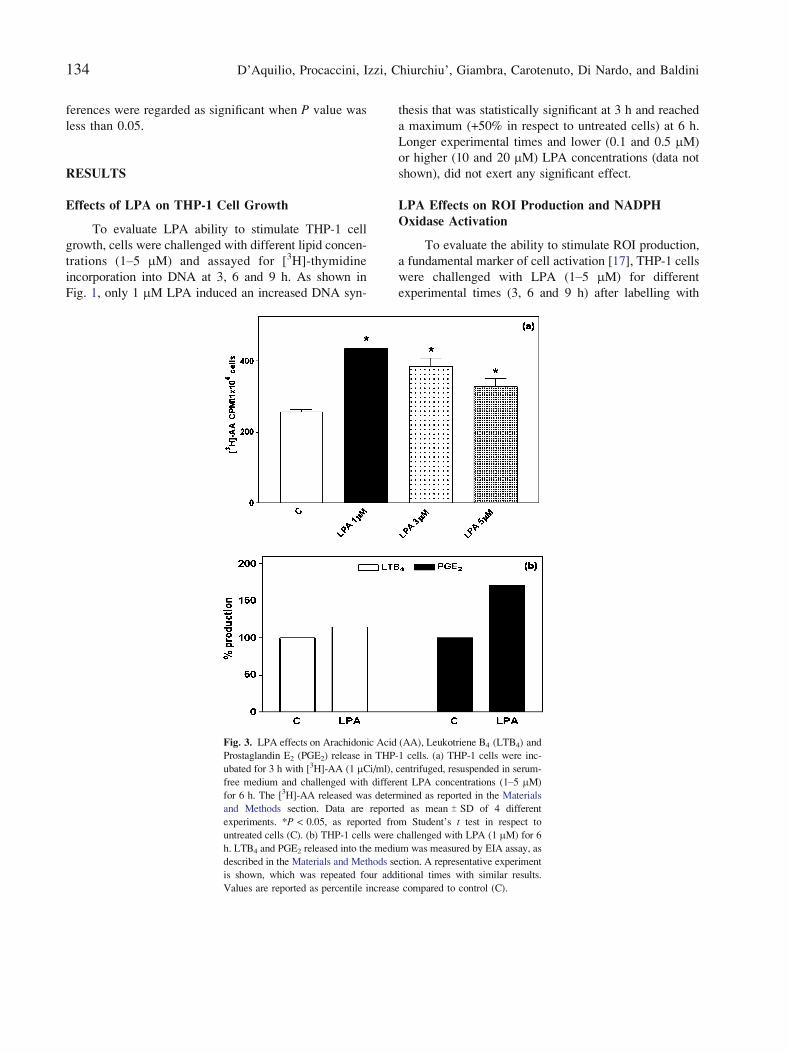
Fig. 3. LPA effects on Arachidonic Acid (AA), Leukotriene B4 (LTB4) and
Prostaglandin E2 (PGE2) release in THP-1 cells. (a) THP-1 cells were inc-
ubated for 3 h with [3H]-AA (1 2Ci/ml), centrifuged, resuspended in serum-
free medium and challenged with different LPA concentrations (1Y5 2M)
for 6 h. The [3H]-AA released was determined as reported in the Materials
and Methods section. Data are reported as mean T SD of 4 different
experiments. *P < 0.05, as reported from Student_s t test in respect to
untreated cells (C). (b) THP-1 cells were challenged with LPA (1 mM) for 6
h. LTB4 and PGE2 released into the medium was measured by EIA assay, as
described in the Materials and Methods section. A representative experiment
is shown, which was repeated four additional times with similar results.
Values are reported as percentile increase compared to control (C).
134 D_Aquilio, Procaccini, Izzi, Chiurchiu_, Giambra, Carotenuto, Di Nardo, and Baldini
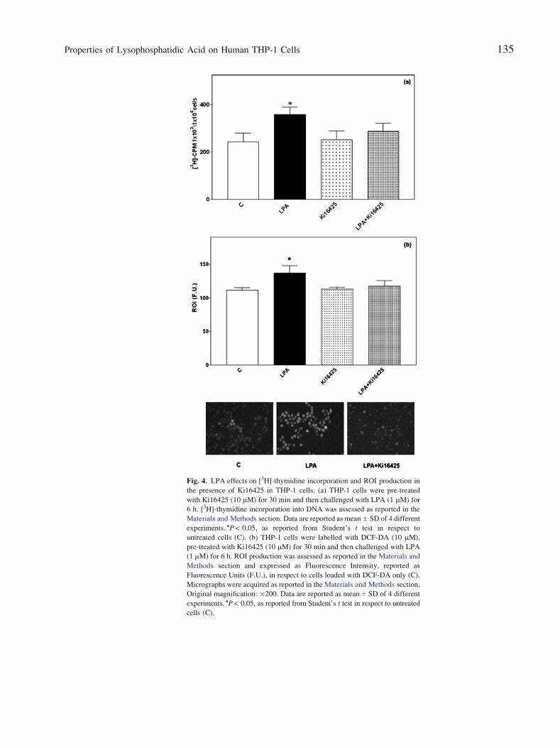
Fig. 4. LPA effects on [3H]-thymidine incorporation and ROI production in
the presence of Ki16425 in THP-1 cells. (a) THP-1 cells were pre-treated
with Ki16425 (10 2M) for 30 min and then challenged with LPA (1 2M) for
6 h. [3H]-thymidine incorporation into DNA was assessed as reported in the
Materials and Methods section. Data are reported as mean T SD of 4 different
experiments. *P < 0.05, as reported from Student_s t test in respect to
untreated cells (C). (b) THP-1 cells were labelled with DCF-DA (10 mM),
pre-treated with Ki16425 (10 mM) for 30 min and then challenged with LPA
(1 mM) for 6 h. ROI production was assessed as reported in the Materials and
Methods section and expressed as Fluorescence Intensity, reported as
Fluorescence Units (F.U.), in respect to cells loaded with DCF-DA only (C).
Micrographs were acquired as reported in the Materials and Methods section.
Original magnification: �200. Data are reported as mean T SD of 4 different
experiments. *P < 0.05, as reported from Student_s t test in respect to untreated
cells (C).
135Properties of Lysophosphatidic Acid on Human THP-1 Cells

DCF-DA (10 mM). The time-course and dose-response of
ROI production are shown in Fig. 2a. Similarly to cell
growth, LPA induced a significant increase in ROI
production only at 1 mM after 3 h with a maximal effect
after 6 h. Longer experimental times and lower (0.1 and
0.5 mM) or higher (10 and 20 mM) LPA concentrations
(data not shown), did not exert any significant effect.
Subsequently, the possible involvement of NADPH
oxidase in LPA-induced ROI production was investigated.
Since the maximal production of ROI occurred after 6 h of
stimulation with 1 mM LPA, such a concentration and time
were used in experiments performed in the presence or
absence of DPI, a specific inhibitor of NADPH oxidase. As
shown in Fig. 2b, cells pre-treatment with DPI (10j8 M)
significantly reduced the production of LPA-induced
ROI in respect to untreated cells as further on
confirmed by fluorescence microscopy analysis.
LPA Effects on Arachidonic Acid, Leukotriene B4
and Prostaglandin E2 Release
Since LPA can promote arachidonic acid (AA)
mobilization [18, 19], doseYresponse experiments were
performed to assess [3H]-AA release into the medium. In
the same way of cell growth and ROI production, assays
were carried out after cell incubation for 6 h with
Fig. 5. LPA effects on Arachidonic Acid (AA) and Prostaglandin E2 (PGE2)
release in the presence of Ki16425 in THP-1 cells. (a) THP-1 cells were
treated as reported in Fig. 3a and challenged with LPA (1 2M) for 6 h after
30 min pre-treatment with Ki16425 (10 2M). The [3H]-AA released was
determined as reported in the Materials and Methods section. Data are
reported as mean T SD of 4 different experiments. *P < 0.05, as reported from
Student_s t test in respect to untreated cells (C). (b) THP-1 cells were treated
as reported in Fig. 3b after 30 min pre-treatment with Ki16425 (10 mM).
PGE2 released into the medium was measured by EIA assay, as described in
the Materials and Methods section. A representative experiment is shown,
which was repeated four additional times with similar results. Values are
reported as percentile increase compared to control (C).
136 D_Aquilio, Procaccini, Izzi, Chiurchiu_, Giambra, Carotenuto, Di Nardo, and Baldini
different LPA concentrations (1Y5 mM). Data shown in
Fig. 3a demonstrate that all LPA concentrations exerted
a significant effect on AA release, the maximal response
occurring at 1 mM and progressively decreasing at higher
lipid concentrations. These results indicated to as
sess LTB4 and PGE2 release in the presence of 1 mM
LPA for 6 h. As shown in Fig. 3b, LPA was able to in-
duce an approximately 15% increase in LTB4 release
(left panel), while PGE2 production increase was
approximately 70% (right panel), suggesting a more
efficient activation of the prostaglandin synthesizing
enzymatic machinery.
LPA Effects on Cell Growth, ROI Production, AA
and PGE2 Release in the Presence of Ki16425
Since LPA1 and LPA3 receptors have been related
to cellular activation and malignant tumour proliferation,
respectively [20, 21], experiments were carried out in the
presence of Ki16425, a selective antagonist of these
receptors [22]. As shown in Fig. 4a, the enhanced LPA-
induced [3H]-thymidine incorporation was significantly
inhibited by Ki16425 (10 mM) pre-treatment. Similarly,
Ki16425 cell pre-treatment totally abolished LPA-stimulated
ROI production, as also confirmed by fluorescence
microscopy analysis (Fig. 4b).
In addition, the antagonist pre-treatment totally
abrogated LPA effects on AA mobilization (Fig. 5a)
and PGE2 release (Fig. 5b). These results suggested that
LPA1 and LPA3 receptors play a fundamental role in
mediating LPA effects on THP-1 cell growth, ROI
production and AA and PGE2 release.
LPA Effects on Receptor Expression Profile
Since cells exposure to LPA can alter the receptor
expression pattern [23], PCR analysis was performed to
further discriminate the relative contribution of each
receptor subtype in LPA effects. RT-PCR analysis
(Fig. 6a) showed that all three LPA receptors are ex-
pressed in THP-1 cells; particularly, un-stimulated cells
expressed predominantly LPA1 and LPA2 mRNAs,
while LPA3 mRNA was poorly expressed. After LPA
(1 mM for 6 h) treatment, a significant increase in
mRNA levels of LPA3 receptor occurred, while no
significant variations in mRNA levels of LPA1 and
LPA2 were detectable. To finely quantify such an
increase, Real Time RT-PCR analysis (Fig. 6b) were
performed demonstrating that LPA addition induced 50-
fold up-regulation in the expression of LPA3 receptor in
respect to control, suggesting a possible role for this
receptor in all the observed effects.
DISCUSSION
The inflammatory response to endothelial injuries is
represented by excessive cell proliferation and activation
[7] that, in turn, can be influenced by several locally
released bioactive factors. Among these substances,
lysophosphatidic acid (LPA), one of the most intriguing
phospholipidic mediators, might act as a powerful
mitogenic and activating factor even for immune cells,
such as T- and B-cells [24, 25].
The present study represents the first attempt to
characterize LPA proinflammatory properties in THP-1
cells. In fact, micromolar concentrations of LPA are able
to stimulate THP-1 cell growth, ROI production and
arachidonic acid metabolites release. Interestingly, a
positive factor in prostaglandins (PGs) and leukotrienes
(LTs) synthesis is considered to be the increased cytosolic
oxidant power [13]. This is generally due to an
enhancement in ROI production, whose primary source
differs depending on cell type [26]. In phagocytes, the
principal ROI source is represented by the NADPH
oxidase enzyme complex [27]. Moreover, ROI release at
inflamed sites causes the activation of different cell types
determining the pathological course of reactions [13]. In
this context, the observed LPA ability to increase both
PGs and LTs production, exerting a stronger effect on
PGE2 synthesis rather than LTB4, and to determine
NADPH oxidase-dependent ROI production accounts for
a possible role of this lipid in the pathogenesis of
different diseases characterized by the development of
local inflammatory reactions. These data are substantiat-
ed by the demonstration that all these effects occurred at
the same concentration and timepoint determining a
remarkable increase in cell proliferation.
To refine our analysis, we tried to determine which
receptor subtype could be mostly involved in mediating
LPA effects. LPA, in fact, regulates a broad variety of
biological processes binding to specific receptors, princi-
pally LPA1, LPA2 and LPA3, which, in turn, activate
pleiotropic signalling pathways [28]. Since previous
studies demonstrated that LPA1 receptor stimulation
was critical for monocyte activation [20] and LPA3
receptor was responsible for cell growth and proliferation
of malignant tumours [21], experiments were carried out
in the presence of Ki16425, a selective LPA1 and LPA3
receptor antagonist [22]. Our results demonstrated that
137Properties of Lysophosphatidic Acid on Human THP-1 Cells

138 D_Aquilio, Procaccini, Izzi, Chiurchiu_, Giambra, Carotenuto, Di Nardo, and Baldini
both receptors were implicated in LPA effects since they
were totally abolished by the addition of a receptor-specific
inhibitor. In fact, preincubating cells with Ki16425, a
significant inhibition of LPA-induced THP-1 proliferation
and ROI production as well as AA release and PGE2
production occurred. Since LPA proinflammatory activity
depends on the individual expression of LPA receptors
[29] and the exposure to mitogens can alter such an
expression profile [23], it is possible that, in THP-1 cells,
the abundance of each LPA receptor may significantly
differ in resting vs activated cells. Therefore, to further
unravel the relative contribute of each receptor subtype in
LPA effects, PCR analysis was performed demonstrating
that the lipid addition caused a 50-fold increase in LPA3
receptor mRNA expression, while the other receptors did
not undergo any significant change. These findings
suggest a possible role for LPA3 receptor in mediating
LPA signalling in THP-1 cells, even though the relative
contribute of LPA1 receptor cannot be excluded.
Taken together, the results obtained demonstrate that
LPA can modulate THP-1 cells proinflammatory activa-
tion implying that this lipid, acting as an autocrine/
paracrine factor, could sustain an ongoing complex
cascade of inflammatory reactions ultimately leading to
a pathological chronic condition. In conclusion, our study
furnishes new evidences on LPA proinflammatory prop-
erties, whose understanding might indicate a new prom-
ising therapeutic strategy in monocytes-related human
diseases management.
REFERENCES
1. Moolenaar, W. H. 2000. Development of our current under-standing of bioactive lysophospholipids. Ann. NY Acad. Sci. 905:1Y10.
2. Fukushima, N., and J. Chun. 2001. The LPA receptors.Prostaglandins 64:21Y32.
3. Goetzl E. J., W. Wang, G. McGiffert, M. G. Huang, and M. H.Graler. 2004. Sphingosine 1-phosphate and its G protein-coupledreceptors constitute a multifunctional immunoregulatory system.J. Cell. Biochem. 92(6):1104Y1114.
4. Graler, M. H., and E. J. Goetzl. 2002. Lysophospholipids and theirG protein-coupled receptors in inflammation and immunity.Biochim. Biophys. Acta. 1582(1Y3):168Y174.
5. Siess, W., K. J. Zangle, M. Essler, M. Bauer, R. Brandl, C.Corrinth, R. Bittman, G. Tigyi, and M. Aepfelbacher. 1999.Lysophosphatidic acid mediates the rapid activation of plateletsand endothelial cells by mildly oxidized low density lipoproteinand accumulates in human atherosclerotic lesions. Proc. Natl.Acad. Sci. USA. 96(12):6931Y6936.
6. Panther E., M. Idzko, S. Corinti, D. Ferrari, Y. Herouy, M.Mockenhaupt, S. Dichmann, P. Gebicke-Haerter, F. Di Virgilio,G. Girolomoni, and J. Norgauer. 2002. The influence oflysophosphatidic acid on the functions of human dendritic cells.J. Immunol. 169(8):4129Y4135.
7. Andres V., and C. Castro. 2003. Antiproliferative strategies for thetreatment of vascular proliferative disease. Curr. Vasc. Pharmacol.1:85Y98.
8. Ross, R. 1999. Atherosclerosis - an inflammatory disease. N. Engl.J. Med. 340(2):115Y126.
9. Osterud, B., and E. Bjorklid. 2003. Role of monocytes inatherogenesis. Physiol. Rev. 83(4):1069Y1112.
10. Melo, R. C., N., D. L. Fabrino, H. D’Avila, H. C. Teixeira, and A. P.Ferreira. 2003. Production of hydrogen peroxide by peripheralblood monocytes and specific macrophages during experimentalinfection with Trypanosoma cruzi in vivo. Cell. Biol. Int. 27(10):853Y861.
11. Babior, B. M. 1999. NADPH oxidase: an update. Blood 93:1464-1476.
12. Hadjigogos, K. 2003. The role of free radicals in the pathogenesisof rheumatoid arthritis. Panminerva Med. 45(1):7Y13.
13. Lu, Y., and L. M. Wahl. 2005. Oxidative stress augments theproduction of matrix metalloproteinase-1, cyclooxygenase-2, andprostaglandin E2 through enhancement of NF-kappa B activity inlipopolysaccharide-activated human primary monocytes. J. Immunol.175(8):5423Y5429.
14. Vila, L. 2004. Cyclooxygenase and 5-lipoxygenase pathways in thevessel wall: role in atherosclerosis. Med. Res. Rev. 24(4):399Y424.
15. Shen, H. M., C. Y. Shi, Y. Shen, and C. N. Ong. 1996. Detectionof elevated reactive oxygen species level in cultured rat hepatocytes treated with aflatoxin B1. Free Radic. Biol. Med. 21:139Y146.
16. Donchenko, V., A. Zannetti, and P. M. Baldini. 1994. Insulin-stimulated hydrolysis of phosphatidylcholine by phospholipase Cand phospholipase D in cultured rat hepatocytes. Biochim.Biophys. Acta 1222(3):492Y500.
17. Johann, A. M., A. von Knethen, D. Lindemann, and B. Brune.2005. Recognition of apoptotic cells by macrophages activates theperoxisome proliferator-activated receptor-gamma and attenuatesthe oxidative burst. Cell Death Differ Epub ahead of print.
18. Inoue, C. N., H. G. Forster, and M. Epstein. 1995. Effects oflysophosphatidic acid, a novel lipid mediator, on cytosolic Ca2+
and contractility in cultured rat mesangial cells. Circ. Res. 77(5):888Y896.
19. Pebay, A., Y. Torrens, M. Toutant, J. Cordier, J. Glowinski, andM. Tence. 1999. Pleiotropic effects of lysophosphatidic acid onstriatal astrocytes. Glia 28(1):25Y33.
20. Fueller, M., D. A. Wang, G. Tigyi, and W. Siess. 2003. Activationof human monocytic cells by lysophosphatidic acid and sphingo-sine-1-phosphate. Cell. Signal 15(4):367Y375.
21. Nakamoto, T., K. Yasuda, M. Yasuhara, T. Yoshimura, T.Kinoshita, T. Nakajima, H. Okada, A. Ikuta, and H. Kanzaki.2005. Expression of the endothelial cell differentiation gene 7(EDG-7), a lysophosphatidic acid receptor, in ovarian tumor. J.Obstet. Gynaecol. Res. 31(4):344Y351.
22. Ohta, H., K. Sato, N. Murata, A. Damirin, E. Malchinkhuu, J.Kon, T. Kimura, M. Tobo, Y. Yamazaki, T. Watanabe, M. Yagi,
Fig. 6. LPA effects on the expression of LPA1, LPA2 and LPA3 re-
ceptors in THP-1 cells. Total RNA was extracted from THP-1 cells
treated or not with LPA (1 2M) for 6 h. RT-PCR (a) and Real-Time
RT-PCR (b) analysis were performed as reported in the Materials and
Methods section. The expression levels of LPA receptors in THP-1
cells were quantified by normalising their respective mRNA levels
with the housekeeping human GAPDH mRNA levels. A representa-
tive experiment is shown, which was repeated three additional times
with similar results.
139Properties of Lysophosphatidic Acid on Human THP-1 Cells
M. Sato, R. Suzuki, H. Murooka, T. Sakai, T. Nishitoba, D. S. Im,H. Nochi, K. Tamoto, H. Tomura, and F. Okajima. 2003.Ki16425, a subtype-selective antagonist for EDG-family lyso-phosphatidic acid receptors. Mol. Pharmacol. 64(4):994Y1005.
23. Baldini, P. M., P. De Vito, F. D’Aquilio, D. Vismara, F. Zalfa, C.Bagni, R. Fiaccavento, and P. Di Nardo. 2005. Role of atrialnatriuretic peptide in the suppression of lysophosphatydic acid-induced rat aortic smooth muscle (RASM) cell growth. Mol. Cell.Biochem. 272(1Y2):19Y28.
24. Wang, L., E. Knudsen, Y. Jin, S. Gessani, and A. A. Maghazachi.2004. Lysophospholipids and chemokines activate distinct signaltransduction pathways in T helper 1 and T helper 2 cells. Cell. Signal.16(9):991Y1000.
25. Rosskopf, D., W. Daelman, S. Busch, M. Schurks, K. Hartung, A.
Kribben, M. C. Michel, and W. Siffert. 1998. Growth factor-likeaction of lysophosphatidic acid on human B lymphoblasts. Am. J.Physiol. 274(6):C1573YC1582.
26. Martindale, J. L. and N. J. Holbrook. 2002. Cellular response tooxidative stress: signalling for suicide and survival. J. Cell. Physiol.192(1):1Y15.
27. Cathcart, M. K. 2004. Regulation of superoxide anion production byNADPH oxidase in monocytes/macrophages: contribution to athero-sclerosis. Arterioscler. Thromb. Vasc. Biol. 24(1):23Y28.
28. Anliker, B., and J. Chun. 2004. Cell surface in lysophospholipidsignalling. Semin. Cell Dev. Biol. 15(5):457Y465
29. Siess, W. 2002. Athero- and thrombogenic actions of lysophos-phatidic acid and sphingosine-1-phosphate. Biochim. Biophys.Acta 1582:204Y215
140 D_Aquilio, Procaccini, Izzi, Chiurchiu_, Giambra, Carotenuto, Di Nardo, and Baldini
Copyright © 2022 FDOKUMEN