Lysophosphatidic acid modulates c-Met redistribution and hepatocyte growth factor/c-Met signaling in...
20
Lysophosphatidic Acid Modulates c-Met Redistribution and Hepatocyte Growth Factor / c-Met Signaling in Human Bronchial Epithelial Cells Through PKC δ and E-cadherin Yutong Zhao a,* , Donghong He a , Randi Stern a , Peter V. Usatyuk a , Ernst Wm. Spannhake b , Ravi Salgia a , and Viswanathan Natarajan a a Department of Medicine, University of Chicago, Chicago, IL b Department of Environmental Health Sciences, Bloomberg School of Public Health, Johns Hopkins University, Baltimore, MD Abstract Previously we demonstrated that ligation of lysophosphatidic acid (LPA) to G protein-coupled LPA receptors induces transactivation of receptor tyrosine kinases (RTKs), such as platelet-derived growth factor receptor beta (PDGF-Rβ) and epidermal growth factor receptor (EGF-R), in primary cultures of human bronchial epithelial cells (HBEpCs). Here we examined the role of LPA on c-Met redistribution and modulation of hepatocyte growth factor (HGF) / c-Met pathways in HBEpCs. Treatment of HBEpCs with LPA induced c-Met serine phosphorylation and redistribution to plasma membrane, while treatment with HGF induced c-Met internalization. Pretreatment with LPA reversed HGF-induced c-Met internalization. Overexpression of dominant negative (Dn)-PKC δ or pretreatment with Rottlerin or Pertussis toxin (PTx) attenuated LPA-induced c-Met serine phosphorylation and redistribution. Co-immnuoprecipitation and immunocytochemistry showed that E-cadherin interacted with c-Met in HBEpCs. LPA treatment induced E-cadherin and c-Met complex redistribution to plasma membranes. Overexpression of Dn-PKC δ attenuated LPA-induced E- cadherin redistribution and E-cadherin siRNA attenuated LPA-induced c-Met redistribution to plasma membrane. Furthermore, pretreatment of LPA attenuated HGF-induced c-Met tyrosine phosphorylation and downstream signaling, such as Akt kinase phosphorylation and cell motility. These results demonstrate that LPA regulates c-Met function through PKC δ- and E-cadherin in HBEpCs, suggesting an alternate function of the cross-talk between G-protein coupled receptors (GPCRs) and RTKs in HBEpCs. Keywords lysophosphatidic acid; G-protein coupled receptor; receptor tyrosine kinase; c-Met; E-cadherin; PKC; signal transduction *Corresponding Author: Dr. Yutong Zhao, Department of Medicine, University of Chicago, Center for Integrative Science Building, Room # W403M, 929 East 57 th Street, Chicago, IL 60637, Tel: 773-834-2385, Fax: 773-834-2687, Email: [email protected]. Publisher's Disclaimer: This is a PDF file of an unedited manuscript that has been accepted for publication. As a service to our customers we are providing this early version of the manuscript. The manuscript will undergo copyediting, typesetting, and review of the resulting proof before it is published in its final citable form. Please note that during the production process errors may be discovered which could affect the content, and all legal disclaimers that apply to the journal pertain. NIH Public Access Author Manuscript Cell Signal. Author manuscript; available in PMC 2008 November 1. Published in final edited form as: Cell Signal. 2007 November ; 19(11): 2329–2338. NIH-PA Author Manuscript NIH-PA Author Manuscript NIH-PA Author Manuscript
-
Upload
independent -
Category
Documents
-
view
0 -
download
0
Transcript of Lysophosphatidic acid modulates c-Met redistribution and hepatocyte growth factor/c-Met signaling in...
Lysophosphatidic Acid Modulates c-Met Redistribution andHepatocyte Growth Factor / c-Met Signaling in Human BronchialEpithelial Cells Through PKC δ and E-cadherin
Yutong Zhaoa,*, Donghong Hea, Randi Sterna, Peter V. Usatyuka, Ernst Wm. Spannhakeb,Ravi Salgiaa, and Viswanathan Natarajana
a Department of Medicine, University of Chicago, Chicago, IL
b Department of Environmental Health Sciences, Bloomberg School of Public Health, Johns HopkinsUniversity, Baltimore, MD
AbstractPreviously we demonstrated that ligation of lysophosphatidic acid (LPA) to G protein-coupled LPAreceptors induces transactivation of receptor tyrosine kinases (RTKs), such as platelet-derivedgrowth factor receptor beta (PDGF-Rβ) and epidermal growth factor receptor (EGF-R), in primarycultures of human bronchial epithelial cells (HBEpCs). Here we examined the role of LPA on c-Metredistribution and modulation of hepatocyte growth factor (HGF) / c-Met pathways in HBEpCs.Treatment of HBEpCs with LPA induced c-Met serine phosphorylation and redistribution to plasmamembrane, while treatment with HGF induced c-Met internalization. Pretreatment with LPAreversed HGF-induced c-Met internalization. Overexpression of dominant negative (Dn)-PKC δ orpretreatment with Rottlerin or Pertussis toxin (PTx) attenuated LPA-induced c-Met serinephosphorylation and redistribution. Co-immnuoprecipitation and immunocytochemistry showed thatE-cadherin interacted with c-Met in HBEpCs. LPA treatment induced E-cadherin and c-Met complexredistribution to plasma membranes. Overexpression of Dn-PKC δ attenuated LPA-induced E-cadherin redistribution and E-cadherin siRNA attenuated LPA-induced c-Met redistribution toplasma membrane. Furthermore, pretreatment of LPA attenuated HGF-induced c-Met tyrosinephosphorylation and downstream signaling, such as Akt kinase phosphorylation and cell motility.These results demonstrate that LPA regulates c-Met function through PKC δ- and E-cadherin inHBEpCs, suggesting an alternate function of the cross-talk between G-protein coupled receptors(GPCRs) and RTKs in HBEpCs.
Keywordslysophosphatidic acid; G-protein coupled receptor; receptor tyrosine kinase; c-Met; E-cadherin;PKC; signal transduction
*Corresponding Author: Dr. Yutong Zhao, Department of Medicine, University of Chicago, Center for Integrative Science Building,Room # W403M, 929 East 57th Street, Chicago, IL 60637, Tel: 773-834-2385, Fax: 773-834-2687, Email:[email protected]'s Disclaimer: This is a PDF file of an unedited manuscript that has been accepted for publication. As a service to our customerswe are providing this early version of the manuscript. The manuscript will undergo copyediting, typesetting, and review of the resultingproof before it is published in its final citable form. Please note that during the production process errors may be discovered which couldaffect the content, and all legal disclaimers that apply to the journal pertain.
NIH Public AccessAuthor ManuscriptCell Signal. Author manuscript; available in PMC 2008 November 1.
Published in final edited form as:Cell Signal. 2007 November ; 19(11): 2329–2338.
NIH
-PA Author Manuscript
NIH
-PA Author Manuscript
NIH
-PA Author Manuscript
1. IntroductionIn eukaryotes extracellular messages are transduced into intracellular signals by activation ofseveral classes of receptors. These include G-protein-coupled receptors (GPCRs), receptortyrosine kinases (RTKs), cytokine receptor-activated kinases, and members of the steroid/thyroid hormone receptor super family [1-3]. Originally, these receptors were thought to actindependently, but in recent years evidence of cross-talk between these different classes ofreceptors has been discovered [4-9]. Among them, the cross-talk between GPCRs and RTKshas been most frequently identified [5-9]. Activation of GPCRs leads to stimulation of RTKs,providing integration and amplification of signaling pathways that regulate cellular responsesand functions [5-9]. In contrast to transactivation of RTKs by GPCRs, increasing evidenceshows that GPCRs also induce inhibition of RTK activity, known as transinactivation[10-11].
Lysophosphatidic acid (LPA), a bioactive lysophospholipid, mediates a wide range of cellularresponses via GPCRs, LPA1 (EDG-2), LPA2 (EDG-4), LPA3 (EDG-7), and LPA4 [12-15]. Wehave previously shown that human bronchial epithelial cells (HBEpCs) express LPA1-3 andthat exposure to LPA stimulated expression and secretion of interleukin-8 (IL-8). The IL-8expression is also partly regulated by protein kinase C δ (PKC δ), Nuclear factor-κB (NF-κB),and c-Jun-N-terminal Kinase (JNK)/AP-1 transcriptional factors [16-18]. Furthermore, LPAinduced IL-13 receptor alpha2 (IL-13Rα2) expression and secretion and attenuated IL-13-induced signaling in HBEpCs [19]. In addition, ligation of LPA to LPA-Rs transactivatedRTKs, such as Platelet-Derived Growth Factor Receptor beta (PDGF-Rβ) [20] and EpidermalGrowth Factor Receptor (EGF-R) [21]. EGF-R transactivation by LPA contributed to LPA-induced IL-8 secretion [21], while PDGF-Rβ transactivation was involved in LPA-inducedphosphorylation of extracelluar signal-regulated kinase (Erk) [20]. LPA also induced PDGF-Rβ transactivation through phospholipase D 2 (PLD2) [19] and EGF-R transactivation throughPKC δ-mediated activation of lyn kinase, matrix metallopreinases, and heparin-binding EGF(HB-EGF) shedding in HBEpCs [21].
c-Met, a proto-oncogene product, is the receptor for hepatocyte growth factor (HGF), and ispredominantly expressed in various types of epithelium [22,23]. Increased expression andactivation of c-Met is associated with tumor invasion and metastasis [23,24]. c-Met has beenshown to be expressed in several non-small cell lung cancer cell (NSCLC) lines, and activationby HGF stimulates cell growth, scattering, and invasion of these cells [24,25]. Ma et al. haveidentified particular mutations in the juxtamembrane and semaphorin domains, and have shownregulation of the cytoskeleton through manipulation of the juxtamembrane mutations of c-Met,especially as related to the focal adhesion protein paxillin [26]. Similar to other RTKs, ligationof HGF to c-Met induced c-Met tyrosine phosphorylation, dimerization, recruitment of severalsignaling molecules, and internalization [22-25,27]. Selective small molecule c-Met inhibitors(SU11274 and PHA665752) inhibited HGF-induced phosphorylation of c-Met, S6K, and Aktin NSCLC cells [28,29].
It is well established that many transmembrane receptors, such as EGF-R and c-Met, becomeinternalized upon ligand stimulation [30-33]. It has been reported that upon HGF stimulation,c-Met is rapidly internalized via clathrin-coated vesicles and traffics through an earlyendosomal compartment, which is independent of the proteasome-mediated degradativepathway [34]. Kermorgant et al. showed that PKC α and PKC ε control c-Met traffic fromplasma membrane to perinuclear compartment and this interlization plays a positive role in c-Met signal output in HeLa cells [35]. In contrast, Gandino et al. showed that PKC activationinhibited tyrosine phosphorylation of c-Met in the gastric carcinoma cell line GTL-16 [36].Recently, Hashigasako et al. demonstrated that HGF-dependent tyrosine phosphorylation ofc-Met was largely suppressed with a reciprocal relationship to Ser-985 phosphorylation by
Zhao et al. Page 2
Cell Signal. Author manuscript; available in PMC 2008 November 1.
NIH
-PA Author Manuscript
NIH
-PA Author Manuscript
NIH
-PA Author Manuscript

PKC δ in A549 cell line [37]. Similar to c-Met, insulin receptors were phosphorylated at serineresidues and tyrosine kinase activity was decreased by PKC [38,39]. These results suggest thatPKC isoforms may play bi-directional roles in regulating c-Met signaling.
Recently, LPA-and sphingosine-1-phosphate (S1P)-induced c-Met tyrosine phosphorylationhas been reported in human colon cancer cell lines and human gastric cancer cells [40,41].Nath et al. reported that shedding of c-Met was regulated by LPA-mediated EGF-Rtransactivation and TIMP-3 sensitive metalloproteinase in A549 cells [37]. The role of LPAin regulating the HGF / c-Met pathways is still not clear. Here we report that LPA, through itsGPCRs, stimulates serine phosphorylation of c-Met in HBEpCs. LPA treatment induced c-Metredistribution to plasma membrane involving PKC δ and E-cadherin while also attenuatedHGF-mediated c-Met internalization. Furthermore, LPA downregulated HGF / c-Met-mediated signaling and cell motility. These results suggest cross-talk between LPA-Rs and c-Met and a possible role of this transactivation in signal integration and epithelial cell function.
2. Materials and Methods2.1. Materials
1-oleoyl (18:1) LPA and dimethyl sulfoxide (DMSO) were purchased from Sigma-Aldrich (St.Louis, MO). Antibody to phophoserine was from Zymed Laboratories Inc. (South SanFranciso, CA). Antibodies to c-Met and E-cadherin were from Upstate (Lake Placid, NY).Antibody to PKC δ was purchased from BD Transduction Laboratories (San Jose, CA).Antibodies to total Akt and phosphor (Ser 473)-Akt were procured from Cell SignalingTechnology (Beverly, MA). E-cadherin siRNAs and protein A/G-agarose were from SantaCruz Biotechnology (Santa Cruz, CA). Scrambled siRNA was from Dharmacon Inc.(Lafayette, CO). Pertussis toxin (PTx), Rottlerin, and bisindolylmaleimide (BIM) were fromCalbiochem (San Diego, CA). HGF and anti phosphor-tyrosine-c-Met (Y1230/1234/1235)antibody were purchased from BIOSOURCE International (Camarillo, CA). Alexa fluor-488goat anti-mouse and Alexa fluor-568 goat anti-rabbit were purchased from Molecular Probes(Eugene, OR). Horseradish peroxidase-conjugated goat anti-rabbit and anti-mouse secondaryantibodies were purchased from Molecular Probes (Eugene, OR). ECL kit for detection ofproteins by Western blotting was obtained from Amersham Pharmacia (Piscataway, NJ).Transfection reagents were from Qiagen (Valencia, CA). Bronchial epithelial cell basalmedium (BEBM) and supplement kit were purchased from Cambrex Bio Science Inc.(Walkersville, MD). All other reagents were of analytical grade.
2.2. Cell cultureHBEpCs were isolated from normal human lung obtained from lung transplant donorsfollowing previously described procedures [16]. The isolated passage 0 (P0) HBEpCs werecultured in serum free basal essential growth medium (BEGM) and supplemented with growthfactors. Cells were incubated at 37 °C in 5 % CO2 and 95 % air to approximately 80 %confluence and subsequently propagated in 100 mm dishes or 6-well collagen-coated plates orcover slips. All experiments were carried out between passages 1 and 3.
2.3. Preparation of cell lysates, immunoprecipitation, and Western blottinCells were rinsed twice with ice-cold PBS and lysed in 500 μl of buffer containing 20 mMTris-HCl (pH 7.4), 150 mM NaCl, 2 mM EGTA, 5 mM β-glycerophosphate, 1 mM MgCl2, 1% Triton X-100, 1 mM sodium orthovanadate, 10 μg/ml protease inhibitors, 1 μg/ml aprotinin,1 μg/ml leupeptin, and 1 μg/ml pepstatin. Cell lysates were incubated at 4 °C for 10 min,sonicated on ice for 20 seconds and centrifuged at 5,000 g for 5 min at 4 °C in a microfuge.Protein concentrations were determined with a BCA protein assay kit (Pierce Chemical Co.,Rockford, IL) using BSA as standard. Equal amounts of protein (300-500 μg) were incubated
Zhao et al. Page 3
Cell Signal. Author manuscript; available in PMC 2008 November 1.
NIH
-PA Author Manuscript
NIH
-PA Author Manuscript
NIH
-PA Author Manuscript
with anti-c-Met or anti-E-cadherin antibody (2 μg/ml) overnight at 4°C. This was followed bythe addition of 40 μl protein A/G-agarose and additional incubation for 2 h at 4°C. The celllysates were centrifuged at 5,000 g for 5 min, washed three times with ice-cold PBS containing1 mM orthovanadate, protein-agarose complexes were dissociated by boiling for 5 min in 2xLammeli buffer, and samples were again centrifuged at 5,000 g for 5 min. The supernatantswere subjected to SDS-PAGE on 10% gels, transferred to polyvinylidene difluoridemembranes, blocked with 5 % (w/v) BSA in TBST (25 mM Tris-HCl, pH 7.4, 137 mM NaCland 0.1 % Tween-20) for 1 h and incubated with primary antibodies in 5 % (w/v) BSA in TBSTfor 1-2 h at room temperature. The membranes were washed at least three times with TBST at15 min intervals and then incubated with either mouse or rabbit horseradish peroxidase-conjugated secondary antibody (1: 3,000) for 1 h at room temperature. The membranes weredeveloped with enhanced chemiluminescence detection system according to manufacturer'sinstructions.
2.4. Infection with adenoviral constructsInfection of HBEpCs (∼50 % confluence) with purified adenoviral empty vector or adenoviralvector containing catalytically inactive mutant of human PKC δ (Dn-PKC δ) were carried outin 100 mm-dishes as described previously [16]. Cells were infected with different MOI in 1ml of BEGM for 48 h.
2.5. Transfection of small interfering RNA (siRNA) for E-cadherinHBEpCs (P1) cultured onto 6-well plates or cover slips (40-50 % confluence) were transientlytransfected with human E-cadherin and scrambled siRNA using Transmessenger TransfectionReagent. Briefly, siRNAs (50 nM) were condensed with Enhancer R and formulated withTransmessenger Reagent, according to the manufacturer's instruction. The transfectioncomplex was diluted into 900 μl of BEBM medium and added directly to the cells. The mediumwas replaced with BEBM medium after 3 h, and cells were used for experiments at 72 h aftertransfection.
2.6. ImmunocytochemistryHBEpCs grown on cover slips to approximately 70-80 % confluence were challenged withLPA or HGF. Coverslips were rinsed with PBS and treated with 3.7 % formaldehyde in PBSat room temperature for 20 min. After washing with PBS, coverslips were incubated in blockingbuffer (1 % BSA in TBST) for 1 h. Cells were then subjected to immunostaining with antibodyto c-Met or E-cadherin (1: 200 dilution) for 1 h, washed three times with TBST, and stainedwith Alexa Fluor 568 or 488 (1: 200 dilution in blocking buffer) for 1 h. After washing at leastthree times with TBST, the coverslips were mounted using commercial mounting medium forfluorescent microscopy (Kirkegaard and Perry Laboratories, Gaithersburg, MD), and wereexamined by immunofluorescent microscope with Hamamatsu digital camera using a 60x oilimmersion objective and MetaVue software.
2.7. Cell motility assayHBEpCs were subcultured on 35 mm dishes in low density. Cells grew to separate coloniesand were treated with or without LPA (1 μM) for 15 min, then challenged with HGF (25 ng/ml) for 3 h. Images were taken using microscopy with Nikon camera and analyzed by NIHImageJ software.
2.8. Statistical analysesAll results were subjected to statistical analysis using one-way ANOVA and, wheneverappropriate, analyzed by Student-Newman-Keuls test. Data are expressed as means ± S.D. of
Zhao et al. Page 4
Cell Signal. Author manuscript; available in PMC 2008 November 1.
NIH
-PA Author Manuscript
NIH
-PA Author Manuscript
NIH
-PA Author Manuscript
triplicate samples from at least three independent experiments and level of significance wastaken to P < 0.01.
3. Results3.1. LPA induces serine phosphorylation of c-Met via Gi and PKC δ in HBEpCs
We have previously shown transactivation of RTKs, such as PDGF-Rβ and EGF-R, by LPA-Rs in HBEpCs [19,20]. Here we examined the cross-talk between LPA-Rs and c-Met inHBEpCs. HBEpCs were challenged with LPA (1 μM) for 2, 15, and 30 min or HGF (50 ng/ml) for 15 min and c-Met was immunoprecipitated with an antibody specific to c-Met. Asshown in Fig. 1A, LPA treatment induced serine phosphorylation of c-Met at 2 min, and forup to 30 min, while HGF had no effect on serine phosphorylation of c-Met. To examine thetyrosine phosphorylation of c-Met, the cell lysates were analyzed by Western blotting withphospho-tyrosine (Y1230/1245/1246) antibody. HGF (50 ng/ml, 15 min) induced tyrosinephosphorylation of c-Met while LPA (1 μM) induced marginal tyrosine phosphorylation of c-Met at 15 min (Fig. 1B). To determine whether LPA-induced serine phosphorylation of c-Metis through G-protein-coupled LPA receptors, HBEpCs were challenged with LPA (1 μM) for15 min with or without pretreatment with PTx (100 ng/ml, 4h), then c-Met wasimmunoprecipitated with a c-Met specific antibody and analyzed with phosphoserine antibody.As shown in Fig. 1C, PTx attenuated LPA-induced serine phosphorylation of c-Met, suggestingLPA-induced c-Met serine phosphorylation is dependent on LPA-R-coupled Gi protein.
Our previous studies showed that PKC δ plays a critical role in LPA-induced IL-8 expression[16] and EGF-R transactivation [20]. Here we examined the role of PKC δ in cross-talk betweenLPA-Rs and c-Met in HBEpCs. HBEpCS were infected with empty control adenoviral vector(20 MOI) or adenoviral vector containing Dn-PKC δ (1, 10, and 20 MOI) for 48 h. Theoverexpression of Dn-PKC δ was confirmed by Western blotting with antibody specific toPKC δ (Fig. 2A). The effect of overexpression of Dn-PKC δ on LPA-induced serinephosphorylation of c-Met was determined by immnuoprecipitation with a c-Met specificantibody and analyzed with phosphoserine antibody. As shown in Fig. 2B, Overexpression ofDn-PKC δ (1, 10, and 20 MOI) attenuated LPA-induced serine phosphorylation of c-Met in adose-dependent fashion. Further, PKC δ inhibitor, Rottlerin blocked LPA-induced serinephosphorylation of c-Met (Fig. 2C). These results suggest that PKC δ regulates LPA-inducedserine phosphorylation of c-Met in HBEpCs.
3.2. LPA induces c-Met redistribution via PKC δ in HBEpCHGF induces c-Met endocytosis and intracellular traffic has been demonstrated in variety ofcell types [33-35]. Here we examined the c-Met redistribution by LPA treatment in HBEpCs.HBEpCs (∼80 - 90% confluence) grown in cover slips were challenged with LPA (1 μM) for15, 30, and 60 minutes. c-Met redistribution was examined by immunofluorescence withantibody to c-Met. As shown in Fig. 3A, LPA (1 μM) treatment induced c-Met redistributionto cell plasma membrane in a time-dependent fashion in HBEpCs.
We previously showed that LPA treatment induced phosphorylation of PKC δ and translocationof PKC δ to plasma membrane [16]. To further determine the mechanisms of LPA-induced c-Met redistribution to plasma membrane, we examined the role of PKC δ in regulating c-Mettraffic in response to LPA treatment. HBEpCs were treated with 0.1 % DMSO or PKC inhibitorbisindolylmaleimide I (BIM, 1.0 μM) for 1 h, then challenged with LPA (0.1, 1.0, and 5.0μM) for an additional 15 min. As shown in Fig. 3B, LPA induced c-Met redistribution to plasmamembrane in a dose-dependent fashion, while BIM blocked the effect of LPA on c-Metredistribution. Overexpression of Dn-PKC δ (1, 10, 20 MOI) attenuated LPA (1 μM, 15 min)-induced c-Met redistribution in a dose-dependent fashion (Fig. 3C). PKC α has been known
Zhao et al. Page 5
Cell Signal. Author manuscript; available in PMC 2008 November 1.
NIH
-PA Author Manuscript
NIH
-PA Author Manuscript
NIH
-PA Author Manuscript
to be involve in HGF-induced c-Met internalization in HeLa cells [35], while overexpressionof Dn-PKC α and Dn-PKC λ had no effect on LPA-induced c-Met redistribution (Y. Zhao andV. Natarajan, unpublished data). These results suggest that PKC δ regulates LPA-induced c-Met redistribution to plasma membrane in HBEpCs.
3.3. Role of E-cadherin in LPA-induced c-Met redistribution in HBEpCsCo-localization of E-cadherin, a cell-cell adhesion molecule, and c-Met in cell-cell adhesionsites has been shown in MDCK cells [43]. In HBEpCs co-localization of E-cadherin and c-Met in plasma membrane and cytoplasm were detected by double immunostaining with anti-E-cadherin and anti-c-Met antibodies. LPA (1 μM) treatment induced both E-cadherin and c-Met complex redistribution to plasma membrane in a time course (Fig. 4A). Further, E-cadherinwas immunoprecipitated from HBEpCs with or without LPA (1 μM, for 10 and 30 min)treatment. As shown in Fig. 4B, interaction of E-cadherin and c-Met were found in E-cadherincomplex from either LPA-treated and untreated HBEpCs. Similar to the effect of Dn-PKC δon LPA-induced c-Met redistribution, overexpression of Dn-PKC δ (20 MOI, 48 h) attenuatedLPA-induced E-cadherin redistribution to plasma membrane in HBEpCs (Fig. 5). These resultssuggest that LPA induced E-cadherin and c-Met complex redistribution through PKC δ inHBEpCs.
Next we examined whether LPA-induced c-Met redistribution to plasma membranes requiredinteraction with E-cadherin. To downregulate E-cadherin expression, HBEpCs weretransfected with scrambled and E-cadherin specific siRNAs (50 nM) for 72 h. As shown inFig. 6A, LPA induced c-Met redistribution in the cells transfected with scrambled siRNA,while E-cadherin siRNA attenuated LPA-induced c-Met redistribution. The effect of E-cadherin siRNA transfection on E-cadherin protein expression was confirmed by Westernblotting with anti E-cadherin antibody (Fig. 6B).
3.4. The effect of LPA on HGF / c-Met-mediated c-Met internalization, signaling, and cellmotility
Consistent with others' results [33-35], in HBEpCs HGF (25 ng/ml) treatment for 30 mininduced c-Met internalization from plasma membrane to cytoplasm and perinuclearcompartment in a dose-dependent fashion (Fig. 7A). To further determine the effect of LPAon c-Met traffic, HBEpCs were pretreated with LPA (1 μM) for 15 min, then challenged with25 ng/ml of HGF for an additional 30 min. LPA pretreatment attenuated HGF-induced c-Metinternalization (Fig. 7A), suggesting that LPA-induced c-Met redistribution to plasmamembrane reversed HGF-induced c-Met internalization.
To determine the role of LPA treatment in c-Met-mediated signaling, HBEpCs were pretreatedwith or without LPA (1 μM) for 15 min, then were challenged with HGF (25 ng/ml) for 15,30, 60, or 120 min. HGF induced tyrosine (1230/1245/1246) phosphorylation of c-Met at 15min and up to 120 min. Pretreatment of HBEpCs with LPA (1 μM) inhibited HGF-inducedtyrosine phosphorylation of c-Met at all the time points (Fig. 7B). To further examine the effectof LPA on HGF-mediated c-Met tyrosine phosphorylation, HBEpCs were pretreated with LPA(1 μM) for 15 min, then challenged with HGF (10 and 25 ng/ml) for 30 min. As shown in Fig.7C, LPA pretreatment completely abrogated 10 ng/ml of HGF-induced tyrosinephosphorylation of c-Met, and attenuated the effect of 25 ng/ml of HGF treatment by around50 % (Fig. 8C). In addition to the effect on HGF-induced phosphorylation of c-Met, weexamined the effect of LPA pretreatment on HGF / c-Met-mediated phosphorylation of Akt.As shown in Fig. 8A, HGF (25 ng/ml) induced phosphorylation of Akt at 30 min and up to 120min. Pretreatment with LPA (1 μM) attenuated HGF / c-Met-mediated phosphorylation of Aktkinase at 30, 60, and 120 min. These results suggest that LPA attenuates HGF-induced c-Metactivation and HGF / c-Met-mediated signaling in HBEpCs.
Zhao et al. Page 6
Cell Signal. Author manuscript; available in PMC 2008 November 1.
NIH
-PA Author Manuscript
NIH
-PA Author Manuscript
NIH
-PA Author Manuscript
HGF has been known to stimulate cell motility in a variety of cell types [23,24]. Withouttreatment, HBEpCs were in contact with each other as a colony. LPA (1 μM) treatment for 3h increased cell-cell interaction and cell clustering while HGF (25 ng/ml) treatment for 3 hstimulated cells to separate and move around. Pretreatment with LPA (1 μM, 15 min) inhibitedHGF-induced cell motility (Fig. 8B, C). These results suggest that LPA pretreatment ofHBEpCs attenuated HGF-induced phosphorylation of c-Met and Akt and cell motility.
4. DiscussionWe previously identified the transactivation of PDGF-Rβ and EGF-R by LPA ligation to G-protein-coupled LPA-Rs and determined that the transactivation of RTKs regulates LPA-mediated cellular responses, such as phosphorylation of Erk and IL-8 gene expression andsecretion in HBEpCs [20,21]. The present study provides the first evidence of cross-talkbetween LPA-Rs and c-Met and the role of this cross-talk in regulating the HGF / c-Metpathway in HBEpCs. Ligation of LPA to LPA-Rs induces serine phosphorylation of c-Met andredistribution of c-Met to plasma membrane through activation of PKC δ and interaction withE-cadherin in HBEpCs. HGF / c-Met mediated phosphorylation of c-Met and Akt and cellmotility is partly down-regulated by LPA.
c-Met is the prototype member of RTKs and the high-affinity receptor for HGF [22,23].Ligation of HGF to the extracellular domain of c-Met results in receptor dimerization,phosphorylation of multiple tyrosine residues at the intracellular region, and binding andactivation of signal transducers such as PI-3-Kinase, PLC, STATs, and Erk1/2 [22,25-27].Transactivation of RTKs by GPCRs induces tyrosine phosphorylation of RTKs and henceresults in further signal transduction [5-9]. We have shown that LPA induced tyrosinephosphorylation of EGF-R and PDGF-Rβ in HBEpCs [20,21]. Here we present evidence thatc-Met is present in HBEpCs and that LPA induces serine phosphorylation and redistributionof c-Met to plasma membrane, while it has no effect on tyrosine phosphorylation of c-Met. c-Met has been known to be transactivated by LPA or S1P receptors in human colon cancer celllines and human gastric cancer cells [40,41]. Our results here show the cross-talk betweenLPA-Rs and RTKs is not limited to tyrosine phosphorylation of RTKs.
The mechanisms of LPA-induced c-Met traffic have been determined in the present study.Ligand-dependent endocytosis of RTKs was the common mechanism of receptor inactivationand recycling [30-35,44,45]. Consistent to the study in HeLa cells [35], the binding of HGF toc-Met triggers internalization and redistribution of c-Met to the perinulear compartment inHBEpCs. In contrast to the HGF treatment, LPA induces c-Met traffic to plasma membraneand attenuates HGF-mediated “traffic in” of c-Met. Kermorgant et al. showed that PKC αregulates HGF-mediated c-Met “traffic in” [35], while the mechanisms of c-Met “traffic out”by LPA has not been studied. We have determined that PKC δ played a central role in LPA-induced EGF-R transactivation [21] as well as IL-8 gene expression and secretion in HBEpCs[16]. Here we present evidence that LPA-induced c-Met serine phosphorylation andredistribution to plasma membrane is dependent on activation of PKC δ in HBEpCs. Theseresults are consistent with the study in A549 cells, in which PKC δ regulates ser-985phosphorylation of c-Met [37]. In conclusion, PKC isoforms play a bi-directional regulatoryrole of c-Met intracellular traffic: PKC α regulates “traffic in”, and PKC δ regulates “trafficout”. In addition to the role of PKC δ in regulation of c-Met traffic, the present study alsodetermines the role of interaction of E-cadherin and c-Met in LPA-induced c-Metredistribution. E-cadherin, a cell surface adhesion molecule, inhibits ligand-dependentactivation of diverse receptor tyrosine kinases such as Neu, EGF-R, insulin growth factor-1receptor (IGF-1R), and c-Met [46]. Association of c-Met with E-cadherin has been reportedin human colon, breast, and prostate cancer cell lines [47-49]. In HBEpCs, c-Met interacts withE-cadherin and the disassociation of c-Met from complex of c-Met and E-cadherin attenuates
Zhao et al. Page 7
Cell Signal. Author manuscript; available in PMC 2008 November 1.
NIH
-PA Author Manuscript
NIH
-PA Author Manuscript
NIH
-PA Author Manuscript
LPA-mediated c-Met redistribution to plasma membrane. These results suggest that interactionwith E-cadherin is necessary for LPA-induced c-Met “traffic out”. Furthermore, the presentstudy determines that LPA-induced c-Met / E-cadherin complex redistribution to plasmamembrane is through PKC δ in HBEpCs. In HaCaT cells, PKC δ is involved in mediating cell-cell contacts via E-cadherin [50]. Further investigation is necessary to determine themechanisms of regulation of E-cadherin by PKC δ in HBEpCs.
HGF / c-Met signaling is involved in a variety of cellular responses, particularly cell motility,migration, and invasion, which are triggered by tyrosine phosphorylation of c-Met in responseto HGF [23,24]. Recently, serine phosphorylation of c-Met by PKC has been associated withdecreased tyrosine phosphorylation of c-Met and has been shown to negatively regulate HGF /c-Met biological function [36,37]. Unlike LPA-mediated tyrosine phosphorylation of PDGF-Rβ and EGF-R [20,21], LPA only induces serine, not tyrosine or threonine, phosphorylationof c-Met in HBEpCs. The present study shows that LPA down regulates HGF / c-Met-mediatedphosphorylation of c-Met and Akt and cell motility in HBEpCs. These results suggest that inthe same type of cells, the same GPCR ligand treatment might induce “yin and yang” responseson different RTKs. The association between transactivation of EGF-R and PDGF-Rβ andtransinactivation of c-Met by LPA are not clear. The mechanisms of downregulation of HGF /c-Met signaling need further investigation. It is possible that serine phosphorylation of c-Met:1) changes confirmation of c-Met, resulting in a decrease of ligand binding affinity; or 2)recruits tyrosine phosphatase to decrease tyrosine phosphorylation of c-Met and adaptorproteins.
Our findings have implications for biologic processes associated with LPA-induced c-Mettransinactivation. Our results demonstrate that LPA-induced c-Met serine phosphorylation andredistribution through PKC δ and E-cadherin and LPA negatively regulates HGF / c-Metsignalings in HBEpCs. Airway epithelium plays a critical role in innate immunity. Ourobservations suggest that the cross-talk between LPA-Rs and c-Met may contribute to theairway protective role of LPA in innate immunity.
Acknowledgements
This work was supported by American Cancer Association Institutional Grant IRG-58-044-47 (to Y. Z.), NationalInstitutes of Health grants HL71152 and HL79396 (to V. N.), and National Institutes of Health / National CancerInstitute grants CA100750 and CA 109640 (to R.S.).
References1. Rompler H, Staubert C, Thor D, Schulz A, Hofreiter M, Schoneberg T. Mol Interv 2007;7:17–25.
[PubMed: 17339603]2. Li E, Hristova K. Biochemistry 2006;45:6241–6251. [PubMed: 16700535]3. Daynes RA, Araneo BA, Hennebold J, Enioutina E, Mu HHJ. Invest Dermatol 1995;105:14S–19S.4. Druker J, Liberman AC, Acuna M, Giacomini D, Refojo D, Silberstein S, Pereda MP, Stalla GK,
Holsboer F, Arzt E. Ann N Y Acad Sci 2006;1088:297–304. [PubMed: 17192575]5. Natarajan K, Berk BC. Methods Mol Biol 2006;332:51–77. [PubMed: 16878685]6. Pyne NJ, Waters CM, Long JS, Moughal NA, Tigyi G, Pyne S. Adv Enzyme Regul. 2007In press7. Luttrell LM, Daaka Y, Lefkowitz RJ. Curr Opin Cell Biol 1999;11:177–83. [PubMed: 10209148]8. Piiper A, Zeuzem S. Curr Pharm Des 2004;10:3539–3545. [PubMed: 15579051]9. Liebmann C, Bohmer FD. Curr Med Chem 2000;7:911–943. [PubMed: 10911023]10. Lin HY, Ballou LM, Lin RZ. FEBS Lett 2003;540:106–110. [PubMed: 12681492]11. Nouet S, Amzallag N, Li JM, Louis S, Seitz I, Cui TX, Alleaume AM, Di Benedetto M, Boden C,
Masson M, Strosberg AD, Horiuchi M, Couraud PO, Nahmias C. J Biol Chem 2004;279:28989–28997. [PubMed: 15123706]
12. Hla T, Lee MJ, Ancellin N, Paik JH, Kluk MJ. Science 2001;294:1875–1878. [PubMed: 11729304]
Zhao et al. Page 8
Cell Signal. Author manuscript; available in PMC 2008 November 1.
NIH
-PA Author Manuscript
NIH
-PA Author Manuscript
NIH
-PA Author Manuscript
13. Lynch KR. Biochim Biophys Acta 2002;1582:70–71. [PubMed: 12069811]14. Toman RE, Spiegel S. Neurochem Res 2002;27:619–627. [PubMed: 12374197]15. Yanagida K, Ishii S, Hamano F, Noguchi K, Shimizu T. J Biol Chem 2007;282:5814–5824. [PubMed:
17172642]16. Cummings R, Zhao Y, Jacoby D, Spannhake EW, Ohba M, Garcia JG, Watkins T, He D, Saatian B,
Natarajan V. J Biol Chem 2004;279:41085–41094. [PubMed: 15280372]17. Zhao Y, Usatyuk PV, Cummings R, Saatian B, He D, Watkins T, Morris A, Spannhake EW, Brindley
DN, Natarajan V. Biochem J 2005;385:493–502. [PubMed: 15461590]18. Saatian B, Zhao Y, He D, Georas SN, Watkins T, Spannhake EW, Natarajan V. Biochem J
2006;393:657–668. [PubMed: 16197369]19. Zhao Y, He D, Zhao J, Wang L, Leff AR, Spannhake EW, Georas S, Natarajan V. J Biol Chem
2007;282:10172–10179. [PubMed: 17287216]20. Wang L, Cummings R, Zhao Y, Kazlauskas A, Sham JK, Morris A, Georas S, Brindley DN, Natarajan
V. J Biol Chem 2003;278:39931–39940. [PubMed: 12890682]21. Zhao Y, He D, Saatian B, Watkins T, Spannhake EW, Pyne NJ, Natarajan V. J Biol Chem
2006;281:19501–19511. [PubMed: 16687414]22. Rubin JS, Bottaro DP, Aaronson SA. Biochim Biophys Acta 1993;1155:357–371. [PubMed:
8268192]23. Rosen EM, Nigam SK, Goldberg ID. J Cell Biol 1994;127:1783–1787. [PubMed: 7806559]24. Jafri NF, Ma PC, Maulik G, Salgia RJ. Environ Pathol Toxicol Oncol 2003;22:147–165.25. Maulik G, Kijima T, Ma PC, Ghosh SK, Lin J, Shapiro GI, Schaefer E, Tibaldi E, Johnson BE, Salgia
R. Clin Cancer Res 2002;8:620–627. [PubMed: 11839685]26. Ma PC, Kijima T, Maulik G, Fox EA, Sattler M, Griffin JD, Johnson BE, Salgia R. Cancer Res
2003;63:6272–6281. [PubMed: 14559814]27. Bolanos-Garcia VM. Mol Cell Biochem 2005;276:149–157. [PubMed: 16132696]28. Christensen JG, Burrows J, Salgia R. Cancer Lett 2005;225:1–26. [PubMed: 15922853]29. Ma PC, Schaefer E, Christensen JG, Salgia R. Clin Cancer Res 2005;11:2312–2319. [PubMed:
15788682]30. Helin K, Beguinot L. J Biol Chem 1991;266:8363–8368. [PubMed: 2022652]31. Orth JD, Krueger EW, Weller SG, McNiven MA. Cancer Res 2006;66:3603–3610. [PubMed:
16585185]32. Hammond DE, Urbe S, Vande Woude GF, Clague MJ. Oncogene 2001;20:2761–2770. [PubMed:
11420688]33. Naka D, Shimomura T, Yoshiyama Y, Sato M, Ishii T, Hara H. FEBS Lett 1993;329:147–152.
[PubMed: 8394828]34. Hammond DE, Carter S, McCullough J, Urbe S, Vande Woude G, Clague MJ. Mol Biol Cell
2003;14:1346–1354. [PubMed: 12686592]35. Kermorgant S, Zicha D, Parker PJ. EMBO J 2004;23:3721–3724. [PubMed: 15385963]36. Gandino L, Munaron L, Naldini L, Ferracini R, Magni M, Comoglio PM. J Biol Chem
1991;266:16094–17104.37. Hashigasako A, Machide M, Nakamura T, Matsumoto K, Nakamura T. J Biol Chem 2004;279:26445–
26452. [PubMed: 15075332]38. Karasik A, Rothenberg PL, Yamada K, White MF, Kahn CR. J Biol Chem 1990;265:10226–10231.
[PubMed: 2354998]39. Greene MW, Ruhoff MS, Roth RA, Kim JA, Quon MJ, Krause JA. Biochem Biophys Res Commun
2006;349:976–986. [PubMed: 16970908]40. Shida D, Kitayama J, Yamaguchi H, Yamashita H, Mori K, Watanabe T, Yatomi Y, Nagawa H. FEBS
Lett 2004;577:333–338. [PubMed: 15556605]41. Shida D, Kitayama J, Yamaguchi H, Yamashita H, Mori K, Watanabe T, Nagawa H. World J
Gastroenterol 2005;11:5638–5643. [PubMed: 16237757]42. Nath D, Williamson NJ, Jarvis R, Murphy GJ. Cell Sci 2001;114:1213–1220.
Zhao et al. Page 9
Cell Signal. Author manuscript; available in PMC 2008 November 1.
NIH
-PA Author Manuscript
NIH
-PA Author Manuscript
NIH
-PA Author Manuscript
43. Kamei T, Matozaki T, Sakisaka T, Kodama A, Yokoyama S, Peng YF, Nakano K, Takaishi K, TakaiY. Oncogene 1999;18:6776–6784. [PubMed: 10597286]
44. Wiley HS, Burke PM. Traffic 2001;2:12–18. [PubMed: 11208164]45. Orth JD, McNiven MA. Cancer Res 2006;66:11094–11096. [PubMed: 17145849]46. Qian X, Karpova T, Sheppard AM, McNally J, Lowy DR. EMBO J 2004;23:1739–1748. [PubMed:
15057284]47. Hiscox S, Jiang WG. Biochem Biophys Res Commun 1999;261:406–411. [PubMed: 10425198]48. Gotte M, Kersting C, Radke I, Kiesel L, Wulfing P. Breast Cancer Res 2007;9In press49. Davies G, Jiang WG, Mason MD. Int J Mol Med 2001;7:385–388. [PubMed: 11254878]50. Dietrich C, Gumpert N, Heit I, Borchert-Stuhltrager M, Oesch F, Wieser R. Biochem Biophys Res
Commun 2001;282:575–579. [PubMed: 11401499]
AbbreviationsLPA
lysophosphatidic acid
HGF hepatocyte growth factor
HBEpCs human bronchial epithelial primary cells
GPCR G-protein-coupled receptor
RTK receptor tyrosine kinase
PDGF-Rβ platelet-derived growth factor receptor beta
EGF-R epidermal growth factor receptor
siRNA small interference RNA
MOI multiplicity of infection
Dn dominant negative
PKC protein kinase C
PTx pertussis toxin
IL interleukin
PBS phosphate buffer solution
BIM
Zhao et al. Page 10
Cell Signal. Author manuscript; available in PMC 2008 November 1.
NIH
-PA Author Manuscript
NIH
-PA Author Manuscript
NIH
-PA Author Manuscript

bisindolylmaleimide
MOI multipul of infection
Zhao et al. Page 11
Cell Signal. Author manuscript; available in PMC 2008 November 1.
NIH
-PA Author Manuscript
NIH
-PA Author Manuscript
NIH
-PA Author Manuscript

Figure 1. LPA induces serine phosphorylation of c-MetA. HBEpCs (70-80 % confluence in 100 mm dishes) were treated with LPA (1 μM) for 2, 10,or 30 min or HGF (25 ng/ml) for 10 min. Cell lysates were immunoprecipitated with anti-c-Met antibody and blotted with anti-phosphoserine and anti-c-Met antibodies. B. HBEpCs(70-80 % confluence in 35 mm-dishes) were treated with LPA (1 μM) for 2, 10, or 30 min orHGF (25 ng/ml) for 10 min. Cell lysates were analyzed by Western blotting with anti-phosphotyrosine (1230/1245/1246)-c-Met and anti-c-Met antibodies. C. HBEpCs (70-80 %confluence in 100 mm dishes) were pretreated with or without PTx (100 ng/ml) for 4 h, thenfurther challenged with or without LPA (1 μM) for 10 min. Lysates were immunoprecipitatedwith anti-c-Met antibody and blotted with anti-phosphoserine and anti-c-Met antibodies.Shown are representative blots of three independent experiments.
Zhao et al. Page 12
Cell Signal. Author manuscript; available in PMC 2008 November 1.
NIH
-PA Author Manuscript
NIH
-PA Author Manuscript
NIH
-PA Author Manuscript
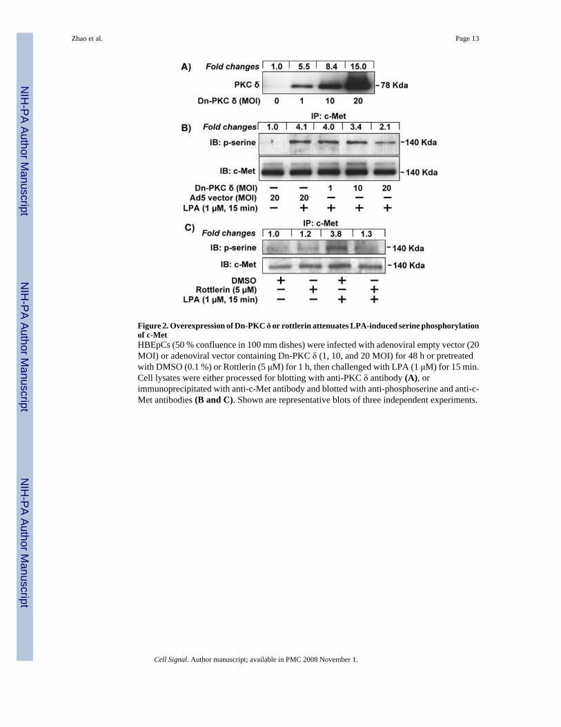
Figure 2. Overexpression of Dn-PKC δ or rottlerin attenuates LPA-induced serine phosphorylationof c-MetHBEpCs (50 % confluence in 100 mm dishes) were infected with adenoviral empty vector (20MOI) or adenoviral vector containing Dn-PKC δ (1, 10, and 20 MOI) for 48 h or pretreatedwith DMSO (0.1 %) or Rottlerin (5 μM) for 1 h, then challenged with LPA (1 μM) for 15 min.Cell lysates were either processed for blotting with anti-PKC δ antibody (A), orimmunoprecipitated with anti-c-Met antibody and blotted with anti-phosphoserine and anti-c-Met antibodies (B and C). Shown are representative blots of three independent experiments.
Zhao et al. Page 13
Cell Signal. Author manuscript; available in PMC 2008 November 1.
NIH
-PA Author Manuscript
NIH
-PA Author Manuscript
NIH
-PA Author Manuscript
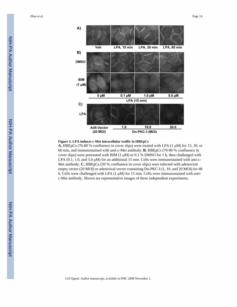
Figure 3. LPA induces c-Met intracellular traffic in HBEpCsA. HBEpCs (70-80 % confluence in cover slips) were treated with LPA (1 μM) for 15, 30, or60 min, and immunostained with anti-c-Met antibody. B. HBEpCs (70-80 % confluence incover slips) were pretreated with BIM (1 μM) or 0.1 % DMSO for 1 h, then challenged withLPA (0.1, 1.0, and 5.0 μM) for an additional 15 min. Cells were immunostained with anti-c-Met antibody. C. HBEpCs (50 % confluence in cover slips) were infected with adenoviralempty vector (20 MOI) or adenoviral vector containing Dn-PKC δ (1, 10, and 20 MOI) for 48h. Cells were challenged with LPA (1 μM) for 15 min. Cells were immunostained with anti-c-Met antibody. Shown are representative images of three independent experiments.
Zhao et al. Page 14
Cell Signal. Author manuscript; available in PMC 2008 November 1.
NIH
-PA Author Manuscript
NIH
-PA Author Manuscript
NIH
-PA Author Manuscript
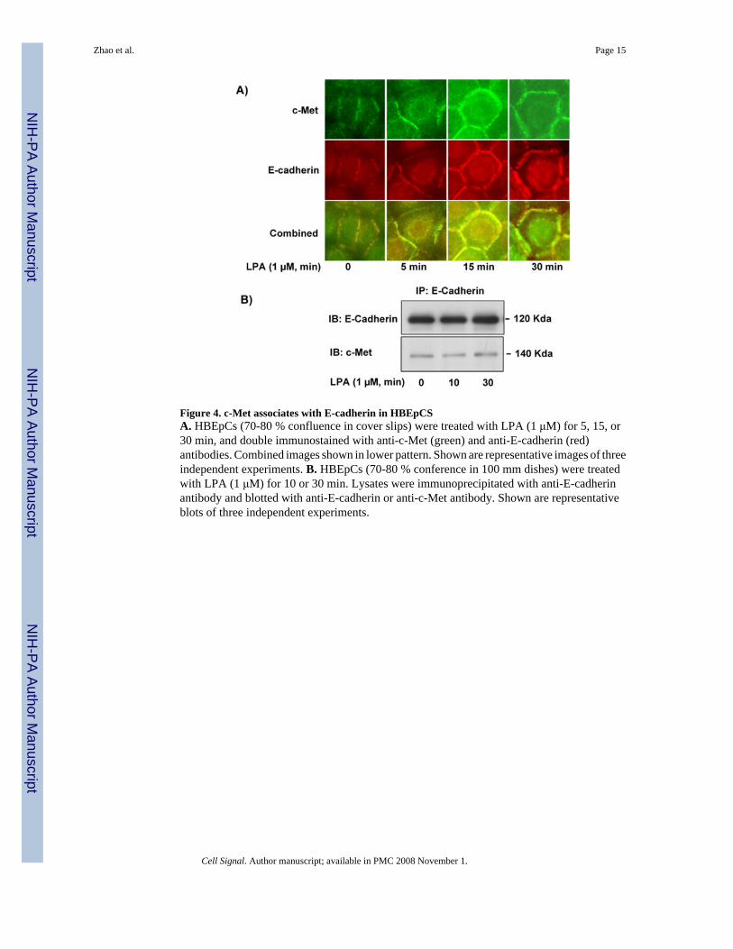
Figure 4. c-Met associates with E-cadherin in HBEpCSA. HBEpCs (70-80 % confluence in cover slips) were treated with LPA (1 μM) for 5, 15, or30 min, and double immunostained with anti-c-Met (green) and anti-E-cadherin (red)antibodies. Combined images shown in lower pattern. Shown are representative images of threeindependent experiments. B. HBEpCs (70-80 % conference in 100 mm dishes) were treatedwith LPA (1 μM) for 10 or 30 min. Lysates were immunoprecipitated with anti-E-cadherinantibody and blotted with anti-E-cadherin or anti-c-Met antibody. Shown are representativeblots of three independent experiments.
Zhao et al. Page 15
Cell Signal. Author manuscript; available in PMC 2008 November 1.
NIH
-PA Author Manuscript
NIH
-PA Author Manuscript
NIH
-PA Author Manuscript
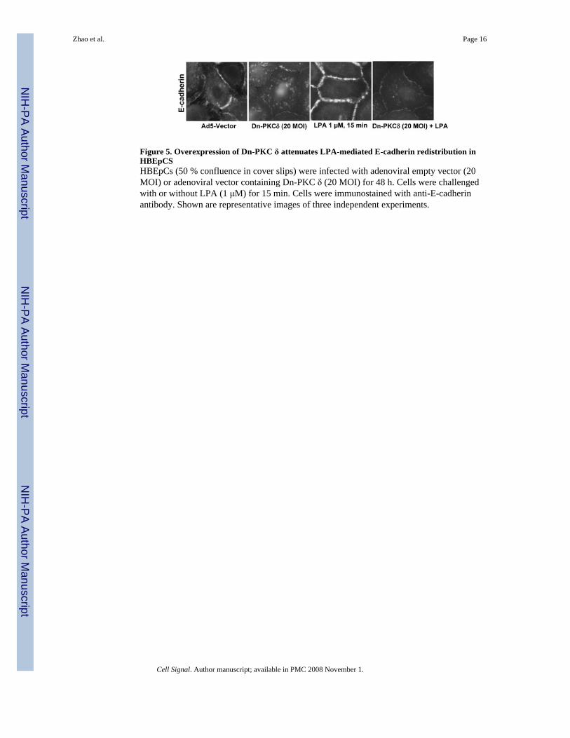
Figure 5. Overexpression of Dn-PKC δ attenuates LPA-mediated E-cadherin redistribution inHBEpCSHBEpCs (50 % confluence in cover slips) were infected with adenoviral empty vector (20MOI) or adenoviral vector containing Dn-PKC δ (20 MOI) for 48 h. Cells were challengedwith or without LPA (1 μM) for 15 min. Cells were immunostained with anti-E-cadherinantibody. Shown are representative images of three independent experiments.
Zhao et al. Page 16
Cell Signal. Author manuscript; available in PMC 2008 November 1.
NIH
-PA Author Manuscript
NIH
-PA Author Manuscript
NIH
-PA Author Manuscript
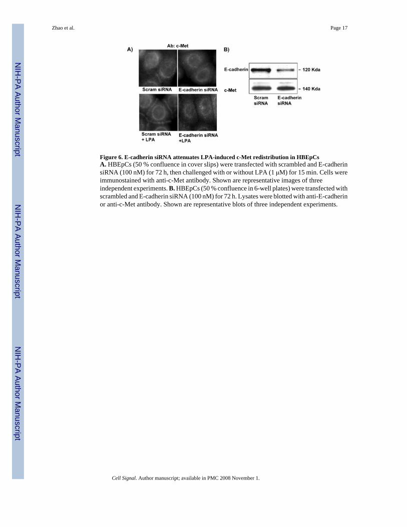
Figure 6. E-cadherin siRNA attenuates LPA-induced c-Met redistribution in HBEpCsA. HBEpCs (50 % confluence in cover slips) were transfected with scrambled and E-cadherinsiRNA (100 nM) for 72 h, then challenged with or without LPA (1 μM) for 15 min. Cells wereimmunostained with anti-c-Met antibody. Shown are representative images of threeindependent experiments. B. HBEpCs (50 % confluence in 6-well plates) were transfected withscrambled and E-cadherin siRNA (100 nM) for 72 h. Lysates were blotted with anti-E-cadherinor anti-c-Met antibody. Shown are representative blots of three independent experiments.
Zhao et al. Page 17
Cell Signal. Author manuscript; available in PMC 2008 November 1.
NIH
-PA Author Manuscript
NIH
-PA Author Manuscript
NIH
-PA Author Manuscript
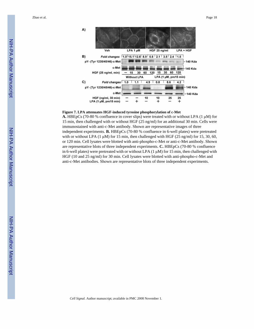
Figure 7. LPA attenuates HGF-induced tyrosine phosphorylation of c-MetA. HBEpCs (70-80 % confluence in cover slips) were treated with or without LPA (1 μM) for15 min, then challenged with or without HGF (25 ng/ml) for an additional 30 min. Cells wereimmunostained with anti-c-Met antibody. Shown are representative images of threeindependent experiments. B. HBEpCs (70-80 % confluence in 6-well plates) were pretreatedwith or without LPA (1 μM) for 15 min, then challenged with HGF (25 ng/ml) for 15, 30, 60,or 120 min. Cell lysates were blotted with anti-phospho-c-Met or anti-c-Met antibody. Shownare representative blots of three independent experiments. C. HBEpCs (70-80 % confluencein 6-well plates) were pretreated with or without LPA (1 μM) for 15 min, then challenged withHGF (10 and 25 ng/ml) for 30 min. Cell lysates were blotted with anti-phospho-c-Met andanti-c-Met antibodies. Shown are representative blots of three independent experiments.
Zhao et al. Page 18
Cell Signal. Author manuscript; available in PMC 2008 November 1.
NIH
-PA Author Manuscript
NIH
-PA Author Manuscript
NIH
-PA Author Manuscript

Figure 8. LPA attenuates HGF / c-Met-mediated signaling and cell motilityA. HBEpCs (70-80 % confluence in 6-well plates) were pretreated with or without LPA (1μM) for 15 min, then challenged with HGF (25 ng/ml) for an additional 30, 60, or 120 min.Cell lysates were blotted with anti-phospho-Akt and anti-Akt antibodies. Shown arerepresentative blots of three independent experiments. B. HBEpCs colonies (grown on 100mm dishes) were pretreated with or without LPA (1 μM) for 15 min, then challenged with orwithout HGF (25 ng/ml) for 3 h. Shown are representative images of five independentexperiments. C. Cells area was measured with NIH imageJ software.
Zhao et al. Page 19
Cell Signal. Author manuscript; available in PMC 2008 November 1.
NIH
-PA Author Manuscript
NIH
-PA Author Manuscript
NIH
-PA Author Manuscript
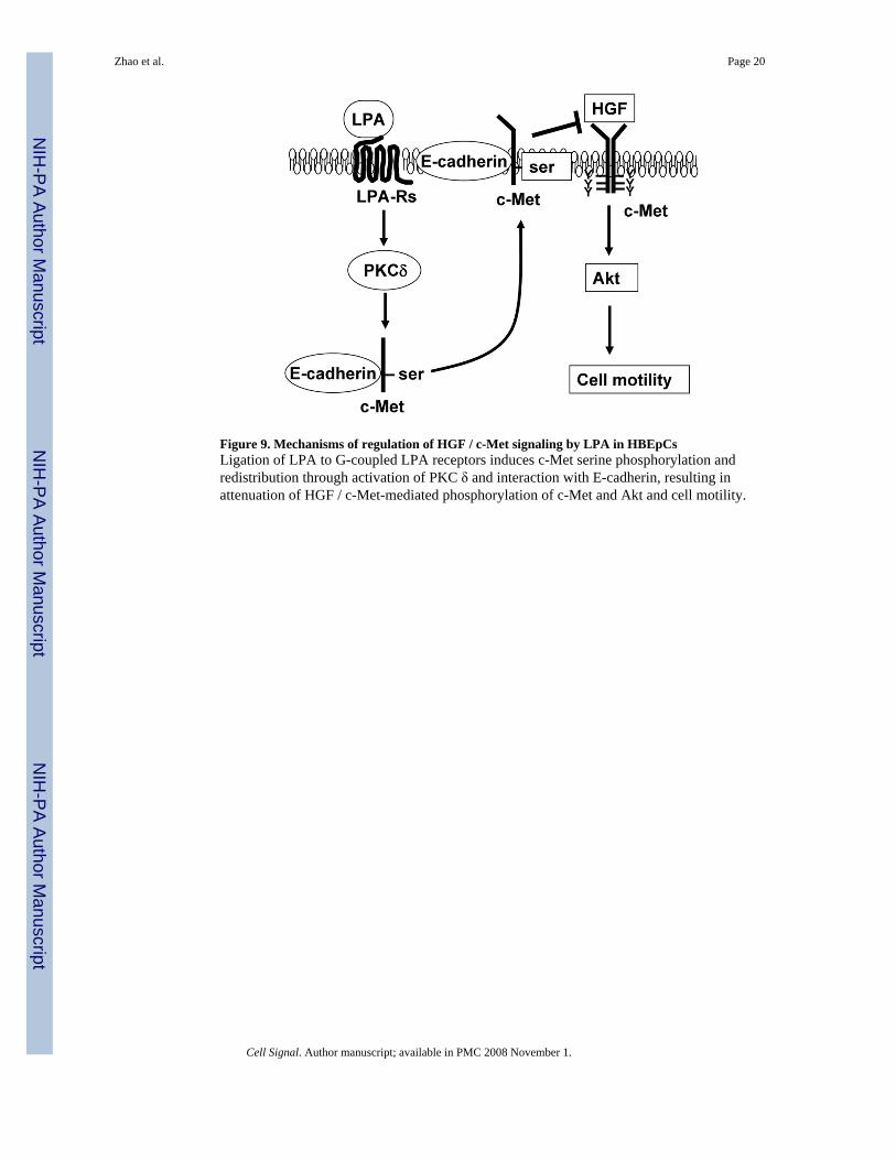
Figure 9. Mechanisms of regulation of HGF / c-Met signaling by LPA in HBEpCsLigation of LPA to G-coupled LPA receptors induces c-Met serine phosphorylation andredistribution through activation of PKC δ and interaction with E-cadherin, resulting inattenuation of HGF / c-Met-mediated phosphorylation of c-Met and Akt and cell motility.
Zhao et al. Page 20
Cell Signal. Author manuscript; available in PMC 2008 November 1.
NIH
-PA Author Manuscript
NIH
-PA Author Manuscript
NIH
-PA Author Manuscript