Substrate-mediated Stabilization of a Tetrameric Drug Target Reveals Achilles Heel in Anthrax
Upload
independentCategory
view
2download
0
SHORT COMMUNICATION
Acid-induced unfolding of didecameric keyhole limpethemocyanin: detection and characterizations of decamericand tetrameric intermediate states
Ankita Varshney • Basir Ahmad • Gulam Rabbani •
Vijay Kumar • Savita Yadav • Rizwan Hasan Khan
Received: 7 August 2009 / Accepted: 10 February 2010 / Published online: 7 March 2010
� Springer-Verlag 2010
Abstract Keyhole limpet hemocyanin (KLH) is widely
used as an immune stimulant and hapten carrier derived
from a marine mollusc Megathura crenulata. To provide
details of the stability and equilibrium of KLH, different
intermediate species were investigated with a series of
biophysical techniques: circular dichroism, binding of
hydrophobic dye, 1-anilino-8-naphthalene sulfonic acid,
acrylamide-induced fluorescence quenching, thermal sta-
bility and dynamic light scattering. KLH in its native state
at pH 7.4 exists in the stable didecameric form with
hydrodynamic radii (Rh) of 28.22 nm, which is approxi-
mately equal to a molecular mass of 8.8 ± 0.6 MDa. The
experimental results demonstrated the presence of two
structurally distinct species in the conformational transition
of KLH under acidic conditions. One species populates at
pH 2.8, characterized as decameric (4.8 ± 0.2 MDa;
Rh = 22.02 nm), molten globule-like state, while the other
accumulates at pH 1.2 and is characterized as a tetramer
(2.4 ± 0.8 MDa; Rh = 16.47 nm) with more organized
secondary and tertiary structures. Our experimental
manipulation of the oligomeric states of KLH has provided
data that correlate well with the known oligomeric forms
obtained from total KLH formed in vivo and extends our
understanding of multimer formation by KLH. The results
are of particular interest in light of the important role of the
mechanistic pathway of pH-dependent structural changes
of Hc stability in the biochemical and medical applications
of these respiratory proteins.
Keywords Acidic pH � Decamer state �Dynamic light scattering � Keyhole limpet hemocyanin �Multimeric protein � Tetramer state
Abbreviations
ANS 1-Anilino-8-naphthalenesulfonate
CD Circular dichroism
DLS Dynamic light scattering
Hc Hemocyanin
KLH Keyhole limpet hemocyanin
MG Molten globule state
MRE Mean residue ellipticity
PFC Packed folded conformational state
Introduction
Interest in the molluscan hemocyanins is well established,
based primarily on the unique immunostimulatory prop-
erties of keyhole limpet hemocyanin (KLH) from the
marine mollusc Megathura crenulata. These extracellular
biopolymers are oxygen carrier copper glycoproteins,
forming freely dissolved aggregates in the hemolymph of
molluscs with extremely high Mw (comparable in size
to ribosomes or small viruses) and complex quaternary
structure (Harris et al. 2000; Sterner and Decker 1994).
They differ fundamentally from arthropod hemocyanins
(Martin et al. 2007; Decker et al. 2007) and are extremely
large proteins (6–7.5 million daltons) that occur either as
decamers (five subunit dimers assembled as a hollow
A. Varshney � B. Ahmad � G. Rabbani � R. H. Khan (&)
Interdisciplinary Biotechnology Unit, Aligarh Muslim
University, Aligarh, UP 202002, India
e-mail: [email protected]; [email protected]
V. Kumar � S. Yadav
Department of Biophysics, All India Institute of Medical
Sciences, Ansari Nagar, New Delhi 110029, India
123
Amino Acids (2010) 39:899–910
DOI 10.1007/s00726-010-0524-4

cylinder), didecamers (face-to-face assembly of two
decamers) or multidecamers (elongated cylinders formed
from a didecamer with added decamers) (Harris and
Markl 1999, 2000; Gatsogiannis and Markl 2009).
Molluscan hemocyanins have been intensively studied for
their structure, function and evolution, and for immuno-
logical and clinical applications (Van Holde et al. 1992;
Van Holde and Miller 1995; Harris and Markl 1999).
Several aspects of their structural–functional peculiarities
make Hcs important materials to address relevant prob-
lems of structural biology, including molecular recogni-
tion among subunits, protein–water interactions or
allosteric regulation (Dolashka-Angelova et al. 2007).
Homogeneous decamer and didecamer structures consist-
ing of only one kind of subunit are found in the bivalves
of the genus Yoldia. In contrast, gastropod Hcs of
M. crenulata (Gebauer et al. 1994), Halotis tuberculata
(Lieb et al. 1999, 2001) and Rapana thomasiana (Gebauer
et al. 1999a) display two distinct homodecameric forms,
attributed to the presence of two different subunits, while
Concholepas concholepas exhibits an unusual hetero-
decameric array of subunits (Ioannes et al. 2004). KLH
consists of two immunologically, physicochemically,
distinct Hc types, termed as KLH1 and KLH2 (Markl
et al. 1991; Gebauer et al. 1994). These act as widely
used immunological tool and promising tumor vaccine
carriers (Kim et al. 2007; Sabbatini et al. 2007). Their
protein structure and disassembly/reassembly behavior
have been extensively studied (Orlova et al. 1997;
Sohngen et al. 1997; Harris et al. 1998, 2000; Gebauer
et al. 1999b, 2002; Mouche et al. 2003). The complete
amino acid sequence of molluscan Hcs H. tuberculata
(Keller et al. 1999; Lieb et al. 2000) and both KLH1 and
KLH2 were found (Lieb and Markl 2004) and, recently,
cryo EM/crystal structure hybrid model of KLH1 was
created (Orlova et al. 1997; Gatsogiannis and Markl
2009). The field of cellular immunology has provided
much relevant biomedical information on KLH and has
led to the expansion of the use of KLH in experimental
immunology and clinically as an immunotherapeutic
agent, because KLH is equal, if not superior, to BCG and
with far fewer side effects (Harris and Markl 1999; Riggs
et al. 2002; Suminoe et al. 2008; Betting et al. 2009).
While the immunological response to KLH has often been
attributed to the carbohydrate moiety, rather than protein
alone, the polypeptide chain of eight globular functional
units constituting the elongated KLH subunit and the
highly organized quaternary structure of the native mol-
ecule could create a scaffold on which multiple carbo-
hydrate epitopes can be initially made available to the
immune system. Optimal steric spacing of sugar residues
could potentiate the observed potent stimulatory response,
both in vivo and in vitro. It is predicted that these
carbohydrate side chains project from the surface of KLH
molecule as well as from the subunits, thereby providing
immunogenicity (Geyer et al. 2005; Beck et al. 2007;
Sandra et al. 2007; Dolashka-Angelova et al. 2009).
Profuse experimental studies, using different dissocia-
tion and reassociation conditions of the native mollusc Hc,
e.g., removal of divalent cations (Bonafe et al. 1994;
Sohngen et al. 1997; Dolashka-Angelova et al. 2003), pH
changes and the addition of denaturing agents (Dolashka-
Angelova et al. 2000), have helped to elucidate its subunit
composition and structure (Van Holde and Miller 1995).
As evident by the very low catalytic activity of molluscs,
Rapana and Octopus Hcs, or arthropods, Carcinus aestu-
arii and Limulus polyphemus Hcs, the entrance to the active
site is probably blocked by Leu or Phe residues, respec-
tively (Hristova et al. 2008). The pH-dependent confor-
mational transition was found to be responsible for the
activation of functional units of Hcs, creating access to the
active site for phenolic substances. There is a wealth of
dissociation-reassembly data at neutral to high pH on KLH
didecamers and dissociation intermediates (Gebauer et al.
1994; Sohngen et al. 1997; Dolashka-Angelova et al.
2003). Thus, we investigated the folding/unfolding of
KLH and its oligomerization into the intermediate struc-
tures at low pH. Previously, molten globule states for
several proteins under various denaturing conditions have
been reported by us (Ahmad et al. 2005, 2006; Varshney
et al. 2008) as well as by several other investigators
(Paci et al. 2005; Greene et al. 2006; Yang et al. 2006;
Gerber et al. 2007, 2008; Georgieva et al. 2008; Kather
et al. 2008; Nishimura et al. 2008; Ramboarina and Redfield
2008; Cremades and Sancho 2008). We found that oligo-
merization of KLH didecamers quantitatively results in
stable decameric and tetrameric forms not previously
observed for KLH under such conditions. These two
structurally distinct isoforms at pH 2.8 and 1.2 behave
similarly at low pH. The results presented here led us to
conclude that KLH has a usual homodecameric organiza-
tion at pH 2.8 resembling the ‘‘molten globule state’’,
which on further protonation results in a stable population
of tetramers (composed of two-subunit dimers).
Materials and methods
Materials
Keyhole limpet hemocyanin, (lot no. H8283) and
1-anilino-8-naphthalene sulfonic acid were purchased
from Sigma Chemical Co., USA. Guanidine hydrochloride
(GnHCl) was obtained from Sisco Research Laboratories,
India. All other reagents used in the study were of analytical
grade.
900 A. Varshney et al.
123
Methods
Protein concentration was determined spectrophotometri-
cally using E1%1 cm of 2.10 at 280 nm (Lowry et al. 1951) on
a Hitachi spectrophotometer, model U-1500.
pH-induced unfolding studies
Stock protein solution was prepared by exhaustive dialysis
of hemocyanin against double distilled water. As much as
50 ll of protein stock solution was mixed with 1,950 ll of
the following buffers: 20 mM sodium phosphate buffer
(pH 7.4–5.8), 20 mM sodium acetate buffer (pH 5.4–3.6)
and 20 mM glycine–HCl buffer (3.4–1.2). The final solu-
tion mixture was incubated for 4–6 h at room temperature
before optical measurements.
Circular dichroism (CD) measurements
CD measurements were carried out with a Jasco spectro-
polarimeter, model J-720, equipped with a microcomputer.
The instrument was calibrated with d-10-camphorsulfonic
acid. All the CD measurements were made at 25�C with a
thermostatically controlled cell holder attached to Neslab’s
RTE-110 water bath with an accuracy of ±0.1�C. Spectra
were collected with a scan speed of 20 nm min-1 and
response time of 1 s. Each spectrum was the average of
four scans. Far UV-CD and near UV-CD spectra were
taken at protein concentrations of 0.25 and 5 mg ml-1 with
0.1 and 1-cm path length cells, respectively. The results
were expressed as mean residue ellipticity (MRE) in
degree cm2 dmol-1, which is defined as:
MRE ¼ hobs �MRW= 10� 1� cð Þ ð1Þ
where hobs is the CD in milli-degree, MRW the mean
residual weight (115), l the path length of the cell and c the
concentration of the protein. Helical content was calculated
from the MRE values at 222 nm using the following
equation as described by Chen et al. (1972):
% a-helix ¼ MRE222nm � 2; 340ð Þ=30; 300½ � � 100: ð2Þ
Circular dichroism data were also analyzed by online
available software, K2d (Andrade et al. 1993).
Fluorescence measurements
Fluorescence measurements were performed on Shimadzu
spectrofluorimeter, model RF-540 equipped with a data
recorder DR-3. The fluorescence spectra were measured at
25 ± 0.1�C with a 1-cm path length cell. The excitation and
emission slits were set at 5 and 10 nm, respectively.
Intrinsic fluorescence was measured by exciting the protein
solution at 280 or 295 nm and emission spectra were
recorded in the range of 300–400 nm. A stock solution of
1-anilino-8-naphthalene sulfonic acid (ANS) was prepared
in distilled water and its concentration was determined
using an extinction coefficient of eM = 5,000 M-1 cm-1 at
350 nm. For ANS fluorescence in the ANS binding experi-
ments, the excitation wavelength was set at 380 nm and the
emission spectra were taken in the range of 400–600 nm.
Acrylamide quenching
In the quenching experiments, aliquots of 5-M quencher
stock solution was added to a protein stock solution (5 mg/
ml), which was further diluted ten times to a total solution
of 3 ml to achieve the desired range of quencher concen-
tration (0.1–1 M). The excitation wavelength was 295 nm
and the emitted light intensity was integrated over the
period of 1 s and detected at 300–400 nm. The results of
the quenching reactions between the excited tryptophan
side chains and acrylamide at corresponding kmax were
analyzed according to the (3) Stern–Volmer and (4) mod-
ified Stern–Volmer equation (Eftink and Ghiron1982):
F0=F ¼ 1þ Ksv Q½ � ð3Þ
F0= F0 � Fð Þ ¼ 1= Kc Q½ ��fað Þ þ 1=fa: ð4Þ
where F0 and F are the fluorescence intensities at an
appropriate wavelength in the absence and presence of
quencher, respectively, Ksv is the Stern–Volmer constant,
fa the fraction of the tryptophans accessible to the
quencher, Kc the collisional quenching constant, and Q the
concentration of the quencher.
Thermal stability studies
To determine the thermal stability of the intermediate state
relative to the native state, ellipticity changes at 222 nm
were measured as a function of temperature. Protein
solutions in 20 mM sodium phosphate buffer of pH 7.4,
and 20 mM glycine–HCl buffer of pH 2.8 and pH 1.2 were
thermostatically controlled using a NESLAB thermostat
water bath model RTE-110. Samples were analyzed after
20 min of incubation at the desired temperature prior to the
measurement to insure the attainment of thermal equili-
bration, and the temperature was continuously varied from
20� to 95�C at a constant rate of 5 ± 0.3�C. The melting
temperature (Tm) values were calculated from the circular
dichroism data.
Dynamic light scattering measurements
Dynamic light scattering measurements were done using
RiNA laser spectroscatter 201 operating at wavelength
660 nm and illuminated by a 100-mW laser diode. The
Acid-induced unfolding of didecameric KLH 901
123

purified and lyophilized samples used for the measure-
ments were first dissolved in buffer solutions of the
required pH values. The protein was dissolved in 20 mM of
sodium phosphate buffer of pH 7.4, 20 mM of glycine–HCl
buffer of pH 2.8 and 20 mM of glycine–HCl buffer of pH
1.2 for DLS measurements. The samples were degassed,
spun down at 14,000 rpm for 10 min and filtered through
0.02-lM polyvinylidene difluoride filters (Millipore). The
protein concentration used was 2 mg ml-1. The samples
were injected manually into the flow cell (30 lL). Data
were acquired over 150 s, where each data point was
averaged over 3 s at a sensitivity of 80%.
Results
To understand the correlation between folding and
assembly of multimeric proteins, it is important to track the
relation between local and global changes that affect the
association/dissociation state of the protein. Hence, we
present the conformational behaviors of whole hemocyanin
molecules in acidic solutions. For this purpose, we used far
UV-CD as a probe for secondary structure, multiple probes
for tertiary structure, and DLS to probe its dissociation
state. These approaches proved to be suitable tools for
studying the Hc structure and conformational changes in
solution. The sensitivity of these techniques allows studies
to be performed using diluted protein solutions. In this
way, problems connected with aggregation and low solu-
bility of the giant keyhole limpet Hc could be avoided.
Far UV-circular dichroism
Changes in the secondary structure of hemocyanin
(KLH) as a function of pH were followed by far UV-CD
measurements in the region between 200 and 250 nm.
Figure 1a shows the CD spectrum for the native hemocyanin
(pH 7.4) possessing two minima, one at 208 and the other
at 222 nm, which is the characteristic feature of a-helical
protein (curve 1). The protein was found to contain
approximately 14% a-helical structure, as calculated by
methods of secondary structure determination (Chen et al.
1972; Andrade et al. 1993). The spectral features at pH 2.8
(curve 2) greatly resembled completely unfolded protein,
but still possessed significant amount of CD signal com-
pared to the 6-M GnHCl denatured protein. However, the
protein at pH 1.2 (curve 3) showed similar features as that
of the native state at pH 7.4 (curve 1). Acid-induced sec-
ondary structural changes of hemocyanin were also moni-
tored by measurements of MRE at 222 nm (Fig. 1b) versus
pH plots. The plot represents a set of smooth and partially
‘‘bell-shaped’’ curve with maxima between pH 2.6 and 3,
without any sigmoid feature at extreme pH. The relative
changes were found to be small and almost noncooperative
(in the vicinity of pH 2.8), indicating that acidic denatur-
ation cannot be achieved as a reversible process for this
intermediate species. But overall, the multimeric intact Hc
had a small interval of partial reversibility under acidic
conditions. Thus, it can be concluded that reversibility was
possible in an acidic pH range, indicating the involvement
of titrable groups responsible for structural stability.
Previously, the effect of acidic and alkaline pH on dichroic
spectra was monitored for RvH2-e (functional unit of
Rapana venosa hemocyanin), but it was found to be pH
independent due to the non-involvement of titrable groups
(Dolashki et al. 2008; Velkova et al. 2009).
ANS fluorescence
Binding of ANS to the hydrophobic region of protein has
been widely used to detect the molten globule state of
-7
-6
-5
0 2 4 6 8
b
MR
E 2
22 x
10-3
(d
eg. c
m2 d
mo
l-1)
pH
a
Fig. 1 a Far UV-CD spectra of hemocyanin: native protein at pH 7.4
(curve 1), molten globule state at pH 2.8 (curve 2), packed folded
conformational state at pH 1.2 (curve 3) and 6-M GnHCl denatured
state (curve 4). b Effect of pH on the mean residual ellipticity (MRE)
of hemocyanin. Ellipticiity was monitored at 222 nm by far UV-CD.
Protein concentration used was 0.25 mg/ml
902 A. Varshney et al.
123
different proteins (Eftink and Ghiron 1982). Figure 2a
shows the acid-induced unfolding of hemocyanin as mon-
itored by ANS fluorescence at 480 nm. We observed
minimum ANS fluorescence in the pH range 7.4–5.0. With
decrease in pH, ANS intensity increased and was highest at
pH 2.8. On further lowering of pH up to 1.2, significant
decrease (about 50%) in the ANS fluorescence intensity
was observed. But the ANS fluorescence at pH 1.2 was still
high as compared to native and/or completely unfolded
protein. Figure 2b shows the comparative emission fluo-
rescence spectra in the 400–600 nm range. We observed
blue shift in the kmax of ANS fluorescence at pH 1.2
(480 nm) relative to native protein (498 nm) and protein at
pH 2.8 (482 nm). This blue shifted fluorescence indicated
burial of bound ANS due to the reorganization of protein
secondary structure. Taken together, we may suggest that
hemocyanin exists in the molten globule state (MG) at pH
2.8, which reorganizes into a packed folded-like confor-
mational state (N*) at pH 1.2.
Intrinsic tryptophan fluorescence
The spectral features of tryptophanyl fluorescence (kmax
and intensity) are dependent on the dynamic and electronic
properties of the chromophore environment; hence, steady
state tryptophan fluorescence has been extensively used to
obtain information on the structural changes of the protein
(Semisotnov et al. 1991; Ahmad et al. 2005). The altera-
tions of microenvironment of tryptophan residues of KLH
have been monitored by studying the changes in the
intensity and kmax of tryptophanyl fluorescence as a func-
tion of pH (Fig. 3, Table 1). The fluorescence intensity at
340 nm and kmax showed no apparent change between pH
0
10
20
30
40
50
0 2 4 6 8pH
AN
S F
luo
resc
ence
inte
nsi
ty a
t 48
0nm
a
b
Fig. 2 a ANS fluorescence of hemocyanin as a function of pH.
b Fluorescence emission spectra of ANS bound to native protein at
pH 7.4 (curve 1), molten globule state at pH 2.8 (curve 2), packed
folded conformational state at pH 1.2 (curve 3) and completely
denatured protein in 6-M GnHCl (curve 4)
0
10
20
30
40
50
60
0 2 4 6 8pH
RF
I 340
nm
339
340
341
342
343
344
345
0 2 4 6 8pH
Wav
elen
gth
(nm
)a
b
Fig. 3 a Tryptophan fluorescence: relative fluorescence intensity of
tryptophan residues at 340 nm in hemocyanin as a function of pH,
kex = 295 nm. b Change in the emission wavelength maximum
(kmax) as function of pH, kex = 295 nm
Acid-induced unfolding of didecameric KLH 903
123
7.4 and 4.0, and when pH decreased below 4.0 up to 2.8,
fluorescence intensity increased markedly with a red shift.
On further lowering of pH up to 1.2, no apparent change
was observed in fluorescence intensity, while kmax was blue
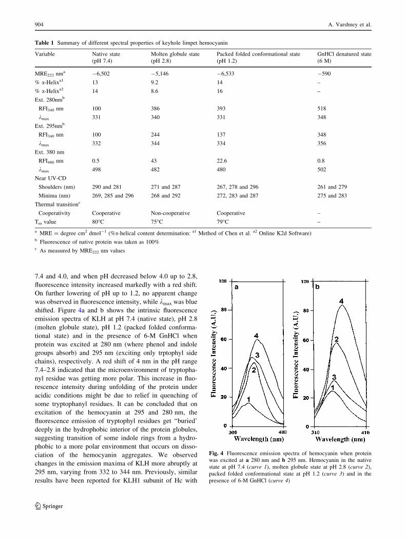
shifted. Figure 4a and b shows the intrinsic fluorescence
emission spectra of KLH at pH 7.4 (native state), pH 2.8
(molten globule state), pH 1.2 (packed folded conforma-
tional state) and in the presence of 6-M GnHCl when
protein was excited at 280 nm (where phenol and indole
groups absorb) and 295 nm (exciting only trptophyl side
chains), respectively. A red shift of 4 nm in the pH range
7.4–2.8 indicated that the microenvironment of tryptopha-
nyl residue was getting more polar. This increase in fluo-
rescence intensity during unfolding of the protein under
acidic conditions might be due to relief in quenching of
some tryptophanyl residues. It can be concluded that on
excitation of the hemocyanin at 295 and 280 nm, the
fluorescence emission of tryptophyl residues get ‘‘buried’
deeply in the hydrophobic interior of the protein globules,
suggesting transition of some indole rings from a hydro-
phobic to a more polar environment that occurs on disso-
ciation of the hemocyanin aggregates. We observed
changes in the emission maxima of KLH more abruptly at
295 nm, varying from 332 to 344 nm. Previously, similar
results have been reported for KLH1 subunit of Hc with
Table 1 Summary of different spectral properties of keyhole limpet hemocyanin
Variable Native state Molten globule state Packed folded conformational state GnHCl denatured state
(pH 7.4) (pH 2.8) (pH 1.2) (6 M)
MRE222 nma -6,502 -5,146 -6,533 -590
% a-Helixa1 13 9.2 14 –
% a-Helixa2 14 8.6 16 –
Ext. 280nmb
RFI340 nm 100 386 393 518
kmax 331 340 331 348
Ext. 295nmb
RFI340 nm 100 244 137 348
kmax 332 344 334 356
Ext. 380 nm
RFI480 nm 0.5 43 22.6 0.8
kmax 498 482 480 502
Near UV-CD
Shoulders (nm) 290 and 281 271 and 287 267, 278 and 296 261 and 279
Minima (nm) 269, 285 and 296 268 and 292 272, 283 and 287 275 and 283
Thermal transitionc
Cooperativity Cooperative Non-cooperative Cooperative –
Tm value 80�C 75�C 79�C –
a MRE = degree cm2 dmol-1 (%a-helical content determination: a1 Method of Chen et al. a2 Online K2d Software)b Fluorescence of native protein was taken as 100%c As measured by MRE222 nm values
Fig. 4 Fluorescence emission spectra of hemocyanin when protein
was excited at a 280 nm and b 295 nm. Hemocyanin in the native
state at pH 7.4 (curve 1), molten globule state at pH 2.8 (curve 2),
packed folded conformational state at pH 1.2 (curve 3) and in the
presence of 6-M GnHCl (curve 4)
904 A. Varshney et al.
123

kmax values of 336–340 nm, while for KLH2 it ranges from
335 to 336 nm. The same effect was observed for dideca-
meric oxy-Hc of R. thomasiana groose molecule and its
subunits (Dolashka et al. 1996). Furthermore, shift in the
emission maxima at 295 nm depicts that Trp residues
might be located in the vicinity of the active site of KLH
functional unit. The partial unfolding of the KLH in these
pH regions was also supported by far UV-CD, further
indicating loss of secondary structure. This finding was
further supported by quenching studies with a neutral
quencher acrylamide.
Acrylamide quenching studies
The fluorescence properties of Trp residues can be used
to obtain topological information of proteins. Fluores-
cence quenching of the tryptophanyl residues by neutral
quencher (acrylamide) has been shown to be useful to
obtain information about the solvent accessibility of
these residues in proteins and the polarity of their
microenvironment, as it can discriminate between ‘bur-
ied’ and ‘exposed’ side chains (Pawar and Deshpande
2000). Its ability to collosionally quench the excited
indole rings depends only on their ‘exposure’ to the
quencher. Figure 5a and b depicts the Stern–Volmer and
modified Stern–Volmer plots for the acrylamide
quenching studies performed on the native, MG, N* and
unfolded (6-M GnHCl) states. The values of Stern–
Volmer constant (Ksv) and fractional accessibility of Trp
residues to quencher (fa) were calculated from the above
plots and presented in Table 2. The observed linearity of
the plots can be explained by the similarity of the
individual Ksv constants. It was interesting to note that
Ksv for MG state (Ksv = 5.2) was found to be markedly
higher compared to the N state (Ksv = 0.70) and was
accompanied by a red shift in kmax from 340 to 344 nm
of Trp, supported by increase of fa from 0.47 to 1 on
decrease of pH from 7.4 to 2.8. The acrylamide
quenching efficiency for the native Hc (Ksv = 0.70 M-1)
was very low compared to Ksv = 16.33 M-1 of trypto-
phan in aqueous solution. These results indicated that
Trp residues in MG state were highly accessible to the
quencher. In the presence of 6-M GnHCl wherein protein
was considered to exist in a random coil conformation,
we found Ksv and fa (11.52 and 1.13) to be much higher
than the acid unfolded state. These results together with
intrinsic fluorescence indicated that N* state at pH 1.2
possesses Trp residues microenvironment, closely
resembling that of native protein (Eftink and Ghiron
1982; Ahmad et al. 2005). As shown in Table 2, frac-
tional accessibility of the acrylamide to the Trp residues
of KLH follows the order:
U [ MG pH 2:8ð Þ[ N� pH 1:2ð Þ[ N:
This also indicates the comparative compactness of
these intermediate states.
Near UV-circular dichroism
To study the tertiary structural alterations in more detail,
CD measurements in the near UV region were performed at
pH 7.4, 2.8, 1.2 and in the presence of 6-M GnHCl
(Fig. 6a). Near UV-CD spectra of native KLH (pH 7.4)
0
2
4
6
8
10
12
14
0 0.2 0.4 0.6 0.8 1 1.2 (Q)
Fo/
F
a
2
6
4
8
10
0 2 4 6 8 10 12
Fo/
Fo-
F
b
(1/Q)
Fig. 5 Acrylamide quenching: Stern–Volmer a and modified Stern–
Volmer b plots for hemocyanin at native pH 7.4 (filled circles),
molten globule state at pH 2.8 (filled squares), packed folded
conformational state at pH 1.2 (open triangles) and 6-M GnHCl state
(multiple symbols)
Table 2 Fluorescence parameters (KSV and fa) for acrylamide
quenching of keyhole limpet hemocyanin
pH Ksv (M-1) fa (%)
7.4 0.7017 0.4766
2.8 5.1913 1.0463
1.2 1.4217 0.5575
6-M GnHCl 11.511 1.1273
Acid-induced unfolding of didecameric KLH 905
123
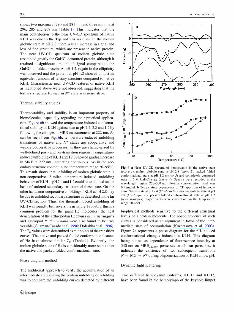
shows two maxima at 290 and 281 nm and three minima at
296, 285 and 269 nm (Table 1). This indicates that the
main contribution to the near UV-CD spectrum of native
KLH was due to the Trp and Tyr residues. In the molten
globule state at pH 2.8, there was an increase in signal and
loss of fine structure, which are present in native protein.
The near UV-CD spectrum of molten globule state
resembled greatly the GnHCl denatured protein, although it
retained a significant amount of signal compared to the
GnHCl unfolded protein. At pH 1.2, regain in the ellipticity
was observed and the protein at pH 1.2 showed almost an
equivalent amount of tertiary structure compared to native
KLH. Characteristic near UV-CD features of native KLH
as mentioned above were not observed, suggesting that the
tertiary structure formed in N* state was non-native.
Thermal stability studies
Thermostability and stability is an important property of
biomolecules, especially regarding their practical applica-
tion. Figure 6b showed the temperature-induced conforma-
tional stability of KLH against heat at pH 7.4, 2.8 and 1.2 by
following the changes in MRE measurements at 222 nm. As
can be seen from Fig. 6b, temperature-induced unfolding
transitions of native and N* states are cooperative and
weakly cooperative processes, as they are characterized by
well-defined post- and pre-transition regions. Temperature-
induced unfolding of KLH at pH 2.8 showed gradual increase
in MRE at 222 nm, indicating continuous loss in the sec-
ondary structure content in the temperature range 20–95�C.
This result shows that unfolding of molten globule state is
non-cooperative. Similar temperature-induced unfolding
behaviors of KLH at pH 7.4 and 1.2 could be explained on the
basis of ordered secondary structure of these state. On the
other hand, non-cooperative unfolding of KLH at pH 2.8 may
be due to unfolded secondary structure as described in the far
UV-CD section. Thus, the thermal-induced unfolding of
KLH was found to be irreversible in nature. Probably, this is a
common problem for the giant Hc molecules; the heat
denaturation of the arthropodan Hc from Palinurus vulgaris
and gastropod R. thomasiana were also found to be irre-
versible (Guzman-Casado et al. 1990; Dolashka et al. 1996).
The Tm values were determined as midpoints of the transition
curves. The native and packed folded conformational states
of Hc have almost similar Tm (Table 1). Evidently, the
molten globule state of Hc is considerably more stable than
the native and packed folded conformational state.
Phase diagram method
The traditional approach to verify the accumulation of an
intermediate state during the protein unfolding or refolding
was to compare the unfolding curves detected by different
biophysical methods sensitive to the different structural
levels of a protein molecule. The noncoincidence of such
curves is considered as an argument in favor of the inter-
mediate state of accumulation (Kuznetsova et al. 2003).
Figure 7a represents a phase diagram for the pH-induced
conformational changes induced in KLH. This diagram
being plotted as dependence of fluorescence intensity at
340 nm on MRE222nm, possesses two linear parts, i.e., it
indicates the existence of two subsequent transitions
N ? MG ? N* during oligomerization of KLH at low pH.
Dynamic light scattering
Two different hemocyanin isoforms, KLH1 and KLH2,
have been found in the hemolymph of the keyhole limpet
-7
-6
-5
-4
20 40 60 80 100
-MR
E 22
2 x 1
0-3 (
deg.
cm
2 dm
ol-1
)
Temperature (°C)
b
a
Fig. 6 a Near UV-CD spectra of hemocyanin in the native state
(curve 1), molten globule state at pH 2.8 (curve 2), packed folded
conformational state at pH 1.2 (curve 3) and completely denatured
state in 6-M GnHCl state (curve 4). Spectra were recorded in the
wavelength region 250–300 nm. Protein concentration used was
0.5 mg/ml. b Temperature dependence of CD spectrum of hemocy-
anin. Native state at pH 7.4 (filled circles), molten globule state at pH
2.8 (filled squares), packed folded conformational state at pH 1.2
(open triangles). Experiments were carried out in the temperature
range 20–95�C
906 A. Varshney et al.
123
M. crenulata with molecular masses of *8 MDa, such that
native didecamers yielded a mass difference of about
800 kDa between KLH l and KLH 2 (8.3 vs. 7.5 MDa)
(Hartmann et al. 2004; Sohngen et al. 1997). KLH displays
different oligomeric states and stabilities. At pH 7.4, KLH
exists as didecamer and shows hydrodynamic radii of
28.22 nm, approximately equal to a molecular mass of
8.8 ± 0.6 MDa, and is stable (Fig. 7b). KLH didecamer
dissociates to decamer (4.8 ± 0.2 MDa; hydrodynamic
radii of 22.02 nm) at pH 2.8. On further lowering of pH to
1.2, it dissociates to a tetramer (2.4 ± 0.8 MDa) corre-
sponding to a hydrodynamic radius of 16.47 nm. Thus, we
conclude that KLH exists primarily in the multimeric state
and dissociates to the decameric state that possesses char-
acteristics of a molten globule, which, on further lowering
the pH to 1.2, forms a tetramer. These tetramers might
form due to dimerization of dimers as reported previously
(Orlova et al. 1997). These dimers could be defined as
packed folded-like conformational state, as they regain the
structure similar to that of native Hc KLH.
Discussion
At present, there is a growing interest in hemocyanins; from a
scientific viewpoint, this attention is focused on their struc-
ture, evolution and diversity (Van Holde et al. 1992; Van
Holde and Miller 1995), whereas from the biomedical
viewpoint, it concerns the relationship among their structural
features and immunotherapeutic effects. Limited number of
literature has been directed toward the biophysical and
structural understanding of Hc stability and function. This
was not surprising because of the complexity of gastropod Hc
structure, created from multiple subunits each containing
seven or eight similar, but not identical functional units. As
reviewed by Herskovits (1988), the dissociation transitions
obtained with the didecameric hemocyanins of land and
marine gastropods were considered to be rather complex. The
occurrence of didecamer as the predominantly oligomeric
form of both KLH1 and KLH2, in in vivo and in vitro con-
ditions, probably relates to the asymmetric nature of the
decamer. Till now, no convincingly defined structural
difference has been detected between didecamers of KLH1
and KLH2. KLH 1 apparently exists as a stable didecamer
with random clusters of didecamers, whereas KLH2 exists as
decamers, didecamers, multidecamers of varying length
(Gebauer et al. 1994; Sohngen et al. 1997). For KLH2, the
production of decamers proved to be slightly more difficult,
as simple adjustment of the stabilizing buffer pH had no
effect. However, dissociation of KLH2 subunits into a mix-
ture of didecamers and multidecamers was found to be
unstable in comparison to that of the KLH1 subunit. It can be
concluded that both isoforms of KLH exhibit the character-
istics of oligomerization features, whether produced natu-
rally or experimentally. Our experimental results described
here allow structural and spectroscopic characterizations of
the keyhole limpet hemocyanin from M. crenulata (Table 1).
Quenching of ‘buried tryptophan’ by acrylamide explains the
structural fluctuation of the protein molecule that facilitates
the inward diffusion of the quencher. The lowered Ksv values
for the KLH at neutral pH in comparison with MG and N*
states reflect the additional limitations of the accessibility to
the tryptophyl side chains imposed by the quaternary struc-
ture of the aggregates. Two classes of ‘‘buried’’ fluorophores
can be considered to be responsible for the Hc fluorescence:
first, Trp residues localized very close to the active site
pocket; and second, Trp moieties involved in intersubunit
contacts (Dolashki et al. 2005). The increase in the quenching
5
15
25
35
45
55
-15 -14 -13 -12 -11 -10MRE 222nm ( deg.cm2 dmol-1)
Flu
ores
cenc
e In
tens
ity
at 3
40 n
m
0
1
2
3
4
5
6
7
8
9
Mol
ecul
ar M
ass
(MD
a)
7.4 2.8 1.2pH
b
N
MG N*
a
Fig. 7 a Phase diagram representation [fluorescence intensity at
340 nm vs. MRE 222nm (degree cm2 dmol-1)] of the pH-induced
conformational changes in KLH. b Measurement of molecular
dimension and hydrodynamic radii of KLH at different pH (7.4, 2.8
and 1.2)
Acid-induced unfolding of didecameric KLH 907
123

efficiently on the tryptophyl emission can be explained by
assuming that upon dissociation, these side chains become
more accessible to the quencher, causing the Stern–Volmer
constant approach values found for ‘exposed’ tryptophans as
in arthropodan and gastropodan Hcs (Dolashka et al. 1996;
Stoeva et al. 1995). As depicted by biophysical techniques,
all transition curves for the state at pH 1.2 were found to be
similar to that of native KLH. It was reasonable to suggest
that in solution the species at pH 1.2 (N*) resembles KLH at
pH 7.4 (N). While for the state at pH 2.8 many secondary and
tertiary structural elements were found to be preserved, the
protein acquires a ‘‘globule state’’. DLS combined with a
variety of biophysical methods, such as fluorescence, phase
diagram and CD, were used to monitor the changes in con-
formation and association state of KLH induced at low pH.
On the basis of these results, we speculate that our protein
contains a mixture of all possible isoforms of Hc (KLH) and
that it remains unclear which isoform was responsible for
which part of the data. Furthermore, as estimated by the
sequence analysis (the KLH sequences were available in the
data banks), the pure polypeptides possess 392 kDa; as
deduced from sugar analysis, the glycans make another 8–
10 kDa. Therefore, it was highly justified to assume a value
of *8.0 MDa for the KLH didecamer. Our direct measure-
ments of the molecular masses of the two structurally distinct
folding intermediates induced by acid, which accumulated at
pH 2.8 (decameric, MG state) and 1.2 (tetrameric, N* state),
further approve this hypothesis and clearly support our con-
cept of oligomerization of the KLH subunit. This knowledge
would be important for a better understanding of oligomer-
ization of KLH under acidic conditions. In view of the bio-
medical application of KLH, our results are of particular
interest in contributing a series of new physicochemical
properties of this complex biopolymer. As shown, KLH
didecamers can be quantitatively split into decamers and
tetramers at low pH in standard buffer solutions.
15
NqN
N State
KLH-Didecamer
(pH 7.4)
• Mol.wt.= 8.8 ± 0.6MDa
• Rh = 28.22 nm
MG State
KLH-Decamer
(pH 2.8)
• Mol.wt.= 4.8 ± 0.2MDa
• Rh = 22.02 nm
N* State
KLH-Tetramer
(pH 1.2)
• Mol.wt.= 2.4 ± 0.8MDa
• Rh = 16.47 nm
Slab/wall interfaces
wall
A proposed model summarizing the dynamic changes in
KLH oligomerization that occurs at different pH values
(7.4, 2.8 and 1.2). A KLH didecamer are depicted
centrally in tilt as well as top view, tagged with putative
carbohydrate trees (red) (Gatsogiannis and Markl 2009).
908 A. Varshney et al.
123
Acknowledgments The facilities provided by AMU are gratefully
acknowledged. A.V. and B.A. thank the Council of Scientific and
Industrial Research, New Delhi, for financial assistance. The authors
are also thankful to DST (FIST) for providing laboratory facilities.
References
Ahmad B, Ankita, Khan RH (2005) Urea induced unfolding of F
isomer of human serum albumin: a case study using multiple
probes. Arch Biochem Biophys 437:159–167
Ahmad B, Ansari MA, Sen P, Khan RH (2006) Low versus high
molecular weight polyethylene glycol induced states of stem
bromelain at low pH: stabilization of molten globule and
unfolded states. Biopolymers 81:350–359
Andrade MA, Chacon P, Merelo JJ, Moran F (1993) Evaluation of
secondary structure of proteins from UV circular dichroism
spectra using an unsupervised learning neural network. Protein
Eng 6:383–390
Beck A, Hillen N, Dolashki A, Stevanovic S, Salvato B, Voelter W,
Dolashka-Angelova P (2007) Oligosaccharide structure of a
functional unit RvH1-b of Rapana venosa hemocyanin using
HPLC/electrospray ionization mass spectrometry. Biochimie
89:938–949
Betting DJ, Mu XY, Kafi K, McDonnel D, Rosas F, Gold DP,
Timmerman JM (2009) Enhanced immune stimulation by a
therapeutic lymphoma tumor antigen vaccine produced in insect
cells involves mannose receptor targeting to antigen presenting
cells. Vaccine 27:250–259
Bonafe CF, Araujo JRV, Silva JL (1994) Intermediate states of
assembly in the dissociation of gastropod hemocyanin by
hydrostatic pressure. Biochemistry 33:2651–2660
Chen YH, Yang JT, Martinez H (1972) Determination of the
secondary structure of proteins by circular dichroism and optical
rotatory dispersion. Biochemistry 11:4120–4131
Cremades N, Sancho J (2008) Molten globule and native state
ensemble of Helicobacter pylori flavodoxin: can crowding,
osmolytes or cofactors stabilize the native conformation relative
to the molten globule? Biophys J 95:1913–1927
Decker H, Hellmann N, Jaenicke E, Lieb B, Meissner U, Markl J
(2007) Minireview: recent progress in hemocyanin research.
Integr Comp Biol 47:631–644
Dolashka P, Genov N, Parvanova K, Voelter W, Geiger M, Stoeva S
(1996) Rapana thomasiana grosse (gastropoda) haemocyanin:
spectroscopic studies of the structure in solution and the
conformational stability of the native protein and its structural
subunits. Biochem J 315:139–144
Dolashka-Angelova P, Schick M, Stoeva S, Voelter W (2000)
Isolation and partial characterization of the N-terminal func-
tional unit of subunit RtH1 from Rapana thomasiana grosse
hemocyanin. Int J Biochem Cell Biol 32:529–538
Dolashka-Angelova P, Schwarz H, Dolashki A, Beltramini M,
Salvato B, Schick M, Saeed M, Voelter W (2003) Oligomeric
stability of Rapana venosa hemocyanin (RvH) and its structural
subunits. Biochim Biophys Acta 1646:77–85
Dolashka-Angelova P, Stevanovic S, Dolashki A, Devreese B,
Tzvetkova B, Voelter W, Van Beeumen J, Salvato B (2007) A
challenging insight on the structural unit 1 of molluscan Rapanavenosa hemocyanin. Arch Biochem Biophys 459:50–58
Dolashka-Angelova P, Lieb B, Velkova L, Heilen N, Sandra K,
Nikolaeva-Glomb L, Dolashki A, Galabov AS, Van Beeumen J,
Stevanovic S, Voelter W, Devreese B (2009) Identification of
glycosylated sites in Rapana hemocyanin by mass spectrometry
and gene sequence, and their antiviral effect. Bioconjug Chem
20:1315–1322
Dolashki A, Schutz J, Hristova R, Voelter W, Dolashka P (2005)
Spectroscopic properties of non-glycosilated functional unit KLH2-
c of keyhole limpet hemocyanin. World J Agric Sci 1:129–136
Dolashki A, Velkova L, Atanasov B, Voelter W, Stevanovic S,
Schwarz H, Muro PD, Dolashka-Angelova P (2008) Reversibility
and ‘‘pH–T phase diagrams’’ of Rapana venosa hemocyanin and
its structural subunits. Biochim Biophys Acta 1784:1617–1624
Eftink MR, Ghiron CA (1982) Fluorescence quenching studies with
proteins. Anal Biochem 114:199–227
Gatsogiannis C, Markl J (2009) Keyhole limpet hemocyanin: the
9 ACryoEM structure and molecular model of the KLH1
didecamer reveal the interfaces and intricate topology of 160
functional units. J Mol Biol 385:963–983
Gebauer W, Harris JR, Heid H, Sueling M, Hillenbrand R, Soehngen
S, Wegener-Strake A, Markl J (1994) Quaternary structure,
subunits and domain patterns of two discrete forms of keyhole
limpet hemocyanin: KLH1 and KLH2. Zoology 98:51–68
Gebauer W, Stoeva S, Voelter W, Dainese E, Salvato B, Beltramini
M, Markl J (1999a) Hemocyanin subunit organization of the
gastropod Rapana thomasiana. Arch Biochem Biophys
372:128–134
Gebauer W, Harris JR, Geisthardt G, Markl J (1999b) Keyhole limpet
hemocyanin type 2 (KLH2): detection and immunolocalization
of a labile functional unit h. J Struct Biol 128:280–286
Gebauer W, Harris JR, Markl J (2002) Topology of the 10 subunits
within the decamer of KLH, the hemocyanin of the marine
gastropod Megathura crenulata. J Struct Biol 139:153–159
Georgieva ER, Narvaez AJ, Hedin N, Graslund A (2008) Secondary
structure conversions of Mycobacterium tuberculosis ribonucle-
otide reductase protein R2 under varying pH and temperature
conditions. Biophys Chem 137:43–48
Gerber R, Tahiri-Alaoui A, Hore PJ, James W (2007) Oligomeriza-
tion of the human prion protein proceeds via a molten globule
intermediate. J Biol Chem 282:6300–6337
Gerber R, Tahiri-Alaoui A, Hore PJ, James W (2008) Conformational
pH dependence of intermediate states during oligomerization of
the human prion protein. Protein Sci 17:537–544
Geyer H, Wuhrer M, Resemann A, Geyer R (2005) Identification and
characterization of keyhole limpet hemocyanin N-glycans
mediating cross-reactivity with Schistosoma mansoni. J Biol
Chem 280:40731–40748
Greene LH, Wijesinha-Bettoni R, Redfield C (2006) Characterization
of the molten globule of human serum retinol-binding protein
using NMR spectroscopy. Biochemistry 45:9475–9484
Guzman-Casado M, Parody-Morreale A, Mateo PL, Sanchez-Ruiz JM
(1990) Differential scanning calorimetry of lobster haemocya-
nin. Eur J Biochem 188:181–185
Harris JR, Markl J (1999) Keyhole limpet hemocyanin (KLH): a
biomedical review. Micron 30:597–623
Harris JR, Markl J (2000) Keyhole limpet hemocyanin: molecular
structure of a potent marine immunoactivator: a review. Euro
Urol 37(suppl 3):24–33
Harris JR, Gebauer W, Adrian M, Markl J (1998) Keyhole limpet
hemocyanin (KLH): slow in vitro reassociation of KLH1 and
KLH2 from Immucothel. Micron 5:329–339
Harris JR, Scheffler D, Gebauer W, Lehnert R, Markl J (2000)
Haliotis tuberculata hemocyanin (HtH): analysis of oligomeric
stability of HtH1 and HtH2 and comparison with keyhole limpet
hemocyanin KLH1 and KLH2. Micron 31:613–622
Hartmann H, Bongers A, Decker H (2004) Small-angle X-ray
scattering based three-dimensional reconstruction of the immu-
nogen KLH1 reveals different oxygen-dependent conformations.
J Biol Chem 279:2841–2845
Herskovits TT (1988) Recent aspects of the subunit organization
and dissociation of hemocyanins. Comp Biochem Physiol B
91:597–611
Acid-induced unfolding of didecameric KLH 909
123
Hristova R, Dolashki A, Voelter W, Stevanovic S, Dolashka-
Angelova P (2008) O-Diphenol oxidase activity of molluscan
hemocyanins. Comp Biochem Phys Part B 149:439–446
Ioannes PD, Moltedo B, Oliva H, Pacheco R, Faunes F, Ioannes AED,
Becker MI (2004) Hemocyanin of the Molluscan Concholepasconcholepas exhibits an unusual heterodecameric array of
subunits. J Biol Chem 279:26134–26142
Kather I, Jakob RP, Dobbek H, Schmid FX (2008) Increased folding
stability of TEM-1 beta-lactamase by in vitro selection. J Mol
Biol 383:238–251
Keller H, Lieb B, Altenhein B, Gebauer D, Richter S, Stricker S,
Markl J (1999) Abalone (Haliotis tuberculata) hemocyanin type
1 (HtH1). Organization of the approximately 400 kDa subunit,
and amino acid sequence of its functional units f, g and h. Eur J
Biochem 264:27–38
Kim JH, Lee Y, Bae YS, Kim WS, Kim K, Im HY, Kang WK, Park
K, Choi HY, Lee HM, Baek SY, Lee H, Doh H, Kim BM, Kim
CY, Jeon C, Jung CW (2007) Phase I/II study of immunotherapy
using autologous tumor lysate-pulsed dendritic cells in patients
with metastatic renal cell carcinoma. Clin Immunol 125:257–267
Kuznetsova IM, Turoverov KK, Uversky VN (2003) Use of the phase
diagram method to analyze the protein unfolding–refolding
reactions: fishing out the ‘‘Invisible’’ intermediates. J Proteome
Res 3:485–494
Lieb B, Markl J (2004) Evolution of molluscan hemocyanins as
deduced from DNA sequencing. Micron 35:117–119
Lieb B, Altenhein B, Lehnert R, Gebauer W, Markl J (1999) Subunit
organization of the abalone Haliotis tuberculata hemocyanin
type 2 (HtH2), and the cDNA sequence encoding its functional
units d, e, f, g and h. Eur J Biochem 265:134–144
Lieb B, Altenhein B, Markl J (2000) The sequence of a gastropod
hemocyanin (HtH1 from Haliotis tuberculata). J Biol Chem
275:5675–5681
Lieb B, Altenhein B, Markl J, Vincent A, Van Olden EV, Van Holde
KE, Miller K (2001) Structures of two molluscan hemocyanin
genes: significance for gene evolution. Proc Natl Acad Sci USA
98:4546–4551
Lowry OH, Rosebrough NJ, Farr AL, Randall RJ (1951) Protein
measurement with the folin–phenol reagent. J Biol Chem
193:265–275
Markl J, Savel-Niemann A, Wegener-Strake A, Sueling M, Schneider
A, Gebauer W, Harris JR (1991) The role of two distinct subunit
types in the architecture of keyhole limpet hemocyanin (KLH).
Naturwissenschaften 78:512–514
Martin A, Depoix F, Stohr M, Meissner U, Hagner-Holler S,
Hammouti K, Burmester T, Heyd J, Wriggers W, Markl J
(2007) Limulus polyphemus hemocyanin: 10 A structure,
sequence analysis, molecular modelling and rigid-body fitting
reveal the interfaces between the eight hexamers. J Mol Biol
366:1332–1350
Mouche F, Zhu YX, Pulokas J, Potter CS, Carragher B (2003)
Automated three-dimensional reconstruction of keyhole limpet
hemocyanin type 1. J Struct Biol 144:301–312
Nishimura C, Dyson HJ, Wright PE (2008) The kinetic and
equilibrium molten globule intermediates of apoleghemoglobin
differ in structure. J Mol Biol 378:715–725
Orlova EV, Dube P, Harris JR, Beckman E, Zemlin F, Markl J, van
Heel M (1997) Structure of keyhole limpet hemocyanin type 1
(KLH1) at 15 A resolution by electron cryomicroscopy and
angular reconstitution. J Mol Biol 271:417–437
Paci E, Greece LH, Jones RM, Smith LJ (2005) Characterization of
the molten globule state of retinol-binding protein using a
molecular dynamics simulation approach. FEBS J 18:4826–4838
Pawar SA, Deshpande VV (2000) Characterization of acid-induced
unfolding intermediates of glucose/xylose isomerase. Eur J
Biochem 267:6331–6338
Ramboarina S, Redfield C (2008) Probing the effect of temperature on
the backbone dynamics of the human a-lactalbumin molten
globule. J Am Chem Soc 130:15318–15326
Riggs DR, Jackson B, Vona-Davis L, McFadden D (2002) In vitro
anticancer effects of a novel immunostimulant: keyhole limpet
hemocyanin. J Surg Res 108:279–284
Sabbatini PJ, Ragupathi G, Hood C, Aghajanian CA, Juretzka M,
Iasonos A, Hensley ML, Spassova MK, Ouerfelli O, Spriggs DR,
Tew WP, Konner J, Clausen H, Abu Rustum N, Dansihefsky SJ,
Livingston PO (2007) Pilot study of a heptavalent vaccine—
keyhole limpet hemocyanin conjugate plus QS21 in patients with
epithelial ovarian, fallopian tube, or peritoneal cancer. Clin
Cancer Res 13:4170–4177
Sandra K, Dolashka-Angelova P, Devreese B, Van Beeumen J (2007)
New insights in Rapana venosa hemocyanin N-glycosylation
resulting from on-line mass spectrometric analyses. Glycobiol-
ogy 17:141–156
Semisotnov GV, Rodionova NA, Razgulyaev OI, Uversky VN,
Gripas AF, Gilmanshin RI (1991) Study of the ‘‘molten globule’’
intermediate state in protein folding by a hydrophobic fluores-
cent probe. Biopolymers 1:119–128
Sohngen MS, Stahalmann A, Harris JR, Muller SA, Engel A, Markl J
(1997) Mass determination, subunit organization and control of
oligomerization states of keyhole limpet hemocyanin (KLH).
Euro J Biochem 248:602–614
Sterner R, Decker H (1994) Inversion of the Bohr effect upon oxygenbinding to 24-meric tarantula hemocyanin. Proc Natl Acad Sci
USA 91:4835–4839
Stoeva S, Dolashka P, Bankov B, Voelter W, Salvato B, Genov N
(1995) Spectroscopic properties of Callinectes sapidus hemocy-
anin subunits. Spectrochim Acta 51A:1965–1974
Suminoe A, Matsuzaki A, Hattori H, Koga Y, Hara T (2008)
Immunotherapy with autologous dendritic cells and tumor
antigens for children with refractory malignant solid tumors.
Pediatr Transpl 13:746–753
Van Holde KE, Miller KI (1995) Hemocyanins. Adv Protein Chem
47:1–81
Van Holde KE, Miller KI, Lang WH (1992) Molluscan hemocyanins:
structure and function. Adv Comp Environ Physiol 13:257–300
Varshney A, Ahmad B, Khan RH (2008) Comparative studies of
unfolding and binding of ligands to human serum albumin in the
presence of fatty acid: spectroscopic approach. Int J Biol
Macromol 42:483–490
Velkova L, Dolashka-Angelova P, Dolashki A, Voelter W, Atanasov
B (2009) Thermodynamic analysis and molecular modeling of
Rapana venosa hemocyanin—functional unit RVH2-E. Biotech-
nol Biotechnol Eq 23:601–605
Yang F Jr, Zhang M, Chen J, Liang Y (2006) Structural changes of
alpha-lactalbumin induced by low pH and oleic acid. Biochim
Biophys Acta 1764:1389–1396
910 A. Varshney et al.
123
Copyright © 2022 FDOKUMEN