The nature of the voltage-dependent conductance of the hemocyanin channel

Spectrochimica Acta Part A 61 (2005) 1207–1217
Structure and stability of arthropodanhemocyaninLimulus polyphemus
Pavlina Dolashka-Angelovaa, ∗, Alexander Dolashkib, Stefan Stevanovicc,Rumijana Hristovaa, Boris Atanasova, 1, Peter Nikolova, Wolfgang Voelterb
a Institute of Organic Chemistry, Bulgarian Academy of Sciences, Acad. G. Bonchev, street, bldg. 9, 1113 Sofia, Bulgariab Abteilung fur Physikalische Biochemie, Physiologisch-chemisches Institut der Universit¨at Tubingen,
Hoppe-Seyler-Straße 4, 72076 T¨ubingen, Germanyc Department of Immunology, Institute for Cell Biology, University of T¨ubingen, Auf der Morgenstelle 15, D-72076 T¨ubingen, Germany
Received 17 March 2004; accepted 14 June 2004
Abstract
In the hemolymph of many arthropodan species, respiratory copper proteins of high molecular weight, termed hemocyanins (Hcs) arerata) using
fast proteinthe active
rp) residuestheir isolatedis stabilizedustacea andely).ing effect ofituated in asociation ofenaturationHc forms.
ef-
dissolved. In this communication, we report on the protein stability of different hemocyanin species (Crustacea and Chelicefluorescence spectroscopy. Five to seven major electrophoretically separable protein chains (structural subunits) were purified byliquid chromatography (FPLC) ion exchange chromatography from different hemocyanins with very high sequence homology ofsite regions binding copper ions (CuA and CuB), and especially the relative sequence positions of histidine (His) and tryptophan (Tof these protein segments are in all cases identical. The conformational stabilities of the native dodecameric aggregates andstructural subunits towards various denaturants (pH and guanidine hydrochloride (Gdn.HCl)) indicate that the quaternary structureby hydrophilic and polar forces, whereby both, the oxy- and apo-forms of the protein are considered. These two classes of CrChelicerata Hcs have the similar Trp-fluorescence quantum yields, but different values ofλmax emission (about 325 and 337 nm, respectivDifferences in the quantum yields are observed of the oxy- and apo-forms, which must be attributed to the fluorescence quenchthe two copper ions (CuA and CuB) in the active site. The position of emission maximum indicates tryptophan side chains are snon-polar environment. Denaturation studies of Hcs by Gdn.HCl indicate that the denaturation process consists of two steps: disthe native molecule into its structural subunits and denaturation of the subunits at concentrations >1.5 M Gdn.HCl. Two steps of dare also observed after keeping the protein in buffer solutions at different pH values with different pH-stability for holo-oxy and apo-© 2004 Published by Elsevier B.V.
Keywords:Arthropods; Molluscs; Hemocyanins; Protein stability/denaturation; Trp-fluorescence
1. Introduction
The circulatory transport of oxygen is essential for
Abbreviations: A, aggregate; AA, amino acid residue(s); N-Ac-Trp.NH2, N-acetyl tryptophan amide; CuA and CuB, copper ions in the ac-ficient aerobic metabolism in most animals. Most inten-
rinsyingper-inciesingedi-
16S
tive sites A and B;λex, excitation wavelength (in nm); Hc(s), hemocyanin(s);His, histidine; Gdn.HCl, guanidine hydrochloride;τ, lifetime; Mn′, nativemonomer; Mu, unfolded monomer; pHd, a and pHd, b, acid and basic pH ofdenaturation; Tn, native tetramer; Tn′, intermediate tetramer; Tyr, tyrosine;Trp, tryptophan
∗ Corresponding author. Tel.: +359 2 9606163; fax: +359 2 8700225.E-mail addresses:[email protected] (P. Dolashka-Angelova),
[email protected] (B. Atanasov).1 Co-corresponding author.
sively investigated are the hemoglobins while hemerythand hemocyanins (Hcs) are less studied oxygen-carrprotein classes. The hemocyanins are extracellular, copcontaining proteins of high molecular weight that occurthe hemolymph of many arthropodan and molluscan spe[1,2]. Arthropodan Hcs are aggregates of hexameric buildblocks, referred to as 16S species, according to their smentation coefficients. Higher aggregation forms of the
1386-1425/$ – see front matter © 2004 Published by Elsevier B.V.doi:10.1016/j.saa.2004.06.043

1208 P. Dolashka-Angelova et al. / Spectrochimica Acta Part A 61 (2005) 1207–1217
species are the 24S dimers (or 2× 6-mers), the 37S tetramers(or 4 × 6-mers) and the 62S octamers (or 8× 6-mers), sta-bilized by Ca2+ ions. A specific aggregation form is typicalfor each species of the phylum; the higher molecular weightoligomers can be reversibly dissociated into the 16S speciesunder mild conditions by removing Ca2+ at neutral pH. The16S species (Mr ∼ 450 kDa) are composed of six 5S subunits(Mr ∼ 75 kDa), each containing one oxygen-binding site andarranged as a trigonal antiprism[2–4]. The 5S subunit con-sists of a single polypeptide chain of about 660 residues,forming three structural domains by folding. Different kindsof 5S subunits exist in the various Hc species differing fromeach other by their association/dissociation behaviours, asdocumented elsewhere[3]. Four subphyla of arthropodanHcs have been identified. Three classes of them, Crustacea,Chelicerata and Myriapoda have been studied, while little isknown from Hexapoda[5]. Limulus polyphemusChelicer-ata Hc is one of the most complex species, composed of8 hexamers or 48 structural subunits[6,7]. Studies on theatomic coordinates of the oxy- and deoxy-forms of subunit IIof Limulushemocyanin has provided intriguing insights con-cerning oxygen binding, allosteric control of oxygen affin-ity and effects that stabilize the oxygen-binding active site[8].
Subunit II of horseshoe crabL. polyphemusshows con-s cer-aoA so -Hcf so oft bunitI
ep , in-c ket.T oride( dif-f o-p -iMp ni ourl
thesg dataw ers:P (In-f elatet ter-n rentf andp
2. Experimentals
2.1. Purification of the native proteins and theirstructural subunits
Hcs were prepared from the hemolymph of the crabCarci-nus aestuarii[18], crabM. squinado[21], lobsterH. amer-icanus[17] and scorpionButhus sindicus[25] by the meth-ods as described in the given references and of the adultL.polyphemusand spiny lobsterP. vulgaris. Before use, theLimulusandPalinurusHcs, previously stored at−20◦C inthe presence of 18% sucrose, was dialyzed at 4◦C against the0.1 M sodium bicarbonate buffer, pH 9.5. The native Hc wasdissociated by 24 h dialysis at the same buffer in the pres-ence of 10 mM EDTA and 1 M urea. The protein was thenloaded onto an ion exchange FPLC Resource 6 ml column(Pharmacia, Sweden), equilibrated with 0.1 M NaHCO3, pH9.5, containing 10 mM EDTA and 1 M urea (Eluent A). Elu-tion was performed with a non-linear NaCl gradient madeby using a 1 M NaCl solution in the buffer mentioned aboveas eluent (Eluent B). The purity and molecular masses ofstructural subunits were determined by SDS-PAGE[26].
2.2. Enzymatic digestion
C2o mbs edb er asi eres har-mu d byr umn( ).Tfl t:9 %Ba ref
2
naa 20%H nsw n ata terd ub-j ocise4 emsG
iderable sequence homology with subunit A of Chelita HcEuyrypelma californicum(99%) and with subunit Af CrustaceaPanulirusHc. Xiphosura (L. polyphemus) andrachnida (E. californicum) Hcs diverged about 450 millionf years ago. The X-ray crystallographic studies of deoxy
romPanulirus interruptus[9] and the deoxy- and oxy-formf L. polyphemus[8,10,11]have elicited a detailed picture
he active site. Crystal structure analysis shows that suI of L. polyphemusHc consists of three domains.
The active site ofLimulusHc is deeply buried within throtein matrix, and a number of hydrophobic residuesluding tryptophans, are involved in the active site poche effects of denaturants such as guanidine hydrochlGdn.HCl), pH, temperature, etc. have been studied byerent methods[12–15]. In addition, the properties of arthrodan Hcs fromCallinectes sapidus[16], Homarus amercanus[17], Palinurus vulgaris, Carcinus maenas[18–20],aia squinado[21] and those of the molluscan Hcs fromRa-ana thomasiana[22,23]and keyhole limpet[24] have bee
nvestigated under different experimental conditions inaboratory.
The current investigations were undertaken to studytability of two arthropodan Hcs,L. polyphemusandP. vul-aris,representatives for two orders, and to compare theith other Hcs from the orders of Crustacea (Infraordalinura, Astacidea and Brachyura) and Chelicerata
raorders: Xiphosura, Araneae and Buthidal) and to corrhem with X-ray data. We analyzed the stability of the quaary structure and its influence on the stability of the diffe
unctional subunits, caused by guanidine hydrochlorideH, using fluorescence spectroscopy.
Three milligrams of one modified structural subunitfC. aestuariiHc was dissolved in 1 ml of 5 mM ammoniuicarbonate buffer, pH 8.2, and incubated with 50�l trypsinolution (1 mg ml−1) at room temperature for 15 h, followy a further addition of 50�l trypsin solution. Then, theaction mixture (enzyme:protein ratio of 1:30 w/w) wncubated overnight at 37◦C. The generated peptides weparated by gel filtration on a Superdex 300 column (Pacia, Freiburg, Germany) at a flow rate of 2 ml min−1,sing water as eluent. The fractions were separateeverse phase HPLC on a Nucleosil 100 RP-18 col250 mm × 10 mm; 7�m; Macherey-Nagel, Germanyhe peptides were eluted (detection atλ = 214 nm) at aow rate of 1 ml min−1 by applying the following gradien0% buffer A, 10% buffer B for 10 min, then 10–100in 70 min; A: 0.1% TFA in H2O; B: 0.085%TFA, 80%
cetonitrile and 20% H2O. The collected fractions weurther subjected to amino acid sequence analysis.
.3. Amino acid sequences
Each subunit ofL. polyphemusHc was further purified oHPLC Nucleosil RP C18 column using 0.1% TFA in H2Os loading buffer and 0.085% TFA, 80% acetonitrile and2O as eluting solution (eluent B). The following conditioere used: 10% B for 10 min; then, 10–100% B in 70 miflow rate of 1 ml min−1. Peak fractions were dried and afissolving in 40% methanol, 1% formic acid, they were s
ected to automated Edman N-terminal sequencing (Pr94A Pulsed Liquid Protein Sequencer, Applied BiosystmbH, Weiterstadt, Germany).

P. Dolashka-Angelova et al. / Spectrochimica Acta Part A 61 (2005) 1207–1217 1209
2.4. Preparation of apo-forms
Apo-Hcs were prepared after 48 h dialysis of the nativeprotein against 50 mM Tris/HCl buffer containing 20 mMEDTA and 20 mM KCN, pH 8.0, at 4◦C, followed by dialysisagainst CN-free buffer.
2.5. UV spectroscopy
Hc concentrations of the proteins were determined spec-trophotometrically (UVIKON spectrophotometer, model930) at 278 nm using the following absorption coeffi-cients (A280): A = 1.35 ml (cm mg)−1 for M. squinado[21], A = 1.17 ml (cm mg)−1 for L. polyphemus, A =1.34 ml (cm mg)−1 for H. americanus [17] and A =1.34 ml (cm mg)−1 for P. vulgaris, all in 50 mM Tris/HClbuffer, pH 8.0. Molar concentrations were referred to 75 kDaas molecular mass for the species containing one bi-nuclearcopper site[27]. The fraction of oxy-Hc was determined spec-trophotometrically by measuring the absorbance ratio at 340and 278 nm and assuming the valuesA340/A278 = 0.21 fornative fully oxygenated protein[27].
2.6. Fluorescence spectroscopy
ith aP ith at 600d exci-t elf-a pre-d ues.S nt.
g N-ayT tionsw ave-l
ut at2 otonc mpw s.T t re-su
R
T en-t atel con-t esec
2.7. pH stability
Different species of Hcs were kept for 24 h in differentbuffers of the pH range 2–11.5 (0.05 M sodium citrate (pH3.0–5.0); 0.05 M sodium phosphate (pH 5.0–7.0); 0.05 MTris/HCl (pH 7.0–9.0); 0.05 M carbonate/bicarbonate pH(9.0–10.5) and 5 M NaOH (pH 11–12).
2.8. Denaturation in Gdn.HCl–water solutions
Fluorescence measurements were performed in 50 mMTris/HCl buffer, pH 8.2, in the presence of 5 mM CaCl2 and5 mM MgCl2 using a 10 mm quartz optical cell. The Gdn.HClconcentrations were stepwise (discrete) increased from 0 to8 M denaturant. The tryptophan emission spectra were mea-sured after equilibration for 24 h from 300 to 420 nm at anexcitation wavelength of 295 nm.
3. Results and discussion
3.1. Purification of the native proteins and theirstructural subunits
The electron microscopic observations of nativeM.sv andm servedf eo9 ato-g ajorft nt oft rac-t jorc form,a
F unitsot ith al
Fluorescence emission spectra were recorded werkin-Elmer Model LS5 spectrofluorimeter, equipped w
hermostatically controlled sample holder and a Model 3ata station. Protein solutions had an absorbance at the
ation wavelength <0.05 to minimize the inner filter or sbsorption effects. Excitation at 295 nm was used forominant measuring the fluorescence of tryptophyl residpectra were corrected for background due to the solveFluorescence quantum yields were calculated usin
cetyl tryptophan amide (N-Ac-Trp.NH2) with a quantumield of 0.13 (λex= 295 nm at 25◦C) as a standard[28].he spectra of the protein and of the standard soluere normalised for their absorbance at the excitation w
engths.Fluorescence lifetime measurements were carried o
0◦C using a System PRA 2000 nanosecond single phounting spectrofluorimeter and a nitrogen-filled flash laith full width at half-maximum (FWHM) of about 2.5 nhe data were analyzed by convoluting the instrumenponse functionY(t′) with an assumed decay functionP(t),sing the following relationship:
c(t) =∫
Y (t′)P(t − t′) dt′ (1)
heRc(t) value obtained, was compared with the experimal time dependenceRm(t). The accuracy in the excited stifetime (τ) determination was 0.1 ns. The decay curvesained 104 counts at the maxima. The time interval for thurves was±0.1 nsec per channel.
quinado[21], H. americanus[17], B. sindicus[25] Hcs re-eal that these proteins exist as a mixture of four-, di-ono-hexameric aggregates. The same results were ob
or Hcs ofL.polyphemusandP. vulgaris. The native moleculf L. polyphemusHc was dissociated at 0.1 M NaHCO3, pH.5, containing 10 mM EDTA and 1 M urea, then chromraphically separated on Resource column into five m
ractions with molecular masses about 75 kDa (Fig. 1). Inhis case, the non-dissociated protein, eluting in the frohe chromatogram, accounts for 5% of total protein. Five fions, indicated inFig. 1as L1–L5, can be separated as maomponents and are present >90% in the oxygenateds indicated by theA340:A278 value 0.20.
ig. 1. Fast protein liquid chromatography (FPLC) of dissociated subn a Resource 6 ml column, equilibrated with 0.1 M NaHCO3 buffer, con-
aining 10 mM EDTA and 1 M urea, pH 9.5. Subunits were separated winear NaCl gradient (0–1 M) at a flow rate of 2.0 ml min−1.

1210 P. Dolashka-Angelova et al. / Spectrochimica Acta Part A 61 (2005) 1207–1217
3.2. Active sit
The high overall sequence similarity between these pro-teins is particularly pronounced at their copper ions in theactive sites A and B (CuA and CuB). The copper-bindingsites of CrustaceaC. aestuariiHc were determined afterdigestion with trypsin followed by separation of the gener-ated peptides via HPLC and sequenced. Two well-separatedcopper-binding regions – the CuA site close to the N-terminusand the CuB site near to the C-terminus have high sequenceidentity (Fig. 2A). According to the X-ray crystal structure,subunit II of theLimulusHc is composed of three domains[7], and a bi-nuclear copper centre that reversibly binds oxy-gen is located in domain 2. Each copper ion is buried inthe core of this domain and is coordinated by three histi-
dine side chains (Fig. 2A). Alignment of partial sequences ofCuA and CuB sites from structural subunit C2 fromC. aes-tuarii and different structural subunits from other Crustacea(Cancer magistersubunit Cm6,P. interruptussubunit Pic,P.vulgaris subunit Pv1) and second order Chelicerata (Limu-lus polyphemussubunit Lp1;Euripelma californicasubunitEce,Androctonus australissubunit Aa6) show considerablehomology. Sixteen positions are conserved in the CuA site(Tyr162, Asn166, Gly168, Asp170, His172, His173, Trp176,His177, Pro181, Trp184, Asn194, Arg195, Lys196, Gly197,Glu198, His205, marked bold inFig. 2B) with even higherhomology, observed for the first three subunits Ca2, Cm6and Pic and the last three subunits (Lp1, Ece and Aa6). Ineach subunit, the His residues in the copper-binding domainA, are conserved at positions 172, 173, 177 and 205 and at
FsPc
ig. 2. (A) X-ray structure of active sites “A” and “B” ofLimulus polyphemusHc; (tructural subunits from the order of Crustacea:Carcinus aestuariisubunit Ca2,Caic (SP P80096), Panulirus vulgarissubunit Pv1 (SP P80888) and from secondalifornicasubunit Ece (GB X16650) andAndroctonus australissubunit Aa6 (SP
B) alignment of partial sequences around CuA and CuB sites from differentncer magistersubunit Cm6 (GB AF091261), Panulirus interruptussubunitorder of ChelicerataL. polyphemussubunit Lp1 (SP P04253), Euripelma
P80476). SP: SwissProt accession no., GB: GenBank accession no.
P. Dolashka-Angelova et al. / Spectrochimica Acta Part A 61 (2005) 1207–1217 1211
positions 324, 328 and 364 in the copper-binding domain B.Seven or eight Trp residues are present in one structural sub-unit of arthropodan Hcs, where by three or four of them arelocated in CuA active site at positions 174, 176, 183, (184 or203). One or two Trp residues are located near to the CuBsite in Lp1, Ece, Aa6, while in Crustacea Hcs no Trp residueis found in the CuB site. Location of Trp residues in the ac-tive sites and near to the Cu-ions is influence fluorescenceemission, which is statically quenched by Cu-ions.
3.3. Fluorescence properties
The fluorescence properties (quantum yield andλmax)of these two orders of Crustacea and Chelicerata Hcs, areexpected to be similar for both orders of Hcs, as a large
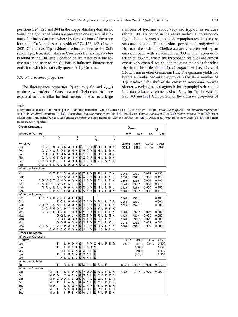
numbers of tyrosine (about 720) and tryptophan residues(about 140) are found in the native molecule, correspond-ing to about 18 tyrosine and 7–8 tryptophan residues in onestructural subunit. The emission spectra ofL. polyphemusHc from the order of Chelicerata are characterized by anemission band with a maximum at 333± 1 nm upon exci-tation at 295 nm, where the tryptophan residues are almostexclusively excited, which is in the same region as for otherHcs from this order (Table 1). P. vulgarisHc has aλmax of326± 1 nm as other crustacean Hcs. The quantum yields forboth are similar because they contain the same number ofTrp residues. The shift of the emission maximum towardsshorter wavelengths is diagnostic for tryptophyl side chainsin a non-polar environment, sinceλmax for Trp in water is355–360 nm[28]. Comparison of the emissive properties of
Table 1N-terminal sequences of different species of arthtropodan hemocyanins: Order Crustacia, Infraorders Palinura:Palinurus vulgaris(Pv);Panulirus interruptus(Pi) [31]; Panulirus japonicus(Pj) [32]; Astacidea:Homarus americanus(Ha) [22]; Brachyura:Carcinus aestuarii(Ca)[24]; Maia squinado(Ms) [25]; OrderChelicerate, Infraorders Xiphosura:Limulus polyphemus(Lp); Buthidae:Buthus sindicus(Bs) [26]; Araneae:Euyrypelma californicum(Ec) [33] and theirfluorescence properties

1212 P. Dolashka-Angelova et al. / Spectrochimica Acta Part A 61 (2005) 1207–1217
the oxy- and apo-forms under the same experimental condi-tions (50 mM Tris/HCl buffer, pH 8.2) shows that the quan-tum yield strongly increases for both,Limulus(Qoxy = 0.025;Qapo- = 0.078) andP. vulgaris(Qoxy = 0.012;Qapo- = 0.082)Hcs upon removal of copper from the active site. This effectis accompanied by a 10 nm shift of the emission maximumfrom 333± 1 nm (oxy-Hc) to 343± 1 nm (apo-Hc) (Table 1).Fig. 2A shows in detail the environment around the activesite ofLimulusHc, where the positions of neighboured Trpresidues are evidenced. Subunit II contains eight Trp residues:Trp65, Trp174, Trp176, Trp184, Trp326, Trp363, Trp538 andTrp563; however, not all of them are present in the varioussubunits of other hemocyanins. All Trp residues, with the ex-ception of Trp563, are located in the second domain ofLimu-lusHc. Two of them (Trp176 and Trp363) are located withina small distance below 6A from CuA and/or CuB, respec-tively (Fig. 2A; Trp1765.36A from CuA site and Trp3635.17from the CuB site). Trp174 (12.02A) and Trp538 (13.88A)are located also in the neighbourhood at the Cu site. Theemission of these Trp residues in the oxy-form is stronglyquenched by the Cu-ions. Removal of O2 and copper ionsdrastically increases the tryptophan fluorescence (343 nm forthe whole molecule and about 347 nm for the structural sub-units).Fig. 2A shows that Trp176. . .CuA O2 CuB-His328and Trp363. . .CuB O2 CuA-His177 are “parallel” to eacho(t ofC ithi rizeC lya bothT ns ino eda int ofv siteio neart onem 538f r dif-f 0.0%a
lys , inc pr threei t int rvedi
3
ropo-d ce, in
the regions involved in inter-subunit contacts, as Trp563 inLimulusand Trp292 inEuripelmaHcs. The exposure of theseresidues to the solvent is connected with short-life time andthey have calculated relative static accessibilities 58.6% and36.3%, respectively. The distances between these two Trpresidues and Cu-ions are four times higher than that betweenCu and the Trp176 and Trp363 (Fig. 2A), and their emis-sion cannot be quenched by internal Cu-ions. The emissionof whole molecule is quenched by the microenvironment ofthe Trp residues and by the interaction of the subunits. Evi-dence for this is that the fluorescence quantum yield of wholemolecule is less than that for its structural subunits (Table 1).The isolated subunits in the oxy-form as and their correspond-ing apo-forms exhibit quantum yields higher as compared tothe whole Hc.
Moreover, the lifetime values of Hc remain almost con-stant upon O2 and copper removal, changes being observedonly in the normalized pre-exponentials A; this supports amechanism of static quenching by copper ions of the activesite [30]. Removing quenching Cu-ions from the active siteof the molecule is connected with an increase of the quan-tum yield but not withτ1. Thus, the increase of fluorescencequantum yield in apo-Hc must be attributed to the demask-ing of Trp fluorophores, which are fluorimetrically almostsilent in the native protein. The fluorescence decay curvef al ins ulti-e merso in-d lvedfl -p ativeTo units,a x-p n byt
P
w fort is oft d fitb val-u sig-n eris-t np th= roms p563a ire 197a fa differb gests
f the “conjugated chains”. The distance of Trp176. . .CuA5.36A) is somewhat larger than Trp363. . .CuB (5.17A), buthe first is “in touch” with the negative dipole side (CE3)u(A) and the second Trp363 is “in touch” with Cu(B) w
ts positive dipole side (near CD1) and thus, both polauA O2 CuB in a way that CuA could be more positivend CuB more negatively charged. The fluorescence ofrp residues has to be strongly quenched by cupric ioxy-Hc and by their environment[29]. In the oxy-state, thistance between both Cu ions is 4.6A (CuA. . .4.6. . .CuB)nd this is very understandable from an electrostatic poiew (repulsion). The Trp distribution around the actives a strict peculiarity ofEurypelmaHc, actually, only 50%f the Trp residues in other Chelicerata Hcs are located
he copper ions and are distributed only in the vicinity ofetal center[3]. The access to Trp174, Trp176 and Trp
or the quenchers is very low and different because theierent static accessibility calculated as small as 2.0%,nd 0.1%, respectively.
These features ofLimulusHc explain the exceptionaltrong quenching of the Trp fluorescence in the oxy-formomparison with other Hcs[21]. Fig. 2B shows that four Tresidues are located in the CuA site of Crustacea Hc andn Chelicerata Hc, while only one or two Trp are presenhe CuB site near to His residues. No Trp residue is obsen the CuB site in Crustacea Hc.
.4. Fluorescence lifetime
Three classes of Trp-s are represented in native arthan Hcs. Some of them are located on the subunit surfa
or Trp residues has been found to be multi-exponentiingle as well as multiple Trp containing proteins. The mxponential decay arises due to the conformational isof the Trp residues, where the local environment of theole chromophore differs substantially. The time-resouorescence decay ofLimulusHc gave two lifetime comonents, which indicate possible presence of two alternrp-positions (“conformational alternates”) (Fig. 3). The flu-rescence decays of the parent molecule, the two subnd N-Ac-Trp.NH2, excited at 295 nm, well fitted to two eonentials. The theoretical fluorescence intensity is give
he function
(t) = A1e−t/τ1 + A2e−t/τ2 (2)
hereA1 andA2 are amplitudes. This function was usedhe determination of the excited state lifetimes. Analyshe data in terms of bi-exponential models give a gooetween the experimental and theoretical curves. Thees show that the environment of these chromophoresificantly influences their dynamic fluorescence charact
ics. The lifetimes for the native molecule of arthropodaL.olyphemusHc are very short (τ1 = 0.3 ns,τ2 = 3.3 ns), buigher than for molluscan keyhole limpet Hc (τ1 = 0.2 ns,τ22.6 ns)[24]. Our experimental data as well as the data f
imilar structures gives us the reason to suggest that Trnd Trp65 seems to be short-lived (τ1 = 0.3 ns) and themission (81%) is almost completely quenched and Trpnd Trp346 are long-lived (τ1 = 2.56 ns) with emission obout 18%. The fluorescence decay properties clearlyetween the whole molecule and the subunits, which sug

P. Dolashka-Angelova et al. / Spectrochimica Acta Part A 61 (2005) 1207–1217 1213
Fig. 3. Fluorescence decay of wholeL. polyphemusHc molecule in 0.05 MTris/HCl, buffer with 5 mM CaCl2 and MgCl2, pH 8.2 after excitation at295 nm.
that the respective tryptophans are affected differently by thelocal tertiary structure. The specific environment of the in-dole groups inLimulusHc decreases the excited state lifetimeby a factor of two in comparison with the value, determinedfor its subunits.
3.5. Denaturation with Gdn.HCl
The unfolding of proteins in denaturing compounds hasbeen widely investigated. In the case of oligomeric proteins,inactivation, dissociation and unfolding have been describedas the structural events that occur subsequently during the de-naturation process. The present paper reports the changes inconformation and aggregation of tetrameric Hcs during un-folding in guanidine hydrochloride. The denaturation of theseproteins takes place in several stages.Fig. 4A shows the fluo-rescence spectra of the whole molecule ofL. polyphemusHcat different concentrations of Gdn.HCl. The addition of denat-urant concentrations between 0 and 1.5 M produced a slightincrease of the fluorescence intensity, but at 2 M Gdn.HCl,the emission is higher and the quenching of Cu-ions is elimi-nated. At 2.0 M guanidine buffer, the whole molecule is fullydissociated into its structural subunits. The results are con-sistent with an aggregation occurring at 0.5 M guanidine, fol-lowed by a similarly drastic dissociation to give a monomericp e ofd con-n s
shown inFig. 4A, which can be explained, by protein un-folding and guanidinium ion quenching.
The curves inFig. 4B insert, expressing the unfolding ofoxy-Hc at different concentrations of Gdn.HCl is analogousto the curve inFig. 4C (expressing the unfolding of apo-Hc),but with specific changes in quaternary structure fluorescentproperties (Fig. 4B insert, limb-a), which seems to representtwo transitions. The first transition has a similar “position”(Cm = 0.52) and amplitude (dQ) typical for very quenchedtotal Trp fluorescence. Obviously, this observation is due toquencher, the effect of which is removed at the followed un-folding step atCm
* = 1.77 and with very big amplitude andsharpness of the transition (within 1.6 M interval). The onlyexplanation is that in this step the CuO2 Cu. . .Trp(s) na-tive structure is destroyed causing a rise of Trp fluorescence.After this apparent “peak” of the amplitude (Q) andCm inthe curve for oxy-Hc, the following limb-b has analogous(but not the same) behaviour as in the apo-Hc case. Threeunfolding steps with apparentCm
* at 2.56, 4.41 and 6.77 M,respectively, could be identified inFig. 4B insert, limb-b. Thefirst of them (at the same amplitude) with�Cm = 1.2 is muchsharper not only than the other two steps, but also in compari-son with the first transition step with�Cm = 3.5 in case of theapo-Hc (Fig. 4C). TheCm
* of the next two transitions are atslightly lower Gdn.HCl concentrations, but with analogouss sum-i ainsi fi andt ons.B ent itc resto theirs
s ofowR wingt II).N falli ared outd
dif-f hingb hert s. Inc ld-i inst M)a M).T s de-s( po-H arys ains
rotein unfolding at 1.5–2 M guanidine. Further increasenaturant concentration decreases the intensity and isected with a shift ofλmax towards 355 nm (red shift) a
mall cooperatively. This behaviour can be explained asng non-symmetric interactions between structural domn the holo-protein, i.e. at the CuO2 Cu site, formation ontra-subunit and chain–chain interactions are changedhe active site formation is coupled with structural alteratiecause the first (less-stable) domain is Cu site-dependan be selected from the known 3D-structure and thef two domains should be easily assigned in terms oftabilities.
For a better understanding of the unfolding procesxy-Hc, we have compared the Q(Cm) curve (Fig. 4B insert)ith theReff(Cm) curve shown inFig. 4B. The “velocity” ofeff changes is different for the first two processes, sho
heir different nature (mark in the figure as regions I andotably, the first domain (at the active site) transition
n the same region II as previous ones and all of themefinitely cooperative. The other two transitions are withrastic changes of Trp environment after the unfolding.
Fig. 4C shows the fluorescence intensity of apo-Hc aterent concentrations of guanidine hydrochloride. Quency Cu-ions is eliminated and the Q at 0 M Gdn.HCl is hig
han for the oxy-form, but less than for structural subunitase of whole multi-chain apo-Hc, (“all apo-Hc”), the unfong curve is more complex, but logically altered. It contawo limbs: (a) at low Gdn.HCl concentrations (0.0–2.4nd (b) at higher concentrations of denaturant (2.5–8.0he last (b) limb-b exhibits also a three-step process acribed above, but only the effectiveCm
* values for oxy-HcFig. 4B insert) are 0.6–0.7 higher than those for the ac (Fig. 4C). This means, the formation of the quaterntructure increases the stability in each of the three dom

1214 P. Dolashka-Angelova et al. / Spectrochimica Acta Part A 61 (2005) 1207–1217
Fig. 4. (A) Emission spectra of oxy-whole molecule ofL. polyphemusHc at different concentrations (0–8 M) of Gdn.HCl. (B)Reff at different concentrations ofGdn.HCl; (b-insert). Effect of guanidine hydrochloride on the stability ofL. polyphemusnative oxy-Hc molecule. (C) Effect of guanidine hydrochloride on thestability ofL. polyphemusapo-Hc molecule. (D) Effect of guanidine hydrochloride on the stability ofL. polyphemusoxy-subunit. The protein concentrationsare about 0.05 mg ml−1 in 0.05 M Tris/HCl buffer with 5 mM CaCl2 and MgCl2, pH 8.2. The fluorescence quantum yield was determined using N-Ac-Trp.NH2
as a standard.
and this alteration is approximately equal and independentfor each of the domains. The presence of limb-a confirmsthe presence of a quaternary structure that is most sensibleand easily destroyed at very low denaturant concentrations.From these results follows that multi-chain complex decom-position leads to an increase of apparent Trp fluorescenceand we have to conclude that chain-chain interactions func-tion as a moderate quencher of Trp fluorescence. This can bedone by introduction of quenching groups to the given Trpresidue(s) and/or by structural changes in the vicinity of theTrp residue(s) after polypeptide complex formation.
In Fig. 4D, the influence on one structural subunit “a” isshown expressing the conformational stability only for thisprotein. The single polypeptide Hc subunit undergoes un-folding in water–Gdn.HCl solutions as a multi-step process.Because the interval of the Q-change is very wide (from 1 to8 M Gdn.HCl) and using the present shape the curve was de-
convoluted into three sigmoid curves with apparent midpoints(Cm
* ) at 1.90, 4.07 and 6.19 M, respectively. It is known from3D X-ray analysis[7] that this subunit contains three struc-tural domains and we can speculate to assign each of theprocesses to each of these domains. This hypothesis requiresdomains to differ considerable in stability at a ratio of ap-proximately 1:2:3. We hope to prove this suggestion fromspecially designed experiments, now in progress. On the ba-sis of the above discussion, a model is given summarizing theresults is as follows:
Tn ⇔ Tn′ ⇔ A ⇔ Mn′ ⇔ Mu (3)
In this model, Tn represents native tetrameric protein that, atlow guanidine concentrations, forms an active intermediate(Tn′) with more hydrophobic surfaces exposed to the solvent.As the guanidine hydrochloride concentration increases, fur-ther exposure of hydrophobic interaction area lead to the

P. Dolashka-Angelova et al. / Spectrochimica Acta Part A 61 (2005) 1207–1217 1215
Fig. 5. (A) Emission spectra of nativeL. polyphemusoxy-Hc in the pH region (9–11.5) and (B) in pH region (2–8.5). (C) Effect of pH on the stability of nativeL. polyphemusoxy-Hc. (D) Effect of pH on the stability ofL. polyphemusapo-whole molecule. (E)Reff at different pH. The protein concentrations are about0.05 mg ml−1 in 0.05 M Tris/HCl buffer with 5 mM CaCl2 and MgCl2, pH 8.2. The fluorescence quantum yield was determined using N-Ac-Trp.NH2 as astandard.

1216 P. Dolashka-Angelova et al. / Spectrochimica Acta Part A 61 (2005) 1207–1217
formation of aggregated species (A). Finally, at concentra-tions higher than 2.0 M guanidine-HCl, the highly aggregatedspecies dissociate into partly unfolded monomers (Mn′) thatare eventually completely unfolded (Mu), accompanied by afurther increasing maximum fluorescence emission.
3.6. Effect of pH on the stability
Finally, we also measured the effect of pH in the rangefrom 2.0 to 12.0 on the tryptophan fluorescence ofL. polyphe-musHc and one structural subunit.Fig. 5A and B shows thefluorescence spectra onL. polyphemusoxy-Hc recorded atdifferent values pH intervals (8–11.4 and 8–2.5). The pH-dependence of Trp-fluorescence quantum yield (Q) of nativeoxy-Hc is shown inFig. 5C. In the acidic pH-region (3.5–6.5),two proton bindings, influencing the quantum yield, were ob-served with crude resolved pKs at 3.7 and 5.4. The first istentatively addressed to Asp (pK = 3.7) or Glu ionization(s)and exhibits cooperative behaviour (it involves more thanone proton uptake within the pH interval, i.e. the sharpnessof Q(pH)-transition is larger than one). Based on cooperativebehaviour, this transition can be also an acid denaturationone and its nature will be proven later based on CD measure-ments. The second acidic pH-dependent Q-change is threetimes smaller in amplitude, with a crude effective pK = 5.4a oxylg ntialfi Hisin s itsq ur att ses:d min-i ndo go bothsi
ingi entsht encei en-b ofoa ti hr-eob ub e tois isont foro tedb ap-
parent pKapp = 8.2, leading to Trp-fluorescence increasing.This single process can be addressed to the “second” one inthe oxy-Hc, but with pK shifted from 9.0 to 8.2 (�pK = 0.8).The effect could be complex: omitting O2 (a bi-radical parti-cle, Trp fluorescence quencher) should increase both, the Trpquantum yield and the “amplitude” of theQ(pH) transitionwith an effective pK = 5.8, which is shifted by 0.4 pH unitscompared to a pK = 5.4 in oxy-Hc. This is connected to smallconformational changes of His and Trp residues in the O2-binding site. Two types of Trp–His couplings by non-contactcoplanarπ–π interactions were identified. One is locatedwithin the Cu center (W176...H173 and W363. . .H324) andthe second one through CuO2 Cu bridge (W176. . .H328and W363. . .H177). Removal of O2 in deoxy-Hc will changethe distances between Trp and His and Trp. . .Cu(O2), and es-pecially their orientations. Both transitions in oxy-Hc (withpK 7.3 and 9.0, respectively) are cooperative (see above),while the single transition in apo-Hc (with pK 8.2) is a non-cooperative one and is addressed to single group ionization.Both, the acidic and alkaline limbs (pK 3.9 and 10.4, respec-tively) in Fig. 5D are slightly drawn together. The calculatedvalues from the curves inFig. 5C and D for the oxy- and apo-Hcs (dpK = pKal − pKac) are 7.0 and 6.5, respectively, i.e.they differ in 0.5 pH units, which qualitatively in agreementthat O2-binding to the active site stabilizes the Hc structure.
enceb mit-t theirs t in-t nmw
R
Bt rea itionw wst e inT tionsw outs
A
ein-s ienst( tedb ) oft thea l forP 286)f flu-o
nd not cooperative. Again, this may be a single carbroup ionization being in a negative electrostatic poteeld. However, and more probably, it is due to a singleonization located close to positively charged Cu(2+) ion andear to Trp, the deprotonation of which partially removeuenching effect. Most interesting is the Q(pH) behavio
he neutral pH region (6.5–9.5) with two contrary proceseprotonation of first site(s) increase Trp quenching (di
shQ) with a crude pK of 7.3 and obvious cooperativity af second one with a crude pK of 9.0, causing decouplinf Trp fluorescence quenching. Probable candidates forites should be His residues in the Cu2+ vicinity or coupledonisation ofα-amino groups at the N-terminus.
Since the equilibrium constant of Hc for oxygen binds expected to change as a function of pH, the experimave been carried out on the Apo-proteins (Fig. 5D) in order
o avoid the complications due to the change in fluorescntensity depending on the shift in the position of the oxyginding equilibrium and compared with pH denaturationxy-Hc. Disappearance of the first transition (pK = 7.3) inpo-Hc leads to the conclusion for O2-binding-dependen
onization (in analogy to well known for hemoglobins Boffect). The last “alkaline ionization” at pK = 10.7 is obvi-usly connected with the loss of the Cu2+ ion(s) and followedy alkaline denaturation. Removal of the oxygen from C2+-inding centre of Hc leads to the characteristic chang
ts pH-dependent intrinsic (Trp-s) fluorescence. TheQ(pH)hape is different in the pH region of 6.5–9.5 in comparo that for oxy-Hc (Fig. 5C). Both “processes”, observedxy-Hc (with pKapp7.3 and 9.0, respectively) are substituy a one “process” in apo-Hc with positive tangent and
The spectral position of the apparent Trp fluorescand which is a sum of individual contributions of each e
ed single Trp residues, is also pH sensitive and definesummary position and shape by the ratio of fluorescenensities (Reff) at two arbitrary wavelengths (335 and 365ere selected):
eff = Iλ(335)
Iλ(365)= f (pH) (4)
oth limbs with pKs 3.7 and 10.7 shown inFig. 5E overlaphe regions of changeable pH/Reff values, i.e. the structure “in transition”. The same could be said for the transith pK = 9.0, but at this pH the protein is native what allo
o speculate that this transition is coupled with changrp orientation(s). It seems that the both others transiith pK values of 5.4 and 7.0, respectively, proceed withtructural changes altering Trp micro-environment(s).
cknowledgments
P. D-A. would like to thank Deutsche Forschungsgemchaft (DFG) and Deutscher Akademischer Austausch DDAAD) for granting a scholarship. This work was suppory grants from DLR 01/001 and from the NCSI (X-1302
he Ministry of Education and Science, Bulgaria. One ofuthors (PN) is deeply grateful to the Swedish Councilanning and Coordination of Research (Grant N a 1-5/2
or the financial support, which allowed performing therescence lifetime measurements.

P. Dolashka-Angelova et al. / Spectrochimica Acta Part A 61 (2005) 1207–1217 1217
References
[1] K.E. van Holde, K.I. Miller, Adv. Protein Chem. 47 (1995) 1.[2] B. Salvato, M. Beltramini, Chem. Rep. 8 (1990) 1.[3] E. Hodgson, J.I. Spicer, Comp. Biochem. Physiol. Part A 128 (2000)
873.[4] K.E. Van Holde, K.I. Miller, H.J. Decker, Biol. Chem. 19 (2001)
15563.[5] D. Sancher, M.D. Gantornin, G. Gutierrer, M.J. Bastiani, Mol. Biol.
Evol. 15 (1998) 415.[6] R. Topham, S. Tesh, G. Cole, D. Mercatante, A. West-
cott, C. Bonaventura, Arch. Biochem. Biophys. 352 (1998)103.
[7] K.A. Magnus, B. Hazes, H. Ton-That, C. Bonaventura, J.Bonaventura, W.G.J. Hol, Proteins Struct Funct Genet 19 (1994)302.
[8] A.K. Shrive, A.M. Metcalf, J.R. Cartwright, T.J. Greenhough, J. Mol.Biol. 290 (1999) 997.
[9] A. Volbeda, W.G.J. Hol, J. Mol. Biol. 209 (1989) 249.[10] B. Hazes, K.A. Magnus, C. Bonaventura, J. Bonaventura, Z. Dauter,
K.H. Kalk, W.G.J. Hol, Protein Sci. 2 (1993) 597.[11] K.A. Magnus, H. Ton-That, J.E. Carpenter, Chem. Rev. 94 (1994)
727.[12] M. Guzman-Casado, A. Parody-Morreale, P.L. Mateo, J.M. Sanchez-
Ruiz, Eur. J. Biochem. 188 (1990) 181.[13] R. Sterner, T. Vogl, H.-J. Hinz, F. Penz, R. Hoff, R. Foll, H. Decker,
FEBS Lett. 364 (1995) 9.[14] R. Hubler, B. Fertl, N. Hellmann, H. Decker, Biochim. Biophys.
Acta 1383 (1998) 327.[15] M.A. Menze, N. Hellmann, H. Decker, K. Grieshaber, Biochemistry
[ nov,
[17] P. Dolashka-Angelova, S. Stoeva, R. Hristova, J. Schuetz, M. Bel-tramini, B. Salvato, W. Voelter, Curr Top. Pept. Protein Res. 3 (1999)19.
[18] R. Hristova, P. Dolashka, S. Stoeva, W. Voelter, B. Salvato, N. Genov,Spectrochim. Acta Part A 53 (1997) 471.
[19] P. Dolashka-Angelova, R. Hristova, S. Stoeva, W. Voelter, Spec-trochim. Acta Part A 55 (1999) 2927.
[20] P. Dolashka-Angelova, M. Beltramini, A. Dolashki, B. Salvato, R.Hristova, W. Voelter, Arch. Biochem. Biophys. 389 (2001) 153.
[21] P. Dolashka-Angelova, S. Stoeva, R. Hristova, J. Schuetz, W. Voelter,Spectrochim. Acta A 56 (2000) 1985.
[22] P. Dolashka, N. Genov, K. Parvanova, W. Voelter, M. Geiger, S.Stoeva, Biochem. J. 315 (1996) 139.
[23] P. Dolashka-Angelova, M. Schick, S. Stoeva, W. Voelter, J. Biochem.Cell Biol. 32 (2000) 529.
[24] J. Schutz, P. Dolashka-Angelova, R. Abrashev, P. Nicolov, S. Stoeva,W. Voelter, Biocim. Biophys. Acta 1546 (2001) 325.
[25] S.A. Ali, A. Abbasi, S. Stoeva, R. Kayed, P. Dolashka-Angelova, H.Schwarz, W. Voelter, Comp. Biochim. Physiol. Part B 126 (2000)361.
[26] U.K. Laemmli, Nature 227 (1970) 680.[27] L. Bubacco, R.S. Magliozzo, M. Beltramini, B. Salvato, J. Peisach,
Biochemistry 31 (1992) 9294.[28] M. Tabak, G. Sartor, P. Neyroz, A. Spisni, P. Cavatorta, J. Luminesc.
46 (1990) 291.[29] N.B. Terwilliger, L. Dangott, M. Ryan, Proc. Natl. Acad. Sci. U.S.A.
96 (1999) 2013.[30] T. Burmester, Mol. Biol. Evol. 18 (2001) 184.[31] B. Neuteboom, P.A. Jekel, J.J. Beintema, Eur. J. Biochem. 206 (15)
(1992) 243.[[ T.
39 (2000) 10806.16] S. Stoeva, P. Dolashka, B. Bankov, W. Voelter, B. Salvato, N. Ge
Spectrochim. Acta Part A 51 (1995) 1965.
32] N. Makino, S. Kimura, Eur. J. Biochem. 173 (2) (1988) 423.33] R. Voit, G. Feldmaier-Fuchs, T. Schweikardt, H. Decker,
Burmester, J. Biol. Chem. 15 (275) (2000) 39339.
Copyright © 2022 FDOKUMEN