ACCEPTED
26
1 Frequent in vitro recombination in the internally transcribed spacers during genotyping of Pneumocystis jirovecii 1 2 3 4 5 6 7 8 9 10 11 12 13 14 15 16 17 18 19 20 21 22 23 Jessica Beser, Per Hagblom and Victor Fernandez* Department of Parasitology, Mycology and Environmental Microbiology, Swedish Institute for Infectious Disease Control, Solna, Sweden and Department of Microbiology, Tumor and Cell Biology, Karolinska Institute, Stockholm, Sweden. Running title: ITS recombinants in P. jirovecii genotyping *Corresponding author. Mailing address: Department of Parasitology, Mycology and Water Microbiology, Swedish Institute for Infectious Disease Control, SE-17182 Solna, Sweden. Phone: +46 8 4572553. Fax: +46 8 318450. E-mail: [email protected] ACCEPTED Copyright © 2007, American Society for Microbiology and/or the Listed Authors/Institutions. All Rights Reserved. J. Clin. Microbiol. doi:10.1128/JCM.02245-06 JCM Accepts, published online ahead of print on 3 January 2007
Transcript of ACCEPTED
1
Frequent in vitro recombination in the internally transcribed spacers during genotyping
of Pneumocystis jirovecii
1
2
3
4
5
6
7
8
9
10
11
12
13
14
15
16
17
18
19
20
21
22
23
Jessica Beser, Per Hagblom and Victor Fernandez*
Department of Parasitology, Mycology and Environmental Microbiology, Swedish Institute
for Infectious Disease Control, Solna, Sweden and Department of Microbiology, Tumor and
Cell Biology, Karolinska Institute, Stockholm, Sweden.
Running title: ITS recombinants in P. jirovecii genotyping
*Corresponding author. Mailing address: Department of Parasitology, Mycology and Water
Microbiology, Swedish Institute for Infectious Disease Control, SE-17182 Solna, Sweden.
Phone: +46 8 4572553. Fax: +46 8 318450. E-mail: [email protected]
ACCEPTED
Copyright © 2007, American Society for Microbiology and/or the Listed Authors/Institutions. All Rights Reserved.J. Clin. Microbiol. doi:10.1128/JCM.02245-06 JCM Accepts, published online ahead of print on 3 January 2007
2
ABSTRACT 24
25
26
27
28
29
30
31
32
33
34
35
36
37
38
39
40
41
42
43
Pneumocystis jirovecii is the causing agent of Pneumocystis pneumonia (PCP) in
immunocompromised persons. Knowledge on the transmission and epidemiology of PCP is
still incipient and investigations on these subjects are based exclusively on applications of
molecular typing techniques. The polymorphic internal transcribed spacers ITS1 and ITS2 in
the rDNA operon, which in the P. jirovecii genome exist as single-copy DNA, are commonly
used as target loci for isolate typing. In the course of genotyping P. jirovecii in respiratory
specimens from PCP patients by amplification and cloning of a large number of ITS
sequences, we found mixed infections (two or more types) in 50% of the samples. In a
majority of the specimens with mixed infections we detected many ITS haplotypes
(combinations of ITS1 and ITS2 types) that appeared to be products of recombination
between globally common ITS haplotypes present in the same sample. Here we present
results of a series of experiments showing that essentially all ITS recombinants are chimeras
formed during the genotyping process. Under standard conditions up to 37% of the amplified
sequences could be hybrid DNA artefacts. We show that by modifying PCR amplification
conditions ITS chimera formation could be largely abolished and the erroneous establishment
of artefactual haplotypes avoided. The accurate assessment of the genetic diversity is
fundamental for a better understanding of the epidemiology and biology of P. jirovecii
infections.
ACCEPTED
3
INTRODUCTION 43
44
45
46
47
48
49
50
51
52
53
54
55
56
57
58
59
60
61
62
63
64
65
66
67
The infectious fungus Pneumocystis jirovecii is the etiological agent of Pneumocystis
pneumonia (PCP) in immunocompromised individuals. Since P. jirovecii cannot be cultivated
in vitro, investigations on the transmission and epidemiology of this organism have been
based on applications of molecular typing techniques (1). In P. jirovecii, sequence analysis of
the internal transcribed spacer (ITS) regions in the nuclear ribosomal DNA (rDNA) gene
complex, which in this fungus exists as a single copy locus (2, 24), is the most informative
epidemiological tool available at present. The ITS1 is located between the coding regions of
the 18S rRNA and the 5.8S rRNA genes, and the ITS2 between the 5.8S and the 26S rRNA
genes. The sequence diversity of ITS1 and ITS2 among different strains of P. jirovecii and the
fact that these segments are removed during ribosomal biogenesis and should not be subjected
to selective pressure make them suitable targets for genotyping (10). To date more than 30
ITS1 genotypes and 40 ITS2 genotypes with over 90 haplotypes (combinations of ITS1 and
ITS2 types) have been described worldwide, based on two different but similar typing
systems (8, 26). Some ITS haplotypes are globally more common and coinfections with
multiple types of P. jiroveci often occur (4, 8, 11, 15, 17, 25, 26). Albeit these typing systems
have been useful, questions have been raised about the extensive polymorphism and stability
of the ITS locus and its use for the genotyping of P. jirovecii.
During the genotyping of specimens from patients with a P. jirovecii infection we noticed the
presence of recombinants between main ITS haplotypes in many of the specimens with mixed
infections. Given the high frequency of this feature we considered the possibility that some of
these recombined haplotypes had been generated during the genotyping procedure. It is well
established that recombination, or chimera formation, can occur during PCR when the
template is a mixture of similar sequences rather than a single target (13, 19, 20). When this
ACCEPTED
4
phenomenon takes place, in vitro generated recombinant molecules will be present in the
amplified product which will result in an overestimation of the diversity in the starting
material (27, 28). Another potential complication is the introduction of point mutations during
PCR amplification due to misincorporation by the DNA polymerase (16). In an effort to
elucidate the origin of recombinants in the clinical material we first performed experiments
that showed the in vitro source of the recombinant sequences. Secondly, we attempted to
resolve in what step and at what level the in vitro generated recombinants were produced.
Thirdly, we made modifications to the standard protocol of Lee et al. (8) to reduce the amount
of erroneous haplotypes in samples with mixed infections. Furthermore, we used exons and
introns of the thioredoxin reductase (TRR1) gene as reference DNA templates to evaluate the
rate of point mutations and performance of DNA polymerases in the ITS and 5.8S loci.
68
69
70
71
72
73
74
75
76
77
78
79
ACCEPTED
5
MATERIALS AND METHODS 79
80
81
82
83
84
85
86
87
88
89
90
91
92
93
94
95
96
97
98
99
100
101
102
103
Processing of specimens and DNA extraction. Respiratory specimens used in this study
were collected and referred to the Swedish Institute for Infectious Disease Control where they
were analysed following routine procedures for the diagnosis of Pneumocystis jirovecii. Nine
volumes of BAL or sputum sample were mixed with one volume of 65 mM DTT and
incubated for 15 min at 37flC. One volume of 50% ethanol was added for inactivation of HIV.
The suspension was centrifuged at 1800 x g for 5 min and the supernatant was discarded.
DNA extractions were performed with the commercial purification system QIAamp DNA
Mini Kit (QIAGEN, Hilden, Germany) according to the protocol supplied by the
manufacturer. The purified DNA (PjDNA) was eluted in 100 ol H2O.
Mix of specimen DNA. The specimens used in these experiments were 232, 790, 926, 1017
and 1874 containing a single ITS haplotype and specimen 308 containing multiple haplotypes
as determined by genotyping carried out according to Lee et al. (8). PjDNA from two
specimens were mixed to an equimolar ratio calculated by real-time PCR quantification of the
gene coding for the large ribosomal subunit (LSU) of P. jirovecii (Fernandez et al.,
unpublished). The mixes of PjDNA were amplified according to the ITS amplification with
Taq polymerase protocol that is described below.
Mix of PCR products. For this experiment PjDNA from specimens 232 and 926 were
amplified independently of each other according to the ITS amplification with Taq
polymerase protocol that is described below. The PCR products were purified with
QIAquickł PCR Purification Kit (QIAGEN, Hilden, Germany), specrophotometrically
quantified and the PCR products were then mixed in a molar ratio of one to one. After mixing
the PCR products were treated in two different ways, either cloned directly or pre-treated
ACCEPTED
6
before cloning. The pretreatment, which was designed to reproduce conditions favouring
heteroduplex formation in later cycles of the regular genotyping PCR, was performed by
taking 10 µl of the PCR mix and combining it with 1xPCR buffer (Invitrogen, Carlsbad,
USA), 3 mM MgCl2, 200 oM of each deoxynucleotide, 20 pmol of each primers FX (5’-
TTCCGTAGGTGAACCTGCG-3’) and RT2 (5’-CTGATTTGAGATTAAAATTCTTG-3’).
The total volume was adjusted to 50 µl with MilliQ water and DNA polymerase was not
added. This mix was then subjected to the following thermal cycling conditions: 5 min at
94flC, 2x (60 sec at 94flC, 60 sec at 56flC, 60 sec at 72flC) and 15 min at 72flC.
104
105
106
107
108
109
110
111
112
113
114
115
116
117
118
119
120
121
122
123
124
125
126
127
ITS amplification with Taq polymerase. PjDNA from individual specimens or in mixes
were amplified with a nested PCR targetting the ITS1-5.8S-ITS2 region as previously
described (8). In short, a first amplification reaction (PCR I) containing 5 µl of template DNA
mix or H2O as negative control, 2.5U Taq polymerase (Invitrogen, Carlsbad, USA), 200 oM
of each deoxynucleotide, 3 mM MgCl2 and 20 pmol each of primers 1724F2 (5’-
AGTTGATCAAATTTGGTCATTTAGAG-3’) and ITS2R (5’-CTCGGACGAG
GATCCTCGCC-3’) in 1xPCR buffer (Invitrogen, Carlsbad, USA) was adjusted with MilliQ
water to a total volume of 50 µl. The PCR was carried out under the following conditions: 3
min at 94flC, 5x (90 sec at 94flC, 90 sec at 62flC, 90 sec at 72flC), 30x (60 sec at 94flC, 60 sec
at 60flC, 60 sec at 72flC) and 15 min at 72flC. A second amplification reaction (PCR II)
containing 2µl of PCR I product, 2.5U Taq polymerase, 200 oM of each deoxynucleotide, 1.5
mM MgCl2, 20 pmol each of primers FX (5’-TTCCGTAGGT GAACCTGCG-3’) and RT2
(5’-CTGATTTGAGATTAAAATTCTTG-3’), 1xPCR buffer and MilliQ water in a total
volume of 50 µl was set up. PCR II was performed under the following conditions: 3 min at
94flC, 5x (90 sec at 94flC, 90 sec at 58flC, 90 sec at 72flC), 30x (60 sec at 94flC, 60 sec at
ACCEPTED
7
56flC, 60 sec at 72flC) and 15 min at 72flC. The amplification products were separated by
electrophoresis on a 1.2% agarose gel containing EtBr and visualized by UV-light.
128
129
130
131
132
133
134
135
136
137
138
139
140
141
142
143
144
145
146
147
148
149
150
151
152
ITS amplification with proofreading Tgo polymerase. A first amplification of the ITS1-
5.8S-ITS2 region was done with the primers 1724F2 and ITS2R, i.e. the same primers as for
amplifications using Taq polymerase. For this PCR, two master mixes were prepared as
follows, five ol of a template DNA mixture (specimens 926+232) were added to master mix 1
containing 20 pmol of each primer, deoxynucleotides (final conc. 200 oM of each) and MilliQ
water, to a final volume of 25 ol. In master mix 2, 1xExpand High Fidelity (Roche, Penzberg,
Germany), 2.6 U Expand High Fidelity enzyme mix containing Taq polymerase and Tgo
polymerase with proofreading activity (Roche, Penzberg, Germany), and MilliQ water were
mixed to a final volume of 25 ol. The two master mixes were subsequently combined and the
PCR was conducted under the same conditions as for the first amplification with Taq
polymerase. For the second amplification, the forward primer FX and the reverse primer RT2
were used. Two microliters of the first PCR were added to a master mix 1 containing 20 pmol
of each primer, deoxynucleotides (final conc. 200 oM of each) and MilliQ water, to a final
volume of 25 ol. In master mix 2, 1xExpand High Fidelity buffer (Roche, Penzberg,
Germany), 2.6 U Expand High Fidelity enzyme mix (Roche, Penzberg, Germany), and MilliQ
water were mixed to a final volume of 25 ol. The two master mixes were subsequently
combined and the PCR was conducted under the same conditions as for the second
amplification with Taq polymerase. The amplified products were separated by electrophoresis
on a 1.2% agarose gel stained with EtBr and visualized under UV-light. Prior to cloning one
ol ATP and 1 ol Taq polymerase were added to the PCR mix and incubated at 37flC for 30
minutes to get A’-overhangs on the fragments.
ACCEPTED
8
ITS amplification with Taq polymerase under modified conditions. In one experiment the
DNA template mixture (specimens 926+232) was amplified with the same protocol as
previously (8) but with longer extension times, i.e. PCR I: 3 min at 94flC, 5x (90 sec at 94flC,
90 sec at 62flC, 4.5 min at 72flC), 5x (60 sec at 94flC, 60 sec at 60flC, 4.5 min at 72flC), 25x
(60 sec at 94flC, 60 sec at 60flC, 60 sec at 72flC) and 15 min at 72flC, and PCR II: 3 min at
94flC, 5x (90 sec at 94flC, 90 sec at 58flC, 4.5 min at 72flC), 5x (60 sec at 94flC, 60 sec at
56flC, 4.5 min at 72flC), 25x (60 sec at 94flC, 60 sec at 56flC, 60 sec at 72flC) and 15 min at
72flC. In another experiment the DNA mixture (926+232) was amplified with the same
protocol but with higher melting temperature, longer extension time and fewer cycles, i.e.
PCRI: 5 min at 96flC, 25x (60 sec at 94flC, 60 sec at 60flC, 4.5 min at 72flC) and 7 min at
72flC, and PCRII: 5 min at 94flC, 20x (60 sec at 94flC, 60 sec at 56flC, 4.5 min at 72flC) and 7
min at 72flC. In the third experiment the DNA mixture (926+232) was amplified using 2-fold
primer concentration in the first PCR and 5-fold primer concentration in the second PCR but
otherwise according to the same protocol as before (8) under the same modified conditions as
just described with higher denature temperatures, longer extension time and fewer cycles.
153
154
155
156
157
158
159
160
161
162
163
164
165
166
167
168
169
170
171
172
173
174
175
176
177
Reconditioning PCR. The PjDNA mix (specimens 926+232) was amplified with Taq
polymerase under the standard conditions and 5 ol of PCR II products were transferred to a
new master mix containing 2.5U Taq polymerase (Invitrogen, Carlsbad, USA), 200 oM of
each deoxynucleotide, 1.5 mM MgCl2, 20 pmol of each primers FX and RT2, 1xPCR buffer
and MilliQ water in a total volume of 50 µl. The reaction mixes were subjected to 3 min at
94flC and 3x (60 sec at 94flC, 60 sec at 56flC, 60 sec at 72flC).
Amplification of the thioredoxin reductase (TRR1) gene. Primers TRR1F1 (5’-
CTTGTTAATCTCTCTAGATCAACGTC-3’) and TRR1R1 (5’-
ACCEPTED

9
TTAATTTGTCCCTCCTAACAAGTAG-3’) were designed to amplify a segment of 888 bp
of the TRR1 gene followed by nested primers TRR1F2 (5’-
GAAGAAAAGAGCCTTTAATAGATAC-3’) and TRR1R2 (5’-
GTAGATATACTTTAGTTGCATATCTCG-3’) amplifying a segment of 817 bp including 5
introns and 5 exons. For amplification with Taq polymerase (Invitrogen, Carlsbad, USA) the
same protocol was used as for ITS but under following conditions in PCRI: 5 min at 95flC, 40
x (30 sec at 95flC, 60 sec at 55flC, 60 sec at 72flC) and 7 min at 72flC and in PCRII: 3 min at
95flC, 35 x (30 sec at 95flC, 30 sec at 53flC, 60 sec at 72flC) and 7 min at 72flC. For
amplification with Expand High Fidelity enzyme mix (Roche, Penzberg, Germany) the same
TRR1F1 and TRR1R1 primers were used for the first PCR, followed by TRRF2 and TRRR2
for the second amplification but with the protocol for proofreading polymerase as described
previously (ITS amplification with proofreading Tgo polymerase). The same PCR conditions
were used as described above, PCRI: 5 min at 95flC, 40 x (30 sec at 95flC, 60 sec at 55flC, 60
sec at 72flC) and 7 min at 72flC and in PCRII: 3 min at 95flC, 35 x (30 sec at 95flC, 30 sec at
53flC, 60 sec at 72flC) and 7 min at 72flC.
178
179
180
181
182
183
184
185
186
187
188
189
190
191
192
193
194
195
196
197
198
199
200
201
202
Cloning and sequencing of PCR products. PCR II products were cloned in the pCRł
2.1-
TOPOł
plasmid vector using the Topo TA Cloning Kit (Invitrogen, Carlsbad, USA)
according to the instructions provided by the manufacturer. Screening of bacterial colonies
was performed with M13 vector-specific primers in a PCR reaction mix containing 2.5U Taq
polymerase (Invitrogen, Carlsbad, USA), 200 oM of each deoxynucleotide, 1.5 mM MgCl2, 5
ng/µl of each primer, 1xPCR buffer and MilliQ water to a final volume of 20 µl. Bacterial
colonies were picked directly into reaction mix and amplified under following conditions: 5
min at 94flC, 25x (30 sec at 94flC, 30 sec at 45flC, 1 min at 72flC) and 7 min at 72flC. The PCR
products were separated in a 1.2% agarose gel containing EtBr and visually inspected to
ACCEPTED
10
confirm the presence of cloned inserts. Recombinants were sequenced in both directions using
M13 primers and the dye terminator chemistry (Applied Biosystems, Warrington, UK).
Sequences were edited and analyzed using the BioEdit Sequence Alignment Editor version
7.0.4.1.
203
204
205
206
207
208
209
210
211
212
213
Statistical analysis and other computer analyses. Statistical analyses were performed using
the SigmaStat software version 3.1. Mutation rate differences between loci were analysed
using the non-parametric Mann-Whitney Rank Sum test. Oligonucleotide primer secondary
and tertiary structure were modelled with NetPrimer (PREMIER Biosoft,
http://www.premierbiosoft.com/) and MacVector (Accelrys software inc.) softwares.
ACCEPTED
11
RESULTS 213
214
215
216
217
218
219
220
221
222
223
224
225
226
227
228
229
230
231
232
233
234
235
236
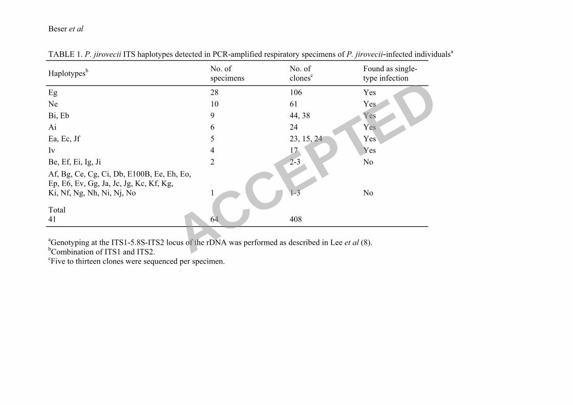
Recombinant ITS sequences in clinical specimens. Genotyping of P. jirovecii by
amplification and cloning of 408 ITS sequences in respiratory specimens from 64 individuals
infected with the fungus revealed the presence of only one single ITS haplotype in half of the
cases (32/64) whereas the remaining samples (32/64) contained two or more haplotypes. In 16
of the 32 specimens with mixed infections we detected what appeared to be recombinant ITS
sequences. This putative recombinant ITS offspring consisted of combinations of ITS1 and
ITS2 seemingly generated from other ITS haplotypes also present in the sample. ITS
haplotypes as well as the number of specimens and number of clones sequenced per haplotype
are shown in Table 1. A total of 41 different ITS haplotypes were identified and 9 of these
haplotypes (17-106 clones each) were found in the amplified DNA of 4 or more specimens.
Each of these 9 haplotypes was detected in at least one single-type specimen verified by the
analysis of 5 or more cloned sequences. Of the remaining, 5 haplotypes were found in 2
specimens (2-3 clones each), and 27 haplotypes in only one sample (1-3 clones each). These
32 less frequent haplotypes were only detected in samples infected with multiple ITS types
which amounted to 25 specimens. In 23 of these 25 specimens the ITS1 or/and ITS2
sequences that were detected in the less frequent haplotypes were the same as those found in
major haplotypes present in that particular sample. The major haplotypes found in these
samples correspond to those that are frequently found in epidemiological studies of P.
jirovecii infections. Therefore, the major dividing line in this material was between single
infections where only one ITS haplotype was found and coinfections where a multitude of
recombination events were evident. It was not possible from this data set to make any
conclusions whether these recombinants were true in vivo phenomena or if they were
generated in vitro during the experimental procedure in the typing protocol.
ACCEPTED
12
237
238
239
240
241
242
243
244
245
246
247
248
249
250
251
252
253
254
255
256
257
258
259
260
261
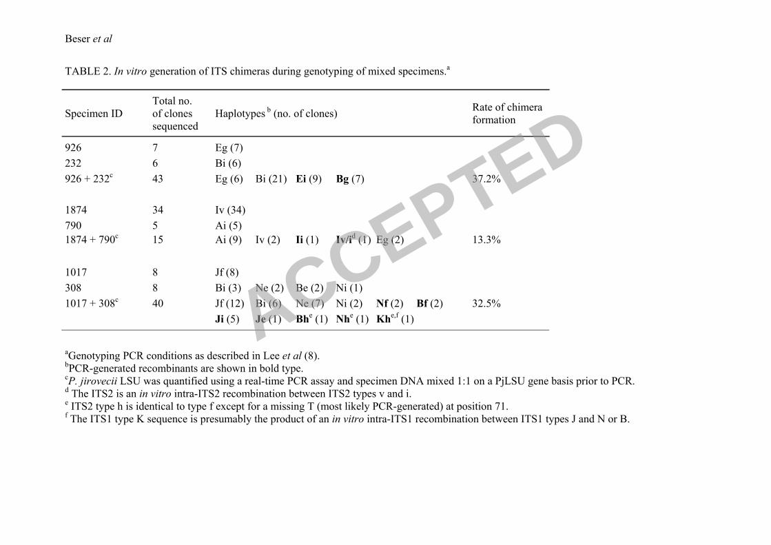
Recombinant sequences are chimeras generated in vitro. To reconstitute a situation where
P. jirovecii DNA (PjDNA) from a coinfected specimen is typed according to the current
protocol by Lee et al. (8), material from two single-infected specimens were mixed in
equimolar amounts and then amplified, cloned and sequenced with this methodology. For this
purpose the PjDNA in various samples were quantified using a real-time PCR assay targeting
the mtLSU gene of the fungus (Fernandez et al, unpublished). Identical amounts of PjDNA
from specimen 926 with haplotype Eg and specimen 232 with haplotype Bi were mixed and
the standard ITS typing procedure was performed. Of 43 clones sequenced 21 were of type
Bi, 6 of haplotype Eg and sixteen sequences were recombinants, 9 Ei and 7 Bg, representing
37.2% of the total clones analyzed (Table 2). To confirm the finding of recombinants
generated in vitro a new haplotype mix was made with specimens 1874 and 790 containing
haplotypes Ai and Iv, respectively (ITS2 v is a new genotype found in the Swedish
population; Beser et al., submitted for publication). Of 15 clones analyzed 9 sequences were
of haplotype Ai and 2 sequences of type Iv. In addition to the parental haplotypes two new
sequences were detected, one the recombinant Ii and the other Iv/i which is a chimera product
of a recombination between the ITS2 types i and v. Unexpectedly, 2 Eg sequences appeared
in the latter experiment and this might represent a contaminant or a real haplotype present in
low amounts in either of the specimens (Table 2). A more complex mixture was also made
with specimen 1017 of type Jf and the multiple infected specimen 308 containing the
haplotypes Bi and Ne. The sequence analysis of this mixture revealed products of
recombination between haplotypes and intra-ITS2 regions, confirming our assumption that an
increased number of PjDNA templates result in an even more varied population of
recombinant PCR products.
ACCEPTED
13
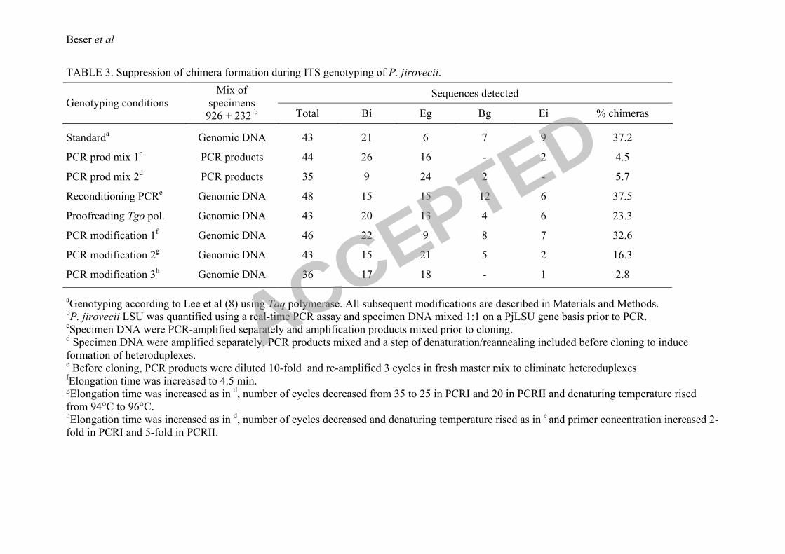
ITS recombinants are generated during the PCR amplification. To further investigate at
what step in the genotyping protocol these in vitro artifacts were produced, experiments were
designed to determine whether the recombinants were generated during PCR amplification or
in the cloning into E. coli. In the first set of these experiments the PCR products from single
haplotype specimens 926 (Eg) and 232 (Bi) were purified, quantified and mixed in equimolar
amounts. To examine if recombination was taking place in the cloning step this mixture was
cloned directly and the analysis showed that 95.5% of the cloned sequences were of parental
type (26 Bi and 16 Eg) and 4.5% recombinants (2 Ei clones) (Table 3). When the mixture was
subjected to two cycles of melting, annealing and elongation, to allow for heteroduplex
formation prior to cloning, the analysis of 35 clones revealed that 94.3% of the sequences
were identical to the parental haplotypes and 5.7% recombinant Bg sequences. These findings
suggested that the vast majority of the chimeras were generated in the PCR amplification but
also that a smaller part could have been produced later on during the cloning step. To confirm
that the recombinants were generated mainly during the elongation step and not as a
consequence of heteroduplex formation in the annealing step of the PCR and subsequent
DNA strand repair in the E. coli, a reconditioning PCR was performed. This was made by
subjecting an equimolar mix of PjDNA from specimens 926 and 232 that had been amplified
according to the ordinary protocol with three cycles of reamplification in a fresh master mix
containing full concentration of primers to minimize the presence of heteroduplexes. Forty-
eight clones were sequenced and, in addition to the original haplotypes, 6 Ei and 12 Bg
chimeric molecules were detected (37.5%, see Table 3) showing that most of the
recombinants were produced already during the elongation step of the PCR.
262
263
264
265
266
267
268
269
270
271
272
273
274
275
276
277
278
279
280
281
282
283
284
285
286
Suppression of in vitro chimera formation. The following experiments were designed as an
attempt to suppress the generation of artifacts during the PCR amplification of the ITS
ACCEPTED
14
genotyping protocol. The standard genotyping method was used as a starting point from
which the amplification protocol was changed in different ways. A modification that was
tested consisted in the introduction of a proofreading polymerase (Tgo) in conjunction with
the Taq polymerase. With this modification the frequency of chimeric sequences decreased to
23.3% (Table 3), which showed that the presence of a polymerase with proofreading activity
in the amplification reaction had a considerable effect on the formation of recombinants. In
another set of experiments several stepwise alterations were made to the standard protocol.
Firstly, the elongation time was increased and this resulted in a minor reduction of chimera
formation to 32.6% (Table 3). Secondly, higher melting temperature and fewer cycles were
used in addition to increased elongation time resulting in a further reduction of chimeras to
16.3% in 43 analyzed clones. Finally, an increase in primer concentration was introduced
together with the extended elongation time, elevated melting temperature and fever cycles.
The primer concentration was increased two fold in the first PCR and five fold in the second
PCR of the nested protocol and the combined effect of these measures drastically decreased
the number of chimeras to one in 36 sequenced clones (2.8%). It was evident from this set of
experiments that relatively simple modifications reduce the amount of in vitro generated
artifacts to a degree that is almost at the level of detection.
287
288
289
290
291
292
293
294
295
296
297
298
299
300
301
302
303
304
305
306
307
308
309
310
311
PCR performance at the ITS and the TRR1 loci. An additional observation throughout this
analysis was that the overall frequency of point mutations in the ITS1-5.8S-ITS2 DNA was
higher than expected. This observation in combination with the finding that the in vitro
generated recombinants could be partially abolished by the addition of a proofreading
polymerase to the reaction raised the question whether intrinsic features in this locus made it
prone to nucleotide misincorporation during replication in vitro. To address this we chose
intronic and exonic regions of the thioredoxin reductase gene (TRR1) as reference locus to
ACCEPTED
15
compare frequencies of nucleotide substitution using the standard PCR conditions for
genotyping of P. jirovecii (8). This showed that using Taq polymerase a significant higher
rate (P<0.05) of nucleotide substitution took place in the ITSs and the 5.8S (2.6 ‒ 1.2
misincorporations/1000 bp of 12558 bp sequenced) compared to the introns and exons of the
TRR1 gene (1.5 ‒ 0.4 misincorporations/1000 bp of 30600 bp sequenced). However, these
differences disappeared when the proofreading DNA polymerase Tgo was added to the
reaction mixture (0.9 ‒ 0.1 misincorporations/1000 bp of 16905 bp sequenced for ITS1-5.8S-
ITS2 and 1.0 ‒ 0.1 misincorporations/1000 bp of 43605 bp sequenced for TRR1). These
results further suggest that a proofreading enzyme should be included in PCR-based typing
schemes for P. jirovecii.
312
313
314
315
316
317
318
319
320
321
322
ACCEPTED
16
DISCUSSION 322
323
324
325
326
327
328
329
330
331
332
333
334
335
336
337
338
339
340
341
342
343
344
345
346
In the course of genotyping P. jirovecii in respiratory samples by amplification and cloning of
a large number of ITS sequences we observed a high frequency of DNA recombinants in
specimens infected with multiple strains of this fungus. Many of the less common ITS1/ITS2
combinations found in these specimens were evident recombinations of two globally common
ITS haplotypes present in the same sample. The question that arose was whether these
recombinants were a consequence of recombinations taking place in vivo or whether
recombinant ITS haplotypes could have been generated in vitro during the genotyping
procedure. To address this subject we mixed specimens of known single haplotypes and after
testing these mixtures with the standard protocol the results showed that ITS chimeras were
generated in the test tube. These processes both generated new haplotypes by combining
different ITS1 and ITS2 genotypes as well as created novel genotypes through recombination
within the ITSs even though the latter was observed infrequently. We therefore concluded that
essentially all the recombinant ITS sequences observed in the clinical material could indeed
be chimeras formed during the genotyping procedure.
The appearance of chimeric DNA molecules is a potential risk when mixed templates of
related sequences are amplified with ordinary PCR protocols. In vitro recombination of rDNA
and other DNA templates with high sequence similarity has been observed in several studies
including those of bacterial diversity (7, 9), viral transcription factors (13), and genes coding
for polymorphic antigens in protozoan parasites (23). These recombinant molecules can be
created as a consequence of chimera formation during the elongation step of the PCR, or by
heteroduplexes formed by base pairing between heterologous molecules during amplification
which in turn will be resolved into chimeras by the DNA repair systems of E. coli after
transformation (6, 14, 18). Our data showed that with the ordinary protocol for genotyping of
P. jirovecii the chimeric ITS sequences are primarily formed during elongation. Under these
ACCEPTED
17
conditions over one out of three amplified sequences could be a hybrid molecule. A factor
that has an effect on the formation of PCR chimeras is the pausing or stalling of the DNA
polymerase along the template which may lead to the dissociation of the polymerase and the
template (3) leaving a prematurely terminated extension product which in turn may act as a
primer on a closely related molecule in the next round of amplification. In this regard, the
concentration of primer in the PCR reaction can be a critical variable influencing the rate of
chimera formation. In the standard protocol used for ITS genotyping of P. jirovecii, the
effective concentration of primer is possibly compromised as hairpin and duplex formation in
three of the primers used in the nested amplification is predicted by various software for
primer design and sequence analysis. Furthermore, alternative target sites for the primers
could be present in non-Pneumocystis DNA in the patient samples since primers flanking the
ITSs are directed to generally conserved sequences. The effect of increasing primer
concentration on the reduction of in vitro generated artifacts is likely to reflect a more
efficient priming in which the oligonucleotides compete out the prematurely terminated
elongation products in the annealing step.
347
348
349
350
351
352
353
354
355
356
357
358
359
360
361
362
363
364
365
366
367
368
369
370
371
The existence of a sexual replication cycle in the genus Pneumocystis is suggested by several
pieces of indirect evidence including the observation in early cysts of P. carinii of structures
resembling the synaptonemal complexes formed during meiotic recombination of sister
chromatids (12), the ultrastructural morphology of binucleated trophic forms of P. carinii
reminiscent of nuclei and nuclear-associated organelles in conjugating Saccharomyces
cerevisiae (5), and the identification in an expressed sequence tag library of meiosis-specific
genes and other genes related to sexual reproduction with a genomic organization in P. carinii
similar to the mating locus described in Cryptococcus neoformans (22). In addition to these
observations, ITS typing data has recently been presented as indicative of the occurrence of
genetic recombination in P. jirovecii populations (17, 21). Although the results reported here
ACCEPTED
18
cannot be used to draw specific conclusions about clonal or recombining modes of
reproduction in P. jirovecii, they do suggest that meiotic recombination in the rDNA of this
fungus is not a frequent event.
372
373
374
375
376
377
378
379
380
381
382
383
The findings described in this paper further suggest that the diversity in P. jirovecii
populations, as estimated by genotyping systems targetting the ITS locus, has in all likelihood
been overestimated. The modifications of the typing protocol described here drastically
reduce the number of sequence artifacts that were produced by the original procedure. This
enables a more correct estimation of the genetic diversity of P. jirovecii and a more accurate
assessment of the epidemiology of P. jirovecii pneumonia. As the phenomenon of in vitro
generated artifacts from mixed templates is common, the procedural modifications presented
herein could be applicable to other diagnostic and typing systems.
ACCEPTED

19
ACKNOWLEDGMENTS 383
384
385
386
This work was supported by grants from the Swedish International Development Cooperation
Agency and the Swedish-South African Health Forum.
ACCEPTED
20
REFERENCES 386
387
388
389
390
391
392
393
394
395
396
397
398
399
400
401
402
403
404
405
406
407
408
409
410
1. Beard, C. B., P. Roux, G. Nevez, P. M. Hauser, J. A. Kovacs, T. R. Unnasch, and
B. Lundgren. 2004. Strain typing methods and molecular epidemiology of
Pneumocystis pneumonia. Emerg Infect Dis 10:1729-35.
2. Giuntoli, D., S. L. Stringer, and J. R. Stringer. 1994. Extraordinarily low number of
ribosomal RNA genes in P. carinii. J Eukaryot Microbiol 41:88S.
3. Hacker, K. J., and B. M. Alberts. 1994. The rapid dissociation of the T4 DNA
polymerase holoenzyme when stopped by a DNA hairpin helix. A model for
polymerase release following the termination of each Okazaki fragment. J Biol Chem
269:24221-8.
4. Helweg-Larsen, J., C. H. Lee, S. Jin, J. Y. Hsueh, T. L. Benfield, J. Hansen, J. D.
Lundgren, and B. Lundgren. 2001. Clinical correlation of variations in the internal
transcribed spacer regions of rRNA genes in Pneumocystis carinii f.sp. hominis. Aids
15:451-9.
5. Itatani, C. A. 1996. Ultrastructural morphology of intermediate forms and forms
suggestive of conjugation in the life cycle of Pneumocystis carinii. J Parasitol 82:163-
71.
6. Jensen, M. A., and N. Straus. 1993. Effect of PCR conditions on the formation of
heteroduplex and single-stranded DNA products in the amplification of bacterial
ribosomal DNA spacer regions. PCR Methods Appl 3:186-94.
7. Kopczynski, E. D., M. M. Bateson, and D. M. Ward. 1994. Recognition of chimeric
small-subunit ribosomal DNAs composed of genes from uncultivated microorganisms.
Appl Environ Microbiol 60:746-8.
8. Lee, C. H., J. Helweg-Larsen, X. Tang, S. Jin, B. Li, M. S. Bartlett, J. J. Lu, B.
Lundgren, J. D. Lundgren, M. Olsson, S. B. Lucas, P. Roux, A. Cargnel, C.
ACCEPTED
21
Atzori, O. Matos, and J. W. Smith. 1998. Update on Pneumocystis carinii f. sp.
hominis typing based on nucleotide sequence variations in internal transcribed spacer
regions of rRNA genes. J Clin Microbiol 36:734-41.
411
412
413
414
415
416
417
418
419
420
421
422
423
424
425
426
427
428
429
430
431
432
433
434
435
9. Liesack, W., S. Sela, H. Bercovier, C. Pitulle, and E. Stackebrandt. 1991.
Complete nucleotide sequence of the Mycobacterium leprae 23 S and 5 S rRNA genes
plus flanking regions and their potential in designing diagnostic oligonucleotide
probes. FEBS Lett 281:114-8.
10. Lu, J. J., M. S. Bartlett, M. M. Shaw, S. F. Queener, J. W. Smith, M. Ortiz-
Rivera, M. J. Leibowitz, and C. H. Lee. 1994. Typing of Pneumocystis carinii
strains that infect humans based on nucleotide sequence variations of internal
transcribed spacers of rRNA genes. J Clin Microbiol 32:2904-12.
11. Matos, O., C. H. Lee, S. Jin, B. Li, M. C. Costa, L. Goncalves, and F. Antunes.
2003. Pneumocystis jiroveci in Portuguese immunocompromised patients: association
of specific ITS genotypes with treatment failure, bad clinical outcome and childhood.
Infect Genet Evol 3:281-5.
12. Matsumoto, Y., and Y. Yoshida. 1984. Sporogony in Pneumocystis carinii:
synaptonemal complexes and meiotic nuclear divisions observed in precysts. J
Protozool 31:420-8.
13. Meyerhans, A., J. P. Vartanian, and S. Wain-Hobson. 1990. DNA recombination
during PCR. Nucleic Acids Res 18:1687-91.
14. Modrich, P. 1987. DNA mismatch correction. Annu Rev Biochem 56:435-66.
15. Nimri, L. F., I. N. Moura, L. Huang, C. del Rio, D. Rimland, J. S. Duchin, E. M.
Dotson, and C. B. Beard. 2002. Genetic diversity of Pneumocystis carinii f. sp.
hominis based on variations in nucleotide sequences of internal transcribed spacers of
rRNA genes. J Clin Microbiol 40:1146-51.
ACCEPTED
22
16. Pienaar, E., M. Theron, M. Nelson, and H. J. Viljoen. 2006. A quantitative model
of error accumulation during PCR amplification. Comput Biol Chem 30:102-11.
436
437
438
439
440
441
442
443
444
445
446
447
448
449
450
451
452
453
454
455
456
457
458
459
460
17. Robberts, F. J., L. D. Liebowitz, and L. J. Chalkley. 2004. Genotyping and
coalescent phylogenetic analysis of Pneumocystis jiroveci from South Africa. J Clin
Microbiol 42:1505-10.
18. Ruano, G., and K. K. Kidd. 1992. Modeling of heteroduplex formation during PCR
from mixtures of DNA templates. PCR Methods Appl 2:112-6.
19. Saiki, R. K., D. H. Gelfand, S. Stoffel, S. J. Scharf, R. Higuchi, G. T. Horn, K. B.
Mullis, and H. A. Erlich. 1988. Primer-directed enzymatic amplification of DNA
with a thermostable DNA polymerase. Science 239:487-91.
20. Shuldiner, A. R., A. Nirula, and J. Roth. 1989. Hybrid DNA artifact from PCR of
closely related target sequences. Nucleic Acids Res 17:4409.
21. Siripattanapipong, S., J. Worapong, M. Mungthin, S. Leelayoova, and P. Tan-
ariya. 2005. Genotypic study of Pneumocystis jirovecii in human immunodeficiency
virus-positive patients in Thailand. J Clin Microbiol 43:2104-10.
22. Smulian, A. G., T. Sesterhenn, R. Tanaka, and M. T. Cushion. 2001. The ste3
pheromone receptor gene of Pneumocystis carinii is surrounded by a cluster of signal
transduction genes. Genetics 157:991-1002.
23. Tanabe, K., N. Sakihama, A. Farnert, I. Rooth, A. Bjorkman, D. Walliker, and L.
Ranford-Cartwright. 2002. In vitro recombination during PCR of Plasmodium
falciparum DNA: a potential pitfall in molecular population genetic analysis. Mol
Biochem Parasitol 122:211-6.
24. Tang, X., M. S. Bartlett, J. W. Smith, J. J. Lu, and C. H. Lee. 1998. Determination
of copy number of rRNA genes in Pneumocystis carinii f. sp. hominis. J Clin
Microbiol 36:2491-4.
ACCEPTED
23
25. Totet, A., J. C. Pautard, C. Raccurt, P. Roux, and G. Nevez. 2003. Genotypes at
the internal transcribed spacers of the nuclear rRNA operon of Pneumocystis jiroveci
in nonimmunosuppressed infants without severe pneumonia. J Clin Microbiol
41:1173-80.
461
462
463
464
465
466
467
468
469
470
471
472
473
474
475
26. Tsolaki, A. G., R. F. Miller, A. P. Underwood, S. Banerji, and A. E. Wakefield.
1996. Genetic diversity at the internal transcribed spacer regions of the rRNA operon
among isolates of Pneumocystis carinii from AIDS patients with recurrent pneumonia.
J Infect Dis 174:141-56.
27. Wang, G. C., and Y. Wang. 1996. The frequency of chimeric molecules as a
consequence of PCR co-amplification of 16S rRNA genes from different bacterial
species. Microbiology 142 ( Pt 5):1107-14.
28. von Wintzingerode, F., U. B. Gobel, and E. Stackebrandt. 1997. Determination of
microbial diversity in environmental samples: pitfalls of PCR-based rRNA analysis.
FEMS Microbiol Rev 21:213-29.
ACCEPTED
Beser et al
TABLE 1. P. jirovecii ITS haplotypes detected in PCR-amplified respiratory specimens of P. jirovecii-infected individualsa
Haplotypesb No. of
specimens
No. of
clonesc
Found as single-
type infection
Eg 28 106 Yes
Ne 10 61 Yes
Bi, Eb 9 44, 38 Yes
Ai 6 24 Yes
Ea, Ec, Jf 5 23, 15, 24 Yes
Iv 4 17 Yes
Be, Ef, Ei, Ig, Ji 2 2-3 No
Af, Bg, Ce, Cg, Ci, Db, E100B, Ee, Eh, Eo,
Ep, E6, Ev, Gg, Ja, Jc, Jg, Kc, Kf, Kg,
Ki, Nf, Ng, Nh, Ni, Nj, No
1
1-3
No
Total
41
64
408
aGenotyping at the ITS1-5.8S-ITS2 locus of the rDNA was performed as described in Lee et al (8).
bCombination of ITS1 and ITS2.
cFive to thirteen clones were sequenced per specimen.
ACCEPTED
Beser et al
TABLE 2. In vitro generation of ITS chimeras during genotyping of mixed specimens.a
Specimen ID
Total no.
of clones
sequenced
Haplotypes b (no. of clones)
Rate of chimera
formation
926 7 Eg (7)
232 6 Bi (6)
926 + 232c
43 Eg (6) Bi (21) Ei (9) Bg (7) 37.2%
1874 34 Iv (34)
790 5 Ai (5)
1874 + 790c
15 Ai (9) Iv (2) Ii (1) Iv/id (1) Eg (2) 13.3%
1017 8 Jf (8)
308
8 Bi (3) Ne (2) Be (2) Ni (1)
1017 + 308c
40 Jf (12) Bi (6) Ne (7) Ni (2) Nf (2) Bf (2) 32.5%
Ji (5) Je (1) Bhe (1) Nh
e (1) Kh
e,f (1)
aGenotyping PCR conditions as described in Lee et al (8).
bPCR-generated recombinants are shown in bold type.
cP. jirovecii LSU was quantified using a real-time PCR assay and specimen DNA mixed 1:1 on a PjLSU gene basis prior to PCR.
d The ITS2 is an in vitro intra-ITS2 recombination between ITS2 types v and i.
e ITS2 type h is identical to type f except for a missing T (most likely PCR-generated) at position 71.
f The ITS1 type K sequence is presumably the product of an in vitro intra-ITS1 recombination between ITS1 types J and N or B.
ACCEPTED
Beser et al
TABLE 3. Suppression of chimera formation during ITS genotyping of P. jirovecii.
Genotyping conditions
Mix of Sequences detected
specimens
926 + 232 b Total Bi Eg Bg Ei
% chimeras
Standarda
Genomic DNA
43 21 6 7 9
37.2
PCR prod mix 1c
PCR products 44 26 16 - 2 4.5
PCR prod mix 2d
PCR products 35 9 24 2 -
5.7
Reconditioning PCRe
Genomic DNA 48 15 15 12 6 37.5
Proofreading Tgo pol. Genomic DNA 43 20 13 4 6 23.3
PCR modification 1f
Genomic DNA 46 22 9 8 7 32.6
PCR modification 2g
Genomic DNA 43 15 21 5 2 16.3
PCR modification 3h
Genomic DNA 36 17 18 - 1 2.8
aGenotyping according to Lee et al (8) using Taq polymerase. All subsequent modifications are described in Materials and Methods.
bP. jirovecii LSU was quantified using a real-time PCR assay and specimen DNA mixed 1:1 on a PjLSU gene basis prior to PCR.
cSpecimen DNA were PCR-amplified separately and amplification products mixed prior to cloning.
d Specimen DNA were amplified separately, PCR products mixed and a step of denaturation/reannealing included before cloning to induce
formation of heteroduplexes. e Before cloning, PCR products were diluted 10-fold and re-amplified 3 cycles in fresh master mix to eliminate heteroduplexes.
fElongation time was increased to 4.5 min.
gElongation time was increased as in
d, number of cycles decreased from 35 to 25 in PCRI and 20 in PCRII and denaturing temperature rised
from 94°C to 96°C. hElongation time was increased as in
d, number of cycles decreased and denaturing temperature rised as in
e and primer concentration increased 2-
fold in PCRI and 5-fold in PCRII.
ACCEPTED