
Accepted Manuscript - Aimen 1 e 2
173
Accepted Manuscript © The Author(s) 2020. Published by Oxford University Press on behalf of the Endocrine Society. All rights reserved. For permissions, please e-mail: [email protected] MULTIPLE ENDOCRINE NEOPLASIA TYPE 1: LATEST INSIGHTS Maria Luisa Brandi 1 , Sunita K. Agarwal 2 , Nancy D. Perrier 3 , Kate E. Lines 4 , Gerlof D. Valk 5 , Rajesh V. Thakker 4 1 University of Florence, Florence, Italy; 2 National Institutes of Health, Bethesda, MD, USA; 3 University of Texas MD Anderson Cancer Center, Houston, TX, USA; 4 University of Oxford, Oxford, UK; and 5 University Medical Center Utrecht, Utrecht, The Netherlands. Disclosure Statement: The authors have nothing to disclose. Corresponding Author: Maria Luisa Brandi, MD, PhD Department of Biomedical, Experimental and Clinical Sciences University of Florence Florence, Italy. [email protected]; Phone: +39 337 685511 Fax: +39 055 2337867 Downloaded from https://academic.oup.com/edrv/advance-article/doi/10.1210/endrev/bnaa031/6009070 by University of New England user on 29 November 2020
-
Upload
khangminh22 -
Category
Documents
-
view
3 -
download
0
Transcript of Accepted Manuscript - Aimen 1 e 2

Accep
ted M
anus
cript
© The Author(s) 2020. Published by Oxford University Press on behalf of the Endocrine Society. All rights reserved. For permissions, please e-mail: [email protected]
MULTIPLE ENDOCRINE NEOPLASIA TYPE 1: LATEST INSIGHTS
Maria Luisa Brandi1, Sunita K. Agarwal2, Nancy D. Perrier3, Kate E. Lines4, Gerlof
D. Valk5, Rajesh V. Thakker4
1 University of Florence, Florence, Italy; 2 National Institutes of Health,
Bethesda, MD, USA; 3 University of Texas MD Anderson Cancer Center,
Houston, TX, USA; 4 University of Oxford, Oxford, UK; and 5University Medical
Center Utrecht, Utrecht, The Netherlands.
Disclosure Statement: The authors have nothing to disclose.
Corresponding Author: Maria Luisa Brandi, MD, PhD
Department of Biomedical, Experimental and Clinical Sciences
University of Florence
Florence, Italy. [email protected]; Phone: +39 337 685511 Fax: +39 055 2337867
Dow
nloaded from https://academ
ic.oup.com/edrv/advance-article/doi/10.1210/endrev/bnaa031/6009070 by U
niversity of New
England user on 29 Novem
ber 2020
Accep
ted M
anus
cript
ABSTRACT
Multiple Endocrine Neoplasia Type 1 (MEN1), a rare tumor syndrome
that is inherited in an autosomal dominant pattern, is continuing to raise great
interest for endocrinology, gastroenterology, surgery, radiology, genetics and
molecular biology specialists. There have been two major clinical practice
guidance papers that were published in the past two decades, with the most
recent publication 8 years ago. Since then, several new insights on the basic
biology and clinical features of MEN1 have appeared in the literature and
those data are discussed in this review. The genetic and molecular interactions
of the MEN1 encoded protein menin with transcription factors and chromatin
modifying proteins in cell signaling pathways mediated by TGF-β/BMP, few
nuclear receptors, Wnt/β-catenin and Hedgehog (Hh), and preclinical studies in
mouse models have facilitated the understanding of the pathogenesis of
MEN1-associated tumors and potential pharmacological interventions. The
advancements in genetic diagnosis have offered a chance to recognize MEN1
related conditions in germline MEN1 mutation negative patients. There is a
rapidly accumulating knowledge about clinical presentation in children,
adolescents and pregnancy that is translatable into the management of these
very fragile patients. The discoveries about the genetic and molecular
signatures of sporadic neuro-endocrine tumors support the development of
clinical trials with novel targeted therapies, along with advancements in
Dow
nloaded from https://academ
ic.oup.com/edrv/advance-article/doi/10.1210/endrev/bnaa031/6009070 by U
niversity of New
England user on 29 Novem
ber 2020

Accep
ted M
anus
cript
diagnostic tools and surgical approaches. Finally, quality of life studies in
patients affected by MEN1 and related conditions represent an effort necessary
to develop a pharmacoeconomic interpretation of the problem. Because
advances are being made both broadly and in focused areas, this timely review
presents and discusses those studies collectively.
Keywords: Multiple Endocrine Neoplasia Type 1; MEN1; MEN1-like;
Phenocopy; Menin; MEN1 Gene Mutations; Mutation-negative;
Neuroendocrine Tumors; Cell Signaling; Epigenetics; Mouse Models;
Pharmacological Therapies; Surgical Approaches; Quality of Life.
Dow
nloaded from https://academ
ic.oup.com/edrv/advance-article/doi/10.1210/endrev/bnaa031/6009070 by U
niversity of New
England user on 29 Novem
ber 2020

Accep
ted M
anus
cript
Graphical abstract
Dow
nloaded from https://academ
ic.oup.com/edrv/advance-article/doi/10.1210/endrev/bnaa031/6009070 by U
niversity of New
England user on 29 Novem
ber 2020
Accep
ted M
anus
cript
INTRODUCTION
Multiple Endocrine Neoplasia Type 1 (MEN1) or Wermer‟s syndrome
(OMIM *131100) is a rare (prevalence 3-20/100,000) highly penetrant
autosomal dominant disorder caused by germline mutations in the tumor
suppressor gene MEN1, which encodes a 610 amino acid protein, menin (1,2).
The diagnosis of MEN1 in a patient has relevant implications for family
members, as first-degree relatives have a 50% risk of developing the syndrome
and can be identified by MEN1 mutational analysis (3). Even though, as an
autosomal dominant disorder, a gender dimorphism is not expected in MEN1,
a female prevalence has been described (4), but the implications of these
findings may need further validation. The age-related penetrance of MEN1 for
all clinical features surpasses 50% by age 20 years and 95% by age 40 years (3).
Also, instances of geographical clustering as a consequence of founder effects
have been reported (5).
MEN1 is characterized by varying combinations of more than 20
endocrine and non-endocrine tumors that show loss of heterozygosity at
11q13, the location of the MEN1 gene, resulting in biallelic loss of MEN1 (3, 4,
6-8) (Fig. 1). Endocrine tumors become evident either by hormonal
overproduction or by growth of the tumor itself. The diagnosis is clinically
suspected by the combined occurrence of two or more of the following
Dow
nloaded from https://academ
ic.oup.com/edrv/advance-article/doi/10.1210/endrev/bnaa031/6009070 by U
niversity of New
England user on 29 Novem
ber 2020
Accep
ted M
anus
cript
classical endocrinopathies: primary hyperparathyroidism (PHPT) due to
parathyroid glands hyperplasia, anterior pituitary tumors, and duodeno-
pancreatic neuroendocrine tumors (DP-NETs). Other MEN1-associated tumors
include thymus and lung NETs, type 2 gastric NETs, adrenocortical tumors,
pheochromocytomas, facial angiofibromas, collagenomas, hibernomas,
meningiomas, ependymomas, leiomyomas, and lipomas, and an increased risk
to develop breast cancer in female patients (9, 10-12). Uncommon neoplasia
associated with MEN1, such as carcinoid tumors, mammary cancer,
parathyroid carcinoma (13), or adrenocortical carcinoma, are the causes of
death among MEN1 patients.
MEN1 patients have a decreased life expectancy and the outcomes of
treatment used in sporadic endocrine tumor counterparts are not as successful
because of tumor multiplicity and aggressiveness (3). The prognosis is
considerably improved by presymptomatic tumor detection and undertaking
treatment specific for MEN1 tumors. This can happen only if clinical care for
MEN1 patients and families is provided by a multidisciplinary MEN1-specialists‟
team, a true permanent task-force fully dedicated to this disorder, as
suggested by the last guidance report published in 2012 (3). In the past 8 years
several important discoveries have been published about the genetic diagnosis
of MEN1, genetics of sporadic endocrine tumors, MEN1 related conditions, the
Dow
nloaded from https://academ
ic.oup.com/edrv/advance-article/doi/10.1210/endrev/bnaa031/6009070 by U
niversity of New
England user on 29 Novem
ber 2020
Accep
ted M
anus
cript
biological functions of menin, potential pharmacological therapies, disease
pathogenesis, surgical advances, and the clinical course and prognosis of
MEN1. Therefore, a review focusing on these latest findings is certainly timely
and useful both for the basic science investigators and clinicians to understand
the molecular basis of MEN1 and for the management of MEN1 patients.
NEW FINDINGS ABOUT THE MEN1-ENCODED PROTEIN MENIN AND ITS
FUNCTIONAL CHARACTERIZATION
Menin, the protein product of the MEN1 gene, consists of 610 amino
acids (menin isoform 2, NCBI Reference Sequence: NM_130799.2). There is a
very rare minor isoform of menin (menin isoform 1, NCBI Reference Sequence:
NM_000244.3), from a potential alternative splice site 15 bp downstream of
exon-2 that inserts five amino acid residues (after amino acid 148). In the gene
and protein databases, the norm is to designate the longest transcript as the
primary isoform; therefore, sometimes menin is described as a 615 amino acid
protein.
All studies of menin and its mutants have been conducted with the 610
amino acid isoform because the rare 615 amino acid isoform has not been
observed in any cell types. The 67 kDa menin is widely expressed, and at the C-
Dow
nloaded from https://academ
ic.oup.com/edrv/advance-article/doi/10.1210/endrev/bnaa031/6009070 by U
niversity of New
England user on 29 Novem
ber 2020
Accep
ted M
anus
cript
terminus contains two nuclear localization signals (NLSs) (NLS1: 479-497 and
NLS2: 588-608) and one accessory NLS (aNLS: 546-572) (9, 14). Therefore,
menin is detected in the nucleus as shown from experiments using Green
Fluorescent Protein (GFP)-tagged menin, immunofluorescence, and western
blot analysis of sub-cellular fractions (9, 14). Post-translational modifications of
menin include phosphorylation at Ser394, Thr397, Thr399, Ser487, Ser543 and
Ser583, SUMOylation and palmitoylation, that may enhance or suppress
menin‟s action as a tumor suppressor in the nucleus or its potential association
with the cell membrane (15-17). The functional contribution of these post-
translational modifications has not been studied in MEN1-associated
endocrine cell types or in a clinical context.
The three-dimensional (3D) crystal structure of human menin (Protein
Data Bank No. 3U84) has been successfully deciphered after the deletion of a
single internal loop region that was predicted to be unstructured (amino acid
residues 460–519) (18). The 3D structure of menin resembles a „curved left
hand‟, with a pocket formed by the „thumb‟ and the „palm‟. The structure
consists of four domains: a long β-hairpin N-terminal domain, a
transglutaminase-like domain („thumb‟), a helical domain that contains three
tetratricopeptide motifs („palm‟), followed by a C-terminal domain („fingers‟)
Dow
nloaded from https://academ
ic.oup.com/edrv/advance-article/doi/10.1210/endrev/bnaa031/6009070 by U
niversity of New
England user on 29 Novem
ber 2020
Accep
ted M
anus
cript
(Fig.2). The pocket or cavity formed by the „thumb‟ and the „palm‟ has been
shown to facilitate protein-protein interactions (18, 19).
The tumor suppressor role of menin in MEN1 and the tissue restricted
pattern of MEN1-associated tumors has been replicated in mouse models.
Germline homozygous knockout of Men1 (Men1-/-) is embryonic lethal at
E11.5-14.5, and germline heterozygous knockout of Men1 (Men1+/-) generates
viable mice that develop (at age >12-15 months) hormone hypersecreting
tumors in the pancreatic islets (mainly insulinoma), anterior pituitary (mainly
prolactinoma), and the parathyroid glands (mainly hyperplasia) (20-24).
Consistent with the tumor suppressor role of menin in human MEN1 tumors, a
second hit to the non-targeted copy of Men1 resulting in loss of heterozygosity
(LOH) is essential for tumor formation in Men1+/- mice. The pancreatic islets
of Men1+/- mice show a pre-tumor stage of hyperplasia and dysplasia prior to
LOH at the Men1 locus (25). Investigating the molecular aspects involved in
these pre-tumor events can be helpful to understand tumor initiation and
progression from tissue-specific menin haploinsufficiency and menin loss.
Tissue-specificity of the tumor suppressor role of menin has been shown
in two conditional mouse models. First, mice with conditional loss of menin in
the liver (Men1f/f;ALB-Cre) do not develop tumors in the liver (26). Second,
mice with conditional loss of menin in the whole pancreas (Men1f/f;PDX1-Cre)
Dow
nloaded from https://academ
ic.oup.com/edrv/advance-article/doi/10.1210/endrev/bnaa031/6009070 by U
niversity of New
England user on 29 Novem
ber 2020
Accep
ted M
anus
cript
develop tumors that originate from the β-cells of endocrine pancreas
(insulinoma), and not from any cells of exocrine pancreas (27). Interestingly,
mice with conditional loss of menin in the glucagon-secreting pancreatic islet
α-cells (Men1f/f;GLU-Cre) do not develop glucagonomas, instead they
predominantly develop β-cell tumors (insulinomas) (28, 29). It is possible that
after menin loss, α-cells may trans-differentiate into β-cells, or paracrine
signals from menin-null α-cells induce β-cell proliferation (28, 29). As expected,
parathyroid-specific Men1-knockout mice (Men1f/f;PTH-Cre) develop
parathyroid hyperplasia and hypercalcemia, and pancreatic islet β-cell-
specific Men1-knockout mice (Men1f/f;RIP-Cre) develop insulinomas (25,30-32).
The Men1f/f;RIP-Cre mice also develop prolactinomas due to the leaky
expression of the RIP-Cre transgene in pituitary lactotroph cells. Similar to
human MEN1, the prolactinomas in mice (Men1+/- orMen1f/f;RIP-Cre) are
frequently observed in females. The However, the reason for the gender bias
of prolactinomas remains unknown. Although the conditional Men1-knockout
mice do not depend on a spontaneous second hit for homozygous loss
of Men1, tumors develop 8-10 months after embryonic menin loss, and the
basis for the delay in tumor formation is not known.
The functional characterization of menin has encountered various
challenges due to the lack of any similarity to known proteins, lack of obvious
Dow
nloaded from https://academ
ic.oup.com/edrv/advance-article/doi/10.1210/endrev/bnaa031/6009070 by U
niversity of New
England user on 29 Novem
ber 2020

Accep
ted M
anus
cript
functional motifs/domains, lack of normal or menin-null endocrine cell lines,
and lack of ex vivo models of MEN1 tumors or their counterpart normal tissues
(organoids or patient derived xenograft (PDX)). Insights into how menin
performs its tumor suppressor activity have been gained from the
identification of interacting partners of menin in cell types unrelated to MEN1-
associated tissues, followed by validation of some relevant targets in
translational studies (33). Even though the sequence of menin does not reveal
any functional attributes, direct or indirect interactions with more than 50
different proteins of known function have helped to provide clues about its
role in various processes and pathways: cell adhesion, cell cycle progression,
cell division, cell motility, cell signaling, cytoskeletal structure, DNA repair,
genomic stability and transcriptional regulation (34). Highly enriched among
the interacting partners of menin are transcription factors and epigenetic
regulators. Menin serves as a multi-functional protein through prominent
functional contributions in transcriptional regulation as a co-activator or co-
repressor.
Dow
nloaded from https://academ
ic.oup.com/edrv/advance-article/doi/10.1210/endrev/bnaa031/6009070 by U
niversity of New
England user on 29 Novem
ber 2020
Accep
ted M
anus
cript
1. Functional contributions of menin in transcriptional regulation
The interactions of menin with various transcription factors and
chromatin modifying proteins have shown a significant functional contribution
in cell signaling pathways mediated by TGF-β/BMP, nuclear receptors, Wnt/β-
catenin, and Hedgehog (Hh) (35, 36). These signaling pathways stimulate
transcription factor recruitment to their cognate DNA binding sites to regulate
gene expression. Menin interacts with SMAD3 or SMAD1/SMAD5 to promote
their transcriptional activity, and loss of menin in these interactions inhibits
TGF-β and BMP signaling pathways, respectively, thus antagonizing their
proliferation-inhibitory effects. Nuclear receptors are transcription factors that
are activated by binding to ligands such as steroid hormones. Menin has been
shown to act as a co-activator of gene expression mediated by some nuclear
receptors (AR, ERα, LXRα, PPARα, PPARϒ, RXR, and VDR), and loss of menin in
these interactions predicts suppression of specific nuclear receptor signaling
affecting cell growth and function. Conversely, menin interacts with β-catenin
and its associated transcription factors TCF3 and TCF4 to suppress their
activity, and loss of menin promotes Wnt/β-catenin signaling that is known to
increase islet β-cell proliferation. Menin interacts with PRMT5 to antagonize
Hh signaling by depositing a PRMT5-dependent repressive histone
modification (H4R3me2s) to suppress the expression of genes in the Hh
Dow
nloaded from https://academ
ic.oup.com/edrv/advance-article/doi/10.1210/endrev/bnaa031/6009070 by U
niversity of New
England user on 29 Novem
ber 2020
Accep
ted M
anus
cript
signaling pathway, GAS1 and GLI1. Loss of menin-PRMT5-mediated repressive
marks in these genes would promote Hh signaling that can upregulate cell
proliferation.
Among the AP1 family of JUN transcription factors (JUNB, cJUN and JUND)
that regulate gene expression downstream of various stimuli, menin only
interacts with JUND and suppresses its transcriptional activity (37, 38). The
menin interacting region of JUND maps to its N-terminus. Synthetic mutations
in this region of JUND (human JUND amino acid residues 33-36), can disrupt
interaction with menin. Such mutants of JUND that lack menin interaction are
oncogenic because they promote cell proliferation, consistent with a tumor
suppressor effect of the menin-JUND interaction (39).
Menin interacts with DAXX (a transcriptional repressor and component of
chromatin remodeling complex) and SUV39H1, a histone methyltransferase
that deposits a repressive histone modification H3K9me3, to repress the
transcription of specific genes associated with the regulation of cell
proliferation - MME, GBX2 and IL6 (40).
H3K4me3 is a histone modification that is primarily located in promoter
regions near the transcriptional start site (TSS) to activate gene transcription.
Among the MLL family members that are responsible for depositing this
Dow
nloaded from https://academ
ic.oup.com/edrv/advance-article/doi/10.1210/endrev/bnaa031/6009070 by U
niversity of New
England user on 29 Novem
ber 2020
Accep
ted M
anus
cript
histone mark, menin interacts with two histone methyltransferases MLL1
(KMT2A) and MLL2 (KMT2B). MLL1 and MLL2 are part of multi-subunit protein
complexes that contain ASH2L, hDPY30, HCF-2, RBBP5, and WDR5, and they
also interact with the 140 kDa subunit of RNA-Pol-II (POLR2B) (41, 42). Loss of
menin in this protein complex results in the transcriptional repression of
specific genes due to gene-specific loss of H3K4me3 in the promoter region
near the TSS (42).
The 3D structure of menin shows a central cavity that forms a binding
pocket for protein interaction but no obvious DNA binding domain, indicating
that to control gene expression menin does not directly bind to DNA and is
dependent on its interactions with components of the transcriptional
regulatory machinery (18, 19). Co-crystallization of menin with peptides from
the interacting region of JUND or MLL1 have shown that their binding to the
pocket region of menin is mutually exclusive (Fig.2) (18). Interestingly, the
menin-binding peptides of JUND and MLL1 are almost identical at the 5
residues that are critical for binding to menin. Also, co-crystallization of menin
with a peptide from the lens epithelium-derived growth factor (LEDGF) (amino
acids 347-429) and MLL1 (amino acids 6-153, which contains motifs for menin-
binding and LEDGF-binding) has shown that the interaction between menin
and MLL1 creates an interface for binding LEDGF which is a protein that directs
Dow
nloaded from https://academ
ic.oup.com/edrv/advance-article/doi/10.1210/endrev/bnaa031/6009070 by U
niversity of New
England user on 29 Novem
ber 2020
Accep
ted M
anus
cript
the MLL-complex to chromatin (Fig.2) (18). Similar structural studies of menin
with other interacting partners may help to determine how menin can interact
one-on-one with individual factors or simultaneously with multiple factors to
control transcriptional regulation.
2. Characterization of menin's target genes and role in cell proliferation
Genome-wide analysis of target genes using a menin antibody by
techniques such as chromatin immunoprecipitation coupled with DNA
microarray chips (ChIP-chip) or coupled with next-generation sequencing
(ChIP-seq), or serial analysis of chromatin occupancy (SACO) have shown that
menin is localized to hundreds of genes near promoter regions and other
regions in the genome (35, 43, 44). Whether all or some of these genes are
relevant to the role of menin as a tumor suppressor have not been
determined. One target gene that was identified in the study by Scacheri et al.
is Hlxb9/Mnx1, encoding an embryonic transcription factor responsible for islet
β-cell differentiation (43). They compared ChIP-chip data of menin occupancy
in human islets to gene expression data from islets of 15 and 25 week (wk) old
mice that were wild type (WT) or menin-null (Men1f/f;RIP-Cre). Hlxb9 was one
of the genes identified among the few genes that were both bound by menin
and altered in expression in menin-null islets. The expression of Hlxb9 was
higher in the menin-null islets. This finding supports the notion that tissue-
Dow
nloaded from https://academ
ic.oup.com/edrv/advance-article/doi/10.1210/endrev/bnaa031/6009070 by U
niversity of New
England user on 29 Novem
ber 2020
Accep
ted M
anus
cript
specificity of MEN1-associated tumors may be related to the regulation of
tissue-specific target genes by menin such as Hlxb9 in islet β-cells. Subsequent
studies have shown menin-dependent regulation of a few β-cell differentiation
factors (Foxa2, Nkx2.2, MafA, MafB, and Hlxb9) (45-48).
Given the association of menin in the MLL-complex for depositing
H3K4me3 in chromatin, another approach to characterize menin‟s target
genes is to analyze WT and menin-null cells for H3K4me3 profiles by ChIP-chip
or ChIP-seq coupled with cDNA microarray analysis for differential gene
expression. This approach in WT and menin-null mouse embryonic stem cells
(mESCs) identified the lncRNA Meg3 as a menin target gene that acts as a
tumor suppressor, and loss of MEG3 expression has been reported in human
sporadic pituitary adenomas (49-51). Similar analysis of WT or menin-null
mouse embryonic fibroblasts (MEFs), and pancreatic islet-like endocrine cells
derived by in vitro differentiation of WT or menin-null mESCs, have
shown Hox genes as targets of menin (42, 49). Menin loss suppresses the
expression of Hox genes. The Hox genes encode essential transcription factors
for embryonic development and tissue homeostasis, and their aberrant
expression has been reported in various cancer types.
One of the obvious processes that could be dysregulated to promote
increased cell proliferation in tumors upon loss of menin is the cell cycle.
Dow
nloaded from https://academ
ic.oup.com/edrv/advance-article/doi/10.1210/endrev/bnaa031/6009070 by U
niversity of New
England user on 29 Novem
ber 2020
Accep
ted M
anus
cript
Various proteins that are involved in the progression, maintenance and
regulation of the cell cycle include cyclins, cyclin-dependent kinases (CDK),
cyclin-dependent kinase inhibitors (CDKI), and tumor suppressors p53 and RB.
Studies using menin-null MEFs have shown accelerated progression from
G0/G1 to S phase, lower expression of two CDKI genes Cdkn2c (p18) and
Cdkn1b (p27), and increased Cdk2 activity compared to WT MEFs or menin-null
MEFs reconstituted with menin expression (52, 53). Similarly, lower expression
of p18 and p27, and higher expression of Cdk4 has been observed in islet
tumors of Men1+/- mice and in hyperplastic islets of mice with acute deletion
of Men1 (Men1f/f;Cre-ER fed with tamoxifen) compared to islets from WT mice
(53, 54). Further studies in mouse models with knockout of each of these cell
cycle genes (Rb1, Tp53, Cdk2, Cdk4, Cdkn2c, and Cdkn1b) in the Men1+/-
background have shown that p18 inactivation and Cdk4 activation may be
critical for islet tumor formation upon menin loss (Table 1) (55-59).
Although the full spectrum of specific target genes of menin from
interaction with various transcription factors that are relevant to MEN1-
associated tumor cell proliferation remain to be determined, translational
studies in mouse models have shown the importance of genetic interaction
between Men1 and Pten, Kmt2a (Mll1), Kdm5a (Rbp2), Ctnnb1 (β-catenin),
Inhbb (Activin-B) or oncogenic Kras in islet tumors and β-cell proliferation
Dow
nloaded from https://academ
ic.oup.com/edrv/advance-article/doi/10.1210/endrev/bnaa031/6009070 by U
niversity of New
England user on 29 Novem
ber 2020
Accep
ted M
anus
cript
(Table-1) (60-65). Combined genetic manipulation of these genes in mice with
β-cell-specific menin loss (Men1f/f;RIP-Cre) have shown that, loss
of Pten or Kmt2a accelerates islet tumor formation and reduces survival, loss
of Ctnnb1 or a histone demethylase (Kdm5a) decreases islet tumor formation
and prolongs survival, loss of Inhbb does not affect tumor formation but
prolongs survival after 10 months of age, and expression of
activated Kras(G12D) enhances rather than inhibits β-cell proliferation.
One approach that has not been explored to identify target genes of
menin dysregulated upon menin loss in tumors, is single-cell RNA-seq analysis
of MEN1-associated tumor cells and their counterpart normal cells.
3. Unexpected function of menin as a pro-oncogenic factor in MLL-rearranged
leukemia and its potential therapeutic applications
The discovery of menin‟s interaction with MLL1 exposed a surprising
functional contribution of menin as an oncogenic co-factor of MLL-fusion
proteins that drive an aggressive form of leukemia (41).
The MLL1/KMT2A gene on chromosome 11q23 is involved in chromosomal
translocations in 10% of acute leukemias with the N-terminus of MLL1 fused to
the C-terminus of over 80 different fusion partners. The most common among
Dow
nloaded from https://academ
ic.oup.com/edrv/advance-article/doi/10.1210/endrev/bnaa031/6009070 by U
niversity of New
England user on 29 Novem
ber 2020
Accep
ted M
anus
cript
the translocations are t(4;11)(q21;q23), t(9;11)(p21;q23), and
t(11;19)(q23;p13) leading to the expression of MLL-AF4, MLL-AF9 and MLL-ENL
fusion proteins (66). The MLL rearranged (MLLr) leukemias are a distinct subset
of acute myeloid leukemia (AML) and acute lymphoblastic leukemia (ALL) that
affect both children and adults.
MLL1 is required to maintain the expression of HOX family genes, that
regulate normal hematopoietic differentiation. Oncogenic MLL-fusion proteins
cause acute leukemia because they upregulate the expression of HOX genes,
including HOXA7, HOXA9 and a HOX cofactor MEIS1, which enhances the
proliferation of hematopoietic stem cells (HSCs) and blocks hematopoietic
differentiation. MLL-fusions that drive MLLr leukemia retain the menin-
interacting part of MLL, and interaction of menin with the MLL-fusion protein
is critical for the maintenance of the MLL-fusion driven gene expression
program (41). Therefore, blocking the interaction of MLL in the central
cavity/pocket of menin with small molecules suggested an important
therapeutic strategy for the treatment of MLLr leukemias. Over the years,
structurally optimized chemical design has resulted in the development of
several small molecule inhibitors of menin-MLL interaction that have greater
potency and specificity, that are orally bioavailable, and with improved
pharmaceutical properties (67-70). These inhibitors have been tested in
Dow
nloaded from https://academ
ic.oup.com/edrv/advance-article/doi/10.1210/endrev/bnaa031/6009070 by U
niversity of New
England user on 29 Novem
ber 2020
Accep
ted M
anus
cript
various experimental model systems where they block HSC proliferation and
promote differentiation - mouse models of MLLr leukemia, patient-derived
leukemia cell lines, and PDX models of these cell lines and human primary
leukemia cells. The promising results from these studies have translated into
ongoing Phase I/II clinical trials for two compounds - Kura Oncology (KO)-539, a
structurally related analog of MI-3454 (NCT04067336), and Syndax (SNDX)-
5613, a close analog of VTP-50469 (NCT04065399). These compounds can also
block the interaction of menin and WT MLL indicating a therapeutic role in
non-MLLr leukemias. In a mouse model of non-MLLr leukemia where AML
development is dependent on a mutation in the nucleophosmin (NPM1) gene
(seen in 30% of AML patients), VTP-50469 elicited a cytotoxic effect in pre-
leukemia AML cells suggesting the potential for preventative therapy (71).
Menin-MLL interaction inhibitors have also been tested in experimental
models of solid tumors where menin has been shown to act as a pro-oncogenic
co-factor, to block the interaction between menin and WT MLL. An earlier
menin–MLL inhibitor, MI-2, has been shown to inhibit tumor cell growth of
pediatric gliomas with a histone H3.3(p.Lys27Met) mutation (72). Antitumor
effects of another menin–MLL inhibitor MI-503 has been demonstrated in
castration resistant prostate cancer, Ewing sarcoma and hepatocellular
carcinoma (68). The utility of menin-MLL interaction inhibitors in MEN1-
Dow
nloaded from https://academ
ic.oup.com/edrv/advance-article/doi/10.1210/endrev/bnaa031/6009070 by U
niversity of New
England user on 29 Novem
ber 2020
Accep
ted M
anus
cript
associated tumor cells are potentially irrelevant, because menin acts as a
tumor suppressor in the context of MEN1 and the tumors show biallelic menin
loss or inactivation. However, these inhibitors have not been tested in sporadic
endocrine tumors that retain WT menin, or sporadic tumors with specific
menin missense mutations that retain interaction with MLL (and
without MEN1 LOH). Availability of experimental models of such tumors could
help to determine whether menin-MLL interaction inhibitors enhance or block
the proliferation of endocrine tumor cells.
4. Role of menin as a tumor suppressor in non-MEN1 target tissues
Approximately, 45-50% of BRAF mutation positive colorectal cancers
show abnormal regulation of the WNT pathway. In a recent study, a somatic
inactivating hotspot mutation at codon R516 in the MEN1 gene (R521 as per
menin isoform 1) was detected in 4% of BRAF mutant colorectal neoplasia
samples (73). These data support a role of menin in colorectal tissues as a
tumor suppressor and adds another WNT pathway associated gene to the
pathology of colorectal cancer, given that menin has been shown to participate
in the regulation of the WNT pathway. In another recent study that
investigated the germline susceptibility of patients with apparently sporadic
Dow
nloaded from https://academ
ic.oup.com/edrv/advance-article/doi/10.1210/endrev/bnaa031/6009070 by U
niversity of New
England user on 29 Novem
ber 2020

Accep
ted M
anus
cript
osteosarcoma (malignant bone tumors), a higher than expected frequency
(0.5%) of pathogenic/likely-pathogenic variants of the MEN1 gene were
observed in patients from European ancestry (74). These findings have
important implications for the genetic testing of osteosarcoma patients and
suggest a role of menin as a tumor suppressor in bone. However, osteogenic
carcinomas are not described in MEN1 patients.
EPIGENETIC REGULATION IN THE TUMORIGENESIS OF MEN1-ASSOCIATED
ENDOCRINE TUMORS
DNA is wrapped around a histone octamer with two copies each of the
four core histone proteins (H2A, H2B, H3 and H4), to form a nucleosome which
is the basic unit of chromatin. Epigenetic modifications to DNA and histone
proteins can impart a closed or open chromatin structure for controlling access
to the transcriptional machinery and to control other processes such as DNA
replication and repair. Various epigenetic factors form multi-protein
complexes, that may also include lncRNAs, to function as enzymes or co-
factors for „writing‟, „reading‟ or „erasing‟ the modifications on DNA and
histones. DNA modifications include methylation, hydroxymethylation and
further oxidation. Posttranslational modifications (PTMs) of histones known as
Dow
nloaded from https://academ
ic.oup.com/edrv/advance-article/doi/10.1210/endrev/bnaa031/6009070 by U
niversity of New
England user on 29 Novem
ber 2020
Accep
ted M
anus
cript
histone „marks‟ include methylation, acetylation, phosphorylation,
ubiquitination and other modifications. The precise regulation of these
epigenetic modifications and their control mechanisms is essential to prevent
abnormal cell proliferation and function that can lead to neoplasia, and other
conditions. Because epigenetic modifications can be written, read and erased,
they offer a therapeutic opportunity to restore aberrant epigenetic changes to
the normal state with drugs that can block or enhance the enzymatic activity or
critical interactions of epigenetic regulators.
1. Epigenetic events in MEN1-associated tumors
Various epigenetic changes have been reported in MEN1-associated
tumors (Fig. 3). Evidence for a role of epigenetic regulation in the tumors of
MEN1 is supported by the interaction of menin with histone modifying
proteins, particularly with histone lysine methyltransferases (KMTs)
MLL1/KMT2A and MLL2/KMT2B in protein complexes that are responsible for
writing the histone mark H3K4me3 (40,41). Histone methyltransferases
methylate lysine or arginine residues in the chain of amino acids that protrude
from the nucleosome (histone tail). Histone H3 can be methylated on lysine
residues at positions 4, 9, 27, 36 and 79 with one, two or three methyl groups.
Dow
nloaded from https://academ
ic.oup.com/edrv/advance-article/doi/10.1210/endrev/bnaa031/6009070 by U
niversity of New
England user on 29 Novem
ber 2020
Accep
ted M
anus
cript
Activation or silencing of gene expression is regulated by the level of
methylation or demethylation of specific histone H3 lysine residues. H3K4me3
is a mark of actively transcribed genes, and H3K9me3 and H3K27me3 are
associated with transcriptional silencing. Specific lysine demethylases (KDMs)
can erase mono-, di- or tri-methylation of H3K4 or H3K9, such as lysine-specific
demethylase 1 (LSD1/KDM1A), lysine-specific demethylase 2 (LSD2/KDM1B)
and Jumonji AT-rich interacting domain 1A (JARID1A/KDM5A/RBP2).
Epigenetic regulation in MEN1-associated islet tumors from H3K4me3 has
been explored in mice with targeted β-cell-specific menin loss (Men1f/f;RIP-
Cre). Genome-wide distribution of the gene activation mark H3K4me3 and its
counterpart recessive mark H3K27me3 has been examined in the pancreatic
islets of 2-month old Men1f/f;RIP-Cre and control RIP-Cre mice (75).
Immunohistochemistry with anti-H3K4me3 showed no significant change in
the global/overall level of H3K4me3 in menin-null islets compared with control
islets. In menin-null islets, loss of H3K4me3 correlated with gain of H3K27me3
within a specific set of genes, and the expression of such genes was
significantly decreased compared to control islets, particularly the gene
encoding insulin-like growth factor 2 mRNA binding protein 2 (Igf2bp2).
Interestingly, the altered epigenetic marks (loss of H3K4me3 and gain of
H3K27me3) and lower expression of Igf2bp2 could be reversed in menin-null
Dow
nloaded from https://academ
ic.oup.com/edrv/advance-article/doi/10.1210/endrev/bnaa031/6009070 by U
niversity of New
England user on 29 Novem
ber 2020

Accep
ted M
anus
cript
islets with simultaneous deletion of the H3K4me3 demethylase Rbp2
(Men1f/f;Kdm5af/f;RIP-Cre) (75). Immunohistochemistry with anti-H3K4me3
showed no significant change in the global/overall level of H3K4me3 in menin-
Rbp2-null islets compared with control islets. The mice with β-cell-specific
combined loss of menin and Rbp2 showed a decreased rate of islet tumor
formation and prolonged survival (62). Therefore, specific epigenetic changes
occurring as a consequence of menin loss could be reversed (by removing the
Rbp2 histone demethylase), and the restoration of the epigenetic changes to
the basal normal state could also reduce tumor formation.
The epigenetic regulation of MEN1-associated islet tumor cell
proliferation has been explored for the interaction of menin with an arginine
methyltransferase PRMT5 that deposits a repressive histone mark, H4R3me2s
(36). In MEFs, menin together with PRMT5 was shown to repress the
expression of the Gas1 gene, which is involved in the activation of the Hh
signaling pathway. GAS1 is required for binding of the Sonic Hedgehog (SHH)
ligand to its receptor PTCH1 for the stimulation of the Hh signaling. Therefore,
in tumors with menin loss, GAS1 repression would be released to activate Hh
signaling. Treatment of 8-month old Men1f/f;RIP-Cre mice with the Hh inhibitor
GDC-0449 for 4 weeks, reduced the proliferation of islet β-cells by
approximately 60%. The effect on tumor size and overall survival was not
Dow
nloaded from https://academ
ic.oup.com/edrv/advance-article/doi/10.1210/endrev/bnaa031/6009070 by U
niversity of New
England user on 29 Novem
ber 2020

Accep
ted M
anus
cript
investigated. This study showed that blocking aberrant signaling due to the loss
of a menin-dependent epigenetic mark could inhibit cell proliferation.
Another histone modification that has been studied in MEN1-associated
islet tumors is histone acetylation that is associated with active transcription.
Acetylation marks at lysine residues in histone tails are deposited by histone
acetyl transferases (HATs) and read by the bromodomain (BRD) contained in
the bromodomain and extra-terminal (BET) proteins. JQ1 is a small-molecule
inhibitor of bromodomain interactions with acetylated histones. Treatment of
30-wk old Men1f/f;RIP-Cre mice with twice weekly injections of JQ1 for one
month reduced the proliferation rate of islet β-cells in the tumors by 49-55%
and significantly increased apoptosis (76). The effect on tumor size and overall
survival was not assessed in this study. Although the specific epigenetic
changes in histone acetylation have not been investigated in MEN1-associated
tumors, this study highlights the potential of targeting histone acetyl marks.
A few studies have investigated DNA methylation in MEN1-associated
tumors which is an epigenetic modification of CpG sites, particularly in gene
promoter regions. DNA hypermethylation usually coincides with gene
silencing. Also, H3K4me3 has been shown to protect CpG islands from DNA
methylation to regulate gene transcription (77). DNA methylation can be
blocked by directly inhibiting DNA methyltransferases (DNMTs)that establish,
Dow
nloaded from https://academ
ic.oup.com/edrv/advance-article/doi/10.1210/endrev/bnaa031/6009070 by U
niversity of New
England user on 29 Novem
ber 2020
Accep
ted M
anus
cript
propagate and maintain the stability of the DNA methylation mark. DNA
hypermethylation was detected in a subset of MEN1 tumors. Global DNA
hypermethylation was detected in parathyroid tumors and non-functioning
pancreatic neuroendocrine tumors (pNETs) (DP-NETs) from MEN1 patients (78,
79). Also, promoter hypermethylation was observed as a frequent event in
MEN1-associated advanced pNETs (80). Using a somatic gene transfer system
in RIP-TVA mice, expression of DNMT1 increased β-cell proliferation,
suggesting that DNMT1 could be targeted to inhibit DNA hypermethylation and
β-cell proliferation (81).
MEN1-associated pNETs have been assessed for telomere length.
Telomeres are specialized chromatin structures that protect chromosome
ends. Alternative lengthening of telomeres (ALT) is a telomerase-independent
process that is activated in cancer cells to prevent telomere shortening that
accompanies normal proliferation of somatic cells. The correlation between
ALT and prognosis is variable in different cancer types. Mutations in chromatin
remodeling genes death domain-associated protein (DAXX) and α-
thalassemia/mental retardation X-linked (ATRX) correlate with ALT activation
in sporadic pNETs (82-85). In a study of non-functioning pNETs, 48% of
sporadic and 25% of MEN1-associated pNETs were ALT-positive, and ALT was
associated with disease relapse (85).
Dow
nloaded from https://academ
ic.oup.com/edrv/advance-article/doi/10.1210/endrev/bnaa031/6009070 by U
niversity of New
England user on 29 Novem
ber 2020
Accep
ted M
anus
cript
Non-coding RNA (ncRNA)-mediated gene silencing is another epigenetic
mechanism that has been investigated in MEN1-associated tumors. There are
two types of ncRNAs - the short ncRNAs (less than 30 nucleotides) and the long
ncRNAs (greater than 200 nucleotides). MicroRNAs (miRNAs) are short ncRNAs
that regulate gene expression at the transcriptional and post-transcriptional
level. In the hyperplastic islets of 8-wk old Men1f/f;RIP-Cre mice, and human
parathyroid tumors, miR-24 (and its immature form miR-24-1) has been shown
to target menin because increased miR-24 level correlated with decreased
menin level (86-88). This mechanism of silencing menin may also contribute to
the second somatic “hit” of MEN1 inactivation in MEN1-associated tumors
that do not show LOH at the MEN1 locus (86).
2. Exploring epigenetic diagnostic and therapeutic options
Epigenetic changes are stable in tumors and thus can be used as
diagnostic markers. Also, epigenetic changes are reversible and can be
targeted to restore normal epigenetic states such as by blocking the enzymatic
activity of epigenetic regulators, disrupting specific interactions in chromatin
modifying protein complexes, interfering with the reading of epigenetic marks
or targeting specific epigenetic factors for degradation.
Dow
nloaded from https://academ
ic.oup.com/edrv/advance-article/doi/10.1210/endrev/bnaa031/6009070 by U
niversity of New
England user on 29 Novem
ber 2020

Accep
ted M
anus
cript
Evidence for epigenetic changes in MEN1-associated tumors
in Men1f/f;RIP-Cre mice or human tumor samples indicates a potential for
exploring epigenetic alterations as biomarkers for diagnostic and therapeutic
options for these tumors. Among the epigenetic alterations that can occur in
MEN1-associated tumors are loss of the active histone mark H3K4me3 in a
sub-set of genes, gain of the repressive histone H3K27me3 in a sub-set of
genes, enhanced Hh signaling due to loss of a repressive histone mark
H4R3me2s in the promoter region of Gas, histone acetylation, DNA
hypermethylation, ALT, and miRNA-mediated silencing of menin expression
(Fig.3 ). Whether the epigenetic changes to DNA and histones, and ALT occur
simultaneously in MEN1-associated tumors has not been investigated.
Experimental evidence for the reversal of changes to histone methylation
in the tumors of Men1f/f;RIP-Cre from combined loss of a demethylase Rbp2,
indicates a therapeutic opportunity with epigenetic drugs to inhibit this
demethylase in MEN1-associated tumors (62). Similarly, the effect of a BETi,
JQ1, to lower cell proliferation in the tumors of Men1f/f;RIP-Cre indicates that
targeting histone acetylation can be further explored as a potential epigenetic
therapeutic option in MEN1-associated tumors (76).
Epigenetic changes to DNA from advanced tumors can be measured in
circulating cell free DNA (cfDNA) in blood and serum samples (89). Because
Dow
nloaded from https://academ
ic.oup.com/edrv/advance-article/doi/10.1210/endrev/bnaa031/6009070 by U
niversity of New
England user on 29 Novem
ber 2020
Accep
ted M
anus
cript
epigenetic changes are stable in tumors and because of the non-invasive
sample acquisition for cfDNA, this potential diagnostic assay (cfDNA liquid
biopsy) is being explored for various cancers but has not yet been applied in a
clinical setting for any type of cancer. Therapeutic options for targeting DNA
hypermethylation in tumors are DNA hypomethylating agents, decitabine and
azacytidine, that target DNA methylating enzymes. These drugs have been
approved by the FDA for the treatment of specific hematological malignancies
and can be explored as potential therapeutic options in experimental models
of MEN1-associated tumors that show DNA hypermethylation.
Telomere-specific FISH has been used to determine ALT status in human
tumor samples and may serve as a diagnostic assay in tissue biopsies (85).
Potential therapeutics targeting ALT have been proposed (90). Results from
miR-24-mediated silencing of the WT MEN1 allele in human parathyroid
tumors without 11q13 LOH can be further investigated to develop RNA
antagomir(s)-based strategies to restore the expression of menin from the
non-mutant copy of the MEN1 gene to control tumorigenesis (86).
Development and use of epigenetic-based therapeutics continue to face
several issues and challenges such as cell-specific targeting in affected tissues,
side effects, drug resistance and achieving constant, consistent and long-
lasting effect on the target. Although several epigenetic drugs are in clinical
Dow
nloaded from https://academ
ic.oup.com/edrv/advance-article/doi/10.1210/endrev/bnaa031/6009070 by U
niversity of New
England user on 29 Novem
ber 2020
Accep
ted M
anus
cript
trials, these challenges need to be overcome for translating drug discovery to
human patients (91).
NOVEL INSIGHTS ON THE GENETICS OF MEN1
The current germline or somatic MEN1 genetic testing consists of DNA
sequence analysis to screen coding exons and splice junctions for mutations,
and Multiplex Ligation-dependent Probe Amplification (MLPA) for
deletion/duplication (del/dup) analysis to screen for larger alterations. Since its
discovery, the availability of genetic testing of the MEN1 gene has become an
essential part in the diagnosis and management of MEN1. Genetic screening in
MEN1 is informative to confirm the clinical diagnosis, for carrier ascertainment
and early monitoring for tumors. Also, in families with clinical and genetic
MEN1, relatives with a negative MEN1 genetic test can be excluded from the
burden of life-long tumor monitoring.
Clinical practice consensus guidelines developed by a panel of experts
including physicians, surgeons, geneticists and other specialists from
international centers outlined recommendations for genetic testing in MEN1
(3). The current guidelines recommend that genetic counseling must be
available to patients before and after genetic testing. In terms of who should
Dow
nloaded from https://academ
ic.oup.com/edrv/advance-article/doi/10.1210/endrev/bnaa031/6009070 by U
niversity of New
England user on 29 Novem
ber 2020
Accep
ted M
anus
cript
be tested, the guidelines state that the test should be offered to: 1) an index
case with clinical MEN1 (presenting with two or more MEN1-associated
endocrine tumors), 2) asymptomatic first-degree relatives of an individual with
genetic MEN1 (known MEN1 mutation carrier) as early as before 5 years of
age, 3) symptomatic first-degree relatives of an individual with genetic MEN1,
who are presenting with at least one MEN1-associated tumor, and 4) patients
with multigland parathyroid disease or parathyroid adenomas before the age
of 30 years, and gastrinoma or multiple pancreatic islet tumors at any age.
Sequencing and del/dup analysis can identify
heterozygous MEN1 germline mutations in 70-90% of families with typical
features of MEN1. A 2015 review of published germline mutations identified
576 unique mutations, and in 2019 the Universal Mutation Database of MEN1
mutations (UMD-MEN1 database) reported an additional 181 unique germline
mutations (92, 93). These 757 unique MEN1 germline mutations cover the
entire coding region with no hot spots.
The obviously pathogenic category of germline mutations (69%) predict
premature truncation of menin from nonsense mutations (14%), frame-shift
mutations (42%), splice site mutations (10.5%), and large deletions (2.5%) (Fig.
4) (92). Missense mutations (25.5%) and in-frame insertion or deletion (indel)
Dow
nloaded from https://academ
ic.oup.com/edrv/advance-article/doi/10.1210/endrev/bnaa031/6009070 by U
niversity of New
England user on 29 Novem
ber 2020
Accep
ted M
anus
cript
of one or more amino acids (5.5%) do not predict obvious inactivation of
menin, and whether they are benign or pathogenic needs further investigation
(92). Across multiple studies and among family members, no clear genotype-
phenotype correlation has emerged from an analysis of mutation types or their
location with the clinical manifestations of MEN1 (94). Similarly,
somatic MEN1 mutations in sporadic tumors have not revealed any hot spots
or a clear genotype-phenotype correlation with specific tumor types.
Germline MEN1 mutations are not found in 10-30% of cases who develop
clinical features consistent with MEN1. These MEN1-mutation negative cases
may carry a germline MEN1 mutation in regions that are not interrogated by
current genetic testing methods (such as untranslated, intronic and regulatory
regions), or the tumors may show somatic mosaicism (post-
zygotic MEN1 mutation), or they carry germline mutations in other genes (such
as CDKN1B (Frederiksen et al., 2019)), or the clinical manifestation of multiple
tumors is a sporadic coincidence with no underlying germline mutation.
Candidate gene analysis, whole genome sequencing (WGS) or whole exome
sequencing (WES) approaches have been applied to decipher the germline
genetic defects in MEN1-mutation negative cases.
Dow
nloaded from https://academ
ic.oup.com/edrv/advance-article/doi/10.1210/endrev/bnaa031/6009070 by U
niversity of New
England user on 29 Novem
ber 2020
Accep
ted M
anus
cript
1. In silico analysis of MEN1 missense mutations
One of the challenges of MEN1 genetic testing is the interpretation of
missense and in-frame indel mutations that do not predict obvious damaging
effects to the protein structure or function. Given that menin is a multi-
functional protein with many interacting partners, missense variants and in-
frame indels could disrupt the function of menin in various ways.
However, reliable functional assays are not available to establish the impact
of MEN1 missense mutations. The effect of amino acid substitutions on the
structure or function of a protein without conducting functional studies can be
assessed by computational (in-silico) predictive tools - SIFT (Sorting Intolerant
From Tolerant), PolyPhen-2 (Polymorphism Phenotyping V-2), MutationTaster,
MutationAssessor, and other similar tools. The prediction programs are based
on various criteria such as sequence homology, physicochemical similarity
between the alternate amino acids, evolutionary conservation, or available 3D
structures. However, these tools are only for predictions and their interpretation
of pathogenic consequence should be used carefully.
The structure of menin has been used to evaluate the impact of missense
mutations. In one study, mapping of 159 unique MEN1 missense mutations on
the 3D structure of menin showed that 66% were located in buried residues
that may destabilize the protein structure (19). The remaining 34% were
Dow
nloaded from https://academ
ic.oup.com/edrv/advance-article/doi/10.1210/endrev/bnaa031/6009070 by U
niversity of New
England user on 29 Novem
ber 2020
Accep
ted M
anus
cript
located at solvent exposed sites and might impair protein-protein interactions
(19). This study did not compare differences between pathogenic and benign
missense mutations. Another study performed an in silico thermodynamic
analysis of 345 MEN1 missense mutations using various structures of menin
alone or in complex with peptides of interacting partners (MLL, JUND or
MLL/LEDGF) or with small molecule inhibitors of menin-MLL interaction, from
the Protein Data Bank (PDB) (95). Thermodynamic destabilization of protein
structure was measured as the change in free energy (G) resulting from an
amino acid substitution that was calculated by the FoldX program. A
higher G value (>4 kcal/mol) co-related with a strong destabilizing effect,
thus providing an in silico positive predictive value to discriminate between
pathogenic and benign missense variants.
In 2015, the American College of Medical Genetics and Genomics (ACMG)
and the Association for Molecular Pathology (AMP) recommended a variant
classification framework in their standards and guidelines (96). This framework
suggested a five-tier variant classification system - pathogenic, likely
pathogenic, uncertain significance, likely benign, and benign. This classification
was based on the allele frequency, segregation, de novo, protein expression,
functional studies, and other factors. For the interpretation of MEN1 missense
variants, the TENGEN network (French oncogenetics network of
Dow
nloaded from https://academ
ic.oup.com/edrv/advance-article/doi/10.1210/endrev/bnaa031/6009070 by U
niversity of New
England user on 29 Novem
ber 2020

Accep
ted M
anus
cript
neuroendocrine tumors) has proposed adjustments to the ACMG-AMP
framework (97). These recommendations can be useful for the classification
of MEN1 missense variants and the genetic diagnosis of MEN1.
2. Advances in molecular genetic studies and their applications to genetic
diagnosis of MEN1
One of the benefits of MEN1 genetic testing is to confirm the diagnosis of
clinical MEN1. However, MEN1 genetic testing is negative in patients with
clinical MEN1 who present with incomplete disease manifestations that
represent phenocopies, or MEN1-like disease characterized by tumor in as few
as one of the three main MEN1-associated endocrine tissues. The identification
of susceptibility genes for endocrine tumor syndromes that have at least one
overlapping feature with MEN1, has helped to extend the genetic diagnosis
of MEN1 mutation-negative cases to include those additional genes in the
genetic testing approach (Fig. 5). Among the 10-30% of
germline MEN1 mutation-negative cases with or without a family history of
clinical MEN1, a few may rarely test positive for germline mutations in genes
for MEN1-like conditions (CDKN1B or other CDKI genes, CDC73, CASR, GNA11,
AP2S1, GCM2 and AIP) (34, 98, 99). Therefore, the genetic predisposition in
Dow
nloaded from https://academ
ic.oup.com/edrv/advance-article/doi/10.1210/endrev/bnaa031/6009070 by U
niversity of New
England user on 29 Novem
ber 2020
Accep
ted M
anus
cript
clinical MEN1-like cases should be further evaluated by genomic approaches to
identify the responsible mutations and genes.
One approach to find the potentially missing MEN1 mutations in clinical
MEN1 cases is to screen the non-coding regions of MEN1 (promoter, introns
and untranslated) that are not part of the current genetic testing methods. In
one study, targeted next generation sequencing (tNGS) of the entire 7.2 Kb
genomic region of MEN1 was performed, and no mutation was detected in
16/76 patients. Also, none of the 76 cases had a point mutation or short indel
mutation in the non-coding regions of MEN1, indicating that such mutations
may be very rare (100).
There is only one study that has performed WGS of constitutional and
tumor DNA samples from patients who were mutation-negative in
prior MEN1 genetic testing (101). Among the six patients analyzed,
surprisingly, pathogenic MEN1 germline heterozygous mutations were
identified in three (two splice-site variants c.1186-2A>G and p.Arg223Arg
(CGG>CGC), and a missense variant p.Pro12Leu) that was missed in the prior
routine genetic testing. One patient showed a pathogenic germline
heterozygous missense mutation in CASR (p.Ile555Val), and one patient had a
germline heterozygous deletion on chromosome 1q which included CDC73. In
the same study, WGS of tumor DNA samples from six other mutation-negative
Dow
nloaded from https://academ
ic.oup.com/edrv/advance-article/doi/10.1210/endrev/bnaa031/6009070 by U
niversity of New
England user on 29 Novem
ber 2020
Accep
ted M
anus
cript
patients did not detect any somatic variants in recurrent genes that may act as
tumor suppressors (101). Therefore, the results of this WGS analysis raises the
possibility of missing a germline mutation in prior routine genetic testing,
perhaps due to older sequencing or variant classification methods. Thus it may
be useful to repeat the MEN1 genetic testing of cases who present with clinical
MEN1 where finding an MEN1 mutation is highly likely (tumors of parathyroid,
pituitary and endocrine pancreas; or tumors of parathyroid and endocrine
pancreas).
Collaborative efforts and other novel approaches can be considered to
identify mutations in MEN1 or other genes in the 10-30% of mutation-negative
cases of clinical MEN1. Blood transcriptome sequencing has been used for the
identification of rare-disease genes, which is RNA-seq analysis of RNA isolated
from whole blood samples to detect any evidence of altered transcription as a
consequence of DNA variants (102, 103). If the transcript is expressed in blood
cells (e.g., MEN1), effect on splicing and expression level can be detected from
missense, synonymous, and loss-of-function (LoF) mutations within the coding
exons. LoF mutations can lead to lower transcript levels through nonsense-
mediated decay. Also, blood transcriptome analysis can identify the decreased
expression of one allele due to a variant in the regulatory region. When
combined with WGS, the corresponding DNA variants in genes of interest can
Dow
nloaded from https://academ
ic.oup.com/edrv/advance-article/doi/10.1210/endrev/bnaa031/6009070 by U
niversity of New
England user on 29 Novem
ber 2020
Accep
ted M
anus
cript
be identified that are responsible for the altered transcription. One of the
limitations of this approach is the inability to identify causal genes due to lack
of expression in blood cells. Also, this approach may not successfully identify
disease susceptibility genes if causal variants do not affect splicing or
expression of the gene.
CLINICAL COURSE OF GENETICALLY (+) AND (-) MEN1 PATIENTS
MEN1 is mainly characterised by the occurrence of parathyroid tumors,
duodeno-pancreatic neuroendocrine tumors (DP-NETs), pituitary adenomas,
and adrenal tumors (3, 104-107). A diagnosis of MEN1 can be made based on
clinical, familial or genetic criteria. Thus, for a clinical diagnosis patients should
have 2 or more MEN1-associated tumors; for a familial diagnosis patients
should have 1 MEN1-associated tumor plus a first degree relative with MEN1;
and for a genetic diagnosis a germline MEN1 mutation needs to be identified
(105, 106). Across all of these diagnostic criteria, up to 90% of patients will be
found to have a MEN1 mutation. To date, however no genotype-phenotype
correlations have been reported, and even within the same family the tumor
types, and age of tumor onset can differ significantly (105). Therefore, long
term radiological and biochemical screening should be performed, and
Dow
nloaded from https://academ
ic.oup.com/edrv/advance-article/doi/10.1210/endrev/bnaa031/6009070 by U
niversity of New
England user on 29 Novem
ber 2020
Accep
ted M
anus
cript
appropriate treatment undertaken, as it has been reported that early diagnosis
of tumors with appropriate interventions can significantly improve patient
survival (3, 105, 108, 109). It is important to note, however,
that MEN1 mutations can also give rise to familial isolated primary
hyperparathyroidism (FIHP)(110). FIHP is characterised by the occurrence of
hereditary primary hyperparathyroidism without occurrence of other MEN1-
associated tumors during a follow up period of >10.4 years, and the
development of primary hyperparathyroidism at 51.414 years of age (110).
Therefore, a diagnosis of FIHP should also be considered in patients that
present with primary hyperparathyroidism at an advanced age and show no
further MEN1 manifestations after 10 year follow up.
In 10-30% of MEN1 patients that are diagnosed based on clinical
criteria, MEN1 mutations are not identified (106, 111). These are referred to as
phenocopies, and are clinically challenging as the manifestations and familial
penetrance of the disease are not defined, and therefore the risk of tumor
occurrence and subsequently the most appropriate screening protocols are
debatable. Furthermore, analysis of a large MEN1 family cohort consisting of
152 members, indicated that 10% of individuals within the family who were
diagnosed as having MEN1 did not have the MEN1 mutation, thereby
indicating that phenocopies can also occur in MEN1 patients with a family
Dow
nloaded from https://academ
ic.oup.com/edrv/advance-article/doi/10.1210/endrev/bnaa031/6009070 by U
niversity of New
England user on 29 Novem
ber 2020
Accep
ted M
anus
cript
history of MEN1. Determining if an individual is an MEN1 is of particular
importance because it has been reported that MEN1 mutation negative
patients have less aggressive disease, with the median age of first tumor
manilfestation 13 years later, and median age of second manifestation 9 years
later compared to MEN1 mutation positive patients (5). Furthermore, no third
manifestations were identified in mutation negative patients, compared to 76
patients in the mutation positive group, and median survival was 14 years
greater in the negative versus positive MEN1 mutation patient groups
(87 versus 73 years of age) (106). The disease course for each MEN1 mutation
negative patient is however likely to differ, and sub-classification of this group
based on genetic analysis will help inform the most appropriate screening and
treatment (112). To date, a number of genes have been reported as causing
MEN1 phenocopies. These genes include the: cyclin dependent kinase inhibitor
1B (CDKN1B) which encodes the cell cycle regulating tumor suppressor protein
p27Kip1
; rearranged during transfection (RET) proto-oncogene which encodes a
receptor tyrosine kinase (RTK) for members of the glial cell line-derived
neurotrophic factor (GDNF) family of extracellular signaling molecules; CDC73
which encodes parafibromin, a subunit of the polymerase II-associated factor
(PAF) protein complex, which associates with the RNA polymerase II subunit
POLR2A and with a histone methyltransferase complex; CASR which encodes a
G-protein coupled receptor (GPCR); and aryl hydrocarbon receptor-interacting
Dow
nloaded from https://academ
ic.oup.com/edrv/advance-article/doi/10.1210/endrev/bnaa031/6009070 by U
niversity of New
England user on 29 Novem
ber 2020
Accep
ted M
anus
cript
protein (AIP), which encodes a tumor suppressor protein that interacts and
colocalises with the protein kinase A (PKA) subunits R1-alpha (PRKAR1A)
and C-alpha (PRKACA) (3, 99, 101, 113).
CDKN1B mutations have been observed in patients with MEN1-
associated tumors. These patients are classified as having MEN4, and to date
>25 cases are reported (114-116). The most common tumors to arise in MEN4
patients are parathyroid tumors, followed by pituitary adenomas and
pancreatic NETs, and then occasional adrenal tumors and non-endocrine
tumors such as lipomas and meningiomas (114-116). Furthermore, details of
the age of onset of these tumors is limited, however primary
hyperparathyroidism has been reported in a 15 year old individual, indicating
that, similar to MEN1, tumors may develop in MEN4 patients during childhood,
or adolescence (117). Although the clinical manifestations appear similar to
MEN1, some key differences have been observed, for example there are
currently no reported cases of prolactinomas in MEN4, and the prevalence of
duodeno-pancreatic NETs in MEN4 patients is only approximately 25%,
compared to up to 70% in MEN1 patients (114). In addition to CDKN1B
mutations, mutations in other CDK family members
including: CDKN2B encoding p15INK4b; CDKN2C encoding p18INK4c;
and CDKN1A encoding p21Cip1 have also been identified in MEN1 patients
Dow
nloaded from https://academ
ic.oup.com/edrv/advance-article/doi/10.1210/endrev/bnaa031/6009070 by U
niversity of New
England user on 29 Novem
ber 2020

Accep
ted M
anus
cript
(118). The number of reported cases for these mutations is low, and therefore
accurate predictions of tumor manifestation is difficult, however parathyroid,
pituitary and adrenal tumors have been reported, as well as prostate and
breast tumors in these patients (118). Moreover, it is predicted that patients
with CDKN1B mutations only account for ~3% of MEN1-like individuals, with
mutations in CDKN2B, CDKN2C and CDKN1A estimated to account for up to 1%,
0.5% and 0.5%, respectively (114, 118). Therefore, mutations in the CDK family
may only occur in a small proportion of patients defined as being MEN1
phenocopies.
Analysis of patients and families diagnosed initially to have MEN1 based
on clinical criteria, has also highlighted additional genes that may represent
MEN1 phenocopies including RET, CDC73, CASR, and AIP (3, 99, 102,
113). RET mutations are usually associated with MEN2 (previously MEN2A) and
MEN2B (previously MEN3). MEN2 is characterised by the occurrence of
medullary thyroid carcinoma (MTC), pheochromocytoma and parathyroid
tumors; while in MEN2 parathyroid tumors are rare, and the occurrence of
MTC and pheochromocytoma is found in association with a marfanoid habitus,
mucosal neuromas, medullated corneal fibers, and intestinal autonomic
ganglion dysfunction leading to megacolon (3). However, a patient presenting
with Cushing’s disease due to a corticotrophinoma at 48 years of age, who
Dow
nloaded from https://academ
ic.oup.com/edrv/advance-article/doi/10.1210/endrev/bnaa031/6009070 by U
niversity of New
England user on 29 Novem
ber 2020
Accep
ted M
anus
cript
later developed primary hyperparathyroidism and was diagnosed clinically to
have MEN1, but in whom a MEN1 mutation was not identified, was
subsequently found to have MTC and pheochromocytoma at 66 years of age,
and a RET mutation (116RETmutation (119), consistent with a diagnosis of
MEN2. MEN1 phenocopies associated with CDC73 mutations, a gene in which
mutations usually cause hyperparathyroidism-jaw tumor (HPT-JT) syndrome
that is characterised by parathyroid tumors, ossifying jaw fibromas, renal
tumors and uterine neoplasms (120), have been reported in two unrelated
patients who had an initial diagnosis of MEN1, but upon genetic analysis were
shown to have CDC73 mutation (99, 100); one had primary
hyperparathyroidism and a prolactinoma (100), and the other had acromegaly,
primary hyperparathyroidism and a pancreatic NET (100). MEN1 phenocopies
associated with CASR mutations, which usually give rise to familial
hypocalciuric hypercalcaemia type 1 (FHH1) and FIHP (121), have been
reported in two unrelated patients; one had acromegaly and hypercalcaemia
possibly due to primary hyperparathyroidism (99), and the other had a NET
hepatic metastasis from an unknown primary tumor, and primary
hyperparathyroidism (102). Thus, these patients represent MEN1 phenocopies
as they have developed MEN1 tumor manifestations on a background of
hereditary disorders associated with parathyroid tumors. MEN1 phenocopy
Dow
nloaded from https://academ
ic.oup.com/edrv/advance-article/doi/10.1210/endrev/bnaa031/6009070 by U
niversity of New
England user on 29 Novem
ber 2020
Accep
ted M
anus
cript
associated with an AIP mutation, which usually gives rise to familial isolated
pituitary adenomas (FIPA), has been reported in a patient with acromegaly and
primary hyperparathyroidism, thereby illustrating the occurrence of a MEN1
phenocopy in which parathyroid tumor development arose on a background of
a hereditary disorder associated with pituitary adenomas (3, 113). These
findings indicate that if a patient is diagnosed with MEN1 based on the
combined occurrence of primary hyperparathyroidism, and a pituitary tumor
then testing for MEN1, RET, CDKN1B, CDKN2B, CDKN2C, CDKN1A,
CDC73, CASR, or AIP mutations would be advisable to determine if the patient
has MEN1, or could potentially have MEN2, MEN4, HPT-JT, FHH, FIHP or FIPA.
In addition, the prevalence of both primary hyperparathyroidism and pituitary
adenomas is rapidly rising, with primary hyperparathyroidism increasing from
76 to 233 per 100,000 women, and 30 to 85 per 100,000 men over the past
two decades (122), and pituitary tumors identified in over 25% of unselected
autopsies and 20% of the population undergoing intracranial imaging (123).
Therefore, the potential of patients to develop co-incidental parathyroid and
pituitary tumors, and thus meeting the MEN1 clinical criteria is also increasing.
This again highlights the need for detailed germline genetic testing, as well as
familial investigation, of MEN1 patients (112).
Dow
nloaded from https://academ
ic.oup.com/edrv/advance-article/doi/10.1210/endrev/bnaa031/6009070 by U
niversity of New
England user on 29 Novem
ber 2020
Accep
ted M
anus
cript
Cohorts of patients who have MEN1 like-syndrome but tested negative
for MEN1, CDKN1A, CDKN1B, CDKN2B, CDKN2C, CDC73, CASR, RET and AIP
mutations have also been reported (100, 102). It is possible that these patients
may have mutations in non-coding regions of the MEN1 gene that affect menin
expression, for example in promotor or enhance regions. Sequencing of cDNA
may therefore be of benefit to identify, for example splicing changes. It is,
however, also probable that additional yet unreported genes are involved in
the pathogenesis of MEN1 phenocopies. To identify novel MEN causing genes
will likely require genetic analysis of large cohorts of patients (102). This is
likely because novel genes will be occurring in less than 20% of clinically
diagnosed MEN1 patients. Thus, in summary the clinical course of patients who
are MEN1 mutation negative differs to that of MEN1 mutation positive
patients. Currently, genetic testing for genes including CDKN1A, CDKN1B,
CDKN2B, CDKN2C, CDC73, CASR, RET, and AIP may highlight MEN1
phenocopies, however these still account for only ~5-10% of the MEN1
mutation negative cases. Therefore, future studies identifying novel genes will
be important for determining the treatment and screening of MEN1 mutation
negative MEN1 patients.
Dow
nloaded from https://academ
ic.oup.com/edrv/advance-article/doi/10.1210/endrev/bnaa031/6009070 by U
niversity of New
England user on 29 Novem
ber 2020
Accep
ted M
anus
cript
CLINICAL PRESENTATION OF MEN1 IN CHILDREN AND ADOLESCENTS
Approximately 12-17% of MEN1 patients are diagnosed with the disease
in the first two decades of life (124-126). Clinical evident disease appears
uncommon before adolescence, with consensus guidelines currently
recommending phenotype screening of confirmed MEN1 carriers commencing
by 5 years of age (3). Even if penetrance of MEN1 mutations is age dependent
clinical manifestations of MEN1 have occurred in some patients by the age of 5
years. Therefore, clinical guidelines suggest the performance of genetic testing
in asymptomatic relatives of MEN1 mutated patients as soon as possible,
certainly within the first decade of life. MEN1 germline mutation la analysis
should be recommended in individuals presenting at an early age with a single,
apparently sporadic MEN1-associated tumor (3).
Early manifestation of the classical MEN1 endocrine disorders in young
patients can be the first manifestation of the syndrome and thus, their
recognition may help not only in the close monitoring of patient’s treatment,
but also direct the screening for other endocrinopathies.
The seminal paper on the role of germline MEN1 mutation causing
pituitary adenomas in children and adolescents described PRL-secreting
tumors as the most frequent manifestation, with GH excess being rare and
Dow
nloaded from https://academ
ic.oup.com/edrv/advance-article/doi/10.1210/endrev/bnaa031/6009070 by U
niversity of New
England user on 29 Novem
ber 2020

Accep
ted M
anus
cript
possibly related to GHRH secreting pancreatic tumor (127). The age at
diagnosis of MEN1 syndrome varies according to the clinical, familial or genetic
diagnosis. Clinically a functional tumor is diagnosed much earlier than a non-
functional tumor (128). The familial diagnosis allows the recognition of gene
carriers at birth with the possibility to follow the natural history of the disease
in a given patient. Future tests based on wide genome analysis will allow the
identification of gene mutations even in sporadic asymptomatic
carriers. Diagnosis in the children has to do with the presymptomatic
screening of at-risk patients which allows for earlier detection and
intervention, with a resultant decrease in mortality and morbidity associated
with these tumors (129). Presymptomatic screening recommendations for
MEN1 management have been based on the youngest age at which disease
manifestations have been reported and this cannot be considered a precise
optimal timing.
1. Diagnosis and Therapy
1a. Primary hyperparathyroidism
Primary hyperparathyroidism (PHPT) is the earliest laboratory and/or
clinical manifestation in patients with MEN1. The youngest reported cases
have been reported at the age of 4 years and with a pediatric prevalence of
Dow
nloaded from https://academ
ic.oup.com/edrv/advance-article/doi/10.1210/endrev/bnaa031/6009070 by U
niversity of New
England user on 29 Novem
ber 2020
Accep
ted M
anus
cript
75% in a study evaluating 122 patients with MEN1 aged <21 years (125). These
data were confirmed in another cohort with a prevalence of 58% for (130).
PHPT is mainly asymptomatic in young patients with MEN1 (125, 130), even
though a higher incidence of rickets and osteomalacia was demonstrated in
the pediatric population with PHPT than in adult patients (131). A single case of
a severe complication of MEN1-associated PHPT has been described: a 14
years old boy with MEN1 experiencing a stroke in the absence of other
recognized causes, but with only PHPT (132).
Development of hypercalcemia during surveillance is suggestive of PHPT
and should be followed-up by the simultaneous measurement of calcium and
intact parathyroid hormone. Diagnostic management is usually the same as for
adult patients.
The therapy for PHPT in MEN1 is surgical parathyroidectomy. In adult
patients this should not be postponed because bone complications are more
severe in MEN1-associated PHPT than in its sporadic counterpart (133). The
published data with 19 MEN1 adolescents developing PHPT before the age of
20 years and undergoing parathyroidectomy before the age of 25 years to
control calcemia support the use of surgery to avoid PHPT complications, even
though two patients developed post surgical hypoparathyroidism (134).
Subtotal parathyroidectomy is always recommended.
Dow
nloaded from https://academ
ic.oup.com/edrv/advance-article/doi/10.1210/endrev/bnaa031/6009070 by U
niversity of New
England user on 29 Novem
ber 2020
Accep
ted M
anus
cript
Treatment with the calcimimetic cinacalcet has been demonstrated to be
successful in children affected by neonatal severe hyperparathyroidism (135),
but reports on the response to monotherapy with cinacalcet in children with
MEN1 are not available.
1b. Pituitary tumors
Pituitary adenomas are the second in frequency in young MEN1 patients
(>30%) with an age at diagnosis as early as 10 years (125, 130). MEN1-
associated pituitary adenomas are known to be more frequent in females than
males (124). Conversely in children macroadenomas and severity of symptoms
are prevalent in young males rather than young females (124, 125, 130, 136-
140). Similar to adults prolactinomas are the most frequent MEN1-associated
pituitary tumors in children (3, 125,130). Pituitary-associated Cushing’s disease
is more frequent than Cushing’s syndrome caused by adrenal lesions in
pediatric MEN1 patients (125, 137, 141). Pituitary tumors have been described
in several publications as aggressive, since they are larger than in sporadic
cases and may be multiple at the time of diagnosis with reduced response to
standard therapy (124). A case of a boy with MEN1 boy who developed a TSH-
secreting pituitary carcinoma at age 19 years was reported in the literature
(142). The aggressive behavior of MEN1-associated pituitary tumors is an
important reason for anticipating the screening at the age of 5 years (3).
Dow
nloaded from https://academ
ic.oup.com/edrv/advance-article/doi/10.1210/endrev/bnaa031/6009070 by U
niversity of New
England user on 29 Novem
ber 2020
Accep
ted M
anus
cript
The treatment of pituitary tumors in young patients should follow the
standard therapy for the sporadic forms of the disease. For
macroprolactinomas the presence of MEN1 mutation was independently
associated with resistance to dopamine therapy in up to 16% of patients (143).
1c. Neuroendocrine tumors
Neuroendocrine tumors (NETs) are the rarest lesion in young MEN1
patients, with the youngest patient diagnosed at 8 years of age
(125, 130, 144). Interestingly, the systematic use of endoscopic ultrasound in
young MEN1 patients has provided evidence for the a prevalence of clinically
occult (non-functioning) pNETs in up to 42% of pediatric cases (145).
Differently than adults, gastrinomas are rare in children with MEN1, but when
present can be very aggressive (125, 130). The parents of MEN1 children are
educated to recognize symptoms related to Zollinger-Ellison syndrome (ZES).
Moreover, MEN1-associated insulinoma, the most frequent neuroendocrine
tumors in the juvenile MEN1 French cohort, is very precocious in onset and its
diagnosis is delayed (125). A possible explantation of this delay is the
overlapping of symptoms related to hypoglycemia and epilepsy, a much less
rare disorder in infancy. Finally, for the few young MEN1 patients affected by
thymic NETs the disease was lethal (125, 146). Altogether these are important
Dow
nloaded from https://academ
ic.oup.com/edrv/advance-article/doi/10.1210/endrev/bnaa031/6009070 by U
niversity of New
England user on 29 Novem
ber 2020
Accep
ted M
anus
cript
elements for deciding when to start clinical surveillance in young MEN1
patients.
The duodeno-pancreatic disease is a major cause of mortality in adult
patients with MEN1, but although rare is also present in the pediatric and
young adult age groups and thus requires active surveillance. Conditions like
insulinoma are cured by surgery- avoiding severe hypoglycemia and brain
damage (147).
2. General Recommedations
The different studies carried out after the publication of the clinical
guidelines in 2012 demonstrate that mortality is rare in children and young
adults, while morbidity is not uncommon for the manifestation of PHPT, DP-
NETs, and pituitary disease. MEN1-associated endocrine tumors in children
and adolescents appear to have clinical manifestations that differ from those
observed in the adults with increased severity. Identification of the endocrine
tumors at an earlier stage in children could potentially reduce morbidity in
MEN1 as observed in other hereditary tumor syndromes. It is important to
remember that any decision related to an active early tumor surveillance and
intervention should take into account patient preference and ascertain any
financial and psychological burden. An important consideration in supporting
Dow
nloaded from https://academ
ic.oup.com/edrv/advance-article/doi/10.1210/endrev/bnaa031/6009070 by U
niversity of New
England user on 29 Novem
ber 2020

Accep
ted M
anus
cript
and managing these younger patients is a risk assessment of the likelihood of
aggressive or incidental functional disease versus the burden of frequent
medical surveillance.
ADVERSE FERTILITY, PREGNANCY OUTCOMES AND IMPACT OF PARENTAL
MEN1
Women affected by MEN1 typically show classical endocrinopathies
during their reproductive years. Some of these endocrine manifestations are
known to potentially affect reproductive health. In a report describing a family
genotyped by linkage analysis with two of the siblings found to be
homozygotes, homozygosity did not result in a more severe phenotype, while
resulting in unexplained infertility possibly at the time of conception
(148). Surprisingly, until very recently there was little published research
regarding the impact of MEN1 on pregnancy and a corresponding lack of data
to guide antenatal management, except for a few case reports. There are,
however, case reports that outline the management of PHPT and sporadic
pituitary tumors and DP-NETs in pregnancy in MEN1. Data concerning the
impact of MEN1 on fertility and pregnancy is based on the accumulated
Dow
nloaded from https://academ
ic.oup.com/edrv/advance-article/doi/10.1210/endrev/bnaa031/6009070 by U
niversity of New
England user on 29 Novem
ber 2020
Accep
ted M
anus
cript
experience with more common sporadic single organ dysfunction, such as
isolated PHPT, pituitary tumors and NETs.
1. Adverse Fertility
Prolactinoma, that represents 70% of pituitary adenomas occurring
before 21 years of age (125, 126), is recognized to reduce fertility, while PHPT
does not affect fertility per se (149, 150). In a historical population-based
analysis in a multigenerational kindred in Tasmania, named Tasman 1 MEN1
kindred, fertility and pregnancy outcomes were analyzed, with no adverse
impact of MEN1 on patient fertility and stillbirth (151). The most likely
explanation for this finding is the lack of symptomatology in parathyroid and
pituitary tumors commonly occurring before 20 years of age in MEN1 (125).
Indeed, in the Tasman population-based analysis the majority of pregnancies
should have occurred in the context of subclinical pituitary or parathyroid
disease. Similarly, in a Finnish MEN1 kindred the reproductive fitness was not
affected by the disease (152). The analysis of the large Tasman kindred made
possible also to exclude any adverse impact of the maternal MEN1 status on
offspring gender or offspring MEN1 (151). In a recent retrospective Australian
analysis that evaluated 96 pregnancies relating to 26 women within the
Tasman 1 cohort, emergency cesarean deliveries and miscarriage rate were
not significantly different in MEN1-positive women (153).
Dow
nloaded from https://academ
ic.oup.com/edrv/advance-article/doi/10.1210/endrev/bnaa031/6009070 by U
niversity of New
England user on 29 Novem
ber 2020
Accep
ted M
anus
cript
Overall the limited published data suggest no adverse impact of MEN1
on patient fertility; however pituitary disease in MEN1 could impair
reproductive potential. Therefore, MEN1 patients should be informed about
the need of a careful antenatal investigation of any potential problem, with
target intervention only in selected patients.
2. Pregnancy outcomes
Surprisingly, there is little published research on the impact of MEN1 on
pregnancy outcomes, making it difficult to guide doctors and patients on the
management of the pregnant MEN1 patient. Case reports published offer a
limited experience to conclude that endocrine disorders in MEN1 pregnant
women are variable with a variable expression of maternofetal complications
(154, 155).
Outcomes of pregnancy in MEN1 women could be weighted indirectly
on the basis of the limited publications reporting on the impact of sporadic
single organ endocrinopathies. Complications of PHPT in pregnancy is
associated with a high incidence of maternal, fetal and neonatal complications
directly proportionate to the degree of maternal calcium levels (156). Maternal
complications include kidney stones, pancreatitis, acute hypercalcemia,
hypertensive crisis, cardiac arrhythmias, pre-eclampsia, and miscarriage (153,
Dow
nloaded from https://academ
ic.oup.com/edrv/advance-article/doi/10.1210/endrev/bnaa031/6009070 by U
niversity of New
England user on 29 Novem
ber 2020

Accep
ted M
anus
cript
156). PHPT in pregnancy can cause - intrauterine growth restriction, preterm
delivery, intrauterine fetal death and neonatal (low birth weight,
hypocalcemia) (157). Adverse outcomes appear to relate to the degree of
hypercalcemia rather than to the presence of PHPT (149, 150).
Calcemia needs to be monitored during pregnancy in MEN1 women and
kept in the mild-moderate levels. Parathyroidectomy is usually avoided when
possible and when necessary is timed in the second trimester. Neonates
should be monitored for hypocalcemia.
Nonparathyroid MEN1-related endocrinopathies occurr less frequently
during pregnancy (158). Pituitary disease was the second most common
endocrinopathy in MEN1 pregnant women, with a prevalence of prolactinomas
and nonfunctioning adenomas, and maternal and neonatal consequences (I.e.
hypertension and low birthweight) (153, 158).
Gestational diabetes mellitus was found at a higher rate in MEN1
pregnant women (156) as a consequence of a high prevalence of hypoglycemia
in infants with an MEN1-positive mother. Therefore, blood glucose should be
actively tested in MEN1 pregnant women and in their offsprings.
Dow
nloaded from https://academ
ic.oup.com/edrv/advance-article/doi/10.1210/endrev/bnaa031/6009070 by U
niversity of New
England user on 29 Novem
ber 2020
Accep
ted M
anus
cript
3. Impact of parental MEN1
In a large survey conducted in the Tasman 1 cohort, parental MEN1 was
shown to be associated with a high vulnerability to of neonates postpartum
and this is not fully explained by the most common metabolic alteration,
hypercalcemia (158). Main neonatal complications encompassed lower birth
weight, longer stay and admission to a higher dependency nursery,
hypoglycemia and more rarely hypocalcemia (158). Children with an MEN1
parent have an elevated risk of postnatal mortality and was not solely
attributable to offsprings of MEN1 mothers, but was also present in offsprings
of MEN1 fathers (158). Infections appear to be an important cause of mortality
in these children and the potential for a role of menin in the control of the
immune system has been proposed (158).
GENETIC AND CLINICAL SIGNATURES OF FUNCTIONING AND NON-
FUNCTIONING DUODENO-PANCREATIC NEURO-ENDOCRINE TUMORS IN
MEN1 AND THEIR IMPACTS IN DIAGNOSIS AND THERAPY OF THESE
NEOPLASIA
In MEN1 patients, duodeno-pancreatic neuroendocrine tumors (DP-
NETs) are highly prevalent and nowadays the major cause of premature MEN1-
related death because of metastasized disease (124, 159, 160). As early as in
Dow
nloaded from https://academ
ic.oup.com/edrv/advance-article/doi/10.1210/endrev/bnaa031/6009070 by U
niversity of New
England user on 29 Novem
ber 2020
Accep
ted M
anus
cript
the first decade of life, a few MEN1 patients have been reported to develop
DP-NETs (125). At the age of 50 years, approximately 50% of patients suffer or
have suffered from a DP-NET rising to a prevalence of almost 90% at an age
higher than 80 years (108). Within MEN1 families, there are some indications
that tumors arise at an earlier age in patients within successive generations
(161). During the lives of patients, multiple functioning as well as non-
functioning tumors occur throughout the pancreas which further complicates
the management for individual patients (162). Therefore, MEN1 carriers are
intensively screened pre-symptomatically from a young age to enable timely
interventions for preventing morbidity and metastasized disease (3). Insights
into the clinical and genetic signatures of the different types of DP-NETs and
the impact on the management of patients is very important for an adequate
management of patients. Because of the complexity, and for well informed and
up-to-date decision making tailored to the individual patients needs, MEN1
care within an experienced multidisciplinary team dedicated to collaborative
research is of utmost importance (163).
Functioning (hormone producing) DP-NETs are often diagnosed because
of elevation of plasma biochemical markers and the endocrine syndrome that
is caused by the hormone produced by the NET. The management of
functioning DP-NETs entails both the treatment of the functional syndrome
Dow
nloaded from https://academ
ic.oup.com/edrv/advance-article/doi/10.1210/endrev/bnaa031/6009070 by U
niversity of New
England user on 29 Novem
ber 2020
Accep
ted M
anus
cript
caused by hormonal hypersecretion as well as mitigating the oncologic risk of
distant metastases, while minimizing treatment related morbidity and
mortality. This is particularly challenging in gastrinoma, the most frequently
occuring functioning DP-NETs in MEN1. Gastrinomas occur in approximately
30% of patients with MEN1 (110, 164-168). These tumors produce gastrin
which, if unopposed, leads to acid hypersecretion of the stomach with
subsequent severe peptic ulceration and gastro-intestinal bleeding, the so
called ZES (169). Gastrinomas in MEN1 are often small and in most cases
located in the submucosa of the duodenum (170, 171). Proton pump inhibitors
are effective for the treatment of the peptic ulcer disease and therefore
suggested for the treatment of the majority of patients with gastrinomas (3,
172). The prognosis of MEN1-related gastrinomas is hard to interpret because
there is a wide variation among studies with regard to diagnosis and treatment
(173-178). In a French cohort study, gastrinomas were associatied with an
increased risk of distant metastases irrespective of tumor size. In this study
metastasized gastrinomas were not significantly associated with the survival of
patients (160). However, in a subgroup of MEN1 patients, gastrinomas
appeared to have an aggressive course of disease with distant metastases and
early death. In this study aggressive tumor behavior was associated with larger
tumor size and higher gastrin levels (173). In a recent population-based study
Dow
nloaded from https://academ
ic.oup.com/edrv/advance-article/doi/10.1210/endrev/bnaa031/6009070 by U
niversity of New
England user on 29 Novem
ber 2020

Accep
ted M
anus
cript
the life expectancy of MEN1 patients with a gastrinoma was shown to be
reduced. Also, in this study fasting serum gastrin levels were associated with
overall survival and could therefore provide a justification for selecting those
patients who might benefit from surgery (179). However, since surgery for
gastrinomas is often extensive, not always curative and is associated with
morbidity, controversies exist about indication and timing (3, 172, 180).
Interestingly, past Helicobacter pylori exposure was associated with
increased prevalence and severity of hypergastrinemia in MEN1 patients (181).
Based on these findings, routine Helicobacter pylori serotyping is suggested in
all MEN1 patients, with eradication therapy for those who demonstrate
evidence of active infection.
The most frequently occurring functioning pNETs in patients with MEN1
are insulinomas with an incidence of 10-15% (33, 108, 182). Insulinomas lead
to symptomatic and potentially life-threatening hypoglycaemia. Patients are
often young and localization of the insulinoma in the presence of multiple
other pNETs is difficult. Therefore, deciding upon the type and extent of
surgery is complex (183). Although only studied in six MEN1 patients with
evidence for an insulinoma, the recently developed 68Ga-Exendin-4 PET-CT
scan appears to be of help for guiding selective pancreas surgery (184). This
functional imaging technique makes use of the glucagon like peptide-1
Dow
nloaded from https://academ
ic.oup.com/edrv/advance-article/doi/10.1210/endrev/bnaa031/6009070 by U
niversity of New
England user on 29 Novem
ber 2020
Accep
ted M
anus
cript
receptor combined with Exendin-4, a synthetic GLP-1 analogue. Although
different surgical strategies are followed, resection of MEN1 related
insulinoma is often effective but depending on the individual patients‟
characteristics (185). The same dilemma applies to the even more seldom
occurring functioning pNETs such as glucagonoma, VIPoma and GHRH-oma
which also lead to symptoms because of overproduction of hormones and can
have a poor prognosis (186).
Non-functioning pNETs (NF-pNET) are the most commonly occurring
pNETs in patients with MEN1 (133, 187, 188). Therefore, the clinical practice
guidelines advice screening for new tumors and monitoring of already existing
tumors by plasma biochemical tumor markers combined with radiological
examinations or endoscopic ultrasound (EUS) (3). Based on the recent
literature, the annual use of tumor markers can no longer be recommended
for diagnosing NF-pNET (189-191). The preferred radiological examination is
the MRI scan, not only because of the lower risk of cumulative ionizing
radiation exposure but also because of the better sensitivity compared with CT
scans (189). Endoscopic ultrasound is the most sensitive imaging modality,
however, it is operator dependent and invasive. Functional imaging using 68Ga-
DOTA PET-CT seems to be most useful for the detection of metastasis of
prevalent NF-pNET(191). Up to now, there is no agreement on the optimal
Dow
nloaded from https://academ
ic.oup.com/edrv/advance-article/doi/10.1210/endrev/bnaa031/6009070 by U
niversity of New
England user on 29 Novem
ber 2020
Accep
ted M
anus
cript
follow-up scheme and the timing of surgical intervention which is the only
curative option. Given the paucity of high-quality evidence, appraisal of the
existing literature still leads to different opinions on the timing and extent of
interventions for surgical interventions (192). Within the multidisciplinary
teams taking care for patients with MEN1 the management of NF-pNETs is one
of the greatest dilemmas. On the one hand, surgery for NF-pNET often leads to
major short- and long-term complications which should be taken into account
in the decision making before proceeding to surgery (193). On the other hand,
up to now, surgery is the only curative option for NF-pNET. In one small, non-
randomized and non-blinded study the somatostatin
analogue lanreotide appeared to improve the progression free survival of
patients with non-metastatic NF-pNET <2 cm (194). However, before this can
be translated to clinical practice more robust evidence is needed.
In MEN1-related NF-pNETs, well informed decision making towards an
optimal follow-up and treatment plan requires insight in natural course and
prognostic factors (163). (NF-pNET was scarce and often at the risk of bias
(195). This emphasizes the caution that is needed, before applying these
scientific findings to clinical practice.
In the follow-up of patients, the size of pNETs is often used for deciding
upon the follow-up scheme and when to proceed to surgery. From the
Dow
nloaded from https://academ
ic.oup.com/edrv/advance-article/doi/10.1210/endrev/bnaa031/6009070 by U
niversity of New
England user on 29 Novem
ber 2020

Accep
ted M
anus
cript
available studies it can be concluded that a larger tumor size is associated with
a higher chance for metastases and a worse survival (195). In addition, from
the studies comparing the effect of surgical resection with a watchful waiting
strategy, it can be concluded that patients with tumors smaller than 2 cm have
an overall low absolute risk for metastases and death (196-198). Therefore, a
watchful waiting strategy for those patients, seems to be oncologically safe
with surgery not lowering the risk for metastases enough when outweighing
the risk for complications.
In daily clinical practice tumor growth over time is often used as a
predictor for the course of the disease in individual patients. In the
aforementioned review, two studies assessing the growth of NF-pNETs were
identified (195, 199, 200). Overall, from the current available literature it can
be concluded that NF-pNETs smaller than 2 cm have a generally stable course
and a watch-and-wait strategy appears to be safe in those patients (195).
However, metastasis of NF-pNETs smaller than 2 cm do seldom occur and
reliable predictors for the course of disease in individual patients are urgently
needed to enable decision making for individual patients (163).
Several studies were undertaken in MEN 1 patients assessing the
prognostic value of tumor tissue-based markers as assessed by the
pathological examination of surgically resected NF-pNETs (85, 201-203). From
Dow
nloaded from https://academ
ic.oup.com/edrv/advance-article/doi/10.1210/endrev/bnaa031/6009070 by U
niversity of New
England user on 29 Novem
ber 2020
Accep
ted M
anus
cript
research in non-MEN 1 related NETs it is clear that tumors with a higher WHO
grade according to the mitotic index, and higher Ki67 labeling index are
associated with a worse survival of patients (204). In only one study, tumor
grade was assessed as a predictor for survival of MEN1 patients who were
operated because of a NF-pNET (201). A higher tumor grade as assessed by
mitotic index, in contrast to Ki 67 labeling index, was associated with a higher
risk of liver metastasis. These patients were generally operated because of
tumors larger than 2 cm.
Epigenetic changes contribute to tumor development. This mechanism
is potentially reversible and might therefore be a new therapeutic target.
Promotor hypermethylation is reported in sporadically occurring pNETs (205).
In MEN1-related pNET, the association between promotor hypermethylation
expressed as cumulative methylation index and clinical outcome was also
studied. Patients with distant metastasis of NF-pNET appeared to have a higher
cumulative methylation index (202).
A study of Cejas et al, showed that within the group of NF-pNETs a
distinction can be made between alpha cell tumors expressing the
transcription factor ARX and beta cell tumors expressing the transcription
factor PDX1 (85). In this study, both transcription factors were assessed by
immunohistochemistry. The occurrence of distant metastasis after surgical
Dow
nloaded from https://academ
ic.oup.com/edrv/advance-article/doi/10.1210/endrev/bnaa031/6009070 by U
niversity of New
England user on 29 Novem
ber 2020

Accep
ted M
anus
cript
resection of MEN1-related NF-pNETs that were often larger than 2 cm was
strongly associated with the expression of ARX and not PDX1. Interestingly,
distant metastasis almost exclusively occurred in cases with alternative
lengthening of telomeres (ALT) within this subgroup of tumors expressing ARX
positivity.
Of the currently available markers the WHO grade of tumors can be used
to assess the risk for metastasis. Since grade 2 tumors are at a higher risk for
metastasis, after resection of these tumors patients should be carefully
followed. In addition, tissue-based prognostic factors such as the transcription
factors ARX and PDX1 can be assessed by immunohistochemistry.
Furthermore, ALT can be an important addition as a biomarker for predicting
the risk for metastasis in individual patients with tumors expressing ARX. New
biomarkers such as multi-analyte circulating transciptomas, tissue-based
molecular factors and image-based markers are needed for predicting the
course of tumors in individual MEN1 patients (195). In the coming decade the
goals for MEN1-related DP-NETs should be to attain the ability to predict
aggressive behaviour early on in the disease course and develop new therapies
to prevent metastastic disease. Where patients with sporadic pNET often
present with metastastic disease and research is aimed at devising novel
treatment options for advanced disease and prediction of treatment response,
Dow
nloaded from https://academ
ic.oup.com/edrv/advance-article/doi/10.1210/endrev/bnaa031/6009070 by U
niversity of New
England user on 29 Novem
ber 2020
Accep
ted M
anus
cript
in MEN1 there is a window of opportunity to prevent metastastic disease.
Since MEN1 is a rare condition and a high quality of research is urgently
needed, patients should be treated in centers of expertise, in which
multidisciplinary dedicated care is combined with collaborative research to
search for new insights into more individualized care for patients (163).
NOVELTIES IN THE SURGICAL APPROACHES IN MEN1 ENDOCRINE TUMORS
Surgical management of MEN1 is complex and controversial, given the
multifocal and multiglandular nature of the disease and the high risk of tumor
recurrence even after surgical intervention. Establishing the diagnosis of MEN1
before making surgical decisions and referring affected individuals to a surgeon
with experience in treating MEN1 can be critical in preventing unnecessary
operations or inappropriate surgical approaches.
1. Treatment for parathyroid tumors
The timing and the extent of surgery for MEN1 PHPT remains
controversial. Once surgical intervention is necessary, subtotal
parathyroidectomy (removal of 3–3.5 glands) is often suggested as the initial
treatment (206). If 3.5 or more glands are removed, the rate of persistent
Dow
nloaded from https://academ
ic.oup.com/edrv/advance-article/doi/10.1210/endrev/bnaa031/6009070 by U
niversity of New
England user on 29 Novem
ber 2020
Accep
ted M
anus
cript
disease is 5% to 6%. Preoperative imaging is not sufficiently reliable to justify
unilateral exploration, with 86% of patients having enlarged contralateral
parathyroid tumors that were missed. Fifty percent of the remaining patients
had the largest parathyroid gland identified intraoperatively on the
contralateral side (207). Reoperation is often necessary as hyperplastic
parathyroid tissue can be stimulated to grow from embryologic locations along
its path along the neck and mediastinum (206). Total parathyroidectomy with
autotransplantation of parathyroid tissue to the forearm risks the devastating
sequelae of rendering the patient hypoparathyroid (207). Concomitant
transcervical thymectomy decreases the rate of recurrence and theoretically
lessens the risk of thymic neuro-endocrine tumor development (which can
occur in males) and is suggested at the initial operation (208). The standard
cervical exploration remains the procedure of choice. At the initial operation,
all 4 parathyroid glands are identified. Occasionally supernumerary parathyroid
tissue will be identified. The size of the parathyroid glands can be different and
gland weight can vary. Factor for choice of remnant creation include relative
macroscopic normality, the accessibility of the preserved gland for subsequent
re-operation should also be considered (inferior parathyroid may be more
suitable lying more anteriorly, away from the recurrent laryngeal nerve) and
vascular viability. The authors choose the remnant early in the exploration in
Dow
nloaded from https://academ
ic.oup.com/edrv/advance-article/doi/10.1210/endrev/bnaa031/6009070 by U
niversity of New
England user on 29 Novem
ber 2020

Accep
ted M
anus
cript
order to confirm viability before resection of the other glands. A remnant
approximately twice the size of a normal gland (60mg) is ideal. Robotic and
videoscopic techniques have been described but the resource allocation and
value have not been proven to be better. However, for recurrent disease or
disease noted within the mediastinum which can not be accessed through the
neck, video-assisted thorascopic approaches are useful.
2. Treatment for DP-NETs
The timing and extent of surgery for DP-NETs are controversial and
depend on many factors, including severity of symptoms, extent of disease,
functional component, location and necessity of simple enucleation, subtotal
or total pancreatectomy, and pancreaticoduodenectomy (Whipple procedure).
Pancreatoduodenectomy has been associated with higher cure rates and
improved overall survival, they also have higher rates of postoperative
complications and long-term morbidity (209). The risks and benefits should be
carefully considered, and surgical decisions should be made on a case-by-case
basis. With regard to open or minimally-invasive (laparoscopic or robotic)
approaches, minimally-invasive pancreatectomy appears to be safe and
associated with a shorter length of stay and fewer complications in selected
patients (210).
Dow
nloaded from https://academ
ic.oup.com/edrv/advance-article/doi/10.1210/endrev/bnaa031/6009070 by U
niversity of New
England user on 29 Novem
ber 2020
Accep
ted M
anus
cript
Minimally invasive surgery techniques have remarkably evolved over the
past few decades in the field of surgical oncology, including minimally-invasive
pancreatectomy. Because of the relative simplicity of distal pancreatectomy
(DP), minimally-invasive DP has been widely accepted and increasingly
performed with reported safety (211). Large retrospective analyses as well as
recent systematic reviews have shown that minimally invasive DP resulted in
less blood loss and shorter hospital stays compared with open DP, and there
were no significant differences in incidence rates of short-term complications,
including postoperative pancreatic fistula, or in mortality rates or complete
gross tumor resection rates between the two techniques (212, 213). Alfieri et
al. reported a large Italian multicenter comparative study, comparing
laparoscopic and robotic DP for pNETs. The study included a total of 181
patients (96 robotic and 85 laparoscopic DP), and reported that both
approaches are safe and efficacious for pNETs treatment, with similar
conversion rate, postoperative morbidity, and pancreatic fistula rate between
groups (214). Robotic approach was reported to have a higher spleen
preservation rate and lower bolood loss, as compared with laparoscopic
pancreaticoduodenectomy (PD) is technically demanding surgical procedure,
associated with high mortality and morbidity rates. Techniques for minimally
Dow
nloaded from https://academ
ic.oup.com/edrv/advance-article/doi/10.1210/endrev/bnaa031/6009070 by U
niversity of New
England user on 29 Novem
ber 2020

Accep
ted M
anus
cript
invasive PD have continued to evolve (215, 216), most significantly with the
emergence of the robotic surgery platform (217).
The LEOPARD-2 trial was conducted in The Netherlands. This multicenter
national study was terminated prematurely after accrual of 99 patients owing
to safety concerns, with reported 10% 90-day mortality in the laparoscopic
group compared with 2% in the open group (218). The results of the LEOPARD-
2 trial clearly demonstrate the safety concerns of laparoscopic PD. Augmented
surgical dexterity, particularly the wide range of instrument articulation
provided by the robotic surgery platform, may improve the safety and
generalizability of robotic PD and several retrospective cohort studies have
reported promising results (219). The accumulating reports support the use of
robotic minimally invasive PD (Fig. 6); however, prospective randomized
controlled trials are warranted to determine the safety and non-inferior
oncologic outcomes of robotic PD compared with open PD for patients with
pNETs.
Recent advances in interventional gastroenterology have allowed for new
treatments as emerging adjuncts to standard care in patients with pNETs.
Multiple applications of different techniques have been demonstrated to
locally treat pNETs. The studies are small and none have had large cohorts of
MEN1 patients but several deserve mention. Local therapies include ethanol
Dow
nloaded from https://academ
ic.oup.com/edrv/advance-article/doi/10.1210/endrev/bnaa031/6009070 by U
niversity of New
England user on 29 Novem
ber 2020
Accep
ted M
anus
cript
ablation using 1-2 or more cycles of ethanol lavage at various concentrations
for local pNETs treatment (220, 221). In 2015, Park et al (222) published results
from a series of 11 patients with fourteen lesions. Ten patients had non-
functional NETs and two patients had symptomatic insulinomas (one with
three lesions). Endoscopic ethanol ablation was performed with 98% ethanol
over multiple sessions, patients were followed for 1 year. Post-procedure
complete resolution was seen in 8/13 lesions (61.5%); both insulinoma patients
were asymptomatic. Three patients had mild pancreatitis and required stent
placement for PD stricture. All three patients with acute pancreatitis had >2 mL
ethanol lavage in a single procedure. Endoscopic US guided brachytherapy,
photodynamic therapy (223), laser ablation therapy (224), “Cyberknife”
frameless radiosurgery, regarded as image guided radiotherapy have also been
described as emerging technologies.
Radiofrequency ablation (RFA) is perhaps the most common endoscopic
treatment considered safe. The RFA technique emits thermal energy resulting
in coagulative necrosis of the surrounding tissue. Recent studies have shown
promising results with RFA in patients with unresectable PC in open,
laparoscopic, or percutaneous setting. EUS-guided RFA (EUS-RFA) allows real-
time imaging of pancreatic tumors and may result in safe tissue ablation. In
2015, Armellini et al (225) demonstrated the safety and feasibility of EUS-RFA
Dow
nloaded from https://academ
ic.oup.com/edrv/advance-article/doi/10.1210/endrev/bnaa031/6009070 by U
niversity of New
England user on 29 Novem
ber 2020
Accep
ted M
anus
cript
in a patient with pNET who refused surgery. Post-ablation, the patient
remained asymptomatic and the CT after one month showed complete
radiological ablation – suggesting RFA can be a potential alternative to surgery
in select cases. Lakhatakia et al. (226) and Waung et al. (227) reported that
EUS-RFA used in 4 cases of insulinoma resulted in complete clinical resolution
in all patients (complete morphological resolution in two patients) following
EUS-RFA with 12 and 10 months follow up, respectively.
Benefits of affecting the systemic immunomodulatory response have also
been studied with RFA of pancreas lesions. The initial results assessing a
systemic response from radio frequency ablation (RFA) have proved feasibility
for locally advanced pancreatic tumors (not specifically pNETs) with regard to
adding the benefit of evidence of immunomodulation. One study group
observed a general activation of adaptive response decrease of
immunosuppression. The plausibility of this occurring with pNETs needs
further study (228).
3. Treatment of NF-pNETs
Approximately 50% of individuals with MEN1 will develop NF-pNETs.
These are often identified incidentally during assessment and exploration for
functioning tumors. As with gastrinomas, the metastatic rate is correlated with
Dow
nloaded from https://academ
ic.oup.com/edrv/advance-article/doi/10.1210/endrev/bnaa031/6009070 by U
niversity of New
England user on 29 Novem
ber 2020
Accep
ted M
anus
cript
larger tumor size. Tumors smaller than 1.5 cm are not likely to have lymph
node metastases (229), although the presence of metastatic disease has been
associated with earlier age at death than in those without DP- NETs (127).
4. Treatment of pituitary tumors
Medical therapy to suppress hypersecretion is often the first line of
therapy for MEN1-associated pituitary tumors. Surgery is often necessary for
patients who are resistant to this treatment. The indications for surgical
removal of pituitary tumors associated with MEN1 are similar to those for non-
syndromic tumors. They include hormone hypersecretion unresponsive to
medical therapy, compression of the optic nerves and/or chiasm endangering
vision, and uncertainty of diagnosis requiring biopsy. Adenomas that secrete
growth hormone (causing acromegaly) or ACTH (causing Cushing‟s disease) are
typically addressed surgically, as medical strategies for those tumor types tend
to palliate rather than cure. Prolactinomas are usually treated with dopamine
agonists, with surgery reserved for patients in whom side effects limit their
ability to take those medications consistently over the long term, or in whom
they are incompletely effective in achieving the dual goals of hormone
normalization and control of tumor growth. In one series of 136 patients,
medical therapy was successful in approximately one-half of patients with
secreting tumors (49 of 116, 42%), and successful suppression was correlated
Dow
nloaded from https://academ
ic.oup.com/edrv/advance-article/doi/10.1210/endrev/bnaa031/6009070 by U
niversity of New
England user on 29 Novem
ber 2020
Accep
ted M
anus
cript
with smaller tumor size (230). For the nonfunctional adenomas that form the
majority of pituitary tumors seen in MEN1, surgery is the main method for
tumor control when such control is needed. Small nonfunctional adenomas
(microadenomas, < 10 mm) are usually watched, and intervention is only
necessary if they grow close enough to the optic chiasm to warrant removal.
The pituitary tumor should be given priority for treatment in patients with
MEN1 only in patients with pituitary apoplexy (hemorrhage into an adenoma
causing loss of vision and/or hormonal collapse) or who present with
significant chiasmal compression and risk of blindness.
Most pituitary surgery today is done by the endonasal transsphenoidal
approach, which can be done with visualization of the sphenoid sinus and sella
by either microscope or endoscope. The microscope confers the advantage of
binocular vision with depth of field; the endoscope provides a wider field of
view. Both techniques are minimally invasive in nature, and such surgery
typically requires short postoperative stays of 1-3 days in hospital. Pituitary
tumors are typically soft and friable, and can be removed selectively with
specially designed curettes that protect the adjacent normal gland, and allow
its preservation. Although such removals are typically piecemeal, in the past
decade en bloc resection has become possible due to the recognition that
tumors > 2 mm are surrounded by a pseudocapsule that provides a plane for
Dow
nloaded from https://academ
ic.oup.com/edrv/advance-article/doi/10.1210/endrev/bnaa031/6009070 by U
niversity of New
England user on 29 Novem
ber 2020

Accep
ted M
anus
cript
the surgeon (231). For ACTH- and GH+ tumors in particular, en bloc resection
provides higher rates of durable remission. Only 1-2% of patients will need
craniotomy, which carries more risk of neurological injury and is reserved for
those with tumors that extend laterally from the sella, encase suprasellar
arteries, invade the skull base, or have a firm consistency that makes
transsphenoidal removal too difficult and less safe.
Rates of success in surgical removal of MEN1-associated adenomas are
similar to those seen in large general series of pituitary tumors in the general
population. Although pituitary tumors in MEN1 patients have been reported as
more invasive or aggressive than in those without MEN1, one recent surgical
series showed that 82% of the tumors were confined to the sella (Knosp grades
0, 1, and 2), and that rates of remission of hormone hypersecretion and of
tumor control matched those in patients without MEN1 (232). The lack of
growth usually seen over time in asymptomatic un-operated microadenomas
in MEN1 is congruent with those findings (233). Applying general principles of
pituitary surgery to MEN1-associated adenomas yields excellent control of the
majority of cases, and that modern techniques (en bloc removal, endoscopic
assistance) have improved the safety and efficacy of such surgery in this group,
with significant benefit to such patients. Radiation therapy is reserved for
patients with incomplete surgical resection (3).
Dow
nloaded from https://academ
ic.oup.com/edrv/advance-article/doi/10.1210/endrev/bnaa031/6009070 by U
niversity of New
England user on 29 Novem
ber 2020

Accep
ted M
anus
cript
PHARMACOLOGICAL THERAPIES IN MEN1 ENDOCRINE TUMORS
MEN1 patients often have multiple, multifocal tumors that occur at a
younger age and have a higher metastatic potential compared to the single
sporadic counterpart tumors (104, 234). This means that surgery may not be
possible, especially in the case of pNETs. In addition, with the increase in
screening and subsequent earlier diagnosis, pharmacological treatments may
also be administered in order to delay surgical intervention, or to control
symptoms in functioning tumors (3). Selecting the optimal pharmacological
therapy is however challenging as clinical trials are often not undertaken in
MEN1 patients, and instead results are extrapolated from those undertaken in
patients with a single endocrine tumor, or based on descriptive small cohort
studies. Nevertheless, there are a number of pharmacological, or medical,
treatments available for MEN1-associated tumors, which can be broadly
divided into biotherapies and chemotherapies.
Biotherapies are agents that target specific tumor-associated receptors,
or signaling pathways. For NETs, these predominately include agents that
target somatostatin receptor (SSTR), mechanistic target of rapamycin (mTOR)
and receptor tyrosine kinase (RTK) signaling (109). The first line
Dow
nloaded from https://academ
ic.oup.com/edrv/advance-article/doi/10.1210/endrev/bnaa031/6009070 by U
niversity of New
England user on 29 Novem
ber 2020

Accep
ted M
anus
cript
pharmacological treatment for MEN1-associated NETs is commonly a
somatostatin analogue (SSA) (235). SSAs bind to SSTRs, a family of G protein-
coupled receptors consisting of SSTR1-5, to activate their downstream
signaling pathways (236). MEN1-associated NETs can express all 5 SSTRs,
however they most commonly express SSTR2 and SSTR5 (235, 237). Targeting
SSTR signaling, which affects anti-secretory and anti-proliferative pathways, is
effective in facilitating symptom control due to hormone hypersecretion and in
reducing tumor burden (104, 235, 236). Three SSAs, octreotide, lanreotide and
pasireotide, have been used clinically for treating NETs. Octreotide and
lanreotide, which primarily bind to SSTR2, have been demonstrated to have
efficacy in pituitary and gastroenteropancreatic tumors NETs (238-243).
Specific evidence for the efficacy of octreotide and lanreotide in MEN1
patients, has also been demonstrated in: a study of 5 patients with
gastroenteropancreatic NETs associated with hypergastrinaemia, in whom 3
months treatment reduced gastrin hypersecretion, and reduced the size of
liver metastasise (244); a retrospective study of 40 individuals with MEN1-
associated DP-NETs, in whom 12-15 months octreotide treatment resulted in
tumor response in 10%, and stable disease in 80% of patients (245); in a
longitudinal open label study of 8 MEN1 patients with <2cm pNETs, in whom
octreotide treatment resulted in a decrease in GEP hormones and stable
Dow
nloaded from https://academ
ic.oup.com/edrv/advance-article/doi/10.1210/endrev/bnaa031/6009070 by U
niversity of New
England user on 29 Novem
ber 2020
Accep
ted M
anus
cript
disease in 80% of patients (246); and in a prospective observational study, in
which lanreotide treatment in 23 MEN1 patients with <2cm pNETs significantly
improved progression free survival (PFS) (204). Pasireotide, which binds to
SSTRs 1-3 and 5, has been reported to be effective in treating pituitary
adenomas and pNETs (247-250). Thus, pasireotide has been reported to
ameliorate hypoglycaemia in insulinoma patients, and to be effective in
patients who are non-responsive to octreotide and lanreotide (247, 248, 251-
253). However, clinical trials have indicated that the efficacy of pasireotide is
not significantly superior to octreotide and lanreotide in controlling disease
progression, and that it is associated with a higher frequency of
hyperglycaemia that requires medical intervention, thereby limiting its use
(243, 247, 250). Furthermore, due to the lack of detailed clinical trials in MEN1
patients the evidence for the use of pasireotide for MEN1-associated tumors is
currently unclear. SSAs have also been utilised in peptide receptor radionuclide
therapy (PRRT), whereby they are labelled with a -emitting radionuclide, for
example 177lutetium (254). One prospective randomized study indicated an
increase in PFS in PRRT versus SSA alone in metastatic midgut NETs, and
currently PRRT is recommended as a third line treatment for metastatic midgut
and pNETs (111). Small retrospective case series have also highlighted the
benefit of PRRT for metastatic MEN1-associated tumors (255, 256).
Dow
nloaded from https://academ
ic.oup.com/edrv/advance-article/doi/10.1210/endrev/bnaa031/6009070 by U
niversity of New
England user on 29 Novem
ber 2020
Accep
ted M
anus
cript
In addition to SSTR‟s, other receptors can be targeted for NET
treatment. For example, NETs are often highly vascularised and express RTKs
including vascular endothelial growth factor receptor (VEGFR), insulin-like
growth factor 1 receptor (IGF1R) and platelet derived growth factor receptor
(PDGFR) (104, 109). The RTK inhibitors sunitinib and pazopanib have been
reported to increase PFS from 5.5 to 11.4 months in patients with pNETs
(107, 257). However, the evidence of the efficacy of these inhibitors in MEN1
patients is inconclusive, as only one study specifically examined sunitinib
treatment in MEN1 patients, and no improvement in PFS was observed in the 2
patients included in the study (258). In addition to SSTR‟s and RTK‟s, pituitary
tumors can also express G-protein coupled dopaminergic type 2 (D2)
receptors, and dopamine agonists (for example cabergoline) are well known to
lower prolactin levels and decrease tumor size in prolactinomas in non-MEN1
and MEN1 patients (259). As well as receptors, molecules targeting
downstream signaling pathways have also been shown to have efficacy in
NETs, including inhibitors of mTOR signaling (83). Everolimus, a mTOR inhibitor
approved for the treatment of advanced NETs, has been demonstrated to
increase PFS of metastatic pancreatic and lung NET patients from 4 to 11
months (260, 261). Evidence for everolimus for the treatment of locally
advanced or metastatic pNETs in MEN1 patients has also been demonstrated,
Dow
nloaded from https://academ
ic.oup.com/edrv/advance-article/doi/10.1210/endrev/bnaa031/6009070 by U
niversity of New
England user on 29 Novem
ber 2020
Accep
ted M
anus
cript
in a retrospective study of 6 patients in which PFS was higher in individuals
with MEN1 mutations (33.1 months) compared to those with sporadic non-
MEN1 disease (12.3 months) (258, 262).
Finally, surgical removal of the multiple parathyroid tumors that occur in
patients with MEN1 is the definitive treatment (3). However, in patients in
whom surgery has either failed or is contraindicated, drugs acting as CaSR
agonists (calcimimetics, e.g. cinacalcet) may be sucessfully used to treat the
hypercalcaemia and primary hyperparathyroidism (263). Thus, in 8 patients up
to 30 mg twice daily cinacalcet treatment has been reported to reduce median
serum calcium by 0.35 mmol/L, and decrease serum PTH by a median of 5.05
pmol/L, with no change in urinary calcium; none of the patients demonstrated
recurrent stone formation (263).
Chemotherapies, including alkylating agents, anti-microtubule agents,
topoisomerase inhibitors, anti-metabolites and cytotoxic antibodies, have been
used to treat NETs. These are however, reserved for pancreatico, thymic and
bronchopulmonary NET patients with metastatic disease, high tumor burden,
or high proliferative index (3, 109). Commonly used agents include the
alkylating agents streptozocin and temozolomide, the anti-microtubule agent
docetaxel, the topoisomerase inhibitor doxorubicin, and the anti-metabolites
capecitabine and gemcitabine, either alone or in combination (3, 109). Thus,
Dow
nloaded from https://academ
ic.oup.com/edrv/advance-article/doi/10.1210/endrev/bnaa031/6009070 by U
niversity of New
England user on 29 Novem
ber 2020
Accep
ted M
anus
cript
for high grade, poorly differentiated pNETs, it has been reported that
streptozocin-based chemotherapy is effective (109), however the benefits for
non-pNETs or for MEN1-associated NETs is unclear due to the lack of detailed
clinical trials in these patients.
Over the last few decades the increased knowledge of menin function,
and the development of robust MEN1 tumor models has led to the preclinical
evaluation of a number of novel pharmacological therapies specifically
targeting MEN1-associated tumor pathways. This includes gene replacement
therapies, epigenetic targeting therapies, Wnt pathway inhibitors, and novel
mTOR and RTK inhibitors. Furthermore, the use of these and existing
pharmacological agents have also been evaluated for their efficacy in
chemoprevention. These preclinical studies are discussed below and shown in
Figure 7.
Menin is a tumor suppressor, and loss of menin expression or function
results in NET tumor formation. Therefore, MEN1 gene replacement therapy
has the potential as a therapy for all MEN1-associated tumors. Thus, injection
of a recombinant non-replicating adenoviral serotype 5 vector (rAd5)
containing Men1 cDNA under the control of a cytomegalovirus promoter, into
pituitary tumors of a conventional heterozygous Men1 knockout mouse
(Men1+/-) resulted in increased menin expression, and decreased proliferation
Dow
nloaded from https://academ
ic.oup.com/edrv/advance-article/doi/10.1210/endrev/bnaa031/6009070 by U
niversity of New
England user on 29 Novem
ber 2020
Accep
ted M
anus
cript
without inducing an immune response (264). Furthermore, systemic delivery of
a hybrid adeno-associated virus and phage vector that displays active
octreotide on the viral surface that allows targeted delivery of a tumor necrosis
factor (TNF) transgene, significantly reduced the size of pNETs in a
conditional Men1 pancreatic knockout mouse model (Men1f/f;RIP-Cre) (265).
Despite the promise of gene therapy representing a potentially curative
treatment for MEN1-assocaited NETs, its translation into clinical trials, may be
complex as challenges around delivery, specificity and off target effects still
need to be clarified.
One of the key functions of menin is the regulation of gene
transcription through epigenetic mechanisms, including the modification of
histones via methyltransferase and deacetylase complexes (described above).
The use of epigenetic-targeting compounds may therefore have utility in
treating MEN1-associated tumors. Preclinical studies have shown that an
inhibitor, JQ1, of the BET protein family that binds to acetylated histone
residues to promote gene transcription, may have efficacy in MEN1-associated
pNETs as it significantly reduced proliferation and increased apoptosis of
pNETs in a conditional Men1 knockout mouse model (76). Furthermore, JQ1
has also been shown to reduce secretion of ACTH from the pituitary tumor cell
line, AtT20 (266). In addition, another BET inhibitor CPI203, was able to
Dow
nloaded from https://academ
ic.oup.com/edrv/advance-article/doi/10.1210/endrev/bnaa031/6009070 by U
niversity of New
England user on 29 Novem
ber 2020

Accep
ted M
anus
cript
significantly reduce proliferation of a BON-1 xenograft model, further
indicating the likely utility of BET inhibition for pNETs (267). The utility of
histone deacetylases inhibitors (HDACi) has also been evaluated in preclinical
NET models. The HDAC5 inhibitor, LMK-235, significantly reduced proliferation
and increased apoptosis of pNET cell lines (268), and etinostat, a HDAC1/3
inhibitor could significantly inhibit the expression of master regulator proteins
of metastatic gastreoenteropancreatic NETs, as well as reduce tumor growth of
the midgut H-STS cell line xenograft mouse model (269). In addition, the HDACi
SAHA has been reported to significantly decrease proliferation, and increase
apoptosis of a growth hormone and prolactin secreting GH3 rat pituitary tumor
cell line, and to decrease cell viability and ACTH secretion from AtT20 and
human derived corticotroph tumor (hCtT) cells (270, 271). Similarly, the HDACi
trichostatin also significantly decreased proliferation, and inhibited ACTH
production in AtT20 cells (272). A number of BET and HDAC inhibitors are
already in clinical trials, and thus these may offer a promising novel
therapeutic approach for MEN1-associated NETs.
Menin has also been reported to promote -catenin
phosphorylation, resulting in inhibition of Wnt signaling, therefore when
menin function is lost -catenin accumulates in the nucleus and promotes
gene transcription (104). The -catenin inhibitor PRI-724 has been shown to
Dow
nloaded from https://academ
ic.oup.com/edrv/advance-article/doi/10.1210/endrev/bnaa031/6009070 by U
niversity of New
England user on 29 Novem
ber 2020

Accep
ted M
anus
cript
significantly decrease pNET cell line viability (273), and a preclinical study in
conditional Men1 knockout mice indicated that the -catenin antagonist
PKF115-584 decreased the number and size of pNETs, as well as increased
overall mouse survival (63). PRI-724 is already in clinical trials for other
conditions (273), and therefore this may provide a novel pharmacological
agent for assessment in MEN1-NETs.
RTK and mTOR inhibitors are already in clinical use, although they may
have limited efficacy and patients often stop responding to these inhibitors.
Therefore, a number of preclinical studies have investigated different
approaches for targeting RTK and mTOR signaling pathways. This includes
targeting angiogenic pathways with an anti-VEGF (the cytokine that binds to
the RTK, VEGFR) monoclonal antibody. However, studies in a RipTag2
insulinoma mouse model indicated that although these compounds reduced
tumor burden, they increased invasiveness and metastasis (274). This
increased invasiveness may be overcome by targeting multiple RTK pathways
simultaneously. For example, concurrent inhibition of VEGF and cMET using
sunitibib and crizotinib, respectively, reduced invasion and metastasis in the
RipTag2 pNET model (275). Furthermore, the VEGF, PDGF and FGF targeting
small kinase inhibitor nindetanib, has been reported to decrease tumor
growth, and prolong survival of Rip1Tag2 insulinomas, without increasing local
Dow
nloaded from https://academ
ic.oup.com/edrv/advance-article/doi/10.1210/endrev/bnaa031/6009070 by U
niversity of New
England user on 29 Novem
ber 2020
Accep
ted M
anus
cript
invasiveness or metastatic spread (276). In addition, inhibition of nitric oxide
(NO) synthase may also prove a novel angiogenic-targeting pharmacological
approach, as the NO synthase inhibitor L-NAME could increase constriction of
tumor supplying arterioles in pNETs of a conventional Men1 knockout mouse
model (277). Also, functionally active angiogenic peptides, including for
thrompospondin 1 (TSP1) have been reported to suppress angiogenesis and
tumor growth in RipTag2 pNETs, and as menin interacts with SMAD3, which is
downstream of TSP1 receptor signaling, this may also provide a menin-
targeted therapy for MEN1 patients (104, 278). Finally, novel mTOR inhibitors
are being investigated, for example sapanisertib may provide a novel
pharmacological agent for everolimus-resistant pNETs. Thus, sapanisertib
caused tumor shrinkage of MEN1 mutant patient xenografts that were
implanted in female athymic nude mice, and these included tumors that were
non-responsive to everolimus (279).
Pharmacological agents have efficacy in reducing proliferation and
hormone secretion, however they may also have utility for chemoprevention,
which would be of particular importance in MEN1 patients, to prevent or delay
tumor occurrence. SSAs may have a role in chemoprevention as paseriotide
treatment of a conventional Men1 knockout mouse model and a Pdx1-
Cre Men1 knockout model (Men1f/f;Pdx1-Cre) significantly reduced pNET
Dow
nloaded from https://academ
ic.oup.com/edrv/advance-article/doi/10.1210/endrev/bnaa031/6009070 by U
niversity of New
England user on 29 Novem
ber 2020
Accep
ted M
anus
cript
occurrence (280, 281). Furthermore, lanreotide treatment of a
conventional Men1 knockout mouse model also significantly decreased the
number of new pNETs, and pNET size compared to vehicle treated mice (282).
In addition, clinical trials indicate that patients treated with octreotide have
stable disease (245, 246). Therefore, the administration of SSAs to familial or
genetically diagnosed MEN1 patients, may provide a novel approach for
preventing or controlling tumor development.
Preclinical studies of novel agents targeting menin specific pathways
that are perturbed upon menin loss in tumors may provide a wealth of
pharmacological approaches for the treatment of MEN1-associated tumors.
These, will however need to be carefully evaluated in prospective clinical trials,
specifically recruiting MEN1 patients.
PROGNOSIS AND QUALITY OF LIFE
MEN1 is linked to a higher mortality when compared to a normal
population or to non-affected members in MEN1 families, with DP-NETs and
thymic NETs representing the main cause (70%) of death (3, 283). Even if
uncommon, other tumors such as adrenal carcinoma and parathyroid
carcinoma are fatal (13, 284). A predisposition to breast cancer, mainly of the
Dow
nloaded from https://academ
ic.oup.com/edrv/advance-article/doi/10.1210/endrev/bnaa031/6009070 by U
niversity of New
England user on 29 Novem
ber 2020

Accep
ted M
anus
cript
luminal type has been reported in female MEN1 patients at a mean age of 45
years, earlier than in the general population, thereby recommending breast
cancer screening in MEN1 patients around the age of 40 years (285).
In females with MEN1, the mortality is lower than in males with MEN1,
probably due to the lower incidence of thymic NETs in females. Moreover,
sporadic MEN1 cases show higher mortality than familial cases. All these
observations impact the surveillance strategies of MEN1 patients.
The publication of the last guidance paper (3) has certainly contributed
to decrease the percentage of deaths related to MEN1 tumors, with a
significant an improvement in the management of MEN1 patients.
The oncological nature of MEN1, the multiplicity of tumors, the
aggressiveness of some of the neoplasms, the lack of solid data on prognosis
and of phenotype genotype correlations increase the potential for a
considerable influence of the disease in the health-related quality of life
(HRQoL) of the affected patients. Recent studies support this hypothesis,
showing a role of the disease and its management in the lower quality of life
scores in adults with MEN1 (286-289). Important variables that influence
HRQoL in MEN1 patients are the possibility to refer to a dedicated
Dow
nloaded from https://academ
ic.oup.com/edrv/advance-article/doi/10.1210/endrev/bnaa031/6009070 by U
niversity of New
England user on 29 Novem
ber 2020

Accep
ted M
anus
cript
mulridisciplinary center and to be supported by a dedicated advocacy group
for of patients.
Unfortunately, specific questionnaires measuring HRQoL for MEN1 or
other hereditary cancers have not yet been developed, contributing to an
under- or over-estimation of specific traits of the syndrome
CONCLUSIONS AND FUTURE PROSPECTS
Considerable progresses have been made in the past decade to study
the natural history, diagnosis and management of MEN1, and in basic and
preclinical research to understand the pathophysiology of MEN1-associated
tumors. This review has outlined the major advances since the publication of
the most recent MEN1 guidance paper (3). The combined efforts of basic
researchers and clinicians have been instrumental to alleviate the morbidity
and mortality of affected individuals. National Registries and Databases have
been developed that can facilitate access to relatively large well-characterized
patient populations for diagnostic and therapeutic developments. Patient
Associations and Federations have been developed worldwide that can
reinforce future screening or intervention programs. In parallel, centers for
excellence are now being recognized and these represent quality contacts for
Dow
nloaded from https://academ
ic.oup.com/edrv/advance-article/doi/10.1210/endrev/bnaa031/6009070 by U
niversity of New
England user on 29 Novem
ber 2020
Accep
ted M
anus
cript
reliable information and clinical care for any type of patients’ needs. Basic
research on the characterization of menin and mouse models on Men1 loss has
revealed protein interactions and pathways affected in tumors that can be
targeted for potential therapeutic options.
However, many important questions are still unanswered, and
appropriate and timely recommendations must be offered to the clinicians
involved in the care of MEN1 patients.
MEN1 is a complex disorder predisposing to over 20 benign and
malignant endocrine and non-endocrine neoplasms. The multidisciplinary team
involved in the clinical care of these patients should represent experts in
endocrinology, radiology, gastroenterology, surgery, and genetics working in
tight connection. This is an essential requirement for the best care of this
difficult disease.
Periodic reassessment of the biomedical literature and raw genetic data
by experts is encouraged that can help to identify new genes or as yet
unrecognized syndromes in individuals with specific syndromic features but
who lack known genetic susceptibility mutations. Further work is needed to
identify epigenetic or modifying factors that may explain even rare
Dow
nloaded from https://academ
ic.oup.com/edrv/advance-article/doi/10.1210/endrev/bnaa031/6009070 by U
niversity of New
England user on 29 Novem
ber 2020
Accep
ted M
anus
cript
associations with uncommon tumors for MEN1. Biobanks of MEN1 biological
materials should be established.
Preclinical and clinical evidences point to the efficacy of potential
pharmacological treatments for MEN1 tumors These promising data are a clear
indication for the development of multicenter clinical trials nationally and
internationally. These studies can benefit from the development of liquid
biopsy assays that could predict specific therapeutic options for patients and
efficacy of the therapy.
Additionally, the design of a dedicated HRQoL questionnaire will
represent a necessary step to increase the analytical effectiveness of any given
treatment in MEN1. Advocacy Associations will be instrumental to carry on
these studies.
Dow
nloaded from https://academ
ic.oup.com/edrv/advance-article/doi/10.1210/endrev/bnaa031/6009070 by U
niversity of New
England user on 29 Novem
ber 2020
Accep
ted M
anus
cript
ACKNOWLEDGMENTS
This paper was supported by FIRMO Foundation (to MLB); the
Intramural Research Program of the National Institutes of Health (NIH)
(National Institute of Diabetes and Digestive and Kidney Diseases (NIDDK)) (to
SKA); RVT is a Wellcome Trust and NIHR senior investigator, and receives
funding from the NIHR Oxford Biomedical Research Centre Programme.
Images, text and cognitive input were graciously contributed by Drs. Naru
Ikoma, Ian McCutchen and Nirav Thosani; Dr. Ikoma (MD Anderson Cancer
Center, Houston, Texas); performed the robotic pancreatectomy and provided
images, and Dr. Thosani (University of Texas Health Science Centet, Houston,
Texas); performed the endoscopic ultrasound ablation and provided images.
Their work has made this review better. We are grateful to Dr. Alison B.
Hickman (LMB/ NIDDK/NIH) for preparing Figure 2.
Dow
nloaded from https://academ
ic.oup.com/edrv/advance-article/doi/10.1210/endrev/bnaa031/6009070 by U
niversity of New
England user on 29 Novem
ber 2020
Accep
ted M
anus
cript
REFERENCES
1. Chandrasekharappa SC, Guru SC, Manickam P, Olufemi SE, Collins FS,
Emmert-Buck MR, Debelenko LV, Zhuang Z, Lubensky IA, Liotta LA, Crabtree JS,
Wang Y, Roe BA, Weisemann J, Boguski MS, Agarwal SK, Kester MB, Kim YS,
Heppner C, Dong Q, Spiegel AM, Burns AL, Marx SJ. 1997 Positional cloning of
the gene for multiple endocrine neoplasia-type 1. Science 276(5311):404-407.
2. Lemmens I, Van de Ven WJ, Kas K, Zhang CX, Giraud S, Wautot
V,Buisson N, De Witte K, Salandre J, Lenoir G, Pugeat M, Calender A, Parente F,
Quincey D, Gaudray P, De Wit MJ, Lips CJ, Ho¨ppener JW, Khodaei S, Grant AL,
Weber G, Kyto¨la¨ S, Teh BT, Farnebo F, Thakker RV. 1997 Identification of the
multiple endocrine neoplasia type 1 (MEN1) gene. The European Consortium
on MEN1. Hum Mol Genet 6 (7):1177–1183.
3. Thakker RV, Newey PJ, Walls GV, Bilezikian J, Dralle H, Ebeling PR,
Melmed S, Sakurai A, Tonelli F, Brandi ML. 2012 Clinical practice guidelines for
Multiple Endocrine Neoplasia Type 1 (MEN1). J Calvin Endocrinol Metab 97:
2990-3012.
4. Giusti F, Cianferotti L, Boaretto F, Cetani F, Cioppi F, Colao A, Davì
MV, Faggiano A, Fanciulli G, Ferolla P, Ferone D, Fossi C, Giudici F, Gronchi G,
Loli P, Mantero F, Marcocci C, Marini F, Masi L, Opocher G, Beck-Peccoz P,
Dow
nloaded from https://academ
ic.oup.com/edrv/advance-article/doi/10.1210/endrev/bnaa031/6009070 by U
niversity of New
England user on 29 Novem
ber 2020

Accep
ted M
anus
cript
Persani L, Scillitani A, Sciortino G, Spada A, Tomassetti P, Tonelli F, Brandi ML.
2017 Multiple Endocrine Neoplasia Type 1: Institution, management, and data
analysis of a nationwide multicenter patient database. Endocrine 58(2):349-
359.
5. Carroll RW. 2013 Multiple Endocrine Neoplasia Type 1 (MEN1). Asia
Pac J Clin Oncol 9: 297-309.
6. Marchand L, Decaussin-Petrucci M, Giraud S, Cotton F, Thivolet C,
Simon C .2017 Hibernoma and Multiple Endocrine Neoplasia Type 1 syndrome:
A non fortuitous association? A case report and literature review. Ann
Endocrinol 78: 194-197.
7. Cuevas-Ocampo AK, Bollen AW, Goode B, Pajtler KW, Chavez L,
Sharma T, Dai SC, McDermott M, Perry A, Korshunov A, Solomon DA. 2017
Genetic confirmation that ependymoma can arise as part of Multiple
Endocrine Neoplasia Type 1 (MEN1) syndrome. Acta Neuropathol (Berl.) 133:
661-663.
8. McKeeby JL, Li X, Zhuang Z, Vortmeyer AO, Huang S, Pirner M,
Skarulis MC, James-Newton L, Marx SJ, Lubensky IA. 2001 Multiple
leiomyomas of the esophagus, lung, and uterus in Multiple Endocrine
Neoplasia Type 1. Am J Pathol 159: 1121-1127.
Dow
nloaded from https://academ
ic.oup.com/edrv/advance-article/doi/10.1210/endrev/bnaa031/6009070 by U
niversity of New
England user on 29 Novem
ber 2020

Accep
ted M
anus
cript
9. Guru SC, Goldsmith PK, Burns AL, Marx SJ, Spiegel AM, Collins FS,
Chandrasekharappa SC. 1998 Menin, the product of the MEN1 gene, is a
nuclear protein. Proc Natl Acad Sci (USA) 95(4):1630-1634.
10. Giusti F, Marini F, Brandi ML. 2017 Multiple Endocrine Neoplasia
Type 1. Adam MP, Ardinger HH, Pagon RA, et al., editors. GeneReviews
[Internet]. Seattle (WA): University of Washington, Seattle; 1993-2020.
11. Dreijerink KMA, Goudet P, Burgess JR, Valk GD & International
Breast Cancer in MEN1 Study Group. 2014 Breast-cancer predisposition in
Multiple Endocrine Neoplasia Type 1. N Engl J Med 371: 583-584.
12. Tepede AA, Welch J, Lee M, Mandl A, Agarwal SK, Nilubol N, Patel D,
Cochran C, Simonds WF Weinstein LS, Jha A, Millo C, Pacak K, Blau JEE. 2020
18F-FDOPA PET/CT accurately identifies MEN1-associated pheochromocytoma.
Endocrinol Diab Metab 2020:19-0156
13. Perrier ND, Arnold A, Costa-Guda J, Busaidy NL, Nguyen H, Chuang HH,
Brandi ML. 2020 New and future perspectives for parathyroid carcinoma.
Endocr Relat Cancer 1;ERC-20-0018.R1.
Dow
nloaded from https://academ
ic.oup.com/edrv/advance-article/doi/10.1210/endrev/bnaa031/6009070 by U
niversity of New
England user on 29 Novem
ber 2020

Accep
ted M
anus
cript
14. La P, Desmond A, Hou Z, Silva AC, Schnepp RW, Hua X. 2006Tumor
suppressor menin: the essential role of nuclear localization signal domains in
coordinating gene expression. Oncogene 25:3537-3546.
15. Francis J, Lin W, Rozenblatt-Rosen O, Meyerson M. 2011 The menin
tumor suppressor protein is phosphorylated in response to DNA damage. PLoS
One 6:e16119.
16. Feng ZJ, Gurung B, Jin GH, Yang XL, Hua XX. 2013 SUMO
modification of menin. Am J Cancer Res 3:96-106.
17. He X, Wang L, Yan J, Yuan C, Witze ES, Hua X. 2016 Menin
localization in cell membrane compartment. Cancer Biol Ther 17:114-122.
18. Huang J, Gurung B, Wan B, Matkar S, Veniaminova NA, Wan K,
Merchant JL, Hua X, Lei M. 2012 The same pocket in menin binds both MLL and
JUND but has opposite effects on transcription. Nature 482:542-546.
19. Murai MJ, Chruszcz M, Reddy G, Grembecka J, Cierpicki T. 2011
Crystal Structure of Menin Reveals Binding Site for Mixed Lineage Leukemia
(MLL) Protein. J Biol Chem 286:31742-31748.
20. Crabtree JS, Scacheri PC, Ward JM, Garrett-Beal L, Emmert-Buck
MR, Edgemon KA, Lorang D, Libutti SK, Chandrasekharappa SC, Marx SJ, Spiegel
Dow
nloaded from https://academ
ic.oup.com/edrv/advance-article/doi/10.1210/endrev/bnaa031/6009070 by U
niversity of New
England user on 29 Novem
ber 2020
Accep
ted M
anus
cript
AM, Collins FS. 2001 A mouse model of Multiple Endocrine Neoplasia, Type 1,
develops multiple endocrine tumors. Proc Natl Acad Sci (USA) 98:1118-1123.
21. Bertolino P, Tong WM, Galendo D, Wang ZQ, Zhang CX. 2003
Heterozygous Men1 mutant mice develop a range of endocrine tumors
mimicking Multiple Endocrine Neoplasia Type 1. Mol Endocrinol 17:1880-1892.
22. Loffler KA, Biondi CA, Gartside M, Waring P, Stark M, Serewko-Auret
MM, Muller HK, Hayward NK, Kay GF. 2007 Broad tumor spectrum in a mouse
model of Multiple Endocrine Neoplasia Type 1. Int J Cancer 120:259-267.
23. Harding B, Lemos MC, Reed AA, Walls GV, Jeyabalan J, Bowl MR,
Tateossian H, Sullivan N, Hough T, Fraser WD, Ansorge O, Cheeseman MT,
Thakker RV. 2009 Multiple Endocrine Neoplasia Type 1 knockout mice develop
parathyroid, pancreatic, pituitary and adrenal tumors with hypercalcaemia,
hypophosphataemia and hypercorticosteronaemia. Endocr Relat Cancer
16:1313-1327.
24. Bertolino P, Radovanovic I, Casse H, Aguzzi A, Wang ZQ, Zhang CX.
2003 Genetic ablation of the tumor suppressor menin causes lethality at mid-
gestation with defects in multiple organs. Mech Dev 120:549-560.
Dow
nloaded from https://academ
ic.oup.com/edrv/advance-article/doi/10.1210/endrev/bnaa031/6009070 by U
niversity of New
England user on 29 Novem
ber 2020
Accep
ted M
anus
cript
25. Crabtree JS, Scacheri PC, Ward JM, McNally SR, Swain GP, Montagna
C, Hager JH, Hanahan D, Edlund H, Magnuson MA, Garrett-Beal L, Burns AL,
Ried T, Chandrasekharappa SC, Marx SJ, Spiegel AM, Collins FS. 2003 Of mice
and MEN1: Insulinomas in a conditional mouse knockout. Mol Cell Biol
23:6075-6085.
26. Scacheri PC, Crabtree JS, Kennedy AL, Swain GP, Ward JM, Marx SJ,
Spiegel AM, Collins FS. 2004 Homozygous loss of menin is well tolerated in
liver, a tissue not affected in MEN1. Mamm Genome 15:872-877.
27. Shen HC, He M, Powell A, Adem A, Lorang D, Heller C, Grover AC,
Ylaya K, Hewitt SM, Marx SJ, Spiegel AM, Libutti SK. 2009 Recapitulation of
pancreatic neuroendocrine tumors in human multiple endocrine neoplasia
type I syndrome via Pdx1-directed inactivation of Men1. Cancer Res 69:1858-
1866.
28. Shen HC, Ylaya K, Pechhold K, Wilson A, Adem A, Hewitt SM, Libutti
SK. 2010 Multiple endocrine neoplasia type 1 deletion in pancreatic alpha-cells
leads to development of insulinomas in mice. Endocrinology 151:4024-4030.
29. Lu J, Herrera PL, Carreira C, Bonnavion R, Seigne C, Calender A,
Bertolino P, Zhang CX. 2010 Alpha cell-specific Men1 ablation triggers the
Dow
nloaded from https://academ
ic.oup.com/edrv/advance-article/doi/10.1210/endrev/bnaa031/6009070 by U
niversity of New
England user on 29 Novem
ber 2020

Accep
ted M
anus
cript
transdifferentiation of glucagon-expressing cells and insulinoma development.
Gastroenterology 138:1954-1965.
30. Libutti SK, Crabtree JS, Lorang D, Burns AL, Mazzanti C, Hewitt SM,
O'Connor S, Ward JM, Emmert-Buck MR, Remaley A, Miller M, Turner E,
Alexander HR, Arnold A, Marx SJ, Collins FS, Spiegel AM. 2003 Parathyroid
gland-specific deletion of the mouse Men1 gene results in parathyroid
neoplasia and hypercalcemic hyperparathyroidism. Cancer Res 63:8022-8028.
31. Bertolino P, Tong WM, Herrera PL, Casse H, Zhang CX, Wang ZQ.
2003 Pancreatic beta-cell-specific ablation of the Multiple Endocrine Neoplasia
Type 1 (MEN1) gene causes full penetrance of insulinoma development in
mice. Cancer Res 63:4836-4841.
32. Biondi CA, Gartside MG, Waring P, Loffler KA, Stark MS, Magnuson
MA, Kay GF, Hayward NK. 2004 Conditional inactivation of the MEN1 gene
leads to pancreatic and pituitary tumorigenesis but does not affect normal
development of these tissues. Mol Cell Biol 24:3125-3131.
33. Pieterman CR, Conemans EB, Dreijerink KM, de Laat JM, Timmers
HT, Vriens MR, Valk GD. 2014 Thoracic and duodenopancreatic
neuroendocrine tumors in Multiple Endocrine Neoplasia Type 1: Natural
Dow
nloaded from https://academ
ic.oup.com/edrv/advance-article/doi/10.1210/endrev/bnaa031/6009070 by U
niversity of New
England user on 29 Novem
ber 2020

Accep
ted M
anus
cript
history and function of menin in tumorigenesis. Endocr Relat Cancer 21:R121-
R142.
34. Agarwal SK. 2017 The future: genetics advances in MEN1
therapeutic approaches and management strategies. Endocr Relat Cancer
24:T119-T134.
35. Dreijerink KMA, Timmers HTM, Brown M. 2017 Twenty years of
menin: emerging opportunities for restoration of transcriptional regulation in
MEN1. Endocr Relat Cancer. 24:T135-T145.
36. Feng Z, Ma J, Hua X. 2017 Epigenetic regulation by the menin
pathway. Endocr Relat Cancer 24:T147-T159.
37. Agarwal SK, Guru SC, Heppner C, Erdos MR, Collins RM, Park SY,
Saggar S, Chandrasekharappa SC, Collins FS, Spiegel AM, Marx SJ, Burns AL.
1999 Menin interacts with the AP1 transcription factor JunD and represses
JunD-activated transcription. Cell 96:143-152.
38. Gobl AE, Berg M, Lopez-Egido JR, Oberg K, Skogseid B, Westin G.
1999 Menin represses JunD-activated transcription by a histone deacetylase-
dependent mechanism. Biochim Biophys Acta 1447:51-56.
Dow
nloaded from https://academ
ic.oup.com/edrv/advance-article/doi/10.1210/endrev/bnaa031/6009070 by U
niversity of New
England user on 29 Novem
ber 2020
Accep
ted M
anus
cript
39. Agarwal SK, Novotny EA, Crabtree JS, Weitzman JB, Yaniv M, Burns
AL, Chandrasekharappa SC, Collins FS, Spiegel AM, Marx SJ. 2003 Transcription
factor JunD, deprived of menin, switches from growth suppressor to growth
promoter. Proc Natl Acad Sci (U S A) 100:10770-10775.
40. Hughes CM, Rozenblatt-Rosen O, Milne TA, Copeland TD, Levine SS,
Lee JC, Hayes DN, Shanmugam KS, Bhattacharjee A, Biondi CA, Kay GF,
Hayward NK, Hess JL, Meyerson M. 2004 Menin associates with a trithorax
family histone methyltransferase complex and with the hoxc8 locus. Mol Cell
13:587-597.
41. Yokoyama A, Somervaille TC, Smith KS, Rozenblatt-Rosen O,
Meyerson M, Cleary ML. 2005 The menin tumor suppressor protein is an
essential oncogenic cofactor for MLL-associated leukemogenesis. Cell 123:207-
218.
42. Wang P, Lin C, Smith ER, Guo H, Sanderson BW, Wu M, Gogol M,
Alexander T, Seidel C, Wiedemann LM, Ge K, Krumlauf R, Shilatifard A. 2009
Global analysis of H3K4 methylation defines MLL family member targets and
points to a role for MLL1-mediated H3K4 methylation in the regulation of
transcriptional initiation by RNA polymerase II. Mol Cell Biol 29:6074-6085.
Dow
nloaded from https://academ
ic.oup.com/edrv/advance-article/doi/10.1210/endrev/bnaa031/6009070 by U
niversity of New
England user on 29 Novem
ber 2020
Accep
ted M
anus
cript
43. Scacheri PC, Davis S, Odom DT, Crawford GE, Perkins S, Halawi MJ,
Agarwal SK, Marx SJ, Spiegel AM, Meltzer PS, Collins FS. 2006 Genome-wide
analysis of menin binding provides insights into MEN1 tumorigenesis. PLoS
Genet 2:e51.
44. Agarwal SK, Impey S, McWeeney S, Scacheri PC, Collins FS, Goodman
RH, Spiegel AM, Marx SJ. 2007 Distribution of menin-occupied regions in
chromatin specifies a broad role of menin in transcriptional regulation.
Neoplasia 9:101-107.
45. Zhang HL, Li WY, Zhang CP, Zhu YX, Wu L, Long HM, Li G, Luo M.
2011 Differentially expressed genes in Men1 knockout and wildtype embryoid
bodies for pancreatic islet development. Mol Med Rep 4:301-305.
46. Hamze Z, Vercherat C, Bernigaud-Lacheretz A, Bazzi W, Bonnavion R,
Lu J, Calender A, Pouponnot C, Bertolino P, Roche C, Stein R, Scoazec JY, Zhang
CX, Cordier-Bussat M. 2013 Altered MENIN expression disrupts the MAFA
differentiation pathway in insulinoma. Endocr Relat Cancer 20:833-848.
47. Lu J, Hamze Z, Bonnavion R, Herath N, Pouponnot C, Assade F,
Fontaniere S, Bertolino P, Cordier-Bussat M, Zhang CX. 2012 Reexpression of
oncoprotein MafB in proliferative beta-cells and Men1 insulinomas in mouse.
Oncogene 31:3647-3654.
Dow
nloaded from https://academ
ic.oup.com/edrv/advance-article/doi/10.1210/endrev/bnaa031/6009070 by U
niversity of New
England user on 29 Novem
ber 2020
Accep
ted M
anus
cript
48. Shi K, Parekh VI, Roy S, Desai SS, Agarwal SK. 2013 The embryonic
transcription factor Hlxb9 is a menin interacting partner that controls
pancreatic beta-cell proliferation and the expression of insulin regulators.
Endocr Relat Cancer. 20:111-122.
48. Agarwal SK, Jothi R. 2012 Genome-Wide Characterization of Menin-
Dependent H3K4me3 Reveals a Specific Role for Menin in the Regulation of
Genes Implicated in MEN1-Like Tumors. PLoS One 7:e37952.
50. Zhou Y, Zhang X, Klibanski A. 2012 MEG3 noncoding RNA: a tumor
suppressor. J Mol Endocrinol 48:R45-R53.
51. Modali SD, Parekh VI, Kebebew E, Agarwal SK. 2015 Epigenetic
regulation of the lncRNA MEG3 and its target c-MET in pancreatic
neuroendocrine tumors. Mol Endocrinol 29:224-237.
52. Milne TA, Hughes CM, Lloyd R, Yang Z, Rozenblatt-Rosen O, Dou Y,
Schnepp RW, Krankel C, Livolsi VA, Gibbs D, Hua X, Roeder RG, Meyerson M,
Hess JL. 2005 Menin and MLL cooperatively regulate expression of cyclin-
dependent kinase inhibitors. Proc Natl Acad Sci (USA) 102:749-754.
Dow
nloaded from https://academ
ic.oup.com/edrv/advance-article/doi/10.1210/endrev/bnaa031/6009070 by U
niversity of New
England user on 29 Novem
ber 2020

Accep
ted M
anus
cript
53. Schnepp RW, Chen YX, Wang H, Cash T, Silva A, Diehl JA, Brown E,
Hua X. 2006 Mutation of tumor suppressor gene Men1 acutely enhances
proliferation of pancreatic islet cells. Cancer Res 66:5707-5715.
54. Karnik SK, Hughes CM, Gu X, Rozenblatt-Rosen O, McLean GW,
Xiong Y, Meyerson M, Kim SK. 2005 Menin regulates pancreatic islet growth by
promoting histone methylation and expression of genes encoding p27Kip1 and
p18INK4c. Proc Natl Acad Sci (USA) 102:14659-14664.
55. Loffler KA, Biondi CA, Gartside MG, Serewko-Auret MM, Duncan R,
Tonks ID, Mould AW, Waring P, Muller HK, Kay GF, Hayward NK. 2007 Lack of
augmentation of tumor spectrum or severity in dual heterozygous Men1 and
Rb1 knockout mice. Oncogene 26:4009-4017.
56. Matoso A, Zhou Z, Hayama R, Flesken-Nikitin A, Nikitin AY. 2008 Cell
lineage-specific interactions between Men1 and Rb in neuroendocrine
neoplasia. Carcinogenesis 29:620-628.
57. Loffler KA, Mould AW, Waring PM, Hayward NK, Kay GF. 2012 Menin
and p53 have non-synergistic effects on tumorigenesis in mice. BMC Cancer
12:252-259.
Dow
nloaded from https://academ
ic.oup.com/edrv/advance-article/doi/10.1210/endrev/bnaa031/6009070 by U
niversity of New
England user on 29 Novem
ber 2020
Accep
ted M
anus
cript
58. Gillam MP, Nimbalkar D, Sun L, Christov K, Ray D, Kaldis P, Liu X,
Kiyokawa H. 2015 MEN1 tumorigenesis in the pituitary and pancreatic islet
requires Cdk4 but not Cdk2. Oncogene 34:932-938.
59. Bai F, Pei XH, Nishikawa T, Smith MD, Xiong Y. 2007 p18Ink4c, but
not p27Kip1, collaborates with Men1 to suppress neuroendocrine organ
tumors. Mol Cell Biol 27:1495-1504.
60. Wong C, Tang LH, Davidson C, Vosburgh E, Chen W, Foran DJ,
Notterman DA, Levine AJ, Xu EY. 2020 Two well-differentiated pancreatic
neuroendocrine tumor mouse models. Cell Death Differ 27:269-283.
61. Lin W, Francis JM, Li H, Gao X, Pedamallu CS, Ernst P, Meyerson M.
2016 Kmt2a cooperates with menin to suppress tumorigenesis in mouse
pancreatic islets. Cancer Biol Ther 17:1274-1281.
62. Lin W, Cao J, Liu J, Beshiri ML, Fujiwara Y, Francis J, Cherniack AD,
Geisen C, Blair LP, Zou MR, Shen X, Kawamori D, Liu Z, Grisanzio C, Watanabe
H, Minamishima YA, Zhang Q, Kulkarni RN, Signoretti S, Rodig SJ, Bronson RT,
Orkin SH, Tuck DP, Benevolenskaya EV, Meyerson M, Kaelin WG, Jr., Yan Q.
2011 Loss of the retinoblastoma binding protein 2 (RBP2) histone demethylase
suppresses tumorigenesis in mice lacking Rb1 or Men1. Proc Natl Acad Sci
(USA) 108:13379-13386.
Dow
nloaded from https://academ
ic.oup.com/edrv/advance-article/doi/10.1210/endrev/bnaa031/6009070 by U
niversity of New
England user on 29 Novem
ber 2020
Accep
ted M
anus
cript
63. Jiang X, Cao Y, Li F, Su Y, Li Y, Peng Y, Cheng Y, Zhang C, Wang W,
Ning G. 2014 Targeting beta-catenin signaling for therapeutic intervention in
MEN1-deficient pancreatic neuroendocrine tumors. Nat Commun. 5:5809-
5822.
64. Ripoche D, Charbord J, Hennino A, Teinturier R, Bonnavion R, Jaafar
R, Goehrig D, Cordier-Bussat M, Ritvos O, Zhang CX, Andersson O, Bertolino P.
2015 ActivinB is induced in Insulinoma to promote tumor plasticity through a
beta-cell induced dedifferentiation. Mol Cell Biol 36:756-64.
65. Chamberlain CE, Scheel DW, McGlynn K, Kim H, Miyatsuka T, Wang
J, Nguyen V, Zhao S, Mavropoulos A, Abraham AG, O'Neill E, Ku GM, Cobb MH,
Martin GR, German MS. 2014 Menin determines K-RAS proliferative outputs in
endocrine cells. J Clin Invest 124:4093-4101.
66. Meyer C, Burmeister T, Groger D, Tsaur G, Fechina L, Renneville A,
Sutton R, Venn NC, Emerenciano M, Pombo-de-Oliveira MS, Barbieri Blunck C,
Almeida Lopes B, Zuna J, Trka J, Ballerini P, Lapillonne H, De Braekeleer M,
Cazzaniga G, Corral Abascal L, van der Velden VHJ, Delabesse E, Park TS, Oh SH,
Silva MLM, Lund-Aho T, Juvonen V, Moore AS, Heidenreich O, Vormoor J,
Zerkalenkova E, Olshanskaya Y, Bueno C, Menendez P, Teigler-Schlegel A, Zur
Stadt U, Lentes J, Gohring G, Kustanovich A, Aleinikova O, Schafer BW,
Dow
nloaded from https://academ
ic.oup.com/edrv/advance-article/doi/10.1210/endrev/bnaa031/6009070 by U
niversity of New
England user on 29 Novem
ber 2020
Accep
ted M
anus
cript
Kubetzko S, Madsen HO, Gruhn B, Duarte X, Gameiro P, Lippert E, Bidet A,
Cayuela JM, Clappier E, Alonso CN, Zwaan CM, van den Heuvel-Eibrink MM,
Izraeli S, Trakhtenbrot L, Archer P, Hancock J, Moricke A, Alten J, Schrappe M,
Stanulla M, Strehl S, Attarbaschi A, Dworzak M, Haas OA, Panzer-Grumayer R,
Sedek L, Szczepanski T, Caye A, Suarez L, Cave H, Marschalek R. 2018 The MLL
recombinome of acute leukemias in 2017. Leukemia 32:273-284.
67. Grembecka J, He S, Shi A, Purohit T, Muntean AG, Sorenson RJ,
Showalter HD, Murai MJ, Belcher AM, Hartley T, Hess JL, Cierpicki T. 2012
Menin-MLL inhibitors reverse oncogenic activity of MLL fusion proteins in
leukemia. Nat Chem Biol 8:277-284.
68. Borkin D, Klossowski S, Pollock J, Miao H, Linhares BM, Kempinska K,
Jin Z, Purohit T, Wen B, He M, Sun D, Cierpicki T, Grembecka J. 2018
Complexity of blocking bivalent protein-protein interactions: development of a
highly potent inhibitor of the menin-mixed-lineage leukemia interaction. J Med
Chem 61:4832-4850.
69. Klossowski S, Miao H, Kempinska K, Wu T, Purohit T, Kim E, Linhares
BM, Chen D, Jih G, Perkey E, Huang H, He M, Wen B, Wang Y, Yu K, Lee SC,
Danet-Desnoyers G, Trotman W, Kandarpa M, Cotton A, Abdel-Wahab O, Lei H,
Dou Y, Guzman M, Peterson L, Gruber T, Choi S, Sun D, Ren P, Li LS, Liu Y,
Dow
nloaded from https://academ
ic.oup.com/edrv/advance-article/doi/10.1210/endrev/bnaa031/6009070 by U
niversity of New
England user on 29 Novem
ber 2020
Accep
ted M
anus
cript
Burrows F, Maillard I, Cierpicki T, Grembecka J. 2020 Menin inhibitor MI-3454
induces remission in MLL1-rearranged and NPM1-mutated models of
leukemia. J Clin Invest 130:981-997.
70. Krivtsov AV, Evans K, Gadrey JY, Eschle BK, Hatton C, Uckelmann HJ,
Ross KN, Perner F, Olsen SN, Pritchard T, McDermott L, Jones CD, Jing D,
Braytee A, Chacon D, Earley E, McKeever BM, Claremon D, Gifford AJ, Lee HJ,
Teicher BA, Pimanda JE, Beck D, Perry JA, Smith MA, McGeehan GM, Lock RB,
Armstrong SA. 2019 A menin-MLL inhibitor induces specific chromatin changes
and eradicates disease in models of MLL-rearranged leukemia. Cancer Cell
36:660-673.
71. Uckelmann HJ, Kim SM, Wong EM, Hatton C, Giovinazzo H, Gadrey
JY, Krivtsov AV, Rucker FG, Dohner K, McGeehan GM, Levine RL, Bullinger L,
Vassiliou GS, Armstrong SA. 2020 Therapeutic targeting of preleukemia cells in
a mouse model of NPM1 mutant acute myeloid leukemia. Science 367:586-
590.
72. Funato K, Major T, Lewis PW, Allis CD, Tabar V. 2014 Use of human
embryonic stem cells to model pediatric gliomas with H3.3K27M histone
mutation. Science 346:1529-1533.
Dow
nloaded from https://academ
ic.oup.com/edrv/advance-article/doi/10.1210/endrev/bnaa031/6009070 by U
niversity of New
England user on 29 Novem
ber 2020

Accep
ted M
anus
cript
73. Fennell LJ, Kane A, Liu C, McKeone D, Fernando W, Su C, Bond C,
Jamieson S, Dumenil T, Patch AM, Kazakoff SH, Pearson JV, Waddell N, Leggett
B, Whitehall VLJ 2020 APC mutation marks an aggressive subtype of BRAF
mutant colorectal cancers. Cancers 12: 1171-1193.
74. Mirabello L, Zhu B, Koster R, Karlins E, Dean M, Yeager M,
Gianferante M, Spector LG, Morton LM, Karyadi D, Robison LL, Armstrong GT,
Bhatia S, Song L, Pankratz N, Pinheiro M, Gastier-Foster JM, Gorlick R, de
Toledo SRC, Petrilli AS, Patino-Garcia A, Lecanda F, Gutierrez-Jimeno M, Serra
M, Hattinger C, Picci P, Scotlandi K, Flanagan AM, Tirabosco R, Amary MF,
Kurucu N, Ilhan IE, Ballinger ML, Thomas DM, Barkauskas DA, Mejia-Baltodano
G, Valverde P, Hicks BD, Zhu B, Wang M, Hutchinson AA, Tucker M, Sampson J,
Landi MT, Freedman ND, Gapstur S, Carter B, Hoover RN, Chanock SJ, Savage
SA. 2020 Frequency of pathogenic germline variants in cancer-susceptibility
genes in patients with osteosarcoma. JAMA Oncol Mar 19:e200197.
75. Lin W, Watanabe H, Peng S, Francis JM, Kaplan N, Pedamallu CS,
Ramachandran A, Agoston A, Bass AJ, Meyerson M. 2015 Dynamic epigenetic
regulation by menin during pancreatic islet tumor formation. Mol Cancer Res
13:689-698.
Dow
nloaded from https://academ
ic.oup.com/edrv/advance-article/doi/10.1210/endrev/bnaa031/6009070 by U
niversity of New
England user on 29 Novem
ber 2020
Accep
ted M
anus
cript
76. Lines KE, Stevenson M, Filippakopoulos P, Muller S, Lockstone HE,
Wright B, Grozinsky-Glasberg S, Grossman AB, Knapp S, Buck D, Bountra C,
Thakker RV. 2017 Epigenetic pathway inhibitors represent potential drugs for
treating pancreatic and bronchial neuroendocrine tumors. Oncogenesis
6:e332.
77. Cedar H, Bergman Y. 2009 Linking DNA methylation and histone
modification: patterns and paradigms. Nat Rev Genet 10:295-304.
78. Yuan Z, Sanchez Claros C, Suzuki M, Maggi EC, Kaner JD, Kinstlinger
N, Gorecka J, Quinn TJ, Geha R, Corn A, Pastoriza J, Jing Q, Adem A, Wu H,
Alemu G, Du YC, Zheng D, Greally JM, Libutti SK. 2016 Loss of MEN1 activates
DNMT1 implicating DNA hypermethylation as a driver of MEN1 tumorigenesis.
Oncotarget 7:12633-12650.
79. Tirosh A, Mukherjee S, Lack J, Gara SK, Wang S, Quezado MM,
Keutgen XM, Wu X, Cam M, Kumar S, Patel D, Nilubol N, Tyagi MV, Kebebew E.
2019 Distinct genome-wide methylation patterns in sporadic and hereditary
nonfunctioning pancreatic neuroendocrine tumors. Cancer 125:1247-1257.
80. Conemans EB, Lodewijk L, Moelans CB, Offerhaus GJA, Pieterman
CRC, Morsink FH, Dekkers OM, de Herder WW, Hermus AR, van der Horst-
Schrivers AN, Drent ML, Bisschop PH, Havekes B, Brosens LAA, Dreijerink KMA,
Dow
nloaded from https://academ
ic.oup.com/edrv/advance-article/doi/10.1210/endrev/bnaa031/6009070 by U
niversity of New
England user on 29 Novem
ber 2020

Accep
ted M
anus
cript
Borel Rinkes IHM, Timmers HTM, Valk GD, Vriens MR. 2018 DNA methylation
profiling in MEN1-related pancreatic neuroendocrine tumors reveals a
potential epigenetic target for treatment. Eur J Endocrinol 179:153-160.
81. Yuan Z, Gardiner JC, Maggi EC, Adem A, Zhang G, Lee S, Romanienko
P, Du YN, Libutti SK. 2019 Tissue-specific induced DNA methyltransferase 1
(Dnmt1) in endocrine pancreas by RCAS-TVA-based somatic gene transfer
system promotes beta-cell proliferation. Cancer Gene Ther 26:94-102.
82. Jiao Y, Shi C, Edil BH, de Wilde RF, Klimstra DS, Maitra A, Schulick RD,
Tang LH, Wolfgang CL, Choti MA, Velculescu VE, Diaz LA, Jr., Vogelstein B,
Kinzler KW, Hruban RH, Papadopoulos N. 2011 DAXX/ATRX, MEN1, and mTOR
pathway genes are frequently altered in pancreatic neuroendocrine tumors.
Science 331:1199-1203.
83. Scarpa A, Chang DK, Nones K, Corbo V, Patch AM, Bailey P, Lawlor
RT, Johns AL, Miller DK, Mafficini A, Rusev B, Scardoni M, Antonello D, Barbi S,
Sikora KO, Cingarlini S, Vicentini C, McKay S, Quinn MC, Bruxner TJ, Christ AN,
Harliwong I, Idrisoglu S, McLean S, Nourse C, Nourbakhsh E, Wilson PJ,
Anderson MJ, Fink JL, Newell F, Waddell N, Holmes O, Kazakoff SH, Leonard C,
Wood S, Xu Q, Nagaraj SH, Amato E, Dalai I, Bersani S, Cataldo I, Dei Tos AP,
Capelli P, Davi MV, Landoni L, Malpaga A, Miotto M, Whitehall VL, Leggett BA,
Dow
nloaded from https://academ
ic.oup.com/edrv/advance-article/doi/10.1210/endrev/bnaa031/6009070 by U
niversity of New
England user on 29 Novem
ber 2020
Accep
ted M
anus
cript
Harris JL, Harris J, Jones MD, Humphris J, Chantrill LA, Chin V, Nagrial AM, Pajic
M, Scarlett CJ, Pinho A, Rooman I, Toon C, Wu J, Pinese M, Cowley M, Barbour
A, Mawson A, Humphrey ES, Colvin EK, Chou A, Lovell JA, Jamieson NB, Duthie
F, Gingras MC, Fisher WE, Dagg RA, Lau LM, Lee M, Pickett HA, Reddel RR,
Samra JS, Kench JG, Merrett ND, Epari K, Nguyen NQ, Zeps N, Falconi M,
Simbolo M, Butturini G, Van Buren G, Partelli S, Fassan M, Khanna KK, Gill AJ,
Wheeler DA, Gibbs RA, Musgrove EA, Bassi C, Tortora G, Pederzoli P, Pearson
JV, Biankin AV, Grimmond SM. 2017 Whole-genome landscape of pancreatic
neuroendocrine tumors. Nature 543:65-71.
84. Chan CS, Laddha SV, Lewis PW, Koletsky MS, Robzyk K, Da Silva E,
Torres PJ, Untch BR, Li J, Bose P, Chan TA, Klimstra DS, Allis CD, Tang LH. 2018
ATRX, DAXX or MEN1 mutant pancreatic neuroendocrine tumors are a distinct
alpha-cell signature subgroup. Nat Commun 9:4158.
85. Cejas P, Drier Y, Dreijerink KMA, Brosens LAA, Deshpande V, Epstein
CB, Conemans EB, Morsink FHM, Graham MK, Valk GD, Vriens MR, Castillo CF,
Ferrone CR, Adar T, Bowden M, Whitton HJ, Da Silva A, Font-Tello A, Long HW,
Gaskell E, Shoresh N, Heaphy CM, Sicinska E, Kulke MH, Chung DC, Bernstein
BE, Shivdasani RA. 2019 Enhancer signatures stratify and predict outcomes of
non-functional pancreatic neuroendocrine tumors. Nat Med 25:1260-1265.
Dow
nloaded from https://academ
ic.oup.com/edrv/advance-article/doi/10.1210/endrev/bnaa031/6009070 by U
niversity of New
England user on 29 Novem
ber 2020
Accep
ted M
anus
cript
86. Luzi E, Marini F, Giusti F, Galli G, Cavalli L, Brandi ML. 2012 The
negative feedback-loop between the oncomir Mir-24-1 and menin modulates
the Men1 tumorigenesis by mimicking the "Knudson's second hit". PLoS One
7:e39767.
87. Vijayaraghavan J, Maggi EC, Crabtree JS. 2014 miR-24 regulates
menin in the endocrine pancreas. Am J Physiol Endocrinol Metab 307:E84-92.
88. Luzi E, Ciuffi S, Marini F, Mavilia C, Galli G, Brandi ML. 2017 Analysis
of differentially expressed microRNAs in MEN1 parathyroid adenomas. Am J
Transl Res. 9:1743-1753.
89. Liu MC, Oxnard GR, Klein EA, Swanton C, Seiden MV, Liu MC, Oxnard
GR, Klein EA, Smith D, Richards D, Yeatman TJ, Cohn AL, Lapham R, Clement J,
Parker AS, Tummala MK, McIntyre K, Sekeres MA, Bryce AH, Siegel R, Wang X,
Cosgrove DP, Abu-Rustum NR, Trent J, Thiel DD, Becerra C, Agrawal M, Garbo
LE, Giguere JK, Michels RM, Harris RP, Richey SL, McCarthy TA, Waterhouse
DM, Couch FJ, Wilks ST, Krie AK, Balaraman R, Restrepo A, Meshad MW,
Rieger-Christ K, Sullivan T, Lee CM, Greenwald DR, Oh W, Tsao C-K, Fleshner N,
Kennecke HF, Khalil MF, Spigel DR, Manhas AP, Ulrich BK, Kovoor PA, Stokoe C,
Courtright JG, Yimer HA, Larson TG, Swanton C, Seiden MV, Cummings SR,
Absalan F, Alexander G, Allen B, Amini H, Aravanis AM, Bagaria S, Bazargan L,
Dow
nloaded from https://academ
ic.oup.com/edrv/advance-article/doi/10.1210/endrev/bnaa031/6009070 by U
niversity of New
England user on 29 Novem
ber 2020
Accep
ted M
anus
cript
Beausang JF, Berman J, Betts C, Blocker A, Bredno J, Calef R, Cann G, Carter J,
Chang C, Chawla H, Chen X, Chien TC, Civello D, Davydov K, Demas V, Desai M,
Dong Z, Fayzullina S, Fields AP, Filippova D, Freese P, Fung ET, Gnerre S, Gross
S, Halks-Miller M, Hall MP, Hartman A-R, Hou C, Hubbell E, Hunkapiller N,
Jagadeesh K, Jamshidi A, Jiang R, Jung B, Kim T, Klausner RD, Kurtzman KN, Lee
M, Lin W, Lipson J, Liu H, Liu Q, Lopatin M, Maddala T, Maher MC, Melton C,
Mich A, Nautiyal S, Newman J, Newman J, Nicula V, Nicolaou C, Nikolic O, Pan
W, Patel S, Prins SA, Rava R, Ronaghi N, Sakarya O, Satya RV, Schellenberger J,
Scott E, Sehnert AJ, Shaknovich R, Shanmugam A, Shashidhar KC, Shen L,
Shenoy A, Shojaee S, Singh P, Steffen KK, Tang S, Toung JM, Valouev A, Venn O,
Williams RT, Wu T, Xu HH, Yakym C, Yang X, Yecies J, Yip AS, Youngren J, Yue J,
Zhang J, Zhang L, Zhang L, Zhang N, Curtis C, Berry DA. 2020 Sensitive and
specific multi-cancer detection and localization using methylation signatures in
cell-free DNA. Ann Oncol S0923-7534(20)39312-1.
90. Kent T, Gracias D, Shepherd S, Clynes D. 2019 Alternative
Lengthening of Telomeres in Pediatric Cancer: Mechanisms to Therapies. Front
Oncol. 9:1518.
91. Mohammad HP, Barbash O, Creasy CL. 2019 Targeting epigenetic
modifications in cancer therapy: erasing the roadmap to cancer. Nat Med
25:403-418.
Dow
nloaded from https://academ
ic.oup.com/edrv/advance-article/doi/10.1210/endrev/bnaa031/6009070 by U
niversity of New
England user on 29 Novem
ber 2020
Accep
ted M
anus
cript
92. Concolino P, Costella A, Capoluongo E. 2015 Multiple endocrine
neoplasia type 1 (MEN1): An update of 208 new germline variants reported in
the last nine years. Cancer Genet 209:36-41.
93. Romanet P, Mohamed A, Giraud S, Odou MF, North MO, Pertuit M,
Pasmant E, Coppin L, Guien C, Calender A, Borson-Chazot F, Beroud C, Goudet
P, Barlier A. UMD-MEN1 Database: An Overview of the 370 MEN1 variants
present in 1676 patients from the french population. J Clin Endocrinol Metab
104:753-764.
94. Marini F, Giusti F, Fossi C, Cioppi F, Cianferotti L, Masi L, Boaretto F,
Zovato S, Cetani F, Colao A, Davi MV, Faggiano A, Fanciulli G, Ferolla P, Ferone
D, Loli P, Mantero F, Marcocci C, Opocher G, Beck-Peccoz P, Persani L, Scillitani
A, Guizzardi F, Spada A, Tomassetti P, Tonelli F, Brandi ML. 2018 Multiple
endocrine neoplasia type 1: analysis of germline MEN1 mutations in the Italian
multicenter MEN1 patient database. Endocrine 62:215-233.
95. Caswell RC, Owens MM, Gunning AC, Ellard S, Wright CF. 2019 Using
Structural analysis in silico to assess the impact of missense variants in MEN1. J
Endocr Soc 3:2258-2275.
96. Richards S, Aziz N, Bale S, Bick D, Das S, Gastier-Foster J, Grody WW,
Hegde M, Lyon E, Spector E, Voelkerding K, Rehm HL, 2015 Committee ALQA.
Dow
nloaded from https://academ
ic.oup.com/edrv/advance-article/doi/10.1210/endrev/bnaa031/6009070 by U
niversity of New
England user on 29 Novem
ber 2020

Accep
ted M
anus
cript
Standards and guidelines for the interpretation of sequence variants: a joint
consensus recommendation of the American College of Medical Genetics and
Genomics and the Association for Molecular Pathology. Genet Med 17:405-
424.
97. Romanet P, Odou MF, North MO, Saveanu A, Coppin L, Pasmant E,
Mohamed A, Goudet P, Borson-Chazot F, Calender A, Beroud C, Levy N, Giraud
S, Barlier A. 2019 Proposition of adjustments to the ACMG-AMP framework for
the interpretation of MEN1 missense variants. Hum Mutat 40:661-674.
98. Turner JJ, Christie PT, Pearce SH, Turnpenny PD, Thakker RV.
Diagnostic challenges due to phenocopies: lessons from Multiple Endocrine
Neoplasia Type1 (MEN1). 2010 Hum Mutat. 31:E1089-E1101.
99. Nachtigall LB, Guarda FJ, Lines KE, Ghajar A, Dichtel L, Mumbach G,
Zhao W, Zhang X, Tritos NA, Swearingen B, Miller KK, Thakker RV. 2020 Clinical
MEN-1 Among a Large Cohort of Patients With Acromegaly. J Clin Endocrinol
Metab 105: e2271-e2281.
100. Carvalho RA, Urtremari B, Jorge AAL, Santana LS, Quedas EPS,
Sekiya T, Longuini VC, Montenegro FLM, Lerario AM, Toledo SPA, Marx SJ,
Toledo RA, Lourenco DM, Jr. 2018 Germline mutation landscape of Multiple
Dow
nloaded from https://academ
ic.oup.com/edrv/advance-article/doi/10.1210/endrev/bnaa031/6009070 by U
niversity of New
England user on 29 Novem
ber 2020
Accep
ted M
anus
cript
Endocrine Neoplasia Type 1 using full gene next-generation sequencing. Eur J
Endocrinol 179:391-407.
101. Backman S, Bajic D, Crona J, Hellman P, Skogseid B, Stalberg P.
2020 Whole genome sequencing of apparently mutation-negative MEN1
patients. Eur J Endocrinol 182:35-45.
102. Fresard L, Smail C, Ferraro NM, Teran NA, Li X, Smith KS, Bonner D,
Kernohan KD, Marwaha S, Zappala Z, Balliu B, Davis JR, Liu B, Prybol CJ, Kohler
JN, Zastrow DB, Reuter CM, Fisk DG, Grove ME, Davidson JM, Hartley T, Joshi R,
Strober BJ, Utiramerur S, Undiagnosed Diseases N, Care4Rare Canada C, Lind L,
Ingelsson E, Battle A, Bejerano G, Bernstein JA, Ashley EA, Boycott KM, Merker
JD, Wheeler MT, Montgomery SB. 2019 Identification of rare-disease genes
using blood transcriptome sequencing and large control cohorts. Nat Med
25:911-919.
103. Lee H, Huang AY, Wang LK, Yoon AJ, Renteria G, Eskin A, Signer RH,
Dorrani N, Nieves-Rodriguez S, Wan J, Douine ED, Woods JD, Dell'Angelica EC,
Fogel BL, Martin MG, Butte MJ, Parker NH, Wang RT, Shieh PB, Wong DA,
Gallant N, Singh KE, Tavyev Asher YJ, Sinsheimer JS, Krakow D, Loo SK, Allard P,
Papp JC, Undiagnosed Diseases N, Palmer CGS, Martinez-Agosto JA, Nelson SF.
Dow
nloaded from https://academ
ic.oup.com/edrv/advance-article/doi/10.1210/endrev/bnaa031/6009070 by U
niversity of New
England user on 29 Novem
ber 2020

Accep
ted M
anus
cript
2020 Diagnostic utility of transcriptome sequencing for rare Mendelian
diseases. Genet Med 22:490-499.
104. Frost M, Lines KE, Thakker RV. 2018 Current and emerging
therapies for pNETs in patients with or without MEN1. Nat Rev Endocrinol
14:216-227.
105. Kamilaris CDC, Stratakis CA. 2019 Multiple Endocrine Neoplasia
Type 1 (MEN1): An Update and the Significance of Early Genetic and Clinical
Diagnosis. Front Endocrinol (Lausanne) 10:339.
106. de Laat JM, van der Luijt RB, Pieterman CR, Oostveen MP, Hermus
AR, Dekkers OM, de Herder WW, van der Horst-Schrivers AN, Drent ML,
Bisschop PH, Havekes B, Vriens MR, Valk GD. 2016 MEN1 redefined, a clinical
comparison of mutation-positive and mutation-negative patients. BMC Med
14:182-191.
107. Ahn HK, Choi JY, Kim KM, Kim H, Choi SH, Park SH, Park JO, Lim
HY, Kang WK, Lee J, Park YS 2013 Phase II study of pazopanib monotherapy in
metastatic gastroenteropancreatic neuroendocrine tumors. Br J Cancer
109:1414-1419.
Dow
nloaded from https://academ
ic.oup.com/edrv/advance-article/doi/10.1210/endrev/bnaa031/6009070 by U
niversity of New
England user on 29 Novem
ber 2020

Accep
ted M
anus
cript
108. Pieterman CR, Schreinemakers JM, Koppeschaar HP, Vriens MR,
Rinkes IH, Zonnenberg BA, van der Luijt RB, Valk GD 2009 Multiple Endocrine
Neoplasia Type 1 (MEN1): its manifestations and effect of genetic screening on
clinical outcome. Clin Endocrinol (Oxf) 70:575-581.
109. Pavel M, O'Toole D, Costa F, Capdevila J, Gross D, Kianmanesh R,
Krenning E, Knigge U, Salazar R, Pape UF, Öberg K, Vienna Consensus
Conference participants 2016 ENETS Consensus Guidelines Update for the
management of distant metastatic disease of intestinal, pancreatic, bronchial
neuroendocrine neoplasms (nen) and nen of unknown primary
site. Neuroendocrinology 103:172-185.
110. Hannan FM, Nesbit MA, Christie PT, Fratter C, Dudley NE, Sadler
GP, Thakker RV 2008 Familial isolated primary hyperparathyroidism caused by
mutations of the MEN1 gene. Nat Clin Pract Endocrinol Metab 4:53-58.
111. Lemos MC, Thakker RV. 2008 Multiple Endocrine Neoplasia Type 1
(MEN1): analysis of 1336 mutations reported in the first decade following
identification of the gene. Hum Mutat 29:22-32.
112. Pieterman CRC, Hyde SM, Wu S-Y, Landry JP, Chiang Y-J,
Christakis I, Grubbs EG, Fisher SB, Graham PH, Wagnerspack SG, Perrier ND.
Dow
nloaded from https://academ
ic.oup.com/edrv/advance-article/doi/10.1210/endrev/bnaa031/6009070 by U
niversity of New
England user on 29 Novem
ber 2020
Accep
ted M
anus
cript
2020 Understanding the clinical course of genotype-negative MEN1 patients
can inform management strategies. Surgery doi:10.1016/j.surg.2020-04.067.
113. Belar O, De La Hoz C, Perez-Nanclares G, Castano L, Gaztambide S.
2012 Novel mutations in MEN1, CDKN1B and AIP genes in patients with
Multiple Endocrine Neoplasia Type 1 syndrome in Spain. Clin Endocrinol (Oxf)
76:719-724.
114. Frederiksen A, Rossing M, Hermann P, Ejersted C, Thakker RV, Frost
M. 2019 Clinical Features of Multiple Endocrine Neoplasia Type 4: Novel
Pathogenic Variant and Review of Published Cases. J Clin Endocrinol Metab
104:3637-3646.
115. Pellegata NS, Quintanilla-Martinez L, Siggelkow H, Samson E, Bink
K, Hofler H, Fend F, Graw J, Atkinson MJ 2006 Germ-line mutations in p27Kip1
cause a multiple endocrine neoplasia syndrome in rats and humans. Proc Natl
Acad Sci (USA) 103:15558-15563.
116. Georgitsi M, Raitila A, Karhu A, van der Luijt RB, Aalfs CM, Sane T,
Vierimaa O, Mäkinen MJ, Tuppurainen K, Paschke R, Gimm O, Koch CA,
Gündogdu S, Lucassen A, Tischkowitz M, Izatt L, Aylwin S, Bano G, Hodgson S,
De Menis E, Launonen V, Vahteristo P, Aaltonen LA 2007 Germline
Dow
nloaded from https://academ
ic.oup.com/edrv/advance-article/doi/10.1210/endrev/bnaa031/6009070 by U
niversity of New
England user on 29 Novem
ber 2020
Accep
ted M
anus
cript
CDKN1B/p27Kip1 mutation in Multiple Endocrine Neoplasia. J Clin Endocrinol
Metab 92:3321-3325.
117. Elston MS, Meyer-Rochow GY, Dray M, Swarbrick M, Conaglen JV.
2015 early onset primary hyperparathyroidism associated with a novel
germline mutation in CDKN1B. Case Rep Endocrinol 2015:510985.
118. Agarwal SK, Mateo CM, Marx SJ. 2009 Rare germline mutations in
cyclin-dependent kinase inhibitor genes in multiple endocrine neoplasia type 1
and related states. J Clin Endocrinol Metab 94:1826-1834.
119. Naziat A, Karavitaki N, Thakker R, Ansorge O, Sadler G, Gleeson F,
Cranston T, McCormack A, Grossman AB, Shine B. 2013 Confusing genes: a
patient with MEN2A and Cushing's disease. Clin Endocrinol (Oxf) 78:966-968.
120. Newey PJ, Bowl MR, Cranston T, Thakker RV. 2010 Cell division
cycle protein 73 homolog (CDC73) mutations in the hyperparathyroidism-jaw
tumor syndrome (HPT-JT) and parathyroid tumors. Hum Mutat 31:295-307.
121. Hannan FM, Kallay E, Chang W, Brandi ML, Thakker RV. 2018 The
calcium-sensing receptor in physiology and in calcitropic and noncalcitropic
diseases. Nat Rev Endocrinol 15:33-51.
Dow
nloaded from https://academ
ic.oup.com/edrv/advance-article/doi/10.1210/endrev/bnaa031/6009070 by U
niversity of New
England user on 29 Novem
ber 2020
Accep
ted M
anus
cript
122. Yeh MW, Ituarte PH, Zhou HC, Nishimoto S, Liu IL, Harari A, Haigh
PI, Adams AL. 2013 Incidence and prevalence of primary hyperparathyroidism
in a racially mixed population. J Clin Endocrinol Metab 98:1122-1129.
123. Daly AF, Tichomirowa MA, Beckers A. 2009 The epidemiology and
genetics of pituitary adenomas. Best Pract Res Clin Endocrinol Metab 23:543-
554.
124. Goudet P, Murat A, Binquet C, Cardot-Bauters C, Costa A,
Ruszniewski P, Niccoli P, Menegaux F, Chabrier G, Borson-Chazot F, Tabarin A,
Bouchard P, Delemer B, Beckers A, Bonithon-Kopp C 2010 Risk factors and
causes of death in MEN1 disease. A GTE (Groupe d’Etude des Tumeurs
Endocrines) cohort study among 758 patients. World J Surg 34: 249-255.
125. Goudet P, Dalac A, Le Bras M, Cardot-Bauters C, Niccoli P, Levy-
Bohbot N, du Boullay H, Bretagna X, Ruszniewski P, Borson-Chazot F, Vergès B,
Sadoul JL, Ménégaux F, Tabarin A, Kühn JM, d'Anella P, Chabre O, Christin-
Maitre S, Cadiot G, BinquetC, Delemer B 2015 MEN1 disease occurring before
21 years old: A 160-patient Cohort study from the Groupe d’Etude de Tumeurs
Endocrines. J Clin Endocrinol Metab 100: 1568-1577.
Dow
nloaded from https://academ
ic.oup.com/edrv/advance-article/doi/10.1210/endrev/bnaa031/6009070 by U
niversity of New
England user on 29 Novem
ber 2020

Accep
ted M
anus
cript
126. Herath M, Paramerwaran V, Thompson M, Williams M, Burgess J.
2019 Paediatric and young adult manifestations and outcomes of Multiple
Endocrine Neoplasia Type 1. Clin Endocrinol 91: 633-638.
127. Stratakis CA, Tichomirowa MA, Boikos S, Azevedo MF, Lodish M,
Martari M, Verma S, Daly AF, Raygada M, Keil MF, Papademetriou J, Drori-
Herishanu L, Horvath A, Tsang KM, Nesterova M, Franklin S, Vanbellinghen J-F,
Bours V, Salvatori R, Beckers A. 2010 The role of germline AIP, MEN1,
PRKAR1A, CDKN1B and CDKN2C mutations in causing pituitary adenomas in a
large cohort of children, adolescents, and patients with genetic syndromes.
Clin Genet 78: 457-463.
128. Machens A, Schaaf L, Karges W, Frank-Raue K, Bartsch DK,
Rothmund M, Schneyer U, Goretzki P, Raue F, Dralle H. 2007 Age-related
penetrance of endocrine tumors in Multiple Endocrine Neoplasia Type 1
(MEN1): A multicentre study of 258 gene carriers. Clin Endocrinol (Oxf.) 67:
613-622.
129. van Leeuwaarde RS, van Nesselrooij BP, Hermes AR, Deckers OM,
de Herder WW, van der Horst-Schrivers AN, Drent ML, Bisschop PH, Havekes B,
Vriens MR, de Laat JM , Pieterman CRC, Valk GD 2016 Impact of delay in
Dow
nloaded from https://academ
ic.oup.com/edrv/advance-article/doi/10.1210/endrev/bnaa031/6009070 by U
niversity of New
England user on 29 Novem
ber 2020
Accep
ted M
anus
cript
diagnosis in outcomes in MEN1: Results from the Dutch MEN1 Study Group. J
Clin Endocrinol Metab 101: 1159-1165.
130. Vannucci L, Marini F, Giusti F, Ciuffi S, Tonelli F, Brandi ML. 2018
MEN1 in children and adolescents: Data from patients of a regional referral
center for hereditary endocrine tumors. Endocrine 59: 438-448.
131. Wang W, Kong J, Nie M, Jiang Y, Li M, Xia W, Meng X, Xiong X,
Wang O. 2017 Primary hyperparathyroidism in Chinese children and
adolescents: A single-center experience at Peking Union Medical College
Hospital. Clin Endocrinol 87: 865-873.
132. Mitre N, Mack K, Babovic-Vuksanovic D, Thompson G, Kumar S.
2008. Ischemic stroke as the presenting symptom of primary
hyperpartahyroidism due to Multiple Endocrine Neoplasia Type 1. J Pediatr
153: 582-585.
133. Maraghelli D, Giusti F, Marini F, Brandi ML. 2020 Bone Tissue and
Mineral Metabolism in Hereditary Endocrine Tumors: Clinical Manifestations
and Genetic Bases Orphanet J Rare Dis 15: 102 doi:10.1186/s13023-020-
01380-1.
Dow
nloaded from https://academ
ic.oup.com/edrv/advance-article/doi/10.1210/endrev/bnaa031/6009070 by U
niversity of New
England user on 29 Novem
ber 2020
Accep
ted M
anus
cript
134. Marini F, Giusti F, Tonelli F, Brandi ML. 2018 When
parathyroidectomy should be indicated or postponed in adolescents with
MEN1-related primary hyperparathyroidism. Front Endocrinol 9: 597
doi:10.3389/fendo.2018.00597.
135. Gannon AW, Monk HM, Levine MA. 2014 Cinacalcet monotherapy
in neonatal severe hyperparathyroidism: a case study and review. J Clin
Endocrinol Metab 99: 7-11.
136. Gan HW, Bulwer C, Jeelani O, Levine MA, Korbonits M, Spoudeas
HA. 2015 Treatment-resistant pediatric giant prolactinoma and Multiple
Endocrine Neoplasia Type 1. Int J Pediatr Endocrinol 2015(1):15.
137. Simonds WF, Varghese S, Marx SJ, Nieman LK. 2012 Cushing’s
syndrome in Multiple Endocrine Neoplasia Type 1. Clin Endocrinol (Oxf.) 76:
379-386.
138. Karageorgiadis AS, Papadakis GZ, Biro J, Keil MF, Lyssikatos C,
Quezado MM, Merino M, Schrump DS, Kebebew E, Patronas NJ, Hunter MK,
Alwazeer MR, Karaviti LP, Balazs AE, Lodish MB, Stratakis CA 2015 J Clin
Endocrinol Metab 100: 141-148.
Dow
nloaded from https://academ
ic.oup.com/edrv/advance-article/doi/10.1210/endrev/bnaa031/6009070 by U
niversity of New
England user on 29 Novem
ber 2020
Accep
ted M
anus
cript
139. Tatsi C, Stratakis CA. 2020 Aggressive pituitary tumors in the young
and elderly. Rev Endocr Metab Dis 21(2):213-223.
140. Delemer B. 2012 MEN1 and pituitary adenomas. Ann Endocrinol
73: 59-61.
141. Rix M, Hertel NT, Nielsen FC, Jacobsen BB, Hoejberg AS, Brixen K,
Hangaard J, Kroustrup JP. 2004 Cushing’s disease in childhood as the first
manifestation of Multiple Endocrine Neoplasia Type 1. Eur J Endocrinol 151:
709-715.
142. Scheithauer BW, Kovacs K, Nose V, Lombardero M, Osamura YR,
Lloyd RV, Horvath E, Pagenstecher JE, Bohl D, Tews S. 2009 Multiple Endocrine
Neoplasia Type 1-associated thyrotropin-producing pituitary carcinoma:
Report of a probable de novo example. Hum Pathol 40: 270-278.
143. Salenave S, Ancelle D, Bahougne T, Raverot G, Kamenicky P,
Bouligand J, Guiochon-Mantel A, Linglart A, Souchon PF, Nicolino M, Young J,
Borson-Chazot F, Delemer B, Chanson P 2015 Macroprolactinomas in children
and adolescents: Factors associated with the response to treatment inn77
patients. J Clin Endocrinol Metab 100: 1177-1186.
Dow
nloaded from https://academ
ic.oup.com/edrv/advance-article/doi/10.1210/endrev/bnaa031/6009070 by U
niversity of New
England user on 29 Novem
ber 2020
Accep
ted M
anus
cript
144. Fabbri HC, Mello MP, Soardi FC, Esquiaveto-Aun AM, Oliveira DM,
DEnardu FC, Moura-Neto A, Garmes HM, Baptista MT, Matos PS, Lemos-
Marini SH, D’Souza-Li LF, Guerra-Junior G. 2010 Long-term follow-up of an8-
year-old boy with insulinoma as the first manifestation of a familial form of
Multiple Endocrine Neoplasia Type 1. Arq Bras Endocrinol Metabol 54: 754-
760.
145. Goncalves TD, Toledo RA, Sekiya T, Matuguma SE, Maluf Filho F,
Rocha MS Siqueira SAC, Glezer A, Bronstein MD, Pereira MAA, Jureidini R,
Bacchella T, Machado MCC, Toledo SPA, Lourenço Jr DM 2014 Penetrance of
functioning and nonfunctioning pancreatic neuroendocrine tumors in Multiple
Endocrine Neoplasia Type 1 in the second decade of life. J Clin Endocrinol
Metab 99: E89-96.
146. Goudet P, Murat A, Cardit-Bauters C, Emy P, Baudin E, du Boullay
Choplin H, Chapuis Y, Kraimps JL, Sadoul JL, Tabarin A, Verges B, Carnaille B,
Niccoli-Sire P, Costa A, Calender A. 2009 GTE network (Groupe des Tumeurs
Endocrines): Thymic neuroendocrine tumors in Multiple Endocrine Neoplasia
Type 1: A comparative study in 21 cases among a series of 761 MEN1 from the
GTE (Groupe des Tumeurs Endocrines). World J Surg 33: 1197-1207.
Dow
nloaded from https://academ
ic.oup.com/edrv/advance-article/doi/10.1210/endrev/bnaa031/6009070 by U
niversity of New
England user on 29 Novem
ber 2020
Accep
ted M
anus
cript
147. Scott Adzick N, De Leon DD, States LJ, Lord K, Bhatti TR, Becker SA,
Stanley CA. 2019 Surgical treatment of congenital hyperinsulinism: Results
from 500 panciate to Inês in neonates and children. J Pediatr Surg 54: 27-32.
148. Brandi ML, Weber G, Svensson A, Falchetti A, Tonelli F, Castello R,
Furlani L, Scappaticci S, Fraccaro M, Larsson C. 1991 Homozygotes for
autosomal dominant neoplasia syndrome (MEN1). Am J Hum Genet 53: 1167-
1172.
149. Abood A, Vestergaard P. 2014 Pregnancy outcomes in women with
primary hyperpartahyroidism. Eur J Endocrinol 171: 69-76.
150. Hirsch D, Kopel V, Nadler V, Levy S, Toledano Y, Tsvetov G.
2015 Pregnancy outcomes in women with primary hyperparathyroidism J Clin
Endocrinol Metab 100: 2115-2122.
151. Thompson M, Burgess J. 2019. No evidence of adverse fertility and
pregnancy outcome in patients with unrecognized and untreated Multiple
1Endocrine Neoplasia Type 1. Clin Endocrinol 90:312-319.
152. Ebeling T, Vierimaa O, Kytola S, Leisti J, Salmela PI. Effect of
Multiple Endocrine Neoplasia Type 1 (MEN1) gene mutation on premature
Dow
nloaded from https://academ
ic.oup.com/edrv/advance-article/doi/10.1210/endrev/bnaa031/6009070 by U
niversity of New
England user on 29 Novem
ber 2020
Accep
ted M
anus
cript
mortality in familial MEN1 syndrome with founder mutations. 2004 J Clin
Endocrinol Metab 89: 3392-3396.
153. Hogg P, Thompson M, Burgess J. 2020 The clinical expression and
impact of Multiple Endocrine Neoplasia Type 1 during pregnancy. Clin
Endocrinol May 17. doi: 10.1111/cen.14252. Online ahead of print.
154. Daglar H, Kirbas A, Biberoglu E, Laleli B, Danisman N. 2016
Managemnet of a Multiple Endocrine Neoplasia Type 1 during pregnancy: A
case report a review of the literature. J Exp Ther Oncol 11: 217-220
155. Mistry M, Gupta M, Kaler M. 2014 Pregnancy in Multipke Endocrine
Neoplasia Type 1 equals multiple complications. Obstet Med 7: 123-125.
156. McCarthy A, Hogarth S, Khoi S, Hale J, Oddy S, Halsall D, Fish B,
Mariathasan S, Andrews K, Oyibo SO, Samyraju M, Gajewska-Knapik K,Park S-
M, Wood D, Moran C, Casey RT. 2019 Management of primary
hyperparathyroidism in pregnancy: A case series Endocrinol Diab Metab
2019:19-0039.
157. Diaz-Soto G, Linglart A, Senat MV, Kamenicky P. Chanson P. 2013
Primary hyperparathyroidism in pregnancy. Endocrine 44:591-597.
Dow
nloaded from https://academ
ic.oup.com/edrv/advance-article/doi/10.1210/endrev/bnaa031/6009070 by U
niversity of New
England user on 29 Novem
ber 2020

Accep
ted M
anus
cript
158. Thompson M, Hogg P, De Paoli A, Burgess J. 2019 Parental Multiple
Endocrine Neoplasia Type 1 (MEN1) is associated with increased offspring
childhood mortality. J Clin Endocrinol Metab 105: 1106-1114.
159. Conemans EB, Nell S, Pieterman CRC, de Herder WW, Dekkers
OM, Hermus AR, van der Horst-Schrivers AN, Bisschop PH, Havekes B, Drent
ML, Vriens MR, Valk GD. 2017 Prognostic factors for survival of men1 patients
with duodenopancreatic tumors metastatic to the liver: results from the
DMSG. Endocr Pract 23:641-648
160. Vinault S, Mariet AS, Le Bras M, Mirallié E, Cardot-Bauters C,
Pattou F, Ruszniewski P, Sauvanet A, Chanson P, Baudin E, Elias D, Menegaux F,
Gaujoux S, Borson-Chazot F, Lifante JC, Caron P, Carrère N, Tabarin A, Laurent
C, Klein M, Brunaud L, Niccoli P, Sebag F, Cadiot G, Kianmanesh R, Luu M,
Binquet C, Goudet P. 2018 Metastatic potential and survival of duodenal and
pancreatic tumors in Multiple Endocrine Neoplasia Type 1: A GTE and AFCE
cohort study (Groupe d'Étude des Tumeurs Endocrines and Association
Francophone de Chirurgie Endocrinienne). Ann Surg doi:
10.1097/SLA.0000000000003162.
161. van den Broek MFM, van Nesselrooij BPM, Pieterman CRC, Verrijn
Stuart AA, van de Ven AC, de Herder WW, Dekkers OM, Drent ML, Havekes B,
Dow
nloaded from https://academ
ic.oup.com/edrv/advance-article/doi/10.1210/endrev/bnaa031/6009070 by U
niversity of New
England user on 29 Novem
ber 2020
Accep
ted M
anus
cript
Kerstens MN, Bisschop PH, Valk GD. 2020 Clues For Genetic Anticipation In
Multiple Endocrine Neoplasia Type 1. J Clin Endocrinol Metab 105: dgaa257.
162. Anlauf M, Schlenger R, Perren A, Bauersfeld J, Koch CA, Dralle H,
Raffel A, Knoefel WT, Weihe E, Ruszniewski P, Couvelard A, Komminoth P,
Heitz PU, Klöppel G. 2006 Microadenomatosis of the endocrine pancreas in
patients with and without the Multiple Endocrine Neoplasia Type 1 syndrome.
Am J Surg Pathol 30:560-574.
163. Pieterman CRC, Sadowski SM, Maxwell JE, Katz MHG, Lines KE,
Heaphy CM, Tirosh A, Blau JE, Perrier N, Lewis MA, Metzcar JP, Halperin DM,
Thakker RV, Valk GD. 2020 Hereditary endocrine tumors: current state-of-the-
art and research opportunities: MEN1-related Pancreatic NETs: identification
of unmet clinical needs and future directives Endocr Relat Cancer 2020 7:T9-
T25.
164. Goudet P, Bonithon-Kopp C, Murat A, Ruszniewski P, Niccoli P,
Ménégaux F, Chabrier G, Borson-Chazot F, Tabarin A, Bouchard P, Cadiot G,
Beckers A, Guilhem I, Chabre O, Caron P, Du Boullay H, Verges B, Cardot-
Bauters C. 2011 Gender-related differences in MEN1 lesion occurrence and
diagnosis: a cohort study of 734 cases from the Groupe d'etude des Tumeurs
Endocrines. Eur J Endocrinol 165:97-105.
Dow
nloaded from https://academ
ic.oup.com/edrv/advance-article/doi/10.1210/endrev/bnaa031/6009070 by U
niversity of New
England user on 29 Novem
ber 2020
Accep
ted M
anus
cript
165. Lourenço DM, Jr., Toledo RA, Coutinho FL, Margarido LC, Siqueira
SA, dos Santos MA, Montenegro FL, Machado MC, Toledo SP. 2007 The impact
of clinical and genetic screenings on the management of the Multiple
Endocrine Neoplasia Type 1. Clinics (Sao Paulo, Brazil) 62:465-476.
166. Sakurai A, Suzuki S, Kosugi S, Okamoto T, Uchino S, Miya A, Imai T,
Kaji H, Komoto I, Miura D, Yamada M, Uruno T, Horiuchi K, Miyauchi A,
Imamura M, Fukushima T, Hanazaki K, Hirakawa S, Igarashi T, Iwatani T,
Kammori M, Katabami T, Katai M, Kikumori T, Kiribayashi K, Koizumi S,
Midorikawa S, Miyabe R, Munekage T, Ozawa A, Shimizu K, Sugitani I,
Takeyama H, Yamazaki M. 2012 Multiple Endocrine Neoplasia Type 1 in Japan:
Establishment and analysis of a multicentre database. Clin Endocrinol 76:533-
539.
167. Vierimaa O, Ebeling TM, Kytölä S, Bloigu R, Eloranta E, Salmi J,
Korpi-Hyövälti E, Niskanen L, Orvola A, Elovaara E, Gynther A, Sane T, Välimäki
M, Ignatius J, Leisti J, Salmela PI. 2007 Multiple Endocrine Neoplasia Type 1 in
Northern Finland: Clinical features and genotype phenotype correlation. Eur J
Endocrinol 157:285-294.
168. Waldmann J, Fendrich V, Habbe N, Bartsch DK, Slater EP, Kann PH,
Rothmund M, Langer P. 2009 Screening of patients with Multiple Endocrine
Dow
nloaded from https://academ
ic.oup.com/edrv/advance-article/doi/10.1210/endrev/bnaa031/6009070 by U
niversity of New
England user on 29 Novem
ber 2020

Accep
ted M
anus
cript
Neoplasia Type 1 (MEN-1): a critical analysis of its value. World J Surg 33:1208-
1218.
169. Zollinger RM, Ellison EH. 1955 Primary peptic ulcerations of the
jejunum associated with islet cell tumors of the pancreas. Ann Surg 142:709-
723; discussion 724-708.
170. Anlauf M, Garbrecht N, Henopp T, Schmitt A, Schlenger R, Raffel A,
Krausch M, Gimm O, Eisenberger CF, Knoefel WT, Dralle H, Komminoth P, Heitz
PU, Perren A, Klöppel G. 2006 Sporadic versus hereditary gastrinomas of the
duodenum and pancreas: distinct clinico-pathological and epidemiological
features. World J Gastroenterol 12:5440-5446.
171. Pipeleers-Marichal M, Somers G, Willems G, Foulis A, Imrie C,
Bishop AE, Polak JM, Häcki WH, Stamm B, Heitz PU, Klöppel G. 1990
Gastrinomas in the duodenums of patients with Multiple Endocrine Neoplasia
Type 1 and the Zollinger-Ellison syndrome. N Engl J Med 322:723-727.
172. Falconi M, Eriksson B, Kaltsas G, Bartsch DK, Capdevila J, Caplin M,
Kos-Kudla B, Kwekkeboom D, Rindi G, Klöppel G, Reed N, Kianmanesh R,
Jensen RT. 2016 ENETS Consensus Guidelines Update for the management of
patients with functional pancreatic neuroendocrine tumors and non-functional
pancreatic neuroendocrine tumors. Neuroendocrinology 103:153-171.
Dow
nloaded from https://academ
ic.oup.com/edrv/advance-article/doi/10.1210/endrev/bnaa031/6009070 by U
niversity of New
England user on 29 Novem
ber 2020
Accep
ted M
anus
cript
173. Cadiot G, Vuagnat A, Doukhan I, Murat A, Bonnaud G, Delemer B,
Thiéfin G, Beckers A, Veyrac M, Proye C, Ruszniewski P, Mignon M. 1999
Prognostic factors in patients with Zollinger-Ellison syndrome and Multiple
Endocrine Neoplasia Type 1. Groupe d'Etude des Néoplasies Endocriniennes
Multiples (GENEM and groupe de Recherche et d'Etude du Syndrome de
Zollinger-Ellison (GRESZE). Gastroenterology 116:286-293.
174. Gibril F, Venzon DJ, Ojeaburu JV, Bashir S, Jensen RT. 2001
Prospective study of the natural history of gastrinoma in patients with MEN1:
definition of an aggressive and a nonaggressive form. J Clin Endocrinl Metab
86:5282-5293.
175. Lopez CL, Falconi M, Waldmann J, Boninsegna L, Fendrich V,
Goretzki PK, Langer P, Kann PH, Partelli S, Bartsch DK. 2013 Partial
pancreaticoduodenectomy can provide cure for duodenal gastrinoma
associated with multiple endocrine neoplasia type 1. Annal Surg 257:308-314
176. Norton JA, Alexander HR, Fraker DL, Venzon DJ, Gibril F, Jensen RT.
2001 Comparison of surgical results in patients with advanced and limited
disease with Multiple Endocrine Neoplasia Type 1 and Zollinger-Ellison
syndrome. Ann Surg 234:495-505; discussion 505-496.
Dow
nloaded from https://academ
ic.oup.com/edrv/advance-article/doi/10.1210/endrev/bnaa031/6009070 by U
niversity of New
England user on 29 Novem
ber 2020

Accep
ted M
anus
cript
177. Singh MH, Fraker DL, Metz DC. 2012 Importance of surveillance for
Multiple Endocrine Neoplasia-1 and surgery in patients with sporadic Zollinger-
Ellison syndrome. Clin Gastroenterol Hepatol 10:1262-1269.
178. Tonelli F, Fratini G, Nesi G, Tommasi MS, Batignani G, Falchetti A,
Brandi ML. 2006 Pancreatectomy in Multiple Endocrine Neoplasia Type 1-
related gastrinomas and pancreatic endocrine neoplasias. Ann Surg 244:61-70.
179. van Beek DJ, Nell S, Pieterman CRC, de Herder WW, van de Ven AC,
Dekkers OM, van der Horst-Schrivers AN, Drent ML, Bisschop PH, Havekes B,
Borel Rinkes IHM, Vriens MR, Valk GD. 2019 Prognostic factors and survival in
MEN1 patients with gastrinomas: Results from the DutchMEN study group
(DMSG). J Surg Oncol 120:966-975.
180. Albers MB, Manoharan J, Bartsch DK. 2019 Contemporary surgical
management of the Zollinger-Ellison syndrome in Multiple Endocrine Neoplasia
Type 1. Best Pract Res Clin Endocrinol Metab 33:101318.
181. Endall R, Thompson M, Parameswaran V, Burgess J. 2020 The
relationship of gastrinoma in MEN1 to Helicobacter pylori infection. J Clin
Endocrinol Metab 105:dgaa004.
Dow
nloaded from https://academ
ic.oup.com/edrv/advance-article/doi/10.1210/endrev/bnaa031/6009070 by U
niversity of New
England user on 29 Novem
ber 2020
Accep
ted M
anus
cript
182. Norton JA, Fang TD, Jensen RT. 2006 Surgery for gastrinoma and
insulinoma in Multiple Endocrine Neoplasia Type 1. J Natl Compr Cancer Netw
4:148-53.
183. O'Riordain DS, O'Brien T, van Heerden JA, Service FJ, Grant CS. 1994
Surgical management of insulinoma associated with Multiple Endocrine
Neoplasia Type I. World J Surg 18:488-493; discussion 493-484.
184. Antwi K, Nicolas G, Fani M, Heye T, Pattou F, Grossman A, Chanson
P, Reubi JC, Perren A, Gloor B, Vogt DR, Wild D, Christ E. 2019 68Ga-Exendin-4
PET/CT Detects insulinomas in patients with endogenous hyperinsulinemic
hypoglycemia in MEN-1. J Clin Endocrinl Metab 104:5843-5852.
185. van Beek DJ, Nell S, Verkooijen HM, Borel Rinkes IHM, Valk GD,
Vriens MR. 2020 Surgery for Multiple Endocrine Neoplasia Type 1-related
insulinoma: Long-term outcomes in a large international cohort. Br J Surg doi:
10.1002/bjs.11632.
186. Lévy-Bohbot N, Merle C, Goudet P, Delemer B, Calender A, Jolly D,
Thiéfin G, Cadiot G. 2004 Prevalence, characteristics and prognosis of MEN1-
associated glucagonomas, VIPomas, and somatostatinomas: Study from the
GTE (Groupe des Tumeurs Endocrines) registry. Gastroenterol Clin Biol
28:1075-81.
Dow
nloaded from https://academ
ic.oup.com/edrv/advance-article/doi/10.1210/endrev/bnaa031/6009070 by U
niversity of New
England user on 29 Novem
ber 2020
Accep
ted M
anus
cript
187. Gonçalves TD, Toledo RA, Sekiya T, Matuguma SE, Maluf Filho F,
Rocha MS, Siqueira SA, Glezer A, Bronstein MD, Pereira MA, Jureidini R,
Bacchella T, Machado MC, Toledo SP, Lourenço DM, Jr. 2014 Penetrance of
functioning and nonfunctioning pancreatic neuroendocrine tumors in Multiple
Endocrine Neoplasia Type 1 in the second decade of life. J Clin Endocrinol
Metab 99:E89-E96.
188. Triponez F, Dosseh D, Goudet P, Cougard P, Bauters C, Murat A,
Cadiot G, Niccoli-Sire P, Chayvialle JA, Calender A, Proye CA. 2006
Epidemiology data on 108 MEN1 patients from the GTE with isolated
nonfunctioning tumors of the pancreas. Ann Surg 243:265-272.
189. de Laat JM, Pieterman CR, Weijmans M, Hermus AR, Dekkers OM,
de Herder WW, van der Horst-Schrivers AN, Drent ML, Bisschop PH, Havekes B,
Vriens MR, Valk GD. 2013 Low accuracy of tumor markers for diagnosing
pancreatic neuroendocrine tumors in Multiple Endocrine Neoplasia Type 1
patients. J Clin Endorinol Metab 98:4143-4151.
190. Qiu W, Christakis I, Silva A, Bassett RL, Jr., Cao L, Meng QH, Gardner
Grubbs E, Zhao H, Yao JC, Lee JE, Perrier ND. 2016 Utility of chromogranin A,
pancreatic polypeptide, glucagon and gastrin in the diagnosis and follow-up of
Dow
nloaded from https://academ
ic.oup.com/edrv/advance-article/doi/10.1210/endrev/bnaa031/6009070 by U
niversity of New
England user on 29 Novem
ber 2020

Accep
ted M
anus
cript
pancreatic neuroendocrine tumors in Multiple Endocrine Neoplasia Type 1
patients. Clin Endocrinol 85:400-407.
191. van Treijen MJC, van Beek DJ, van Leeuwaarde RS, Vriens MR, Valk
GD. 2018 Diagnosing nonfunctional pancreatic NETs in MEN1: The evidence
base. J End Soc 2:1067-1088.
192. Challis BG, Casey RT, Grossman A, Newell-Price J, Newey P, Thakker
RV. 2019 What is the appropriate management of nonfunctioning pancreatic
neuroendocrine tumors disclosed on screening in adult patients with multiple
endocrine neoplasia type 1? Clin Endocrinol 91:708-715.
193. Nell S, Borel Rinkes IHM, Verkooijen HM, Bonsing BA, van Eijck CH,
van Goor H, de Kleine RHJ, Kazemier G, Nieveen van Dijkum EJ, Dejong CHC,
Valk GD, Vriens MR. 2018 Early and late complications after surgery for MEN1-
related nonfunctioning pancreatic neuroendocrine tumors. Ann Surg 267:352-
356.
194. Faggiano A, Modica R, Lo Calzo F, Camera L, Napolitano V, Altieri B,
de Cicco F, Bottiglieri F, Sesti F, Badalamenti G, Isidori AM, Colao A. 2020
Lanreotide therapy vs active surveillance in MEN1-related pancreatic
neuroendocrine tumors < 2 centimeters. J Clin Endocrinol Metab 105:dgz007.
Dow
nloaded from https://academ
ic.oup.com/edrv/advance-article/doi/10.1210/endrev/bnaa031/6009070 by U
niversity of New
England user on 29 Novem
ber 2020
Accep
ted M
anus
cript
195. Sadowski SM, Pieterman CRC, Perrier ND, Triponez F, Valk GD. 2020
Prognostic factors for the outcome of nonfunctioning pancreatic
neuroendocrine tumors in MEN1: A systematic review of literature. Endocr
Relat Cancer 27:R145-161.
196. Nell S, Verkooijen HM, Pieterman CRC, de Herder WW, Hermus AR,
Dekkers OM, van der Horst-Schrivers AN, Drent ML, Bisschop PH, Havekes B,
Borel Rinkes IHM, Vriens MR, Valk GD. 2018 Management of MEN1 related
nonfunctioning pancreatic NETs: A shifting paradigm: results from the
DutchMEN1 Study Group. Anna Surg 267:1155-1160.
197. Partelli S, Tamburrino D, Lopez C, Albers M, Milanetto AC, Pasquali
C, Manzoni M, Toumpanakis C, Fusai G, Bartsch D, Falconi M. 2016 Active
surveillance versus surgery of nonfunctioning pancreatic neuroendocrine
neoplasms ≤2 cm in MEN1 Patients. Neuroendocrinology 103:779-786.
198. Triponez F, Goudet P, Dosseh D, Cougard P, Bauters C, Murat A,
Cadiot G, Niccoli-Sire P, Calender A, Proye CA. 2006 Is surgery beneficial for
MEN1 patients with small (< or = 2 cm), nonfunctioning pancreaticoduodenal
endocrine tumor? An analysis of 65 patients from the GTE. World J Surg
30:654-662; discussion 663-654.
Dow
nloaded from https://academ
ic.oup.com/edrv/advance-article/doi/10.1210/endrev/bnaa031/6009070 by U
niversity of New
England user on 29 Novem
ber 2020
Accep
ted M
anus
cript
199. Dʼsouza SL, Elmunzer BJ, Scheiman JM. 2014 Long-term follow-up
of asymptomatic pancreatic neuroendocrine tumors in Multiple Endocrine
Neoplasia Type I syndrome. J Clin Gastroenterol 48:458-461.
200. Pieterman CRC, de Laat JM, Twisk JWR, van Leeuwaarde RS, de
Herder WW, Dreijerink KMA, Hermus A, Dekkers OM, van der Horst-Schrivers
ANA, Drent ML, Bisschop PH, Havekes B, Borel Rinkes IHM, Vriens MR, Valk GD.
2017 Long-term natural course of small nonfunctional pancreatic
neuroendocrine tumors in MEN1-Results from the Dutch MEN1 Study Group. J
Clin Endocrinol Metab 102:3795-3805.
201. Conemans EB, Brosens LAA, Raicu-Ionita GM, Pieterman CRC, de
Herder WW, Dekkers OM, Hermus AR, van der Horst-Schrivers AN, Bisschop
PH, Havekes B, Drent ML, Timmers HTM, Offerhaus GJ, Valk GD, Vriens MR.
2017 Prognostic value of WHO grade in pancreatic neuro-endocrine tumors in
Multiple Endocrine Neoplasia type 1: Results from the DutchMEN1 Study
Group. Pancreatology :17:766-772.
202. Conemans EB, Lodewijk L, Moelans CB, Offerhaus GJA, Pieterman
CRC, Morsink FH, Dekkers OM, de Herder WW, Hermus AR, van der Horst-
Schrivers AN, Drent ML, Bisschop PH, Havekes B, Brosens LAA, Dreijerink KMA,
Borel Rinkes IHM, Timmers HTM, Valk GD, Vriens MR. 2018 DNA methylation
Dow
nloaded from https://academ
ic.oup.com/edrv/advance-article/doi/10.1210/endrev/bnaa031/6009070 by U
niversity of New
England user on 29 Novem
ber 2020
Accep
ted M
anus
cript
profiling in MEN1-related pancreatic neuroendocrine tumors reveals a
potential epigenetic target for treatment. Eur J Endocrinol 179:153-160.
203. Conemans EB, Raicu-Ionita GM, Pieterman CRC, Dreijerink KMA,
Dekkers OM, Hermus AR, de Herder WW, Drent ML, van der Horst-Schrivers
ANA, Havekes B, Bisschop PH, Offerhaus GJ, Borel Rinkes IHM, Valk GD,
Timmers HTM, Vriens MR. 2018 Expression of p27(Kip1) and p18(Ink4c) in
human Multiple Endocrine Neoplasia Type 1-related pancreatic
neuroendocrine tumors. J Endocrinol Invest 41:655-661.
204. Lee L, Ito T, Jensen RT. 2019 Prognostic and predictive factors on
overall survival and surgical outcomes in pancreatic neuroendocrine tumors:
recent advances and controversies. Exp Rev Anticancer Ther 19:1029-1050.
205. Karpathakis A, Dibra H, Thirlwell C. 2013 Neuroendocrine tumors:
Cracking the epigenetic code. Endocr Relat Cancer 20:R65-82.
206. Romero Arenas MA, Morris LF, Rich TA, Cote GJ, Grubbs EG,
Waguespack SG, Perrier ND. 2014 Preoperative multiple endocrine neoplasia
type 1 diagnosis improves the surgical outcomes of pediatric patients with
primary hyperparathyroidism. J Pediatr Surg 49:546-550.
Dow
nloaded from https://academ
ic.oup.com/edrv/advance-article/doi/10.1210/endrev/bnaa031/6009070 by U
niversity of New
England user on 29 Novem
ber 2020
Accep
ted M
anus
cript
207. Nilubol N, Weinstein LS, Simonds WF, Jensen RT, Marx SJ,
Kebebew E. 2016 Limited Parathyroidectomy in Multiple Endocrine Neoplasia
Type 1-Associated Primary Hyperparathyroidism: A Setup for Failure. Ann Surg
Oncol 23:416-423.
208. Pieterman CR, van Hulsteijn LT, den Heijer M, van der Luijt RB,
Bonenkamp JJ, Hermus AR, Borel Rinkes IH, Vriens MR, Valk GD, 2012 Dutch
MENSG. Primary hyperparathyroidism in MEN1 patients: a cohort study with
longterm follow-up on preferred surgical procedure and the relation with
genotype. Ann Surg 255:1171-1178.
209. Lopez CL, Waldmann J, Fendrich V, Langer P, Kann PH, Bartsch DK.
2011 Long-term results of surgery for pancreatic neuroendocrine neoplasms in
patients with MEN1. Langenbecks Arch Surg 396:1187-1196.
210. Drymousis P, Raptis DA, Spalding D, Fernandez-Cruz L, Menon D,
Breitenstein S, Davidson B, Frilling A. 2014 Laparoscopic versus open pancreas
resection for pancreatic neuroendocrine tumors: a systematic review and
meta-analysis. HPB (Oxford) 16:397-406.
211. Asbun HJ, Moekotte AL, Vissers FL, Kunzler F, Cipriani F, Alseidi A,
D'Angelica MI, Balduzzi A, Bassi C, Bjornsson B, Boggi U, Callery MP, Del Chiaro
M, Coimbra FJ, Conrad C, Cook A, Coppola A, Dervenis C, Dokmak S, Edil BH,
Dow
nloaded from https://academ
ic.oup.com/edrv/advance-article/doi/10.1210/endrev/bnaa031/6009070 by U
niversity of New
England user on 29 Novem
ber 2020

Accep
ted M
anus
cript
Edwin B, Giulianotti PC, Han HS, Hansen PD, van der Heijde N, van Hilst J,
Hester CA, Hogg ME, Jarufe N, Jeyarajah DR, Keck T, Kim SC, Khatkov IE,
Kokudo N, Kooby DA, Korrel M, de Leon FJ, Lluis N, Lof S, Machado MA,
Demartines N, Martinie JB, Merchant NB, Molenaar IQ, Moravek C, Mou YP,
Nakamura M, Nealon WH, Palanivelu C, Pessaux P, Pitt HA, Polanco PM,
Primrose JN, Rawashdeh A, Sanford DE, Senthilnathan P, Shrikhande SV,
Stauffer JA, Takaori K, Talamonti MS, Tang CN, Vollmer CM, Wakabayashi G,
Walsh RM, Wang SE, Zinner MJ, Wolfgang CL, Zureikat AH, Zwart MJ, Conlon
KC, Kendrick ML, Zeh HJ, Hilal MA, Besselink MG, 2020 International Study
Group on Minimally Invasive Pancreas S. The Miami International Evidence-
based Guidelines on minimally invasive pancreas resection. Ann Surg 271:1-14.
212. van Hilst J, de Rooij T, Klompmaker S, Rawashdeh M, Aleotti F, Al-
Sarireh B, Alseidi A, Ateeb Z, Balzano G, Berrevoet F, Bjornsson B, Boggi U,
Busch OR, Butturini G, Casadei R, Del Chiaro M, Chikhladze S, Cipriani F, van
Dam R, Damoli I, van Dieren S, Dokmak S, Edwin B, van Eijck C, Fabre JM,
Falconi M, Farges O, Fernandez-Cruz L, Forgione A, Frigerio I, Fuks D, Gavazzi F,
Gayet B, Giardino A, Groot Koerkamp B, Hackert T, Hassenpflug M, Kabir I, Keck
T, Khatkov I, Kusar M, Lombardo C, Marchegiani G, Marshall R, Menon KV,
Montorsi M, Orville M, de Pastena M, Pietrabissa A, Poves I, Primrose J,
Pugliese R, Ricci C, Roberts K, Rosok B, Sahakyan MA, Sanchez-Cabus S,
Dow
nloaded from https://academ
ic.oup.com/edrv/advance-article/doi/10.1210/endrev/bnaa031/6009070 by U
niversity of New
England user on 29 Novem
ber 2020
Accep
ted M
anus
cript
Sandstrom P, Scovel L, Solaini L, Soonawalla Z, Souche FR, Sutcliffe RP, Tiberio
GA, Tomazic A, Troisi R, Wellner U, White S, Wittel UA, Zerbi A, Bassi C,
Besselink MG, Abu Hilal M, 2019 European Consortium on Minimally Invasive
Pancreatic S. Minimally invasive versus open distal pancreatectomy for ductal
adenocarcinoma (DIPLOMA): A pan-european propensity score matched study.
Ann Surg 269:10-17.
213. Riviere D, Gurusamy KS, Kooby DA, Vollmer CM, Besselink MG,
Davidson BR, van Laarhoven CJ. 2016 Laparoscopic versus open distal
pancreatectomy for pancreatic cancer. Cochrane Database Syst Rev
4:CD011391.
214. Alfieri S, Butturini G, Boggi U, Pietrabissa A, Morelli L, Vistoli F,
Damoli I, Peri A, Fiorillo C, Pugliese L, Ramera M, De Lio N, Di Franco G,
Esposito A, Landoni L, Rosa F, Menghi R, Doglietto GB, Quero G, 2019 Italian
Robotic p NETG. Short-term and long-term outcomes after robot-assisted
versus laparoscopic distal pancreatectomy for pancreatic neuroendocrine
tumors (pNETs): a multicenter comparative study. Langenbecks Arch Surg
404:459-468.
215. Croome KP, Farnell MB, Que FG, Reid-Lombardo KM, Truty MJ,
Nagorney DM, Kendrick ML. 2014 Total laparoscopic
Dow
nloaded from https://academ
ic.oup.com/edrv/advance-article/doi/10.1210/endrev/bnaa031/6009070 by U
niversity of New
England user on 29 Novem
ber 2020
Accep
ted M
anus
cript
pancreaticoduodenectomy for pancreatic ductal adenocarcinoma: oncologic
advantages over open approaches? Ann Surg 260:633-638; discussion 638-640.
216. Kendrick ML, van Hilst J, Boggi U, de Rooij T, Walsh RM, Zeh HJ,
Hughes SJ, Nakamura Y, Vollmer CM, Kooby DA, Asbun HJ, 2017 Minimally
Invasive Pancreatic Resection Organizing C. Minimally invasive
pancreatoduodenectomy. HPB (Oxford) 19:215-224.
217. Watkins AA, Kent TS, Gooding WE, Boggi U, Chalikonda S, Kendrick
ML, Walsh RM, Zeh HJ, 3rd, Moser AJ. 2018 Multicenter outcomes of robotic
reconstruction during the early learning curve for minimally-invasive
pancreaticoduodenectomy. HPB (Oxford) 20:155-165.
218. van Hilst J, de Rooij T, Bosscha K, Brinkman DJ, van Dieren S,
Dijkgraaf MG, Gerhards MF, de Hingh IH, Karsten TM, Lips DJ, Luyer MD, Busch
OR, Festen S, Besselink MG, 2019 Dutch Pancreatic Cancer G. Laparoscopic
versus open pancreatoduodenectomy for pancreatic or periampullary tumors
(LEOPARD-2): A multicentre, patient-blinded, randomised controlled phase 2/3
trial. Lancet Gastroenterol Hepatol 4:199-207.
219. Shi Y, Wang W, Qiu W, Zhao S, Wang J, Weng Y, Huo Z, Jin J, Wang
Y, Deng X, Shen B, Peng C. 2019 Learning curve from 450 cases of robot-
assisted pancreaticoduocectomy in a high-volume pancreatic center:
Dow
nloaded from https://academ
ic.oup.com/edrv/advance-article/doi/10.1210/endrev/bnaa031/6009070 by U
niversity of New
England user on 29 Novem
ber 2020
Accep
ted M
anus
cript
Optimization of operative procedure and a retrospective study. Ann Surg doi:
10.1097/SLA.0000000000003664.
220. Paik WH, Seo DW, Dhir V, Wang HP. 2016 Safety and efficacy of
EUS-guided ethanol ablation for treating small solid pancreatic neoplasm.
Medicine (Baltimore) 95:e2538.
221. Yang D, Inabnet WB, 3rd, Sarpel U, DiMaio CJ. 2015 EUS-guided
ethanol ablation of symptomatic pancreatic insulinomas. Gastrointest Endosc
82:1127.
222. Park DH, Choi JH, Oh D, Lee SS, Seo DW, Lee SK, Kim MH. 2015
Endoscopic ultrasonography-guided ethanol ablation for small pancreatic
neuroendocrine tumors: Results of a pilot study. Clin Endosc 48:158-164.
223. Choi JH, Oh D, Lee JH, Park JH, Kim KP, Lee SS, Lee YJ, Lim YS, Song
TJ, Lee SS, Seo DW, Lee SK, Kim MH, Park DH. 2015 Initial human experience of
endoscopic ultrasound-guided photodynamic therapy with a novel
photosensitizer and a flexible laser-light catheter. Endoscopy 47:1035-1038.
224. Di Matteo FM, Saccomandi P, Martino M, Pandolfi M, Pizzicannella
M, Balassone V, Schena E, Pacella CM, Silvestri S, Costamagna G. 2018
Dow
nloaded from https://academ
ic.oup.com/edrv/advance-article/doi/10.1210/endrev/bnaa031/6009070 by U
niversity of New
England user on 29 Novem
ber 2020
Accep
ted M
anus
cript
Feasibility of EUS-guided Nd:YAG laser ablation of unresectable pancreatic
adenocarcinoma. Gastrointest Endosc 88:168-174.
225. Armellini E, Crino SF, Ballare M, Occhipinti P. 2015 Endoscopic
ultrasound-guided radiofrequency ablation of a pancreatic neuroendocrine
tumor. Endoscopy. 47 Suppl 1 UCTN:E600-601.
226. Lakhtakia S, Seo DW. 2017 Endoscopic ultrasonography-guided
tumor ablation. Dig Endosc 29:486-494.
227. Waung JA, Todd JF, Keane MG, Pereira SP. 2016 Successful
management of a sporadic pancreatic insulinoma by endoscopic ultrasound-
guided radiofrequency ablation. Endoscopy. 48:E144-E145.
228. Giardino A, Innamorati G, Ugel S, Perbellini O, Girelli R, Frigerio I,
Regi P, Scopelliti F, Butturini G, Paiella S, Bacchion M, Bassi C. 2017
Immunomodulation after radiofrequency ablation of locally advanced
pancreatic cancer by monitoring the immune response in 10 patients.
Pancreatology 17:962-966.
229. Zhang IY, Zhao J, Fernandez-Del Castillo C, Braun Y, Razmdjou S,
Warshaw AL, Lillemoe KD, Ferrone CR. 2016 operative versus nonoperative
Dow
nloaded from https://academ
ic.oup.com/edrv/advance-article/doi/10.1210/endrev/bnaa031/6009070 by U
niversity of New
England user on 29 Novem
ber 2020
Accep
ted M
anus
cript
management of nonfunctioning pancreatic neuroendocrine tumors. J
Gastrointest Surg 20:277-283.
230. Verges B, Boureille F, Goudet P, Murat A, Beckers A, Sassolas G,
Cougard P, Chambe B, Montvernay C, Calender A. 2002 Pituitary disease in
MEN type 1 (MEN1): Data from the France-Belgium MEN1 multicenter study. J
Clin Endocrinol Metab 87:457-465.
231. Oldfield EH, Vortmeyer AO. 2006 Development of a histological
pseudocapsule and its use as a surgical capsule in the excision of pituitary
tumors. J Neurosurg 104:7-19.
232. Wu Y, Gao L, Guo X, Wang Z, Lian W, Deng K, Lu L, Xing B, Zhu H.
2019 Pituitary adenomas in patients with Multiple Endocrine Neoplasia Type 1:
A single-center experience in China. Pituitary 22:113-123.
233. de Laat JM, Dekkers OM, Pieterman CR, Kluijfhout WP, Hermus AR,
Pereira AM, van der Horst-Schrivers AN, Drent ML, Bisschop PH, Havekes B, de
Herder WW, Valk GD. 2015 Long-term natural course of pituitary tumors in
patients with MEN1: Results from the DutchMEN1 Study Group (DMSG). J Clin
Endocrinol Metab 100:3288-3296.
Dow
nloaded from https://academ
ic.oup.com/edrv/advance-article/doi/10.1210/endrev/bnaa031/6009070 by U
niversity of New
England user on 29 Novem
ber 2020
Accep
ted M
anus
cript
234. Yates CJ, Newey PJ, Thakker RV. 2015 Challenges and controversies
in management of pancreatic neuroendocrine tumors in patients with MEN1.
Lancet Diabetes Endocrinol 3:895-905.
235. Grozinsky-Glasberg S, Shimon I, Korbonits M, Grossman AB. 2008
Somatostatin analogues in the control of neuroendocrine tumors: Efficacy and
mechanisms. Endocr Relat Cancer 15:701-720.
236. Theodoropoulou M, Stalla GK. 2013 Somatostatin receptors: from
signaling to clinical practice. Front Neuroendocrinol 34:228-252.
237. Mizutani G, Nakanishi Y, Watanabe N, Honma T, Obana Y, Seki T,
Ohni S, Nemoto N 2012 Expression of somatostatin receptor (SSTR) subtypes
(SSTR-1, 2A, 3, 4 and 5) in neuroendocrine tumors using real-time RT-PCR
method and immunohistochemistry. Acta Histochem Cytochem 45:167-176.
238. Rinke A, Wittenberg M, Schade-Brittinger C, Aminossadati B,
Ronicke E, Gress TM, Müller HH, Arnold R 2017 Placebo-controlled, double-
blind, prospective, randomized study on the effect of octreotide LAR in the
control of tumor growth in patients with metastatic neuroendocrine midgut
tumors (PROMID): Results of long-term survival. Neuroendocrinology 104:26-
32.
Dow
nloaded from https://academ
ic.oup.com/edrv/advance-article/doi/10.1210/endrev/bnaa031/6009070 by U
niversity of New
England user on 29 Novem
ber 2020
Accep
ted M
anus
cript
239. Melmed S. 2016 New therapeutic agents for acromegaly. Nat Rev
Endocrinol 12:90-98.
240. Caplin ME, Pavel M, Cwikla JB, Phan AT, Raderer M, Sedlackova E,
Cadiot G, Wolin EM, Capdevila J, Wall L, Rindi G, Langley A, Martinez S,
Blumberg J, Ruszniewski P, CLARINET Investigators. 2014 Lanreotide in
metastatic enteropancreatic neuroendocrine tumors. N Engl J Med 371:224-
233.
241. Caplin ME, Pavel M, Dwikła JB, Phan AT, Raderer M, Sedláčková E,
et al. 2016 Anti-tumor effects of lanreotide for pancreatic and intestinal
neuroendocrine tumors: The CLARINET open-label extension study. Endocr
Relat Cancer 23:191-199.
242. Hofland J, Herrera-Martinez AD, Zandee WT, de Herder WW. 2019
Management of carcinoid syndrome: A systematic review and meta-analysis.
Endocr Relat Cancer 26:R145-R156.
243. Yao JC, Chan JA, Mita AC, Kundu MG, Hermosillo Resendiz K, Hu K,
Ravichandran S , J Strosberg JR , Wolin EM . 2017 Phase I dose-escalation study
of long-acting pasireotide in patients with neuroendocrine tumors. Onco
Targets Ther 10:3177-3186.
Dow
nloaded from https://academ
ic.oup.com/edrv/advance-article/doi/10.1210/endrev/bnaa031/6009070 by U
niversity of New
England user on 29 Novem
ber 2020

Accep
ted M
anus
cript
244. Burgess JR, Greenaway TM, Parameswaran V, Shepherd JJ. 1999
Octreotide improves biochemical, radiologic, and symptomatic indices of
gastroenteropancreatic neoplasia in patients with multiple endocrine
neoplasia type 1 (MEN-1). Implications for an integrated model of MEN-1
tumorigenesis. Cancer 86:2154-2159.
245. Ramundo V, Del Prete M, Marotta V, Marciello F, Camera L,
Napolitano V, De Luca L, Circelli L, Colantuoni V, Di Sarno A, Carratù AC, de
Luca di Roseto C, Colao A, Faggiano A, Multidisciplinary Group for
Neuroendocrine Tumors of Naples 2014 Impact of long-acting octreotide in
patients with early-stage MEN1-related duodeno-pancreatic neuroendocrine
tumors. Clin Endocrinol (Oxf) 80:850-855.
246. Cioppi F, Cianferotti L, Masi L, Giusti F, Brandi ML. 2017 The LARO-
MEN1 study: A longitudinal clinical experience with octreotide Long-Acting
Release in patients with Multiple Endocrine Neoplasia type 1 Syndrome. Clin
Cases Miner Bone Metab 14:123-130.
247. Kvols LK, Oberg KE, O'Dorisio TM, Mohideen P, de Herder WW,
Arnold R, Hu K, Zhang Y, Hughes G, Anthony L, Wiedenmann B 2012
Pasireotide (SOM230) shows efficacy and tolerability in the treatment of
Dow
nloaded from https://academ
ic.oup.com/edrv/advance-article/doi/10.1210/endrev/bnaa031/6009070 by U
niversity of New
England user on 29 Novem
ber 2020
Accep
ted M
anus
cript
patients with advanced neuroendocrine tumors refractory or resistant to
octreotide LAR: results from a phase II study. Endocr Relat Cancer 19:657-666.
248. Hendren NS, Panach K, Brown TJ, Peng L, Beg MS, Weissler J,
Mirfakhraee S 2018 Pasireotide for the treatment of refractory hypoglycaemia
from malignant insulinoma. Clin Endocrinol (Oxf) 88:341-343.
249. Cives M, Kunz PL, Morse B, Coppola D, Schell MJ, Campos T,
Nguyen PT, Nandoskar P, Khandelwal V, Strosberg JR 2015 Phase II clinical trial
of pasireotide long-acting repeatable in patients with metastatic
neuroendocrine tumors. Endocr Relat Cancer 22:1-9.
250. Wolin EM, Jarzab B, Eriksson B, Walter T, Toumpanakis C, Morse
MA, Tomassetti P, Weber MM, Fogelman DR, Ramage J, Poon D, Gadbaw B, Li
J, Pasieka JL, Mahamat A, Swahn F , Newell-Price J, Mansoor W, Öberg K. 2015
Phase III study of pasireotide long-acting release in patients with metastatic
neuroendocrine tumors and carcinoid symptoms refractory to available
somatostatin analogues. Drug Des Devel Ther 9:5075-5086.
251. Tirosh A, Stemmer SM, Solomonov E, Elnekave E, Saeger W, Ravkin
Y, Nir K, Talmor Y, Shimon I. 2016 Pasireotide for malignant insulinoma.
Hormones (Athens) 15:271-276.
Dow
nloaded from https://academ
ic.oup.com/edrv/advance-article/doi/10.1210/endrev/bnaa031/6009070 by U
niversity of New
England user on 29 Novem
ber 2020

Accep
ted M
anus
cript
252. Colao A, Bronstein MD, Freda P, Gu F, Shen CC, Gadelha M, Fleseriu
M, van der Lely AJ, Farrall AJ, Hermosillo Reséndiz K, Ruffin M, Chen Y,
Sheppard M, Pasireotide C2305 Study Group. 2014 Pasireotide versus
octreotide in acromegaly: a head-to-head superiority study. J Clin Endocrinol
Metab 99:791-799.
253. Gadelha MR, Bronstein MD, Brue T, Coculescu M, Fleseriu M,
Guitelman M, Pronin V , Raverot G , Shimon I , Kodama LievreK , Fleck J , Aout
M , Pedroncelli AM , Colao A , Pasireotide C2402 Study Group 2014 Pasireotide
versus continued treatment with octreotide or lanreotide in patients with
inadequately controlled acromegaly (PAOLA): a randomised, phase 3 trial.
Lancet Diabetes Endocrinol 2:875-884.
254. Ezziddin S, Khalaf F, Vanezi M, Haslerud T, Mayer K, Al Zreiqat A,
Willinek W, Biersack HJ, Sabet A. 2014 Outcome of peptide receptor
radionuclide therapy with 177Lu-octreotate in advanced grade 1/2 pancreatic
neuroendocrine tumors. Eur J Nucl Med Mol Imaging 41:925-933.
255. Naik C, Basu S. 2017 Peptide Receptor Radionuclide Therapy with
(177)Lu-DOTATATE for metastatic neuroendocrine tumor occurring in
association with Multiple Endocrine Neoplasia Type 1 and Cushing's Syndrome.
World J Nucl Med 16:126-132.
Dow
nloaded from https://academ
ic.oup.com/edrv/advance-article/doi/10.1210/endrev/bnaa031/6009070 by U
niversity of New
England user on 29 Novem
ber 2020

Accep
ted M
anus
cript
256. Gezer E, Cetinarslan B, Canturk Z, Tarkun I, Sozen M, Selek A. 2019
Metastatic MEN1 Syndrome Treated with Lutetium-177 - A case report. Eur J
Endocrinol 15:92-94.
257. Raymond E, Dahan L, Raoul JL, Bang YJ, Borbath I, Lombard-Bohas
C, Valle J, Metrakos P, Smith D, Vinik A, Chen JS, Hörsch D, Hammel P,
Wiedenmann B, Van Cutsem E, Patyna S, Ray Lu D, Blanckmeister C, Chao R,
Ruszniewski P 2011 Sunitinib malate for the treatment of pancreatic
neuroendocrine tumors. N Engl J Med. 364:501-513.
258. Nunez JE, Donadio M, Filho DR, Rego JF, Barros M, Formiga MN,
Lopez R , Riechelmann R . 2019 The efficacy of everolimus and sunitinib in
patients with sporadic or germline mutated metastatic pancreatic
neuroendocrine tumors. J Gastrointest Oncol 10:645-651.
259. Gillam MP, Molitch ME, Lombardi G, Colao A. 2006 Advances in the
treatment of prolactinomas. End Rev 27:485-534.
260. Yao JC, Lombard-Bohas C, Baudin E, Kvols LK, Rougier P,
Ruszniewski P, Hoosen S, St Peter J, Haas T, Lebwohl D, Van Cutsem E, Kulke
MH, Hobday TJ, O'Dorisio TM, Shah MH, Cadiot G, Luppi G, Posey JA,
Wiedenmann B 2010 Daily oral everolimus activity in patients with metastatic
Dow
nloaded from https://academ
ic.oup.com/edrv/advance-article/doi/10.1210/endrev/bnaa031/6009070 by U
niversity of New
England user on 29 Novem
ber 2020

Accep
ted M
anus
cript
pancreatic neuroendocrine tumors after failure of cytotoxic chemotherapy: A
phase II trial. J Clin Oncol 28:69-76.
261. Yao JC, Fazio N, Singh S, Buzzoni R, Carnaghi C, Wolin E, Tomasek J,
Raderer M, Lahner H, Voi M, Pacaud LB, Rouyrre N, Sachs C, Valle JW, Fave GD,
Van Cutsem E, Tesselaar M, Shimada Y, Oh DY, Strosberg J, Kulke MH, Pavel
ME; RAD001 in Advanced Neuroendocrine Tumors, Fourth Trial (RADIANT-4)
Study Group. 2016 Everolimus for the treatment of advanced, non-functional
neuroendocrine tumors of the lung or gastrointestinal tract (RADIANT-4): A
randomised, placebo-controlled, phase 3 study. Lancet 387:968-77.
262. Maia MC, Muniz Lourenco D, Jr., Riechelmann R. 2016 Efficacy and
long-term safety of everolimus in pancreatic neuroendocrine tumor associated
with Multiple Endocrine Neoplasia Type I: Case report. Oncol Res Treat 39:643-
645.
263. Moyes VJ, Monson JP, Chew SL, Akker SA. 2010 Clinical use of
cinacalcet in MEN1 hyperparathyroidism. Int J Endocrinol 2010:906163.
264. Walls GV, Lemos MC, Javid M, Bazan-Peregrino M, Jeyabalan J,
Reed AA, Harding B, Tyler DJ, Stuckey DJ, Piret S, Christie PT, Ansorge O, Clarke
K, Seymour L, Thakker RV 2012 MEN1 gene replacement therapy reduces
Dow
nloaded from https://academ
ic.oup.com/edrv/advance-article/doi/10.1210/endrev/bnaa031/6009070 by U
niversity of New
England user on 29 Novem
ber 2020
Accep
ted M
anus
cript
proliferation rates in a mouse model of pituitary adenomas. Cancer Res
72:5060-5068.
265. Smith TL, Yuan Z, Cardó-Vila M, Sanchez Claros C, Adem A, Cui MH,
Branch CA, Gelovani JG, Libutti SK, Sidman RL, Pasqualini R, Arap W 2016 AAVP
displaying octreotide for ligand-directed therapeutic transgene delivery in
neuroendocrine tumors of the pancreas. Proc Natl Acad Sci (USA) 113:2466-
2471.
266. Lines KE, Filippakopoulos P, Stevenson M, Müller S, Lockstone HE,
Wright B, Knapp S, Buck D, Bountra C, Thakker RV 2020 Effects of epigenetic
pathway inhibitors on corticotroph tumor AtT20 cells. Endocr Relat Cancer
27:163-174.
267. Wong C, Laddha SV, Tang L, Vosburgh E, Levine AJ, Normant E,
Sandy P , Harris CR, Chan CS, Xu EY 2014 The bromodomain and extra-terminal
inhibitor CPI203 enhances the antiproliferative effects of rapamycin on human
neuroendocrine tumors. Cell Death Dis 5:e1450.
268. Wanek J, Gaisberger M, Beyreis M, Mayr C, Helm K, Primavesi F,
Jäger T, Di Fazio P, Jakab M, Wagner A, Neureiter D , Kiesslich T. 2018
Pharmacological inhibition of class IIA HDACs by LMK-235 in pancreatic
neuroendocrine tumor cells. Int J Mol Sci 19: 3128.
Dow
nloaded from https://academ
ic.oup.com/edrv/advance-article/doi/10.1210/endrev/bnaa031/6009070 by U
niversity of New
England user on 29 Novem
ber 2020

Accep
ted M
anus
cript
269. Alvarez MJ, Subramaniam PS, Tang LH, Grunn A, Aburi M, Rieckhof
G, Komissarova EV, Hagan EA, Bodei L, Clemons PA, Dela Cruz FS, Dhall D,
Diolaiti D, Fraker DA, Ghavami A, Kaemmerer D, Karan C, Kidd M, Kim KM, Kim
HC, Kunju LP, Langel Ü, Li Z, Lee J, Li H, LiVolsi V, Pfragner R, Rainey AR,
Realubit RB, Remotti H, Regberg J, Roses R, Rustgi A, Sepulveda AR, Serra S, Shi
C, Yuan X, Barberis M, Bergamaschi R, Chinnaiyan AM, Detre T, Ezzat S, Frilling
A, Hommann M, Jaeger D, Kim MK, Knudsen BS, Kung AL, Leahy E, Metz DC,
Milsom JW, Park YS, Reidy-Lagunes D, Schreiber S, Washington K, Wiedenmann
B, Modlin I, Califano A. 2018 A precision oncology approach to the
pharmacological targeting of mechanistic dependencies in neuroendocrine
tumors. Nat Genet 50:979-989.
270. Sangeetha SR, Singh N, Vender JR, Dhandapani KM. 2009
Suberoylanilide hydroxamic acid (SAHA) induces growth arrest and apoptosis in
pituitary adenoma cells. Endocrine 35:389-396.
271. Lu J, Chatain GP, Bugarini A, Wang X, Maric D, Walbridge S, Zhuang
Z, Chittiboina P. 2017 Histone Deacetylase Inhibitor SAHA Is a Promising
Treatment of Cushing Disease. J Clin Endocrinol Metab 102:2825-2835.
272. Nakada Y, Kageyama K, Sugiyama A, Desaki R, Takayasu S, Niioka K,
Murasawa S, Ishigame N, Asari Y, Iwasaki Y, Daimon M 2015 Inhibitory effects
Dow
nloaded from https://academ
ic.oup.com/edrv/advance-article/doi/10.1210/endrev/bnaa031/6009070 by U
niversity of New
England user on 29 Novem
ber 2020

Accep
ted M
anus
cript
of trichostatin A on adrenocorticotropic hormone production and proliferation
of corticotroph tumor AtT-20 cells. Endocr J 62:1083-1090.
273. Jin XF, Spoettl G, Maurer J, Nölting S, Auernhammer CJ. 2020
Inhibition of Wnt/β-catenin signaling in neuroendocrine tumors in vitro:
Antitumoral effects. Cancers (Basel). 12: 345.
274. Casanovas O, Hicklin DJ, Bergers G, Hanahan D. 2005 Drug
resistance by evasion of antiangiogenic targeting of VEGF signaling in late-stage
pancreatic islet tumors. Cancer Cell 8:299-309.
275. Sennino B, Ishiguro-Oonuma T, Wei Y, Naylor RM, Williamson CW,
Bhagwandin V, Tabruyn SP, You WK, Chapman HA, Christensen JG, Aftab DT,
McDonald DM 2012 Suppression of tumor invasion and metastasis by
concurrent inhibition of c-Met and VEGF signaling in pancreatic
neuroendocrine tumors. Cancer Discov 2:270-287.
276. Bill R, Fagiani E, Zumsteg A, Antoniadis H, Johansson D, Haefliger S,
Albrecht I, Hilberg F, Christofori G 2015 Nintedanib is a highly effective
therapeutic for neuroendocrine carcinoma of the pancreas (pNET) in the
Rip1Tag2 transgenic mouse model. Clin Cancer Res 21:4856-4867.
Dow
nloaded from https://academ
ic.oup.com/edrv/advance-article/doi/10.1210/endrev/bnaa031/6009070 by U
niversity of New
England user on 29 Novem
ber 2020

Accep
ted M
anus
cript
277. Chu X, Gao X, Jansson L, Quach M, Skogseid B, Barbu A. 2013
Multiple microvascular alterations in pancreatic islets and neuroendocrine
tumors of a Men1 mouse model. Am J Pathol 182:2355-2367.
278. Xie L, Duncan MB, Pahler J, Sugimoto H, Martino M, Lively J,
Mundel T, Soubasakos M, Rubin K, Takeda T, Inoue M, Lawler J, Hynes RO,
Hanahan D, Kalluri R. 2011 Counterbalancing angiogenic regulatory factors
control the rate of cancer progression and survival in a stage-specific manner.
Proc Natl Acad Sci (U S A) 108:9939-9944.
279. Chamberlain CE, German MS, Yang K, Wang J, VanBrocklin H, Regan
M, Shokat KM , Ducker GS, Kim GE, Hann B , Donner DB, Warren RS, Venook
AP, Bergsland EK, Lee D, Wang Y, Nakakura EK. 2018 A patient-derived
xenograft model of pancreatic neuroendocrine tumors identifies sapanisertib
as a possible new treatment for everolimus-resistant tumors. Mol Cancer Ther
17:2702-2709.
280. Quinn TJ, Yuan Z, Adem A, Geha R, Vrikshajanani C, Koba W, Fine E,
Hughes DT, Schmid HA, Libutti SK 2012 Pasireotide (SOM230) is effective for
the treatment of pancreatic neuroendocrine tumors (pNETs) in a Multiple
Endocrine Neoplasia Type 1 (MEN1) conditional knockout mouse model.
Surgery 152:1068-1077.
Dow
nloaded from https://academ
ic.oup.com/edrv/advance-article/doi/10.1210/endrev/bnaa031/6009070 by U
niversity of New
England user on 29 Novem
ber 2020
Accep
ted M
anus
cript
281. Walls GV, Stevenson M, Soukup BS, Lines KE, Grossman AB, Schmid
HA, Thakker RV 2016 Pasireotide therapy of Multiple Endocrine Neoplasia Type
1-associated neuroendocrine tumors in female mice deleted for an Men1 allele
improves survival and reduces tumor progression. Endocrinology 157:1789-
1798.
282. Lopez CL, Joos B, Bartsch DK, Manoharan J, Albers M, Slater EP,
Bollmann C, Roth S, Bayer A, Fendrich V 2019 Chemoprevention with
somatuline© delays the progression of pancreatic neuroendocrine neoplasms
in a mouse model of Multiple Endocrine Neoplasia Type 1 (MEN1). World J
Surg 43:831-838.
283. Ito T, Igarashi H, Uehara H, Berna MJ, Jens3n RT. 2013 Causes
ofdeath and prognostic factors in Multipke Endocrine Neoplasia Type 1: A
prospective study: Comparison of 106 MEN1/Zollinger-Ellison syndrome patients
with 1613 literature MEN1 patients with or without pancreatic endocrine tumors.
Medicine (Baltimore) 92: 135-181.
284. Gatta-Cherifi B, Chabre O, Murat A, Niccoli P, Cardot-Bauters C,
Rohmer V, Young J, Delemer B, Du Boullay H, Verger MF, Kuhn JM, Sadoul JL,
Ruszniewski Ph, Beckers A, Monsaingeon M, Baudin E, Goudet P, Tabarin A. 2012
Adrena involvement in MEN1. Analysis of 715 cases from the Groupe d’Etude des
Tumeurs Endocrines database. Eur J Endocrinol 166: 269-279.
Dow
nloaded from https://academ
ic.oup.com/edrv/advance-article/doi/10.1210/endrev/bnaa031/6009070 by U
niversity of New
England user on 29 Novem
ber 2020

Accep
ted M
anus
cript
285. van Leeuwaarde RS, Dreijerink KM, Ausems MG, Beijers HG, Dekkers
OM, de Herder WW, van der Horst-Schrivers AN, Drent 8ML, Bisschop PH,
Havekes B, Peeters PHM, Pijnappel RM, Vriens MR, Valk GD. 2017 MEN1-
dependent breast cancer: Indication for early screening? Results from the Dutch
MEN1 Study Group. J Clin Endocrinol Metab 102: 2083-2090.
286. Goswami S, Peipert BJ, Helenowski I, Yount SE, Sturgeon C. 2017
Disease a d treatment factors associated with lower quality of life scores in adults
with Multuple Endocrine Neoplasia Type 1. Surgery 162: 1270-1277.
287. Peipert BJ, Goswami S, Yount SE, Sturgeon C. 2018 Healthy-related
quality of life in MEN1 patients compared with other chronic conditions and the
United States general population. Surgery 163: 205-211.
288. Peipert BJ, Goswami S, Helenowski I, Yount SE, Sturgeon C. 2017
Financila burd3n is associated with worse health-related quality of life in adults
with Multipke Endocrine Neoplasia Type 1. Surgery 162: 1278-1285.
289. van Leeuwaarde RS, Pieterman CRC, Bleiker EMA , Dekkers OM, van
der Horst-Schrivers AN, Hermus AR, de Herder WW , Drent ML, Bisschop PH ,
Havekes B , Vriens MR , Valk GD. 2018 High fear of disease occurrence is
associated with low quality of life in patients with Multiple Endocrine Neoplasia
Dow
nloaded from https://academ
ic.oup.com/edrv/advance-article/doi/10.1210/endrev/bnaa031/6009070 by U
niversity of New
England user on 29 Novem
ber 2020

Accep
ted M
anus
cript
Type 1: Results from the Dutch MEN1 Study Group. J Clin Endocrinol Metab 103:
2354-2361.
Dow
nloaded from https://academ
ic.oup.com/edrv/advance-article/doi/10.1210/endrev/bnaa031/6009070 by U
niversity of New
England user on 29 Novem
ber 2020

Accep
ted M
anus
cript
Table 1. Mouse models of menin loss to study functional interactions*
Double knockout with conventional Men1+/-
GENOTYPE PHENOTYPE (Islet tumorigenesis) Men1 LOH
Rb1+/-
Similar to Men1+/-
Yes
Tp53+/-
Similar to Men1+/-
Yes
Cdkn2c-/-
Accelerated No
Cdkn1b-/-
Similar to Men1+/-
Yes
Cdk2-/-
Similar to Men1+/-
Yes
Cdk4-/-
No tumors (islet and pituitary hypoplasia from Cdk4-/-
) No
Double knockout with conditional menin loss in β-cells (Men1f/f
;RIP-Cre)
GENOTYPE PHENOTYPE (Islet tumorigenesis and survival)
Ptenf/f
Accelerated tumorigenesis and shorter lifespan
Kmt2af/f
Accelerated tumorigenesis and shorter lifespan
Kdm5af/f
Decreased tumorigenesis and prolonged survival
Ctnnb1f/f
Decreased tumorigenesis and prolonged survival
Inhbb-/-
No effect on tumorigenesis but prolonged survival
Oncogenic KRas mutant with conditional menin deficiency in β-cells (Men1+/f
;RIP-Cre)
GENOTYPE PHENOTYPE (Islets of P5 neonates)
Kras(G12D) Kras(G12D) expression enhanced, rather than inhibited β-cell proliferation
*References are cited in the main text
Dow
nloaded from https://academ
ic.oup.com/edrv/advance-article/doi/10.1210/endrev/bnaa031/6009070 by U
niversity of New
England user on 29 Novem
ber 2020
Accep
ted M
anus
cript
FIGURES LEGENDS
Fig. 1 Tumors associated with MEN1
Fig. 2. 3D crystal structure of human menin alone or together with
interacting partners. (A) Structure of menin showing a pocket/cavity for
protein-protein interaction (PDB ID 3U84). The different domains of menin are
color coded: „N-terminus‟ in pale green (1-101), „thumb‟ in green-cyan (102-
230), „palm‟ in olive green (231-386), and „fingers‟ in dark green (387-end). (B)
Structure of menin interacting with the menin-binding motif (MBM) of JUND
(amino acid 27-47, in purple) (PDB ID 3U86). (C) Structure of menin interacting
with the MBM of MLL1 (amino acid 6-13, in gold) (PDB ID 3U85). (D) The
ternary complex of menin with interacting regions in LEDGF (amino acid 347-
435, in red) and MLL1 (MBM-LEGDF binding motif (LBM), amino acid 6-153, in
gold) (PDB ID 3U88).
To facilitate crystallization, the following regions were deleted (and not
present in the structures shown in this Figure): an unstructured loop (amino
acid 460–519) in menin, a short fragment (amino acid 40–45) in the JUND-
MBM and two loop regions (amino acids 16–22 and 36–102) in the MLL1-
MBM-LBM.
Dow
nloaded from https://academ
ic.oup.com/edrv/advance-article/doi/10.1210/endrev/bnaa031/6009070 by U
niversity of New
England user on 29 Novem
ber 2020

Accep
ted M
anus
cript
The structural images were generated by using PyMOL (Schrodinger, Inc)
(https://pymol.org).
Fig. 3 Schematic representation of epigenetic regulation in MEN1-
associated tumors. Epigenetic modification in normal cells is shown on the
left. Aberrant epigenetic change observed or predicted upon menin loss in
tumors is shown in the middle. Consequence of the aberrant epigenetic
change is shown on the right. Green and red indicates the nature of the
specific histone or DNA epigenetic modification, active or repressive mark of
gene expression, respectively. Open black oval with a slanted line indicates loss
of that histone mark. Alternative lengthening of telomeres (ALT) is activated in
tumors and it is absent in normal cells (indicated by the blue open oval with a
slanted line). Not shown is miR-24-mediated epigenetic regulation.
Fig. 4 Percent distribution of the different types
of MEN1 mutations. Obvious inactivation of menin in predicted by
nonsense, frameshift, and splice mutations, and large deletions that constitute
69% of the mutations. Missense and in-frame insertion or deletion (indel)
mutations are sliced out of the pie chart to indicate the potential of variants of
unknown significance among these two types of mutations.
Dow
nloaded from https://academ
ic.oup.com/edrv/advance-article/doi/10.1210/endrev/bnaa031/6009070 by U
niversity of New
England user on 29 Novem
ber 2020
Accep
ted M
anus
cript
Fig. 5 Schematic diagram for a suggested approach to germline genetic
screening in MEN1 and MEN1-like disease. MEN1: a patient with two or more
MEN1-associated endocrine tumors. MEN1-like: a patient with as few as any
one of the three main MEN1-associated endocrine tumors. Clinical MEN1:
patients with MEN1 or MEN1-like disease features. Genetic MEN1:
germline MEN1 mutation-positive. WGS: whole genome sequencing. WES:
whole exome sequencing. +ve = genetic test is positive, -ve = genetic test is
negative, and +ve* = genetic test is positive as per ACMG-AMP guidelines.
Fig. 6 Intraoperative view after robotic pancreaticoduodenectomy of a
pNET in the head of the pancreas before reconstruction .
CHA: common hepatic atery, IVC: inferior vene cava, SMA: superior
mesenteric artery, SMV: superior mesenteric vein
Fig. 7 Emerging therapies for MEN1. Menin, encoded by the MEN1 gene
has roles in multiple pathways associated with cell proliferation. These can be
targeted by emerging compounds, including receptor tyrosin kinase (RTKs)
inhibitors, novel mTOR inhibitors, β-catenin antagonists, epigenetic
modulators, and thrombospondin analogues. In addition, preclinical studies
indicate MEN1 gene replacement may have efficacy in MEN1 patients, and
somatostatin (SST) analogues may have chemopreventative efficacy.
Dow
nloaded from https://academ
ic.oup.com/edrv/advance-article/doi/10.1210/endrev/bnaa031/6009070 by U
niversity of New
England user on 29 Novem
ber 2020

Accep
ted M
anus
cript
Essential point
The discovered pathways regulated by menin are opening new
opportunities for novel therapeutical interventions in MEN1.
Genetic diagnosis of MEN1 is making possible a distinct management of
the genetically positive and negative patients.
Sensitive areas, as MEN1 clinical course in the youngsters and in
pregnancy, can be clinically managed treasuring the accumulated
experiences.
MEN1 pancreatic neuroendocrine tumors are better understood in their
pathogenesis and in their impact in the management of the affected
patients.
Surgical and pharmacological therapies are opening to a brighter future in
the clinical course of MEN1.
Dow
nloaded from https://academ
ic.oup.com/edrv/advance-article/doi/10.1210/endrev/bnaa031/6009070 by U
niversity of New
England user on 29 Novem
ber 2020
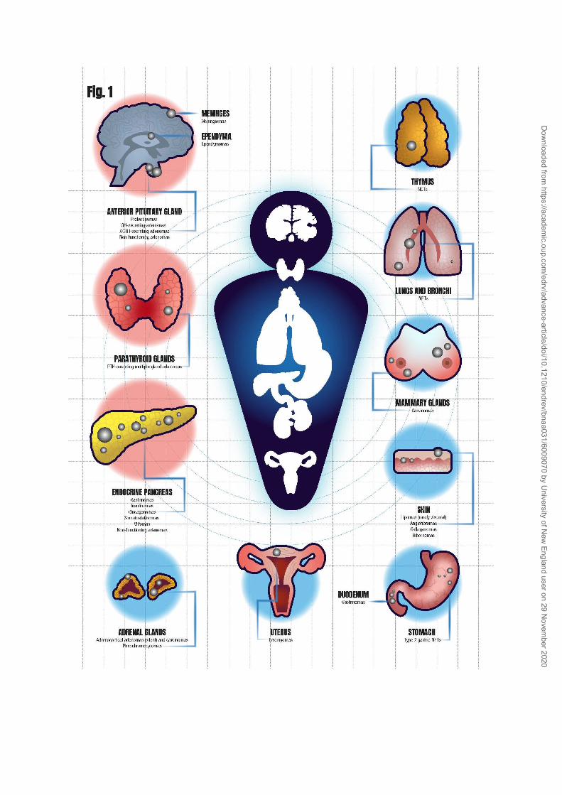
Accep
ted M
anus
cript
Dow
nloaded from https://academ
ic.oup.com/edrv/advance-article/doi/10.1210/endrev/bnaa031/6009070 by U
niversity of New
England user on 29 Novem
ber 2020

Accep
ted M
anus
cript
Dow
nloaded from https://academ
ic.oup.com/edrv/advance-article/doi/10.1210/endrev/bnaa031/6009070 by U
niversity of New
England user on 29 Novem
ber 2020
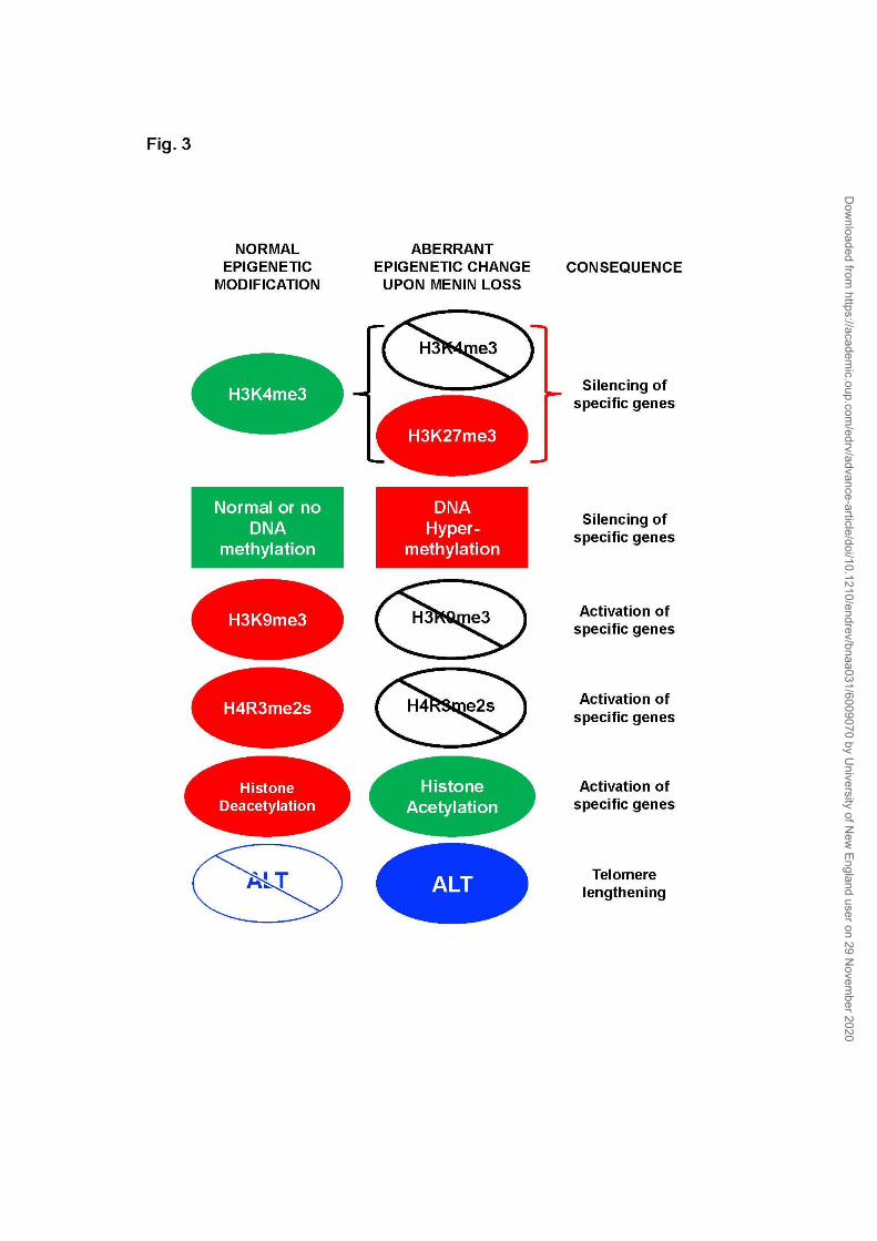
Accep
ted M
anus
cript
Dow
nloaded from https://academ
ic.oup.com/edrv/advance-article/doi/10.1210/endrev/bnaa031/6009070 by U
niversity of New
England user on 29 Novem
ber 2020
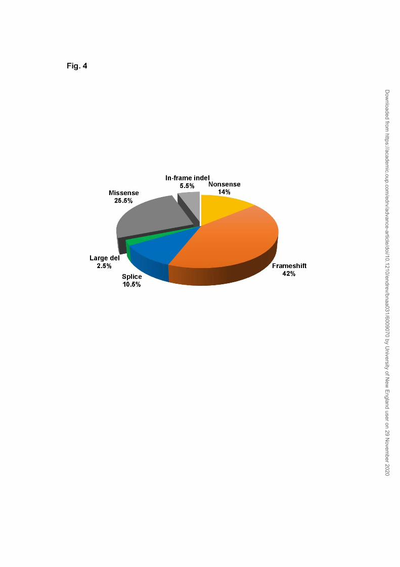
Accep
ted M
anus
cript
Dow
nloaded from https://academ
ic.oup.com/edrv/advance-article/doi/10.1210/endrev/bnaa031/6009070 by U
niversity of New
England user on 29 Novem
ber 2020
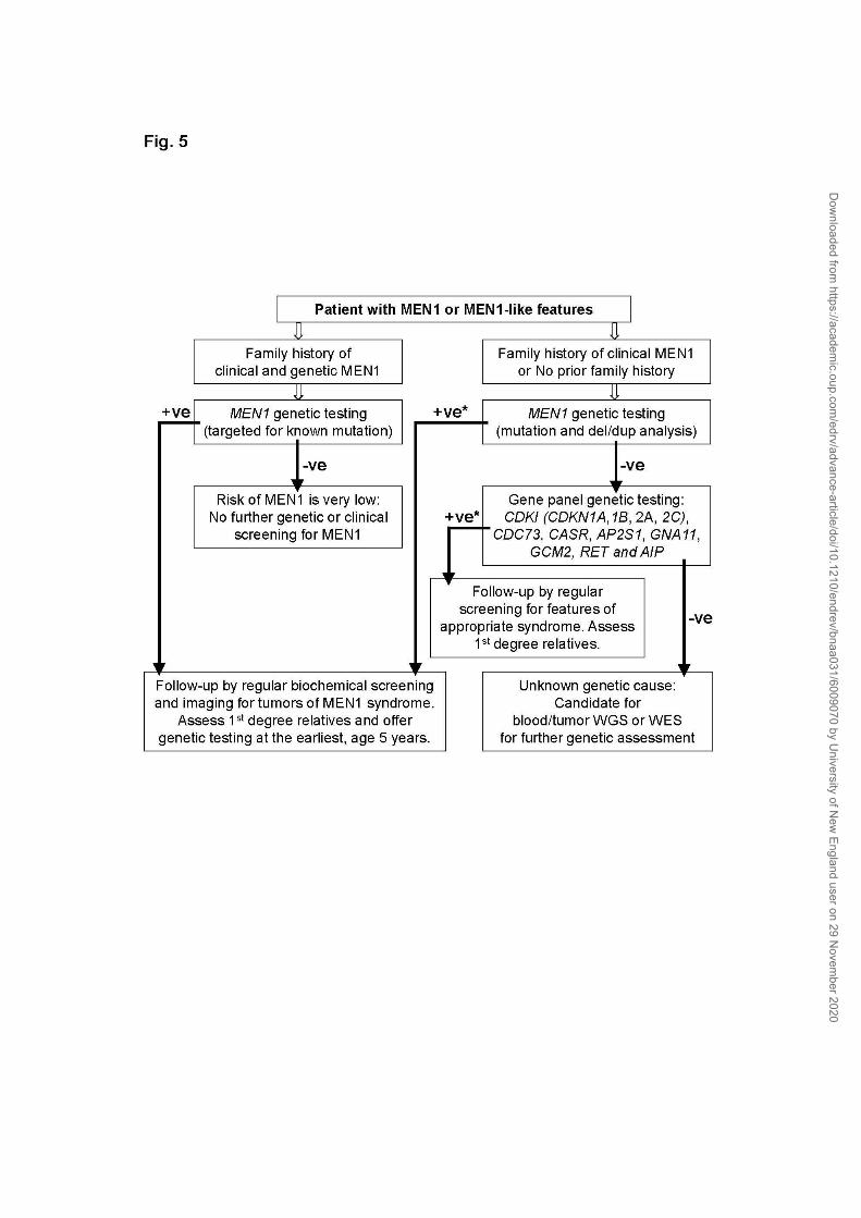
Accep
ted M
anus
cript
Dow
nloaded from https://academ
ic.oup.com/edrv/advance-article/doi/10.1210/endrev/bnaa031/6009070 by U
niversity of New
England user on 29 Novem
ber 2020

Accep
ted M
anus
cript
Dow
nloaded from https://academ
ic.oup.com/edrv/advance-article/doi/10.1210/endrev/bnaa031/6009070 by U
niversity of New
England user on 29 Novem
ber 2020
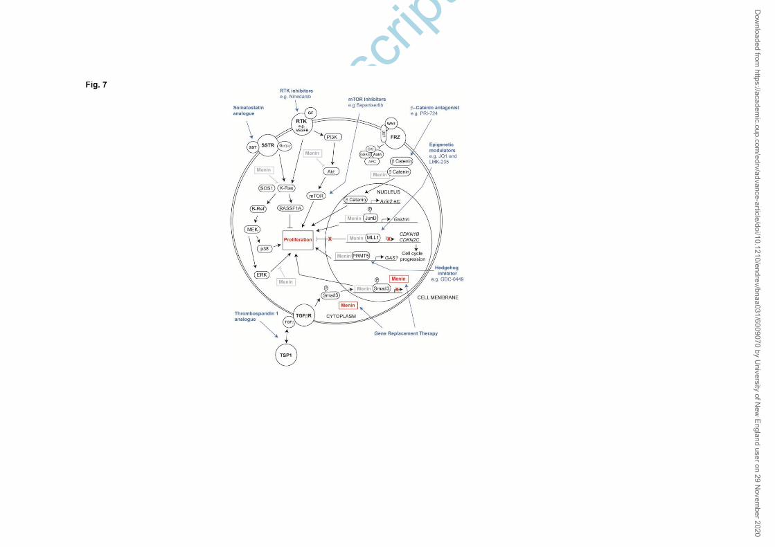
Accep
ted M
anus
cript
Dow
nloaded from https://academ
ic.oup.com/edrv/advance-article/doi/10.1210/endrev/bnaa031/6009070 by U
niversity of New
England user on 29 Novem
ber 2020