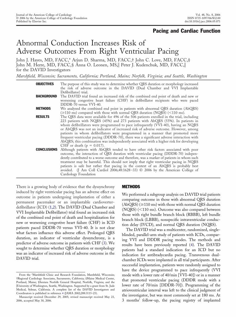
Abnormal Conduction Increases Risk of Adverse Outcomes From Right Ventricular Pacing
6
Pacing and Cardiac Function Abnormal Conduction Increases Risk of Adverse Outcomes From Right Ventricular Pacing John J. Hayes, MD, FACC,* Arjun D. Sharma, MD, FACC,† John C. Love, MD, FACC,‡ John M. Herre, MD, FACC,§ Anna O. Leonen, MS, Peter J. Kudenchuk, MD, FACC, for the DAVID Investigators Marshfield, Wisconsin; Sacramento, California; Portland, Maine; Norfolk, Virginia; and Seattle, Washington OBJECTIVES The purpose of this study was to determine whether QRS duration or morphology increased the risk of adverse outcome in the DAVID (Dual Chamber and VVI Implantable Defibrillator) trial. BACKGROUND The DAVID trial found an increased risk of the combined end point of death and new or worsening congestive heart failure (CHF) in defibrillator recipients who were paced DDDR-70 versus VVI-40. METHODS We analyzed the combined end point in patients with abnormal QRS duration (AbQRS) (110 ms) compared with those with normal QRS duration (NQRS) (110 ms). RESULTS The QRS data were available for 496 of the 506 patients enrolled in the trial, including 223 patients with NQRS (45%) and 273 patients with AbQRS (55%). In patients in whom defibrillators were programmed to pace infrequently (VVI-40), having an NQRS or AbQRS was not an indicator of increased risk of adverse outcome. However, among patients in whom defibrillators were programmed in a manner that promoted more frequent ventricular pacing (DDDR-70), there was a significant adverse interaction with AbQRS; this combination was independently associated with a higher risk for developing CHF or death (p 0.017). CONCLUSIONS Although patients with AbQRS tended to have other risk factors associated with poor outcome, the interaction of QRS duration with ventricular pacing (DDDR-70) indepen- dently contributed to a worse outcome and therefore, was a marker of patients in whom such treatment may be harmful. This should not imply that right ventricular pacing in NQRS patients is safe but rather that pacing in the context of an AbQRS is probably best avoided. (J Am Coll Cardiol 2006;48:1628 –33) © 2006 by the American College of Cardiology Foundation There is a growing body of evidence that the dyssynchrony induced by right ventricular pacing has an adverse effect on outcome in patients undergoing implantation of either a permanent pacemaker or an implantable cardioverter- defibrillator (ICD) (1,2). The DAVID (Dual Chamber and VVI Implantable Defibrillator) trial found an increased risk of the combined end point of death and hospitalization for new or worsening congestive heart failure (CHF) in ICD patients paced DDDR-70 versus VVI-40. It is not clear what factors influence this adverse effect. Prolonged QRS duration, an indicator of ventricular dyssynchrony, is a predictor of adverse outcome in patients with CHF (3). We sought to determine whether QRS duration or morphology was an indicator of increased risk of adverse outcome in the DAVID trial. METHODS We performed a subgroup analysis on DAVID trial patients comparing outcome in those with abnormal QRS duration (AbQRS) (110 ms) with those with normal QRS duration (NQRS) (110 ms). Outcome was also compared between those with right bundle branch block (RBBB), left bundle branch block (LBBB), nonspecific intraventricular conduc- tion delay (IVCD), and normal QRS duration (NQRS). The DAVID trial was a multicenter, randomized, single- blinded, parallel-arm study of patients with ICDs, compar- ing VVI and DDDR pacing modes. The methods and results have been previously reported (4). The DAVID patients had a standard indication for an ICD but no indication for antibradycardia pacing. Transvenous dual- chamber ICDs were implanted in all trial participants. After successful implantation, patients were randomly assigned to have the device programmed to pace infrequently (VVI mode with a lower rate of 40/min [VVI-40]) or in a manner that promoted ventricular pacing (DDDR mode with a lower rate of 70/min [DDDR-70]). Programming of the atrioventricular interval was left to the clinical judgment of the investigator, but was most commonly set at 180 ms. At 3 months’ follow-up, the pacing registry of implanted From the *Marshfield Clinic and Research Foundation, Marshfield, Wisconsin; †Regional Cardiology Associates, Sacramento, California; ‡Maine Medical Center, Portland, Maine; §Sentara Norfolk General Hospital, Norfolk, Virginia; and the University of Washington, Seattle, Washington. Supported by a grant from St. Jude Medical, Sylmar, California. A complete list of the DAVID Investigators and Coordinators is published in reference 4 (JAMA 2002;288:3115–23). Manuscript received December 29, 2005; revised manuscript received May 23, 2006, accepted May 30, 2006. Journal of the American College of Cardiology Vol. 48, No. 8, 2006 © 2006 by the American College of Cardiology Foundation ISSN 0735-1097/06/$32.00 Published by Elsevier Inc. doi:10.1016/j.jacc.2006.05.071
-
Upload
independent -
Category
Documents
-
view
0 -
download
0
Transcript of Abnormal Conduction Increases Risk of Adverse Outcomes From Right Ventricular Pacing
AAJJfM
TiopdVonpwdpswD
†P�MC
2
Journal of the American College of Cardiology Vol. 48, No. 8, 2006© 2006 by the American College of Cardiology Foundation ISSN 0735-1097/06/$32.00P
Pacing and Cardiac Function
bnormal Conduction Increases Risk ofdverse Outcomes From Right Ventricular Pacing
ohn J. Hayes, MD, FACC,* Arjun D. Sharma, MD, FACC,† John C. Love, MD, FACC,‡ohn M. Herre, MD, FACC,§ Anna O. Leonen, MS,� Peter J. Kudenchuk, MD, FACC,�or the DAVID Investigators
arshfield, Wisconsin; Sacramento, California; Portland, Maine; Norfolk, Virginia; and Seattle, Washington
OBJECTIVES The purpose of this study was to determine whether QRS duration or morphology increasedthe risk of adverse outcome in the DAVID (Dual Chamber and VVI ImplantableDefibrillator) trial.
BACKGROUND The DAVID trial found an increased risk of the combined end point of death and new orworsening congestive heart failure (CHF) in defibrillator recipients who were pacedDDDR-70 versus VVI-40.
METHODS We analyzed the combined end point in patients with abnormal QRS duration (AbQRS)(�110 ms) compared with those with normal QRS duration (NQRS) (�110 ms).
RESULTS The QRS data were available for 496 of the 506 patients enrolled in the trial, including223 patients with NQRS (45%) and 273 patients with AbQRS (55%). In patients inwhom defibrillators were programmed to pace infrequently (VVI-40), having an NQRSor AbQRS was not an indicator of increased risk of adverse outcome. However, amongpatients in whom defibrillators were programmed in a manner that promoted morefrequent ventricular pacing (DDDR-70), there was a significant adverse interaction withAbQRS; this combination was independently associated with a higher risk for developingCHF or death (p � 0.017).
CONCLUSIONS Although patients with AbQRS tended to have other risk factors associated with pooroutcome, the interaction of QRS duration with ventricular pacing (DDDR-70) indepen-dently contributed to a worse outcome and therefore, was a marker of patients in whom suchtreatment may be harmful. This should not imply that right ventricular pacing in NQRSpatients is safe but rather that pacing in the context of an AbQRS is probably bestavoided. (J Am Coll Cardiol 2006;48:1628–33) © 2006 by the American College of
ublished by Elsevier Inc. doi:10.1016/j.jacc.2006.05.071
Cardiology Foundation
M
Wc((tbt
birpicshmtlat
here is a growing body of evidence that the dyssynchronynduced by right ventricular pacing has an adverse effect onutcome in patients undergoing implantation of either aermanent pacemaker or an implantable cardioverter-efibrillator (ICD) (1,2). The DAVID (Dual Chamber andVI Implantable Defibrillator) trial found an increased riskf the combined end point of death and hospitalization forew or worsening congestive heart failure (CHF) in ICDatients paced DDDR-70 versus VVI-40. It is not clearhat factors influence this adverse effect. Prolonged QRSuration, an indicator of ventricular dyssynchrony, is aredictor of adverse outcome in patients with CHF (3). Weought to determine whether QRS duration or morphologyas an indicator of increased risk of adverse outcome in theAVID trial.
From the *Marshfield Clinic and Research Foundation, Marshfield, Wisconsin;Regional Cardiology Associates, Sacramento, California; ‡Maine Medical Center,ortland, Maine; §Sentara Norfolk General Hospital, Norfolk, Virginia; and the
University of Washington, Seattle, Washington. Supported by a grant from St. Judeedical, Sylmar, California. A complete list of the DAVID Investigators andoordinators is published in reference 4 (JAMA 2002;288:3115–23).
3Manuscript received December 29, 2005; revised manuscript received May 23,
006, accepted May 30, 2006.
ETHODS
e performed a subgroup analysis on DAVID trial patientsomparing outcome in those with abnormal QRS durationAbQRS) (�110 ms) with those with normal QRS durationNQRS) (�110 ms). Outcome was also compared betweenhose with right bundle branch block (RBBB), left bundleranch block (LBBB), nonspecific intraventricular conduc-ion delay (IVCD), and normal QRS duration (NQRS).
The DAVID trial was a multicenter, randomized, single-linded, parallel-arm study of patients with ICDs, compar-ng VVI and DDDR pacing modes. The methods andesults have been previously reported (4). The DAVIDatients had a standard indication for an ICD but nondication for antibradycardia pacing. Transvenous dual-hamber ICDs were implanted in all trial participants. Afteruccessful implantation, patients were randomly assigned toave the device programmed to pace infrequently (VVIode with a lower rate of 40/min [VVI-40]) or in a manner
hat promoted ventricular pacing (DDDR mode with aower rate of 70/min [DDDR-70]). Programming of thetrioventricular interval was left to the clinical judgment ofhe investigator, but was most commonly set at 180 ms. At
months’ follow-up, the pacing registry of implanted
dvt(DaiaaN1bdRIbaaEffwciahbAnsiscFSvceSwrai
tSfiL�
R
PaT9a5hlnafactpratbldaavt[EdibtmpabqsN4ia(sRswt
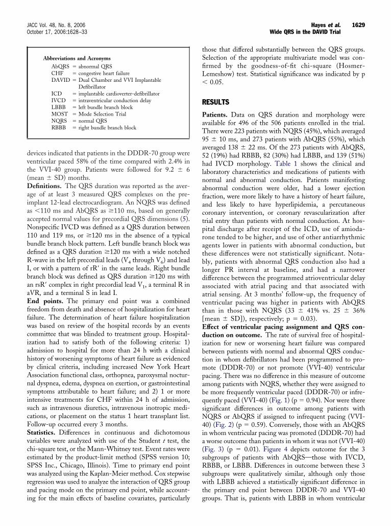
1629JACC Vol. 48, No. 8, 2006 Hayes et al.October 17, 2006:1628–33 Wide QRS in the DAVID Trial
evices indicated that patients in the DDDR-70 group wereentricular paced 58% of the time compared with 2.4% inhe VVI-40 group. Patients were followed for 9.2 � 6mean � SD) months.
efinitions. The QRS duration was reported as the aver-ge of at least 3 measured QRS complexes on the pre-mplant 12-lead electrocardiogram. An NQRS was defineds �110 ms and AbQRS as �110 ms, based on generallyccepted normal values for precordial QRS dimensions (5).onspecific IVCD was defined as a QRS duration between
10 and 119 ms, or �120 ms in the absence of a typicalundle branch block pattern. Left bundle branch block wasefined as a QRS duration �120 ms with a wide notched-wave in the left precordial leads (V4 through V6) and lead
, or with a pattern of rR= in the same leads. Right bundleranch block was defined as QRS duration �120 ms withn rsR= complex in right precordial lead V1, a terminal R inVR, and a terminal S in lead I.nd points. The primary end point was a combined
reedom from death and absence of hospitalization for heartailure. The determination of heart failure hospitalizationas based on review of the hospital records by an events
ommittee that was blinded to treatment group. Hospital-zation had to satisfy both of the following criteria: 1)dmission to hospital for more than 24 h with a clinicalistory of worsening symptoms of heart failure as evidencedy clinical criteria, including increased New York Heartssociation functional class, orthopnea, paroxysmal noctur-al dyspnea, edema, dyspnea on exertion, or gastrointestinalymptoms attributable to heart failure; and 2) 1 or morentensive treatments for CHF within 24 h of admission,uch as intravenous diuretics, intravenous inotropic medi-ations, or placement on the status 1 heart transplant list.ollow-up occurred every 3 months.tatistics. Differences in continuous and dichotomousariables were analyzed with use of the Student t test, thehi-square test, or the Mann-Whitney test. Event rates werestimated by the product-limit method (SPSS version 10;PSS Inc., Chicago, Illinois). Time to primary end pointas analyzed using the Kaplan-Meier method. Cox stepwise
egression was used to analyze the interaction of QRS groupnd pacing mode on the primary end point, while account-
Abbreviations and AcronymsAbQRS � abnormal QRSCHF � congestive heart failureDAVID � Dual Chamber and VVI Implantable
DefibrillatorICD � implantable cardioverter-defibrillatorIVCD � intraventricular conduction delayLBBB � left bundle branch blockMOST � Mode Selection TrialNQRS � normal QRSRBBB � right bundle branch block
ng for the main effects of baseline covariates, particularly g
hose that differed substantially between the QRS groups.election of the appropriate multivariate model was con-rmed by the goodness-of-fit chi-square (Hosmer-emeshow) test. Statistical significance was indicated by p0.05.
ESULTS
atients. Data on QRS duration and morphology werevailable for 496 of the 506 patients enrolled in the trial.here were 223 patients with NQRS (45%), which averaged5 � 10 ms, and 273 patients with AbQRS (55%), whichveraged 138 � 22 ms. Of the 273 patients with AbQRS,2 (19%) had RBBB, 82 (30%) had LBBB, and 139 (51%)ad IVCD morphology. Table 1 shows the clinical and
aboratory characteristics and medications of patients withormal and abnormal conduction. Patients manifestingbnormal conduction were older, had a lower ejectionraction, were more likely to have a history of heart failure,nd less likely to have hyperlipidemia, a percutaneousoronary intervention, or coronary revascularization afterrial entry than patients with normal conduction. At hos-ital discharge after receipt of the ICD, use of amioda-one tended to be higher, and use of other antiarrhythmicgents lower in patients with abnormal conduction, buthese differences were not statistically significant. Nota-ly, patients with abnormal QRS conduction also had aonger PR interval at baseline, and had a narrowerifference between the programmed atrioventricular delayssociated with atrial pacing and that associated withtrial sensing. At 3 months’ follow-up, the frequency ofentricular pacing was higher in patients with AbQRShan in those with NQRS (33 � 41% vs. 25 � 36%mean � SD]), respectively; p � 0.03).ffect of ventricular pacing assignment and QRS con-uction on outcome. The rate of survival free of hospital-
zation for new or worsening heart failure was comparedetween patients with normal and abnormal QRS conduc-ion in whom defibrillators had been programmed to pro-ote (DDDR-70) or not promote (VVI-40) ventricular
acing. There was no difference in this measure of outcomemong patients with NQRS, whether they were assigned toe more frequently ventricular paced (DDDR-70) or infre-uently paced (VVI-40) (Fig. 1) (p � 0.94). Nor were thereignificant differences in outcome among patients withQRS or AbQRS if assigned to infrequent pacing (VVI-
0) (Fig. 2) (p � 0.59). Conversely, those with an AbQRSn whom ventricular pacing was promoted (DDDR-70) hadworse outcome than patients in whom it was not (VVI-40)
Fig. 3) (p � 0.01). Figure 4 depicts outcome for the 3ubgroups of patients with AbQRS—those with IVCD,BBB, or LBBB. Differences in outcome between these 3
ubgroups were qualitatively similar, although only thoseith LBBB achieved a statistically significant difference in
he primary end point between DDDR-70 and VVI-40
roups. That is, patients with LBBB in whom ventricular
phwaAAfblct(a
TovtFt(tvbop
T
NAEHHHHPPHHHIT
R
L
E
M
M ventric
1630 Hayes et al. JACC Vol. 48, No. 8, 2006Wide QRS in the DAVID Trial October 17, 2006:1628–33
acing was promoted (DDDR-70) appeared to have aigher likelihood of death or heart failure than those inhom it was not (VVI-40) (p � 0.03), although there wasvisually apparent divergence in the survival curves for all 3bQRS subgroups.djusted outcomes. Cox regression analysis was per-
ormed, adjusting the primary outcome for differences inaseline variables, including age, ejection fraction, revascu-arization, history of heart failure, history of diabetes, serumreatinine, PR interval, and use of nitrates, followed, inurn, by QRS duration, promoted ventricular pacingDDDR-70), and the interaction between QRS duration
able 1. Clinical and Laboratory Characteristics of Patients With
umber of patients, n (%)ge at randomization, yrs (mean � SD)jection fraction (mean � SD)istory of MI, n (%)istory of heart failure, n (%)istory of atrial fibrillation or flutter, n (%)istory of diabetes, n (%)rior VF, n (%)rior VT, n (%)istory of unexplained syncope, n (%)istory of hypertension, n (%)istory of hyperlipidemia, n (%)
schemic cardiomyopathy, n (%)ype of index arrhythmiaVF, n (%)Syncopal VT, n (%)Symptomatic VT, n (%)Syncope with inducible VT or VF, n (%)Hemodynamically stable VT, n (%)Symptomatic NSVT with inducible VT or VF, n (%)Asymptomatic NSVT with inducible VT or VF, n (%)
evascularization historyHistory of coronary revascularization surgery or percutaneous coronary
prior to index arrhythmia, n (%)Coronary revascularization surgery or percutaneous coronary interventioAny revascularization, n (%)
aboratory findingsBlood urea nitrogen, mg/dl (mean � SD)Serum creatinine, mg/dl (mean � SD)
lectrocardiographic and pacing characteristicsHeart rate, beats/min (mean � SD)PR interval, msec (mean � SD)PR not measurable (AF or paced VVI), n (%)QRS duration, msec (mean � SD)Final: pacing AV delay, msec (mean � SD)AV delay � PR interval (mean � SD)edication usage at baseline hospital dischargeNitrates, n (%)Any antiarrhythmic medications, n (%)Amiodarone, n (%)Amiodarone total daily dose, mg (mean � SD)Sotalol, n (%)Other antiarrhythmic medications, n (%)Antidepressant, n (%)
I � myocardial infarction; NSVT � nonsustained ventricular tachycardia; VF �
nd ventricular pacing (DDDR-70), as depicted in Table 2. i
he regression analysis identified a significant effect uponutcome stemming from the combination of an AbQRS,entricular pacing (DDDR-70), and the interaction be-ween AbQRS and pacing corresponding to that seen inigures 3 and 4 (p � 0.017). That is, the adverse effects on
he primary outcome when ventricular pacing was promotedDDDR-70) were much more pronounced in the AbQRShan in the NQRS group. When not adjusted for baselineariables, there was only a trend toward an interactionetween QRS duration and treatment upon the primaryutcome (p � 0.13), probably owing to imbalances in otherredictive characteristics between groups. There was an
mal and Abnormal Conduction
QRS Duration
p Value>110 ms <110 ms
273 (55%) 223 (45%)66 � 10 63 � 12 0.00426 � 8 29 � 7 �0.001
187 (69%) 150 (68%) 0.78171 (63%) 108 (49%) 0.00244 (16%) 24 (11%) 0.0987 (32%) 71 (32%) 1.007 (3%) 11 (5%) 0.16
40 (15%) 24 (11%) 0.2037 (14%) 30 (14%) 0.98
169 (62%) 144 (65%) 0.53145 (53%) 141 (64%) 0.02226 (83%) 189 (85%) 0.54
0.8953 (19%) 38 (17%)17 (6%) 15 (7%)58 (21%) 39 (17%)42 (15%) 37 (17%)24 (9%) 21 (9%)20 (7%) 16 (7%)59 (22%) 57 (26%)
ention 156 (57%) 124 (56%) 0.74
r index arrhythmia, n (%) 32 (12%) 43 (19%) 0.02171 (63%) 147 (66%) 0.44
24 � 11 22 � 11 0.031.3 � 1 1.4 � 2 0.80
71 � 15 73 � 15 0.17191 � 40 174 � 34 �0.001
5 (2%) 0 (0%) 0.07138 � 22 95 � 10 �0.001201 � 30 206 � 36 0.31
7 � 53 32 � 46 0.001
72 (27%) 56 (26%) 0.7894 (35%) 63 (29%) 0.1587 (32%) 53 (24%) 0.05
364 � 242 426 � 248 0.156 (2%) 6 (3%) 0.712 (1%) 6 (3%) 0.15
34 (13%) 26 (12%) 0.81
ular fibrillation; VT � ventricular tachycardia.
Nor
interv
n afte
nsufficient number of cases to distinguish whether this

acIabtenttFcQsm
wo
D
AQmtsdoIbdjogahd
athf((wNQopt(c
Fow(
Ffinc0
Fow(
1631JACC Vol. 48, No. 8, 2006 Hayes et al.October 17, 2006:1628–33 Wide QRS in the DAVID Trial
dverse effect was independently predicted by the specificonduction abnormality (LBBB, RBBB, or nonspecificVCD), as was suggested by the Kaplan-Meier survivalnalysis. For example, despite the apparent relationshipetween LBBB and treatment on outcome observed inhe univariate analysis, the interaction between the two,ither unadjusted or adjusted for baseline variables, wasot statistically significant (p � 0.45 and 0.42, respec-ively). This may have been because no such interactionruly existed or because the analysis was underpowered.inally, in support of our prospective selection of aonventional cut point of �110 ms to define abnormalRS duration, when the regression analysis was re-
tricted to patients who already manifested such abnor-al conduction, further increments in QRS duration
igure 1. Kaplan-Meier curve depicting the primary end point of deathr first hospitalization for new or worsening heart failure in patientsith normal conduction who were programmed in a manner that did
DDDR-70) or did not (VVI-40) promote ventricular pacing. p � 0.94.
igure 2. Kaplan-Meier curve depicting the primary end point of death orrst hospitalization for new or worsening heart failure in patients withormal QRS (�110 ms) (NQRS) or abnormal QRS (�110 ms) (AbQRS)
ronduction who were programmed to pace infrequently (VVI-40) (p �.59).
ere not in themselves found to be a significant predictorf the primary end point (p � 0.89).
ISCUSSION
cross the spectrum from narrow to markedly prolonged,RS duration has been shown to be an independentarker of mortality in patients with and without conges-
ive heart failure (1,3,6). Recent studies have also ob-erved that, when combined with severe left ventricularysfunction, right ventricular pacing worsens clinicalutcome (4,7), particularly when it occurs frequently (8).n addition, multiple studies have shown the benefits ofiventricular pacing in patients with prolonged QRSuration and CHF suggesting that a widened QRS is not
ust a marker of disease severity, but a possible indicatorf a treatable functional impairment (9,10). Taken to-ether, these observations support the existence of andverse and potentially modifiable interaction betweeneart failure, QRS duration, and pacing that was ad-ressed by the present study.When ventricular pacing was promoted (DDDR-70)
mong patients with impaired heart function, we found thathose with a prolonged QRS duration (�110 ms) had aigher risk of death or new or worsening congestive heartailure compared with patients with a baseline narrow QRS�110 ms). When ventricular pacing was not promotedVVI-40), having a normal or an abnormal QRS durationas not an indicator of increased risk of adverse outcome.or, apart from pacing considerations, was increasingRS duration found to independently predict an adverse
utcome among patients in whom QRS was alreadyrolonged at baseline (�110 ms). However, in combina-ion with assignment to more frequent ventricular pacingDDDR-70), a prolonged QRS was independently asso-iated with a higher risk of an adverse outcome. These
igure 3. Kaplan-Meier curve depicting the primary end point of deathr first hospitalization for new or worsening heart failure in patientsith abnormal conduction who were programmed in a manner that did
DDDR-70) or did not (VVI-40) promote ventricular pacing. p � 0.01.
esults suggest that patients with some degree of baseline

deu
t(hamufsPecvbnkLapoawddupigwvwcfbPfw(nen(owOM(dwotwapMp
Ffianb7fo
1632 Hayes et al. JACC Vol. 48, No. 8, 2006Wide QRS in the DAVID Trial October 17, 2006:1628–33
yssynchrony may be more susceptible to the adverseffects of further dyssynchrony induced by right ventric-
igure 4. Kaplan-Meier curve depicting the primary end point of death orrst hospitalization for new or worsening heart failure in patients withbnormal conduction due to right bundle branch block (RBBB) (A), aonspecific conduction abnormality (IVCD) (B), or left bundle branchlock (LBBB) (C) who were programmed in a manner that did (DDDR-0) or did not (VVI-40) promote ventricular pacing. Statistical significanceor an adverse outcome associated with ventricular pacing (DDDR-70) wasbserved only in patients with LBBB (p � 0.03).
lar pacing. Although patients with a wide QRS tended M
o have other risk factors associated with poor outcomesuch as increasing age, worse ejection fraction, andigher likelihood of prior congestive heart failure), thedverse interaction of QRS duration with assignment toore frequent ventricular pacing (DDDR-70) contrib-
ted to a worse outcome independent of these other riskactors and, as such, was a marker of patients in whomuch treatment should be avoided.acing in bundle branch block. Although it could not bestablished as an independent predictor, the adverse out-ome in patients with increased QRS duration in whomentricular pacing was promoted (DDDR-70) appeared toe more strongly associated with LBBB than with RBBB oronspecific IVCD. This observation is consistent with thenown mechanical and hemodynamic consequences ofBBB on left ventricular function, which is simulated, andrguably might even be exaggerated, by right ventricularacing (11). However, nonsignificant trends toward worseutcome in patients with RBBB and IVCD when pacedlso were observed which were qualitatively similar to thoseith LBBB. It is possible that ventricular pacing isetrimental in the presence of an underlying conductionisorder in any of its forms, and this trial was simplynderpowered to establish these differences. It is alsoossible that the subgroup of patients with IVCD, whichncluded those with more minor degrees of QRS prolon-ation, might be expected to behave more like patientsith NQRS and have less detrimental effects fromentricular pacing. Similarly patients with RBBB (inhom the benefit from resynchronization therapy is less
ertain (12) may, by the same token, sustain less harmrom the change in ventricular activation sequence causedy right ventricular pacing.acing in patients with a normal QRS. In our study we
ound no difference in the primary end point among patientsith a normal QRS in whom ventricular pacing was
DDDR-70) or was not (VVI-40) promoted. This shouldot necessarily be taken to indicate there are no adverseffects from right ventricular pacing in patients with aarrow baseline QRS. A subgroup analysis of the MOSTMode Selection Trial) pacing trial showed an increased riskf development of CHF and atrial fibrillation in patientsith a “normal” QRS who received ventricular pacing (13).ne explanation for this apparent discrepancy betweenOST and DAVID lies in their selection of a wider QRS
�120 ms) to define normal than the more conventionalefinition (�110 ms) used in our trial. In the context of aider than normal QRS, both trials could be said to havebserved ventricular pacing to be detrimental, whereashis may or may not have been the case when the QRSas truly narrow. Another explanation for this discrep-
ncy is the difference in the frequency of ventricularacing between the 2 studies. The DDDR group inOST was 90% ventricular paced compared with 58%
aced in DAVID. The VVI group was 58% paced in
OST compared with 2.4% in DAVID. These differ-
ewisatQtdSndoheatatbr
R4dHOm
R
1
1
1
1
T
EHHASPNAADA
TssIws†
reedom
1633JACC Vol. 48, No. 8, 2006 Hayes et al.October 17, 2006:1628–33 Wide QRS in the DAVID Trial
nces stem from the different programming parameters asell as the fact that MOST patients had a primary
ndication for antibradycardia pacing. The MOST sub-tudy observed an increasing risk of adverse outcomesssociated with the frequency of ventricular pacing. It isherefore possible that patients in DAVID with a narrowRS complex would have had a higher event rate had
heir frequency of ventricular pacing been greater or theiruration of follow-up been longer.tudy limitations. This study is limited by the post-hocature of the analysis. The DAVID trial was not primarilyesigned to study the effects of QRS duration on outcomer in relation to ventricular pacing. The results do suggest,owever, that patients with baseline electrocardiographicvidence of dyssynchrony may be particularly prone to thedverse effects of right ventricular pacing. Increased atten-ion to programming and novel pacing modes are needed tovoid right ventricular stimulation where possible. Prospec-ive studies are required to define better alternatives, such asiventricular pacing, for broader groups of patients whoequire ventricular pacing.
eprint requests: DAVID Clinical Trial Center, 1107 NE5th St., Suite 505, Seattle, Washington 98105. E-mail:[email protected]. Correspondence: Dr. John J.ayes, Department of Cardiology, Marshfield Clinic, 1000 N.ak Avenue, Marshfield, Wisconsin 54449. E-mail: [email protected].
EFERENCES
1. Sweeney MO, Hellkamp AS, Lee KL, Lamas GA; MOST Investi-gators. Association of prolonged QRS duration with death in a clinical
able 2. Multivariate Proportional Hazards Regression Model Re
VariablesUnivariate
p Value
Step 1
p Value RR
jection fraction �0.01 0.96istory of heart failure �0.01 2.12istory of diabetes 0.011 1.56ny revascularization 0.159 �0.01 0.63erum creatinine 0.002 (8 df) 1.11R interval 0.005 1.01itrates �0.01 2.23ge 0.111 1.01bQRS 0.339DDR pacing* 0.063bQRS � DDDR pacing†
his table depicts the univariate statistical significance upon the primary study end pignificance of adding these variables to the regression model. In the first step, all 8econd and third steps, AbQRS and programming in a manner that promoted ventrn the final step, the interaction between AbQRS and promoted ventricular pacihen AbQRS, promoted ventricular pacing (DDDR-70), and the interaction betw
tep (i.e., steps 2, 3, and 4 combined), statistical significance was observed at p � 0Interaction between abnormal QRS duration and promoted ventricular pacing.
AbQRS � abnormal QRS duration; CI � confidence interval; df � degrees of f
trial of pacemaker therapy for sinus node dysfunction. Circulation2005;111:2418–23.
2. Moss AJ, Zareba W, Hall WJ, et al. Prophylactic implantation of adefibrillator in patients with myocardial infarction and reduced ejec-tion fraction. N Engl J Med 2002;346:877–83.
3. Hofmann M, Bauer R, Handrock R, Weidinger G, Goedel-MeinenL. Prognostic value of the QRS duration in patients with heart failure:a subgroup analysis from 24 centers of Val-HeFT. J Card Fail2005;11:523–8.
4. DAVID Trial Investigators. Dual chamber pacing or ventricularpacing in patients with an implantable defibrillation: the Dual Cham-ber and VVI Implantable Defibrillator (DAVID) trial. JAMA 2002;288:3115–23.
5. Rowlands DJ. Clinical electrocardiography. Philadelphia, PA: Lippin-cott, 1991:40.
6. Iuliana S, Fischer SG, Karasik PE, Fletcher RD, Singh SN. Depart-ment of Veterans Affairs Survival Trial of Antiarrhythmic Therapy inCongestive Heart Failure: QRS duration and mortality in patientswith congestive heart failure. Am Heart J 2002;143:1085–91.
7. Steinberg JS, Fischer A, Wang P, et al.; MADIT II Investigators. Theclinical implications of cumulative right ventricular pacing in theMulticenter Automatic Defibrillator Trial II. J Cardiovasc Electro-physiol 2005;16:359–65.
8. Sharma AD, Rizo-Patron C, Hallstrom AP, et al.; DAVID Investi-gators. Percent right ventricular pacing predicts outcomes in theDAVID trial. Heart Rhythm 2005;2:830–4.
9. Young JB, Abraham WT, Smith AJ, et al.; MIRACLE ICD TrialInvestigators. Combined cardiac resynchronization and implantablecardioversion defibrillation in advanced chronic heart failure: theMIRACLE ICD trial. JAMA 2003;289:2685–94.
0. Cleland JGF, Daubert JC, Erdmann E, et al.; CARE-HF StudyInvestigators. The effect of cardiac resynchronization on morbidityand mortality in heart failure. N Engl J Med 2005;352:1539 – 49.
1. Gerber TC, Nishimura RA, Holmes DR, et al. Left ventricular andbiventricular pacing in congestive heart failure. Mayo Clin Proc2001;76:803–12.
2. Egoavil CA, Ho RT, Greenpon AJ, Pavri BB. Cardiac resynchroni-zation therapy in patients with right bundle branch block: analysis ofpooled data from the MIRACLE and Contak CD trials. HeartRhythm 2005;6:616–8.
3. Sweeney MO, Hllkamp AS, Ellenbogen KA, et al.; MOST Investi-gators. Adverse effect of ventricular pacing on heart failure and atrialfibrillation among patients with normal baseline QRS duration in aclinical trial of pacemaker therapy for sinus node dysfunction. Circu-
Step 2 Step 3 Step 4
ue RR p Value RR p Value RR 95% CI
0.96 0.96 0.95 0.93, 0.982.14 2.10 2.17 1.29, 3.651.56 1.49 1.46 0.94, 2.280.63 0.63 0.62 0.40, 0.961.10 1.10 1.10 1.00, 1.221.01 1.01 1.01 1.00, 1.012.22 2.27 2.38 1.50, 3.771.01 1.01 1.02 0.99, 1.04
7 0.92 0.95 0.46 0.23, 0.900.112 1.42 0.67 0.34, 1.33
0.006 3.47 1.42, 8.47
eath or hospitalization for heart failure) for the variables shown and the incrementall variables entered the model, with statistical significance shown at p � 0.01. In theacing (DDDR-70), respectively, entered the model, without statistical significance.
DDR-70) entered the model with statistical significance at p � 0.006. Notably,ese 2 variables upon outcome were entered into the multivariate model as a singlesee text). *Programming in a manner that promoted ventricular pacing (DDDR-70).
; RR � relative risk.
sults
p Val
0.70
oint (dclinicaicular png (Deen th.017 (
lation 2003;107:2932–7.