Temperature-controlled slow pathway ablation for treatment of ...

Ablation of PGC-1b Results in DefectiveMitochondrial Activity, Thermogenesis,Hepatic Function, and Cardiac PerformanceChristopher J. Lelliott
1,2[, Gema Medina-Gomez
1[, Natasa Petrovic
3, Adrienn Kis
1, Helena M. Feldmann
3,
Mikael Bjursell2
, Nadeene Parker1
, Keira Curtis1
, Mark Campbell1
, Ping Hu4
, Dongfang Zhang4
, Sheldon E. Litwin4
,
Vlad G. Zaha5
, Kimberly T. Fountain5
, Sihem Boudina5
, Mercedes Jimenez-Linan1
, Margaret Blount1
, Miguel Lopez1
,
Aline Meirhaeghe6
, Mohammad Bohlooly-Y2
, Leonard Storlien2
, Maria Stromstedt2
, Michael Snaith2
, Matej Oresic7
,
E. Dale Abel5
, Barbara Cannon3
, Antonio Vidal-Puig1*
1 Department of Clinical Biochemistry, University of Cambridge, Cambridge, United Kingdom, 2 AstraZeneca R&D, Molndal, Sweden, 3 The Wenner-Gren Institute, Stockholm
University, Stockholm, Sweden, 4 Division of Cardiology, University of Utah, Salt Lake City, Utah, United States of America, 5 Division of Endocrinology, Metabolism, and
Diabetes and Program in Human Molecular Biology and Genetics, University of Utah, Salt Lake City, Utah, United States of America, 6 INSERM, Institut Pasteur de Lille, Lille
Cedex, France, 7 VTT Technical Research Centre of Finland, Espoo, Finland
The transcriptional coactivator peroxisome proliferator-activated receptor-gamma coactivator-1b (PGC-1b) has beenimplicated in important metabolic processes. A mouse lacking PGC-1b (PGC1bKO) was generated and phenotypedusing physiological, molecular, and bioinformatic approaches. PGC1bKO mice are generally viable and metabolicallyhealthy. Using systems biology, we identified a general defect in the expression of genes involved in mitochondrialfunction and, specifically, the electron transport chain. This defect correlated with reduced mitochondrial volumefraction in soleus muscle and heart, but not brown adipose tissue (BAT). Under ambient temperature conditions, PGC-1b ablation was partially compensated by up-regulation of PGC-1a in BAT and white adipose tissue (WAT) that lead toincreased thermogenesis, reduced body weight, and reduced fat mass. Despite their decreased fat mass, PGC1bKOmice had hypertrophic adipocytes in WAT. The thermogenic role of PGC-1b was identified in thermoneutral and cold-adapted conditions by inadequate responses to norepinephrine injection. Furthermore, PGC1bKO hearts showed ablunted chronotropic response to dobutamine stimulation, and isolated soleus muscle fibres from PGC1bKO mice haveimpaired mitochondrial function. Lack of PGC-1b also impaired hepatic lipid metabolism in response to acute high fatdietary loads, resulting in hepatic steatosis and reduced lipoprotein-associated triglyceride and cholesterol content.Altogether, our data suggest that PGC-1b plays a general role in controlling basal mitochondrial function and alsoparticipates in tissue-specific adaptive responses during metabolic stress.
Citation: Lelliott C, Medina-Gomez G, Petrovic N, Kis A, Feldmann HM, et al. (2006) Ablation of PGC-1b results in defective mitochondrial activity, thermogenesis, hepaticfunction, and cardiac performance. PLoS Biol 4(11): e369. DOI: 10.1371/journal.pbio.0040369
Introduction
Transcriptional coactivators (TCs) have emerged as keyproteins that are capable of regulating entire metabolicnetworks [1]. TCs are often selective with respect to therepertoire of transcription factor partners. The mechanismsused to modulate transcription may vary according to thespecific TC, their transcription factor partners, and thenucleic acid environment in which they operate. TCs providean additional layer of control by facilitating temporal andtissue-specific regulation of metabolic pathways [1].
Peroxisome proliferator-activated receptor-gamma coacti-vator-1a (PGC-1a, previously known as PGC-1) [2] is involvedin coordinating diverse organ-specific transcription pro-grams. For example, PGC-1a not only activates the thermo-genic program in brown adipose tissue (BAT) in response toenvironmental cold exposure but also modulates the meta-bolic transition from the fed to the fasted state in the liver [2–4]. Two other PGC-1a-related genes have subsequently beenfound, PGC-1-related coactivator (PRC) [5] and PGC-1b(PERC/ERRL-1) [6–8]. All three family members share asimilar basic structure; an N-terminal domain containingnuclear hormone receptor–interacting motifs (the LXXLL
motifs) and a C-terminal region containing RNA-bindingmotifs (RMM) and serine-arginine rich (RS) domains. Where-as PRC is primarily an activator of nuclear respiratory factor-
Academic Editor: Gregory S. Barsh, Stanford University School of Medicine, UnitedStates of America
Received June 2, 2006; Accepted September 5, 2006; Published November 7,2006
DOI: 10.1371/journal.pbio.0040369
Copyright: � 2006 Lelliott et al. This is an open-access article distributed under theterms of the Creative Commons Attribution License, which permits unrestricteduse, distribution, and reproduction in any medium, provided the original authorand source are credited.
Abbreviations: BAT, brown adipose tissue; Cox, cytochrome oxidase subunit; EScell, embryonic stem cell; ETC, electron transport chain; HDL, high-densitylipoprotein; HFD, high-fat diet; KEGG, kyoto encyclopedia genes and genomes;LDL, low-density lipoprotein; NE, norepinephrine; NS, not significant; OxPhos,oxidative phosphorylation; PGC, peroxisome proliferator-activated receptor-gam-ma coactivator; RER, respiratory exchange ratio; VLDL, very low-density lipoprotein;WAT, white adipose tissue; WT, wild-type
* To whom correspondence should be addressed. E-mail: [email protected]
[ These authors contributed equally to this work.
PLoS Biology | www.plosbiology.org November 2006 | Volume 4 | Issue 11 | e3692042
PLoS BIOLOGY

1 [5], PGC-1a and PGC-1b have a broader repertoire ofpartners [7,9–12].
The gene expression level of PRC is conserved betweentissues [5], but the highest expression of PGC-1a and PGC-1bis found in oxidative tissues, including BAT, heart, andskeletal muscle [2,6,7]. However, despite this overlap ofexpression pattern, PGC-1a and �1b display differentresponses to physiological stimuli. Whereas PGC-1a is tran-scriptionally up-regulated in BAT and skeletal muscle duringcold exposure, PGC-1b expression remains stable under theseconditions [2,6,7]. Furthermore, up-regulation of PGC-1a isessential for a normal response to acute cold exposure,indicating that PGC-1b is not able to compensate for allaspects of a lack of PGC-1a [13,14]. Similarly PGC-1a is up-regulated in liver in response to fasting, whereas PGC-1bexpression remained stable [6,7,9], further suggesting thatPGC-1a is more often regulated transcriptionally than PGC-1b.
Overexpression studies both in vivo and in vitro haverevealed that both PGC-1a and�1b are capable of stimulatingmitochondrial biogenesis, increasing mitochondrial oxygenconsumption, and elevating gene expression of fatty acidoxidation and mitochondrial electron transport chain (ETC)components [7,9,15]. In addition, bioenergetic studies ofC2C12 myotubes demonstrated that overexpression of PGC-1a or �1b enhances mitochondrial proton leak [15]. ThusPGC-1b increases mitochondrial energy production througha combination of activated fatty acid oxidation geneexpression with increased mitochondrial activity. A role forPGC-1b enhancing mitochondrial activity is further sup-ported by the fact that transgenic overexpression of PGC-1bin mice results in elevated energy expenditure, resistance tohigh-fat diet (HFD), and genetically induced obesity [10].
Despite their functional similarities, it has recently beensuggested that PGC-1a and�1b may have differing metabolicroles in liver. PGC-1a stimulates expression of gluconeogenicgenes both in vivo and in vitro [3,9,13,16], and based on datafrom mice exposed to a diet enriched in saturated fat for ashort period of time, it appears that PGC-1b but not PGC-1ainteracts with SREBP and LXRa to enhance hepatic lipo-genesis and lipoprotein secretion [11]. In addition, PGC-1binteracts with Foxa2 to facilitate hepatic secretion of very lowdensity lipoprotein (VLDL) [17], suggesting a central role forPGC-1b in hepatic lipid metabolism. The second organ wherePGC-1a and �1b are also highly expressed is the heart [2,6],but only the role of PGC-1a in cardiac energy metabolism hasbeen studied in detail. Overexpression of PGC-1a in cardiacmuscle in vivo leads to uncontrolled mitochondrial prolifer-ation, leading to cardiomyopathy and congestive heart failuredue to contractile dysfunction [18,19]. Similarly, PGC-1aknockout mice also develop heart failure with abnormal heartrate and impaired ventricular function [14,20], again suggest-ing that despite their similar mode of action, PGC-1b isunable to compensate fully for a lack of PGC-1a.
To investigate the function of PGC-1b, we have generatedand phenotyped a mouse model lacking PGC-1b (thePGC1bKO mouse). Using a combination of physiologicalexperiments guided by a systems biology approach, weshowed that PGC-1b plays a general role in regulating themitochondrial activity in tissues with active oxidativemetabolism. In all tissues studied, lack of PGC-1b wasassociated with a reduction in the expression of mitochon-
drial genes, including intermediary subunits and enzymesconnected to ETC and oxidative phosphorylation (OxPhos).Despite these mitochondrial defects, the PGC1bKO mousewas able to cope when challenged with a range ofphysiological stressors including cold acclimatisation andadrenergically-mediated BAT and cardiac stimulation butwith abnormal compensatory responses. Altogether, thesedata suggest that PGC-1b is an important regulator of basalenergy homeostasis and essential for the proper metabolictuning in acute stress situations in multiple organs.
Results
Generation of a PGC1bKO MouseA conditional PGC1bKO was generated using a triple LoxP
targeting vector (Figure 1A) as detailed in the Material andMethods. Murine PGC-1b is alternatively spliced at the 59 end[7] and at exon 4 [8,10]. Exons 4 and 5, which encode two ofthe nuclear hormone receptor interacting motifs (LXXLL),were deleted. Ablation of these exons also introduced apremature stop codon. PCR and Southern analyses of theselected clone confirmed the presence of the single LoxP inintron 5 and that the embyronic stem (ES) cell clonecontained all three LoxP sites as designed (Figure 1B and1C). Germ line transmission of the modified PGC-1b allele wasconfirmed by PCR analysis of the agouti pups obtained fromchimeras bred to C57BL/6 wild-type (WT) mice. Heterozygoustriple-LoxP–containing mice were then bred with ROSA26-Cre mice to generate mice heterozygous for the PGC-1bdeletion. Total LoxP site recombination was detected usingPCR from genomic DNA (Figure 1D) and RT-PCR fromisolated RNA. Intercross breeding of heterozygous germ-lineknockout animals produced litters that did not deviatesignificantly from the expected Mendelian ratio of genotypes,and the size of litters from PGC1bKO heterozygous crosseswas normal (averaging seven to eight pups per breeding eventper female).
Effect of PGC-1b Ablation on Energy Balance: ReducedBody Weight and WAT Content Consistent with IncreasedEnergy ExpenditureNo differences in food intake, total energy intake, total
energy output as faeces, and no change in the ratio of energyin (as food) to energy out (as faeces) (Table S1) were detectedin 12-wk-old male mice selected for equal body weight.Oxymax analysis revealed increased minimum metabolic rateat 22 8C in 9-wk-old male PGC1bKO mice (Table 1). Nodifferences in spontaneous activity (unpublished data) or inrespiratory exchange ratio (RER) were detected, suggestingthat there was no difference in whole-body substrate-typeutilization. Consistent with an elevated minimum metabolicrate at ambient temperature, PGC1bKO mice weighed lessthan their WT littermates (Figure 2A). A similar pattern wasalso observed in female PGC1bKO mice.Body composition analysis using dual energy x-ray absorp-
tiometry (DEXA) confirmed decreased body weight at theexpense of fat mass rather than lean mass in both young (8-wk) and older (27-wk) male PGC1bKO mice compared to WTlittermates (Figure 2B). This was confirmed in an independ-ent group of weight-matched 14-wk-old male PGC1bKO micethat had smaller gonadal adipose tissue depots than WTlittermates. Other organs examined did not show differences
PLoS Biology | www.plosbiology.org November 2006 | Volume 4 | Issue 11 | e3692043
Phenotype of the PGC-1b Knockout Mouse

in their weights relative to body weight (Table S2). Histo-logical analysis of male gonadal WAT from PGC1bKO miceshowed a reduced fraction of small adipocytes (up to an areaof 2,000 lm2) and an elevated fraction of larger adipocytes inthe PGC1bKO WAT (Figure 2C and 2D). This pattern forlarger white adipocytes was also seen in male subcutaneousand omental WAT and in female gonadal WAT (Figure S1).
Therefore, when on a chow diet, the PGC1bKO mice had lesstotal WAT content than WT did, but it was composed oflarger adipocytes. Histological analysis of other metabolicallyrelevant tissues—including BAT, liver, skeletal muscle, andpancreas—did not reveal morphological alterations inPGC1bKO mice under basal conditions (Figure S2).
Gene expression analysis of gonadal WAT from male
Figure 1. Generation of the PGC1bKO Mouse
(A) Design of the targeting construct for the generation of the PGC1bKO mouse using a phosphoglycerate kinase-neomycin phosphotransferase (PGK-Neo)–based LoxP cassette inserted between exons III and IV and a third LoxP site between exons V and VI. Total collapse of the LoxP sites deletes exons4 and 5 of the PGC-1b gene.(B) A Southern blot confirming the generation of the targeted allele in ES cells. WT, wild type ES cells;þ/T, ES cells heterozygous for the targeted allele, T.(C) Schematic showing the restriction digest strategy for the southern blot confirming the generation of the targeted allele in ES cells. P, PsiI; X, XmaI; S,SpeI.(D) Confirmation of the total LoxP collapse and generation of KO mice by PCR from genomic DNA using primers F1, R1, and R2 shown in (A).DOI: 10.1371/journal.pbio.0040369.g001
Table 1. Assessment of Energy Expenditure on 9-wk-Old Male Chow-Fed PGC1bKO and WT Mice
Measurement 9-wk-old WT 9-wk-old PGC1bKO p Value
Minimum VO2 (ml/min) 0.39 6 0.05 0.52 6 0.03 p ¼ 0.07
Maximum VO2 (ml/min) 1.47 6 0.06 1.40 6 0.05 NS
Minimum VO2 (ml/kg/min) 16.1 6 2.3 22.6 6 1.8 p ¼ 0.05
Maximum VO2 (ml/kg/min) 59.9 6 1.7 60.8 6 3.0 NS
Minimum VO2 (ml/kg[lean]/min) 20.0 6 2.7 27.3 6 2.0 p ¼ 0.05
Maximum VO2 (ml/kg[lean]/min) 74.5 6 2.0 73.5 6 3.2 NS
Minimum VO2 (ml/kg0.75/min) 6.4 6 0.9 8.8 6 0.6 p ¼ 0.05
Maximum VO2 (ml/kg0.75/min) 23.7 6 0.7 23.7 6 1.0 NS
RER 0.88 6 0.01 0.90 6 0.01 NS
Oxygen consumption and the respiratory exchange ratio of male WT and PGC1bKO mice was measured at 9 wk old, in mice housed and measured at 22 8C. Minimum values refer to theaverage of the three lowest VO2 value recorded during the final 24 h of the experimental procedure, with maximum values referring to those corresponding to the highest three values ofVO2 during this period. VO2 measurements are given related per mouse (ml/min) to total body weight (ml/kg/min), lean body weight (ml/kg [lean]/min), and using the scaling factor of0.75 (ml/kg0.75/min). The RER was calculated as the average RER measurement during the final 24 h of the procedure. n¼ 5–7 mice per group.NS, not significant.DOI: 10.1371/journal.pbio.0040369.t001
PLoS Biology | www.plosbiology.org November 2006 | Volume 4 | Issue 11 | e3692044
Phenotype of the PGC-1b Knockout Mouse

PGC1bKO mice and WT littermates showed no significantchange in markers of adipogenesis including C/EBPa,PPARc2, aP2, and GLUT4 (Figure 2E). There was a significantup-regulation of PGC-1a expression in WAT in the absenceof PGC-1b (Figure 2E). Previously, increased PGC-1a ex-pression in WAT has been associated with the development ofa BAT-like phenotype [21]. However, increased expression of
PGC-1a was not sufficient to increase UCP1 and D2-deiodinase expression in PGC1bKO WAT (unpublished data).Combined with the WAT morphology data, elevation of PGC-1a in the PGC1bKO WAT is not enough to result in clearBAT-like adipose tissue. However, ablation of PGC-1bresulted in reduced expression in the ETC genes CytC andCytochrome Oxidase subunit (Cox) 5b and also in leptin (p¼ 0.06).
Figure 2. The PGC1bKO Mouse Has Altered Metabolism under Standard Environmental Conditions
(A) Growth curves of male (left panel) and female (right panel) mice on normal diet. WT (solid circles) and PGC1bKO (open circles) mice, n¼ 18–21 miceper group.(B) Assessment of fat content by DEXA in 8- and 32-wk-old male WT (solid bars) and PGC1bKO mice (open bars), n¼ 8–12 mice per group.(C) Representative histological sections of tissues from WAT (n ¼ 6).(D) Size distribution of adipocytes from WT and PGC1bKO mice. Two fields from each section from epididymal adipose tissue depot (n¼ 4 mice pergenotype) were analysed to obtain the mean cell area per animal.(E) Epididymal WAT gene expression from 12-wk-old PGC1bKO (white bars) and WT littermates (black bars). Individual measurements are standardizedusing 18S, and then the average of the WT group was set to 1. n ¼ 5–8 mice per group.DOI: 10.1371/journal.pbio.0040369.g002
PLoS Biology | www.plosbiology.org November 2006 | Volume 4 | Issue 11 | e3692045
Phenotype of the PGC-1b Knockout Mouse

Ablation of PGC-1b did not result in differences in basallevels of plasma glucose or insulin. Similarly, glucosetolerance tests and insulin tolerance tests (GTTs and ITTs,respectively) performed on 16-wk male and female miceeither on chow or after a 13-wk HFD did not revealdifferences among genotypes (Figure S3). Therefore, thePGC1bKO mouse is leaner, has increased energy expenditure,and remains insulin sensitive.
Reduced Expression of ETC Components DespiteIncreased PGC-1a mRNA Expression in the BAT ofPGC1bKO mice
Examination of BAT in chow-fed 12-wk-old PGC1bKOmale mice and WT littermates showed major changes in theexpression of genes involved in intermediary metabolism(Figure 3). At ambient temperature, we identified a 4-foldcompensatory increase in the expression of PGC-1a whenPGC-1b was ablated. We also observed a 5.9-fold elevation inthe thyroid-dependent pro-thermogenic enzyme D2-deiodi-nase. These changes were associated with up-regulation inPGC1bKO mice of genes involved in glucose and fatty acidmetabolism (including GLUT4, PPARa, ADRP, LPL, and HSL)as well as elevations in PPARc1 and 2 and all three UCPisoforms. These changes suggested that increased PGC-1aexpression was overcompensating for a lack of PGC-1b.Despite this up-regulation of PGC-1a, ablation of PGC-1b stillresulted in a specific decreased expression of genes involvedin mitochondrial ETC such as Cox5b and CytC. In addition, afurther range of mitochondrially encoded (Figure 4A) andnuclear-encoded (Figure 4B) ETC mRNAs were also reducedin PGC1bKO mouse BAT. The mitochondrially encoded Cox2was the only ETC subunit measured in PGC1bKO BAT withincreased expression. Western blotting analysis of ETCcomponents in BAT lysates from 15-wk-old female micerevealed decreases in subunits from complexes I to IV inPGC1bKO mice (Figure 4C). Similarly, assessment of succi-nate dehydrogenase subunit B (SDHB, complex II) and Cox4protein levels in isolated mitochondrial preparations wereboth reduced in PGC1bKO mice (Figure 4D). ATP synthasesubunit b was unchanged. Taken together, these results
demonstrate defective ETC expression and protein compo-sition in the BAT of PGC1bKO mice, which cannot be fullycompensated for by up-regulation of PGC-1a. Despite thesedefective changes, mitochondrial volume fraction was onlymarginally affected in BAT of PGC1bKO mice (Figure 4E, WTversus PGC1bKO: 5.0 6 0.3% versus 4.4 6 0.2%, n ¼ 4 miceper group, p ¼ not significant [NS]) and there was nodifference in BAT lipid content as determined by lightmicroscopy (WT versus PGC1bKO: 13.7 6 0.8 versus 13.1 6
0.9, n¼ 4 mice per group, p¼NS). This suggests that the up-regulation of PGC-1a in BAT can compensate some, but notall, aspects of the mitochondrial system in this tissue.
PGC1bKO Mice Are Cold-Tolerant but Display AbnormalResponses to Norepinephrine-Induced EnergyExpenditureA stepwise acclimatisation to cold exposure (4 8C) and
towards thermoneutrality (30 8C) was performed on malemice that were 16 wk old at the end of the 3-wk cold-acclimatisation process. The rationale for these experimentswas to identify the contribution of PGC-1b to energyexpenditure in these two directly opposing thermogenicscenarios. PGC1bKO mice survived at 4 8C after thisacclimatisation process. BAT pad weight was similarlyincreased by cold-acclimatisation in both WT (BAT weightin mg for 30 8C versus 4 8C: WT 117.8 6 7.1 versus 145.5 6
9.4, p , 0.05, n¼5 mice per group) and PGC1bKOmice (107.96 9.1 versus 160.0 6 10.3, p , 0.01, n ¼ 4 mice per group),without differences in BAT weight between the genotypes ineither condition. Cold-acclimatised PGC1bKO mice haddecreased basal metabolic rate compared to WT (Figure 5A)and had a higher RER than WT mice, suggesting that PGC1b-deficient adaptation to cold exposure may involve prefer-ential use of carbohydrates (Figure 5B). Thermoneutralitystudies showed that contrary to the increased restingmetabolic rate observed at ambient temperature, whenPGC1bKO mice are housed and measured at thermoneu-trality, they have reduced basal metabolic rate compared toWT (Figure 5A). Of note, PGC-1a up-regulation observed inPGC1bKO mice at ambient temperature was prevented atthermoneutrality (see below).To test the maximal BAT-based nonshivering thermo-
genesis capacity, we used a norepinephrine (NE) challenge(Figure 5C). Whereas NE provoked the expected large (almost4-fold) increase in O2 consumption in cold-adapted WT mice,the same stimulus in cold-adapted PGC1bKO mice onlyreached 60% of the WT response. (Figure 5C). NE admin-istration to mice kept at thermoneutrality revealed thatwhereas WT mice exhibited a physiological small increase inO2 consumption after being given NE, O2 consumption in thePGC1bKO mice housed at thermoneutrality was essentiallyunaffected by NE. This demonstrates that the ability of theBAT to mediate thermoregulatory thermogenesis after aperiod of cold acclimatisation or during thermoneutrality isblunted by the absence of PGC-1b.Gene expression of BAT from cold-acclimatised and
thermoneutrally maintained mice was analysed. Comparisonof gene expression patterns between WT and PGC1bKO micehoused at 30 8C (Figure 5D) revealed the same differencesbetween the genotypes as previously seen in mice housed atambient temperatures (Figure 3). In addition, many of thegenes examined concerning fuel handling (e.g., SREBP1c,
Figure 3. PGC-1b Ablation Results in Major Changes in BAT Metabolic
Gene Expression
Expression levels of mRNA were assessed on interscapular BAT from 12-wk-old male WT (black bars) and PGC1bKO (white bars) mice. Individualmeasurements are standardized using 18S, and the average of the WTgroup set to 1. n¼ 5–7 mice per group.DOI: 10.1371/journal.pbio.0040369.g003
PLoS Biology | www.plosbiology.org November 2006 | Volume 4 | Issue 11 | e3692046
Phenotype of the PGC-1b Knockout Mouse

GLUT4, LPL, PPARc) were similar in cold-acclimatised WTand PGC1bKO mice (Figure 5D, left panel). However, cold-acclimatised PGC1bKO BAT still had decreased expression ofthe ETC genes Cox4, Cox5b, and CytC (Figure 5D, right panel).Similarly, there was significantly lower expression of UCP1and MCAD in cold-acclimatised PGC1bKO BAT than in WT.With cold exposure, PGC-1a mRNA was increased to a similarextent in both WT and PGC1bKO mice, and PGC-1bexpression was increased in WT mice. Also, as indicated
above, there was no difference in PGC-1a expression betweenthe genotypes at thermoneutrality. As expected, cold accli-matisation increased levels of UCP1 protein in WT (totalinterscapular BAT UCP1 protein 30 8C versus 4 8C, stand-ardised to WT 4 8C: 12.9 6 2.8 versus 100.0 6 7.4; n¼ 5 miceper group) and also in PGC1bKO mice (14.9 6 2.9 versus140.9 6 19.9; n¼ 4 mice per group). Total BAT UCP1 proteinwas near-significantly elevated (p¼ 0.07) in cold-acclimatisedPGC1bKO mice compared to cold-acclimatised WT mice.
Figure 4. PGC-1b Ablation Reduces ETC Gene and Protein Expression but Mitochondrial Volume Fraction is Unaffected
(A and B) Expression levels of (A) nuclear-encoded and (B) mitochondrially encoded genes were assessed on interscapular BAT from 12-wk-old male WT(black bars) and PGC1bKO (white bars) mice. Individual measurements are standardized using 18S, and the average of the WT group set to 1. n¼ 5–7mice per group.(C and D) BAT protein levels of ETC and OxPhos components from 15-wk-old female mice were assessed by western blotting of samples from (C) tissuesand (D) mitochondrial fractions. WT, black bars and PGC1bKO, white bars. Complex I, a-subcomplex 9 (a-s9); complex II, succinate dehydrogenasesubunit B (SDHB); complex III, Fe-S core protein (Fe-S); complex IV, Cox4; complex V, ATP synthase subunit b (ATPb). n¼5–6 mice for each protein, withthe average value of the WT group set to 1. Representative blots showing two samples from each genotype.(E) Representative electron micrographs from BAT of WT (left panel) and PGC1bKO (right panel) mice.DOI: 10.1371/journal.pbio.0040369.g004
PLoS Biology | www.plosbiology.org November 2006 | Volume 4 | Issue 11 | e3692047
Phenotype of the PGC-1b Knockout Mouse
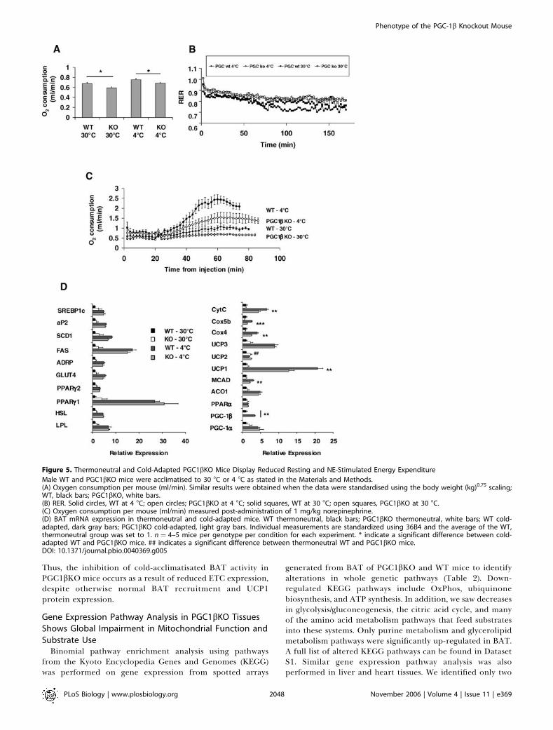
Thus, the inhibition of cold-acclimatisated BAT activity inPGC1bKO mice occurs as a result of reduced ETC expression,despite otherwise normal BAT recruitment and UCP1protein expression.
Gene Expression Pathway Analysis in PGC1bKO Tissues
Shows Global Impairment in Mitochondrial Function and
Substrate UseBinomial pathway enrichment analysis using pathways
from the Kyoto Encyclopedia Genes and Genomes (KEGG)was performed on gene expression from spotted arrays
generated from BAT of PGC1bKO and WT mice to identifyalterations in whole genetic pathways (Table 2). Down-regulated KEGG pathways include OxPhos, ubiquinonebiosynthesis, and ATP synthesis. In addition, we saw decreasesin glycolysis/gluconeogenesis, the citric acid cycle, and manyof the amino acid metabolism pathways that feed substratesinto these systems. Only purine metabolism and glycerolipidmetabolism pathways were significantly up-regulated in BAT.A full list of altered KEGG pathways can be found in DatasetS1. Similar gene expression pathway analysis was alsoperformed in liver and heart tissues. We identified only two
Figure 5. Thermoneutral and Cold-Adapted PGC1bKO Mice Display Reduced Resting and NE-Stimulated Energy Expenditure
Male WT and PGC1bKO mice were acclimatised to 30 8C or 4 8C as stated in the Materials and Methods.(A) Oxygen consumption per mouse (ml/min). Similar results were obtained when the data were standardised using the body weight (kg)0.75 scaling;WT, black bars; PGC1bKO, white bars.(B) RER. Solid circles, WT at 4 8C; open circles; PGC1bKO at 4 8C; solid squares, WT at 30 8C; open squares, PGC1bKO at 30 8C.(C) Oxygen consumption per mouse (ml/min) measured post-administration of 1 mg/kg norepinephrine.(D) BAT mRNA expression in thermoneutral and cold-adapted mice. WT thermoneutral, black bars; PGC1bKO thermoneutral, white bars; WT cold-adapted, dark gray bars; PGC1bKO cold-adapted, light gray bars. Individual measurements are standardized using 36B4 and the average of the WT,thermoneutral group was set to 1. n ¼ 4–5 mice per genotype per condition for each experiment. * indicate a significant difference between cold-adapted WT and PGC1bKO mice. ## indicates a significant difference between thermoneutral WT and PGC1bKO mice.DOI: 10.1371/journal.pbio.0040369.g005
PLoS Biology | www.plosbiology.org November 2006 | Volume 4 | Issue 11 | e3692048
Phenotype of the PGC-1b Knockout Mouse

KEGG pathways, OxPhos, and ATP synthesis that weredecreased in all three tissues—BAT, heart, and liver (TableS3). This suggests a global role for PGC-1b in the main-tenance of normal gene expression encoding for mitochon-drial proteins.
Tissue Mitochondrial Activity, but Not IsolatedMitochondrial Function, Is Impaired in PGC1bKO SoleusMuscle
We assessed whether reduced expression of ETC geneswere associated with functional defects in permeabilisedsoleus muscle fibres (Figure 6). Both state 3 and state 4mitochondrial respiration were reduced in PGC1bKO fibres,suggesting reduced electron chain capacity and OxPhosactivity (Figure 6A). There was no change in the respiratorycontrol ratio (RCR; state 3/state 4), suggesting that thedifferences between WT and PGC1bKO mice were notrelated to intrinsic differences in mitochondria functionbut to reduced numbers of mitochondria per fibre. Inagreement with this, ATP synthesis was decreased inPGC1bKO fibres (Figure 6B), and a trend for decreasedATP/O ratio in PGC1bKO muscle fibres was also observed(Figure 6C). This suggested that PGC1bKO soleus fibres notonly produce less ATP than WT fibres did, but they were alsoless efficient at converting reduced substrate into ATP. Geneexpression analysis of 12-wk-old male soleus muscles demon-strated that PGC1bKO mice have reduced ETC expression(Cox4 and Cox5b), which corresponds to reduced soleusmitochondrial capacity (Figure S4). These results weresupported by a decreased mitochondrial volume fraction inthe soleus of PGC1bKO mice performed by stereologicalanalysis (WT versus PGC1bKO: 20.4 6 0.7% versus 16.8 6
0.8%, p ¼ 0.012) (Figure 6D).To assess whether this oxidative defect could be attributed
to the functionality of individual mitochondria, we measuredoxidative activity of isolated soleus mitochondria. In all states
examined, there was no significant difference between WTand PGC1bKO-derived soleus mitochondria (Figure S5). Inaddition, no differences amongst genotypes were observedwhen RCR (Figure S5B) or proton leak were measured (FigureS5C).
PGC-1b Ablation Reduces Mitochondrial Content of the
Heart and Blunts the Effect of Adrenergic Stimulation on
Heart RateWe hypothesized that defective PGC-1b may result in
electromechanical myocardial dysfunction in the hearts ofPGC1bKO mouse. Heart weights from PGC1bKO mice weresimilar to WT in chow-fed 14-wk-old male mice (Table S2). Inaddition, under basal conditions, PGC1bKO hearts showedno histological abnormalities compatible with fibrosis or
Table 2. Major KEGG Pathways Are Changed in PGC1bKO MouseBAT
Pathway Type Pathway Name p Value
DOWN Oxidative phosphorylation 0.000
Citrate cycle (TCA cycle) 0.000
ATP synthesis 0.001
Pyruvate metabolism 0.015
Galactose metabolism 0.002
Cysteine metabolism 0.001
Reductive carboxylate cycle (CO2 fixation) 0.000
Tyrosine metabolism 0.002
Ubiquinone biosynthesis 0.001
Glycolysis gluconeogenesis 0.004
Phenylalanine metabolism 0.001
Arginine and proline metabolism 0.004
Starch and sucrose metabolism 0.001
Phenylalanine tyrosine and tryptophan biosynthesis 0.003
UP Glycerolipid metabolism 0.042
Purine metabolism 0.046
RNA was isolated from 14-wk-old normal diet-fed male PGC1bKO and WT mice and thenanalysed using spotted arrays as described in the Materials and Methods. Data aredisplayed as pathway name followed by p value for the binomial test. A pathway p valueof ,0.05 was considered significant.DOI: 10.1371/journal.pbio.0040369.t002
Figure 6. Isolated Soleus Fibres from PGC1bKO Mice Have Reduced
Mitochondrial Activity
Soleus fibres were isolated from WT (black bars) and PGC1bKO mice(white bars) and permeabilised to allow measurement of tissue-associated mitochondrial function.(A) Mitochondrial respiratory parameters for state 2 (V0), state 3 (VADP),state 4 (Voligomycin), and respiratory control ratio (RC).(B) ATP synthesis rates in permeabilised soleus fibres.(C) ATP/O ratio in permeabilised soleus fibres. Data are standardised tomg of muscle dry weight (mgdw). n¼ 9 WT mice, 11 PGC1bKO mice.(D) A representative electron micrograph of soleus muscle from WT (leftpanel) and PGC1bKO (right panel) mice.DOI: 10.1371/journal.pbio.0040369.g006
PLoS Biology | www.plosbiology.org November 2006 | Volume 4 | Issue 11 | e3692049
Phenotype of the PGC-1b Knockout Mouse

inflammatory infiltrates (unpublished data). The expressionof molecular markers of cardiac hypertrophy, such as a-actin,natriuretic peptide precursor type B, or troponin I, were all normalin 24-wk-old male PGC1bKO hearts (Figure 7A). The onlytranscriptional change that can be associated with myocardialdysfunction was the up-regulation of skeletal muscle actin inPGC1bKO compared with WT mice. PGC-1a mRNA expres-sion was also unchanged in PGC1bKO hearts. PGC1bKOhearts had a decreased mitochondrial volume fraction asindicated by stereological analysis (42.8 6 2.8 % versus 35.36 0.3 %, p ¼ 0.05: Figure 7B). Despite this result, PGC1bKOand WT heart mitochondria were similarly packed betweenmyofibres, and there were no changes in cristae surfacedensity, Sv (per unit volume of mitochondria). This suggeststhat the internal structure of the mitochondria was normal inthe PGC1bKO hearts. In agreement with the gene expressionpathway analysis, the expression of Cox4 (p ¼ 0.07) and Cox5bwas down-regulated in heart of 14-wk-old PGC1bKO com-pared with WT mice, whereas Cox2 expression was unchanged(Figure 7C).
Male, 26-wk-old PGC1bKO and WT littermates werechallenged with acute infusion of the b1,a1-adrenergicselective agonist dobutamine. Haemodynamic recordings
showed that baseline heart rate and left ventricular contrac-tility (þdP/dt or –dP/dt) were similar between genotypes. Asexpected during infusion of 10 ng dobutamine per min per gbody weight (ng/min/g BW), WT mice increased their heartrate, an effect that was blunted in PGC1bKO mice (Figure8A). This inability to increase heart rate was even moreevident using a 40 ng/min/g BW dobutamine infusion (p, 0.05for 8, 10, and 12 min post-injections). Ventricular perform-ance during the dobutamine challenge was the same in WTand PGC1bKO mice (Figure 8B and 8C).
PGC-1b Modulates the Response of the Liver to an AcuteHFD ChallengeGene expression analysis of fed-state 12-wk-old male mouse
livers showed ablation of PGC-1b but did not result in majorchanges in mRNA expression levels (Figure S6). Only Cox4 wasdecreased in the PGC1bKO livers, a result that was inagreement with gene expression pathway analysis (TableS3). When 8-wk-old female PGC1bKO mice were challengedwith a 24-h period of Surwit HFD, liver mass increased as aproportion of body weight (Figure 9A). This effect was notobserved in WT mice fed the same diet. Histological analysisrevealed that PGC1bKO mice fed the Surwit diet developedsevere hepatic steatosis compared to WT mice (Figure 9B).
Figure 7. Alterations in Gene Expression and Mitochondrial Dimensions in Hearts of PGC1bKO Mice
(A) Expression levels of mRNA were assessed on hearts from 24-wk-old male WT (black bars) and PGC1bKO (white bars) mice. Individual measurementsare standardized using 18S, and the average of the WT group set to 1. n¼ 5–7 mice per group.(B) A representative electron micrograph of mitochondria from WT (left panel) and PGC1bKO (right panel) hearts. The bar indicates a measurement of200 nm.(C) mRNA expression of key genes for mitochondrial function in 24-wk-old WT and PGC1bKO mouse hearts.(D) mRNA expression of key genes for metabolic function in 24-wk-old male WT and PGC1bKO mouse hearts.DOI: 10.1371/journal.pbio.0040369.g007
PLoS Biology | www.plosbiology.org November 2006 | Volume 4 | Issue 11 | e3692050
Phenotype of the PGC-1b Knockout Mouse

Increased fat deposition in PGC1bKO livers was associatedwith normal plasma non-esterified fatty acids (NEFA) levels(Table 3). As expected, plasma triglyceride levels increased inWT mice in response to 24-h HFD, however plasmatriglyceride levels were significantly lower in PGC1bKO thanWT mice when fed the Surwit diet. When compared to WTmice, the PGC1bKO mice had lower basal levels of high-density lipoprotein (HDL) and LDL-associated cholesteroland elevated VLDL-associated cholesterol. Administration ofa fat-enriched diet increased the amount of cholesterolassociated with VLDL, HDL, and LDL particles in WT mice(Table 3). However, PGC1bKO mice had inappropriately lowlevels of VLDL and blunted increases on total cholesterol andHDL- and LDL-associated cholesterol in plasma. Thus,defective PGC-1b results in lipid accumulation in liver anddecreased circulating triglyceride-rich VLDL lipoproteinsfollowing a high-dietary lipid load.
Of interest and contrary to previous reports, we wereunable to detect increases in PGC-1a and PGC-1b mRNAs inresponse to acute HFD in WT liver (Figure S7). We observedthat SCD-1 and HMG-CoA reductase were significantly elevatedin WT livers in response to Surwit diet compared to chow WTcontrols and that similar changes were not observed inPGC1bKO mouse livers after the Surwit diet (Figure S7).
Discussion
Accumulating evidence indicated that PGC-1b may play arole in energy homeostasis through its effects on substratemetabolism and mitochondrial activity [4,6,7,13–15,19].However, the function and relevance of PGC-1b in thecontrol of whole-organism energy metabolism is not well
defined. To address this question, we generated andphenotyped a PGC1bKO mouse model. The PGC1bKOmouse is viable and apparently healthy. However, using aphenotyping strategy that combines physiological stresschallenges guided by information obtained from a systemsbiology approach, we found that PGC1bKO mice have ageneral defect in mitochondrial function, a defect that ispartly compensated for by mechanisms that are usuallyinvolved in adaptation to increased energy demands, such asup-regulation of PGC-1a.The effect of PGC-1b controlling mitochondrial activity
was investigated at the mRNA level using spotted arraytechnology and bioinformatics pathway analysis. This ap-proach showed that deletion of PGC-1b results in a significantmitochondrial phenotype. Multi-tissue comparisons demon-strated that PGC-1b controls the level of expression of ETCand OxPhos genes across all the organs studied and that thisdefect can only be partially compensated for in BAT andWAT by up-regulation of PGC-1a. However, the decrease inmRNA expression of ETC components is not necessarily seenat the protein level. These data suggest that although PGC-1bmay be a controller of mitochondrial gene expression, in theabsence of PGC-1b, additional factors such as proteindegradation may be counter-regulated in specific tissues inan attempt to normalise levels of mitochondrial activity.Our initial hypothesis was that dysregulation of ETC gene
Figure 8. PGC1bKO Hearts Display a Blunted Heart Rate Response to
Dobutamine Stimulation In Vivo
PGC1bKO and WT littermates (male, 26-wk-old) were treated as stated inMaterial and Methods and infused with 10 or 40 ng/min/g BWdobutamine to measure in vivo hemodynamic responses WT, solid line;KO, dashed line.(A) Percentage change in heart rate from basal during dobutamineinfusion.(B and C) Measurement of ventricular performance, dP/dt, duringinfusion. The arrow marks the increase in dobutamine concentration inthe infusion from 10 to 40 ng/min/g BW. n¼ 5 mice per genotype.DOI: 10.1371/journal.pbio.0040369.g008
Figure 9. PGC1bKO Mice Demonstrate Increased Liver Mass and
Development of Fatty Liver after 24 h HFD
Female 8-wk-old mice were given normal chow or Surwit HFD (Sur) for24 h. Tissues were then collected for analysis.(A) Liver weight, as standardized by body weight (LW/BW) for WT (blackbars) and PGC1bKO (white bars) after 24 h diets.(B) Representative histological sections from mice given normal or Surwitdiet for 24 h. n¼ 6–7 mice per group.DOI: 10.1371/journal.pbio.0040369.g009
PLoS Biology | www.plosbiology.org November 2006 | Volume 4 | Issue 11 | e3692051
Phenotype of the PGC-1b Knockout Mouse

expression would increase the likelihood that the PGC1bKOmouse would become obese. However, despite its mitochon-drial phenotype, the PGC1bKO mouse showed elevatedresting metabolic rate and lower body weight compared towild type littermates under ambient room temperatureconditions. This phenotype is possibly due to the compensa-tory increase in expression of PGC-1a and its target genes inBAT. The increase in BAT PGC-1a expression may cause thehigher degree of energy expenditure and relatively conservedmitochondria volume fraction despite reductions in ETCgene and protein expression. PGC-1a was also up-regulatedin PGC-1b–deficient WAT, but its induction was not robustenough to produce a BAT-like histology or induction oftypical BAT genes. Instead, within the reduced WAT contentof the PGC1bKO mice, there was a greater proportion oflarger, hypertrophic adipocytes. This result suggests thatPGC-1b may play an as yet unknown role in white adipocytebiology, and the nature of this role requires furtherinvestigation.
Of interest, despite marked elevation of PGC-1a levels inPGC1bKO BAT, we did not observe a full restoration of theexpression of mitochondrial genes back to WT levels.However, unlike the heart or skeletal muscle, organs wherePGC-1a was not up-regulated, the mitochondrial fraction ofBAT tissue was preserved in PGC1bKO mice. Thus, there maybe partial functional overlap between PGC-1a and PGC-1b,concerning mitochondrial structure and function, whichallows the development of an appropriate mitochondrialBAT content but fails to correct deficiencies in ETCcomposition. BAT was the only tissue out of three metabol-ically relevant tissues examined using electron microscopythat displayed relatively normal mitochondrial fraction,suggesting that further tissue function–specific mechanismsmay be able to correct for PGC-1b ablation. However, theBAT gene expression pattern in PGC1bKO mice also showedaltered expression of genes involved in intermediary metab-olism, a pattern of gene expression that may also be primarilythe result of PGC-1a up-regulation. Our conclusion fromthese gene expression analyses is that whereas PGC-1b may berequired to set the basal level of mitochondrial ETC geneexpression, it is not essential for normal expression ofmetabolic pathways such as glycolysis, the citric acid cycle,and fatty acid oxidation.
We hypothesized that the elevated resting metabolic rate
seen in ambient-temperature housed PGC1bKO mice may bereversed under conditions of thermoneutrality, particularly ifit was caused by compensatory up-regulation of PGC-1a.Under thermoneutral conditions, levels of PGC-1a geneexpression are suppressed in BAT in response to the lowerthermogenic and therefore oxidative demands of life at 30 8C.At thermoneutral conditions, PGC-1a mRNA expression wasequivalent in WT and PGC1bKO BAT. Interestingly, underthermoneutral conditions, the PGC1bKO mouse exhibitedreduced basal metabolic rate compared to the WT mouse.The energy expenditure measurements with mice acclima-tised to 4 8C and 30 8C were performed at 33 8C. Thismeasurement at thermoneutrality gives an indication of thebasal metabolic rate. Under these experimental conditions,we can conclude that the PGC1bKO mouse has a reducedbasal metabolic rate. The data from mice housed in ambient(22 8C) conditions were also generated at ambient conditions(Table 1). Therefore, this measurement represents energyexpenditure in ‘‘normal’’ environmental conditions, when thebody weights of the PGC1bKO mice are lower than WT.Under these experimental conditions, this measurement iscomposed of both basal metabolic rate and any additionalmetabolic effort required to maintain body temperature inthis suboptimal thermal environment. Given that basalmetabolic rate is reduced in 30 8C– and 4 8C–acclimatisedPGC1bKO mice, it could be assumed that at ambienttemperatures, basal metabolic rate is also reduced, but thatthe metabolic adaptation required for body temperaturedefense is actually greater in the PGC1bKO mouse. Thissuggests that PGC-1b may contribute to BAT thermogenesisin states of low metabolic demand (as is the case for BAT at 308C), whereas induction of PGC-1a regulates BAT thermo-genesis as environmental temperatures fall.It has been suggested that PGC-1b plays a role in BAT
accumulation and function during cold exposure [6].PGC1bKO mice tolerated cold acclimatisation, despite thefact that under these conditions, PGC1bKO mice haddecreased ETC gene expression and had decreased maximalthermogenic capacity in BAT, as shown by the lower oxygenconsumption rate in response to adrenergic stimulation.Both WT and PGC1bKO mice up-regulated PGC-1a to asimilar extent under conditions of cold exposure, suggestingthat there is a maximal capacity of PGC-1a up-regulation inBAT. Alternatively, survival in cold may also be facilitated by
Table 3. Serum Lipid Biochemistry from 24-h Surwit Diet–Fed WT and PGC1bKO Mice
Lipid WT - Chow WT - Surwit PGC1bKO - Chow PGC1bKO - Surwit
Triglycerides (mM) 0.39 6 0.03 a 0.66 6 0.12 a,b 0.21 6 0.04 0.35 6 0.03 b
NEFA (mM) 0.64 6 0.05 0.82 6 0.12 0.60 6 0.05 0.71 6 0.07
Cholesterol (mM) 3.34 6 0.20 a,b 5.37 6 0.24 a,c 2.20 6 0.23 b,d 3.45 6 0.30 c,d
VLDL (mM) 0.026 0.049 0.037 0.025
HDL (mM) 0.29 0.46 0.23 0.35
LDL (mM) 3.06 4.86 1.93 3.07
Female WT and PGC1bKO mice were placed on Surwit diet for 24 h or maintained on normal diet. At the end of the time period, the mice were sacrificed and blood serum analysed. n¼5–7 mice per condition. For cholesterol-associated lipoproteins, serum samples for each group were pooled and analysed.aSignificant comparisons between groups.bSignificant comparisons between groups.cSignificant comparisons between groups.dSignificant comparisons between groups.DOI: 10.1371/journal.pbio.0040369.t003
PLoS Biology | www.plosbiology.org November 2006 | Volume 4 | Issue 11 | e3692052
Phenotype of the PGC-1b Knockout Mouse

the ability of the mice to maintain some fraction of increasedmetabolic activity through continued shivering [22–24].Therefore, although the total metabolic capacity providedby PGC-1a in cold-exposed PGC1bKO mice was sufficient fortheir survival, our results indicate that PGC-1b regulates alarge fraction of the thermogenic capacity of the BAT tissue.
Studies focused in other metabolically relevant organswhere PGC-1b is well expressed also indicated that ablationof PGC-1b resulted in impaired metabolic capacity. Forexample, mitochondrial volume fraction in soleus ofPGC1bKO mice was decreased, and permeabilised soleusmuscle fibres from PGC1bKO mice had reduced oxygenconsumption and ATP synthesis compared to WT-derivedfibres. Similar experiments using isolated soleus mitochon-dria from PGC1bKO mice failed to show any abnormalities inmetabolic performance, indicating that metabolic defects inPGC1bKO soleus tissue relate to lower mitochondrial densityrelative to WT, although individual mitochondria retainsimilar metabolic properties.
The heart is the third oxidative tissue that is a major site ofPGC-1b expression. Absence of PGC-1b in heart also led to areduction of mitochondrial fraction, consistent with reducedcardiac expression of ETC genes. However, despite thesepotential bioenergetic defects, PGC1bKO mice have normalbasal heart rates and ventricular contractility. Left ventric-ular function was also maintained after dobutamine treat-ment, suggesting that despite their mitochondrial defect, thePGC-1b–deficient hearts are able to adapt normally toimposed hemodynamic loads. The only difference in adre-nergic stress that we observed between the PGC1bKO miceand WT was a blunting of the expected increase in heart ratein the PGC1bKO mice. A similar defect in heart rateregulation was also found in the PGC-1a knockout mice[14], suggesting that both PGC-1a and PGC-1b may have adirect effect in controlling the activity of heart pacemakers.Overall, our results indicate that ablation of PGC-1b impairsheart mitochondrial function but that this defect is not severeenough to induce heart failure. However, a more chronicintervention study would be required to establish the relativeimportance of these PGC isoforms in cardiac energy supplyand control.
To this point, we have discussed exclusively a role for PGC-1b in oxidative metabolism. Nonetheless, PGC-1b may alsoplay an important role in regulating hepatic lipid production[11,17]. Contrary to previous reports, we did not observeeither PGC-1b induction after 24-h HFD in the liver of ourcontrol mice or differences in FAS and SREBP1c geneexpression between controls and PGC1bKO mice. However,we did observe a reduction in circulating total cholesteroland alterations in the lipoprotein-associated cholesterolpattern in chow-fed conditions. Feeding for 24 h with asaturated fat–enriched HFD was associated with severehepatic lipid accumulation and decreased total triglycerides,cholesterol, and VLDL and LDL-cholesterol plasma levels inPGC1bKO mice compared to WT. These results agree with arecent report suggesting that PGC-1b is a likely mediator ofVLDL secretion by acting via interactions with Foxa2 to altermicrosomal transfer protein expression [17]. However, it isalso possible that PGC-1b is necessary for additional aspectsof hepatic lipid handling, including modification of lipidstorage pathways and for controlling the balance betweenfatty acid synthesis and oxidation. Indeed, our pathway
analysis suggests that the ETC in liver is expressed at a lowerlevel and this may contribute to the steatosis after acute HFDtreatment.Our results indicate that PGC-1b has a well-defined role in
controlling mitochondrial gene expression and function inmany different organs. Despite this, PGC-1b can be ablatedwithout overt metabolic failure, at least in unstressedconditions. This may be due in part to robust compensatorymechanisms, as demonstrated by the up-regulation of PGC-1a expression in WAT and BAT. Interestingly, other organssuch as liver, muscle, or heart did not show up-regulation ofPGC-1a under the conditions investigated. Following fromthese differences in PGC-1a expression, it is likely that thelean phenotype of this mouse model at ambient temperatureshould be considered the result of overcompensationmediated by up-regulation of PGC-1a, at least in BAT andWAT. Conversely, defects observed in skeletal muscle, heart,and liver are more likely to be the result of the absence ofPGC-1b given the lack of PGC-1a induction in those tissues.When considering the roles played by PGC-1a and PGC-1b,our results show that there are specific effects of PGC-1b thatcannot be compensated for by PGC-1a. Taken altogether, ourresults indicate that PGC-1b seems to cover basal bioen-ergetic needs whereas PGC-1a provides the extra bioener-getic support required under conditions of increased energydemand.
Materials and Methods
Materials and reagents. All reagents used in this paper weresupplied from Sigma-Aldrich (St. Louis, Missouri, United States),unless stated.
Animal care. Animals were housed in a temperature-controlledroom with a 12-h light/dark cycle. Food and water were available adlibitum unless noted. All animal protocols used in this study wereapproved by the UK Home Office, The Institutional Animal Care andUse Committee of the University of Utah, United States, and theAnimal Ethics Committees of Gothenburg and North Stockholm,Sweden. Mice were cared for according to the Guiding Principles forResearch Involving Animals and Human Beings.
Generation of PGC1bKO mice. A triple LoxP strategy was used totarget the PGC-1b locus in order to generate mice with bothstandard and conditional KO alleles at this locus. The targetingvector was a ;8 kilobase (kb) 129/SvJ mouse genomic subclonecontaining a floxed neomycin phosphotransferase selectable markercassette inserted into intron 3 and a single LoxP site inserted intointron 5 (Figure 1A). The targeting construct was electroporatedinto R1 ES cells, and neomycin-resistant clones were selected in G-418-containing media. The clone was injected into C57Bl/6blastocysts and chimeric males were crossed to C57Bl/6 females.Heterozygous triple LoxP mice were then bred to ROSA26Cre [25]mice in order to generate heterozygous PGC-1b KO mice which hadundergone Cre-mediated deletion of the intervening region of DNAbetween the outermost LoxP sites. PCR analysis gave a product ofapproximately 0.5 kb for the Cre-recombined allele and products ofapproximately 2.8 and 0.6 kb with the WT allele (Figure 1D).Heterozygous PGC-1b KO mice were then intercrossed to generatemice that were homozygous for the PGC-1b deletion. Null mutantswere verified using RNA prepared from heart and skeletal muscle todemonstrate that exons 4 and 5 had been deleted in the PGC1bKOmice. Mice used for this research were backcrossed between threeand six times to a C57BL6/J background. Littermate controls wereused for all experiments. A more complete description for thissection can be found in the Protocol S1.
Long-term feeding and growth studies. Mice were placed atweaning (3-wk-old) on normal chow diet (12% fat, 62% carbohy-drates, and 26% protein with a total energy content of 12.6 kJ/g) (R3diet, Lactamin AB, Stockholm, Sweden). Mouse weights were taken atthe same time each week, until the end of the specific protocolperiod. Mice were routinely housed at 22 8C, except for those used forthe BAT activity and cold acclimatisation experiments (see below).
PLoS Biology | www.plosbiology.org November 2006 | Volume 4 | Issue 11 | e3692053
Phenotype of the PGC-1b Knockout Mouse

Acute dietary measurements and interventions. To examine 48-hfood intake, cages (233 16 cm) were prepared with normal chow andincubated at 80 8C for 1 h to correct for any differences in humidity.After 2 h at room temperature, the cages were accurately weighed.12-h-fasted mice were put in preweighed cages with free access tofood and water. After 48 h, the mice were removed and all faecalmatter was collected. The cages were reincubated at 80 8C in order todry out waterspill and urine, and then reweighed after 2 h cooling.The difference in weights of the cage before and after the 48-hassessment produced the weight of food consumed. For measurementof energy content of faeces and food, samples were dried at 55 8Covernight and stored in airtight containers at �20 8C until assayed.The gross energy content of the dried samples was determined usinga bomb calorimeter (C5000, IKA Werke GmbH & Co., KG, Germany).To assess the effect of 24-h high-fat feeding, 8-wk-old female micewere assigned into two groups: ad libitum normal food or ad libitumSurwit diet (58% of calories derived from fat, predominantlyhydrogenated coconut oil; D12331, Research Diets, New Brunswick,New Jersey, United States). Start of the 24-h period was 9 am. At theend of the time period, mice were killed and dissected as above.Water was freely available during all procedures.
Body composition and indirect calorimetry. For body compositionanalysis, dual energy x-ray absorptiometry (DEXA, GE MedicalSystems Lunar Corporation, Madison, Wisconsin, United States) wasperformed on isoflurane anaesthetized mice as previously described[26]. Oxygen consumption (VO2) and carbon dioxide production(VCO2) were measured using the Oxymax system (Columbus Instru-ments International, Columbus, Ohio, United States) [27]. Furtherdetails can be found in Protocol S1.
GTTs and ITTs on mice fed chow and HFDs were performed aspreviously described [26].
Cold acclimatisation and BAT activity. PGC1bKO mice and WTlittermates were exposed to 30 8C for 3 wk, placed at 18 8C for 1 wk,and then exposed to 4 8C for 3 wk or kept at 30 8C for the duration ofthe protocol. For both 30 8C and 4 8C acclimated animals, restingmetabolic rate was measured in awake animals at 30 8C. In addition,NE-stimulated (1 mg NE/kg body weight in saline, (�) arterenolbitartrate, intraperitoneally administered) energy expenditure wasevaluated in anaesthetized (pentobarbital, 90 mg/kg) animals at 33 8C.Animals were allowed to recover from NE administration for 2–3 h at30 8C. The mice where then rehoused at their acclimatisationtemperature for 1 wk prior to tissue collection for gene and proteinexpression analysis to avoid effects of NE on gene expression. Oxygenconsumption in conscious animals was followed for 3 h using an opencircuit system with a chamber volume of 3 l and a flow rate of 1 l/min(Somedic, Horby, Sweden). This system allowed the ambient temper-ature of the instruments to be adjusted between 5 8C and 40 8C,together with the volume and flow rates to optimise the system forthe particular investigation. Oxygen consumption, carbon dioxiderelease, and ambient temperature data were collected every secondminute via MacLab/2e (AD Instruments Pty. Ltd., Castle Hill,Australia). Resting metabolic rate was defined as the average of thelowest metabolic rates observed at three time points. Determinationsin WT and PGC1bKO mice were performed in alternating order.
Catherization and dobutamine treatment. Mice were anesthetizedwith isoflurane and underwent endotracheal intubation. The airwaywas connected to mouse ventilator (Model 687, Harvard Apparatus,Holliston, Massachusetts, United States) to control breathing. Theoxygen flow rate was 1 l/min. The left jugular vein was identified andaccessed by cut down method using a 25 G needle connected to asyringe with dobutamine hydrochloride (Sigma) that was mounted ona Standard Infuse/Withdraw Harvard 33 Twin Syringe Pump(Harvard Apparatus). A micromanometer-tipped catheter (MillarInstruments, Houston, Texas, United States) was then inserted intothe left ventricle via right carotid artery, and hemodynamicmeasurements was obtained as described [28]. After obtainingbaseline left ventricular pressure and heart rate readings, thedobutamine infusion was commenced. The initial infusion rate was10 ng/min/g BW, with hemodynamic recordings taken at 2, 4, and 6min. The infusion rate was then increased to 40 ng/min/g BW, andadditional readings were obtained at 2, 4, and 6 min after the doseadjustment.
Tissue collection and RNA extractions. Mice were anaesthetisedusing isoflourane. Blood, tissues used for RNA, protein extraction,and histology were prepared as previously published [26,29].
Biochemical analysis. Enzymatic assay kits were used for determi-nation of basic blood biochemical parameters as described in [26,29].The size distribution profiles of serum lipoproteins were measured inpooled plasma samples using a high-performance liquid chromatog-
raphy system (HPLC), SMART, and a Superose 6 PC 3.2/30 column asdescribed before [30].
Histological sample preparation and analysis. Tissue samples formorphological analysis were prepared according to publishedprotocols [29]. For light microscopy, sections were stained withhaematoxylin and eosin. For adipose tissue, images of each sectionwere acquired using a digital camera and microscope (OlympusBX41, Olympus Corporation, Tokyo, Japan) and adipocyte area wasmeasured using AnalySIS software (Soft Imaging System, Munster,Germany). Two fields from each section from gonadal, subcuta-neous, and omental adipose tissue depots (n ¼ 7–8 mice pergenotype) were analysed to obtain the mean cell area per animal.For preparation of BAT, soleus muscle, and hearts for electronmicroscopy, mice were exsanguinated by perfusion with physiolog-ical saline containing 0.1% sodium nitrate until no blood was left.Mice were then perfused with 60–90 ml of fixative (3% glutaralde-hyde and 1% formaldehyde in 0.1 mol/l 1,4-piperazine diethanesulfonic acid (PIPES) buffer (pH 7.4) containing 2 mol/l calciumchloride). Isotropic uniform random planes of section through theleft ventricular wall in hearts were prepared using the orientatorprinciple as described in [31]. Soleus samples were prepared atequal lengths along the long axis of the muscle. The blocks of tissuewere sectioned in small fragments with a razor blade to ,1 mm inone dimension and fixed by immersion at 4 8C for a further 3–4 h.Samples were then washed in 0.1 M PIPES and post-fixed in 1%osmium tetroxide, dehydrated in acetone, and embedded in Spurr’sepoxy resin. Thin sections were obtained with a Leica UCTultramicrotome (Leica Microsystems, Milton Keynes, United King-dom) and examined with a transmission electron microscope(CM100, Philips, Netherlands).
Stereological assessment of mitochondria. Stereological assess-ments of mitochondrial volume fractions (Vv) and the surface density(SV) of their inner and outer membranes in heart were performed asdescribed in [31] and in [32].
Permeabilised tissue and isolated mitochondrial respirationstudies. Isolation of mitochondria was performed in hearts andskeletal muscle from 10-wk-old male PGC-1b mice and their WTlittermates. Mice were killed by cervical dislocation. The hearts orhind limb skeletal muscle of four mice per genotype were pooledand immediately placed in ice-cold isolation medium (for hearts,250 mM sucrose, 5 mM Tris, 2 mM EGTA at pH 7.4; for skeletalmuscle, 100 mM KCl, 50 mM Tris, 2 mM EGTA at pH 7.4).Mitochondria were prepared essentially as described in [33].Mitochondrial respiration was assessed in saponin-skinned soleusfibres prepared as in [14,34].
Measurement of proton conductance and ETC function in isolatedmitochondria. The kinetics of proton conductance and ETC functionwere measured in mitochondria in the presence of oligomycin (1 lg/ml), where that rate of respiration is directly proportional to the leakof protons across the mitochondrial inner membrane rather than acombination of ADP phosphorylation and proton leak. Mitochon-drial function was assessed as described previously [35,36].
Quantitative RT-PCR analysis of gene expression. Total RNA wasisolated from tissues as described and reverse-transcribed using theSuperscriptII kit (Invitrogen, Frederick, Maryland, United States) orHi-Capacity cDNA archive kit (Applied Biosystems, Foster City,California, United States), following the manufacturers protocol.Oligonucleotide primers and TaqMan probe were designed usingPrimer Express, version 2.0 (Applied Biosytems). Primer and probesequences, together with gene abbreviations, can be found in TablesS4–S6.
Microarray analysis. Male 14-wk-old mice were killed and dissectedas above. The tissues were extracted for RNA as above and purifiedusing the RNA clean-up protocol from the RNeasy Mini Kit (QiagenLtd, Crawley, UK). RNA was quantified spectroscopically at 260 nmusing a GeneQuant Nucleotide calculator (Amersham Biosciences,Little Chalfont, UK) and checked for integrity on a 1% TBE gel usingethidium bromide staining. cDNA was amplified from total RNAusing template-switching PCR and labeled with Cy3 or Cy5 dyes aspreviously described [37]. Binomial pathway enrichment analysis wasthen performed. A more detailed protocol can be found in ProtocolS1. Raw data from the analysis are presented in Dataset S1 (pathwaycomparison) and Dataset S2 (raw data from the chip analysis).
Mitochondrial isolation, protein extraction, and Western analysis.Female 15-wk-old chow-fed mice were anaesthetised using isofluraneand killed by cervical dislocation and heart removal. BAT, heart,and soleus muscle tissue was removed and either snap-frozen inliquid nitrogen or used to prepare a mitochondrial extract asdescribed in Protocol S1. In general, 10 lg of total lysate or 3 lg ofmitochondrial preparation was separated by SDS-PAGE on 14%
PLoS Biology | www.plosbiology.org November 2006 | Volume 4 | Issue 11 | e3692054
Phenotype of the PGC-1b Knockout Mouse

gels. After transfer, the membranes were cut and placed in blockingbuffer (5% powdered milk in 13 PBS and 0.1% Tween-20). We usedanti-OxPhos complex I (a-subcomplex 9) and anti-OxPhos complexIII (Fe-S core protein) (Molecular Probes, Carlsbad, California,United State) and anti-succinate dehydrogenase subunit B, anti-Cox4, and anti-ATP synthase b-subunit from Abcam, Cambridge,UK. All primary antibodies were used at ratios of 1:2,000, exceptanti-Cox4, which was used at 1:20,000. Goat anti-mouse horseradishperoxidase secondary antibody (Pierce, Rockford, Illinois, UnitedStates) was used at 1:40,000 for all blots, the membranes werereassembled, and the proteins detected using the ECL-Plus system(Amersham Biosystems). Films were scanned and analysed using NIHImage 1.34s.
Statistical analysis. The data presented here were analysed usingthe Student t-test or analysis of variance (ANOVA) with post-hoc testson the program StatView Version 4.5 (Abacus Concepts, Berkley,California, United States). Data are presented as mean 6 SEM unlessstated. p , 0.05 was considered significant. The significance levelsdisplayed on figures are as follows: * indicates p, 0.05, ** indicates p, 0.01, *** indicates p , 0.001.
Supporting Information
Dataset S1. Data Used for the Comparative Pathway Analysis of BAT,Heart, and Liver Tissues Using KEGG and KEGG2 Pathways
Found at DOI: 10.1371/journal.pbio.0040369.sd001 (62 KB XLS).
Dataset S2. Raw Data Text Files from the Spotted Array Experimentson Heart, Liver, and BAT Used in the Comparative Pathway Analysis
Found at DOI: 10.1371/journal.pbio.0040369.sd002 (3.2 MB XLS).
Figure S1. Size Distribution of Adipocytes from WT and PGC1bKOMice
Two fields from each section from (A) omental WAT, (B) subcuta-neous WAT, and (C) female gonadal WAT depots (age 24 wk, n¼ 7–8mice per genotype) were analysed to obtain the mean cell area peranimal.
Found at DOI: 10.1371/journal.pbio.0040369.sg001 (43 KB PPT).
Figure S2. Histological Sections of Tissues
Sections are as follows: BAT (A and B), liver (C and D), soleus muscle(E and F), pancreatic sections stained for glucagon (G and H), orpancreatic sections stained for insulin (I and J). A representativesection fromWT (A, C, E, G, and I) or PGC1bKO (B, D, F, H, and J) areshown, n¼6.Found at DOI: 10.1371/journal.pbio.0040369.sg002 (1.7 MB PDF).
Figure S3. Deletion of PGC-1b Does Not Affect Insulin Sensitivity
Mice were placed on normal diet or HFD for 13 wk. GTTs (left panels)or ITTs (right panels) were performed on the mice at 16 wk of age.Plasma glucose levels of WT mice (open circles) and PGC1bKO (solidcircles) were measured during the protocol.
Found at DOI: 10.1371/journal.pbio.0040369.sg003 (115 KB PPT).
Figure S4. Expression of mRNAs Assessed on Soleus from 12-wk-OldMale WT (Black Bars) and PGC1bKO (White Bars) Mice
Individual measurements were standardised using 18S, and theaverage of the WT group set to 1. n ¼ 6 mice per group.
Found at DOI: 10.1371/journal.pbio.0040369.sg004 (34 KB PPT).
Figure S5. Assessment of Isolated Soleus Mitochondrial Functionfrom WT and PGC1bKO Mice
SoleustissuewaspooledfromfourWTmice(blackbars)orfourPGC1bKOmice(whitebars)andmitochondriaisolatedonfiveseparateoccasions(n¼5). Respiration states (A) were calculated using oxygen consumptionrates in isolated mitochondria energised with succinate (4 mM).Respiratorycontrolratios(B)weredeterminedformitochondriaasstate3dividedby state 4. Proton leak kinetics (C)weremeasuredas describedunderMaterials andMethods.
Found at DOI: 10.1371/journal.pbio.0040369.sg005 (35 KB PPT).
Figure S6. PGC-1b Ablation Results in Few Changes in GeneExpression in Liver
Gene expression was analysed in 12-wk-old fed-state male WT (blackbars) and PGC1bKO (white bars) livers. Individual measurements arestandardised using 18S and then the average of the WT group was setto 1. n ¼ 6 mice per group.
Found at DOI: 10.1371/journal.pbio.0040369.sg006 (36 KB PPT).
Figure S7. Gene Expression of PGC-1a and PGC-1b (A) and for aSelection of Genes Involved with Fatty Acid and CholesterolHandling in the Liver (B) in 8-wk-Old Female Mice Fed Surwit Dietfor 24 h
WT chow, black bars; WT Surwit HFD, white bars; PGC1bKO chow,dark gray bars; PGC1bKO Surwit HFD, light gray bars. Individualmeasurements are standardised using 36B4 and then the average ofthe WT chow group was set to 1. n ¼ 4–7 per condition.
Found at DOI: 10.1371/journal.pbio.0040369.sg007 (35 KB PPT).
Protocol S1. Supplementary Protocols
Found at DOI: 10.1371/journal.pbio.0040369.sd003 (99 KB DOC).
Table S1. WT and PGC1bKO Mouse Parameters for Energy Intakeand Faecal Output over 48 h
The food intake and energetic content of faeces from 12-wk-old malemice was determined over a 48-h period. n ¼ 5–8 per group.
Found at DOI: 10.1371/journal.pbio.0040369.st001 (21 KB DOC).
Table S2. WT and PGC1bKO Tissue Weights in 14-wk-Old Male Mice
The data are presented both comparing raw weights and as tissueweight/body weight. Statistical comparison between WT andPGC1bKO mice was performed using t-tests. n ¼ 6–8 per group. pvalues are shown where p is less than or equal to 0.2.
Found at DOI: 10.1371/journal.pbio.0040369.st002 (25 KB DOC).
Table S3. Comparison of KEGG Pathways Significantly Down-regulated in all Three Arrayed Tissues (Liver, BAT, and Heart)
RNA was isolated from 14-wk-old normal diet-fed male PGC1bKOand WT mice and then analysed using spotted arrays as described inthe Materials and Methods. Data from these chips were then analysedusing for pathway changes using the KEGG pathway database.Pathways with only one changed gene were discounted. Data aredisplayed as pathway name followed by p value for the binomial test.A pathway p value of ,0.05 was considered significant.
Found at DOI: 10.1371/journal.pbio.0040369.st003 (22 KB DOC).
Table S4. Sequences for Taqman Primers and Probes
Sybr as a probe indicates the use of a Sybr-Green system instead ofTaqman.
Found at DOI: 10.1371/journal.pbio.0040369.st004 (31 KB DOC).
Table S5. Sequences for Taqman Primers and Probes
Sybr as a probe indicates the use of a Sybr-Green system instead ofTaqman.
Found at DOI: 10.1371/journal.pbio.0040369.st005 (29 KB DOC).
Table S6. Sequences for Taqman Primers and Probes
Sybr as a probe indicates the use of a Sybr-Green system instead ofTaqman.
Found at DOI: 10.1371/journal.pbio.0040369.st006 (29 KB DOC).
Acknowledgments
The authors would like to thank all lab staff who performed mousebreeding and biochemical analyses at AstraZeneca and the Universityof Cambridge for this project. We wish to thank Jane Lofvenmark forinvaluable help during the early stages of the project. We would liketo thank the animal care staff at the institutions involved for theirwork on this project. We also thank Ian McFarlane, Lyn Carter andJeremy Skepper for providing technical help.
Author contributions. C. Lelliott, G. Medina-Gomez, M. Snaith, B.Cannon, andA.Vidal-Puig conceived and designed the experiments. C.Lelliott, G. Medina-Gomez, N. Petrovic, A. Kis, H. Feldmann, M.Bjursell, N. Parker, M. Campbell, P. Hu, D. Zhang, S. Litwin, V. Zaha, K.Fountain, S. Boudina, M. Jimenez-Linan, M. Blount, M. Lopez, A.Meirhaeghe, M. Bohlooly-Y, and M. Stromstedt performed the experi-ments. C. Lelliott, G. Medina-Gomez, N. Petrovic, A. Kis, H. Feldmann,N. Parker, K. Curtis, K. Fountain, S. Boudina, M. Jimenez-Linan, M.Blount, M. Oresic, E. Abel, B. Cannon, and A. Vidal-Puig analyzed thedata. K. Curtis, M. Bohlooly-Y, M. Snaith, and M. Oresic contributedreagents/materials/analysis tools. C. Lelliott, G. Medina-Gomez, M.Snaith, E. Abel, B. Cannon, and A. Vidal-Puig wrote the paper.
Funding. The work presented in this paper was supported by
PLoS Biology | www.plosbiology.org November 2006 | Volume 4 | Issue 11 | e3692055
Phenotype of the PGC-1b Knockout Mouse

grants from the British Heart Foundation, Wellcome Trust Integra-tive Physiology, and Diabetes Wellness Research Foundation (to AVPlab); NIH grants RO1HL73167 and UO1HL70525 from the NationalInstitutes of Health and the Ben and Iris Margolis Foundation (toEDA - Established Investigator of the American Heart Association).
Support was from the European Union (DLARFID), the SwedishResearch Council, and the Swedish Cancer Society (to BC lab).
Competing interests. The authors having affiliations to AstraZe-neca are paid employees of this company.
References1. Spiegelman BM, Heinrich R (2004) Biological control through regulated
transcriptional coactivators. Cell 119: 157–167.2. Puigserver P, Wu Z, Park CW, Graves R, Wright M, et al. (1998) A cold-
inducible coactivator of nuclear receptors linked to adaptive thermo-genesis. Cell 92: 829–839.
3. Yoon JC, Puigserver P, Chen G, Donovan J, Wu Z, et al. (2001) Control ofhepatic gluconeogenesis through the transcriptional coactivator PGC-1.Nature 413: 131–138.
4. Wu Z, Puigserver P, Andersson U, Zhang C, Adelmant G, et al. (1999)Mechanisms controlling mitochondrial biogenesis and respiration throughthe thermogenic coactivator PGC-1. Cell 98: 115–124.
5. Andersson U, Scarpulla RC (2001) Pgc-1-related coactivator, a novel,serum-inducible coactivator of nuclear respiratory factor 1-dependenttranscription in mammalian cells. Mol Cell Biol 21: 3738–3749.
6. Lin J, Puigserver P, Donovan J, Tarr P, Spiegelman BM (2002) Peroxisomeproliferator-activated receptor gamma coactivator 1beta (PGC-1beta ), anovel PGC-1-related transcription coactivator associated with host cellfactor. J Biol Chem 277: 1645–1648.
7. Meirhaeghe A, Crowley V, Lenaghan C, Lelliott C, Green K, et al. (2003)Characterization of the human, mouse and rat PGC1 beta (peroxisome-proliferator-activated receptor-gamma co-activator 1 beta) gene in vitroand in vivo. Biochem J 373: 155–165.
8. Kressler D, Schreiber SN, Knutti D, Kralli A (2002) The PGC-1-relatedprotein PERC is a selective coactivator of estrogen receptor alpha. J BiolChem 277: 13918–13925.
9. Lin J, Tarr PT, Yang R, Rhee J, Puigserver P, et al. (2003) PGC-1beta in theregulation of hepatic glucose and energy metabolism. J Biol Chem 278:30843–30848.
10. Kamei Y, Ohizumi H, Fujitani Y, Nemoto T, Tanaka T, et al. (2003)PPARgamma coactivator 1beta/ERR ligand 1 is an ERR protein ligand,whose expression induces a high-energy expenditure and antagonizesobesity. Proc Natl Acad Sci U S A 100: 12378–12383.
11. Lin J, Yang R, Tarr PT, Wu PH, Handschin C, et al. (2005) Hyperlipidemiceffects of dietary saturated fats mediated through PGC-1beta coactivationof SREBP. Cell 120: 261–273.
12. Lin J, Handschin C, Spiegelman BM (2005) Metabolic control through thePGC-1 family of transcription coactivators. Cell Metab 1: 361–370.
13. Lin J, Wu PH, Tarr PT, Lindenberg KS, St-Pierre J, et al. (2004) Defects inadaptive energy metabolism with CNS-linked hyperactivity in PGC-1alphanull mice. Cell 119: 121–135.
14. Leone TC, Lehman JJ, Finck BN, Schaeffer PJ, Wende AR, et al. (2005) PGC-1alpha deficiency causes multi-system energy metabolic derangements:muscle dysfunction, abnormal weight control and hepatic steatosis. PLoSBiology 3: e101. DOI: 10.1371/journal.pbio.0030101.
15. St-Pierre J, Lin J, Krauss S, Tarr PT, Yang R, et al. (2003) Bioenergeticanalysis of peroxisome proliferator-activated receptor gamma coactivators1alpha and 1beta (PGC-1alpha and PGC-1beta) in muscle cells. J Biol Chem278: 26597–26603.
16. Koo SH, Satoh H, Herzig S, Lee CH, Hedrick S, et al. (2004) PGC-1promotes insulin resistance in liver through PPAR-alpha-dependentinduction of TRB-3. Nature Med 10: 530–534.
17. Wolfrum C, Stoffel M (2006) Coactivation of Foxa2 through Pgc-1[beta]promotes liver fatty acid oxidation and triglyceride/VLDL secretion. CellMetab 3: 99–110.
18. Russell LK, Mansfield CM, Lehman JJ, Kovacs A, Courtois M, et al. (2004)Cardiac-specific induction of the transcriptional coactivator peroxisomeproliferator-activated receptor gamma coactivator-1alpha promotes mito-chondrial biogenesis and reversible cardiomyopathy in a developmentalstage-dependent manner. Circ Res 94: 525–533.
19. Lehman JJ, Barger PM, Kovacs A, Saffitz JE, Medeiros DM, et al. (2000)
Peroxisome proliferator-activated receptor gamma coactivator-1 promotescardiac mitochondrial biogenesis. J Clin Invest 106: 847–856.
20. Arany Z, He H, Lin J, Hoyer K, Handschin C, et al. (2005) Transcriptionalcoactivator PGC-1 alpha controls the energy state and contractile functionof cardiac muscle. Cell Metab 1: 259–271.
21. Tiraby C, Tavernier G, Lefort C, Larrouy D, Bouillaud F, et al. (2003)Acquirement of brown fat cell features by human white adipocytes. J BiolChem 278: 33370–33376.
22. Golozoubova V, Hohtola E, Matthias A, Jacobsson A, Cannon B, et al. (2001)Only UCP1 can mediate adaptive nonshivering thermogenesis in the cold.FASEB J 15: 2048–2050.
23. Golozoubova V, Cannon B, Nedergaard J (2006) UCP1 is essential foradaptive adrenergic nonshivering thermogenesis. Am J Physiol EndocrinolMetab: E350–357.
24. Golozoubova V, Gullberg H, Matthias A, Cannon B, Vennstrom B, et al.(2004) Depressed thermogenesis but competent brown adipose tissuerecruitment in mice devoid of all hormone-binding thyroid hormonereceptors. Mol Endocrinol 18: 384–401.
25. Soriano P (1999) Generalized lacZ expression with the ROSA26 Crereporter strain. Nature Genet 21: 70–71.
26. Medina-Gomez G, Virtue S, Lelliott C, Boiani R, Campbell M, et al. (2005)The link between nutritional status and insulin sensitivity is dependent onthe adipocyte-specific peroxisome proliferator-activated receptor-gamma2isoform. Diabetes 54: 1706–1716.
27. Bohlooly YM, Olsson B, Bruder CE, Linden D, Sjogren K, et al. (2005)Growth hormone overexpression in the central nervous system results inhyperphagia-induced obesity associated with insulin resistance anddyslipidemia. Diabetes 54: 51–62.
28. McQueen AP, Zhang D, Hu P, Swenson L, Yang Y, et al. (2005) Contractiledysfunction in hypertrophied hearts with deficient insulin receptorsignaling: Possible role of reduced capillary density. J Mol Cell Cardiol39: 882–892.
29. Lelliott CJ, Lopez M, Curtis RK, Parker N, Laudes M, et al. (2005) Transcriptand metabolite analysis of the effects of tamoxifen in rat liver revealsinhibition of fatty acid synthesis in the presence of hepatic steatosis. FASEBJ 19: 1108–1119.
30. Linden D, William-Olsson L, Ahnmark A, Ekroos K, Hallberg C, et al. (2006)Liver-directed overexpression of mitochondrial glycerol-3-phosphateacyltransferase results in hepatic steatosis, increased triacylglycerolsecretion and reduced fatty acid oxidation. FASEB J 20: 434–443.
31. Howard CV, Reed MG (1998) Unbiased stereology: Three dimensionalmeasurement in microscopy. Oxford: Bios Scientific Publishers. 256 p.
32. Mattfeldt T, Mall G, Gharehbaghi H, Moller P (1990) Estimation of surfacearea and length with the orientator. J Microscopy 159: 301–317.
33. Rolfe DF, Hulbert AJ, Brand MD (1994) Characteristics of mitochondrialproton leak and control of oxidative phosphorylation in the major oxygen-consuming tissues of the rat. Biochim Biophys Acta 1188: 405–416.
34. Saks VA, Veksler VI, Kuznetsov AV, Kay L, Sikk P, et al. (1998)Permeabilized cell and skinned fiber techniques in studies of mitochondrialfunction in vivo. Mol Cell Biochem 184: 81–100.
35. Cadenas S, Echtay KS, Harper JA, Jekabsons MB, Buckingham JA, et al.(2002) The basal proton conductance of skeletal muscle mitochondria fromtransgenic mice overexpressing or lacking uncoupling protein-3. J BiolChem 277: 2773–2778.
36. Echtay KS, Esteves TC, Pakay JL, Jekabsons MB, Lambert AJ, et al. (2003) Asignalling role for 4-hydroxy-2-nonenal in regulation of mitochondrialuncoupling. EMBO J 22: 4103–4110.
37. Petalidis L, Bhattacharyya S, Morris GA, Collins VP, Freeman TC, et al.(2003) Global amplification of mRNA by template-switching PCR: Linearityand application to microarray analysis. Nucleic Acids Res 31: e142.
PLoS Biology | www.plosbiology.org November 2006 | Volume 4 | Issue 11 | e3692056
Phenotype of the PGC-1b Knockout Mouse
Copyright © 2022 FDOKUMEN