
Ab Initio Folding of Proteins with All-Atom Discrete Molecular Dynamics
17
Ab initio folding of proteins using all-atom discrete molecular dynamics Feng Ding 1 , Douglas Tsao 2 , Huifen Nie 1 , and Nikolay V. Dokholyan 1 1Department of Biochemistry and Biophysics, University of North Carolina, School of Medicine, Chapel Hill, NC 27599 2Department of Chemistry, University of North Carolina, Chapel Hill, NC 27599 Summary Discrete molecular dynamics (DMD) is a rapid sampling method used in protein folding and aggregation studies. Until now, DMD was used to perform simulations of simplified protein models in conjunction with structure-based force fields. Here, we develop an all-atom protein model and a transferable force field featuring packing, solvation, and environment-dependent hydrogen bond interactions. Using the replica exchange method, we perform folding simulations of six small proteins (20–60 residues) with distinct native structures. In all cases, native or near-native states are reached in simulations. For three small proteins, multiple folding transitions are observed and the computationally-characterized thermodynamics are in quantitative agreement with experiments. The predictive power of all-atom DMD highlights the importance of environment-dependent hydrogen bond interactions in modeling protein folding. The developed approach can be used for accurate and rapid sampling of conformational spaces of proteins and protein-protein complexes, and applied to protein engineering and design of protein-protein interactions. Keywords ab initio protein folding; environment-dependent hydrogen bond; replica exchange; free energy landscape; conformational sampling Introduction Computer simulations, from simple lattice Monte Carlo to all-atom molecular dynamics methods, have proven to be essential in our understanding of proteins (Chen et al., 2007). Among these simulation techniques is discrete molecular dynamics (DMD; see Methods), in which the interaction potentials are approximated by discontinuous step functions, and the simulations are driven by collisions (Rapaport, 1997). The discrete nature of the collision- driven DMD simulations is akin the distinct move set in Monte Carlo simulations; thus, the DMD algorithm features the fast sampling efficiency (Ding et al., 2005b) characteristic of Monte Carlo algorithms. DMD has been used in studies of protein folding thermodynamics and kinetics, protein evolution, protein domain-swapping, and amyloid fibril formation (Hall et al., 2006;Urbanc et al., 2006;Dokholyan et al., 2000). Publisher's Disclaimer: This is a PDF file of an unedited manuscript that has been accepted for publication. As a service to our customers we are providing this early version of the manuscript. The manuscript will undergo copyediting, typesetting, and review of the resulting proof before it is published in its final citable form. Please note that during the production process errors may be discovered which could affect the content, and all legal disclaimers that apply to the journal pertain. NIH Public Access Author Manuscript Structure. Author manuscript; available in PMC 2009 July 1. Published in final edited form as: Structure. 2008 July ; 16(7): 1010–1018. doi:10.1016/j.str.2008.03.013. NIH-PA Author Manuscript NIH-PA Author Manuscript NIH-PA Author Manuscript
Transcript of Ab Initio Folding of Proteins with All-Atom Discrete Molecular Dynamics
Ab initio folding of proteins using all-atom discrete moleculardynamics
Feng Ding1, Douglas Tsao2, Huifen Nie1, and Nikolay V. Dokholyan1
1Department of Biochemistry and Biophysics, University of North Carolina, School of Medicine, Chapel Hill,NC 27599
2Department of Chemistry, University of North Carolina, Chapel Hill, NC 27599
SummaryDiscrete molecular dynamics (DMD) is a rapid sampling method used in protein folding andaggregation studies. Until now, DMD was used to perform simulations of simplified protein modelsin conjunction with structure-based force fields. Here, we develop an all-atom protein model and atransferable force field featuring packing, solvation, and environment-dependent hydrogen bondinteractions. Using the replica exchange method, we perform folding simulations of six small proteins(20–60 residues) with distinct native structures. In all cases, native or near-native states are reachedin simulations. For three small proteins, multiple folding transitions are observed and thecomputationally-characterized thermodynamics are in quantitative agreement with experiments. Thepredictive power of all-atom DMD highlights the importance of environment-dependent hydrogenbond interactions in modeling protein folding. The developed approach can be used for accurate andrapid sampling of conformational spaces of proteins and protein-protein complexes, and applied toprotein engineering and design of protein-protein interactions.
Keywordsab initio protein folding; environment-dependent hydrogen bond; replica exchange; free energylandscape; conformational sampling
IntroductionComputer simulations, from simple lattice Monte Carlo to all-atom molecular dynamicsmethods, have proven to be essential in our understanding of proteins (Chen et al., 2007).Among these simulation techniques is discrete molecular dynamics (DMD; see Methods), inwhich the interaction potentials are approximated by discontinuous step functions, and thesimulations are driven by collisions (Rapaport, 1997). The discrete nature of the collision-driven DMD simulations is akin the distinct move set in Monte Carlo simulations; thus, theDMD algorithm features the fast sampling efficiency (Ding et al., 2005b) characteristic ofMonte Carlo algorithms. DMD has been used in studies of protein folding thermodynamicsand kinetics, protein evolution, protein domain-swapping, and amyloid fibril formation (Hallet al., 2006;Urbanc et al., 2006;Dokholyan et al., 2000).
Publisher's Disclaimer: This is a PDF file of an unedited manuscript that has been accepted for publication. As a service to our customerswe are providing this early version of the manuscript. The manuscript will undergo copyediting, typesetting, and review of the resultingproof before it is published in its final citable form. Please note that during the production process errors may be discovered which couldaffect the content, and all legal disclaimers that apply to the journal pertain.
NIH Public AccessAuthor ManuscriptStructure. Author manuscript; available in PMC 2009 July 1.
Published in final edited form as:Structure. 2008 July ; 16(7): 1010–1018. doi:10.1016/j.str.2008.03.013.
NIH
-PA Author Manuscript
NIH
-PA Author Manuscript
NIH
-PA Author Manuscript
DMD simulations of simplified protein models with structure-based force fields have beenused in previous studies of protein folding and aggregation (Dokholyan et al., 2000). Despitethe simplicity of the utilized protein models, the DMD simulations show strikingly predictivepower in uncovering the underlying molecular mechanisms of various biological processes(Ding et al., 2005b;Dokholyan, 2006). With continued advances in our understanding ofproteins, there is an ever growing interest in the application of our knowledge toward medicallyrelevant studies (Chen et al., 2007), such as designing novel protein-protein interactions anddrug discovery. Such a shift of research focus requires higher resolution protein models andtransferable force fields (Shimada et al., 2001). Borreguero et al. devised an all-atom DMDmodel to study the thermodynamic structure of a short ten-residue peptide from an amyloidβ polypeptide (Borreguero et al., 2005). The interactions were assigned according to theexperimentally-determined hydrophobicity. Additional examination of the hydrophobicity-based force field with additional systems is necessary to assess the transferability. Zhou et al.have developed an all-atom DMD model to study the folding dynamics of proteins using astructure-based interaction function (Luo et al., 2007;Zhou et al., 2003). The built-in structuralinformation hinders broader applications due to the lack of transferability. Here, we developan all-atom DMD model with a transferable interaction function.
In the all-atom DMD force field, we use the van der Waals potential to model packing, andLazaridis-Karplus effective energy (Lazaridis et al., 1999) to model solvation. We alsoexplicitly model hydrogen bond interactions (Ding et al., 2003). Hydrogen bonds play a pivotalrole in protein folding (Baldwin, 2007b;Rose et al., 2006b). It has been experimentally shownthat hydrogen bonds stabilize globular proteins (Myers et al., 1996). Recent experimentalevidence (Deechongkit et al., 2004a) suggests that stability contribution of a backbonehydrogen bond depends on its solvent-exposure in the native structure. Mutating backboneamides with esters in the WW domain, Deechongkit et al. (Deechongkit et al., 2004b) illustratedthat a solvent-exposed hydrogen bond has a stability contribution of 1.0 to 2.0 kcal/mol whilea buried hydrogen bond contributes as much as 3.1±1.0 kcal/mol to the stability. To model theenvironment-dependent hydrogen bond interaction, we assume that a hydrogen-bondedbackbone peptide has a weaker desolvation energy (approximately 2 kcal/mol) than that of thenon-hydrogen bonded one. As a result, the buried hydrogen bond will be effectively strongerthan the solvent-exposed one, therefore, mimicking the environment-dependent effect.
Given the vast conformational space available to proteins, the ability to capture protein nativestates (Dinner et al., 2000) provides an important benchmark test for a computational samplingmethod. Using all-atom DMD method, we perform ab initio folding simulations (Yang et al.,2007) of six structurally diverse proteins: Trp-cage (20 residues; a mini α/β protein; PDB code:1L2Y), WW domain (26 residues; the central three-strand β-sheet (GLY5-GLU30) of the all-β protein; PDB code: 1I6C), villin headpiece (35 residues; an all-α protein; PDB code: 1WY3),GB1 domain (56 residues; an α/β protein; PDB code: 1GB1), bacterial ribosomal protein L20(60 residues; an all-α protein; PDB code: 1GYZ), and the engrailed homeodomain (54 residues;an all-α protein; PDB code: 1ENH). We demonstrate that our method enables proteins to reachthe native or near-native states in all cases. For three small proteins: Trp-cage, WW domain,and villin headpiece, multiple folding transitions are observed and the computationally-characterized thermodynamics are in quantitative agreement with experiments. Due to thecomplex nature of protein folding and the fact that tested proteins are all small in size withrelatively simple topology, we do not expect our method to fully resolve the protein foldingproblem. We do posit that our new all-atom DMD method can be used for the accurate samplingof conformational spaces of proteins and protein-protein complexes, which is crucial forprotein engineering and design of protein-protein and protein-ligand interactions.
Ding et al. Page 2
Structure. Author manuscript; available in PMC 2009 July 1.
NIH
-PA Author Manuscript
NIH
-PA Author Manuscript
NIH
-PA Author Manuscript
ResultsThe all-atom DMD method employs a united atom protein model, where heavy atoms and polarhydrogen atoms are explicitly modeled (Methods). We include van der Waals, solvation, andenvironment-dependent hydrogen bond interactions. We adopt the Lazaridis-Karplus solvationmodel and use the fully-solvated conformation as the reference state. The desolvation energyof each atom is decomposed into pair-wise interactions with its surrounding atoms. Forexample, unfavorable to be buried, a hydrophilic atom has repulsive Lazaridis-Karplusinteractions with other atoms. For simplicity, we do not include the long-range charge-chargeinteractions in the current model. Due to the strong screening effect of solvent, charges faraway have weak polar interactions. For salt-bridges, we expect the hydrogen bonds to partiallyaccount for their polar interactions. Similar neutralization of charged residues were alsoemployed in the implicit solvent model of the effective energy function of CHARMM19(Lazaridis et al., 1999). In DMD, the interaction potential between two atoms is a step functionof their distance. We adapt the continuous energy functions of Medusa into step functions bymimicking the attractions and repulsions (Methods). The Medusa force field has been used torecapitulate the sequence diversity of protein folding families (Ding et al., 2006) and to predictprotein stability changes upon mutation (Yin et al., 2007).
In modern molecular dynamics force fields, the hydrogen bond interaction is often modeledimplicitly by the electrostatic interaction between dipoles. In contrast, our method explicitlymodels hydrogen bond formation (Ding et al., 2003) by effectively considering the distanceand angular dependence of a hydrogen bond (Methods). To account for the environment-dependent effect of hydrogen bonds, we assign weaker solvation energy to a hydrogen-bondedbackbone carbonyl oxygen atom compared to that of a non-hydrogen-bonded atom (Methods)
To efficiently explore the conformational space, we utilize replica exchange DMD (REXDMD)simulations (Methods). In REXDMD simulations, replicas perform DMD simulations at agiven set of temperatures in parallel. The temperatures range from low to high. Periodically,replicas with neighboring temperature values exchange their temperatures in a Metropolis-based stochastic manner. Thus, each replica effectively follows a random walk in temperaturespace (Supplementary Figure S2). A temporarily trapped state in a replica can be rescued bysimulating at a higher temperature, thereby enhancing the sampling efficiency of DMDsimulations.
For each of the six proteins, we start from fully extended conformations and perform REXDMDsimulations (Methods). Native or near-native conformations are observed for all six proteinsin at least one replica of REXDMD simulations. In Fig. 1, the computational structures withthe lowest root-mean-square deviation (RMSD) for the native states are aligned withcorresponding experimentally-determined structures. For three small proteins (Trp-cage, WWdomain, and villin headpiece), we observe multiple folding transitions in different replicas(e.g., the trajectories of Trp-cage folding in the Supplementary Fig. S2A), suggesting anequilibrium sampling of conformational space during DMD simulations. The remaining threelarger proteins (GB1 domain, bacterial ribosomal protein L20, and engrailed homeodomain)take a long simulation time to reach the native or near-native states (Supplementary Fig. S1)and lack multiple folding/unfolding transitions. The folding transition into lowest-RMSDstructures only occurs in one or two replicas, where temperatures remain low for the rest ofthe simulations. However, the ability of the all-atom DMD model to capture the native or near-native states in simulations for all six proteins highlights its predictive power.
We use the weighted histogram analysis method (WHAM; see Methods) to compute the foldingthermodynamics from REXDMD simulation trajectories. The WHAM method computes thedensity of states in a self-consistent manner (Kumar et al., 1992). An accurate estimation of
Ding et al. Page 3
Structure. Author manuscript; available in PMC 2009 July 1.
NIH
-PA Author Manuscript
NIH
-PA Author Manuscript
NIH
-PA Author Manuscript
the density of states requires sufficient data points along the reaction coordinates. Therefore,we do not attempt to determine the folding thermodynamics of GB1 domain, bacterialribosomal protein L20, and engrailed homeodomain due to insufficient sampling insimulations. However, the achievement of equilibrium sampling of the three small proteins inREXDMD simulations enables us to study the folding thermodynamics of these three proteinsand compare the results with experimental studies.
Trp-CageTrp-cage is a thermodynamically stable 20 residue mini-protein (Neidigh et al., 2001). Due toits simple topology and fast folding nature, Trp-cage has been successfully folded in computersimulations using different computational methods (Ding et al., 2005a;Pitera et al., 2003;Schuget al., 2005;Snow et al., 2002;Zhou, 2004), including DMD simulations of a simplified proteinmodel (Ding et al., 2005a).
Starting from the fully extended conformation, the mini-protein is able to reach its native state(Fig. 1a). In the lowest-RMSD structure of the folded state in simulations (Fig. 1a), we findthat the protein core is well packed and the sidechain rotamers of core residues are alsoconsistent with the NMR structure. The protein folds consistently in all replicas(Supplementary Fig. S2A). For each replica, we observe multiple folding events during thesimulations and the protein is able to fold early in the simulation (within 20,000 time units,Supplementary Fig. S2A), indicating that the Trp-cage is a fast folding protein (Neidigh et al.,2001).
We use WHAM to analyze the folding thermodynamics (Fig. 2) from all the replica exchangesimulation trajectories. We find that the specific heat of the protein features a broad peak atthe temperature Tpeak~320K (Fig. 2A). To closely examine the folding thermodynamics, wecompute the two-dimensional potential mean force (2D-PMF) with respect to the fraction ofnative contacts (Q) and radius of gyration (Rg) at T=320K (Fig. 2B). Here, the contacts aredefined by positions of Cβ atoms and a cutoff distance of 7.5 Å is used. We find that the PMFfeatures a broad peak with a wide range of Q values but compact dimensions. We also computethe PMF as a function of the RMSD of the N-terminal α-helix (1–10) and the whole structureat T=320K (Fig. 2C). Here, we choose the RMSD of the N-terminal α-helix as one of thereaction coordinates since we observe independent folding of the α-helix at high temperatures.At temperature Tpeak (Fig. 2C), we find that the 2D-PMF has two basins that correspond to thefolding/unfolding of the N-terminal α-helix. Interestingly, these two basins are almostinterconnected. The 1D-PMF with respect to the N-terminal RMSD at Tpeak shows that thereis a small barrier (< 1 kBT) for the N-terminal α-helix formation (Fig. 2D). At a lowertemperature T=300K (Supplementary Fig. 2B), the 2D-PMF has only one basin, which featuresa wide spread of the RMSD (from 1.5 Å to 6 Å), corresponding to the non-cooperative dockingof the C-terminal coil to the N-terminal α-helix. Therefore, our simulations of Trp-cage suggestthat the protein features a small folding barrier, and thus, fast folding rate.
Villin HeadpieceThe villin headpiece is a 35-residue α-helical protein. It has been heavily studiedexperimentally (Buscaglia et al., 2005;Kubelka et al., 2003;Wang et al., 2003) and throughcomputational simulations (Pitera et al., 2003;Schug et al., 2005;Snow et al., 2002;Steinbach,2004;Zhou, 2004;Duan et al., 1998) since it is perhaps one of the smallest, fastest folding, andnaturally occurring proteins. Folding-kinetics studies of villin headpiece in experimentsindicated the existence of a biphasic folding kinetics (Kubelka et al., 2003). Further solid-stateNMR studies suggests a two-step folding mechanism (Havlin et al., 2005). Severalcomputational groups have been investigating the folding of villin headpiece using all-atommolecular dynamics simulations. Many of these computational studies were able to fold the
Ding et al. Page 4
Structure. Author manuscript; available in PMC 2009 July 1.
NIH
-PA Author Manuscript
NIH
-PA Author Manuscript
NIH
-PA Author Manuscript
protein with a RMSD from the native state of 3–4 Å. Notably, a recent MD simulation of villinheadpiece (Lei et al., 2007) using a replica exchange sampling technique is able to reach thenative state with sub-angstrom accuracy. Hence, this small protein serves an excellentbenchmark for the test of all-atom DMD methods.
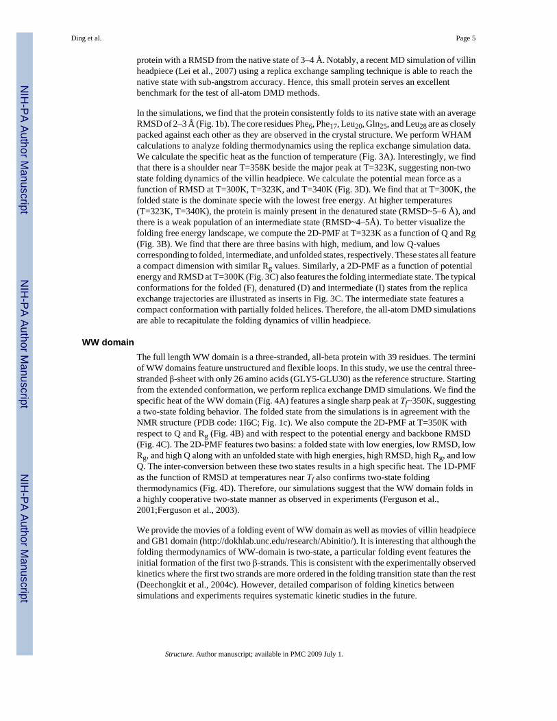
In the simulations, we find that the protein consistently folds to its native state with an averageRMSD of 2–3 Å (Fig. 1b). The core residues Phe6, Phe17, Leu20, Gln25, and Leu28 are as closelypacked against each other as they are observed in the crystal structure. We perform WHAMcalculations to analyze folding thermodynamics using the replica exchange simulation data.We calculate the specific heat as the function of temperature (Fig. 3A). Interestingly, we findthat there is a shoulder near T=358K beside the major peak at T=323K, suggesting non-twostate folding dynamics of the villin headpiece. We calculate the potential mean force as afunction of RMSD at T=300K, T=323K, and T=340K (Fig. 3D). We find that at T=300K, thefolded state is the dominate specie with the lowest free energy. At higher temperatures(T=323K, T=340K), the protein is mainly present in the denatured state (RMSD~5–6 Å), andthere is a weak population of an intermediate state (RMSD~4–5Å). To better visualize thefolding free energy landscape, we compute the 2D-PMF at T=323K as a function of Q and Rg(Fig. 3B). We find that there are three basins with high, medium, and low Q-valuescorresponding to folded, intermediate, and unfolded states, respectively. These states all featurea compact dimension with similar Rg values. Similarly, a 2D-PMF as a function of potentialenergy and RMSD at T=300K (Fig. 3C) also features the folding intermediate state. The typicalconformations for the folded (F), denatured (D) and intermediate (I) states from the replicaexchange trajectories are illustrated as inserts in Fig. 3C. The intermediate state features acompact conformation with partially folded helices. Therefore, the all-atom DMD simulationsare able to recapitulate the folding dynamics of villin headpiece.
WW domainThe full length WW domain is a three-stranded, all-beta protein with 39 residues. The terminiof WW domains feature unstructured and flexible loops. In this study, we use the central three-stranded β-sheet with only 26 amino acids (GLY5-GLU30) as the reference structure. Startingfrom the extended conformation, we perform replica exchange DMD simulations. We find thespecific heat of the WW domain (Fig. 4A) features a single sharp peak at Tf~350K, suggestinga two-state folding behavior. The folded state from the simulations is in agreement with theNMR structure (PDB code: 1I6C; Fig. 1c). We also compute the 2D-PMF at T=350K withrespect to Q and Rg (Fig. 4B) and with respect to the potential energy and backbone RMSD(Fig. 4C). The 2D-PMF features two basins: a folded state with low energies, low RMSD, lowRg, and high Q along with an unfolded state with high energies, high RMSD, high Rg, and lowQ. The inter-conversion between these two states results in a high specific heat. The 1D-PMFas the function of RMSD at temperatures near Tf also confirms two-state foldingthermodynamics (Fig. 4D). Therefore, our simulations suggest that the WW domain folds ina highly cooperative two-state manner as observed in experiments (Ferguson et al.,2001;Ferguson et al., 2003).
We provide the movies of a folding event of WW domain as well as movies of villin headpieceand GB1 domain (http://dokhlab.unc.edu/research/Abinitio/). It is interesting that although thefolding thermodynamics of WW-domain is two-state, a particular folding event features theinitial formation of the first two β-strands. This is consistent with the experimentally observedkinetics where the first two strands are more ordered in the folding transition state than the rest(Deechongkit et al., 2004c). However, detailed comparison of folding kinetics betweensimulations and experiments requires systematic kinetic studies in the future.
Ding et al. Page 5
Structure. Author manuscript; available in PMC 2009 July 1.
NIH
-PA Author Manuscript
NIH
-PA Author Manuscript
NIH
-PA Author Manuscript
DiscussionThe contribution of backbone hydrogen bonds to protein stability has been controversial. Somebelieve that the peptide hydrogen bond is destabilizing since formation of intra-peptidehydrogen bonds break peptide-water hydrogen bonds despite desolvation of the backbonepeptide (Honig et al., 1995;Yang et al., 1995). Others propose that backbone hydrogen bondsstabilize proteins given the experimentally observed α-helix propensity of short poly-alaninepeptides at low temperatures (Baldwin, 2007a;Rose et al., 2006a). Kelly and co-workers(Deechongkit et al., 2004d) designed elegant experiments to target specific backbone-hydrogenbond donors or acceptors by mutating the backbone amides to esters. They found that indeedthe disruption of a buried hydrogen bond destabilizes the proteins more than a solvent exposedhydrogen bond does, and the difference of ΔΔG can be as large as 2–3 kcal/mol. Such adifference can be explained by the redistribution of partial charges in the hydrogen bondedpeptides, which in turn, affects their solvation energies. Such an environment-dependenceeffect of the hydrogen bond interaction can be readily modeled using the “reaction” algorithmfor hydrogen bonds in DMD (Ding et al., 2003), where donors and acceptors change their typesupon hydrogen bond formation (see Methods). With the environment-dependent hydrogenbond model, we are able to reach native or near-native conformations in DMD simulations ofsix proteins. As a control, we also perform DMD simulations without the solvent-dependenteffect: With weak hydrogen bond strength (1–2 kcal/mol), proteins neither fold into specificstructures nor form regular secondary structures. In contrast, a strong hydrogen (>3 kcal/mol)bond strength tends to fold proteins into all-α helices, including the natively all-β proteins (datanot shown). Therefore, our study suggests that the environment-dependent hydrogen bond isimportant for protein folding.
Since multiple folding/unfolding transitions are observed for three small proteins, we are ableto analyze the folding thermodynamics from simulations. We found qualitative agreementbetween the simulation-derived thermodynamics and experimental observations. In oursimulations, we discover that native states always correspond to the lowest free energy stateat room temperature (300K; Fig. 2–Fig 4). Although these native states often have low potentialenergies, there are still individual conformations with low potential energies but high RMSDvalues. This observation suggests that potential energy alone is not an appropriate reactioncoordinate for protein folding. Hence, ensemble analysis of the protein conformations, such asclustering, is necessary for structure determination applications (Bradley et al., 2005b).
We attribute the success of the all-atom DMD method to its ability to rapidly sample proteinconformational space. Proteins usually fold in the milliseconds to seconds range: the fast-folding Trp-cage protein was experimentally shown to fold within microseconds (~µs). Duringour simulations we find that this mini protein folds very rapidly, where the folding time is onthe order of 104 time units (104 × 50 fs = 0.5 ns; see Methods), and multiple folding events areobserved in all replicas (Fig. 2a). The observation of multiple folding events during Trp-cageDMD simulations is mainly due to faster protein dynamics in the absence of explicit solvent.The speed-up in this case is over 1,000-fold. Additionally, the application of replica exchangeincreases the conformational sampling efficiency (Okamoto, 2004). As a result, we are able toobserve the folding of all six proteins to their native or near-native states within an accumulative1.6×107 time units in REXDMD simulations.
We believe that the success of the current model is also due to the fact that these six proteinsare fast folders and their topologies are relatively simple. As the protein size increases and thetopology becomes more sophisticated, longer simulations will be required and the folding ofthese proteins may become practically intractable, even in all-atom DMD simulations. Forexample, we do not observe multiple folding events of the relatively larger proteins (GB1domain, bacterial ribosomal protein L20, and the engrailed homeodomain) in the DMD
Ding et al. Page 6
Structure. Author manuscript; available in PMC 2009 July 1.
NIH
-PA Author Manuscript
NIH
-PA Author Manuscript
NIH
-PA Author Manuscript
simulations due to the insufficient sampling. Therefore, a multi-scale folding method may berequired where simplified protein models are used to sample the large scale conformationalchanges and the all-atom protein model is used to sample the conformational spaces at smallertime scales (Bradley et al., 2005a). The applicability of the current approach to folding of largerproteins requires further investigation.
Protein flexibility modeling with accurate sampling of the protein conformations near its nativestates is essential in protein design (Kuhlman et al., 2003), protein stability estimation (Yin etal., 2007), and protein-protein, protein-ligand designs (Kortemme et al., 2004). Due to the fastconformational sampling efficiency of DMD and the ability to capture the folding free energylandscape of proteins under study, we believe that the current all-atom model is able to rapidlyand accurately sample the available conformations near the target states of proteins. We expectapplications of the all-atom DMD method in protein engineering, protein-protein interfacedesign, and protein-ligand design by combining the dynamics sampling method with proteindesign methods.
MethodsDiscrete molecular dynamics
A detailed description of the DMD algorithm can be found elsewhere (Dokholyan et al.,1998;Rapaport, 1997;Zhou et al., 1997). Briefly, inter-atomic interactions in DMD aregoverned by square-well potential functions. Neighboring interactions (such as bonds, bondangles, and dihedrals) are modeled by infinitely high square well potentials. During asimulation, an atom’s velocity remains constant until a potential step is encountered, where itchanges instantaneously according to the conservations of energy, momentum and angularmomentum. Simulations proceed as a series of such collisions, with a rapid sorting algorithmemployed at each step to determine the following collision.
The difference between discrete molecular dynamics and traditional molecular dynamics is inthe interaction potential functions. Approximating the continuous potential functions with step-functions of pair-wise distances, DMD simulations are reduced to event-driven (collision)molecular dynamics simulation. The sampling efficiency of DMD over traditional MD ismainly due to rapid processing of collision events and localized updates of collisions (onlycollided atoms are required to update at each collision). At an adequately small step size, thediscrete step-function approaches the continuous potential function and DMD simulationsbecome equivalent to traditional molecular dynamics.
All-atom protein modelWe use a united-atom representation to model proteins, in which all heavy atoms and polarhydrogen atoms of each amino acid are included (Fig. 5a). In order to maintain the proteinbackbone and sidechain geometries, we introduce three types of bonded constraints betweenneighboring atoms: (a) consecutive atoms (i, i+1) covalently bonded, (b) next-nearestneighbors (i, i+2) under angular constraints, and (c) atom pairs (i, i+3) linked by dihedralinteractions. For covalent bonds and bond angles, we use a single-well potential (Fig. 5b) withtwo parameters: effective bond length dAB, and its variance, σAB. The dihedral interactions aremodeled by multi-step potential functions of pair-wise distance as introduced in Ref. (Ding etal., 2005a), which is characterized by a set of distance parameters, {dmin, d0, d1, d2, dmax} (Fig.5b). We obtain these parameters by sampling the corresponding distance distribution in a non-redundant database of high-resolution protein structures. These bonded interaction parametersare listed in the Supplementary Table S1. For the non-bonded interactions, we include the vander Waals (VDW), solvation, and hydrogen bond interactions (Ding et al., 2003):
Ding et al. Page 7
Structure. Author manuscript; available in PMC 2009 July 1.
NIH
-PA Author Manuscript
NIH
-PA Author Manuscript
NIH
-PA Author Manuscript
VDW and solvation interactions—The VDW and solvation interactions are pair-wisefunctions of distances, while the hydrogen bond interactions are angular- and distance-dependent, making them multi-body interactions. Therefore, we combine the VDW andsolvation together as the pair-wise interactions. We use a standard 12-6 Lennard-Jones
potential to model the Van der Waals interactions: Here, the van der Waals radii σij and interaction strengths εij betweem atoms i and j are takenfrom CHARMM19 force field: We use the Lazaridis-Karplus (Lazaridiset al., 1999) solvation model:
Here, parameters of reference solvation energy (ΔGfree), volume of atoms (V), correlationlength (λ) and atomic radius (σ). are taken from Lazaridis and Karplus (Lazaridis et al.,1999). The discrete potential functions mimic the continuous potential
by capturing the attractions and repulsions (Fig. 5c). We keep thenumber of steps minimal since increasing the amount of steps reduces the computationalefficiency of DMD. The discrete potential function is characterized by the hardcore distancedhc and a series of potential steps {di, ei}. Here, di is the distance where potential energy E hasa step E(di−1,di)-E(di,di+1)=ei (dhc<d1<d2<⋯<dn). We use a cutoff of 6.5 Å as the interactionrange between all atom pairs. Details of the discrete potential function are provided in theSupplementary Table S2.
Hydrogen bonds—We use the reaction algorithm to model the hydrogen bond interactionas described in Ref. (Ding et al., 2003). Briefly, after the formation of a hydrogen bond, theacceptor (A) and hydrogen (H) change their types to A' and H', respectively. The interactionpotential between an atom and A(H) can be different from its interaction potential with respectto A'(H'). Thus, the formation of a hydrogen bond depends on its neighbors. To mimic theorientation-dependent hydrogen bond interaction, we introduce auxiliary interactions inaddition to the distance-dependent interaction between the hydrogen and the acceptor (Fig.5d). The auxiliary interactions are between the acceptor (A') and the donor (D), and betweenthe hydrogen (H') and the nearest heavy atoms bonded to the acceptor (X). For example, oncethe hydrogen Hi and the acceptor Aj (Fig. 5d) reach the interaction range, we evaluate thedistances between HiXj and DiAj which define the orientations of the hydrogen bond. The totalpotential energy change, ΔE, between Hi/Aj and other surrounding atoms are also evaluatedbefore and after the putative hydrogen bond formation:
Here, σk is the other atoms. If thesedistances satisfy the pre-determined range and the total kinetic energy is enough to overcomethe potential energy change ΔE, we allow the hydrogen bond to be formed, and forbid itsformation otherwise. We include all possible interactions between backbone-backbone,backbone-sidechain, and sidechain-sidechain. The donors include backbone amide hydrogenatoms and sidechain polar hydrogen atoms of His, Trp, Tyr, Asn, Gln, Arg, and Lys. Theacceptors include backbone carbonyl oxygens; sidechain oxygens of Asp, Glu, Ser, Thr, andTyr; and the sidechain nitrogen of His. The interaction parameters of both donor-acceptor andauxiliary interactions are described in the Supplementary Table S3A.
Environment-dependence of hydrogen bonds—To model the environment-dependenteffect, we assume that the hydrogen bonded peptide has weaker solvation energy than the non-hydrogen bonded backbone peptide. For simplicity, we use the carbonyl oxygen as the
Ding et al. Page 8
Structure. Author manuscript; available in PMC 2009 July 1.
NIH
-PA Author Manuscript
NIH
-PA Author Manuscript
NIH
-PA Author Manuscript
solvation center of a peptide. We assign a weaker reference solvation energy ΔGfree value (3.85kcal/mol) to a hydrogen-bonded backbone carbonyl oxygen atom than that of a non-hydrogen-bonded atom (5.85 kcal/mol). In the Lazaridis-Karplus solvation model, it is unfavorable tobury a backbone carbonyl oxygen atom. The desolvation energy depends on its environment:the more it is buried, the higher the total desolvation energy. The formation of a buried hydrogenbond leads to a less unfavorable desolvation of the carbonyl oxygen, and thus, results in ahigher potential energy gain ΔE than a solvent-exposed hydrogen bond. The environment-dependent hydrogen bond model features the multiple body interaction, which is akin to thepolarizable force field. Therefore, this approach effectively models the environment-dependenteffect of a hydrogen bond. The discontinuous potentials between a hydrogen bonded carbonyloxygen atom and other atoms are listed in the Supplementary Table S3B.
Units in all-atom DMD—In the all-atom DMD simulations, the units of mass, length, andenergy are dalton (1.66×10⁻²⁴ gram), angstrom (10−10 meter), and kcal/mol (6.9×10⁻²² joule),respectively. Given the units of mass [M], length [L], and energy [E], the time unit can be
obtain as which is approximately 50 femtoseconds. The temperature unit iskcal/mol·kB or 5.03×10² Kelvin, where kB is the Boltzmann constant.
Replica exchange DMDEfficient exploration of the potential energy landscape of molecular systems is the centraltheme of most molecular modeling applications. The ruggedness and the slope toward theenergy minimum in the landscape govern sampling efficiency at a given temperature. Althoughescape out of local minima is accelerated at higher temperatures, the free energy landscape isaltered due to larger entropic contributions. To efficiently overcome energy barriers whilemaintaining conformational sampling corresponding to a relevant free energy surface, weutilize the replica exchange sampling scheme (Okamoto, 2004;Zhou et al., 2001). In replicaexchange computing, multiple simulations or replicas of the same system are performed inparallel at different temperatures. Individual simulations are coupled through MonteCarlobased exchanges of simulation temperatures between replicas at periodic time intervals.Temperatures are exchanged between two replicas, i and j, maintained at temperatures Ti andTj and with energies Ei and Ej according to the canonical Metropolis criterion with the exchangeprobability p, where p=1 if Δ=(1/kBTi−1/kBTj)(Ej − Ei) ≤ 0,and p=exp(−Δ), if Δ > 0. In DMDsimulations, we use an Anderson thermostat to maintain constant temperature in simulations(Andersen, 1980).
For each protein, we start from a fully extended conformation. We perform eight replicas withtemperatures ranging from 0.50 (~250 Kelvin) to 0.75 (~375 Kelvin) with an increment of0.035 (~17.5 Kelvin). Here, the temperature unit is kcal/mol·kB or 5.03×10² Kelvin. Theexchange takes place every 1×10³ time units. The length of each simulation is 2×10⁶ time units.
Weighted Histogram Analysis MethodWe use the MMTSB tool (Feig et al., 2004) to perform WHAM analysis using replica-exchangetrajectories. In short, the WHAM method utilizes multiple simulation trajectories withoverlapping sampling along the reaction coordinates. The density of states ρ(E) is self-consistently computed by combining histograms from different simulation trajectories (Kumaret al., 1992). Given the density of states, the folding specific heat (Cv) can be computed atdifferent temperatures according to the partition function, Z = ∫ρ(E)exp(−E/KBT)dE. Tocompute the potential of mean force (PMF) as the function of reaction coordinate A, wecompute the conditional probability P(A | E)of observing A at given energy E, which isevaluated from all the simulation trajectories. The PMF is computed as PMF(A) = −ln(∫P(A |E)ρ(E)exp(−E/KBT)dE) + C. Here, C is the reference constant and we set it in such a way that
Ding et al. Page 9
Structure. Author manuscript; available in PMC 2009 July 1.
NIH
-PA Author Manuscript
NIH
-PA Author Manuscript
NIH
-PA Author Manuscript
the lowest PMF always corresponds to zero. Since our simulations start from fully extendedconformations, we exclude the trajectories from the first 5×10⁵ time units and use those of thelast 1.5×10⁶ time units for WHAM analysis. We use the trajectories from all replicas to computethe histograms.
Supplementary MaterialRefer to Web version on PubMed Central for supplementary material.
AcknowledgementsWe thank Brittany M. Fotsch for suggestions on the manuscript. This work is supported in part by the American HeartAssociation grant No. 0665361U and the National Institutes of Health grant R01GM080742.
Reference ListAndersen HC. Molecular-Dynamics Simulations at Constant Pressure And-Or Temperature. Journal of
Chemical Physics 1980;72:2384–2393.Baldwin RL. Energetics of protein folding. J Mol. Biol 2007a;371:283–301. [PubMed: 17582437]Baldwin RL. Energetics of protein folding. J Mol. Biol 2007b;371:283–301. [PubMed: 17582437]Borreguero JM, Urbanc B, Lazo ND, Buldyrev SV, Teplow DB, Stanley HE. Folding events in the 21–
30 region of amyloid beta-protein (Abeta) studied in silico. Proc. Natl. Acad. Sci. U. S. A2005;102:6015–6020. [PubMed: 15837927]
Bradley P, Misura KM, Baker D. Toward high-resolution de novo structure prediction for small proteins.Science 2005b;309:1868–1871. [PubMed: 16166519]
Bradley P, Misura KM, Baker D. Toward high-resolution de novo structure prediction for small proteins.Science 2005a;309:1868–1871. [PubMed: 16166519]
Buscaglia M, Kubelka J, Eaton WA, Hofrichter J. Determination of ultrafast protein folding rates fromloop formation dynamics. J. Mol. Biol 2005;347:657–664. [PubMed: 15755457]
Chen Y, Ding F, Nie H, Serohijos AW, Sharma S, Wilcox KC, Yin S, Dokholyan NV. Protein folding:Then and now. Arch. Biochem. Biophys. 2007
Deechongkit S, Nguyen H, Powers ET, Dawson PE, Gruebele M, Kelly JW. Context-dependentcontributions of backbone hydrogen bonding to beta-sheet folding energetics. Nature 2004d;430:101–105. [PubMed: 15229605]
Deechongkit S, Nguyen H, Powers ET, Dawson PE, Gruebele M, Kelly JW. Context-dependentcontributions of backbone hydrogen bonding to beta-sheet folding energetics. Nature 2004c;430:101–105. [PubMed: 15229605]
Deechongkit S, Nguyen H, Powers ET, Dawson PE, Gruebele M, Kelly JW. Context-dependentcontributions of backbone hydrogen bonding to beta-sheet folding energetics. Nature 2004b;430:101–105. [PubMed: 15229605]
Deechongkit S, Nguyen H, Powers ET, Dawson PE, Gruebele M, Kelly JW. Context-dependentcontributions of backbone hydrogen bonding to beta-sheet folding energetics. Nature 2004a;430:101–105. [PubMed: 15229605]
Ding F, Borreguero JM, Buldyrey SV, Stanley HE, Dokholyan NV. Mechanism for the alpha-helix tobeta-hairpin transition. Proteins 2003;53:220–228. [PubMed: 14517973]
Ding F, Buldyrev SV, Dokholyan NV. Folding Trp-cage to NMR resolution native structure using acoarse-grained protein model. Biophys. J 2005a;88:147–155. [PubMed: 15533926]
Ding F, Dokholyan NV. Simple but predictive protein models. Trends Biotechnol 2005b;23:450–455.[PubMed: 16038997]
Ding F, Dokholyan NV. Emergence of protein fold families through rational design. PLoS. Comput. Biol2006;2:e85. [PubMed: 16839198]
Dinner AR, Sali A, Smith LJ, Dobson CM, Karplus M. Understanding protein folding via free-energysurfaces from theory and experiment. Trends Biochem. Sci 2000;25:331–339. [PubMed: 10871884]
Ding et al. Page 10
Structure. Author manuscript; available in PMC 2009 July 1.
NIH
-PA Author Manuscript
NIH
-PA Author Manuscript
NIH
-PA Author Manuscript
Dokholyan NV. Studies of folding and misfolding using simplified models. Curr. Opin. Struct. Biol2006;16:79–85. [PubMed: 16413773]
Dokholyan NV, Buldyrev SV, Stanley HE, Shakhnovich EI. Identifying the protein folding nucleus usingmolecular dynamics. J. Mol. Biol 2000;296:1183–1188. [PubMed: 10698625]
Dokholyan NV, Buldyrev SV, Stanley HE, Shakhnovich EI. Discrete molecular dynamics studies of thefolding of a protein-like model. Fold. Des 1998;3:577–587. [PubMed: 9889167]
Duan Y, Wang L, Kollman PA. The early stage of folding of villin headpiece subdomain observed in a200-nanosecond fully solvated molecular dynamics simulation. Proc. Natl. Acad. Sci. U. S. A1998;95:9897–9902. [PubMed: 9707572]
Feig M, Karanicolas J, Brooks CL III. MMTSB Tool Set: enhanced sampling and multiscale modelingmethods for applications in structural biology. J. Mol. Graph. Model 2004;22:377–395. [PubMed:15099834]
Ferguson N, Berriman J, Petrovich M, Sharpe TD, Finch JT, Fersht AR. Rapid amyloid fiber formationfrom the fast-folding WW domain FBP28. Proc. Natl. Acad. Sci. U. S. A 2003;100:9814–9819.[PubMed: 12897238]
Ferguson N, Johnson CM, Macias M, Oschkinat H, Fersht A. Ultrafast folding of WW domains withoutstructured aromatic clusters in the denatured state. Proc. Natl. Acad. Sci. U. S. A 2001;98:13002–13007. [PubMed: 11687613]
Hall CK, Wagoner VA. Computational approaches to fibril structure and formation. Methods Enzymol2006;412:338–365. [PubMed: 17046667]
Havlin RH, Tycko R. Probing site-specific conformational distributions in protein folding with solid-state NMR. Proc. Natl. Acad. Sci. U. S. A 2005;102:3284–3289. [PubMed: 15718283]
Honig B, Yang AS. Free energy balance in protein folding. Adv. Protein Chem 1995;46:27–58. [PubMed:7771321]
Kortemme T, Kim DE, Baker D. Computational alanine scanning of protein-protein interfaces. Sci. STKE2004;2004:l2.
Kubelka J, Eaton WA, Hofrichter J. Experimental tests of villin subdomain folding simulations. J. Mol.Biol 2003;329:625–630. [PubMed: 12787664]
Kuhlman B, Dantas G, Ireton GC, Varani G, Stoddard BL, Baker D. Design of a novel globular proteinfold with atomic-level accuracy. Science 2003;302:1364–1368. [PubMed: 14631033]
Kumar S, Bouzida D, Swendsen RH, Kollman PA, Rosenberg JM. The Weighted Histogram AnalysisMethod for Free-Energy Calculations on Biomolecules .1. the Method. Journal of ComputationalChemistry 1992;13:1011–1021.
Lazaridis T, Karplus M. Effective energy function for proteins in solution. Proteins 1999;35:133–152.[PubMed: 10223287]
Lei H, Wu C, Liu H, Duan Y. Folding free-energy landscape of villin headpiece subdomain frommolecular dynamics simulations. Proc. Natl. Acad. Sci. U. S. A 2007;104:4925–4930. [PubMed:17360390]
Luo Z, Ding J, Zhou Y. Temperature-Dependent Folding Pathways of Pin1 WW Domain: An All-AtomMolecular Dynamics Simulation of a Go Model. Biophys. J. 2007
Myers JK, Pace CN. Hydrogen bonding stabilizes globular proteins. Biophys. J 1996;71:2033–2039.[PubMed: 8889177]
Neidigh JW, Fesinmeyer RM, Prickett KS, Andersen NH. Exendin-4 and glucagon-like-peptide-1: NMRstructural comparisons in the solution and micelle-associated states. Biochemistry 2001;40:13188–13200. [PubMed: 11683627]
Okamoto Y. Generalized-ensemble algorithms: enhanced sampling techniques for Monte Carlo andmolecular dynamics simulations. J. Mol. Graph. Model 2004;22:425–439. [PubMed: 15099838]
Pitera JW, Swope W. Understanding folding and design: replica-exchange simulations of "Trp-cage"miniproteins. Proc. Natl. Acad. Sci. U. S. A 2003;100:7587–7592. [PubMed: 12808142]
Rapaport, DC. The art of molecular dynamics simulations. Cambridge: Cambridge University Press;1997.
Rose GD, Fleming PJ, Banavar JR, Maritan A. A backbone-based theory of protein folding. Proc. Natl.Acad. Sci. U. S. A 2006b;103:16623–16633. [PubMed: 17075053]
Ding et al. Page 11
Structure. Author manuscript; available in PMC 2009 July 1.
NIH
-PA Author Manuscript
NIH
-PA Author Manuscript
NIH
-PA Author Manuscript
Rose GD, Fleming PJ, Banavar JR, Maritan A. A backbone-based theory of protein folding. Proc. Natl.Acad. Sci. U. S. A 2006a;103:16623–16633. [PubMed: 17075053]
Schug A, Wenzel W, Hansmann UH. Energy landscape paving simulations of the trp-cage protein. J.Chem. Phys 2005;122:194711. [PubMed: 16161610]
Shimada J, Kussell EL, Shakhnovich EI. The folding thermodynamics and kinetics of crambin using anall-atom Monte Carlo simulation. J. Mol. Biol 2001;308:79–95. [PubMed: 11302709]
Snow CD, Zagrovic B, Pande VS. The Trp cage: folding kinetics and unfolded state topology viamolecular dynamics simulations. J. Am. Chem. Soc 2002;124:14548–14549. [PubMed: 12465960]
Steinbach PJ. Exploring peptide energy landscapes: a test of force fields and implicit solvent models.Proteins 2004;57:665–677. [PubMed: 15390266]
Urbanc B, Borreguero JM, Cruz L, Stanley HE. Ab initio discrete molecular dynamics approach to proteinfolding and aggregation. Methods Enzymol 2006;412:314–338. [PubMed: 17046666]
Wang M, Tang Y, Sato S, Vugmeyster L, McKnight CJ, Raleigh DP. Dynamic NMR line-shape analysisdemonstrates that the villin headpiece subdomain folds on the microsecond time scale. J. Am. Chem.Soc 2003;125:6032–6033. [PubMed: 12785814]
Yang AS, Honig B. Free energy determinants of secondary structure formation: II. Antiparallel beta-sheets. J Mol. Biol 1995;252:366–376. [PubMed: 7563057]
Yang JS, Chen WW, Skolnick J, Shakhnovich EI. All-atom ab initio folding of a diverse set of proteins.Structure 2007;15:53–63. [PubMed: 17223532]
Yin S, Ding F, Dokholyan NV. Eris: an automated estimator of protein stability. Nat. Methods2007;4:466–467. [PubMed: 17538626]
Zhou R. Exploring the protein folding free energy landscape: coupling replica exchange method withP3ME/RESPA algorithm. J. Mol. Graph. Model 2004;22:451–463. [PubMed: 15099840]
Zhou R, Berne BJ, Germain R. The free energy landscape for beta hairpin folding in explicit water. Proc.Natl. Acad. Sci. U. S. A 2001;98:14931–14936. [PubMed: 11752441]
Zhou Y, Karplus M. Folding thermodynamics of a model three-helix-bundle protein. Proc. Natl. Acad.Sci. U. S. A 1997;94:14429–14432. [PubMed: 9405629]
Zhou Y, Zhang C, Stell G, Wang J. Temperature dependence of the distribution of the first passage time:results from discontinuous molecular dynamics simulations of an all-atom model of the second beta-hairpin fragment of protein G. J. Am. Chem. Soc 2003;125:6300–6305. [PubMed: 12785863]
Ding et al. Page 12
Structure. Author manuscript; available in PMC 2009 July 1.
NIH
-PA Author Manuscript
NIH
-PA Author Manuscript
NIH
-PA Author Manuscript
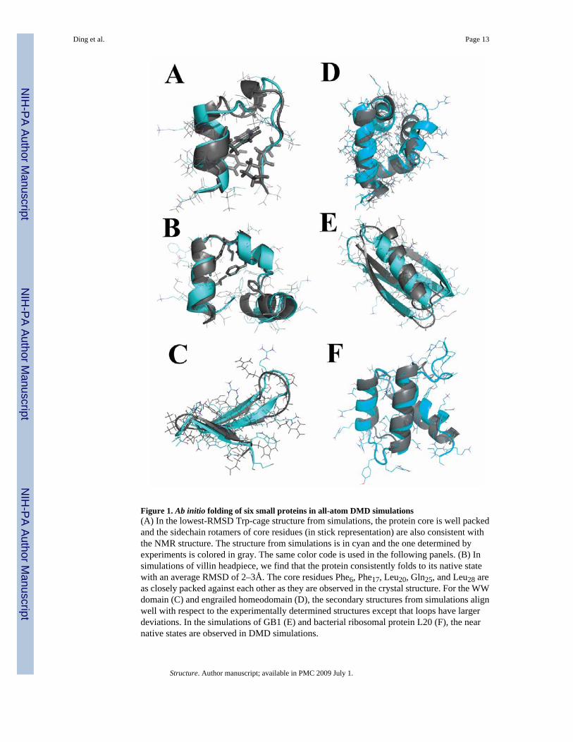
Figure 1. Ab initio folding of six small proteins in all-atom DMD simulations(A) In the lowest-RMSD Trp-cage structure from simulations, the protein core is well packedand the sidechain rotamers of core residues (in stick representation) are also consistent withthe NMR structure. The structure from simulations is in cyan and the one determined byexperiments is colored in gray. The same color code is used in the following panels. (B) Insimulations of villin headpiece, we find that the protein consistently folds to its native statewith an average RMSD of 2–3Å. The core residues Phe6, Phe17, Leu20, Gln25, and Leu28 areas closely packed against each other as they are observed in the crystal structure. For the WWdomain (C) and engrailed homeodomain (D), the secondary structures from simulations alignwell with respect to the experimentally determined structures except that loops have largerdeviations. In the simulations of GB1 (E) and bacterial ribosomal protein L20 (F), the nearnative states are observed in DMD simulations.
Ding et al. Page 13
Structure. Author manuscript; available in PMC 2009 July 1.
NIH
-PA Author Manuscript
NIH
-PA Author Manuscript
NIH
-PA Author Manuscript
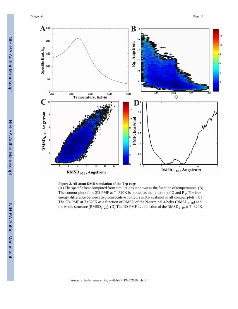
Figure 2. All-atom DMD simulation of the Trp-cage(A) The specific heat computed from simulations is shown as the function of temperatures. (B)The contour plot of the 2D-PMF at T=320K is plotted as the function of Q and Rg. The freeenergy difference between two consecutive contours is 0.6 kcal/mol in all contour plots. (C)The 2D-PMF at T=320K as a function of RMSD of the N-terminal a-helix (RMSD1–10) andthe whole structure (RMSD1–20). (D) The 1D-PMF as a function of the RMSD1–10 at T=320K.
Ding et al. Page 14
Structure. Author manuscript; available in PMC 2009 July 1.
NIH
-PA Author Manuscript
NIH
-PA Author Manuscript
NIH
-PA Author Manuscript
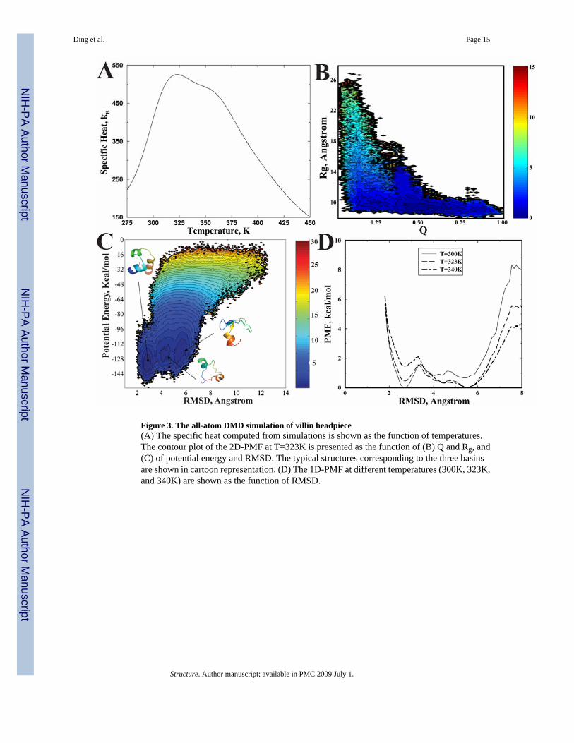
Figure 3. The all-atom DMD simulation of villin headpiece(A) The specific heat computed from simulations is shown as the function of temperatures.The contour plot of the 2D-PMF at T=323K is presented as the function of (B) Q and Rg, and(C) of potential energy and RMSD. The typical structures corresponding to the three basinsare shown in cartoon representation. (D) The 1D-PMF at different temperatures (300K, 323K,and 340K) are shown as the function of RMSD.
Ding et al. Page 15
Structure. Author manuscript; available in PMC 2009 July 1.
NIH
-PA Author Manuscript
NIH
-PA Author Manuscript
NIH
-PA Author Manuscript
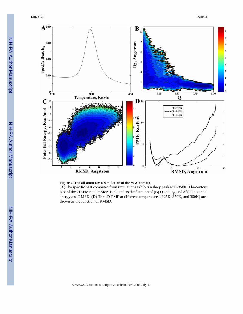
Figure 4. The all-atom DMD simulation of the WW domain(A) The specific heat computed from simulations exhibits a sharp peak at T~350K. The contourplot of the 2D-PMF at T=348K is plotted as the function of (B) Q and Rg, and of (C) potentialenergy and RMSD. (D) The 1D-PMF at different temperatures (325K, 350K, and 360K) areshown as the function of RMSD.
Ding et al. Page 16
Structure. Author manuscript; available in PMC 2009 July 1.
NIH
-PA Author Manuscript
NIH
-PA Author Manuscript
NIH
-PA Author Manuscript
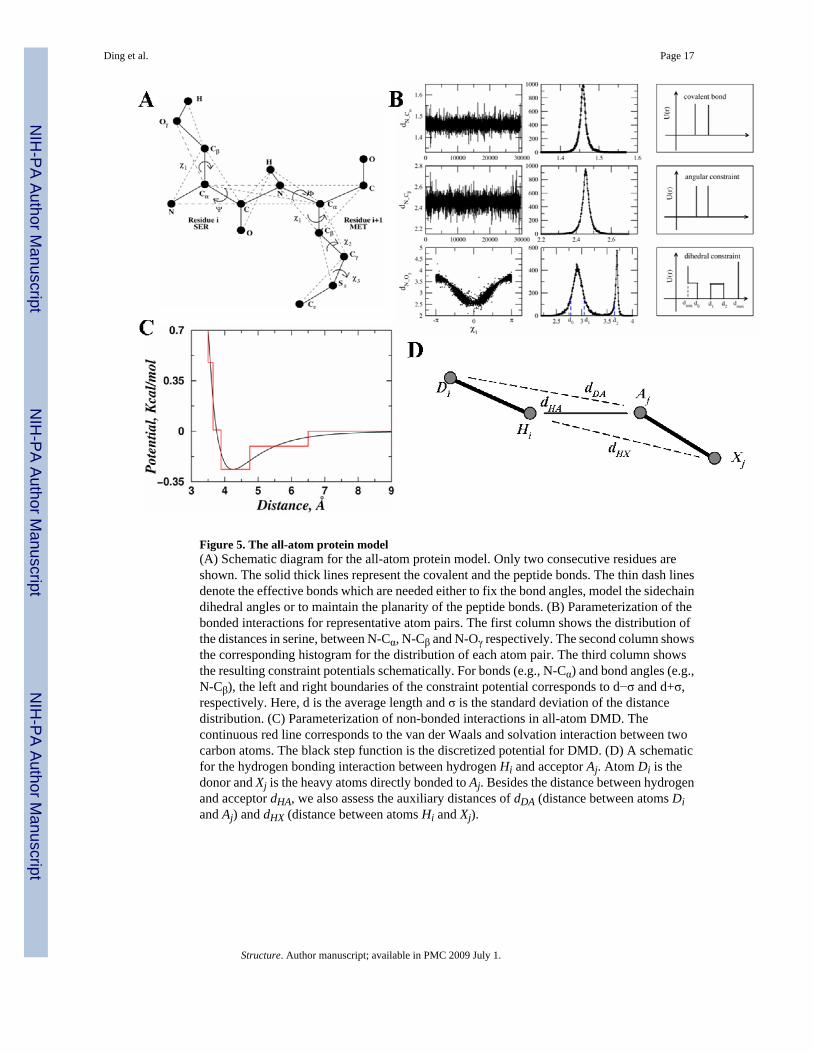
Figure 5. The all-atom protein model(A) Schematic diagram for the all-atom protein model. Only two consecutive residues areshown. The solid thick lines represent the covalent and the peptide bonds. The thin dash linesdenote the effective bonds which are needed either to fix the bond angles, model the sidechaindihedral angles or to maintain the planarity of the peptide bonds. (B) Parameterization of thebonded interactions for representative atom pairs. The first column shows the distribution ofthe distances in serine, between N-Cα, N-Cβ and N-Oγ respectively. The second column showsthe corresponding histogram for the distribution of each atom pair. The third column showsthe resulting constraint potentials schematically. For bonds (e.g., N-Cα) and bond angles (e.g.,N-Cβ), the left and right boundaries of the constraint potential corresponds to d−σ and d+σ,respectively. Here, d is the average length and σ is the standard deviation of the distancedistribution. (C) Parameterization of non-bonded interactions in all-atom DMD. Thecontinuous red line corresponds to the van der Waals and solvation interaction between twocarbon atoms. The black step function is the discretized potential for DMD. (D) A schematicfor the hydrogen bonding interaction between hydrogen Hi and acceptor Aj. Atom Di is thedonor and Xj is the heavy atoms directly bonded to Aj. Besides the distance between hydrogenand acceptor dHA, we also assess the auxiliary distances of dDA (distance between atoms Diand Aj) and dHX (distance between atoms Hi and Xj).
Ding et al. Page 17
Structure. Author manuscript; available in PMC 2009 July 1.
NIH
-PA Author Manuscript
NIH
-PA Author Manuscript
NIH
-PA Author Manuscript