
Ab initio Discrete Molecular Dynamics Approach to Protein Folding and Aggregation
25
Williams, A., Portelius, E., Kheterpal, I., Guo, J.‐T., Cook, K., Xu, Y., and Wetzel, R. (2004). Mapping Abeta amyloid fibril secondary structure using scanning proline mutagenesis. J. Mol. Biol. 335, 833–842. Xu, Y., and Xu, D. (2000). Protein threading using PROSPECT: Design and evaluation. Proteins 40, 343–354. Yankner, B. A. (1996). Mechanism of neuronal degeneration in Alzheimer‘s disease. Neuron 16, 921–932. Zanuy, D., Gunasekaran, K., Ma, B., Tsai, H. H., Tsai, C. J., and Nussinov, R. (2004). Insights into amyloid structural formation and assembly through computational approaches. Amyloid 11, 143–161. [19] Ab initio Discrete Molecular Dynamics Approach to Protein Folding and Aggregation By BRIGITA URBANC,JOSE M. BORREGUERO, LUIS CRUZ, and H. EUGENE STANLEY Abstract Understanding the toxicity of amyloidogenic protein aggregates and designing therapeutic approaches require the knowledge of their structure at atomic resolution. Although solid‐state NMR, X‐ray diffraction, and other experimental techniques are capable of discerning the protein fibrillar struc- ture, determining the structures of early aggregates, called oligomers, is a challenging experimental task. Computational studies by all‐atom molecular dynamics, which provides a complete description of a protein in the solvent, are typically limited to study folding of smaller protein or aggregation of a small number of short protein fragments. We review an efficient ab initio computer simulation approach to protein folding and aggregation using discrete molecular dynamics (DMD) in combi- nation with several coarse‐grained protein models and implicit solvent. This approach involves different complexity levels in both the protein model and the interparticle interactions. Starting from the simplest protein model with minimal interactions, and gradually increasing its complexity, while guided by in vitro findings, we can systematically select the key features of the protein model and interactions that drive protein folding and aggregation. Because the method used in this DMD approach does not require any knowledge of the native or any other state of the protein, it can be applied to study degenerative disorders associated with protein misfolding and aberrant protein aggregation. The choice of the coarse‐grained model depends on the complexity of the protein and specific questions to be addressed, which are mostly suggested by in vitro findings. Thus, we illustrate our approach on amyloid ‐protein (A) 314 computational approaches in theory [19] METHODS IN ENZYMOLOGY, VOL. 412 0076-6879/06 $35.00 Copyright 2006, Elsevier Inc. All rights reserved. DOI: 10.1016/S0076-6879(06)12019-4
-
Upload
independent -
Category
Documents
-
view
1 -
download
0
Transcript of Ab initio Discrete Molecular Dynamics Approach to Protein Folding and Aggregation
Williams, A., Portelius, E., Kheterpal, I., Guo, J.‐T., Cook, K., Xu, Y., and Wetzel, R. (2004).
Mapping Abeta amyloid fibril secondary structure using scanning proline mutagenesis.
J. Mol. Biol. 335, 833–842.
Xu, Y., and Xu, D. (2000). Protein threading using PROSPECT: Design and evaluation.
Proteins 40, 343–354.Yankner, B. A. (1996). Mechanism of neuronal degeneration in Alzheimer‘s disease. Neuron
16, 921–932.
Zanuy, D., Gunasekaran, K., Ma, B., Tsai, H. H., Tsai, C. J., and Nussinov, R. (2004). Insights
into amyloid structural formation and assembly through computational approaches.
Amyloid 11, 143–161.
314 computational approaches in theory [19]
[19] Ab initio Discrete Molecular Dynamics Approachto Protein Folding and Aggregation
By BRIGITA URBANC, JOSE M. BORREGUERO,LUIS CRUZ, and H. EUGENE STANLEY
Abstract
Understanding the toxicity of amyloidogenic protein aggregates anddesigning therapeutic approaches require the knowledge of their structureat atomic resolution. Although solid‐state NMR, X‐ray diffraction, and otherexperimental techniques are capable of discerning the protein fibrillar struc-ture, determining the structures of early aggregates, called oligomers, is achallenging experimental task. Computational studies by all‐atom moleculardynamics, which provides a complete description of a protein in the solvent,are typically limited to study folding of smaller protein or aggregation of asmall number of short protein fragments.
We review an efficient ab initio computer simulation approach to proteinfolding and aggregation using discrete molecular dynamics (DMD) in combi-nation with several coarse‐grained protein models and implicit solvent. Thisapproach involves different complexity levels in both the protein model andthe interparticle interactions. Starting from the simplest protein model withminimal interactions, and gradually increasing its complexity, while guidedby in vitro findings, we can systematically select the key features of theprotein model and interactions that drive protein folding and aggregation.Because the method used in this DMD approach does not require anyknowledge of the native or any other state of the protein, it can be appliedto study degenerative disorders associated with protein misfolding andaberrant protein aggregation.
The choice of the coarse‐grained model depends on the complexity of theprotein and specific questions to be addressed, which are mostly suggested byin vitro findings. Thus, we illustrate our approach on amyloid �‐protein (A�)
METHODS IN ENZYMOLOGY, VOL. 412 0076-6879/06 $35.00Copyright 2006, Elsevier Inc. All rights reserved. DOI: 10.1016/S0076-6879(06)12019-4
[19] AB INITIO DMD APPROACH 315
associated with Alzheimer’s disease (AD). Despite the simplificationsintroduced in the DMD approach, the predicted A� conformations are inagreement with existing experimental data. The in silico findings also providefurther insights into the structure and dynamics of A� folding and oligomerformation that are amenable to in vitro testing.
Introduction
An increasing number of neuropathological disorders, such asAlzheimer’s,Parkinson’s, Creutzfeldt‐Jakob, motor neuron diseases, and polyglutaminedisorders, are known to be associated with protein misfolding, followed bydeposition of toxic protein aggregates in tissue (Dobson, 2004; Koo et al.,1999). Alzheimer’s disease (AD) is a progressive, neurodegenerative disor-der, pathologically characterized by senile plaques and neurofibrillarytangles (Selkoe, 2001). The primary component of senile plaques is amyloid�‐protein (A�), which has been strongly linked to the etiology and patho-genesis of AD. A� aggregates into small assemblies (oligomers), protofi-brils, and fibrils rich in �‐sheet content. In the past decade, compellingevidence has emerged indicating that soluble oligomeric assemblies andprotofibrillar intermediates that form before senile plaque deposition maybe determinant pathogenetic factors (Klein et al., 2004).
Determination of oligomer conformation at the atomic level and track-ing pathways of assembly from monomers to oligomers requires efficientcomputational approaches. With the dramatic increase of computer powerin recent decades, it has become possible to study the behavior of largebiological molecular systems by computer simulations (Ash et al., 2004; Feigand Brooks, 2004; Fersht and Daggett, 2002; Karplus and McCammon,2002). Traditional, all‐atom molecular dynamics (MD) with atomic‐detailforce fields in a physiological solution (which would be ideal for studyingA�oligomerization) is not computationally accessible with current technology.An aggregation process amenable to study by all‐atomMD should occur ontime scales of�10�7 sec and would require the use of advanced technologiessuch as worldwide distributed computing (Snow et al., 2002; Zagrovic andPande, 2003; Zagrovic et al., 2002). However, in vivo and in vitro studiessuggest that the initial stages of oligomerization occur on time scale of hours(Bitan et al., 2003a; Kayed et al., 2003).
The idea of applying a fast and efficient discrete molecular dynamicsmethod (DMD) (Rapaport, 1997) to study protein folding was proposed in1996 (Zhou et al., 1996). Soon after, the method was combined with a one‐bead proteinGomodel to study folding of amodel three‐helix bundle protein(Dokholyan et al., 1998, 2000; Zhou and Karplus, 1997, 1999; Zhou et al.,1997). The interparticle interactions in theGomodel are assigned on the basis
316 computational approaches in theory [19]
of the knowledge of a native state of a protein (Taketomi et al., 1975). Thus,Go models are not ab initio, because they require the knowledge of a nativestate of a protein. Despite this drawback, they are the simplest thermodynam-ic models that yield a unique native state of a protein and describe foldingreminiscent of a first‐order phase transition (Dokholyan et al., 2003). Recent-ly, a two‐beadGo model was applied by Peng et al. to study aggregation of anensemble of 28 A�40 peptides into a fibrillar structure (Peng et al., 2004).
In 2001, a four‐bead proteinmodel in combinationwith theDMDmethodwas first introduced by Smith and Hall (Smith and Hall, 2001a) inspired byearlier studies (Takada et al., 1999). This model accounts for a rather accuratebackbone description and is able to describe a cooperative transition of apolyalanine chain into an �‐helical conformation without any a priori knowl-edge of the native state (Smith and Hall, 2001b). With the four‐bead modelwith hydrogen bond interactions on a single 16‐residue polyalanine chain,Ding et al. (2003) demonstrated a temperature‐induced conformationalchange from the �‐helix to the �‐hairpin conformation. Because of theseproperties, the four‐bead model with hydrogen bond interactions representsa base on which the ab initio modeling can be realized. The ab initio DMDcomputational approach introduces simplifications to the protein description,interparticle interactions, and treatment of the solvent. These simplificationsmake theDMDapproach at least six orders ofmagnitude faster than all‐atomMDwith explicit solvent. To ensure biological relevance of the approach thattargets different aspects of A� folding, oligomer, and fibril formation, up‐to‐date experimental findings need to be integrated into the development of theproteinmodel and interactions, creating a much‐needed partnership betweencomputation and experiment as recognized by others [e.g., the review byMa and Nussinov (2004)].
This review is organized in two main sections. In the first section, wedescribe the applications of the DMD approach to model A� folding andaggregation. The goal of this section is to give an idea of the kind ofinformation we can obtain using the proposed DMD approach. In addition,we present the main hypotheses on the structure and dynamics of foldingand assembly that emerged from these applications. The second sectionintroduces in detail the implementation of the DMD method, coarse‐grained protein models, interparticle interactions, and limitations thatoriginate in simplifications associated with the approach.
Applications to A� Folding and Aggregation
We describe the applications of the DMD approach with the four‐beadand the united‐atom model to A� folding and aggregation to demonstratethe variety of information that can be obtained. We also review selected
[19] AB INITIO DMD APPROACH 317
in vitro findings that shed light on diffe rent structura l aspe cts of A � foldingand aggrega tion and help guide the de velopment of the DMD app roach.
In Vitro Findings
The sequen ce DAEFRH DSG YEVHH QKL VFFAED VGSNKG AII-GLMVG GVV IA de fines the primary structure of A � 42. A � 40 lacks thelast two amin o acids , I41 and A42. The secon dary struc ture of A� mono-mer conform ations depen ds strongly on the enviro nment. In an apo larmembrane ‐ like en vironment , A � 40 and A� 42 monom ers ad opt predomi-nantly an �‐ he lical conform ation (Col es et al ., 1998; Cre scenzi et a l., 2002 ),whereas in an aqu eous so lution A� prefer s a collap sed coil monom erstructure wi th a bend in the V24 ‐ K28 regi on ( Zhang et a l., 2000 ).
Rec ent limited proteol ysis exp eriment s on A � 40 and A � 42 have shownthat the regi on A21–A 30 is highl y resista nt to proteol ytic attack unde rconditions favoring oligo merization, suggest ing the presenc e of a folde dstructure ( Lazo et al., 2005 ). Sim ilar resul ts were obs erved for the A �fragment A � (21–30) in monom eric solution. Lazo et a l. pos tulated that thisdecapept ide ad opts a structure that nuc leates the intramol ecula r foldin g ofthe full ‐ lengt h A � monom er. The so lution dynami cs of A � (21–30) , asdetermi ned by NMR studies, yielded two fam ilies of folded A � (21–30)structure s both containing a turn ‐lik e motif center ed at G25–S2 6 ( Lazoet al., 20 05 ). Thes e in vitro results raise que stions that can be address edin silico : (1) what is the drivi ng force of foldi ng, and (2) how does the folde dstructure a ffect the pathw ay of A � assembly ?
A � 40 and A� 42 both have high tendenci es to aggrega te into fibrils,which makes studies of oligomer ic inte rmediat es difficult . To study A �oligomer ization in v itro , the techniqu e photo ‐ induced cross ‐ linking of un-modified protein s (PICUP) ha s been applied to coval ently stabi lize oligo-mers (see Chapter 12 by Bitan, 2006 in Volume 413). Using PICUP coupledwith size‐exclusion chromatography, Bitan et al. (2003a) showed that A�40and A�42 display distinct oligomer size distributions. Solutions of A�40display a rapid equilibrium among monomers, dimer, trimers, and tetra-mers, whereas A�42 preferentially forms pentamer/hexamer units (para-nuclei), which further assemble into beaded superstructures similar to earlyprotofibrils (Bitan et al., 2003a). Further studies of primary structure ele-ments controlling early oligomerization demonstrate that I41 is critical forparanucleus formation in solutions of A�42 and that A42 is necessary forfurther assembly of A�42 into larger oligomers (Bitan et al., 2003b). Inaddition, oxidation of M35 blocks paranucleus formation in A�42 but doesnot alter the A�40 oligomer size distribution (Bitan et al., 2003c). Massspectroscopy and ion mobility measurements of A�42, which was subjected

318 computational approaches in theory [19]
to filtration to remov e larg e assem blies and im mediately elec trosprayed ,indicate d the presenc e of dim ers, tetr amers, paranucl ei, and pa irs ofparanu clei in agreem ent with PICUP resul ts ( Bern stein et al ., 2005 ).
Solid ‐ state NMR studies yielded high ‐ resol ution informat ion on theA � 40 fibrilla r structure , in which each indiv idual pepti de displays a bend ,stabilized by a salt ‐ bridge between D23 and K28 ( Petkova et al., 200 2; seeChapter 6 by Tycko, 2006 in Volume 413). The kinetics of A� 40 fibrilformation is typical ly preceded by a lag pha se that is not present in a recent lysynthesized A�40‐lactam (D23/K28) that contains a lactam bridge betweenD23 and K28 (Sciarretta et al., 2005). This experimental finding explains theimportance of the bend in the V24‐K28 region and the associated salt‐bridgeD23‐K28 in A�40 fibrillogenesis and suggests that A�40‐lactam (D23/K28)bypasses an unfavorable folding step, leading to �1000‐fold greater rate offibril formation (Sciarretta et al., 2005). The role of the salt‐bridge D23‐K28formation at different stages ofA� folding and assembly can be addressed inthe DMD approach by systematically varying the effective electrostaticpotential.
Four‐Bead Model with Hydrogen Bonding: Planar b‐sheet Assembliesand the Role of Glycines
Urbanc et al. (2004a) applied a four‐bead protein model with backbonehydrogen bond interactions to study A�40 versus A�42 dimer formation.The A�42 sequence was simplified to a polyalanine chain with glycines atpositions 9, 25, 29, 33, 37, and 38. This model exhibited conformationalchanges with increasing temperature. The monomer adopted an �‐helicalconformation at low temperatures, several types of �‐strand conformationsincluding �‐hairpin conformation at intermediate temperatures, and ran-dom coil‐like conformation at high temperatures. A turn between G25 andG29 was consistently observed at intermediate temperatures and was shownto be induced by the presence of glycines, in particular G25. The importanceof glycines was recently confirmed by an all‐atomMD study of A�42 foldingin explicit aqueous solution, which demonstrated that glycines induced localturns in the peptide and consequently caused the �‐helical to �‐strandconformational change (Xu et al., 2005).
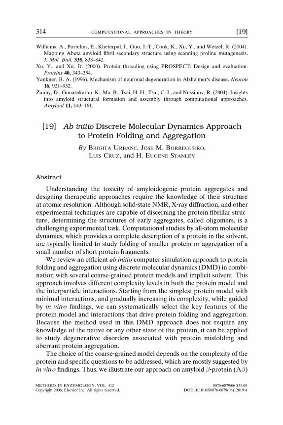
The turn between G25 and G29 occurred in the same protein region asthe bend in the model of A� fibrils by Petkova et al. (2002). The localstructure of a typical peptide within the fibril is quite different from thefour‐bead model prediction. Hydrogen bonds in the fibril are orientedalong the fibrillar axis and link neighboring peptides with no significantintramolecular hydrogen bonding. In the simplified four‐bead model,intramolecular hydrogen bonds first give rise to �‐hairpin monomer

FIG. 1. An A�42 octamer as found within the four‐bead model with hydrogen bond, but no
amino acid–specific interactions. The octamer is an extended planar �‐sheet with several
domains that are slightly rotated with respect to one another.
[19] AB INITIO DMD APPROACH 319
conformations, which then further assemble into extended planar �‐sheets.These planar �‐sheet aggregates are held together exclusively by intramo-lecular and intermolecular hydrogen bonding (Fig. 1.). A critical observa-tion was the lack of stacking among the �‐sheets, in contradiction with themodel of A� fibril formation (Petkova et al., 2002). This result suggests thatamino acid–specific interactions between pairs of side‐chains are responsi-ble for a correct description of the stacked �‐sheet structure.
Four‐Bead Model with Amino Acid–Specific Hydropathic Interactions:Ab40 versus Ab42 Oligomer Formation
Ding et al. (2003) studied the effect of hydrophobic side chain interac-tions on the �‐helix and �‐hairpin monomer conformations in a 16‐residuepolyalanine. They found that above a certain strength of effective hydro-phobic interactions (EHP/EHB > 0.20), the �‐hairpin monomer conforma-tion disappears, and it is replaced by a globular monomer conformation(Ding et al., 2003). Nguyen and Hall demonstrated that the presence of aweak effective hydrophobic attraction (EHP/EHB < 1/6) between the sidechains of 16‐residue polyalanine peptides leads to formation of a stacked�‐sheet structure, consistent with the basic structural features of the fibrilformation ( Nguye n an d Hal l, 2004a ,b, 2005 ; see Chapt er 20 by Hal l andWagoner, 2006 in this volum e).
320 computational approaches in theory [19]
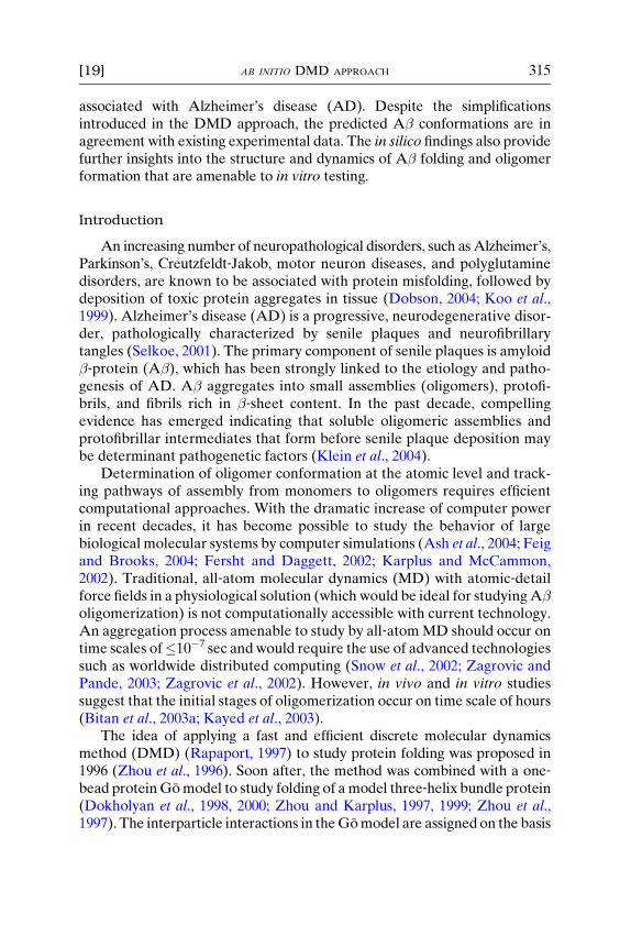
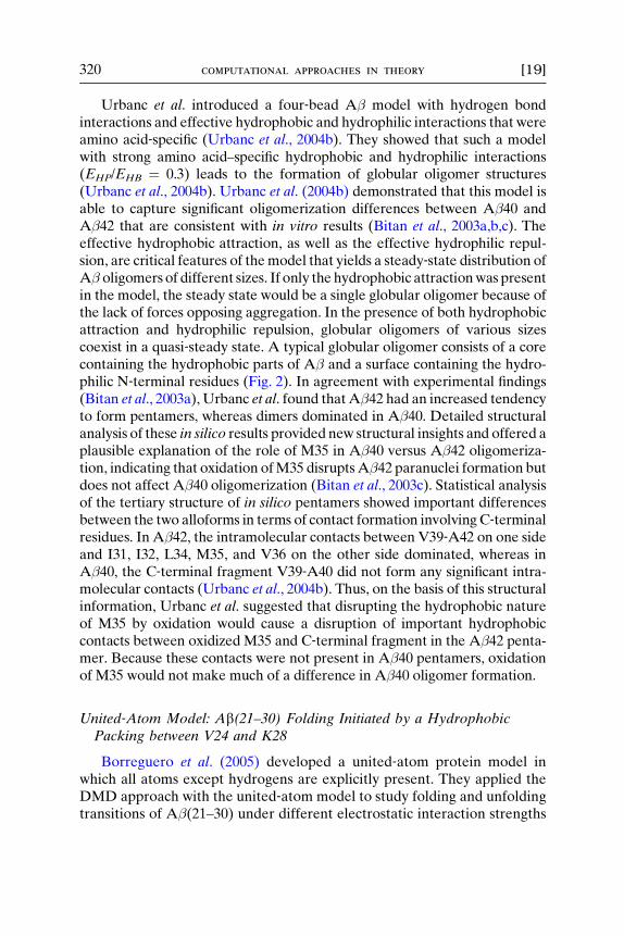
Urbanc et al. introduced a four‐bead A� model with hydrogen bondinteractions and effective hydrophobic and hydrophilic interactions that wereamino acid‐specific (Urbanc et al., 2004b). They showed that such a modelwith strong amino acid–specific hydrophobic and hydrophilic interactions(EHP/EHB ¼ 0.3) leads to the formation of globular oligomer structures(Urbanc et al., 2004b). Urbanc et al. (2004b) demonstrated that this model isable to capture significant oligomerization differences between A�40 andA�42 that are consistent with in vitro results (Bitan et al., 2003a,b,c). Theeffective hydrophobic attraction, as well as the effective hydrophilic repul-sion, are critical features of themodel that yields a steady‐state distribution ofA� oligomers of different sizes. If only the hydrophobic attractionwas presentin the model, the steady state would be a single globular oligomer because ofthe lack of forces opposing aggregation. In the presence of both hydrophobicattraction and hydrophilic repulsion, globular oligomers of various sizescoexist in a quasi‐steady state. A typical globular oligomer consists of a corecontaining the hydrophobic parts of A� and a surface containing the hydro-philic N‐terminal residues (Fig. 2). In agreement with experimental findings(Bitan et al., 2003a), Urbanc et al. found thatA�42 had an increased tendencyto form pentamers, whereas dimers dominated in A�40. Detailed structuralanalysis of these in silico results provided new structural insights and offered aplausible explanation of the role of M35 in A�40 versus A�42 oligomeriza-tion, indicating that oxidation ofM35 disruptsA�42 paranuclei formation butdoes not affect A�40 oligomerization (Bitan et al., 2003c). Statistical analysisof the tertiary structure of in silico pentamers showed important differencesbetween the two alloforms in terms of contact formation involvingC‐terminalresidues. In A�42, the intramolecular contacts between V39‐A42 on one sideand I31, I32, L34, M35, and V36 on the other side dominated, whereas inA�40, the C‐terminal fragment V39‐A40 did not form any significant intra-molecular contacts (Urbanc et al., 2004b). Thus, on the basis of this structuralinformation, Urbanc et al. suggested that disrupting the hydrophobic natureof M35 by oxidation would cause a disruption of important hydrophobiccontacts between oxidized M35 and C‐terminal fragment in the A�42 penta-mer. Because these contacts were not present in A�40 pentamers, oxidationof M35 would not make much of a difference in A�40 oligomer formation.
United‐Atom Model: Ab(21–30) Folding Initiated by a HydrophobicPacking between V24 and K28
Borreguero et al. (2005) developed a united‐atom protein model inwhich all atoms except hydrogens are explicitly present. They applied theDMD approach with the united‐atom model to study folding and unfoldingtransitions of A�(21–30) under different electrostatic interaction strengths
F IG. 2. Globular structure of A� 42 hexamer as found within the four‐bead peptide model
with amino acid–specific interactions caused by hydropathy. D1 is represented by four red
spheres to illustrate the hydrophilic N‐termini at the surface of the hexamer. I41 (four green
spheres) and A42 (four blue spheres), as part of the C‐ terminal region, are at the hydrophobic
core of the hexamer. Yellow ribbons represent a �‐strand, cyan tube a turn, and silver tube a
random coil‐like secondary structure. The image was generated within the VMD software
package (Humphrey et al., 1996), which includes the STRIDE algorithm for calculating the
secondary structure‐propensity per residue (Heinig and Frishman, 2004).
[19] AB INITIO DMD APPROACH 321
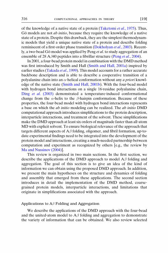
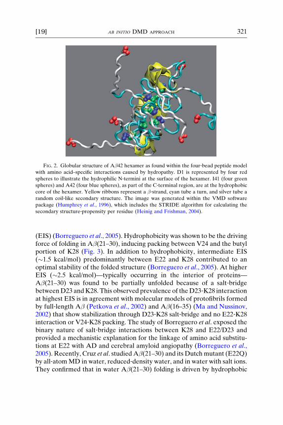
(EIS) (Borr eguero et al ., 2005 ). Hydrop hobicity was shown to be the drivingforce of folding in A � (21–30) , induci ng packing betwe en V24 an d the butylportion of K28 ( Fig. 3). In addition to hydropho bicity, interme diate EIS(� 1.5 kcal/mo l) pr edominan tly be tween E22 and K28 con tributed to anoptimal stability of the folde d struc ture ( Borregu ero et al., 2005 ). At higherEIS ( � 2.5 kcal/m ol)— typical ly occu rring in the inte rior of pr oteins—A� (21–30) was found to be partial ly unfolde d be cause of a salt ‐ bridgebetween D23 and K28. This observed preval ence of the D23 ‐ K28 inte ractionat highes t EIS is in agreemen t with mole cular mod els of protofibri ls form edby full ‐lengt h A � ( Petkova et al., 2002 ) and A� (16–35) (Ma an d Nussinov,2002) that sho w stabi lization through D23‐ K28 salt ‐ bridge and no E22 ‐K28interaction or V24 ‐K28 pack ing. The study of Borregu ero et al. exp osed thebinary na ture of salt ‐ bridge interact ions between K28 and E22/D23 an dprovide d a mecha nistic explana tion for the lin kage of ami no acid substi tu-tions at E22 with AD and cerebr al amyloid angiopath y ( Borr eguero et al.,2005 ). Recen tly, Cruz et al . studi ed A � (21–30) and its Dutch muta nt (E2 2Q)by all ‐ atom MD in water, reduced ‐ den sity wate r, and in wat er with salt ions.They confirm ed that in water A � (21–30) folding is drive n by hy dropho bic
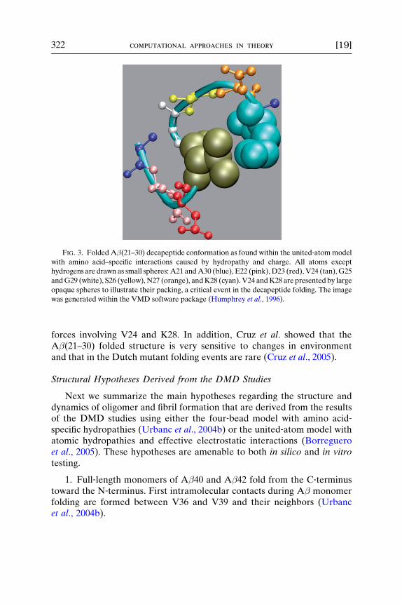
FIG. 3. Folded A�(21–30) decapeptide conformation as found within the united‐atommodel
with amino acid–specific interactions caused by hydropathy and charge. All atoms except
hydrogens are drawn as small spheres: A21 andA30 (blue), E22 (pink),D23 (red),V24 (tan),G25
andG29 (white), S26 (yellow), N27 (orange), andK28 (cyan).V24 andK28 are presentedby large
opaque spheres to illustrate their packing, a critical event in the decapeptide folding. The image
was generated within the VMD software package (Humphrey et al., 1996).
322 computational approaches in theory [19]
forces involving V24 and K28. In addition, Cruz et al. showed that theA�(21–30) folded structure is very sensitive to changes in environmentand that in the Dutch mutant folding events are rare (Cruz et al., 2005).
Structural Hypotheses Derived from the DMD Studies
Next we summarize the main hypotheses regarding the structure anddynamics of oligomer and fibril formation that are derived from the resultsof the DMD studies using either the four‐bead model with amino acid‐specific hydropathies (Urbanc et al., 2004b) or the united‐atom model withatomic hydropathies and effective electrostatic interactions (Borregueroet al., 2005). These hypotheses are amenable to both in silico and in vitrotesting.
1. Full‐length monomers of A�40 and A�42 fold from the C‐terminustoward the N‐terminus. First intramolecular contacts during A� monomerfolding are formed between V36 and V39 and their neighbors (Urbancet al., 2004b).
[19] AB INITIO DMD APPROACH 323
2. A�40 andA�42monomers fold in different ways. TheA�42monomerfolding is associatedwitha turnlike element centeredatG37–G38,which is notpresent in theA�40monomer. TheA�40monomer has an additional parallel�‐strand between A2‐F4 and the central hydrophobic cluster (L17‐A21), notpresent in the A�42 monomer (Urbanc et al., 2004b). The prediction of theturn atG37‐G38 inA�42 (but not inA�40) is consistent with in vitro results oflimited proteolysis, which shows that the region V39‐A42 in A�42 is proteaseresistant, whereas the region V39‐A40 in A�40 is not (Lazo et al., 2005).
3. In A�40 oligomers, the most significant intermolecular contacts existbetween pairs of central hydrophobic clusters (L17‐V18‐F19‐F20‐A21),whereas in A�42 oligomers, contacts between pairs of C‐terminal regions(V39‐A42) are the most important (Urbanc et al., 2004b).
4. Despite similar globular structure with hydrophobic C‐terminalresidues in the core and hydrophilic N‐terminal residues at the surface,the structure of A�40 and A�42 pentamers differs. The parallel �‐strandstructure at the N‐termini of A�40 (as described in 2) persists in all assemblystates and is completely absent from A�42 oligomers. Consequently, theN‐termini of A�42 are spatially less restricted and can be found on averagefurther away from the core of the oligomer. This difference in the N‐terminiproperties might contribute to a more exposed hydrophobic core of A�42oligomers, rendering A�42 more prone to further aggregate (Urbanc et al.,2004b).
5. Hydrophobic attraction between V24 and the butyl portion of K28drives the folding of A�(21–30), whereas the salt‐bridge E22‐K28contributes to the stability of the folded structure (Borreguero et al., 2005).
6. Because experiments show that the same region V24‐G25‐S26‐N27‐K28 is protease resistant in the full‐length A� (Lazo et al., 2005), fibrilformation should be preceded by an event that disrupts the folded loopV24‐G25‐S26‐N27‐K28. Because the E22‐K28 salt‐bridge contributes to the loopstability, a substitution ofE22by anon‐negatively charged amino acid shouldenhance the fibril formation through: (1) decrease of the loop stability, and(2) increase of the rate of D23‐K28 salt‐bridge formation due to the absenceof competition between E22 and D23 (Borreguero et al., 2005).
Based on the above predictions, one can introduce selected amino acidsubstitutions in A�40 and A�42 that would hypothetically disrupt orchange monomer and oligomer conformations. Should in vitro and otherexperimental findings that target the structure of A� folded monomers andoligomers determine that any of the above hypotheses is not valid, theDMD approach can be refined in two ways: (a) by introducing more detailinto the protein model; and (b) by refining the interactions between theside‐chain atoms and possibly introducing locally modified interactions.

324 computational approaches in theory [19]
Methods
Discrete Molecular Dynamics Method
MD is a computer simulation method in which particles move accordingto specific interparticle forces on the basis of classical dynamics. Newton’sequations of motion must be numerically integrated at each time step forall particles to update instantaneous velocities and positions. DMD is asimplified version of MD and is applicable whenever the interparticlepotentials can be represented by one or more square wells (Rapaport,1997). Within each well, the potential is constant, because the forcebetween the two particles is zero, and thus the particles move with constantvelocities until they reach a distance at which the potential is discontinu-ous. At that moment, an elastic or inelastic collision occurs, and the twoparticles change their velocities instantaneously while conserving the totalenergy, momentum, and angular momentum. No numerical integration isneeded. The only events are two‐particle collisions, and the main challengeis to keep track of collision times. Consequently, DMD simulations areconsiderably faster than continuous MD simulations.
During a DMD simulation, the number of particles, volume, and tem-perature are held constant. Periodic boundary conditions are implementedto avoid interactions with the walls of the simulation box. The size of thebox is chosen to be larger than the stretched protein under study. Weimplement temperature control in our model using the method proposedby Berendsen et al. (1984). In this method, a heating rate coefficient, �, isintroduced. The temperature is rescaled at regular intervals �t: T(t þ �t)¼ T(t) þ ��t [T1 � T(t)] where T(t) is the instantaneous temperature,T(t þ �t) is the rescaled temperature, and T1 is the target temperature ofthe heat bath; ��1 is a characteristic time, in which the temperatureequilibrates. The time interval �t corresponds to about N collisions,where N is the number of particles. Temperature is defined by the totalkinetic energy of particles as follows 3
2 kBT ¼ 1N
PNi¼1
miv2i2 ;where kB is the
Boltzmann constant, vi are the velocities of each of the N particles, and mi
their masses. Rescaling temperature requires rescaling all N velocities vi bya factor
ffiffiffiffiffiffiffiffiffiffiffiffiffiffiffiffiffiffiffiffiffiffiffiffiffiffiffiffiffiffiffiTðt þ DtÞ=TðtÞp
;which is followed by recalculation of the collisiontimes. To avoid the time‐consuming task of recalculating collision times, weintroduce a rescaled time variable and rescaled potentials, keeping thevelocities and collision times intact. This transformation does not alterthe trajectory in any way. We keep track of the original simulation timeby keeping track of the rescaling factors, so that results are expressedin original units. A more detailed description has been given elsewhere(Borreguero, 2004).

[19] AB INITIO DMD APPROACH 325
Four‐Bead Model Implementation
We use the four‐bead protein model introduced by Ding et al. (2003).In the four‐bead model, the backbone is represented by three beads,corresponding to the amide (N), the �‐carbon (C�), and the carbonyl (C)groups. Each side‐chain (except G, which lacks the side‐chain group) isrepresented by one side‐chain bead (C�). Each bead (atom) is character-ized by its mass and hard‐core radius. In the simplest version of the model,all atoms have equal mass and their hard‐core radii are set to their van derWaals radii (Creighton, 1993). Each side‐chain atom is characterized by atype, which determines its interactions with other atoms. Any two atomscan only be at a distance d > dmin, where dmin is the sum of their hard‐coreradii. Thus, the potential is set to an ‘‘infinitely’’ large value for d < dmin.Pairs of atoms can be linked by a covalent bond or an angular constraintto account for the protein geometry as shown in Fig. 4A. If two atomsare linked in this way, there is a distance dmax such that for d > dmax thepotential is infinite to prevent the two atoms from breaking the bond. Thelengths of bonds and angular constraints are determined phenomenologi-cally by calculating their distributions using known folded protein struc-tures of �7700 proteins from the Protein Data Bank (PDB) (http://www.rcsb.org/pdb). The values of the lengths of covalent bonds and angularconstraints, which are allowed to vary around their average values by 2%,were reported elsewhere (Ding et al., 2003).
Backbone Hydrogen Bond. In proteins, the most ubiquitous hydrogenbond interaction involves the carbonyl oxygen and the amide hydrogen oftwo amino acids. In the four‐bead model, because the carbonyl oxygen andthe amide hydrogen are not explicitly present, an effective backbone
FIG. 4. (A) Covalent bonds (solid lines) and constraints (dashed lines) that define the four‐bead peptidemodel. (B)Hydrogen bond betweenCj andNi (dotted line) and the corresponding
auxiliary bonds (dashed lines) that define the geometry of hydrogen bonding in the four‐beadmodel. (C) Interparticle potential U(r) for the four auxiliary bonds shown in (B).
326 computational approaches in theory [19]
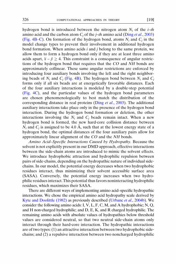
hydrogen bond is introduced between the nitrogen atom Ni of the i–thamino acid and the carbon atom Cj of the j–th amino acid (Ding et al., 2003)(Fig. 4B–C). On formation of the hydrogen bond, atoms Ni and Cj in themodel change types to prevent their involvement in additional hydrogenbond formation. When amino acids i and j belong to the same protein, weallow them to form a hydrogen bond only if they are at least three aminoacids apart, |i – j| � 4. This constraint is a consequence of angular restric-tions of the hydrogen bond that requires that the CO and NH bonds areapproximately collinear. These same angular restrictions are enforced byintroducing four auxiliary bonds involving the left and the right neighbor-ing beads of Ni and Cj (Fig. 4B). The hydrogen bond between Ni and Cj
forms only if all six beads are at energetically favorable distances. Eachof the four auxiliary interactions is modeled by a double‐step potential(Fig. 4C), and the particular values of the hydrogen bond parametersare chosen phenomenologically to best match the distribution of thecorresponding distance in real proteins (Ding et al., 2003). The additionalauxiliary interactions take place only in the presence of the hydrogen bondinteraction. During the hydrogen bond formation or deletion, the otherinteractions involving the Ni and Cj beads remain intact. When a newhydrogen bond is formed, the new hard‐core collision distance betweenNi and Cj is assigned to be 4.0 A, such that at the lowest energy state of ahydrogen bond, the optimal distances of the four auxiliary pairs allow forapproximately linear alignment of the CO and the NH bonds.
Amino Acid–Specific Interactions Caused by Hydropathy. Because thesolvent is not explicitly present in our DMD approach, effective interactionsbetween the side‐chain atoms are introduced to mimic the solvent effects.We introduce hydrophobic attraction and hydrophilic repulsion betweenpairs of side‐chains, depending on the hydropathic nature of individual side‐chains. In our model, the potential energy decreases when two hydrophobicresidues interact, thus minimizing their solvent accessible surface area(SASA). Conversely, the potential energy increases when two hydro-philic residues interact.This potential thus favors noninteracting hydrophilicresidues, which maximizes their SASA.
There are different ways of implementing amino acid–specific hydropathicinteractions. We chose the empirical amino acid hydropathy scale derived byKyte and Doolittle (1982) as previously described (Urbanc et al., 2004b). Weconsider the following amino acids: I, V, L, F, C, M, and A hydrophobic; N, Q,and H non‐charged hydrophilic; and D, E, K, and R charged hydrophilic. Theremaining amino acids with absolute values of hydropathies below thresholdvalues are considered neutral, so that two neutral side‐chain atoms onlyinteract through their hard‐core interaction. The hydropathic interactionsare of two types: (1) an attractive interaction between two hydrophobic side‐chains; and (2) a repulsive interaction between two noncharged hydrophilic

[19] AB INITIO DMD APPROACH 327
or a charged hydrophilic and a noncharged hydrophilic side‐chain. Interac-tions are implemented using a square‐well potential between the pairs ofside‐chain beads C�,i and C�,j, so that they interact if the distance betweentheir centers is less than the interaction range distance 7.5 A (Fig. 5A,B).The potential energy of the effective attractive hydrophobic interactionEHP is proportional to the mean of the relative hydrophobic strengths,I (�1.0), V (�0.93), L (�0.84), F (�0.62), M (�0.42), and A (�0.40), wherethe negative sign reflects the attractive nature of the interaction. The poten-tial energy of the effective repulsive hydrophilic interaction is proportionalto the mean of the relative hydrophilic strengths, R (1.0), K (0.87), E (0.78),D (0.78), N (0.78), Q (0.78), andH (0.71), where the positive sign reflects therepulsive nature of the interaction. H, with a pKa value �6.0, is considereda noncharged hydrophilic amino acid because at physiological conditions(pH ¼ 7.4) only about 4% of H is charged.
FIG. 5. (A) Effective hydrophobic interaction with the potential energy –EHP between two
hydrophobic side‐chain atoms as implemented in the four‐bead model. (B) Effective
hydrophilic interaction with the potential energy EHP between two hydrophilic side‐chainatoms as implemented in the four‐bead model. The range of the hydrophobic and hydrophilic
interactions is set to rR ¼ 7.5 A. (C) Effective electrostatic interaction with the electrostatic
potential energy –ECH between two oppositely charged atoms as implemented in the four‐bead model. (D) Effective electrostatic interaction with the electrostatic potential energy ECH
between two atoms of the same charge as implemented in the four‐bead model. The range of
the interaction is rR ¼ 7.5 A and the soft range is rSR ¼ 6 A. The hard‐core repulsion distance
is denoted by rHC.
328 computational approaches in theory [19]
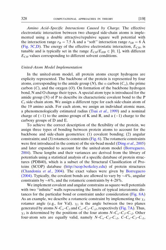
Amino Acid–Specific Interactions Caused by Charge. The effectiveelectrostatic interaction between two charged side‐chain atoms is imple-mented using a double attractive/repulsive square well potential withthe interaction range rR ¼ 7.5 A and a ‘‘soft’’ interaction range rSR ¼ 6 A(Fig. 5C,D). The energy of the effective electrostatic interaction, ECH, istunable and is typically set in the range ECH/EHB 2 [0, 1], with differentECH values corresponding to different solvent conditions.
United‐Atom Model Implementation
In the united‐atom model, all protein atoms except hydrogens areexplicity represented. The backbone of the protein is represented by fouratoms, corresponding to the amide group (N), the � carbon (C�), the primecarbon (C), and the oxygen (O). On formation of the backbone hydrogenbond, N and O change their types. A special atom type is introduced for theamide group (N) of P to describe its characteristic covalent bond to the PC� side‐chain atom. We assign a different type for each side‐chain atom ofthe 19 amino acids. For each atom, we assign an individual atomic mass,a phenomenologically estimated radius (Tsai et al., 1999) and a nominalcharge of (þ1) to the amino groups of K and R, and a (�1) charge to thecarboxy groups of D and E.
To achieve the correct description of the flexibility of the protein, weassign three types of bonding between protein atoms to account for thebackbone and side‐chain geometries: (1) covalent bonding; (2) angularconstraints; and (3) rotameric constraints (Fig. 6). The rotameric constraintswere first introduced in the context of the six‐bead model (Ding et al., 2005)and later expanded to account for the united‐atom model (Borreguero,2004). These lengths and their variances are derived from the library ofpotentials using a statistical analysis of a specific database of protein struc-tures (PDB40), which is a subset of the Structural Classification of Pro-teins (SCOP) database (http://scop.berkeley.edu) of protein structures(Chandonia et al., 2004). The exact values were given by Borreguero(2004). Typically, the covalent bonds are allowed to vary by �4%, angularconstraints by �6%, and the rotameric constraints by 4–28%.
We implement covalent and angular constrains as square‐well potentialswith two ‘‘infinite’’ walls representing the limits of typical interatomic dis-tances for the particular bond or constraint under consideration (Fig. 8A)As an example, we describe a rotameric constraint by implementing the �1
rotamer angle (e.g., for Val). �1 is the angle between the two planesgenerated by atomsN–C�–C� and C�–C�–C�2, respectively (Fig. 7A). Thus,�1 is determined by the positions of the four atoms N–C�–C�–C�2. Otherfour‐atom sets are equally valid, namely N–C�–C�–C�1, C–C�–C�–C�1,
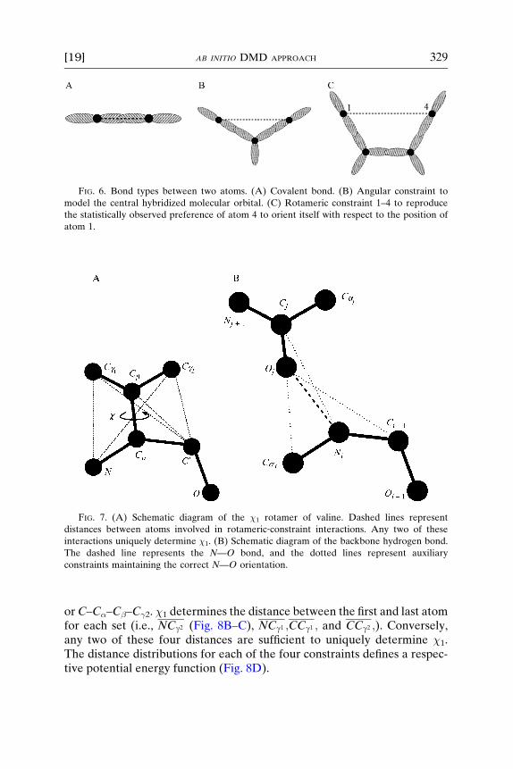
FIG. 6. Bond types between two atoms. (A) Covalent bond. (B) Angular constraint to
model the central hybridized molecular orbital. (C) Rotameric constraint 1–4 to reproduce
the statistically observed preference of atom 4 to orient itself with respect to the position of
atom 1.
FIG. 7. (A) Schematic diagram of the �1 rotamer of valine. Dashed lines represent
distances between atoms involved in rotameric‐constraint interactions. Any two of these
interactions uniquely determine �1. (B) Schematic diagram of the backbone hydrogen bond.
The dashed line represents the N—O bond, and the dotted lines represent auxiliary
constraints maintaining the correct N—O orientation.
[19] AB INITIO DMD APPROACH 329
orC–C�–C�–C�2. �1 determines the distance between the first and last atomfor each set (i.e., NCg2 (Fig. 8B–C), NCg1 ;CCg1 ; and CCg2 ;). Conversely,any two of these four distances are sufficient to uniquely determine �1.The distance distributions for each of the four constraints defines a respec-tive potential energy function (Fig. 8D).
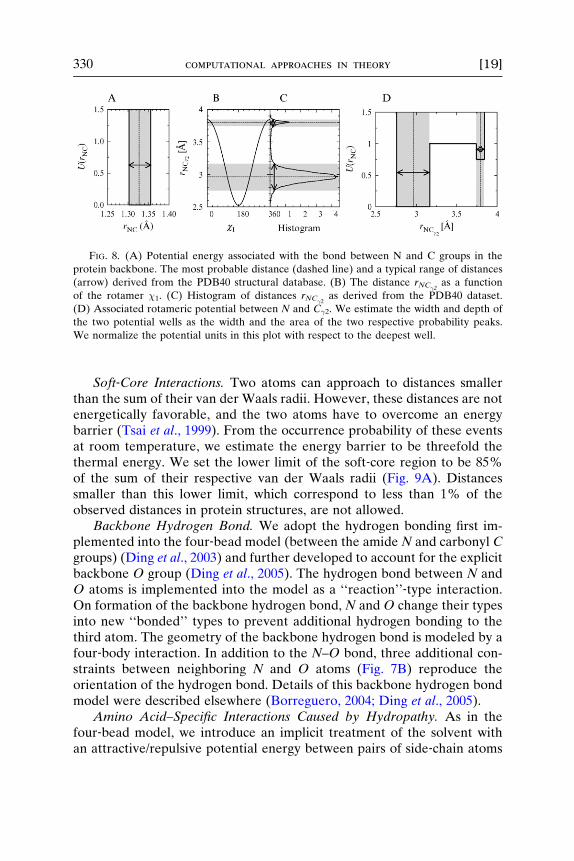
FIG. 8. (A) Potential energy associated with the bond between N and C groups in the
protein backbone. The most probable distance (dashed line) and a typical range of distances
(arrow) derived from the PDB40 structural database. (B) The distance rNC�2as a function
of the rotamer �1. (C) Histogram of distances rNC�2as derived from the PDB40 dataset.
(D) Associated rotameric potential between N and C�2. We estimate the width and depth of
the two potential wells as the width and the area of the two respective probability peaks.
We normalize the potential units in this plot with respect to the deepest well.
330 computational approaches in theory [19]
Soft‐Core Interactions. Two atoms can approach to distances smallerthan the sum of their van der Waals radii. However, these distances are notenergetically favorable, and the two atoms have to overcome an energybarrier (Tsai et al., 1999). From the occurrence probability of these eventsat room temperature, we estimate the energy barrier to be threefold thethermal energy. We set the lower limit of the soft‐core region to be 85%of the sum of their respective van der Waals radii (Fig. 9A). Distancessmaller than this lower limit, which correspond to less than 1% of theobserved distances in protein structures, are not allowed.
Backbone Hydrogen Bond. We adopt the hydrogen bonding first im-plemented into the four‐bead model (between the amide N and carbonyl Cgroups) (Ding et al., 2003) and further developed to account for the explicitbackbone O group (Ding et al., 2005). The hydrogen bond between N andO atoms is implemented into the model as a ‘‘reaction’’‐type interaction.On formation of the backbone hydrogen bond, N and O change their typesinto new ‘‘bonded’’ types to prevent additional hydrogen bonding to thethird atom. The geometry of the backbone hydrogen bond is modeled by afour‐body interaction. In addition to the N–O bond, three additional con-straints between neighboring N and O atoms (Fig. 7B) reproduce theorientation of the hydrogen bond. Details of this backbone hydrogen bondmodel were described elsewhere (Borreguero, 2004; Ding et al., 2005).
Amino Acid–Specific Interactions Caused by Hydropathy. As in thefour‐bead model, we introduce an implicit treatment of the solvent withan attractive/repulsive potential energy between pairs of side‐chain atoms
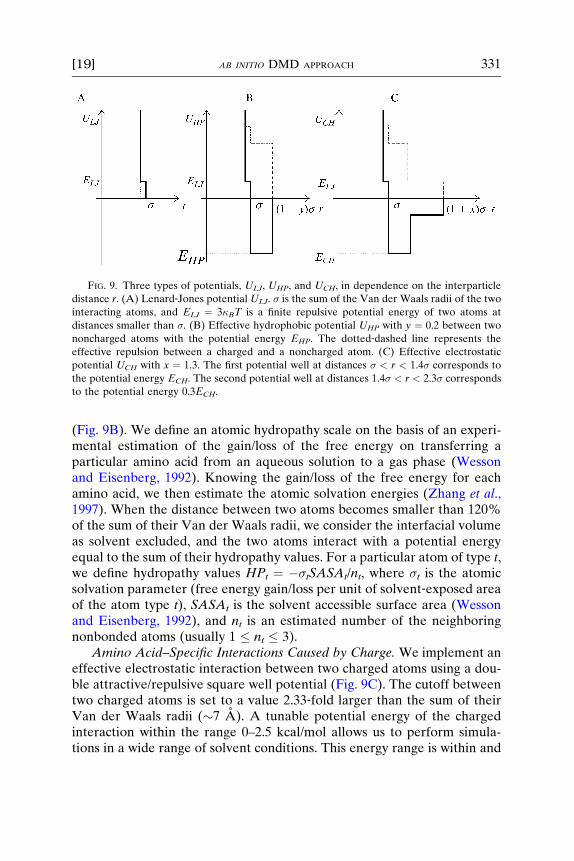
FIG. 9. Three types of potentials, ULJ, UHP, and UCH, in dependence on the interparticle
distance r. (A) Lenard‐Jones potential ULJ. � is the sum of the Van der Waals radii of the two
interacting atoms, and ELJ ¼ 3�BT is a finite repulsive potential energy of two atoms at
distances smaller than �. (B) Effective hydrophobic potential UHP with y ¼ 0.2 between two
noncharged atoms with the potential energy EHP. The dotted‐dashed line represents the
effective repulsion between a charged and a noncharged atom. (C) Effective electrostatic
potential UCH with x ¼ 1.3. The first potential well at distances � < r < 1.4� corresponds to
the potential energy ECH. The second potential well at distances 1.4� < r < 2.3� corresponds
to the potential energy 0.3ECH.
[19] AB INITIO DMD APPROACH 331
(Fig. 9B). We define an atomic hydropathy scale on the basis of an experi-mental estimation of the gain/loss of the free energy on transferring aparticular amino acid from an aqueous solution to a gas phase (Wessonand Eisenberg, 1992). Knowing the gain/loss of the free energy for eachamino acid, we then estimate the atomic solvation energies (Zhang et al.,1997). When the distance between two atoms becomes smaller than 120%of the sum of their Van der Waals radii, we consider the interfacial volumeas solvent excluded, and the two atoms interact with a potential energyequal to the sum of their hydropathy values. For a particular atom of type t,we define hydropathy values HPt ¼ ��tSASAt/nt, where �t is the atomicsolvation parameter (free energy gain/loss per unit of solvent‐exposed areaof the atom type t), SASAt is the solvent accessible surface area (Wessonand Eisenberg, 1992), and nt is an estimated number of the neighboringnonbonded atoms (usually 1 � nt � 3).
Amino Acid–Specific Interactions Caused by Charge. We implement aneffective electrostatic interaction between two charged atoms using a dou-ble attractive/repulsive square well potential (Fig. 9C). The cutoff betweentwo charged atoms is set to a value 2.33‐fold larger than the sum of theirVan der Waals radii (�7 A). A tunable potential energy of the chargedinteraction within the range 0–2.5 kcal/mol allows us to perform simula-tions in a wide range of solvent conditions. This energy range is within and

332 computational approaches in theory [19]
above the experimentally measured values for the free energy gain on salt‐bridge formation on the surface of proteins, 0.24–1.26 kcal/mol (Horovitzand Fersht, 1992; Searle et al., 1999). A more detailed description was givenelsewhere (Borreguero et al., 2005).
Limitations of the DMD Approach
As described previously, the peptide model parameters (the peptidebonds and constraints) and the parameters of the hydrogen bonding aredefined phenomenologically using the known crystalline structure of pro-teins from the Protein Data Bank. Such a phenomenological approach tomodeling (Ding et al., 2002) has been discussed extensively by Zhang et al.(2004). The phenomenologically derived force‐field was shown to be essen-tial for successful folding of the Trp‐cage protein (Ding et al., 2005). As thePDB expands, however, these parameters could change. Different proteindatabases (�‐protein database, �‐protein database, �/�‐protein database,etc.) could yield different parameters as well. It has been shown thatknowledge‐based potentials yield different results when trained on eitherNMR or X‐ray resolved structures (Godzik et al., 1995). We considered thefiltered protein structural database PDB40, which contains representativesof all known protein folds. The filter ensures that no two proteins havemore than 40% sequence identity, preventing any bias in the statisticalanalysis toward overrepresented homologous sequences. For our purposes,the peptide model parameters obtained from this database are ‘‘fixed’’ andrepresent the definition of the model peptide.
In our approach, the hydrogen bond interaction is not amino acid‐specific. The hydrogen‐bond potential energy, EHB, is the energy unit. Thischoice does not imply that we treat the hydrogen bond interaction as afixed interaction independent of the environment. In fact, the free energycost of breaking a hydrogen bond strongly depends on the local environ-ment (Honig and Yang, 1995). This cost may be small in aqueous solutionsbut is typically large in organic solvents. In aqueous solution, the effectivehydrogen bond is strong at the hydrophobic core and weak at the surface ofa protein or a protein assembly. These local variations of the hydrogenbonding caused by variations in the dielectric constant within a solvent areneglected in our approach. However, our simulation approach allows fordifferent environments by assigning different nonvariable dielectric con-stants to different solvent conditions and correspondingly renormalizinginteractions in the model to the reference energy given by the hydrogenbond energy, EHB.
Amino acid–specific interactions caused by hydropathy and charge alsodepend on the particular environment under study. The strength of the
[19] AB INITIO DMD APPROACH 333
effective hydropathic interactions depends on the solvent and other globalvariables, such as temperature, molar concentration, and pH. Our modelaccounts for these global variables by varying the amino acid–specific hy-dropathies and charge, temperature, number of peptides, and the volume ofthe simulation box. In an explicit solvent, however, the hydrophobic/hydro-philic effect and electrostatic interactions depend strongly on the localvariations of SASA and the dielectric constant. As in the case of the hydro-gen bond, in water the hydrophobic/hydrophilic effect is strongest at thesurface of the protein or protein assembly. The opposite is true for theelectrostatic interactions. The free energy change associated with breakinga salt‐bridge within the hydrophobic core of a protein is much higher than atits surface, where polar water molecules shield the charged atoms and thuseffectively weaken the electrostatic interactions between the side‐chains.
The DMD approach described in this review neglects local variations inthe dielectric constant and in the SASA of each side‐chain. The effectivehydrophobic/hydrophilic interactions in coarse‐grained models are basedon hydropathy scales. A number of different hydropathy scales exist, someof them phenomenological (for example, Kyte and Doolittle [1982]); othersare based on the in vitro gain/loss of the free energy when a particular atomis transferred from an aqueous solvent to a gas phase (for example, Wessonand Eisenberg [1992]). A question that needs to be addressed in the futureis how robust the results of the DMD approach are with respect to differenthydropathy scales.
The effective electrostatic interaction is modeled by a two‐step square‐well potential.Weneglect the long‐range nature of theCoulombic interactionbetween two charged particles (and/or two dipoles). Implementing a truelong‐range electrostatic interaction would require a considerable computa-tional effort with a potential approximated by a multistep square‐well of an‘‘infinite’’ range. The interaction range problem is addressed in all‐atomMDeither by using Ewald sums in combination with periodic boundary condi-tions, multipole expansions, or a field‐reaction method (Rapaport, 1997).However, even these sophisticated algorithms neglect the electrostatic forcesabove a certain cutoff distance. In the DMD approach, the solvent is implicit.When implementing the effective interaction between charged atoms, oneneeds to consider effects of the aqueous solution. The charged groups of apeptide, surrounded by water molecules, are effectively shielded because ofthe polar nature of watermolecules. Because of this shielding, we can approx-imate the effective electrostatic potential by a two‐step square‐well potentialwith a finite distance range as a first‐order approximation. In addition, one canassign an effective charge to a given side‐chain. As the charge of a particularside‐chain depends on pH, we can model different pH environments byreassigning the charge of a particular amino acid. For example, H, which is
334 computational approaches in theory [19]
considered neutral at pH ¼ 7, would be considered positively charged at lowpH. Again, our approach neglects the fact that in the core of a peptideassembly the electrostatic interactions are stronger than at the surface of theassembly.
In principle, it is possible to model local variations of interparticleinteractions that depend on SASA of individual side‐chain atoms in theDMD approach by keeping track of the neighborhood of each atom. Allinterparticle interactions except the hard‐core and soft‐core interactionswould need to be rescaled at regular simulation time intervals to accountfor atom‐to‐atom variability of SASA. However, this would requirecomputational effort and could significantly slow down the simulations.
Conclusion
In this chapter, we described in detail the DMDmethod, coarse‐grainedprotein models, and interparticle interactions that were developed to studyA� folding and aggregation. We also addressed weaknesses of the DMDapproach that originate in simplifications of the peptide description, inter-particle interactions, and the absence of explicit solvent. The strengths ofthe DMD approach are: (1) its ab initio nature that does not require anyexperimental parameters specific to A� as input parameters; (2) its effi-ciency that allows for study of not only folding but also of oligomerizationand fibril formation of full‐length A�; and (3) its biological relevance thatcan be achieved through structural in vitro $ in silico feedback‐guideddevelopment of the model and interactions. Because of these advantages,this approach is also applicable to studies of proteins associated with otherneurodegenerative diseases.
Soluble oligomers are a common feature of amyloid assembly, but theirsignificance in the pathway of fibril assembly is not clear (Glabe, 2004).Are oligomers obligatory intermediates on a single pathway from mis-folded monomers to fibrils or are oligomers reversible off‐pathway inter-mediates that only buffer the concentration of misfolded monomers?Another fundamental question is how the assembly state of A� correlateswith its function and toxicity (Klein et al., 2004). The DMD approachcan provide detailed structural information on different assembly statesand explain the pathways of A� oligomer and fibril formation. Assumingthat the assembly structure is directly correlated to toxicity, in silico find-ings could yield mechanistic hypotheses about why certain assemblies aremore toxic than others and thus provide directions for further in vitrotesting and identifying drug targets that would disrupt formation of theseassemblies.
[19] AB INITIO DMD APPROACH 335
Acknowledgments
We thank S. V. Buldyrev, F. Ding, and N. V. Dokholyan for helpful discussions on the
implementation of coarse‐grained models and for continued intellectual support. We greatly
appreciate many valuable discussions with G. Bitan, N. D. Lazo, and D. B. Teplow that guided
us in our approach. We are thankful to the NIH for support on the grant AG023661 and
Alzheimer’s Association for the Zenith Fellows award. We are grateful to Stephen Bechtel,
Jr. for a private donation.
References
Ash, W. L., Zlomislic, M. R., Oloo, E. O., and Tieleman, D. P. (2004). Computer simulations
of membrane proteins. Biochim. Biophys. Acta 1666, 158–189.
Berendsen, H. J. C., Postma, J. P. M., Vangunsteren, W. F., Dlinola, A., and Haak, J. R. (1984).
Molecular dynamics with coupling to an external bath. J. Chem. Phys. 81, 3684–3690.Bernstein, S. L., Wyttenbach, T., Baumketner, A., Shea, J.‐E., Bitan, G., Teplow, D. B., and
Bowers, M. T. (2005). Amyloid �‐protein: Monomer structure and early aggregation states
of A�42 and its Pro19 alloform. J. Am. Chem. Soc. 127, 2075–2084.
Bitan, G. (2006). Structural study of metastable amyloidogenic protien oligomers by photo‐induced cross‐linking of unmodified protiens. Methods Enzymol. 413, in press.
Bitan, G., Kirkitadze, M. D., Lomakin, A., Vollers, S. S., Benedek, G. B., and Teplow, D. B.
(2003a). Amyloid �‐protein (A�) assembly: A�–40 and A�–42 oligomerize through
distinct pathways. Proc. Natl. Acad. Sci. USA 100, 330–335.
Bitan, G., Vollers, S. S., and Teplow, D. B. (2003b). Elucidation of primary structure elements
controlling early amyloid �‐protein oligomerization. J. Biol. Chem. 278, 34882–34889.
Bitan, G., Tarus, B., Vollers, S. S., Lashuel, H. A., Condron, M. M., Straub, J. E., and Teplow,
D. B. (2003c). A molecular switch in amyloid assembly: Met35 and amyloid �‐proteinoligomerization. J. Am. Chem. Soc. 125, 15359–15365.
Borreguero, J. M. (2004). Computational studies of protein stability and folding kinetics. PhD.
Thesis, Boston University.
Borreguero, J. M., Urbanc, B., Lazo, N. D., Buldyrev, S. V., Teplow, D. B., and Stanley, H. E.
(2005).Discretemolecular dynamics study of the amyloid�‐protein decapeptideA�(21–30).
Proc. Natl. Acad. Sci. USA 102, 6015–6020.
Chandonia, J. M., Hon, G., Walker, N. S., Lo Conte, L., Koehl, P., Levitt, M., and Brenner,
S. E. (2004). The ASTRAL compendium in 2004. Nucleic Acids Res. 32, D189–D192.
Coles, M., Bicknell, W., Watson, A. A., Fairlie, D. P., and Craik, D. J. (1998). Solution
structure of amyloid‐� peptide (1–40) in a water‐micelle environment. Is the membrane
spanning domain where we think it is? Biochemistry 37, 11064–11077.
Creighton, T. E. (1993). ‘‘Proteins: Structures and Molecular Properties,’’ 2nd Ed. Freeman
and Company.
Crescenzi, O., Tomaselli, S., Guerrini, R., Salvatori, S., D’Ursi, A. M., Temussi, P. A., and
Picone,D. (2002). Solution structure of theAlzheimer amyloid�‐peptide (1–42) in an apolarmicroenvironment. Similarity with a virus fusion domain. Eur. J. Biochem. 269, 5642–5648.
Cruz, L., Urbanc, B., Borreguero, J. M., Lazo, N. D., Teplow, D. B., and Stanley, H. E. (2005).
Solvent and mutation effects on the nucleation of amyloid �‐protein folding. Proc. Natl.
Acad. Sci. USA 102, 18258–18263.
Ding, F., Dokholyan, N. V., Buldyrev, S. V., Stanley, H. E., and Shakhnovich, E. I. (2002).
Direct molecular dynamics observation of protein folding transition state ensemble.
Biophys. J. 83, 3525–3532.
336 computational approaches in theory [19]
Ding, F., Borreguero, J. M., Buldyrev, S. V., Stanley, H. E., and Dokholyan, N. V. (2003).
A mechanism for the �‐helix to �‐hairpin transition. Proteins: Struct. Func. Genet. 53,220–228.
Ding, F., Buldyrev, S. V., and Dokholyan, N. V. (2005). Folding Trp‐cage to NMR resolution
native structure using a coarse‐grained protein model. Biophys. J. 88, 147–155.Dobson, C. M. (2004). Principles of protein folding, misfolding, and aggregation. Cell Dev.
Biol. 15, 3–16.
Dokholyan, N. V., Buldyrev, S. V., Stanley, H. E., and Shakhnovich, E. I. (1998). Discrete
molecular dynamics studies of folding of a protein‐like model. Folding Design 3, 577–587.Dokholyan, N. V., Buldyrev, S. V., Stanley, H. E., and Shakhnovich, E. I. (2000). Identifying
the protein folding nucleus using molecular dynamics. J. Mol. Biol. 296, 1183–1188.
Dokholyan, N. V., Borreguero, J. M., Buldyrev, S. V., Ding, F., Stanley, H. E., and
Shakhnovich, E. I. (2003). Identifying importance of amino acids for protein folding from
crystal structures. Methods Enzymol. 374, 616–638.
Feig, M., and Brooks, C. L., III (2004). Recent advances in the development and application
of implicit solvent models in biomolecule simulations. Curr. Opin. Struct. Biol. 14, 217–224.
Fersht, A. R., and Daggett, V. (2002). Protein folding and unfolding at atomic resolution. Cell
108, 573–582.
Glabe, C. G. (2004). Conformation‐dependent antibodies target diseases of protein
misfolding. Trends Biochem. Sci. 29, 542–547.Godzik, A., Kolinski, A., and Skolnick, J. (1995). Are proteins ideal mixtures of amino acids?
Analysis of energy parameter sets. Prot. Sci. 4, 2107–2117.
Hall, C. K., and Wagoner, V. A. (2006). Computational approaches to fibril structure and
formation. Methods Enzymol. 412, 338–365.Heinig, M., and Frishman, D. (2004). STRIDE: A web server for secondary structure
assignment from known atomic coordinates of proteins. Nucl. Aci. Res. 32, W500–W502.
Honig, B., and Yang, A. S. (1995). Free energy balance in protein folding. Adv. Protein Chem.
46, 27–58.Horovitz, A., and Fersht, A. R. (1992). Co‐operative interactions during protein folding.
J. Mol. Biol. 224, 733–740.
Humphrey, W., Dalke, A., and Schulten, K. (1996). VMD: Visual molecular dynamics. J. Mol.
Graphics 14, 33–38.
Karplus, M., and McCammon, J. A. (2002). Molecular dynamics simulations of biomolecules.
Nat. Struct. Biol. 9, 646–652.
Kayed, R., Head, E., Thompson, J. L., McIntire, T. M., Milton, S. C., Cotman, C. W., and
Glabe, C. G. (2003). Common structure of soluble amyloid oligomers implies common
mechanisms of pathogenesis. Science 300, 486–489.
Klein,W.L., Stine,W.B., Jr., andTeplow,D.B. (2004).Small assembliesofunmodifiedamyloid�‐protein are the proximate neurotoxin in Alzheimer’s disease. Neurobiol. Aging 25, 569–580.
Koo, E. H., Lansbury, P. T., Jr., and Kelly, J. W. (1999). Amyloid diseases: Abnormal protein
aggregation in neurodegeneration. Proc. Natl. Acad. Sci. USA 96, 9989–9990.
Kyte, J., and Doolittle, R. F. (1982). A simple method for displaying the hydropathic character
of a protein. J. Mol. Biol. 157, 105–132.
Lazo, N. D., Grant, M. A., Condron, M. C., Rigby, A. C., and Teplow, D. B. (2005). On
nucleation of amyloid �‐protein monomer folding. Prot. Sci. 14, 1581–1596.
Ma, B., and Nussinov, R. (2002). Stabilities and confomations Alzheimer’s �‐amyloid peptide
oligomers (A�16–22, A�16–35, and A�10–35): Sequence effect. Proc. Natl. Sci. USA 99,
14126–14131.
Ma, B., and Nussinov, R. (2004). From computational quantum chemistry to computational
biology: Experiments and computations are (full) partners. Phys. Biol. 1, P23–P26.
[19] AB INITIO DMD APPROACH 337
Nguyen, H. D., and Hall, C. K. (2004a). Molecular dynamics simulations of spontaneous fibril
formation by random‐coil peptides. Proc. Natl. Acad. Sci. USA 101, 16180–16185.Nguyen, H. D., and Hall, C. K. (2004b). Phase diagrams describing fibrillization by
polyalanine peptides. Biophys. J. 87, 4122–4134.
Nguyen, H. D., and Hall, C. K. (2005). Kinetics of fibril formation by polyalanine peptides.
J. Biol. Chem. 280, 9074–9082.
Peng,S.,Ding,F.,Urbanc,B.,Buldyrev,S.V.,Cruz,L.,Stanley,H.E.,andDokholyan,N.V.(2004).
Discrete molecular dynamics simulations of peptide aggregation. Phys. Rev.E69, 041908.
Petkova, A., Ishii, T. Y., Balbach, J. J., Antzutkin, O. N., Leapman, R. D., Delaglio, F., and
Tycko, R. (2002). A structural model for Alzheimer’s �‐amyloid fibrils based on
experimental constraints from solid stateNMR.Proc.Natl. Acad. Sci. USA 99, 16742–16747.
Rapaport, D. C. (1997). ‘‘The Art of Molecular Dynamics Simulation.’’ Cambridge University
Press.
Sciarretta, K. L., Gordon, D. J., Petkova, A. T., Tycko, R., and Meredith, S. C. (2005). A�40‐Lactam (D23/K28) models a conformation highly favorable for nucleation of amyloid.
Biochemistry 44, 6003–6014.
Searle, M. S., Griffiths‐Jones, S. R., and Skinner‐Smith, H. (1999). Energetics of weak
interactions in a �‐hairpin peptide: Electrostatic and hydrophobic contributions to stability
from lysine salt bridges. J. Acad. Chem. Soc. 121, 11615–11620.
Selkoe,D. J. (2001). Alzheimer’s disease: Genes, proteins, and therapy.Physiol. Rev. 81, 741–766.Smith, A. V., and Hall, C. K. (2001a). �‐Helix formation: Discontinuous molecular dynamics
on an intermediate‐resolution protein model. Proteins 44, 344–360.
Smith, A. V., and Hall, C. K. (2001b). Assembly of a tetrameric �‐helical bundle: Computer
simulations on an intermediate‐resolution protein model. Proteins 44, 376–391.Snow, C. D., Nguyen, N., Pande, V. S., and Gruebele, M. (2002). Absolute comparison of
simulated and experimental protein‐folding dynamics. Nature 420, 102–106.
Takada, S., Luthey‐Schulten, Z., and Wolynes, P. G. (1999). Folding dynamics with
nonadditive forces: A simulation study of a designed helical protein and a random
heteropolymer. J. Chem. Phys. 110, 11616–11629.
Taketomi, H., Ueda, Y., and Go, N. (1975). Studies on protein folding, unfolding and
fluctuations by computer simulations. Int. J. Peptide Protein Res. 7, 445–459.Tsai, J., Taylor, R., Chothia, C., and Gerstein, M. (1999). The packing density in proteins:
Standard radii and volumes. J. Mol. Biol. 290, 253–266.
Tycko, R. (2006). Characterization of amyloid structures at the molecular level by solid state
nuclear magnetic resonance spectroscopy. Methods Enzymol. 413, in press.
Urbanc, B., Cruz, L., Ding, F., Sammond, D., Khare, S., Buldyrev, S. V., Stanley, H. E., and
Dokholyan, N. V. (2004a). Molecular dynamics simulation of amyloid‐� dimer formation.
Biophys. J. 87, 2310–2321.
Urbanc, B., Cruz, L., Yun, S., Buldyrev, S. V., Bitan, G., Teplow, D. B., and Stanley, H. E.
(2004b). In silico study of amyloid �‐protein (A�) folding and oligomerization. Proc. Natl.
Acad. Sci. USA 101, 17345–17350.
Wesson, L., and Eisenberg, D. (1992). Atomic solvation parameters applied to molecular
dynamics of proteins in solution. Protein Sci. 1, 227–235.
Xu, Y., Shen, J., Luo, X., Zhu, W., Chen, K., Ma, J., and Jiang, H. (2005). Conformational
transition of amyloid �‐peptide. Proc. Natl. Acad. Sci. USA 102, 5403–5407.
Zagrovic, B., Snow, C. D., Shirts, M. R., and Pande, V. S. (2002). Simulation of folding of a
small �‐helical protein in atomistic detail using worldwide‐distributed computing. J. Mol.
Biol. 323, 927–937.
Zagrovic, B., and Pande, V. S. (2003). Solvent viscosity dependence of the folding rate of a
small protein: Distributed computing study. J. Comput. Chem. 24, 1432–1436.
338 computational approaches in theory [20]
Zhang, C., Vasmatzis, G., Cornette, J. L., and DeLisi, C. (1997). Determination of atomic
desolvation energies from the structures of crystallized proteins. J. Mol. Biol. 267, 707–726.Zhang, C., Liu, S., Zhou, H., and Zhou, Y. (2004). The dependence of all‐atom statistical
potentials on structural training database. Biophys. J. 86, 3349–3358.
Zhang, S., Iwata, K., Lachenmann, M. J., Peng, J. W., Li, S., Stimson, E. R., Lu, Y., Felix,
A. M., Maggio, J. E., and Lee, J. P. (2000). The Alzheimer’s peptide A� adopts a collapsed
coil structure in water. J. Struct. Biol. 130, 130–141.
Zhou, Y., Hall, C. K., and Karplus, M. (1996). First‐order disorder‐to‐order transition in an
isolated homopolymer model. Phys. Rev. Lett. 77, 2822–2825.Zhou, Y., Karplus, M., Wichert, J. M., and Hall, C. K. (1997). Equilibrium thermodynamics of
homopolymers and clusters: Molecular dynamics and Monte‐Carlo simulations of system
with square‐well interactions. J. Chem. Phys. 107, 10691–10708.
Zhou, Y., and Karplus, M. (1997). Folding thermodynamics of a three‐helix‐bundle protein.
Proc. Natl. Acad. Sci. USA 94, 14429–14432.
Zhou, Y., and Karplus, M. (1999). Folding of a model three‐helix bundle protein:
A thermodynamic and kinetic analysis. J. Mol. Biol. 293, 917–951.
[20] Computational Approaches to FibrilStructure and Formation
By CAROL K. HALL and VICTORIA A. WAGONER
Abstract
Assembly of normally soluble proteins into amyloid fibrils is a cause orassociated symptom of numerous human disorders. Although some progresstoward understanding the molecular‐level details of fibril structure has beenmade through in vitro experiments, the insoluble nature of fibrils make themdifficult to study experimentally. We describe two computational approachesused to investigate fibril formation and structure: intermediate‐resolutiondiscontinuous molecular dynamics simulations and atomistic molecular dy-namics simulations. Each method has its strengths and weaknesses, but takentogether the two approaches provide a useful molecular‐level picture of fibrilstructure and formation.
Introduction
The hallmark ofmany neurodegenerative diseases, includingAlzheimer’sdisease, is the accumulation and deposition of protein plaques in specifictissues within various organs in the body (Koo, 2002). These plaques arecomposed of ordered protein aggregates known as amyloid fibrils that formwhen normally soluble disease‐specific proteins undergo a conformationalchange that leads to their aberrant assembly. Many of the 24 known so‐called
METHODS IN ENZYMOLOGY, VOL. 412 0076-6879/06 $35.00Copyright 2006, Elsevier Inc. All rights reserved. DOI: 10.1016/S0076-6879(06)12020-0