Reversible Aggregation Plays a Crucial Role on the Folding Landscape of p53 Core Domain
10
Reversible Aggregation Plays a Crucial Role on the Folding Landscape of p53 Core Domain Daniella Ishimaru,* Luis M. T. R. Lima, y Lenize F. Maia,* Priscila M. Lopez,* Ana P. Ano Bom,* Ana P. Valente,* and Jerson L. Silva* *Laborato ´ rio de Termodina ˆmica de Proteı ´nas e Estruturas Virais Gregorio Weber, Centro Nacional de Ressona ˆ ncia Magne ´ tica Nuclear de Macromole ´ culas, Departamento de Bioquı ´mica Me ´dica, Instituto de Cie ˆncias Biome ´ dicas, and y Departamento de Medicamentos, Faculdade de Farma ´cia, Universidade Federal do Rio de Janeiro, Rio de Janeiro, Brazil ABSTRACT The role of tumor suppressor protein p53 in cell cycle control depends on its flexible and partially unstructured conformation, which makes it crucial to understand its folding landscape. Here we report an intermediate structure of the core domain of the tumor suppressor protein p53 (p53C) during equilibrium and kinetic folding/unfolding transitions induced by guanidinium chloride. This partially folded structure was undetectable when investigated by intrinsic fluorescence. Indeed, the fluorescence data showed a simple two-state transition. On the other hand, analysis of far ultraviolet circular dichroism in 1.0 M guanidinium chloride demonstrated a high content of secondary structure, and the use of an extrinsic fluorescent probe, 4,4#- dianilino-1,1# binaphthyl-5,5#-disulfonic acid, indicated an increase in exposure of the hydrophobic core at 1 M guanidinium chloride. This partially folded conformation of p53C was plagued by aggregation, as suggested by one-dimensional NMR and demonstrated by light-scattering and gel-filtration chromatography. Dissociation by high pressure of these aggregates reveals the reversibility of the process and that the aggregates have water-excluded cavities. Kinetic measurements show that the intermediate formed in a parallel reaction between unfolded and folded structures and that it is under fine energetic control. They are not only crucial to the folding pathway of p53C but may explain as well the vulnerability of p53C to undergo departure of the native to an inactive state, which makes the cell susceptible to malignant transformation. INTRODUCTION The wild-type tumor suppressor protein p53C is a nuclear phosphoprotein that plays a key role in cell cycle control (Hall et al., 1996). In normal conditions, p53C has a short half-life, being directed to ubiquitin-mediated degradation by binding to MDM2 protein (Lane and Hall, 1997). The occurrence of cellular stress (ultraviolet radiation, genomic damage, hypoxia) activates p53C and causes it to remain stable in the cell for a longer period of time; this leads to cell cycle arrest or apoptosis. It is the failure of these actions that has been related to tumor progression (Prives and Hall, 1999). In fact, mutations in the p53C gene constitute the most frequent genetic alteration in human cancers. More than 50% of all cancers—including lung, colon, bladder, breast, and ovary—reassociated with a mutant, nonfunctional p53C; ;90% of these mutants have a single amino-acid residue altered at p53C’s core domain (p53C), making this domain one of the most investigated proteins during the last decade. Therefore understanding of folding/unfolding properties of the native state of p53C is an important prerequisite for directing successful therapeutic approaches. The classical view of protein folding holds that it occurs through specific pathways, where partially folded interme- diate conformations gradually drive the protein to its native state (Kim and Baldwin, 1990). However, several proteins have been shown to fold apparently in a single step, without detectable intermediates. An alternative hypothesis suggests the concept of a smooth or rugged funnel to represent the energy landscape for protein folding without and with intermediate structures, respectively (Bryngelson et al., 1995; Dill and Chan, 1997; Plotkin and Onuchic, 2002). In accordance with the rugged funnel hypothesis, in- termediate structures have been detected for several proteins (Plotkin and Onuchic, 2002). They are important since they can either be related to real intermediates of protein folding or be identified as important precursors along protein misfold- ing and aggregation pathways. Many intermediate con- formers are associated with human diseases such as Alzheimer’s, Parkinson’s, prion-related encephalopathies, and some types of cancer (Dobson, 1999; Bullock and Fersht, 2001; Cordeiro et al., 2001; Fa ¨ndrich et al., 2001; Lashuel et al., 2002; Sacchettini and Kelly, 2002; Foguel et al., 2003). Intermediates of protein folding/unfolding processes can be captured by mild denaturing conditions, such as changes in pressure, temperature, small variations in pH, and addition of small amounts of chaotropic agents such as urea and GdmCl Submitted April 19, 2004, and accepted for publication July 23, 2004. Daniella Ishimaru and Luis M. T. R. Lima contributed equally to this work. Address reprint requests to Jerson L. Silva, Universidade Federal do Rio de Janeiro, Departamento de Bioquı ´mica Me ´dica, Instituto de Cie ˆncias Biome ´dicas, CCS Sala E10, Cidade Universita ´ria, Rio de Janeiro, RJ 21941-590 Brazil. Tel.: 55-21-2562-6756; Fax: 55-21-2562-6756; E-mail: [email protected]. Abbreviations used: p53C, core domain of the tumor suppressor protein p53C; GdmCl, guanidinium chloride; CD, circular dichroism; bis-ANS, 4,4#-dianilino-1,1# binaphthyl-5,5#-disulfonic acid. Ó 2004 by the Biophysical Society 0006-3495/04/10/2691/10 $2.00 doi: 10.1529/biophysj.104.044685 Biophysical Journal Volume 87 October 2004 2691–2700 2691
Transcript of Reversible Aggregation Plays a Crucial Role on the Folding Landscape of p53 Core Domain
Reversible Aggregation Plays a Crucial Role on the FoldingLandscape of p53 Core Domain
Daniella Ishimaru,* Luis M. T. R. Lima,y Lenize F. Maia,* Priscila M. Lopez,* Ana P. Ano Bom,*Ana P. Valente,* and Jerson L. Silva**Laboratorio de Termodinamica de Proteınas e Estruturas Virais Gregorio Weber, Centro Nacional de Ressonancia Magnetica Nuclearde Macromoleculas, Departamento de Bioquımica Medica, Instituto de Ciencias Biomedicas, and yDepartamento de Medicamentos,Faculdade de Farmacia, Universidade Federal do Rio de Janeiro, Rio de Janeiro, Brazil
ABSTRACT The role of tumor suppressor protein p53 in cell cycle control depends on its flexible and partially unstructuredconformation, which makes it crucial to understand its folding landscape. Here we report an intermediate structure of the coredomain of the tumor suppressor protein p53 (p53C) during equilibrium and kinetic folding/unfolding transitions induced byguanidinium chloride. This partially folded structure was undetectable when investigated by intrinsic fluorescence. Indeed, thefluorescence data showed a simple two-state transition. On the other hand, analysis of far ultraviolet circular dichroism in 1.0 Mguanidinium chloride demonstrated a high content of secondary structure, and the use of an extrinsic fluorescent probe, 4,4#-dianilino-1,1# binaphthyl-5,5#-disulfonic acid, indicated an increase in exposure of the hydrophobic core at 1 M guanidiniumchloride. This partially folded conformation of p53C was plagued by aggregation, as suggested by one-dimensional NMR anddemonstrated by light-scattering and gel-filtration chromatography. Dissociation by high pressure of these aggregates revealsthe reversibility of the process and that the aggregates have water-excluded cavities. Kinetic measurements show that theintermediate formed in a parallel reaction between unfolded and folded structures and that it is under fine energetic control.They are not only crucial to the folding pathway of p53C but may explain as well the vulnerability of p53C to undergo departureof the native to an inactive state, which makes the cell susceptible to malignant transformation.
INTRODUCTION
The wild-type tumor suppressor protein p53C is a nuclear
phosphoprotein that plays a key role in cell cycle control
(Hall et al., 1996). In normal conditions, p53C has a short
half-life, being directed to ubiquitin-mediated degradation
by binding to MDM2 protein (Lane and Hall, 1997). The
occurrence of cellular stress (ultraviolet radiation, genomic
damage, hypoxia) activates p53C and causes it to remain
stable in the cell for a longer period of time; this leads to cell
cycle arrest or apoptosis. It is the failure of these actions that
has been related to tumor progression (Prives and Hall,
1999). In fact, mutations in the p53C gene constitute the
most frequent genetic alteration in human cancers. More than
50% of all cancers—including lung, colon, bladder, breast,
and ovary—reassociated with a mutant, nonfunctional p53C;
;90% of these mutants have a single amino-acid residue
altered at p53C’s core domain (p53C), making this domain
one of the most investigated proteins during the last decade.
Therefore understanding of folding/unfolding properties of
the native state of p53C is an important prerequisite for
directing successful therapeutic approaches.
The classical view of protein folding holds that it occurs
through specific pathways, where partially folded interme-
diate conformations gradually drive the protein to its native
state (Kim and Baldwin, 1990). However, several proteins
have been shown to fold apparently in a single step,
without detectable intermediates. An alternative hypothesis
suggests the concept of a smooth or rugged funnel to
represent the energy landscape for protein folding without
and with intermediate structures, respectively (Bryngelson
et al., 1995; Dill and Chan, 1997; Plotkin and Onuchic,
2002).
In accordance with the rugged funnel hypothesis, in-
termediate structures have been detected for several proteins
(Plotkin and Onuchic, 2002). They are important since they
can either be related to real intermediates of protein folding or
be identified as important precursors along protein misfold-
ing and aggregation pathways. Many intermediate con-
formers are associated with human diseases such as
Alzheimer’s, Parkinson’s, prion-related encephalopathies,
and some types of cancer (Dobson, 1999; Bullock and Fersht,
2001; Cordeiro et al., 2001; Fandrich et al., 2001; Lashuel
et al., 2002; Sacchettini and Kelly, 2002; Foguel et al., 2003).
Intermediates of protein folding/unfolding processes can be
captured by mild denaturing conditions, such as changes in
pressure, temperature, small variations in pH, and addition of
small amounts of chaotropic agents such as urea and GdmCl
Submitted April 19, 2004, and accepted for publication July 23, 2004.
Daniella Ishimaru and Luis M. T. R. Lima contributed equally to this work.
Address reprint requests to Jerson L. Silva, Universidade Federal do Rio de
Janeiro, Departamento de Bioquımica Medica, Instituto de Ciencias
Biomedicas, CCS Sala E10, Cidade Universitaria, Rio de Janeiro, RJ
21941-590 Brazil. Tel.: 55-21-2562-6756; Fax: 55-21-2562-6756; E-mail:
Abbreviations used: p53C, core domain of the tumor suppressor protein
p53C; GdmCl, guanidinium chloride; CD, circular dichroism; bis-ANS,
4,4#-dianilino-1,1# binaphthyl-5,5#-disulfonic acid.
� 2004 by the Biophysical Society
0006-3495/04/10/2691/10 $2.00 doi: 10.1529/biophysj.104.044685
Biophysical Journal Volume 87 October 2004 2691–2700 2691
(Brems, 1988; Carra and Privalov, 1996; Fersht, 1999;
Dobson, 2000; Kuwajima and Arai, 2000; Silva et al., 2001;
Ishimaru et al., 2003a). The protein conformational changes
induced by these treatments can be accompanied by means of
spectroscopic techniques such as fluorescence, CD, and
NMR (Sanz and Fersht, 1993; Bullock et al., 1997; Foguel
et al., 1998; Fersht, 1999; Mateu et al., 1999; Dobson, 2000;
Kuwajima and Arai, 2000; Neira and Mateu, 2001; Silva
et al., 2001; Kuwata et al., 2002; Ishimaru et al., 2003a).
However, some intermediates are not observable by most
usual spectroscopic methods, as in the case of barnase (Sanz
and Fersht, 1993).
Kinetic intermediates of p53C have been described only for
the folding of the C-terminal region; they are transient, highly
structured, but spectroscopically silent (Mateu et al., 1999).
We recently described that denaturation by high temperature
or high pressure leads to generation of irreversible aggregates
(Ishimaru et al., 2003b). The isolation of a monomeric
intermediate by high pressure was only attained by the
combination of pressure with subzero temperatures (Ishimaru
et al., 2003a).
In the context of the cell, the unfolding process participates
in several important steps in cellular physiology, such as
protein turnover in proteasomes (Fersht and Daggett, 2002).
In addition, several diseases related to misfolded proteins are,
in fact, diseases of protein unfolding, since the protein is
initially correctly folded (Ferrao-Gonzales et al., 2000;
Cordeiro et al., 2001). Therefore the detection of intermediate
species, in special aggregating ones, during p53C unfolding
might be important for the cellular homeostasis since its
accumulation could interfere with the proper integration of
cellular functions.
Here we describe an intermediate structure during
equilibrium and kinetic folding and unfolding of p53C. This
intermediate is obtained by low concentrations of GdmCl
and has a high tendency to form aggregates. The reversibility
of the aggregation sheds light into the mechanism of folding
of p53C and its role on the well-known plasticity of this
tumor suppressor protein. The elution from the gel filtration
chromatography and the light-scattering value indicates that
this aggregate is small (10–20 subunits). This intermediate
also occurred with the hot-spot mutant R248Q (one of the
most prominent somatic mutations in different types of
cancer) which points to the medical impact of this finding.
The kinetic data show how high concentration of protein
favors the aggregation, a condition that would be present in
a cellular situation of loss-of-function mutation, such as the
R248Q. These polymeric intermediates may be the site
where p53 mutants (translated from a single mutant allele)
are able to drive wild-type p53 protein (translated from
the remaining wild-type p53 allele) into a mutant confor-
mation. Thus, these intermediates may be targets to the
development of lead compounds capable of destabilizing
them with potential therapeutic action against tumor
diseases.
MATERIALS AND METHODS
Chemicals
All reagents were of analytical grade. Distilled water was deionized and
filtered through a Millipore (Billerica, MA) water purification system before
use. Bis-ANS was purchased from Molecular Probes (Eugene, OR). All
solutions were prepared just before use. Stock solutions of GdmCl were
checked for exact concentration by refractive measurements (Pace, 1986).
p53C subcloning, expression, and purification
The core domain of the tumor suppressor human protein p53C was obtained
as described elsewhere (Ishimaru et al., 2003a). Purification was performed
as described in Bullock et al. (1997). Protein samples were stored in 50 mM
Tris.Cl, pH 7.2, 150 mM NaCl, 5 mM DTT, and 5% glycerol in liquid
nitrogen. p53C comprises amino-acid residues 94–312.
Equilibrium folding and unfolding measurements
p53C at 5 mM was used in all equilibrium measurements. All experiments
were carried out at least three times, in buffer Tris.Cl 50 mM, pH 7.2, 150
mM NaCl, 5 mM DTT, and 5% glycerol. Intrinsic fluorescence spectra were
obtained by excitation at 278 nm and emission was monitored from 295 to
415 nm in an ISS (Champaign, IL) spectrometer. Because of the high protein
concentration used, and since the buffer contributed ,2% to the emission,
no correction was required. For light-scattering data, excitation was at 320
nm and emission was collected from 300 to 340 nm. For experiments in the
presence of bis-ANS (5 mM) the excitation wavelength was at 360 nm and
emission was collected from 400 to 600 nm.
Equilibrium unfolding in GdmCl was performed at 25�C 6 0.2�C in the
presence of the concentrations indicated in the figures. Data were collected
after overnight incubation with the denaturing agent at 25�C.
Circular dichroism
Experiments were carried out at least three times with 5 mM p53C in 50 mM
Tris-HCl, pH 7.2, 150 mM NaCl, 5 mM DTT, and 5% glycerol. Far-
ultraviolet spectra were monitored from 200 to 260 nm in a 2.00 mm quartz
cuvette and recorded in a Jasco J-715 spectropolarimeter (Jasco, Tokyo,
Japan) at 25�C 6 0.2�C.
Kinetic measurements
Kinetic unfolding experiments were performed using a SX18MV stopped-
flow apparatus (Applied Photophysics, Leatherhead, UK). All experiments
were performed at 25�C 6 0.2�C, with 5 mM p53C, unless otherwise stated.
Intrinsic fluorescence was followed by setting excitation to 278 nm,
collecting emission through cut-off filter (WG320, with 50% transmittance
at 320 nm).
All data presented are an average of 5 to 10 runs, and all concentration
refers to those resulting from mixing equal volumes of protein and GdmCl
from each syringe (both in 50 mM Tris-HCl, pH 7.2, 150 mM NaCl, 5 mM
DTT, and 5% glycerol). Data were fitted to obtain the rates using nonlinear
least-squares fitting softwares provided by the manufacturer, using single
exponential equation. Kinetic refolding experiments were performed
incubating 50 mM p53C in GdmCl for at least 2 h. Later, samples were
diluted 10 times with buffer and different amounts of GdmCl to provide
indicated GdmCl final concentration. Aggregation was immediately followed
in a spectrofluorimeter (Cary Eclipse Spectrofluorimeter, Varian, Palo Alto,
CA) over time by light-scattering measurements with excitation and emission
set at 320 nm. All experiments were performed at 25�C 6 0.2�C, in 50 mM
Tris-HCl, pH 7.2, 150 mM NaCl, 5 mM DTT, and 5% glycerol.
2692 Ishimaru et al.
Biophysical Journal 87(4) 2691–2700

Gel filtration chromatography
Gel filtration chromatography was performed using a GPC100 column
(SynChropak, Synchrom, Lafayette, IN) attached to a high-pressure liquid
chromatography system (Shimadzu, Tokyo, Japan) with absorbance
recorded at 280 nm. The system was equilibrated with 50 mM sodium
phosphate, pH 7.2, 5 mM DTT, 150 mM NaCl, and 5% glycerol, in the
absence or presence of the indicated GdmCl concentration, with a flow rate
of 0.6 mL/min.
NMR data
For 1D-NMR experiments, 10% D2O (Isotec., Miamisburg, OH) was added
to purified p53C samples. 1H-NMR measurements were performed at 25�Con a Bruker Avance DRX 600 spectrometer (Bruker Biospin GmbH,
Rheinstetten, Germany), equipped with a 5-mm inverse detection triple
resonance probe with z gradient. Water suppression was achieved using the
watergate technique (Piotto et al., 1992).
RESULTS
The presence of one tryptophan and eight tyrosine residues
in p53C allowed us to follow the unfolding process through
the fluorescence emission spectra based on these intrinsic
probes (Fersht, 1999; Dobson, 2000; Nichols and Matthews,
2001; Silva et al., 2001; Ishimaru et al., 2003a,b). From each
spectrum, we calculated the spectral center of mass, an index
of the average energy value of the spectrum. Since aromatic
amino-acid residues are sensitive to the polarity of their
immediate environment, changes in center of mass will
reflect conformational changes induced in the protein. To
investigate the structural stability of p53C, we performed
isothermal unfolding experiments by employing GdmCl as
denaturant, at 25�C (Fig. 1). GdmCl has been widely used
for protein denaturation studies providing a more pro-
nounced effect as compared with urea, but in general similar
results with both are obtained (Pace, 1986). The fluorescence
emission spectra of p53C at high GdmCl concentrations
(from 3 M to 6 M), clearly showed both Tyr and Trp
contributions (see 6 M spectrum in inset to Fig. 1),
suggesting that aromatic residues were highly exposed to
the aqueous environment and allowing us to consider them
as completely unfolded. Fitting the data shown in Fig. 1,
a [GdmCl]50% value equal to 1.00 6 0.03 M was obtained,
which is similar to previous reports (Bullock et al., 2000;
Bell et al., 2002), presenting no apparent accumulated
intermediate.
Intrinsic fluorescence probes the local environments of the
Trp and Tyr, providing a measure of the tertiary conforma-
tion. A well-known test of a two-state transition is to examine
whether changes in secondary structure occur in parallel with
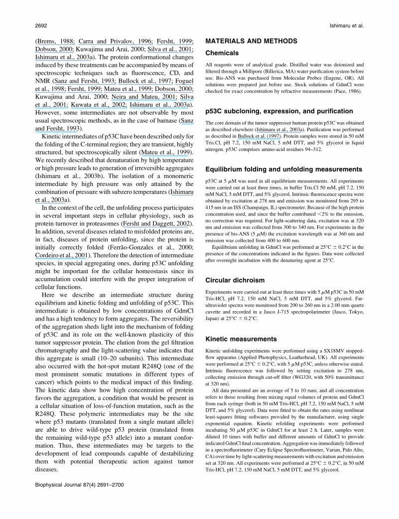
the changes in fluorescence. When we examined the
secondary structure of p53C by far ultraviolet circular
dichroism (Fig. 2) as a function of GdmCl, the changes were
not coincident with the intrinsic fluorescence data. When we
followed b-sheet structure at 218 nm (Fig. 2 A), we observed
a slight loss of secondary structure up to 0.4 M GdmCl,
followed by an increase around 0.9–1.2 M GdmCl and
a second and abrupt loss of structure above 1.5 M. This result
directly entails the existence of an intermediate conformation
with a significantly higher content of secondary structure
around 1 M GdmCl. The ellipticity at 1 M GdmCl increased
about twice (from �25 to �50 mdeg). Examples of spectra at
0, 1, and 6 M GdmCl are shown in Fig. 2 C. When a hot-spot
mutant (R248Q) was utilized, we observed the same pattern
(Fig. 2 B), where the protein presented the higher degree of
secondary structure at 1 M GdmCl.
Because intrinsic fluorescence and CD data are non-
coincident, a two-state transition can be ruled out. To further
characterize the properties of this intermediate, we utilized
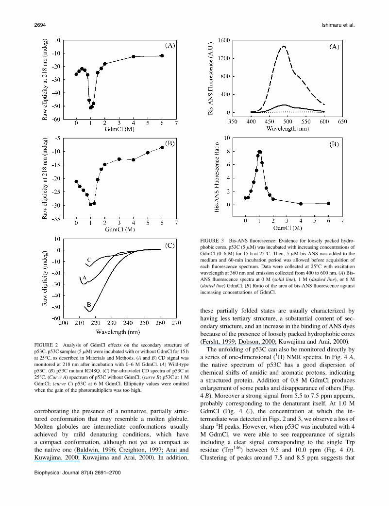
an extrinsic fluorescent probe (bis-ANS) for analysis of
GdmCl-induced denaturation of p53C (Fig. 3). The hydro-
phobic probe bis-ANS binds noncovalently to hydrophobic
regions of proteins. It is a sensitive probe for structural
changes promoted by different treatments (Silva et al.,
1992a; Ishimaru et al., 2003a) because it undergoes a large
increase in the fluorescence quantum yield when bound to
proteins. Fig. 3 A shows bis-ANS spectra in the presence of
p53C and no GdmCl (solid line), 1 M (dashed line) and 6 M
(dotted line) GdmCl. At 1 M GdmCl, we can observe a large
increase in bis-ANS fluorescence, which can be better
visualized in Fig. 3 B. The binding of this extrinsic
fluorescent probe is eightfolded increased at 1–1.1 M GdmCl
and decreases as the GdmCl concentration is further raised.
This large increase in bis-ANS binding by the intermediate
indicates that it exposes structured hydrophobic pockets to
the solvent around 1 M GdmCl. The increase in bis-ANS
binding coincides with the gain in secondary structure,
FIGURE 1 Effects of GdmCl-induced denaturation on the tertiary
structure of p53C. p53C samples (5 mM) were exposed to 0–6 M GdmCl
at 25�C (15 h in each case) in a buffer containing 50 mM Tris.Cl, pH 7.2,
150 mM NaCl, 5 mM DTT, and 5% glycerol. Data were analyzed as center
of spectral mass. Line in the main panel corresponds to fitted data as
described in Materials and Methods. (Inset) Self-normalized spectra
obtained in 1 M GdmCl (dashed line), 6 M GdmCl (dotted line), and no
GdmCl (solid line). Excitation wavelength was 278 nm and emission was
collected from 295 to 415 nm.
Intermediate States of Tumor Suppressor p53C 2693
Biophysical Journal 87(4) 2691–2700
corroborating the presence of a nonnative, partially struc-
tured conformation that may resemble a molten globule.
Molten globules are intermediate conformations usually
achieved by mild denaturing conditions, which have
a compact conformation, although not yet as compact as
the native one (Baldwin, 1996; Creighton, 1997; Arai and
Kuwajima, 2000; Kuwajima and Arai, 2000). In addition,
these partially folded states are usually characterized by
having less tertiary structure, a substantial content of sec-
ondary structure, and an increase in the binding of ANS dyes
because of the presence of loosely packed hydrophobic cores
(Fersht, 1999; Dobson, 2000; Kuwajima and Arai, 2000).
The unfolding of p53C can also be monitored directly by
a series of one-dimensional (1H) NMR spectra. In Fig. 4 A,
the native spectrum of p53C has a good dispersion of
chemical shifts of amidic and aromatic protons, indicating
a structured protein. Addition of 0.8 M GdmCl produces
enlargement of some peaks and disappearance of others (Fig.
4 B). Moreover a strong signal from 5.5 to 7.5 ppm appears,
probably corresponding to the denaturant itself. At 1.0 M
GdmCl (Fig. 4 C), the concentration at which the in-
termediate was detected in Figs. 2 and 3, we observe a loss of
sharp 1H peaks. However, when p53C was incubated with 4
M GdmCl, we were able to see reappearance of signals
including a clear signal corresponding to the single Trp
residue (Trp146) between 9.5 and 10.0 ppm (Fig. 4 D).
Clustering of peaks around 7.5 and 8.5 ppm suggests that
FIGURE 3 Bis-ANS fluorescence: Evidence for loosely packed hydro-
phobic cores. p53C (5 mM) was incubated with increasing concentrations of
GdmCl (0–6 M) for 15 h at 25�C. Then, 5 mM bis-ANS was added to the
medium and 60-min incubation period was allowed before acquisition of
each fluorescence spectrum. Data were collected at 25�C with excitation
wavelength at 360 nm and emission collected from 400 to 600 nm. (A) Bis-
ANS fluorescence spectra at 0 M (solid line), 1 M (dashed line), or 6 M
(dotted line) GdmCl. (B) Ratio of the area of bis-ANS fluorescence against
increasing concentrations of GdmCl.
FIGURE 2 Analysis of GdmCl effects on the secondary structure of
p53C. p53C samples (5 mM) were incubated with or without GdmCl for 15 h
at 25�C, as described in Materials and Methods. (A and B) CD signal was
monitored at 218 nm after incubation with 0–6 M GdmCl. (A) Wild-type
p53C. (B) p53C mutant R248Q. (C) Far-ultraviolet CD spectra of p53C at
25�C. (Curve A) spectrum of p53C without GdmCl; (curve B) p53C at 1 M
GdmCl; (curve C) p53C at 6 M GdmCl. Ellipticity values were omitted
when the gain of the photomultipliers was too high.
2694 Ishimaru et al.
Biophysical Journal 87(4) 2691–2700
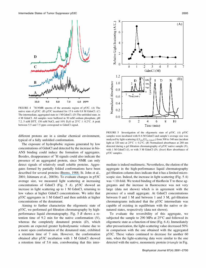
different protons are in a similar chemical environment,
typical of a fully unfolded conformation.
The exposure of hydrophobic regions generated by low
concentrations of GdmCl and detected by the increase in bis-
ANS binding could induce the formation of aggregates.
Besides, disappearance of 1H signals could also indicate the
presence of an aggregated protein, since NMR can only
detect signals of relatively small soluble proteins. Aggre-
gates formed by partially folded conformations have been
described for several proteins (Brems, 1988; St. John et al.,
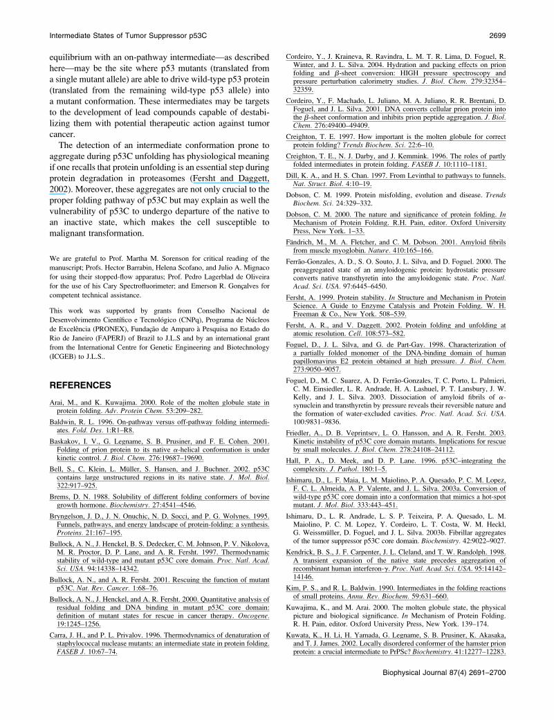
2001; Ishimaru et al., 2003b). To evaluate changes in p53C
average size, we measured light scattering at increasing
concentrations of GdmCl (Fig. 5 A). p53C showed an
increase in light scattering up to 1 M GdmCl, returning to
low values at higher GdmCl concentrations. We infer that
p53C aggregates in 1 M GdmCl and then unfolds at higher
concentrations of the denaturant.
Aiming to further characterize the oligomeric state of
p53C, we performed gel filtration chromatography by high-
performance liquid chromatography. Fig. 5 B shows a re-
tention time of 9.2 min for the native conformation (N),
whereas the completely denatured protein (D), which
presents an expected greater hydrodynamic volume due to
a more open conformation of the denatured state, exhibited
a retention time of 7 min. However, the conformation
obtained after p53C incubation with 1 M GdmCl showed
a retention time of 5.6 min, corroborating that this inter-
mediate is indeed multimeric. Nevertheless, the elution of the
aggregate in the high-performance liquid chromatography
gel filtration column does indicate that it has a limited micro-
scopic size. Indeed, the increase in light scattering (Fig. 5 A)
was ,10-fold. We tested binding of thioflavin T to these ag-
gregates and the increase in fluorescence was not very
large (data not shown) which is in agreement with the
presence of a small aggregate. At GdmCl concentrations
between 0 and 1 M and between 1 and 3 M, gel-filtration
chromatograms indicated that the p53C intermediate was
capable of existing in equilibrium with the native or de-
natured states, respectively (data not shown).
To evaluate the reversibility of this aggregate, we
subjected the sample to 290 MPa at 25�C and followed its
oligomeric state as a function of time (Fig. 6 A). Immediately
after pressurization, the light-scattering value decreased 50%
in comparison with the one obtained with the aggregated
p53C. These values continued to decrease for another 60
min, when the light-scattering data were similar to the one
detected with the native, monomeric protein (triangle in Fig.
FIGURE 5 Investigation of the oligomeric state of p53C. (A) p53C
samples were incubated with 0–6 M GdmCl and sample’s average size was
analyzed by light scattering (LSobs/LSno GdmCl) from 300 to 340 nm (incident
light at 320 nm) at 25�C 6 0.2�C. (B) Normalized absorbance at 280 nm
detected during a gel filtration chromatography of p53C native sample (N),
with 1 M GdmCl (I), or with 3 M GdmCl (D). (Inset) Raw absorbance of
p53C samples.
FIGURE 4 1H-NMR spectra of the aromatic region of p53C. (A) The
native state of p53C. (B) p53C incubated for 15 h with 0.8 M GdmCl. (C)
The intermediate, aggregated state in 1 M GdmCl. (D) The unfolded state, in
4 M GdmCl. All samples were buffered in 50 mM sodium phosphate, pH
7.2, 5 mM DTT, 150 mM NaCl, and 10% D2O at 25�C 6 0.2�C. A peak
between 5.5 and 7.5 ppm correspond to GdmCl signal.
Intermediate States of Tumor Suppressor p53C 2695
Biophysical Journal 87(4) 2691–2700

6). When we returned to atmospheric pressure, the p53C
began to aggregate again (Fig. 6 B), indicating that the p53C
aggregates induced by incubation with 1 M GdmCl were
highly specific. The susceptibility to pressure of these
aggregates indicates that they have a structure that excludes
water as found with some aggregates (Ferrao-Gonzales et al.,
2000; Silva et al., 2001; St. John et al., 2001; Foguel et al.,
2003). The liability to pressure is the characteristic of some
aggregates. However, there are some fibrillar structures that
are highly resistant to pressure. For example, at some
conditions, PrP fibrils become insensitive to pressure
(Cordeiro et al., 2004; Torrent et al., 2004) because of very
tight packing. Aggregates obtained by combined thermal and
pressure denaturation of p53C are also resistant to pressure
(Ishimaru et al., 2003b). As pointed out by Torrent et al.
(2004), different pathways of aggregation and amyloid
formation can lead to a different degree of compactness of
the final structures. It seems that early aggregates tend to be
more susceptible to pressure as recently shown by Niraula
et al. (2004) for the multimeric precursor of amyloid fibrils of
the disulfide-deficient mutant of hen lysozyme.
To further characterize the formation of the aggregates, we
performed kinetic unfolding measurements. Using a stopped-
flow fluorimeter, p53C was mixed with different amounts of
GdmCl and changes in intrinsic fluorescence emission were
followed over time. We observed an increase of the
emission, which can be well adjusted to a single exponential
profile (Fig. 7 A). There are no further changes in
fluorescence even up to 15 min (data not shown).
As generally expected, unfolding rate constants present
a dependence on GdmCl concentration (Fig. 7 B). In
principle, a single exponential behavior indicates no
apparent kinetic unfolding intermediate. However, measure-
ments of p53C unfolding by GdmCl show a dependence of
the rate constant on protein concentration (Fig. 8). These data
indicate the existence of at least one intermediate step in
FIGURE 6 Investigation of the reversibility of the p53C aggregates. (A)
p53C at 5 mM was allowed to aggregate through incubation with 1 M
GdmCl at 25�C 6 0.2�C for 24 h. The aggregated sample was subjected to
2.9 kbar at 25�C and light-scattering values from 300 to 340 nm (incident
light at 320 nm) were accompanied over time. (B) Light-scattering values
after return to atmospheric pressure.
FIGURE 7 Kinetic unfolding of p53C. (A) p53C (5 mM final concentra-
tion) was mixed with GdmCl (3.5 M final concentration) and intrinsic
fluorescence was followed. Signal shows a simple exponential dependence
on time. (B) Kinetic fluorescence changes rate upon p53C unfolding in the
indicated concentration of GdmCl. Experiments performed at 25�C 6
0.2�C. Details in Materials and Methods.
2696 Ishimaru et al.
Biophysical Journal 87(4) 2691–2700

which protein association occurs. The fact that increasing
protein concentration leads to a slower kinetic indicate that
p53C association/collapse is a step that anticipates sub-
sequent protein unfolding. The existence of a protein-
concentration dependence on the kinetics of p53 unfolding is
indicative of a high-order (bimolecular or higher) association
event. Thus, at low protein concentration, the unfolding is
faster because it occurs with no concurrent association. In
contrast, at high protein concentration the association
competes with the unfolding.
The kinetic data seem to corroborate the finding at
equilibrium conditions of an association/aggregation process
during unfolding. However, we could not identify any
change in light scattering during the unfolding kinetics,
indicating that the lifetime of the aggregated intermediate is
very short. Nevertheless, aggregation occurs and the in-
termediate accumulates in the refolding pathway (Fig. 9).
Under refolding conditions, the kinetic data show the
accumulation of aggregated material when the protein is
diluted from the unfolding condition (3 M GdmC) to low
concentrations of the denaturant (Fig. 9).
DISCUSSION
One of the first established principles for protein folding was
that the process should not be random (Levinthal, 1969).
Therefore, a number of theories have been proposed to
explain the protein folding and several of them are based on
the formation of intermediates (Kim and Baldwin, 1990;
Plotkin and Onuchic, 2002). Bovine growth hormone has
been shown to undergo a multistate denaturation process
with stable intermediates (Brems, 1988); human recombi-
nant g-interferon has an expanded intermediate at 0.9 M
GdmCl (Kendrick et al., 1998); Arc repressor protein has
a molten-globule conformation in its pressure-induced
monomeric state (Silva et al., 1992b). Nevertheless, each
intermediate is characteristic for a specific condition and
a specific protein. For instance, whereas p53C showed an
increase in secondary structure (a more negative CD signal)
for the intermediate state (Fig. 2), the recombinant human
growth hormone had much less intermolecular b-sheet in the
aggregates prepared in 0.75 M GdmCl compared with those
formed in buffer alone under high pressure (St. John et al.,
2001).
The relation structure-function is a fundamental subject
for the study of proteins. In this view, intermediates
constitute an issue of intense debate as to whether they
represent conformations essential for directing the protein
folding to the native state, or misfolded structures trapped in
physiologically nonrelevant local energy minima (Kim and
Baldwin, 1990; Bryngelson et al., 1995; Carra and Privalov,
1996; Creighton et al., 1996; Dill and Chan, 1997; Wagner
and Kiefhaber, 1999). Several experimental data have shown
that some intermediates are indeed specific folding inter-
mediates. Besides, studies of transition states have demon-
strated that intermediate conformations are very close to the
native state, although somewhat distorted (Oltzen et al.,
1994). On the other hand, some intermediate conformations
could represent energetically trapped, misfolded structures.
In the case of p53C, protein-engineering studies have
detected important kinetic intermediates for the proper
folding of the tetrameric C-terminal (Mateu et al., 1999).
For this domain, a transient, highly structured dimeric
intermediate was observed, although the transition from the
monomeric intermediate to the tetrameric native protein was
undetectable spectroscopically. The initial step of folding
behaved as a nucleation-condensation mechanism with an
FIGURE 8 Dependence of kinetic unfolding on p53C concentration.
p53C (final concentration as indicated) was mixed with GdmCl (to give
3.5 M final concentration) and data were analyzed as described in Materials
and Methods. (A) Kinetic rate constant versus protein concentration.
(Inset) Double log plot. Experiments performed at 25�C 6 0.2�C.
FIGURE 9 Formation of intermediate p53C aggregates from unfolded
protein. p53C unfolded in 3.0 M GdmCl was diluted into buffer to 5 mM
p53C (final concentration), containing GdmCl for (final concentrations)
0.050 M (a), 0.10 M (b), 0.20 M (c), and 0.40 M (d), and light scattering was
followed over time. All experiments were performed at 25�C 6 0.2�C.
Details in Materials and Methods.
Intermediate States of Tumor Suppressor p53C 2697
Biophysical Journal 87(4) 2691–2700

early transition state, whereas the spectroscopically silent
step followed the framework mechanism (Mateu et al.,
1999). Still, hydrogen exchange studies probed by chemical
denaturant and temperature have shown that no particular
folding intermediate is populated for this same domain
(Neira and Mateu, 2001).
A denaturing midpoint of 1.00 6 0.03 M GdmCl at 25�Cand a two-state transition for the p53C denaturation process
are consistent with a previous report (Bullock et al., 1997).
However, the CD signal and the bis-ANS binding both
reached a minimum and a maximum, respectively, at 1.0 M
GdmCl indicating that an intermediate structure exists at
a GdmCl concentration coincident with the midpoint value
obtained by intrinsic fluorescence. Intermediate conforma-
tions of p53C have been previously achieved by high
pressure (Ishimaru et al., 2003a,b), high temperatures
(Ishimaru et al., 2003b), and low pH (Bullock et al., 2000).
The conformation achieved by pH values below 5.5 has been
described as having large increases in tryptophan as well as in
ANS fluorescence, indicating a transition to an acid molten-
globule state (Bullock et al., 2000). However the species
obtained by high pressure at subzero temperatures resembles
that of the hot-spot mutant R248Q and exists in a preag-
gregating state (Ishimaru et al., 2003a). In contrast, those
achieved by high pressure at 37�C and high temperatures (up
to 55�C) are highly aggregating conformations (Nichols and
Matthews, 2001). In addition, a recent report by Friedler and
colleagues (Friedler et al., 2003) showed that the wild-type
p53C is a kinetically unstable protein at 37�C exhibiting an in
vitro unfolding rate of 1.9 3 10�5 s�1. Moreover, they also
showed that p53C unfolding induced by this temperature was
accompanied by the formation of large aggregates.
Interestingly, under GdmCl conditions where the in-
termediate was detected, the extension of aggregation was
intense, as demonstrated by light-scattering data and gel-
filtration chromatography (Fig. 5), and almost no peaks
could be detected by NMR (Fig. 4). On the other hand,
increasing the GdmCl concentration up to 4.0 M reversed the
aggregation and a soluble, although denatured, protein was
observed. A similar behavior was described with urea-
induced denaturation (Bullock et al., 1997). Therefore, p53C
denaturation by GdmCl occurred in two stages: formation of
an intermediate, partially folded, and aggregating structure at
1.0 M GdmCl, and further denaturation of the protein at
higher GdmCl concentrations.
Partially folded denatured structures are generally the
conformers responsible for protein aggregation (Brems,
1988; Sanz and Fersht, 1993), and aggregation is usually
considered to be an irreversible process (Kendrick et al.,
1998; Friedler et al., 2003). Consequently, the nature of the
intermediate state is essential for the understanding of the
aggregation pathway (Kendrick et al., 1998). Interestingly,
some human diseases, as senile systemic amyloidosis and
some types of cancer, involve wild-type proteins prone to
aggregate (Moll et al., 1996; Ostermeyer et al., 1996; Ferrao-
Gonzales et al., 2000). In the case of senile systemic
amyloidosis, aggregation of wild-type protein occurs
because of an intermediate, alternative conformation of the
protein (Ferrao-Gonzales et al., 2000). Exact protein folding
in vivo is assisted by a more complex system, which
obviously is not obtained in any in vitro condition, even for
a cell-free pull-down preparation. However, our equilibrium
and kinetic data strongly indicate that this off-pathway
oligomer is a stable intermediate, that once formed has a high
energetic barrier to be overcome. This behavior resembles
that of the prion protein, which gives origin to an aggregated
amyloid structure after incubating the native protein for
a period of time with different subdenaturing concentration
of urea (Baskakov et al., 2001).
Our results suggest that the equilibrium denaturation
processes of p53C induced by GdmCl is not a simple two-
state transition, but one that occurs via a partially folded
conformation, indicating the presence of an intermediate
state on the p53C folding/unfolding pathway. This in-
termediate has the propensity to undergo aggregation into
a state whose most interesting feature is the high content of
secondary structure. Assembly into aggregates in the
folding/unfolding pathway may contribute to restrict the
folding landscape. The reversible dissociation of these
aggregates by pressure is a clear indication that they have
water-excluded cavities (Silva et al., 2001; Foguel et al.,
2003). The limited changes in light scattering (;7-fold) also
indicate a prominent role of this aggregate to the folding
pathway. Small angle x-ray scattering are currently being
performed to characterize the precise size and shape of this
aggregate.
The reaction scheme in Fig. 10 is based on the equilibrium
and kinetic data described here. The aggregate is formed off-
pathway from the intermediate and imposes a constraint in
the folding landscape of the protein. The kinetic data clearly
demonstrate how high concentration of protein favors the
aggregation, a condition that would be present in a cellular
situation of loss-of-function mutation, such as that present
for the hot-spot mutant R248Q. The small aggregate in
FIGURE 10 Reaction scheme for p53C folding/unfolding pathway with
concurrent formation of aggregates from the intermediate. Although less
likely, the aggregates can also be formed from the unfolded protein (dashed
arrows).
2698 Ishimaru et al.
Biophysical Journal 87(4) 2691–2700
equilibrium with an on-pathway intermediate—as described
here—may be the site where p53 mutants (translated from
a single mutant allele) are able to drive wild-type p53 protein
(translated from the remaining wild-type p53 allele) into
a mutant conformation. These intermediates may be targets
to the development of lead compounds capable of destabi-
lizing them with potential therapeutic action against tumor
cancer.
The detection of an intermediate conformation prone to
aggregate during p53C unfolding has physiological meaning
if one recalls that protein unfolding is an essential step during
protein degradation in proteasomes (Fersht and Daggett,
2002). Moreover, these aggregates are not only crucial to the
proper folding pathway of p53C but may explain as well the
vulnerability of p53C to undergo departure of the native to
an inactive state, which makes the cell susceptible to
malignant transformation.
We are grateful to Prof. Martha M. Sorenson for critical reading of the
manuscript; Profs. Hector Barrabin, Helena Scofano, and Julio A. Mignaco
for using their stopped-flow apparatus; Prof. Pedro Lagerblad de Oliveira
for the use of his Cary Spectrofluorimeter; and Emerson R. Goncxalves for
competent technical assistance.
This work was supported by grants from Conselho Nacional de
Desenvolvimento Cientıfico e Tecnologico (CNPq), Programa de Nucleos
de Excelencia (PRONEX), Fundacxao de Amparo a Pesquisa no Estado do
Rio de Janeiro (FAPERJ) of Brazil to J.L.S and by an international grant
from the International Centre for Genetic Engineering and Biotechnology
(ICGEB) to J.L.S..
REFERENCES
Arai, M., and K. Kuwajima. 2000. Role of the molten globule state inprotein folding. Adv. Protein Chem. 53:209–282.
Baldwin, R. L. 1996. On-pathway versus off-pathway folding intermedi-ates. Fold. Des. 1:R1–R8.
Baskakov, I. V., G. Legname, S. B. Prusiner, and F. E. Cohen. 2001.Folding of prion protein to its native a-helical conformation is underkinetic control. J. Biol. Chem. 276:19687–19690.
Bell, S., C. Klein, L. Muller, S. Hansen, and J. Buchner. 2002. p53Ccontains large unstructured regions in its native state. J. Mol. Biol.322:917–925.
Brems, D. N. 1988. Solubility of different folding conformers of bovinegrowth hormone. Biochemistry. 27:4541–4546.
Bryngelson, J. D., J. N. Onuchic, N. D. Socci, and P. G. Wolynes. 1995.Funnels, pathways, and energy landscape of protein-folding: a synthesis.Proteins. 21:167–195.
Bullock, A. N., J. Henckel, B. S. Dedecker, C. M. Johnson, P. V. Nikolova,M. R. Proctor, D. P. Lane, and A. R. Fersht. 1997. Thermodynamicstability of wild-type and mutant p53C core domain. Proc. Natl. Acad.Sci. USA. 94:14338–14342.
Bullock, A. N., and A. R. Fersht. 2001. Rescuing the function of mutantp53C. Nat. Rev. Cancer. 1:68–76.
Bullock, A. N., J. Henckel, and A. R. Fersht. 2000. Quantitative analysis ofresidual folding and DNA binding in mutant p53C core domain:definition of mutant states for rescue in cancer therapy. Oncogene.19:1245–1256.
Carra, J. H., and P. L. Privalov. 1996. Thermodynamics of denaturation ofstaphylococcal nuclease mutants: an intermediate state in protein folding.FASEB J. 10:67–74.
Cordeiro, Y., J. Kraineva, R. Ravindra, L. M. T. R. Lima, D. Foguel, R.Winter, and J. L. Silva. 2004. Hydration and packing effects on prionfolding and b-sheet conversion: HIGH pressure spectroscopy andpressure perturbation calorimetry studies. J. Biol. Chem. 279:32354–32359.
Cordeiro, Y., F. Machado, L. Juliano, M. A. Juliano, R. R. Brentani, D.Foguel, and J. L. Silva. 2001. DNA converts cellular prion protein intothe b-sheet conformation and inhibits prion peptide aggregation. J. Biol.Chem. 276:49400–49409.
Creighton, T. E. 1997. How important is the molten globule for correctprotein folding? Trends Biochem. Sci. 22:6–10.
Creighton, T. E., N. J. Darby, and J. Kemmink. 1996. The roles of partlyfolded intermediates in protein folding. FASEB J. 10:1110–1181.
Dill, K. A., and H. S. Chan. 1997. From Levinthal to pathways to funnels.Nat. Struct. Biol. 4:10–19.
Dobson, C. M. 1999. Protein misfolding, evolution and disease. TrendsBiochem. Sci. 24:329–332.
Dobson, C. M. 2000. The nature and significance of protein folding. InMechanism of Protein Folding. R.H. Pain, editor. Oxford UniversityPress, New York. 1–33.
Fandrich, M., M. A. Fletcher, and C. M. Dobson. 2001. Amyloid fibrilsfrom muscle myoglobin. Nature. 410:165–166.
Ferrao-Gonzales, A. D., S. O. Souto, J. L. Silva, and D. Foguel. 2000. Thepreaggregated state of an amyloidogenic protein: hydrostatic pressureconverts native transthyretin into the amyloidogenic state. Proc. Natl.Acad. Sci. USA. 97:6445–6450.
Fersht, A. 1999. Protein stability. In Structure and Mechanism in ProteinScience. A Guide to Enzyme Catalysis and Protein Folding. W. H.Freeman & Co., New York. 508–539.
Fersht, A. R., and V. Daggett. 2002. Protein folding and unfolding atatomic resolution. Cell. 108:573–582.
Foguel, D., J. L. Silva, and G. de Part-Gay. 1998. Characterization ofa partially folded monomer of the DNA-binding domain of humanpapillomavirus E2 protein obtained at high pressure. J. Biol. Chem.273:9050–9057.
Foguel, D., M. C. Suarez, A. D. Ferrao-Gonzales, T. C. Porto, L. Palmieri,C. M. Einsiedler, L. R. Andrade, H. A. Lashuel, P. T. Lansbury, J. W.Kelly, and J. L. Silva. 2003. Dissociation of amyloid fibrils of a-synuclein and transthyretin by pressure reveals their reversible nature andthe formation of water-excluded cavities. Proc. Natl. Acad. Sci. USA.100:9831–9836.
Friedler, A., D. B. Veprintsev, L. O. Hansson, and A. R. Fersht. 2003.Kinetic instability of p53C core domain mutants. Implications for rescueby small molecules. J. Biol. Chem. 278:24108–24112.
Hall, P. A., D. Meek, and D. P. Lane. 1996. p53C–integrating thecomplexity. J. Pathol. 180:1–5.
Ishimaru, D., L. F. Maia, L. M. Maiolino, P. A. Quesado, P. C. M. Lopez,F. C. L. Almeida, A. P. Valente, and J. L. Silva. 2003a. Conversion ofwild-type p53C core domain into a conformation that mimics a hot-spotmutant. J. Mol. Biol. 333:443–451.
Ishimaru, D., L. R. Andrade, L. S. P. Teixeira, P. A. Quesado, L. M.Maiolino, P. C. M. Lopez, Y. Cordeiro, L. T. Costa, W. M. Heckl,G. Weissmuller, D. Foguel, and J. L. Silva. 2003b. Fibrillar aggregatesof the tumor suppressor p53C core domain. Biochemistry. 42:9022–9027.
Kendrick, B. S., J. F. Carpenter, J. L. Cleland, and T. W. Randolph. 1998.A transient expansion of the native state precedes aggregation ofrecombinant human interferon-g. Proc. Natl. Acad. Sci. USA. 95:14142–14146.
Kim, P. S., and R. L. Baldwin. 1990. Intermediates in the folding reactionsof small proteins. Annu. Rev. Biochem. 59:631–660.
Kuwajima, K., and M. Arai. 2000. The molten globule state, the physicalpicture and biological significance. In Mechanism of Protein Folding.R. H. Pain, editor. Oxford University Press, New York. 139–174.
Kuwata, K., H. Li, H. Yamada, G. Legname, S. B. Prusiner, K. Akasaka,and T. J. James. 2002. Locally disordered conformer of the hamster prionprotein: a crucial intermediate to PrPSc? Biochemistry. 41:12277–12283.
Intermediate States of Tumor Suppressor p53C 2699
Biophysical Journal 87(4) 2691–2700
Lane, D. P., and P. A. Hall. 1997. MDM2—arbiter of p53C’s destruction.Trends Biochem. Sci. 22:372–374.
Lashuel, H. A., D. Hartley, B. M. Petre, T. Walz, and P. T. Lansbury, Jr.2002. Neurodegenerative disease: amyloid pores from pathogenicmutations. Nature. 418:291.
Levinthal, C. 1969. How to fold graciously. In Proceedings of a MeetingHeld at Allerton House. I. L. Monticello, editor. University of IllinoisPress, Champaign, IL. 22–24.
Mateu, M. G., M. M. S. del Pino, and A. R. Fersht. 1999. Mechanism offolding and assembly of a small tetrameric protein domain from tumorsuppressor p53C. Nat. Struct. Biol. 6:191–198.
Moll, U. M., A. G. Ostermeyer, R. Haladay, B. Winkfield, M. Frazier, andG. Zambetti. 1996. Cytoplasmic sequestration of wild-type p53C proteinimpairs the G1 checkpoint after DNA damage. Mol. Cell. Biol. 16:1126–1137.
Neira, J. L., and M. G. Mateu. 2001. Hydrogen exchange of thetetramerization domain of the human tumor suppressor p53C probedby denaturants and temperature. Eur. J. Biochem. 268:4868–4877.
Nichols, N. M., and K. S. Matthews. 2001. p53C unfolding detected by CDbut not by tryptophan fluorescence. Biochem. Biophys. Res. Commun.288:111–115.
Niraula, T. N., T. Konno, H. Li, H. Yamada, K. Akasaka, and H.Tachibana. 2004. Pressure-dissociable reversible assembly of intrinsi-cally denatured lysozyme is a precursor for amyloid fibrils. Proc. Natl.Acad. Sci. USA. 23:4089–4093.
Oltzen, D. E., L. S. Itzhaki, N. F. El Masry, S. E. Jackson, and A. R. Fersht.1994. Structure of the transition state for the folding/unfolding of thebarley chymotrypsin inhibitor 2 and its implications for mechanisms offolding. Proc. Natl. Acad. Sci. USA. 91:10422–10425.
Ostermeyer, A. G., E. Runko, B. Winkfield, B. Ahn, and U. M. Moll. 1996.Cytoplasmically sequestered wild-type p53C protein in neuroblastoma isrelocated to the nucleus by a C-terminal peptide. Proc. Natl. Acad. Sci.USA. 93:15190–15194.
Pace, C. N. 1986. Determination and analysis of urea and guanidinehydrochloride denaturation curves. Meth. Enzymol. 131:266–280.
Piotto, M., V. Saudek, and V. Sklenar. 1992. Gradient-tailored excitationfor single-quantum NMR spectroscopy of aqueous solutions. J. Biomol.NMR. 2:661–665.
Plotkin, S. S., and J. N. Onuchic. 2002. Understanding protein folding withenergy landscape theory. Part I: Basic concepts. Q. Rev. Biophys.35:111–167.
Prives, C., and P. A. Hall. 1999. The p53C pathway. J. Pathol. 187:112–126.
Sacchettini, J. C., and J. W. Kelly. 2002. Therapeutic strategies for humanamyloid diseases. Nat. Rev. Drug Discov. 1:267–275.
Sanz, J. M., and A. R. Fersht. 1993. Rationally designing the accumulationof a folding intermediate of barnase by protein engineering. Bio-chemistry. 32:13584–13592.
Silva, J. L., D. Foguel, and C. A. Royer. 2001. Pressure provides newinsights into protein folding, dynamics and structure. Trends Biochem.Sci. 26:612–618.
Silva, J. L., L. Peng, M. Glaser, E. W. Voss, and G. Weber. 1992a. Effectsof hydrostatic pressure on a membrane-enveloped virus: high immuno-genicity of the pressure-inactivated virus. J. Virol. 66:2111–2117.
Silva, J. L., C. F. Silveira, A. Correia Jr., and L. Pontes. 1992b.Dissociation of a native dimer to a molten globule monomer. J. Mol.Biol. 223:545–555.
St. John, R. J., J. F. Carpenter, C. Balny, and T. W. Randolph. 2001. Highpressure refolding of recombinant human growth hormone frominsoluble aggregates. J. Biol. Chem. 276:46856–46863.
Torrent, J., M. T. Alvarez-Martinez, M. C. Harricane, F. Heitz, J. P.Liautard, C. Balny, and R. Lange. 2004. High pressure induces scrapie-like prion protein misfolding and amyloid fibril formation. Biochemistry.43:7162–7170.
Wagner, C., and T. Kiefhaber. 1999. Intermediates can accelerate proteinfolding. Proc. Natl. Acad. Sci. USA. 96:6716–6721.
2700 Ishimaru et al.
Biophysical Journal 87(4) 2691–2700