On Utilising Template and Feature-based Correspondence in Multi-view Appearance Models

A Viral Discovery Methodology for Clinical BiopsySamples Utilising Massively Parallel Next GenerationSequencingGordon M. Daly1, Nick Bexfield1, Judith Heaney1, Sam Stubbs1, Antonia P. Mayer1, Anne Palser2, Paul
Kellam2, Nizar Drou3, Mario Caccamo3, Laurence Tiley1, Graeme J. M. Alexander4, William Bernal5,
Jonathan L. Heeney1*
1 Department of Veterinary Medicine, The University of Cambridge, Cambridge, United Kingdom, 2 Wellcome Trust Sanger Institute, Wellcome Trust Genome Campus,
Hinxton, Cambridge, United Kingdom, 3 The Genome Analysis Centre, Norwich Research Park, Norwich, United Kingdom, 4 Department of Hepatology, Addenbrookes
Hospital, Cambridge University Hospitals National Health Services Foundation Trust, Cambridge, United Kingdom, 5 Institute of Liver Studies, King’s College London
School of Medicine, King’s College Hospital, Denmark Hill, London, United Kingdom
Abstract
Here we describe a virus discovery protocol for a range of different virus genera, that can be applied to biopsy-sized tissuesamples. Our viral enrichment procedure, validated using canine and human liver samples, significantly improves viral readcopy number and increases the length of viral contigs that can be generated by de novo assembly. This in turn enables theIllumina next generation sequencing (NGS) platform to be used as an effective tool for viral discovery from tissue samples.
Citation: Daly GM, Bexfield N, Heaney J, Stubbs S, Mayer AP, et al. (2011) A Viral Discovery Methodology for Clinical Biopsy Samples Utilising Massively ParallelNext Generation Sequencing. PLoS ONE 6(12): e28879. doi:10.1371/journal.pone.0028879
Editor: Patricia V. Aguilar, University of Texas Medical Branch, United States of America
Received August 26, 2011; Accepted November 16, 2011; Published December 21, 2011
Copyright: � 2011 Daly et al. This is an open-access article distributed under the terms of the Creative Commons Attribution License, which permits unrestricteduse, distribution, and reproduction in any medium, provided the original author and source are credited.
Funding: GD, NB, SS and AM are funded by the Wellcome Trust. AP and PK are funded by the Wellcome Trust and part of this work is supported by funding fromthe European Community’s Seventh Framework Programme (FP7/2007–2013) under the project EMPERIE, EC grant agreement number 223498’. The funders hadno role in study design, data collection and analysis, decision to publish, or preparation of the manuscript.
Competing Interests: The authors have declared that no competing interests exist.
* E-mail: [email protected]
Introduction
A variety of methods for identifying unknown viruses have been
reported, such as: degenerate primer PCR/amplification [1], viral
microarrays [2–4] and conventional sequencing. Low abundance
of viral sequences relative to total host nucleic acids usually
requires the use of viral enrichment and concentration procedures.
These include: filtration, ultracentrifugation and nuclease treat-
ment followed by random priming and amplification using the
sequence-independent single primer amplification (SISPA) method
or variations thereof [1,5–10] and/or with the Viral Discovery
cDNA-AFLP (VIDISCA) method [11–15]. These approaches
have generally been limited to liquid based samples (body fluids,
eluted swabs, culture supernatants and environmental samples).
NGS has shown great potential for novel virus discovery [16–19].
The use of NGS alone can be sufficient if the viral nucleic acids are
in sufficient abundance relative to host nucleic acids. However, as
we confirm here, clinical biopsy samples can present a problem
where even the depth of sequencing provided by NGS may be
insufficient to generate useful viral sequence contigs by de novo
assembly.
We have now established a broadly applicable approach for
viral nucleic acid enrichment from small biopsy sized clinical liver
tissue (e.g Tru-Cut), which combined with the Illumina NGS
platform could provide an effective tool for viral discovery.
Results
Detection of HCV reads from HCV infected human biopsysamples using the Illumina platform
We analysed frozen Hepatitis C virus (HCV) infected Tru-Cut
liver biopsies without viral enrichment to ascertain the limitations
of detection of virus in a small liver biopsy using the Roche 454
and the Illumina NGS platforms. Total RNA was extracted from
six biopsy samples (RNA integrity (RIN) values between 6 and 8)
and HCV infection was confirmed by PCR. 0.5 mg of RNA from
each sample were pooled and underwent SISPA (detailed in
materials and methods). The minimally amplified pooled material
was then mass sequenced on a single Illumina NGS lane. Mapping
of the short reads to HCV reference genomes from the Los Alamos
HCV database confirmed HCV infection with sub-type 3a.
However, the mapping clearly showed a paucity of viral
genome-coverage (12.5%) with a total of 32 HCV reads out of
,8 million (Fig. 1). tBLASTx analysis of the complete dataset of
viral fragments against all HCV genomes in the EMBL database
did not identify any further HCV reads. The lack of overlapping
viral sequence reads prevented de novo assembly of viral contigs,
making the use of the Illumina NGS platform and the SISPA
protocol alone a potentially ineffective technique for novel virus
discovery. The same process using the Roche 454 platform failed
to identify any HCV sequences.
PLoS ONE | www.plosone.org 1 December 2011 | Volume 6 | Issue 12 | e28879

Liver Cytosol/Pellet fractionation for viral enrichmentTo improve on published tissue extraction methods for viral
discovery [20] we compared different homogenization procedures.
We found the optimal procedure was the use of a TH Omni-
homogeniser/hard-tissue probe combination (Omni-International)
using a short pulse (15 seconds) in cold PBS with a dry ice freeze
thaw cycle, repeated three times, followed by RNAse and DNAse
digestion of the host nucleic acids as illustrated in figure 2a and
detailed in materials and methods. We found that pestle grinding
with Alumina did not break the liver cells as reliably as our freeze/
thaw-mechanical approach (determined microscopically).
We compared the two methods with dissected (2 mm3) biopsy
sized fragments of an HCV infected liver sample with and without
the presence of Benzonase as a nuclease to remove host nucleic
acids (Fig. 2b). Following nuclease treatment with both Turbo-
DNAse and RNAse1 (+/2 Benzonase) to remove host nucleic
acids, we extracted viral genomic material and residual host
nucleic acids using the Trizol RNA extraction method with
glycogen as a carrier. A modified SISPA protocol (materials and
methods) was used to amplify a minimum amount of material
needed for the Illumina NGS platform (1–3 mg) after cleaning and
size fractionation to remove sub-200 bp fragments. By removing
the sub-200 bp fragments we were effectively comparing NGS
readable nucleic acids whilst removing fragments likely to
represent residual host material that had survived exposure to
the nuclease treatment. We determined that the improved cell
breakage (determined microscopically) with the probe homogeni-
zation and freeze thaw cycles, correlated with an improved
concentration of HCV nucleic acids using identical amounts of
post-amplification dsDNA as an input to an HCV qPCR assay.
Furthermore, with both methods the addition of Benzonase
together with RNAse1 and Turbo DNAse reduced the HCV copy
number 2–6 fold.
To validate our extraction/enrichment approach (high-speed
tissue homogenization and freeze/thaw cycles) with Illumina
NGS, we used four pieces of liver tissue infected with canine
parvovirus 2 (CPV2), canine adenovirus 1 (CAV1), human
hepatitis B virus (HBV) and hepatitis C virus (HCV). Liver
samples were dissected into 2 mm3 pieces (equivalent to
approximately half of a Tru-Cut needle biopsy sample). Estimates
of the viral sequence copy number per liver sample (2 mm3) were
made by qPCR (Fig. 3) to confirm the presence of the viral nucleic
acids within the liver tissue and to estimate the maximum possible
starting viral copy number of the sample with the subsequent
processed fractions.
Next, our enrichment procedure and the total RNA and DNA
extractions were carried out on separate 2 mm3 dissected
fragments of each liver sample (materials and methods). Following
nuclease treatment with turbo-DNAse and RNAse1 (without
Benzonase) the SISPA based protocol was used to amplify a
minimum amount of material needed for the Illumina NGS
platform (1–3 mg) after cleaning and size fractionation to remove
sub-200 bp fragments. The level of enrichment achieved, per
nanogram of amplified material, for all four different virus infected
liver samples was quantified by qPCR (Fig. 4). The DNA virus
enrichment achieved a 104 increase in CPV viral nucleic acids
relative to the highest non-enriched CPV sample extract (total
RNA) and a similar enrichment level for CAV relative to the
highest non-enriched CAV sample extract (total DNA). The RNA
virus enrichment process achieved a 106 increase in both HBV
and HCV nucleic acids relative to the highest non-enriched
samples (total RNA).
Illumina NGS viral read and de novo assemblycomparison
Based on the qPCR results, ten samples were selected for mass
sequencing on the Illumina NGS platform with one sample library
per lane, indicated by an asterisk in Fig. 4. Mapping sequence
reads (Fig. 5) revealed complete or near complete (.95%)
coverage of the reference genomes for the virally enriched
samples. Viral nucleic acid point-coverage was orders of
magnitude (46101–8.56104) greater than total RNA (non-
Figure 1. NGS of HCV biopsy RNA. Extracted and pooled (66) HCV infected biopsy RNA was reverse transcribed and amplified prior to IlluminaNGS and mapped to the HCV reference 3a.NZL. NC_0009824 (Los Alamos HCV database).doi:10.1371/journal.pone.0028879.g001
Viral Discovery Using Clinical Biopsy Tissue
PLoS ONE | www.plosone.org 2 December 2011 | Volume 6 | Issue 12 | e28879

enriched) samples processed in the same manner. The increase in
viral sequences in enriched samples as a percentage of the total
NGS read set is shown in Fig. 6. Mass-sequencing of total DNA
from CAV and CPV infected samples was performed since qPCR
data indicated these samples had a higher viral genome copy
number than the RNA transcriptome samples (Fig. 4). This
correlated with the NGS read mapping analysis (Figs. 5 & 6) for
both the CPV and CAV total DNA samples.
In order to ascertain whether the increased viral read copy
number and reference coverage yielded significant improvement
in the size and viral reference coverage of the contigs that could be
generated by de novo assembly, we utilised CLC Genomics v4
(Katrinebjerg, 8200 Aarhus N, Denmark) and ABySS [21] de novo
assemblers in tandem and with varying stringencies. The data
from the unenriched CAV and HCV infected total RNA samples
indicated that RNA extraction and SISPA processing alone was
insufficient to generate contigs with significant viral reference
coverage. The HBV infected RNA fraction produced two contigs
covering one third of the HBV genome. In contrast, the virally
enriched samples produced contigs covering between 73–100% of
Figure 2. Enrichment methodologies. a: Illustration of the key steps in isolating and enriching viral nucleic acids relative to host nucleic acidsprior to sequencing by NGS. Liver tissue cells were broken in an isotonic buffer supplemented with BSA using a hard tissue probe and an Omni-Homogeniser with a dry-ice freeze/thaw step (repeated 36). To ensure that cells membranes were broken the lysate was checked microscopically. b:Comparison of Alumina/pestle-grinding against hard-tissue probe homogenisation with freeze/thaw cycles (+/2 the addition of Benzonase). (2 mm3
fragments were dissected from an HCV infected liver sample. Duplicate tissue samples were used for each protocol. HCV nucleic acids were measuredin triplicate by dual labelled qPCR assay with NIBSC standard.doi:10.1371/journal.pone.0028879.g002
Viral Discovery Using Clinical Biopsy Tissue
PLoS ONE | www.plosone.org 3 December 2011 | Volume 6 | Issue 12 | e28879

their reference genome (Fig. 7), with the HBV enriched
preparation producing a single contig spanning 100% of the
genome. The canine parvovirus was clearly transcriptionally active
and the contig coverage from the total RNA (non-enriched)
fraction was comparable to the virally enriched sample. For the
CAV and CPV total DNA extracts, the resulting assembled
contigs (1–2/sample) covered the reference genomes between
83.6% and 97% respectively, lower than, but comparable to the
enriched viral DNA samples.
Discussion
Identifying unknown viruses in clinical samples is technically
challenging and this is especially true for solid tissues when
compared with using less complex samples such as body fluids.
Additionally, whilst biopsy sampling is routine in the clinic it can
be limited for research purposes by ethical and safety consider-
ations. Often, the amount of material available for virological
analysis may be limited to a fraction of a small needle biopsy left-
over after diagnostic histopathological analysis. Detection of virus
is not always possible in serum and plasma even with nucleic acid
tests whilst the ‘occult’ virus may still be detectable in the viral
reservoir tissue [22–23].
Mass sequencing technologies provide a new avenue for viral
discovery as highlighted by Feng and Palacios [17–18] using the
Roche 454 NGS platform. The Illumina NGS platform also has
scope for novel viral discovery, particularly with recent technical
developments yielding improved sequence length and quality now
complementing its exceptional sequencing depth. However, our
preliminary work (Fig. 1) demonstrated that virally infected clinical
biopsy samples exist where extracted total nucleic acids processed
for NGS with the Illumina platform contain viral sequence at such
a low number relative to host nucleic acids that viral reference
coverage was sparse with no overlapping reads, precluding the
possibility of assembling larger contiguous viral sequences. This is
key in facilitating the discovery and characterization of novel
viruses by nucleotide and amino acid similarity to viral database
sequences or by predicted structural domain conservation.
Concurrent NGS with the Roche 454 platform failed to sequence
any viral nucleic acids that we could detect and for both platforms
the only assembled contigs over 200 bp corresponded to host
sequences.
We have shown that our enrichment methodology together with
the Illumina NGS platform works for a range of viruses using
sample sizes equivalent to a small needle biopsy. Cells in the
sample are disrupted and the cytosol isolated with encapsidated
virus intact. The resulting cytosolic fraction can be separated from
the cell debris and nuclei leaving a low viscosity solution that can
be nuclease treated directly or readily size fractionated by filtration
methods. qPCR analysis of equivalent samples processed for DNA,
RNA and with the viral enrichment method clearly demonstrate
the extent of enrichment (Fig. 4) as did mapping of the subsequent
NGS reads to the respective viral genomes to assess coverage
(Fig. 5) and target viral sequence as a percentage of the whole
NGS read set (Fig. 6). Subsequent de novo assembly of the NGS
reads using different assembly algorithms (fig. 7) demonstrates the
advantage of the enrichment procedure over whole RNA
transcriptome sequencing. Enriched sample de novo assembled
contigs using two different algorithms resulted in 73–100%
coverage of the viral reference genome with one to three contigs.
For the un-enriched RNA samples, only the CPV and the HBV
sample contigs included contigs to the reference virus (97% and
32.5% respectively). Interestingly, it is clear that viral DNA
genomes, from the CAV and CPV samples we used for validation,
could be reconstituted from total extracted DNA using NGS
alone. This is of particular interest if one considers that an
unknown (DNA) virus may be latent, in a non-encapsidated, non-
replicative and transcriptionally repressed form at a given time
point or cell type. This is of course a hypothetical, but in such a
case, total RNA and viral enrichment analysis would be unlikely to
work. Furthermore, with this methodology, the DNA can be
concurrently extracted from the nuclei containing pellet after
homogenization and prior to nuclease treatment, thus a three way
strategy might be considered when attempting to find an unknown
virus in a solid tissue biopsy. Our technique readily allows total
RNA, virus enrichable cytosolic fraction and total DNA to be
extracted with minimal loss of sample and minimal sample size.
In summary, our method can enrich a range of virus types that
can be sequenced using the Illumina NGS platform. Furthermore,
viral genomes can be largely reconstituted by currently available de
novo assembly algorithms. This approach is robust, enabling the
use of NGS for the detection and identification of novel viral
pathogens from small diagnostic biopsy samples without the
requirement to culture or isolate the virus first. We show this
technique works for liver tissue, which can be a difficult tissue for
extracting high quality RNA from and the HCV sample we used
for enrichment validation and NGS was approximately 8 mm3 of
very hard fibrotic tissue that had a low viral genome copy number
(Fig. 3) suggesting that the technique could probably be applied
successfully to most tissue types including other fibrotic diseased
tissues or joint collagen for example. Importantly, this methodol-
ogy is rapid and results in very little loss of sample.
Methods
Liver samples and processingHuman liver samples were acquired from the Institute of Liver
Studies, Kings College Hospital, London, University of London,
UK. Samples were obtained with patient written consent. This
Figure 3. Quantitative PCR (viral genome copy number in liversamples). A SYBR-green assay and plasmid standard was used for theCAV and CPV samples, whilst the HCV and HBV assays used duallabelled probed with NIBSC standards. Total RNA extracted for HCV andTotal DNA extracted for HBV, CPV and CAV. The Genomic strand copynumber for HCV was estimated by performing the RT step in thepresence of the reverse primer only.doi:10.1371/journal.pone.0028879.g003
Viral Discovery Using Clinical Biopsy Tissue
PLoS ONE | www.plosone.org 4 December 2011 | Volume 6 | Issue 12 | e28879

work forms part of a broader project with ethical approval
provided by the UK National Research Ethics Service, Cambridge
3 Research Ethics Committee, Cambridge CB21 5XB (REC
reference numbers 09/HO306/52, 09/HO306/60) and Kings
College Hospital Research Ethics Committee, London SE5 9RS
(REC reference number 04/Q0703/27).
Canine liver samples were acquired from the Blue Cross Animal
Hospital (CPV) and the Royal Veterinary College (CAV) with
informed and written owner consent acquired by both centers as per
the guidelines of the Royal College of Veterinary Surgeons (RCVS),
UK. The CAV and CPV liver samples represented legacy material
for which no ethical committee approval was required in the UK.
Individual samples were screened +ve for the following viruses
(Table. 1). After collection, liver tissue was stored at 280uCpending further analysis. Liver samples were divided into sections
measuring 2 mm3 and weighing ,15 mg comparable to approx-
imately half a Tru-Cut needle biopsy. Size rather than weight was
used for handling reasons in order to keep the samples as cold as
possible to minimize nucleic acid degradation.
No Enrichment. Total RNA extraction was performed on
liver tissue (Liver biopsy or 2 mm3) using the RNeasy Lipid Tissue
Mini Kit (Qiagen) according to the manufacturer’s instructions
with on column DNAse treatment. Total DNA extraction was
performed on liver tissue (2 mm3 or from nuclear pellet) using the
QIAamp DNA Mini Kit (Qiagen) and according to the
manufacturer’s instructions. Liver tissue was homogenized in the
relevant denaturant, according to the kit manufacturers
instructions, using a micropestle (Eppendorf).
Enrichment. Liver tissue (2 mm3) was immersed in 250 ml
ice cold 0.7% bovine albumen supplemented buffered saline
pH.7.2 and homogenised for 15 seconds on ice using an Omni
TH - Tissue Homogenizer and disposable (7 mm6110 mm)
‘Omni Tip’ hard-tissue probe (Omni-International). The
resulting homogenate was placed on dry ice for approximately
two minutes until frozen, and thawed quickly before returning to
ice. Homogenization followed by freezing and thawing was
repeated a further two times to disrupt the cells (.90%) while
leaving the nucleus intact (.90%) determined microscopically.
Samples were then spun at 6006g for 10 minutes at 4uC to pellet
the nuclei and large cellular aggregate. Non-particle protected
viral DNA and RNA was removed from the supernatant by
digestion with 30 U of turbo DNase [Ambion] and 25 U of RNase
Figure 4. Quantitative PCR (viral copy number estimations of prepared samples for NGS). For each viral liver sample, 2 mm3 of liver wasused separately for total RNA extraction, total DNA extraction, DNA viral enrichment and RNA viral enrichment. qPCR assays were performed using20 ng of each sample (estimated by Pico-green assay) as an input in triplicate. The samples subsequently mass sequenced are indicated with anasterisk.doi:10.1371/journal.pone.0028879.g004
Viral Discovery Using Clinical Biopsy Tissue
PLoS ONE | www.plosone.org 5 December 2011 | Volume 6 | Issue 12 | e28879
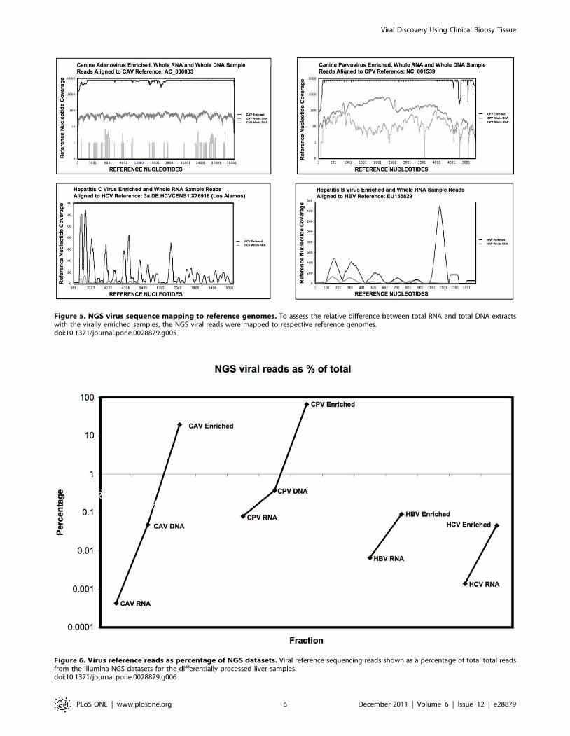
Figure 5. NGS virus sequence mapping to reference genomes. To assess the relative difference between total RNA and total DNA extractswith the virally enriched samples, the NGS viral reads were mapped to respective reference genomes.doi:10.1371/journal.pone.0028879.g005
Figure 6. Virus reference reads as percentage of NGS datasets. Viral reference sequencing reads shown as a percentage of total total readsfrom the Illumina NGS datasets for the differentially processed liver samples.doi:10.1371/journal.pone.0028879.g006
Viral Discovery Using Clinical Biopsy Tissue
PLoS ONE | www.plosone.org 6 December 2011 | Volume 6 | Issue 12 | e28879

One [Promega]) in 16DNAse buffer (Ambion) and incubated at
37uC for 90 minutes. Viral DNA (virally enriched) was extracted
using the High Pure Viral Nucleic Acid Kit (Roche) according to
the manufacturer’s instructions and eluted with 30 ml water. The
DNA virus extraction method used a polyA carrier not suitable for
the viral RNA extraction with the necessity for subsequent random
priming and amplification. We further established that glycogen
was not an efficient substitute for polynucleotides as a carrier in
silica column based methods. Therefore, viral RNA from the
cytosolic extract was extracted using Trizol LS (Invitrogen)
according to the manufacturer’s instructions with modifications.
20 mg glycogen was added prior to precipitation and left over-
night at 220uC and vortexed after 1 hour and 24 hours. The
Figure 7. De novo assembly of viral contigs. NGS reads for infected liver samples and their processed fractions, were used to generate contigsusing ABySS and CLC de novo assembly algorithms. Contigs (length over 200 nt) were mapped to the viral reference genomes with the length andcoverage indicated (solid black lines). Regions of the reference genomes not covered by the contigs are indicated with dashed lines.doi:10.1371/journal.pone.0028879.g007
Table 1. Characteristics of the different viral genera presentin the liver tissue used in this study.
Virus Group Envelope +/2 Genome Size (Kb)
CAV-1 I 2 dsDNA 30.5
CPV-2 II 2 ssDNA 2/+ 5
HCV-3 IV + ssRNA + 10.5
HBV VII + (Partial) dsDNA 3.1
doi:10.1371/journal.pone.0028879.t001
Viral Discovery Using Clinical Biopsy Tissue
PLoS ONE | www.plosone.org 7 December 2011 | Volume 6 | Issue 12 | e28879

precipitation was then frozen at 280uC and vortexed again prior
to centrifugation at 10 Kg for 30 minutes at 4uC. The RNA pellet
was rinsed three times in 75% ethanol, dried at room temperature
and the pellet re-suspended in 20 ml water supplemented with
80 U RNAse OUT (Invitrogen). RNA was passed through a
NucleoSpin RNA Clean-up XS column (Machery-Nagel) accor-
ding to the manufacturer’s instructions and eluted with 10 ml water
supplemented with 80 U RNAse OUT.
Alumina grinding and the addition of Benzonase was used in a
comparison study with a HCV infected liver sample using a
previously reported protocol [20]. This was adapted for use with a
needle biopsy sized tissue sample, obviating the need for
ultracentrifugation to pellet the virus. Briefly, the liver tissue
2 mm3 was ground with a micropestle (Eppendorf) with 20 mg of
100-mesh Alumina (Sigma-Aldrich) and 250 ml of ice cold 0.7%
bovine albumen supplemented buffered saline pH.7.2. The
homogenized sample was spun at 2.5 k rpm for 20 minutes at
4uC and passed through a 0.45 micron low bind filter (Millipore).
This was compared to an identical sized sample from the same
specimen processed for enrichment using our freeze/thaw
protocol described. Additionally, Benzonase was tested as an
additional nuclease to remove host nucleic acids, with both
processed samples using 20 U benzonase (Novagen).
Sequence independent Single Primer Amplification(SISPA): RT/2nd strand synthesis and productamplification
The protocol was adapted from previously reported SISPA
based protocols10,21–22 and using the SISPA primers/adaptors:
FR26RV-N GCCGGAGCTCTGCAGATATCNNNNNN
FR20RV GCCGGAGCTCTGCAGATATC.
First-strand cDNA synthesis was performed using 100 U of
Superscript III and the primer FR26RV-N (20 pmoles) using the
manufacturers recommended random primer protocol (Invitro-
gen, UK) at 50uC for 60 minutes in the presence of RNase- OUT
(Invitrogen, UK). cDNA and DNA were incubated with 2.5 U of
Klenow DNA polymerase (NEB) at 37uC for 60 minutes with an
inactivation step of 75uC for 20 minutes. PCR of the above
extension products was performed using 5 ml of cDNA or DNA in
a total reaction volume of 50 ml containing 2.5 mM MgCl2,
0.2 mM dNTPs, 16 Advantage 2 kit PCR buffer (Takara-
Clontech), 0.8 mM primer FR20RV and 1 U Advantage 2 kit
Polymerase Mix. Temperature cycling was performed as follows:
1 cycle of 95uC for 2 minutes, 20 cycles (minimum) of denaturing
at 95uC for 30 seconds, 65uC for 1 minute, 68uC for 30 seconds.
An additional extension of 3 minutes at 68uC was performed.
Further cycling was used when necessary to generate a final
output of 1–3 mg of dsDNA post-fractionation and clean-up.
Amplified DNA/cDNA was cleaned and fractionated using
Chroma-Spin-200 columns (Takara-Clontech) to remove sub-
200 bp nucleotide fragments to effectively standardise the
samples for Illumina NGS platform sequencing. Integrity and
quantity was assessed on a gel chip 7500 (Agilent) using a 2100
Bioanalyzer (Agilent Technologies UK Ltd). Concentration of
dsDNA was determined using the Quant-iT PicoGreen assay
(Invitrogen).
PCR diagnosticsA HCV PCR diagnostic kit (GeneAmpH EZ rTth RNA PCR,
AB life technologies) was used to confirm the presence of the virus,
according to the manufacturers instructions, in the HCV infected
biopsy samples and the post-transplantation excised HCV infected
liver sample used for the enrichment protocol.
qPCR AssaysViral nucleotide sequence copy numbers were measured in the
amplified material from the various fractions prior to NGS
sequencing as well as to estimate the viral genome sequence copy
number in the original samples. All liver samples were dissected
into equally sized, 2 mm3 pieces. Equivalent samples were used for
the different extraction procedures and all assays used a Rotorgene
6000 qPCR machine (Rotor-Gene Version 6.1 build 93, 2009,
Corbett Research/Qiagen). Data was analysed using Rotor gene
software, version 1.7 (Corbett Research/Qiagen).
CAV and CPV qPCR. CAV and CPV viral sequence copy
number was estimated relative to a standard to determine the
genome copy number of CAV and CPV in the liver tissue samples
and to acquire a viral nucleic acid copy number estimate from the
processed fractions. To produce the standard, amplified fragments of
CAV and CPV were individually cloned into pJET1.2 (Fermentas)
according to the manufacturer’s instructions. Chemically competent
E.coli cells (One Shot Top10 E.coli; Invitrogen) were transformed with
this construct and plasmid DNA was extracted from E.coli grown
overnight in liquid culture (Plasmid mini kit; Qiagen). Plasmid DNA
was linearised and quantified using the Quant-iT PicoGreen assay
(Invitrogen). A 10-fold dilution series was made by diluting the
plasmid DNA in polyinosinic-polycytidylic acid (Poly I:C) from 108
to 1 copy. qPCR was performed on each of the standards, DNA
extracted from infected tissue (for genome copy number) as well as
the processed DNA and cDNA fractions in triplicate. Amplification
was performed using 1 ml of template and 0.3 mM of each primer
using the QuantiTect SYBR Green Master Mix (Qiagen) and
distilled water to a final volume of 25 ml. After an initial PCR
activation step at 95uC for 15 minutes, 45 cycles of amplification
were performed consisting of 95uC for 15 seconds, 60uC for
30 seconds and 72uC for 30 seconds.
Hexon gene CAV1 primers
(forward) 59-TGCTGCCACAATGGTCTTAC-39 (reverse) 59-
CCACAGTGGGGTTTCTGAAC-39
NS1 gene CPV2 primers
(forward) 59- GACTGGGAATCGGAAGTTGA-39 (reverse)
59-CAATGCCAGCCTTGATCTTT-39
HCV and HBV qPCR viral genome copy number
estimation. These assays are based on the primers and probes
previously reported [24–25] and modified to fit the QuantiTect Virus
qPCR kit (Qiagen) according to the manufacturers instructions.
Briefly, the primer final concentrations were at 0.4 mM and the probe
final concentrations were at 0.2 mM. Polymerase activation/strand
denaturation was at 95uC for 5 minutes, and two step cycling was at
95uC for 15 seconds, and 60uC for 45 seconds for 45 cycles. Total
DNA or RNA was extracted from 2 mm3 of HBV and HCV liver
respectively.
HCV (forward) 59TGCTAGCCGAGTAGYGTTGG39
HCV (reverse) 59ACTCGCAAGCACCCTATCAG39
HCV (Probe) 59-[JOE] ACCACAAGGCCTTTCGCGAC
[BHQ1] – 3
HBV (forward)59CAACCTCCAATCACTCACCAAC39
HBV (reverse) 59ATATGATAAAACGCCGCAGACAC39
HBV (Probe) 59[CY3.5] TCCTCCAATTTGTCCTGGT-
TATCGCT [BHQ2] 39
qPCR standards and controls. HCV genotype 1a WHO
International Standard, NIBSC. 154,881 IU/ml. HBV Eurohep
standard reference 1, genotype A, HBsAg subtype adw, WHO
International Standard, NIBSC. 1,000,000 IU/ml. Viral nucleic
acids were extracted using the High Pure Viral Nucleic Acid Kit,
Roche. Viral load was calculated per ml of eluate with no
adjustment for kit extraction efficiency. Triplicate dilutions of the
HCV and HBV standards were run at neat and 1in 5 serial
Viral Discovery Using Clinical Biopsy Tissue
PLoS ONE | www.plosone.org 8 December 2011 | Volume 6 | Issue 12 | e28879

dilutions together with the samples in triplicate. HCV RNA
genome copy number was estimated by using single primers (not
pooled) for the reverse transcription step prior to the addition of
the second primer and the PCR cycles in order to exclude the –ve
strand from the genome copy estimation. The reverse tran-
scription step was at 50uC for 20 minutes.HCV and HBV qPCR viral sequence quantitation in
processed sample fractions. HCV and HBV viral
sequences in each fraction (RNA or DNA virus enriched, total
RNA and total DNA) were quantified against NIBSC controls
(HCV 06/100 and HBV 97/750) essentially as described
previously with minor modifications [24,26]. All reactions were
carried out in a total volume of 25 ml using Jumpstart Taq
readymix (Sigma) and specific forward and reverse primers (HBV1
and HBV2, MAD1 and MAD2 for HCV) were used at a final
concentration of 400 nM, and labelled taqman probes at 0.2 mM
(BS1 and MAD3 for HBV and HCV respectively). Following a
10 minute denaturation step at 95uC, 50 cycles of 30 seconds at
60uC and 30 seconds at 95uC were performed.
Illumina NGS protocolSequencing was performed using standard Illumina methods.
Libraries were created with the Illumina Paired End Genomic
DNA Sample Prep kit. Briefly, DNA was sheared into 200–400 bp
fragments using a Covaris AFA (Covaris, Woburn, MA), end
repaired and an A-overhang added. Illumina paired end adapters
were A-T ligated onto the ends of the fragments. Libraries were
PCR amplified and each sample sequenced using one lane of an
Illumina GA II sequencer generating 76 bp paired end reads. For
more detail see Quail et al [27].
Illumina NGS read set trimmingRead set trimming of the NGS data was performed using the
CLC Bio Trimming algorithms with the parameters: Failed reads
removed on import, ambiguous limit (2), 39 terminal nucleotides
removed (2), homopolymeric tracts of 30 bp removed, SISPA
primers removed and minimum number of nucleotides in read
allowed (38 nt).
Illumina NGS Viral Sequence copy number and referencecoverage estimation
To assess overall genome coverage and reference nucleotide
coverage, the Illumina reads were mapped to complete viral
reference genomes (.95% similarity) PVCCP-N for canine
parvovirus, AC_000003 for canine adenovirus and EU155829
for hepatitis B virus. Hepatitis C virus reads were first mapped to
the 61 reference genomes from the Los Alamos National
Laboratory HCV sequence database and the HCV genotype 3
reference sequence NC_009824 with the HCV sub-type 3a
consensus genome Ref.3a.DE.HCVCENS1.X76918 used subse-
quently for best coverage (,85%) and sample comparison using
CLC Bio Genomics Workbench v4 (Katrinebjerg, 8200 Aarhus N,
Denmark) and BWA [28]. The references were indexed using bwa
index –a IS with bwa aln and bwa sampe used to achieve the
paired end alignments. No read mapping was possible to the
59UTR and the first 400 bp of the core protein sequence for any of
the confirmed or unconfirmed HCV geno/subtypes indicative of a
novel subtype as defined by this hypervariable region.
Illumina NGS viral contig de novo assemblyDe novo assemblies of viral contigs were generated using the CLC
Bio Genomics Workbench v4 (Katrinebjerg, 8200 Aarhus N,
Denmark) and ABySS 1.2.7 [21] in tandem. For CLC de novo
assembly both paired and unpaired were co-assembled with a
paired read distance min/max at 180/380. For Abyss, a K-mer
size of 37 was used, K = 37 and the sequences were assembled as
pairs. Contig consensus sequences were mapped to the viral
reference genomes to determine total coverage as well as contig
size relative to genome size. Sequence alignment to the reference
genomes was performed using the sequence alignment algorithms
from CLC Genomics workbench v4.0 and bwa. For the CAV and
CPV enriched NGS data sets the viral reads were greater than
20% of the total. De novo assembly was ineffective probably due to
‘noise’ as a result of accumulated errors in the NGS data sets
containing millions of target viral reads. This occurred with both de
novo assemblers used and was overcome by a combination of
splitting the read sets to #1 million reads and increasing the
alignment stringency of the reduced sets.
Author Contributions
Conceived and designed the experiments: GD. Performed the experiments:
GD NB SS J. Heaney. Analyzed the data: GD AM AP ND. Contributed
reagents/materials/analysis tools: WB GA NB PK MC. Wrote the paper:
GD J. Heeney LT.
References
1. Reyes GR, Kim J (1991) Sequence-independent, single-primer amplification
(SISPA) of complex DNA populations. Mol Cell Probes 5: 473–481.
2. Palacios G, Quan P, Jabado OJ, Conlan S, Hirschberg DL, et al. (2007)
Panmicrobial oligonucleotide array for diagnosis of infectious diseases. Emerging
Inf Dis 13: 73–81.
3. Wang D, Urisman A, Liu YT, Springer M, Ksiazek TG, et al. (2003) Viral
discovery and sequence recovery using DNA microarrays. PLoS Biol 1: E2
(2003).
4. Wang D, Coscoy L, Zylberberg M, Avila PC, Boushey HA, Ganem D, et al.
(2002) Microarray-based detection and genotyping of viral pathogens. Proc Natl
Acad Sci USA 99: 15687–15692.
5. Allander T, Emerson SU, Engle RE, Purcell RH, Bukh J (2001) A virus
discovery method incorporating DNase treatment and its application to the
identification of two bovine parvovirus species. Proc Natl Acad Sci USA 98:
11609–11614 (2001).
6. Ambrose H, Clewley J (2006) Virus discovery by sequence-independent genome
amplification. Rev Med Virol 16: 365–383.
7. Delwart E (2007) Viral metagenomics. Rev Med Virol 17: 115–131.
8. Djikeng A, Halpin R, Kuzmickas R, DePasse J, Feldblyum J, et al. (2008) Viral
genome sequencing by random priming methods. BMC genomics 9: 5.
9. Telenius H, Carter NP, Bebb CE, Nordenskjold M, Ponder BA, et al. (1992)
Degenerate oligonucleotide-primed PCR: general amplification of target DNA
by a single degenerate primer. Genomics 13: 718–725.
10. Stang A, Korn K, Wildner O, Uberla K (2005) Characterization of virus isolates
by particle-associated nucleic acid PCR. J Clin Microb 43: 716–720.
11. De Souza Luna LK, Baumgarte S, Grywna K, Panning M, Drexler JF, et al.
(2008) Identification of a contemporary human parechovirus type 1 by
VIDISCA and characterisation of its full genome. Virol J 5: 26.
12. De Vries M, Pyrc K, Berkhout R, Vermeulen-Oost W, Dijkman R, et al. (2008)
Human parechovirus type 1, 3, 4, 5, and 6 detection in picornavirus cultures.
J Clin Microbiol 46: 759–762.
13. De Vries M, Deijs M, Canuti M, van Schaik BD, Faria NR, et al. (2011) A
sensitive assay for virus discovery in respiratory clinical samples. PLoS ONE 6:
e16118.
14. Pyrc K, Jebbink MF, Berkhout B, Van der Hoek L (2008) Detection of new
viruses by VIDISCA. Virus discovery based on cDNA-amplified fragment length
polymorphism. Meth Mol Biol 454: 73–89.
15. Tan LH, van Doorn R, van der Hoek L, Hien VM, Jebbink MF, et al. (2011)
Random PCR and ultracentrifugation increases sensitivity and throughput of
VIDISCA for screening of pathogens in clinical specimens. J Inf Dev Count 5:
142–148.
16. Feng H, Taylor JL, Benos PV, Newton R, Waddell K, et al. (2007) Human
transcriptome subtraction by using short sequence tags to search for tumor
viruses in conjunctival carcinoma. J Virol 81: 11332–11340.
17. Feng H, Masahiro Shuda, Yuan Chang, Moore PS (2008) Clonal integration of
a polyomavirus in human Merkel cell carcinoma. Science 319: 1096–1100.
Viral Discovery Using Clinical Biopsy Tissue
PLoS ONE | www.plosone.org 9 December 2011 | Volume 6 | Issue 12 | e28879

18. Palacios G, Druce J, Du L, Tran T, Birch C, et al. (2008) A new arenavirus in a
cluster of fatal transplant-associated diseases. N Engl J Med 358: 991–998.19. Weber G, Shendure J, Tanenbaum DM, Church GM, Meyerson M (2002)
Identification of foreign gene sequences by transcript filtering against the human
genome. Nat Gen 30: 141–142.20. Victoria J, Kapoor A, Dupuis K, Schnurr DP, Delwart EL, et al. (2008) Rapid
identification of known and new RNA viruses from animal tissues. PLoS Path 4:e1000163.
21. Simpson JT, Wong K, Jackman SD, Schein JE, Jones SJ, et al. (2009) ABySS: a
parallel assembler for short read sequence data. Genome Res 19: 1117–1123.22. Castillo I, Pardo M, Bartolome J, Ortiz-Movilla N, Rodrıguez-Inigo E, et al.
(2004) Occult hepatitis C infection in patients in whom the etiology ofpersistently abnormal results of liver-function tests is unknown. J Inf Dis 189:
7–14.23. De Marco L, Gillio-Tos A, Fiano V, Ronco G, Krogh V, et al. (2009) Occult
HCV infection: an unexpected finding in a population unselected for hepatic
disease. PloS1 4: e8128.
24. Candotti D, Temple J, Owusu-Ofori S, Allain JP (2004) Multiplex real-time
quantitative RT-PCR assay for hepatitis B virus, hepatitis C virus, and human
immunodeficiency virus type 1. J Virol Meth 118: 39–47.
25. Hsia CC, Purcell RH, Farshid M, Lachenbruch PA, Yu MW (2006)
Quantification of hepatitis B virus genomes and infectivity in human serum
samples. Transfusion 46: 1829–1835.
26. Weinberger KM, Bauer T, Bohm S, Jilg W (2000) High genetic variability of the
group-specific a-determinant of hepatitis B virus surface antigen (HBsAg) and
the corresponding fragment of the viral polymerase in chronic virus carriers
lacking detectable HBsAg in serum. J Gen Virol 81: 1165–1174.
27. Quail MA, Kozarewa I, Smith F, Scally F, Stephens PJ, et al. (2008) A large
genome center’s improvements to the Illumina sequencing system. Nat Meth 5:
1005–1010.
28. Li H, Durbin R (2009) Fast and accurate short read alignment with Burrows-
Wheeler transform. Bioinformatics 25: 1754–1760.
Viral Discovery Using Clinical Biopsy Tissue
PLoS ONE | www.plosone.org 10 December 2011 | Volume 6 | Issue 12 | e28879
Copyright © 2022 FDOKUMEN