A Twin Study of Metabolic Syndrome and Autonomic Tone
14
A Twin Study of Metabolic Syndrome and Autonomic Tone Anil Gehi, MD 1 , Rachel Lampert, MD 3 , Emir Veledar, PhD 2 , Forrester Lee, MD 3 , Jack Goldberg, PhD 4 , Linda Jones, BS 2 , Nancy Murrah, RN 2 , Ali Ashraf, MD 2 , and Viola Vaccarino, MD, PhD 2,5 1 Department of Medicine, Division of Cardiology, University of North Carolina at Chapel Hill School of Medicine, Chapel Hill, NC 2 Department of Medicine, Division of Cardiology, Emory University School of Medicine, Atlanta, GA 3 Department of Medicine, Yale University School of Medicine, New Haven, CT 4 Vietnam Era Twin Registry, Seattle, WA 5 Department of Epidemiology, Rollins School of Public Health, Emory University, Atlanta, GA Abstract Introduction—One possible mechanism of higher cardiovascular mortality associated with the metabolic syndrome (MetS) may be through abnormal modulation in autonomic tone. Methods and Results—We examined the association between the MetS and autonomic tone as measured by heart rate variability (HRV) among 288 twins from the Twins Heart Study. Of the 288 participants, 151 (52%) had the MetS. The MetS was associated with decreased HRV across all frequency ranges, and each additional MetS risk factor was associated with lower HRV. After adjustment for several potential confounders, very-low frequency (p<0.001), low frequency (p<0.001), and total power (p=.02) spectra of HRV remained significantly lower in twins with a progressively higher number of MetS components (18-50% decrease comparing twins with 5 risk factors to those with no risk factors). Among 87 twin pairs who were discordant for the number of MetS components, a one-unit increment in MetS components was associated with an 8% smaller very-low frequency (p=0.03) and a 15% smaller low frequency spectrum (p=0.002) comparing each twin with his brother. Conclusion—MetS was associated with lower HRV in a well-characterized sample of middle- aged male twins. This association persisted even after controlling for genetic and shared environmental factors accounted for by comparison within twin pairs. Abnormalities of autonomic tone, as evidenced by lower HRV, may be partly responsible for the higher rate of atrial fibrillation, coronary heart disease, cardiac death, and overall mortality seen in patients with the MetS. Keywords metabolic syndrome; heart rate variability; autonomic nervous system; twins Address for correspondence: Anil K. Gehi, MD Assistant Professor Cardiac Electrophysiology Division of Cardiology CB 7075 160 Dental Circle 6025 Burnett Womack Bldg Chapel Hill, NC 27599-7075 919-966-4743 [email protected]. NIH Public Access Author Manuscript J Cardiovasc Electrophysiol. Author manuscript; available in PMC 2010 April 1. Published in final edited form as: J Cardiovasc Electrophysiol. 2009 April ; 20(4): 422–428. doi:10.1111/j.1540-8167.2008.01363.x. NIH-PA Author Manuscript NIH-PA Author Manuscript NIH-PA Author Manuscript
-
Upload
independent -
Category
Documents
-
view
0 -
download
0
Transcript of A Twin Study of Metabolic Syndrome and Autonomic Tone

A Twin Study of Metabolic Syndrome and Autonomic Tone
Anil Gehi, MD1, Rachel Lampert, MD3, Emir Veledar, PhD2, Forrester Lee, MD3, JackGoldberg, PhD4, Linda Jones, BS2, Nancy Murrah, RN2, Ali Ashraf, MD2, and ViolaVaccarino, MD, PhD2,5
1Department of Medicine, Division of Cardiology, University of North Carolina at Chapel HillSchool of Medicine, Chapel Hill, NC2Department of Medicine, Division of Cardiology, Emory University School of Medicine, Atlanta,GA3Department of Medicine, Yale University School of Medicine, New Haven, CT4Vietnam Era Twin Registry, Seattle, WA5Department of Epidemiology, Rollins School of Public Health, Emory University, Atlanta, GA
AbstractIntroduction—One possible mechanism of higher cardiovascular mortality associated with themetabolic syndrome (MetS) may be through abnormal modulation in autonomic tone.
Methods and Results—We examined the association between the MetS and autonomic tone asmeasured by heart rate variability (HRV) among 288 twins from the Twins Heart Study. Of the288 participants, 151 (52%) had the MetS. The MetS was associated with decreased HRV acrossall frequency ranges, and each additional MetS risk factor was associated with lower HRV. Afteradjustment for several potential confounders, very-low frequency (p<0.001), low frequency(p<0.001), and total power (p=.02) spectra of HRV remained significantly lower in twins with aprogressively higher number of MetS components (18-50% decrease comparing twins with 5 riskfactors to those with no risk factors). Among 87 twin pairs who were discordant for the number ofMetS components, a one-unit increment in MetS components was associated with an 8% smallervery-low frequency (p=0.03) and a 15% smaller low frequency spectrum (p=0.002) comparingeach twin with his brother.
Conclusion—MetS was associated with lower HRV in a well-characterized sample of middle-aged male twins. This association persisted even after controlling for genetic and sharedenvironmental factors accounted for by comparison within twin pairs. Abnormalities of autonomictone, as evidenced by lower HRV, may be partly responsible for the higher rate of atrialfibrillation, coronary heart disease, cardiac death, and overall mortality seen in patients with theMetS.
Keywordsmetabolic syndrome; heart rate variability; autonomic nervous system; twins
Address for correspondence: Anil K. Gehi, MD Assistant Professor Cardiac Electrophysiology Division of Cardiology CB 7075 160Dental Circle 6025 Burnett Womack Bldg Chapel Hill, NC 27599-7075 919-966-4743 [email protected].
NIH Public AccessAuthor ManuscriptJ Cardiovasc Electrophysiol. Author manuscript; available in PMC 2010 April 1.
Published in final edited form as:J Cardiovasc Electrophysiol. 2009 April ; 20(4): 422–428. doi:10.1111/j.1540-8167.2008.01363.x.
NIH
-PA Author Manuscript
NIH
-PA Author Manuscript
NIH
-PA Author Manuscript

IntroductionThe metabolic syndrome (MetS) is associated with the development of diabetes mellitus andincreases the likelihood of cardiovascular disease (CVD) events, including atrial fibrillation,acute coronary syndrome, cardiac death, and overall mortality. 1-6 The risk of future CVDevents is more strongly correlated with the MetS than its individual components,2, 4although this issue is still debated. One possible mechanism underlying the link betweenMetS and CVD events is through abnormal modulation in autonomic tone, which has beenassociated both with MetS and CVD risk. 7-9 Specifically, increased sympathetic activity10and decreased parasympathetic activity11 have been associated with a higher risk of suddendeath. Both the sympathetic and parasympathetic nervous system have been implicated inmediating atrial fibrillation.12 Experimental evidence also suggests an interaction of theautonomic nervous system with the development of vascular atheroma and occlusion.13
Heart rate variability (HRV) is a complex measure of heart rate modulation that incorporatesboth sympathetic and parasympathetic effects as well as their interaction.14, 15 Whendetermined from 24-hour electrocardiographic recordings, depressed HRV predicts adversecardiovascular events.16-18 In particular, reduced HRV is associated with an increased riskof atrial fibrillation, myocardial infarction, congestive heart failure, coronary heart diseasedeath, and total mortality.16-19
While several pilot studies suggest an association between the MetS and HRV, these studiesare limited by modest sample size and short-term HRV recordings.7, 9, 20 Short-term HRVrecordings (i.e., 2-5 minutes rather than 24-hour) do not quantify the very-low and ultra-lowfrequency components of HRV, which are the most powerful predictors of adverse CVDevents.14, 16 In addition, prior studies have not adjusted for important confounders such asdepression and physical activity, nor have these studies standardized the schedule andactivity of participants during ambulatory ECG recording21, 22
Studies using discordant twin pairs provide unique insight because similarities in geneticbackground and early environment between twin siblings reduce variation resulting fromunmeasured potential confounders. In addition, a study utilizing twins may provide insightinto the genetic or other familial component of any association. The purpose of this study,therefore, was to further examine the association between the MetS and autonomic tone, asmeasured by HRV, using a well-controlled study of twins. In addition, a study of MetS andautonomic function utilizing twins provides insight into the genetic and other familialcomponent of any association.
MethodsParticipants
The Twins Heart Study (THS) is an investigation of psychological, behavioral andbiological risk factors for subclinical cardiovascular disease using twins. Participants wereselected from the Vietnam Era Twin (VET) Registry, a cohort of 7369 middle-aged male-male twin pairs both of whom served in the United States military during the Vietnam War.23, 24 Characteristics of the VET Registry have been previously reported.25 The THSincluded 360 twins from the VET Registry all born between 1946 and 1956 (>90% of thetwins in the VET registry fall into this range). The methods of construction of this samplewere also described before 26-28. Briefly, the twins were free of a selfreported previousdiagnosis of cardiovascular disease based on survey data collected in 199029, including aprevious diagnosis of myocardial infarction, coronary heart disease, angina, congestive heartfailure or stroke, or previous coronary angioplasty or coronary bypass surgery. From thisgroup, random samples of twins in two strata were selected: one stratum included twins
Gehi et al. Page 2
J Cardiovasc Electrophysiol. Author manuscript; available in PMC 2010 April 1.
NIH
-PA Author Manuscript
NIH
-PA Author Manuscript
NIH
-PA Author Manuscript

discordant for a lifetime history of major depression; and in a second stratum, neither twinhad a history of depression. All twins were examined, in pairs, at the Emory UniversityGeneral Clinical Research Center between March 2002 and March 2006. The GCRCadmission lasted approximately 27 hours and the entire data collection for the studyoccurred during this time. The two twins maintained an identical schedule while in theGCRC under the supervision of study personnel. Activity was limited to ambulation withinthe Emory facilities. All assessment, including ambulatory ECG monitoring, began andended at the same time. This protocol was approved by the Institutional Review Board atEmory University, and all participants provided written informed consent.
Metabolic syndromeWe used the 2005 American Heart Association (AHA), National Heart Lung and BloodInstitute (NHLBI) definition of the MetS,30 which is based on any 3 of the following 5criteria: waist circumference of ≥40 inches, triglycerides ≥150 mg/dL or drug treatment forelevated triglycerides, HDL-C <40 mg/dL or drug treatment for reduced HDL-C, systolicblood pressure (BP) ≥130 mmHg or diastolic BP ≥85 mmHg or drug treatment forhypertension, and fasting glucose ≥100 mg/dL or drug treatment for elevated glucose.30
Heart rate variabilityFor the measurement of HRV, participants wore an ambulatory ECG (Holter) monitor (GEMarquette SEER digital system) for 24 hours. Twin pairs were studied at the same time andtheir recording times, schedule, and activity level during Holter monitoring were matched.The methodology of HRV acquisition and analysis has been previously described.16, 31-33All recordings were manually processed and edited to ensure accurate identification of QRScomplexes. For frequency domain analysis, the heart rate spectrum was computed using afast Fourier transformation with a Parzen window. As described previously,16 the variousbands of power spectrum were defined as follows: ultra-low frequency (ULF) < 0.0033 Hz,very-low frequency (VLF) 0.0033 to <0.04 Hz, low frequency (LF) 0.04 to <0.15 Hz, highfrequency (HF) 0.15 to <0.40 Hz, and total power (TP) <0.40 Hz. Recordings with >20%interpolation or <18 recorded hours of Holter data were excluded from the analysis.
Other measurementsA thorough medical history and a physical exam were obtained from all participants.Abdominal and hip circumference was measured. Systolic and diastolic blood pressure wasmeasured using a standard sphygmomanometer on the right arm with the participant in theseated position after 10 minutes of rest. Blood pressure was taken as an average of twomeasurements 5 minutes apart. Venous blood samples were drawn for the measurements ofglucose and lipid profile after an overnight fast. Total triglycerides were determined byenzymatic methods (Beckman Coulter Diagnostics, Fullerton, CA). Direct high-densitylipoprotein (HDL) and low-density lipoprotein (LDL) cholesterol were measured withhomogeneous assays (Equal Diagnostics, Exton, PA). Glucose levels were measured on theBeckman CX7 chemistry autoanalyzer. Cigarette smoking was classified into current versusnot current smoker. Physical activity was determined by means of a modified version of theBaecke Questionnaire of Habitual Physical Activity.34 For this analysis, we used the globalphysical activity score, higher scores indicating more physically active. To measuredepressive symptoms, we administered the Beck Depression Inventory-II (BDI-II), astandardized scale of depressive symptoms with higher scores indicating more severedepression.35 For this analysis, we used a cut point of 14 to indicate the presence of at leastmild depression.35
Gehi et al. Page 3
J Cardiovasc Electrophysiol. Author manuscript; available in PMC 2010 April 1.
NIH
-PA Author Manuscript
NIH
-PA Author Manuscript
NIH
-PA Author Manuscript

Statistical analysisBaseline characteristics were compared between study participants with and without theMetS. P values were corrected for the correlation between co-twins using generalizedestimating equations (GEE) for categorical variables and mixed effects models forcontinuous variables. All HRV measurements were log-transformed to normalize theirdistribution for analysis.
Two approaches were used to examine the association between MetS (and its individualcomponents) and HRV: a) considering twins as separate individuals, in which all twins wereeligible for inclusion regardless of whether their brother was available for analysis; and b)within twin pairs approach (co-twin control analysis). The within-pair effects are inherentlycontrolled for demographic, shared familial (including genetics) and environmentalinfluences; in addition, daily activities and other environmental factors during theambulatory ECG recording are controlled since co-twins were examined at the same timeand under the same conditions. If an association is present both when analyzing twins asindividuals and within twin pairs, this suggests that there are not familial confoundingfactors.
In the analysis of all twins as individuals, we used mixed effects regression models andaccounted for the twin pairs using a random effect term for each pair. Analyses wererepeated after adjusting for age, current smoking, depression, physical activity, medications(beta-blocker, renin-angiotensin blocker, statin, aspirin, anti-depressant), and creatinineclearance. To see if there was a gradient pattern to the association between MetS and HRV,we compared the mean HRV in each frequency band for each additional component of theMetS (from 0 components to all 5 components). To ease interpretation, and in order todisplay the actual magnitude of the difference, we expressed the results as percent differenceof the nontransformed values by using the following formula: [1-(expβ)] × 100 (%).Spearman correlation coefficients were also calculated between the individual componentsof the MetS and the spectral components of HRV as continuous variables. Finally, wecompared mean HRV between individuals with and without the MetS, again adjusting forpotential confounding factors.
We then performed within-pair analyses, which examined differences in HRV between co-twins in each pair who were discordant for the MetS component of interest. Initially weconstructed twin pair subgroups who were discordant for the MetS and for each individualMetS component and compared HRV values between the discordant twins. Next, to examinethe cumulative impact of increasing MetS risk factors on HRV, we defined discordance as adifferent number of MetS components within a twin pair. We fitted mixed effects modelsadapted for twin research36 in which the within-pair parameter expressed the differencebetween twins who were discordant for number of MetS components, and was analyzed asan ordinal variable describing the departure of each twin from the pair average. Theseanalyses were repeated after stratification according to zygosity. A twin study design is auseful tool in uncovering genetic influences not only on specific traits, but also on specificassociations. If the association is due to genetic factors, then it should be found withindizygotic (DZ) twin pairs, who share on average only 50% of their genes, but not withinmonozygotic (MZ) twin pairs, who share 100% of their genes. All of the analyses wereperformed using Statistical Analysis Software (version 9.13, SAS Institute, Inc, Cary, NC).
ResultsAnalyses of all twins as individuals
From the 360 twins in this study, 288 (80%) had Holter recordings adequate for HRVanalyses (18 hours or more of recording with at least 80% noninterpolated intervals). Of
Gehi et al. Page 4
J Cardiovasc Electrophysiol. Author manuscript; available in PMC 2010 April 1.
NIH
-PA Author Manuscript
NIH
-PA Author Manuscript
NIH
-PA Author Manuscript

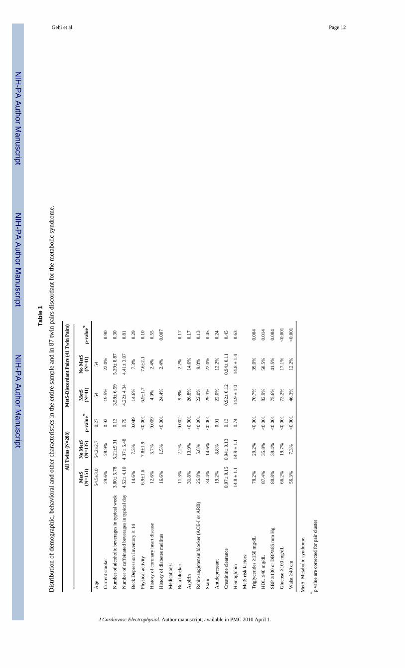
these 288 participants, 151 (52%) had the MetS. This sample included 115 twin pairs and 58unpaired twins (resulting from exclusion of the co-twin). Twins with the MetS had higherlevels of depressive symptoms, were less physically active, and were more likely to betaking beta-blocker, aspirin, renin-angiotensin blocker, statin, and antidepressantmedications (Table 1).
When the individual components of the MetS were considered as dichotomous variables (asdefined by the updated AHA/NHLBI criteria) in the overall sample, most of them showedassociations with at least one HRV variable. In adjusted analyses, associations persisted forhypertriglyceridemia (p<0.05 for ULF, VLF, LF, TP, ranging from 16% to 21% lowerHRV), hypertension (p<0.05 for VLF, LF, 16% and 20% lower, respectively),hyperglycemia (p<0.05 for LF, 17% lower), and waist circumference (p<0.05 for LF, 18%lower). In contrast, after adjustment for possible known confounders such as physicalactivity and smoking, HDL did not show significant associations with any of the HRVfrequency bands in multivariable analysis.37-40
The MetS was associated with decreased HRV across all frequency ranges when consideredas a dichotomous variable (Table 2). After adjustment for possible confounders, the VLF,LF, and TP spectra of HRV remained significantly lower in participants with MetS. Inaddition, HRV decreased progressively with higher number of MetS components (Figure 1),and participants with all 5 components had HRV values between 18% and 50% lower thanthose with no risk factors. Exclusion of participants with diabetes or coronary heart diseaseresulted in no substantial change in these results.
Analyses within twin pairsOf all twin pairs, 41 were discordant for metabolic syndrome. In general, differences insubject characteristics showed trends similar to the analyses of twins as individuals but wereless pronounced within these matched pairs. When analyses were restricted to twin pairswho were discordant for each MetS component, analyses of the individual components ofthe MetS revealed a similar pattern to analyses of twins as individuals, although differenceswere somewhat less marked. The limited number of twin pairs who were discordant for eachof the MetS components (n=24 to 42, depending on the MetS component), however,minimized the power of this analysis. Only hypertension and waist circumference weresignificantly associated (p<0.05) with HRV (VLF and LF, 24% to 40% lower forhypertension, respectively, and from 20% to 25% lower HRV for waist circumference) intwin pairs who were discordant for these components. When the analysis was repeatedamong the 41 twin pairs who were discordant for the MetS, VLF and LF were significantlyassociated with the MetS in unadjusted analysis and showed a borderline association inadjusted analysis (Table 3).
When we considered the number of MetS components, there were 87 pairs who werediscordant for number of MetS components. Within-pair analyses for differences in HRVbetween co-twins who differed for number of components confirmed a significantly lowerVLF and LF spectra of HRV for each incremental component of the MetS. After adjustmentfor other covariables as above, a one-unit increment in MetS components was associatedwith an 8% lower VLF (p=0.03) and a 15% lower LF spectrum (p=0.002) comparing eachtwin with his brother (Table 3). Results were no different within MZ and DZ twin pairsexamined separately. Among MZ twins, who share 100% of their genetic material, for eachadditional MetS component there was a 10% lower VLF (p=0.03) and a 15% lower LF(p=0.01) HRV comparing each twin with his brother.
Gehi et al. Page 5
J Cardiovasc Electrophysiol. Author manuscript; available in PMC 2010 April 1.
NIH
-PA Author Manuscript
NIH
-PA Author Manuscript
NIH
-PA Author Manuscript

We repeated analyses without adjusting for medications, which may have been prescribedfor treatment of MetS risk factors (beta-blockers, renin-angiotensin blockers and statins).The results were very similar and are not shown.
DiscussionWe found a strong and consistent association between the MetS and lower HRV in a well-characterized sample of middle-aged male twins. There was a gradient to the associationsuch that participants with an increasing number of components of the MetS hadprogressively lower HRV. In addition, within-pair analyses of co-twins, which accounted forunmeasured sociodemographic, lifestyle and familial factors shared by twins being raised inthe same family, as well as environmental influences during ECG monitoring, demonstrateda robust association of the MetS with HRV (specifically VLF and LF). Because wedemonstrated that an association between MetS and lower HRV was present in bothanalyses of twins as individuals as well as within twin pairs, we also can eliminate familialconfounding factors. Of all the HRV spectra, VLF and LF showed persistent associations inwithin-pair analyses, and each incremental MetS component was associated withprogressively lower VLF and LF HRV comparing co-twins.
Several prior studies have examined the association between the MetS and HRV.7, 9, 20,41-43 The presence of the MetS in 2359 patients from the Atherosclerosis Risk inCommunities (ARIC) study was associated with reduced LF and HF components of HRV.20In a study of 2197 participants in the Whitehall II study, each individual component of theMetS and the MetS as a whole were associated with reduced LF and HF power componentsof HRV.9 In the Cardiovascular Health Study, the presence of >2 components of the MetSwas associated with decreased ULF and TP components of HRV.43 Most of these priorstudies, however, have been limited by their measurement of short-term (LF and HF) HRV.Importantly, short-term HRV does not measure fluctuation in beat-to-beat intervals withcycles longer than 5 minutes. It is the VLF and ULF spectra that have the greatest prognosticutility for CVD.14, 16 Although VLF can be calculated in short-term recordings (≤ 5 min),it is a dubious measure and should be avoided.31 Other prior studies did not adjust forimportant potential confounders, such as depression and physical activity, nor was HRVrecorded in a controlled setting.43 Our study also has the advantage of twins analyses,which may eliminate other unmeasured confounders.
There are several important implications of our findings. These results support the integralrole of the autonomic nervous system in patients with the MetS. Whether the inciting factorleading to the MetS is obesity44 or a primary abnormality of autonomic tone leading to adisturbance in the hypothalamus-pituitary-adrenal axis45 is unclear. Due to the cross-sectional design of our study, this cannot be determined from our results. As demonstratedin the postmyocardial infarction setting and in patients without overt coronary heart disease,abnormal HRV predicts mortality, cardiac death, and arrhythmic death.16, 18, 46 Inparticular, the ULF and VLF components of HRV are more important predictors than the LFand HF components. The VLF component, which showed a robust association with theMetS in our study, is dependent on the presence of parasympathetic activity47 and isassociated with arrhythmic death.16
There are several strengths of our study. We assessed and adjusted for importantconfounding factors associated with HRV. In addition, because participants in our studywere twins, within-pair analyses of co-twins accounted for familial and genetic factorsshared by twins. Because co-twins were examined at the same time, environmentalinfluences during ECG monitoring were also controlled. There are, however, severallimitations of our study to consider. Despite our controlled design, it is possible that the
Gehi et al. Page 6
J Cardiovasc Electrophysiol. Author manuscript; available in PMC 2010 April 1.
NIH
-PA Author Manuscript
NIH
-PA Author Manuscript
NIH
-PA Author Manuscript

association between the MetS and HRV may be influenced by other unmeasuredconfounding variables. In particular, obstructive sleep apnea has been associated with boththe MetS and HRV but was unmeasured in our population.48, 49 Also, the participantsincluded in our study were all male middle-aged military veterans, so the results may notgeneralize to other populations.
In conclusion, in predominantly healthy middle-aged men, the MetS is associated withdecreased HRV. These findings suggest that abnormalities of the autonomic tone asevidenced by HRV may be partly responsible for the adverse cardiovascular events such asatrial fibrillation, sudden death, or acute coronary syndrome seen in patients with the MetS.
AcknowledgmentsWe gratefully acknowledge the continued cooperation and participation of the members of the Vietnam Era TwinRegistry. Without their contribution this research would not have been possible.
This work was supported by NIH (R01 HL68630, R01 AG026255, and K24HL077506); the American HeartAssociation (0245115N), and Emory University General Clinical Research Center (MO1-RR00039). The UnitedStates Department of Veterans Affairs has provided financial support for the development and maintenance of theVietnam Era Twin (VET) Registry. Numerous organizations have provided invaluable assistance, including: VACooperative Study Program; Department of Defense; National Personnel Records Center, National Archives andRecords Administration; the Internal Revenue Service; NIH; National Opinion Research Center; National ResearchCouncil, National Academy of Sciences; the Institute for Survey Research, Temple University.
References[1]. Aguilar D, Fisher MR, O'Connor CM, Dunne MW, Muhlestein JB, Yao L, Gupta S, Benner RJ,
Cook TD, Edwards D, Pfeffer MA. Metabolic syndrome, C-reactive protein, and prognosis inpatients with established coronary artery disease. Am Heart J. 2006; 152:298–304. [PubMed:16875914]
[2]. Gami AS, Witt BJ, Howard DE, Erwin PJ, Gami LA, Somers VK, Montori VM. Metabolicsyndrome and risk of incident cardiovascular events and death: a systematic review and meta-analysis of longitudinal studies. J Am Coll Cardiol. 2007; 49:403–414. [PubMed: 17258085]
[3]. Hu G, Qiao Q, Tuomilehto J, Balkau B, Borch-Johnsen K, Pyorala K. Prevalence of the metabolicsyndrome and its relation to all-cause and cardiovascular mortality in nondiabetic European menand women. Arch Intern Med. 2004; 164:1066–1076. [PubMed: 15159263]
[4]. Malik S, Wong ND, Franklin SS, Kamath TV, L'Italien GJ, Pio JR, Williams GR. Impact of themetabolic syndrome on mortality from coronary heart disease, cardiovascular disease, and allcauses in United States adults. Circulation. 2004; 110:1245–1250. [PubMed: 15326067]
[5]. Wilson PW, D'Agostino RB, Parise H, Sullivan L, Meigs JB. Metabolic syndrome as a precursorof cardiovascular disease and type 2 diabetes mellitus. Circulation. 2005; 112:3066–3072.[PubMed: 16275870]
[6]. Watanabe H, Tanabe N, Watanabe T, Darbar D, Roden DM, Sasaki S, Aizawa Y. Metabolicsyndrome and risk of development of atrial fibrillation: the Niigata preventive medicine study.Circulation. 2008; 117:1255–1260. [PubMed: 18285562]
[7]. Brunner EJ, Hemingway H, Walker BR, Page M, Clarke P, Juneja M, Shipley MJ, Kumari M,Andrew R, Seckl JR, Papadopoulos A, Checkley S, Rumley A, Lowe GD, Stansfeld SA, MarmotMG. Adrenocortical, autonomic, and inflammatory causes of the metabolic syndrome: nestedcase-control study. Circulation. 2002; 106:2659–2665. [PubMed: 12438290]
[8]. Grassi G, Seravalle G. Autonomic imbalance and metabolic syndrome: unravelling interactions,mechanisms and outcomes. J Hypertens. 2006; 24:47–49. [PubMed: 16331100]
[9]. Hemingway H, Shipley M, Brunner E, Britton A, Malik M, Marmot M. Does autonomic functionlink social position to coronary risk? The Whitehall II study. Circulation. 2005; 111:3071–3077.[PubMed: 15939818]
Gehi et al. Page 7
J Cardiovasc Electrophysiol. Author manuscript; available in PMC 2010 April 1.
NIH
-PA Author Manuscript
NIH
-PA Author Manuscript
NIH
-PA Author Manuscript

[10]. Miyazaki T, Zipes DP. Pericardial prostaglandin biosynthesis prevents the increased incidence ofreperfusion-induced ventricular fibrillation produced by efferent sympathetic stimulation in dogs.Circulation. 1990; 82:1008–1019. [PubMed: 2118429]
[11]. Vanoli E, De Ferrari GM, Stramba-Badiale M, Hull SS Jr. Foreman RD, Schwartz PJ. Vagalstimulation and prevention of sudden death in conscious dogs with a healed myocardialinfarction. Circ Res. 1991; 68:1471–1481. [PubMed: 2019002]
[12]. Chen PS, Tan AY. Autonomic nerve activity and atrial fibrillation. Heart Rhythm. 2007; 4:S61–64. [PubMed: 17336887]
[13]. Hamaad A, Lip GY, MacFadyen RJ. Heart rate variability estimates of autonomic tone:relationship to mapping pathological and procedural stress responses in coronary disease. Annalsof medicine. 2004; 36:448–461. [PubMed: 15513296]
[14]. Kleiger RE, Stein PK, Bigger JT Jr. Heart rate variability: measurement and clinical utility. AnnNoninvasive Electrocardiol. 2005; 10:88–101. [PubMed: 15649244]
[15]. Lahiri MK, Kannankeril PJ, Goldberger JJ. Assessment of autonomic function in cardiovasculardisease: physiological basis and prognostic implications. J Am Coll Cardiol. 2008; 51:1725–1733. [PubMed: 18452777]
[16]. Bigger JT Jr. Fleiss JL, Steinman RC, Rolnitzky LM, Kleiger RE, Rottman JN. Frequencydomain measures of heart period variability and mortality after myocardial infarction.Circulation. 1992; 85:164–171. [PubMed: 1728446]
[17]. Kleiger RE, Miller JP, Bigger JT Jr. Moss AJ. Decreased heart rate variability and its associationwith increased mortality after acute myocardial infarction. Am J Cardiol. 1987; 59:256–262.[PubMed: 3812275]
[18]. Tsuji H, Larson MG, Venditti FJ Jr. Manders ES, Evans JC, Feldman CL, Levy D. Impact ofreduced heart rate variability on risk for cardiac events. The Framingham Heart Study.Circulation. 1996; 94:2850–2855. [PubMed: 8941112]
[19]. Vikman S, Makikallio TH, Yli-Mayry S, Nurmi M, Airaksinen KE, Huikuri HV. Heart ratevariability and recurrence of atrial fibrillation after electrical cardioversion. Annals of medicine.2003; 35:36–42. [PubMed: 12693611]
[20]. Liao D, Sloan RP, Cascio WE, Folsom AR, Liese AD, Evans GW, Cai J, Sharrett AR. Multiplemetabolic syndrome is associated with lower heart rate variability. The Atherosclerosis Risk inCommunities Study. Diabetes Care. 1998; 21:2116–2122. [PubMed: 9839103]
[21]. Carney RM, Blumenthal JA, Stein PK, Watkins L, Catellier D, Berkman LF, Czajkowski SM,O'Connor C, Stone PH, Freedland KE. Depression, heart rate variability, and acute myocardialinfarction. Circulation. 2001; 104:2024–2028. [PubMed: 11673340]
[22]. Pannier B, Thomas F, Eschwege E, Bean K, Benetos A, Leocmach Y, Danchin N, Guize L.Cardiovascular risk markers associated with the metabolic syndrome in a large Frenchpopulation: the “SYMFONIE” study. Diabetes Metab. 2006; 32:467–474. [PubMed: 17110902]
[23]. Eisen S, True W, Goldberg J, Henderson W, Robinette CD. The Vietnam Era Twin (VET)Registry: method of construction. Acta Genet Med Gemellol (Roma). 1987; 36:61–66. [PubMed:3673478]
[24]. Goldberg J, Curran B, Vitek ME, Henderson WG, Boyko EJ. The Vietnam Era Twin Registry.Twin Res. 2002; 5:476–481. [PubMed: 12537880]
[25]. Henderson WG, Eisen S, Goldberg J, True WR, Barnes JE, Vitek ME. The Vietnam Era TwinRegistry: a resource for medical research. Public Health Rep. 1990; 105:368–373. [PubMed:2116638]
[26]. Dai J, Miller AH, Bremner JD, Goldberg J, Jones L, Shallenberger L, Buckham R, Murrah NV,Veledar E, Wilson PW, Vaccarino V. Adherence to the mediterranean diet is inversely associatedwith circulating interleukin-6 among middle-aged men: a twin study. Circulation. 2008;117:169–175. [PubMed: 18086924]
[27]. Vaccarino V, Lampert R, Bremner JD, Lee FA, Su S, Maisano C, Murrah NV, Jones L, Jawed F,Afzal N, Ashraf A, Goldberg J. Depressive symptoms and heart rate variability: evidence for ashared genetic substrate in a study of twins. Psychosom Med. 2008 in press.
Gehi et al. Page 8
J Cardiovasc Electrophysiol. Author manuscript; available in PMC 2010 April 1.
NIH
-PA Author Manuscript
NIH
-PA Author Manuscript
NIH
-PA Author Manuscript

[28]. Zhao J, Cheema FA, Reddy U, Bremner JD, Su S, Goldberg J, Snieder H, Vaccarino V.Heritability of flow-mediated dilation: a twin study. J Thromb Haemost. 2007; 5:2386–2392.[PubMed: 17848176]
[29]. Scherrer JF, Xian H, Bucholz KK, Eisen SA, Lyons MJ, Goldberg J, Tsuang M, True WR. Atwin study of depression symptoms, hypertension, and heart disease in middle-aged men.Psychosom Med. 2003; 65:548–557. [PubMed: 12883104]
[30]. Grundy SM, Cleeman JI, Daniels SR, Donato KA, Eckel RH, Franklin BA, Gordon DJ, KraussRM, Savage PJ, Smith SC Jr. Spertus JA, Costa F. Diagnosis and management of the metabolicsyndrome: an American Heart Association/National Heart, Lung, and Blood Institute ScientificStatement. Circulation. 2005; 112:2735–2752. [PubMed: 16157765]
[31]. Heart rate variability. standards of measurement, physiological interpretation and clinical use.Task Force of the European Society of Cardiology and the North American Society of Pacingand Electrophysiology. Circulation. 1996; 93:1043–1065. [PubMed: 8598068]
[32]. Rottman JN, Steinman RC, Albrecht P, Bigger JT Jr. Rolnitzky LM, Fleiss JL. Efficientestimation of the heart period power spectrum suitable for physiologic or pharmacologic studies.Am J Cardiol. 1990; 66:1522–1524. [PubMed: 2252008]
[33]. Lampert R, Ickovics JR, Viscoli CJ, Horwitz RI, Lee FA. Effects of propranolol on recovery ofheart rate variability following acute myocardial infarction and relation to outcome in the Beta-Blocker Heart Attack Trial. Am J Cardiol. 2003; 91:137–142. [PubMed: 12521623]
[34]. Richardson MT, Ainsworth BE, Wu HC, Jacobs DR Jr. Leon AS. Ability of the AtherosclerosisRisk in Communities (ARIC)/Baecke Questionnaire to assess leisure-time physical activity. Int JEpidemiol. 1995; 24:685–693. [PubMed: 8550264]
[35]. Beck, A.; Steer, R.; Brown, G. Beck Depression Inventory Manual. 2nd ed.. PsychologicalCorporation; San Antonio, TX: 1996.
[36]. Carlin JB, Gurrin LC, Sterne JA, Morley R, Dwyer T. Regression models for twin studies: acritical review. Int J Epidemiol. 2005; 34:1089–1099. [PubMed: 16087687]
[37]. Aadahl M, Kjaer M, Jorgensen T. Associations between overall physical activity level andcardiovascular risk factors in an adult population. European journal of epidemiology. 2007;22:369–378. [PubMed: 17333472]
[38]. Carnethon MR, Prineas RJ, Temprosa M, Zhang ZM, Uwaifo G, Molitch ME. The associationamong autonomic nervous system function, incident diabetes, and intervention arm in theDiabetes Prevention Program. Diabetes Care. 2006; 29:914–919. [PubMed: 16567837]
[39]. Chen CC, Li TC, Chang PC, Liu CS, Lin WY, Wu MT, Li CI, Lai MM, Lin CC. Associationamong cigarette smoking, metabolic syndrome, and its individual components: the metabolicsyndrome study in Taiwan. Metabolism. 2008; 57:544–548. [PubMed: 18328358]
[40]. Poulsen PL, Ebbehoj E, Hansen KW, Mogensen CE. Effects of smoking on 24-h ambulatoryblood pressure and autonomic function in normoalbuminuric insulin-dependent diabetes mellituspatients. Am J Hypertens. 1998; 11:1093–1099. [PubMed: 9752895]
[41]. Aso Y, Wakabayashi S, Nakano T, Yamamoto R, Takebayashi K, Inukai T. High serum high-sensitivity C-reactive protein concentrations are associated with relative cardiac sympatheticoveractivity during the early morning period in type 2 diabetic patients with metabolic syndrome.Metabolism. 2006; 55:1014–1021. [PubMed: 16839835]
[42]. Kuch B, Hense HW, Sinnreich R, Kark JD, von Eckardstein A, Sapoznikov D, Bolte HD.Determinants of short-period heart rate variability in the general population. Cardiology. 2001;95:131–138. [PubMed: 11474158]
[43]. Stein PK, Barzilay JI, Domitrovich PP, Chaves PM, Gottdiener JS, Heckbert SR, Kronmal RA.The relationship of heart rate and heart rate variability to non-diabetic fasting glucose levels andthe metabolic syndrome: the Cardiovascular Health Study. Diabet Med. 2007; 24:855–863.[PubMed: 17403115]
[44]. Tentolouris N, Liatis S, Katsilambros N. Sympathetic system activity in obesity and metabolicsyndrome. Ann N Y Acad Sci. 2006; 1083:129–152. [PubMed: 17148737]
[45]. Buijs RM, Kreier F. The metabolic syndrome: a brain disease? J Neuroendocrinol. 2006; 18:715–716. [PubMed: 16879171]
Gehi et al. Page 9
J Cardiovasc Electrophysiol. Author manuscript; available in PMC 2010 April 1.
NIH
-PA Author Manuscript
NIH
-PA Author Manuscript
NIH
-PA Author Manuscript

[46]. Sandercock GR, Brodie DA. The role of heart rate variability in prognosis for different modes ofdeath in chronic heart failure. Pacing Clin Electrophysiol. 2006; 29:892–904. [PubMed:16923007]
[47]. Taylor JA, Carr DL, Myers CW, Eckberg DL. Mechanisms underlying very-low-frequency RR-interval oscillations in humans. Circulation. 1998; 98:547–555. [PubMed: 9714112]
[48]. Coughlin SR, Mawdsley L, Mugarza JA, Calverley PM, Wilding JP. Obstructive sleep apnoea isindependently associated with an increased prevalence of metabolic syndrome. Eur Heart J.2004; 25:735–741. [PubMed: 15120883]
[49]. Narkiewicz K, Montano N, Cogliati C, van de Borne PJ, Dyken ME, Somers VK. Alteredcardiovascular variability in obstructive sleep apnea. Circulation. 1998; 98:1071–1077. [PubMed:9736593]
Gehi et al. Page 10
J Cardiovasc Electrophysiol. Author manuscript; available in PMC 2010 April 1.
NIH
-PA Author Manuscript
NIH
-PA Author Manuscript
NIH
-PA Author Manuscript

Figure 1.Percent Change in HRV by number of components compared with no components of themetabolic syndrome. P values listed are for the trend of increasing number of metabolicsyndrome components. The results are adjusted for age, current smoking, depression,physical activity, medications (beta-blocker, renin-angiotensin blocker, statin, aspirin,antidepressant), and creatinine clearance.
Gehi et al. Page 11
J Cardiovasc Electrophysiol. Author manuscript; available in PMC 2010 April 1.
NIH
-PA Author Manuscript
NIH
-PA Author Manuscript
NIH
-PA Author Manuscript
NIH
-PA Author Manuscript
NIH
-PA Author Manuscript
NIH
-PA Author Manuscript
Gehi et al. Page 12
Tabl
e 1
Dis
tribu
tion
of d
emog
raph
ic, b
ehav
iora
l and
oth
er c
hara
cter
istic
s in
the
entir
e sa
mpl
e an
d in
87
twin
pai
rs d
isco
rdan
t for
the
met
abol
ic sy
ndro
me.
All
Tw
ins (
N=2
88)
Met
S-D
isco
rdan
t Pai
rs (4
1 T
win
Pai
rs)
Met
S(N
=151
)N
o M
etS
(N=1
37)
p-va
lue*
Met
S(N
=41)
No
Met
S(N
=41)
p-va
lue*
Age
54.5
±3.0
54.2
±2.7
0.27
5454
Cur
rent
smok
er29
.6%
28.9
%0.
9219
.5%
22.0
%0.
90
Num
ber o
f alc
ohol
ic b
ever
ages
in ty
pica
l wee
k3.
80±
5.78
5.21
±9.3
10.
133.
58±
6.59
5.39
± 8.
870.
30
Num
ber o
f caf
fein
ated
bev
erag
es in
typi
cal d
ay4.
52±
4.10
4.37
± 5.
480.
794.
22±
4.34
4.41
± 3.
070.
81
Bec
k D
epre
ssio
n In
vent
ory ≥
14
14.6
%7.
3%0.
049
14.6
%7.
3%0.
29
Phys
ical
act
ivity
6.9±
1.6
7.8±
1.9
<0.0
016.
9±1.
77.
6±2.
10.
10
His
tory
of c
oron
ary
hear
t dis
ease
12.6
%3.
7%0.
009
4.9%
2.4%
0.55
His
tory
of d
iabe
tes m
ellit
us16
.6%
1.5%
<0.0
0124
.4%
2.4%
0.00
7
Med
icat
ions
:
Bet
a bl
ocke
r11
.3%
2.2%
0.00
29.
8%2.
2%0.
17
Asp
irin
31.8
%13
.9%
<0.0
0126
.8%
14.6
%0.
17
Ren
in-a
ngio
tens
in b
lock
er (A
CE-
I or A
RB
)25
.8%
5.8%
<0.0
0122
.0%
9.8%
0.13
Stat
in34
.4%
14.6
%<0
.001
29.3
%22
.0%
0.45
Ant
idep
ress
ant
19.2
%8.
8%0.
0122
.0%
12.2
%0.
24
Cre
atin
ine
clea
ranc
e0.
97±
0.15
0.94
± 0.
130.
130.
92±
0.12
0.94
± 0.
110.
45
Hem
oglo
bin
14.8
± 1
.114
.9 ±
1.1
0.74
14.9
± 1
.014
.8 ±
1.4
0.63
Met
S ris
k fa
ctor
s:
Trig
lyce
rides
≥15
0 m
g/dL
78.2
%29
.2%
<0.0
0170
.7%
39.0
%0.
004
HD
L ≤4
0 m
g/dL
87.4
%35
.8%
<0.0
0182
.9%
58.5
%0.
014
SBP ≥
130
or D
BP≥
85 m
m H
g80
.8%
39.4
%<0
.001
75.6
%41
.5%
0.00
4
Glu
cose
≥10
0 m
g/dL
66.2
%19
.7%
<0.0
0173
.2%
17.1
%<0
.001
Wai
st ≥
40 c
m56
.3%
7.3%
<0.0
0146
.3%
12.2
%<0
.001
Met
S: M
etab
olic
synd
rom
e.
* p va
lue
are
corr
ecte
d fo
r pai
r clu
ster
J Cardiovasc Electrophysiol. Author manuscript; available in PMC 2010 April 1.

NIH
-PA Author Manuscript
NIH
-PA Author Manuscript
NIH
-PA Author Manuscript
Gehi et al. Page 13
Tabl
e 2
Una
djus
ted
and
adju
sted
mea
n (a
nd 9
5% C
I) H
RV
in 2
88 p
artic
ipan
ts w
ith a
nd w
ithou
t the
met
abol
ic sy
ndro
me
Una
djus
ted
Adj
uste
d *
Met
abol
icsy
ndro
me
(n=1
51)
No
met
abol
icsy
ndro
me
(n=1
37)
%D
iffer
ence
†p-
valu
eM
etab
olic
synd
rom
e
No
met
abol
icsy
ndro
me
%D
iffer
ence
†p-
valu
e
LnU
LF9.
06 (8
.97-
9.16
)9.
22 (9
.12-
9.32
)-1
4.8
0.02
9.11
(8.7
9-9.
43)
9.24
(8.9
0-9.
58)
-12.
20.
059
LnV
LF7.
47 (7
.37-
7.57
)7.
74 (7
.64-
7.85
)-2
3.7
<0.0
017.
61 (7
.29-
7.94
)7.
84 (7
.49-
8.19
)-2
0.5
0.00
2
LnLF
6.47
(6.3
4-6.
60)
6.83
(6.7
0-6.
97)
-30.
2<0
.001
6.65
(6.2
3-7.
07)
6.91
(6.4
7-7.
36)
-22.
90.
005
LnH
F5.
26 (5
.11-
5.41
)5.
50 (5
.34-
5.66
)-2
1.3
0.02
5.43
(4.9
5-5.
91)
5.59
(5.0
8-6.
10)
-14.
80.
14
LnTP
9.36
(9.2
7-9.
45)
9.55
(9.4
5-9.
64)
-17.
30.
003
9.44
(9.1
5-9.
74)
9.60
(9.2
8-9.
91)
-14.
80.
02
LnU
LF=
log
ultra
low
freq
uenc
y, L
nVLF
= lo
g ve
ry lo
w fr
eque
ncy,
lnLF
= lo
g lo
w fr
eque
ncy,
lnH
F =
log
high
freq
uenc
y, ln
TP =
log
tota
l pow
er
* Adj
uste
d fo
r age
, cur
rent
smok
ing,
dep
ress
ion,
phy
sica
l act
ivity
, med
icat
ions
(bet
a-bl
ocke
r, re
nin-
angi
oten
sin
bloc
ker,
stat
in, a
spiri
n, a
ntid
epre
ssan
t), a
nd c
reat
inin
e cl
eara
nce.
† Diff
eren
ce in
HR
V e
xpre
ssed
in o
rigin
al (n
ot lo
g-tra
nsfo
rmed
) uni
ts.
J Cardiovasc Electrophysiol. Author manuscript; available in PMC 2010 April 1.

NIH
-PA Author Manuscript
NIH
-PA Author Manuscript
NIH
-PA Author Manuscript
Gehi et al. Page 14
Tabl
e 3
Una
djus
ted
and
adju
sted
mea
n (a
nd 9
5% C
I) H
RV
in 4
1 tw
in p
airs
dis
cord
ant f
or th
e m
etab
olic
synd
rom
e.
Una
djus
ted
Adj
uste
d *
Diff
eren
ce fo
rM
etS
Yes
vs.
No
Diff
eren
ce fo
r 1
Ris
k Fa
ctor
Incr
emen
tD
iffer
ence
for
Met
S Y
es v
s. N
o
Diff
eren
ce fo
r 1
Ris
k Fa
ctor
Incr
emen
t
Met
abol
icsy
ndro
me
(n=4
1)
No
met
abol
icsy
ndro
me
(n=4
1)%
Diff
eren
ce†
p-va
lue
%D
iffer
ence
†p-
valu
e
Met
abol
icsy
ndro
me
(n=4
1)
No
met
abol
icsy
ndro
me
(n=4
1)%
Diff
eren
ce†
p-va
lue
%D
iffer
ence
†p-
valu
e
LnU
LF9.
15 (8
.96-
9.33
)9.
13 (8
.94-
9.32
)2.
00.
872.
10.
609.
01 (8
.49-
9.53
)8.
98 (8
.46-
9.51
)3.
00.
793.
00.
46
LnV
LF7.
59 (7
.39-
7.79
)7.
78 (7
.58-
7.97
)-1
7.3
0.04
-10.
10.
017.
79 (7
.31-
8.27
)7.
95 (7
.46-
8.44
)-1
4.8
0.09
-8.4
0.03
LnLF
6.64
(6.3
8-6.
91)
6.90
(6.6
3-7.
17)
-22.
90.
03-1
7.5
0.00
037.
24 (6
.58-
7.91
)7.
46 (6
.78-
8.14
)-1
9.7
0.10
-14.
60.
002
LnH
F5.
40 (5
.13-
5.67
)5.
54 (5
.27-
5.81
)-1
3.1
0.28
-8.4
0.13
5.90
(5.1
9-6.
62)
6.05
(5.3
1-6.
78)
-13.
90.
32-5
.00.
35
LnTP
9.46
(9.2
8-9.
63)
9.49
(9.3
2-9.
76)
-3.0
0.63
-1.3
0.72
9.44
(8.9
6-9.
91)
9.45
(8.9
6-9.
94)
-1.0
0.86
-0.2
0.95
LnU
LF=
log
ultra
low
freq
uenc
y, L
nVLF
= lo
g ve
ry lo
w fr
eque
ncy,
lnLF
= lo
g lo
w fr
eque
ncy,
lnH
F =
log
high
freq
uenc
y, ln
TP =
log
tota
l pow
er
* Adj
uste
d fo
r age
, cur
rent
smok
ing,
dep
ress
ion,
phy
sica
l act
ivity
, med
icat
ions
(bet
a-bl
ocke
r, re
nin-
angi
oten
sin
bloc
ker,
stat
in, a
spiri
n, a
ntid
epre
ssan
t), a
nd c
reat
inin
e cl
eara
nce.
† Diff
eren
ce in
HR
V e
xpre
ssed
in o
rigin
al (n
ot lo
g-tra
nsfo
rmed
) uni
ts.
J Cardiovasc Electrophysiol. Author manuscript; available in PMC 2010 April 1.