A Systems Neuroscience Perspective of Schizophrenia and Bipolar Disorder
9
523 Schizophrenia Bulletin vol. 40 no. 3 pp. 523–531, 2014 doi:10.1093/schbul/sbu017 Advance Access publication March 8, 2014 © The Author 2014. Published by Oxford University Press on behalf of the Maryland Psychiatric Research Center. All rights reserved. For permissions, please email: [email protected] A Systems Neuroscience Perspective of Schizophrenia and Bipolar Disorder Sophia Frangou* ,1 1 Department of Psychiatry, Icahn School of Medicine at Mount Sinai, New York, NY *To whom correspondence should be addressed; Department of Psychiatry, Icahn School of Medicine at Mount Sinai, 1425 Madison Avenue, New York, NY 10029, US; tel: 212-659-1668, fax: 212-659-8576, e-mail: [email protected] Neuroimaging studies have generated a large body of knowledge regarding the neural correlates of schizophrenia (SZ) and bipolar disorder (BD). However, the initial goal of identifying disease-specific topographical mappings to localized brain regions or to distinct neural networks has not materialized and may be untenable. This contribution will argue that a systems neuroscience approach may prove more fruitful. The supporting evidence presented covers (a) brain structural, functional, and connectivity alterations and their implication for the clinical and cognitive manifes- tations of SZ and BD, (b) the prevailing system neurosci- ence models of the 2 disorders, and (c) key hypotheses likely to produce new insights into the mechanisms of underlying psychotic disorders. Key words: neuroimaging/schizophrenia/bipolar disorder Introduction The relationship between the main psychotic disorders, schizophrenia (SZ) and bipolar disorder (BD), has been the focus of much research and debate. Both disorders are genetically related 1–3 and have overlapping clinical phenomenology. 4–6 They continue to rank among the leading causes of disability worldwide largely because current clinical syndromal definitions are inadequately aligned with underlying pathophysiology and hence of limited therapeutic and prognostic value. 7 Accordingly, substantial research efforts are being directed toward developing biologically informed constructs of psycho- sis. 8,9 The difficulty reaching this objective stems from the relative paucity of data to link clinical phenomenology with underlying mechanisms. Neuroimaging has arguably had a transformative influ- ence on the field as it has firmly established SZ and BD as brain disorders involving multiple, spatially distributed structural and functional brain abnormalities. The neuro- imaging literature that either describes or contrasts the 2 disorders is expansive and has been subjected to equally extensive quantitative and narrative reviews (notable recent examples, references 10–18 ). However, the goal of identifying disease-specific mappings to localized brain regions or to distinct neural networks has not material- ized and may prove untenable. What could then be the way forward? This contribution will argue that a systems neurosci- ence approach may prove particularly fruitful. First, key structural, functional, and connectivity alterations in SZ and BD are presented based on a synthesis of the evidence from the relevant neuroimaging literature. Then, proposed system neuroscience models of SZ and BD are described and discussed. Finally, key hypoth- eses likely to produce new insights into the mechanisms of underlying psychotic disorders are highlighted for future research. Clinical Phenomenology as an Emergent Property of Neural Network Disruption in SZ and BD Studies of psychopathology in SZ and BD have identified separable, transdiagnostic symptom dimensions which include hallucinations and delusions (referred to jointly as reality distortion), disorganization, amotivation/nega- tive symptoms (otherwise known as psychomotor pov- erty), depression, and mania. 5,6 This factor structure is present at the first psychotic episode and remains stable at least over the ensuing 5–10 years. 6 Neuroimaging has provided firm evidence linking symptom dimensions to dysfunction in multiple aspects of brain function. The mapping of 3 symptom fac- tors, reality distortion, disorganization, and psychomo- tor poverty, to different patterns of cerebral blood flow in patients with SZ is a classic early example. 19 Recent studies have relied mostly on magnetic resonance imag- ing (MRI) techniques (eg, Strakowski et al 12 , Bora et al 15 , Koutsouleris et al 20 , Nenadic et al 21 , Goghari 22 ). Disorganization in SZ has been associated with bilateral gray matter alterations in temporal, insular, cerebellar, at King's College London on October 18, 2014 http://schizophreniabulletin.oxfordjournals.org/ Downloaded from
Transcript of A Systems Neuroscience Perspective of Schizophrenia and Bipolar Disorder
523
Schizophrenia Bulletin vol. 40 no. 3 pp. 523–531, 2014 doi:10.1093/schbul/sbu017Advance Access publication March 8, 2014
© The Author 2014. Published by Oxford University Press on behalf of the Maryland Psychiatric Research Center. All rights reserved. For permissions, please email: [email protected]
A Systems Neuroscience Perspective of Schizophrenia and Bipolar Disorder
Sophia Frangou*,1 1Department of Psychiatry, Icahn School of Medicine at Mount Sinai, New York, NY
*To whom correspondence should be addressed; Department of Psychiatry, Icahn School of Medicine at Mount Sinai, 1425 Madison Avenue, New York, NY 10029, US; tel: 212-659-1668, fax: 212-659-8576, e-mail: [email protected]
Neuroimaging studies have generated a large body of knowledge regarding the neural correlates of schizophrenia (SZ) and bipolar disorder (BD). However, the initial goal of identifying disease-specific topographical mappings to localized brain regions or to distinct neural networks has not materialized and may be untenable. This contribution will argue that a systems neuroscience approach may prove more fruitful. The supporting evidence presented covers (a) brain structural, functional, and connectivity alterations and their implication for the clinical and cognitive manifes-tations of SZ and BD, (b) the prevailing system neurosci-ence models of the 2 disorders, and (c) key hypotheses likely to produce new insights into the mechanisms of underlying psychotic disorders.
Key words: neuroimaging/schizophrenia/bipolar disorder
Introduction
The relationship between the main psychotic disorders, schizophrenia (SZ) and bipolar disorder (BD), has been the focus of much research and debate. Both disorders are genetically related1–3 and have overlapping clinical phenomenology.4–6 They continue to rank among the leading causes of disability worldwide largely because current clinical syndromal definitions are inadequately aligned with underlying pathophysiology and hence of limited therapeutic and prognostic value.7 Accordingly, substantial research efforts are being directed toward developing biologically informed constructs of psycho-sis.8,9 The difficulty reaching this objective stems from the relative paucity of data to link clinical phenomenology with underlying mechanisms.
Neuroimaging has arguably had a transformative influ-ence on the field as it has firmly established SZ and BD as brain disorders involving multiple, spatially distributed structural and functional brain abnormalities. The neuro-imaging literature that either describes or contrasts the 2 disorders is expansive and has been subjected to equally
extensive quantitative and narrative reviews (notable recent examples, references10–18). However, the goal of identifying disease-specific mappings to localized brain regions or to distinct neural networks has not material-ized and may prove untenable. What could then be the way forward?
This contribution will argue that a systems neurosci-ence approach may prove particularly fruitful. First, key structural, functional, and connectivity alterations in SZ and BD are presented based on a synthesis of the evidence from the relevant neuroimaging literature. Then, proposed system neuroscience models of SZ and BD are described and discussed. Finally, key hypoth-eses likely to produce new insights into the mechanisms of underlying psychotic disorders are highlighted for future research.
Clinical Phenomenology as an Emergent Property of Neural Network Disruption in SZ and BD
Studies of psychopathology in SZ and BD have identified separable, transdiagnostic symptom dimensions which include hallucinations and delusions (referred to jointly as reality distortion), disorganization, amotivation/nega-tive symptoms (otherwise known as psychomotor pov-erty), depression, and mania.5,6 This factor structure is present at the first psychotic episode and remains stable at least over the ensuing 5–10 years.6
Neuroimaging has provided firm evidence linking symptom dimensions to dysfunction in multiple aspects of brain function. The mapping of 3 symptom fac-tors, reality distortion, disorganization, and psychomo-tor poverty, to different patterns of cerebral blood flow in patients with SZ is a classic early example.19 Recent studies have relied mostly on magnetic resonance imag-ing (MRI) techniques (eg, Strakowski et al12, Bora et al15, Koutsouleris et al20, Nenadic et al 21, Goghari22). Disorganization in SZ has been associated with bilateral gray matter alterations in temporal, insular, cerebellar,
at King's C
ollege London on O
ctober 18, 2014http://schizophreniabulletin.oxfordjournals.org/
Dow
nloaded from

524
S. Frangou
and medial prefrontal cortices (MPFC)20,21 and functional deficits in the dorsolateral prefrontal cortex (DLPFC).22 In BD, disorganization has been linked to hypofunc-tion in the ventrolateral prefrontal cortex (VLPFC)10,12,13 and MPFC regions centered in the anterior cingulate cortex (ACC).12 Reality distortion in SZ has been asso-ciated with gray matter loss in perisylvian20,21 and tha-lamic regions21 and with functional abnormalities in the MPFC, amygdala (AMY), and hippocampus/parahip-pocampal region.22 Reality distortion in patients with BD is also associated with functional disruption in pre-frontal and thalamic regions.23 Psychomotor poverty has been associated with the most extensively distributed gray matter changes in SZ encompassing the MPFC,15,20 the DLPFC and VLPFC and lateral temporal cortices,20 and subcortical structures (striatum, AMY, and thala-mus).20,21 Functional deficits associated with psychomo-tor poverty in BD have been most consistently reported in the VLPFC and ventral striatum.22 Collectively this evidence confirms that clinically recognizable symptoms in psychotic disorders emerge from the loss of structural and functional integrity within large-scale neural net-works. At the same time they argue against the notion of disease-specific networks. Instead they highlight the significance of key brain regions, known to support a variety of processes as expanded below, whose internal function and spatial and temporal interaction generate brain states and corresponding behaviors that we catego-rize as clinical symptom dimensions.
Cognitive Dysfunction as an Emergent Property of Neural Network Disruption in SZ and BD
Cognitive dysfunction is another phenotypic dimension of SZ and BD documented in a wide array of experi-mental tasks in multitudinous studies.24–30 The upshot is that patients with SZ present with severe and generalized cognitive deficits.28–30 The largest and more recent meta-analysis to date, which included 9,048 patients with SZ and 8,814 healthy individuals, reported global deficits of an average weighted effect size of about 1 across tasks and across domains, that appear remarkably unaffected by geographic or temporal variation.29 In addition, SZ is associated with significant premorbid cognitive impair-ment and further decline prior to disease onset.31,32 Premorbid cognitive impairment is not a key feature of BD; in contrast several studies suggest better than aver-age premorbid cognitive ability.27,31 Postonset, however, patients with BD show cognitive deficits that are quali-tatively similar but quantitatively milder than those seen in patients with SZ24–27; cognitive dysfunction is further exacerbated in BD patients with a history of psychosis.33–37
The neural circuitry underlying cognitive dysfunc-tion in SZ and BD has been subjected to intense scru-tiny using a wide array of cognitive tasks in functional MRI (t-fMRI) studies.10,13,16,38–41 Different tasks are often
discussed in terms of their relevance to specific cogni-tive domains although there is no general agreement for either the task groupings or the conceptual boundaries of the domains themselves.28,29 Here I focus on executive function and affect processing which are considered core domains both in SZ and BD. Neuroimaging studies of executive function (encompassing initiation, inhibition, switching, working memory, performance monitoring, planning, and sustained attention) reliably implicate the DLPFC, VLPFC, and the dorsal ACC in both dis-orders.13,38–40 During affect processing, most commonly examined in response to facial expressions, patients with either SZ or BD, show abnormalities in the visual asso-ciation cortices, the AMY and parahippocampal gyrus, the VLPFC and the MPFC.10,13,41–43 The general pattern emerging from this line of research indicates that neu-ral abnormalities associated with cognitive dysfunction in SZ and BD show significant topographical overlap between domains and across the 2 disorders. Moreover, the evidence reinforces the involvement of the same dis-tributed set of brain regions implicated by studies of symptom dimensions. These observations raise the pos-sibility that psychotic disorders involve a key set of brain regions whose internal function and spatial and temporal interaction generate brain states that we recognize both as clinical and cognitive symptoms.
Functional Dysconnectivity in SZ and BD
Since, symptomatic expression and cognitive dysfunction involve spatiotemporal interactions among brain regions, there has been a major shift in neuroimaging research toward defining and quantifying parameters relating to brain functional connectivity.
fMRI can be used to detect localized blood oxygen level–dependent signal change either in response to a particular experimental task or during task-free (resting state) periods. In the first instance, connectivity measures reflect interactions among the regions engaged by the experimental task. In contrast, resting state networks are defined based on correlated spontaneous fluctuations in regional brain activity.44
Task-Dependent Connectivity
During executive function tasks, patients with SZ show abnormally increased connectivity between the DLPFC and temporal regions (hippocampus or superior tempo-ral gyrus)44–46 and between the right and left DLPFC47 with additional recruitment of the VLPFC.48 Conversely, connectivity within the PFC and striatal regions is decreased.48 A similar pattern of decreased frontostriatal connectivity and increased ventral-dorsal PFC connectiv-ity during executive function tasks has also been reported in patients with BD.49 Both in SZ and BD, connectiv-ity abnormalities in task-dependent networks for affect
at King's C
ollege London on O
ctober 18, 2014http://schizophreniabulletin.oxfordjournals.org/
Dow
nloaded from

525
A Systems Neuroscience Perspective
processing seem to converge on the interactions between the PFC and the AMY (and related limbic regions); frontolimbic connectivity, however, appears abnormally reduced in SZ50,51 and abnormally increased in BD.52–55
Resting State Connectivity
The first resting state network identified in healthy adults involved motor function.56 Additional resting state net-works have since been described which show a close cor-respondence to known task-dependent networks involved in visual processing, auditory processing, memory, atten-tion, as well as the default mode network which is task negative.57–61 These networks are highly reproducible across individuals58 and within individuals across time.57
Examination of resting state connectivity in patients with SZ or BD has, to a large extent, reaffirmed abnor-malities originally reported within task-dependent net-works. Despite significant between-study variability (driven by methodology and sample composition), the evidence collectively confirms that both disorders are most consistently associated with dysfunctional connec-tivity within PFC-linked networks.11,62–68 Current findings implicate a disruption in frontoparietal connectivity pri-marily in SZ,11,62 reduced fronto-occipital connectivity in SZ and BD,11,62,63 and increased fronto-AMY connectiv-ity primarily in BD.65,67,68
There is also strong empirical support implicating the default mode network in psychotic disorders.11,69 The default mode network is perhaps the best researched brain network.69,70 However, it is also the only network, ie, abnormal in every neuropsychiatric condition it has been studied in, including (but not limited to) traumatic brain injury, autism, Alzheimer’s disease, SZ, major depres-sive disorder, and BD.69,70 These findings argue in favor of default mode network abnormalities being a rather generic marker of brain dysfunction.
Integrative System Neuroscience Models for SZ and BD
Several system neuroscience models have been proposed to provide an explanatory framework for the spectrum of phenotypic expression (clinical symptoms and cog-nitive dysfunction) in SZ and BD. These models rely mostly on findings from the literature on SZ. They focus on the clinical dimension of reality distortion and par-ticularly delusional beliefs. Delusions are conceptualized as inaccurate inferences about the external environment including abnormalities in the attribution of salience (ie, meaning) to external stimuli or events.
The simplest model considers psychosis as a disor-der of abnormal salience resulting from dysregulated dopamine neurotransmission within frontoparietal and frontostriatal networks.71,72 Increased dopaminergic release in the striatum has been convincingly linked to
the emergence73,74 and exacerbation of reality distor-tion symptoms.75 Inappropriate allocation of salience is thought to underlie the formation of positive psy-chotic symptoms as well as the processing of reward-related information thus leading to negative symptoms.76 Consistent with this, patients with SZ appear minimally impaired in terms of hedonic experience77,78 but show significant deficits in encoding reward-related associa-tions.79,80 Cognitive dysfunction is also considered in the context of dopamine dysregulation because executive function is known to depend on optimal dopaminer-gic signaling.81,82 This model has been of great heuristic value in SZ research but its transdiagnostic relevance is less clear. Direct evidence for dopamine dysregulation in BD is currently lacking although aspects of the disor-der, such as mania, may involve changes in dopaminergic signaling.83
Cameron and colleagues, anchoring their model on cognitive neuroscience concepts, have proposed that psy-chotic disorders are disorders of deficient cognitive con-trol.84 Cognitive control refers to processes involved in integrating sensory and motor information with higher order representations of goals or rules in order to select appropriate responses.84,85 The concept of cognitive con-trol is therefore multifaceted and encompasses a number of mental processes often referred to as executive func-tion; these include initiation, inhibition, switching, work-ing memory, performance monitoring, planning, and sustained attention.84,85 The deficient cognitive control model is, at least in part, informed by recent paradigm shifts in our understanding of brain-behavior relation-ships. It was assumed until recently that the component mental processes of cognitive control were realized within topographically distinct regions or networks. Attempts to map these networks have generated an expansive body of evidence which, however, points to a domain-general, superordinate cognitive control network. As Niendam et al86 have most recently demonstrated, the various executive function tasks activate a shared net-work of regions in the frontopolar cortex, in the lateral and medial PFC bilaterally, in the dorsal ACC, and in the inferior and superior parietal lobes. The key empiri-cally supported premise of the cognitive control deficit model of psychosis is the complete overlap between the regions that are most consistently implicated in SZ (already highlighted in the preceding sections) and those of the domain-general, superordinate cognitive control network. The model then speculates that in SZ structural and functional abnormalities seen within this network result in the loss of efficient integration of mental pro-cesses which can then lead to both cognitive dysfunction and clinical symptoms, particularly disorganization and delusional beliefs. The assertion that cognitive control dysfunction is the core unitary mechanism underlying both cognitive dysfunction and psychotic symptoms is however problematic. This is because cognitive control
at King's C
ollege London on O
ctober 18, 2014http://schizophreniabulletin.oxfordjournals.org/
Dow
nloaded from

526
S. Frangou
deficits (and abnormalities within the associated neural network) are highly prevalent in psychiatric and neuro-logical conditions, even those not associated with psy-chosis, with depression,87 childhood neurodevelopmental disorders,88 addiction,89 and Parkinson’s disease90 being notable examples.
The hierarchical predictive coding model, proposed by Friston and colleagues,91,92 and the hierarchical tem-poral processing deficit model, proposed by Krishnan and colleagues93 will be considered together as they both emphasize aberrant prediction as the key unitary mecha-nism underlying psychosis. However, the predictive cod-ing model uses concepts derived from computational neuroscience based on Bayesian principles while the hierarchical temporal processing deficit model employs linguistic conventions from the field of cognitive psychol-ogy. The conceptual similarity of these models is there-fore obscured by the use of different terminology. For that reason, when describing the models below, different terms that relate to the same underlying concepts are jux-taposed. To begin with, both models emphasize that the normal micro- and macro-anatomical94,95 and functional organization96–98 of the human brain supports mental processes through hierarchical strategies. Forward con-nections from sensory to associative cortices depend on local neuronal adaptations to sensory stimuli. Backward connections from higher order cortical areas provide sig-nals that disambiguate activity in sensory areas through the process of comparing the incoming information to preexisting representations. These representations incor-porate future expectations (ie, predictions) either in terms of the next sensory input or the likely behavioral output. Comparisons between preexisting representa-tions and incoming sensory information are also hierar-chically organized. Highly associative cortical areas are involved in integrating information from multiple sen-sory domains, in maintaining representations that allow predictions over longer timescales and in modulating the function of lower brain regions through changing “post-synaptic gain” (predictive coding) or through “priming” (hierarchical temporal processing deficit model). New evidence is incorporated into the higher order representa-tions resulting in updating the “posterior beliefs” (predic-tive coding) about the new evidence, or in the “resolution of the mismatch” (hierarchical temporal processing deficit model) between the new input and preexisting represen-tations. According to the hierarchical temporal process-ing deficit model, reality distortion arises from impaired “memory-based prediction of perception.” The predic-tive coding model identifies this impairment as “aberrant encoding of precision,” whereby precision represents the likelihood of the predicted event. The model further pos-tulates that this impairment is likely to arise because of deficient identification of prediction errors by principal or pyramidal neurons within the superficial layers of the associative cortical regions.
Common across all the models discussed is the notion that environmental insults (eg, trauma, nutritional defi-cits) and genetic variation (eg, DISC1, NRG1) that are known to increase the risk for psychosis, disrupt pro-cesses responsible for the orderly neuronal configuration (ie, migration, differentiation, and adhesion) and for the efficient neuronal communication (ie, synaptic integrity, neurotransmission). An important advantage of the pre-dictive coding model is that it allows computational for-mulations of its tenants and predictions. This is because biologically restrained computational models of the brain may prove invaluable in allowing us to test hypotheses regarding the effect of microscale mechanisms on synap-tic and global processes that may generate new insights regarding the pathogenesis of psychosis and could poten-tially identify and evaluate new interventions.
Also common across all the models is that disturbances in affect processing are either not discussed or implicitly assumed to arise from the same mechanisms postulated to give rise to psychotic symptoms and cognitive dysfunc-tion. However, affect processing is highly relevant as it may prove particularly informative in defining syndro-mal boundaries between SZ and BD. Affect processing is a multifaceted concept that involves the generation, the behavioral, and experiential inhibition of emotional states and their recognition in oneself and in others. As is the case with cognitive control, it was assumed until recently that that these component processes were realized within topographically distinct regions or networks comprising the “affective” and the “social” brain. However, such dis-tinctions are beginning to give way to more unitary con-siderations of affect as the physiological, behavioral, and experiential outcome of domain-general, superordinate networks.99 Facial affect processing will be used here to exemplify this new approach as it is the most extensively studied aspect of affect processing in terms of its neural correlates and its relevance to psychotic disorders.100 The domain-general, superordinate network engaged in facial affect processing also shows evidence of hierarchical organization into perceptual and higher order associative brain regions.101–103 Perceptual regions in the visual and inferior temporal cortices (mainly the fusiform gyrus) are primarily concerned with the processing of facial fea-tures; higher order regions include the AMY and VLPFC which are primarily concerned with contextual appraisal and regulation.103,104 Similarities but, importantly, also differences between SZ and BD have been documented in the facial affect processing network based on meta-anal-yses of the relevant fMRI literature.10,41–43 Both diagnoses were associated with similar reduction in VLPFC engage-ment but differed in the degree of engagement of the AMY (which was greater in BD) and the visual associa-tion cortices (which was greater in SZ). These differences in activation are also reflected in altered connectivity. In both disorders, there is evidence of a reduction in regu-latory signals from the PFC.105,106,52 In contrast, forward
at King's C
ollege London on O
ctober 18, 2014http://schizophreniabulletin.oxfordjournals.org/
Dow
nloaded from
527
A Systems Neuroscience Perspective
connectivity between AMY and VLPFC has been found to be increased in BD55,107 while in SZ increased connec-tivity has been noted within the perceptual regions of the network.108 The patterns of abnormalities in the regional engagement and connectivity of the facial affect process-ing network could be accommodated within each of the system neuroscience models described above. In general terms, in patients with SZ, abnormal inferences about the facial stimuli (be they attributable to abnormal salience, impaired memory-based perceptual prediction, or abnor-mal encoding of precision) could lead to inefficient engagement and connectivity within this domain-general network. However, it is difficult to apply these concepts to patients with BD because if a unitary mechanism applied to both disorders then this should be evident at the neural system level.
Conclusions and Future Directions
We have accumulated a large body of knowledge regarding the clinical and cognitive manifestations of SZ and BD and their neural underpinnings. Substantial evidence points to the involvement of multiple large-scale neural networks and alterations in local microscale circuitry within associa-tive and sensory cortices. Nonetheless, a fully mechanistic description of either psychotic disorder remains elusive.
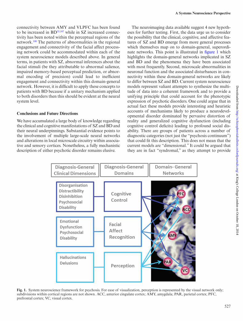
The neuroimaging data available suggest 4 new hypoth-eses for further testing. First, the data urge us to consider the possibility that the clinical, cognitive, and affective fea-tures of SZ and BD emerge from more general processes which themselves map on to domain-general, superordi-nate networks. This point is illustrated in figure 1 which highlights the domain-general networks implicated in SZ and BD and the phenomena they have been associated with most frequently. Second, microscale abnormalities in neuronal function and the associated disturbances in con-nectivity within these domain-general networks are likely to differ between SZ and BD. Current system neuroscience models represent valiant attempts to synthesize the multi-tude of data into a coherent framework and to provide a unifying principle that could account for the phenotypic expression of psychotic disorders. One could argue that in actual fact these models provide interesting and heuristic accounts of mechanisms likely to produce a neurodevel-opmental disorder dominated by pervasive distortion of reality and generalized cognitive dysfunction (including cognitive control deficits) leading to profound social dis-ability. There are groups of patients across a number of diagnostic categories (not just the “psychosis continuum”) that could fit this description. This does not mean that the current models are “dimensional.” It could be argued that they are in fact “syndromal,” as they attempt to provide
Fig. 1. System neuroscience framework for psychosis. For ease of visualization, perception is represented by the visual network only; subdivisions within cortical regions are not shown. ACC, anterior cingulate cortex; AMY, amygdala; PAR, parietal cortex; PFC, prefrontal cortex; VC, visual cortex.
at King's C
ollege London on O
ctober 18, 2014http://schizophreniabulletin.oxfordjournals.org/
Dow
nloaded from
528
S. Frangou
an explanatory framework for multiple dimensions of phe-nomenology. This leads to the third hypothesis. A system neuroscience approach may be usefully employed to define a more biological informed nosology for psychiatric disor-ders. Neuroimaging research could play a crucial part in such an endeavor because disturbances within neural net-works are the necessary condition for the clinical expres-sion of any mental disorder or dysfunction. This has at least 2 important ramifications. One is the possibility that neuroimaging data can differentiate between patients and healthy individuals or among patients with different psy-chiatric disorders. The feasibility of this approach has been demonstrated by the successful application of multivari-ate pattern classification analyses in SZ and BD.109,110 In principle, this type of analyses could lead to the identifica-tion of more homogeneous groups of patients, in terms of their neural patterns, who would also be expected to share similar disturbances at the level of microscale circuits. The use of unsupervised classifiers represents another avenue of research, ie, likely to prove particular fruitful. Unsupervised classifiers attempt to distinguish groups of individuals defined solely by their neural profiles while being agnostic about their diagnostic status. This is funda-mentally different from supervised multivariate decoding that relies on diagnostic categorization during the train-ing phase of the algorithm. The presence of subgroups of patients defined by their neural architecture could lead to new and more biologically meaningful phenotypes for psychosis. The final hypothesis acknowledges that not all processes relevant to the pathogenesis of psychosis can be examined in biological systems. The hierarchical predictive coding model exemplifies a new approach to brain model-ing that allows computational formulations of brain func-tion at multiple levels, from changes to synaptic gain to connectivity alterations within large-scale networks. A suf-ficiently detailed model of neuronal groupings and their connections could help define the set of conditions under which phenomena relevant to psychosis might emerge. Potentially it could also help explore the effect of diverse risk factors on neuronal computations in order to under-stand the mechanisms that render neural networks vulner-able to psychosis. Conversely, it could be used to model the effect of treatments and even assist in the selection of new pharmacological interventions based on their predicted effect on the model.
Acknowledgment
The authors have declared that there are no conflicts of interest in relation to the subject of this study.
References
1. Cross-Disorder Group of the Psychiatric Genomics Consortium, Smoller JW, Craddock N, Kendler K, et al. Identification of risk loci with shared effects on five major
psychiatric disorders: a genome-wide analysis. Lancet. 2013;381:1371–1379.
2. International Schizophrenia Consortium, Purcell SM, Wray NR, Stone JL, et al. Common polygenic variation contrib-utes to risk of schizophrenia and bipolar disorder. Nature. 2009;460:748–752.
3. Lichtenstein P, Yip BH, Björk C, et al. Common genetic deter-minants of schizophrenia and bipolar disorder in Swedish families: a population-based study. Lancet. 2009;373:234–239.
4. Tamminga CA, Ivleva EI, Keshavan MS, et al. Clinical phe-notypes of psychosis in the Bipolar-Schizophrenia Network on Intermediate Phenotypes (B-SNIP). Am J Psychiatry. 2013;170:1263–1274.
5. Peralta V, Moreno-Izco L, Calvo-Barrena L, Cuesta MJ. The low- and higher-order factor structure of symptoms in patients with a first episode of psychosis. Schizophr Res. 2013;147:116–124.
6. Russo M, Levine SZ, Demjaha A, et al. Association between symptom dimensions and categorical diagnoses of psychosis: a cross-sectional and longitudinal investigation. Schizophr Bull. 2014;40:111–119. doi:10.1093/schbul/sbt055
7. Collins PY, Insel TR, Chockalingam A, Daar A, Maddox YT. Grand challenges in global mental health: integration in research, policy, and practice. PLoS Med. 2013;10:e1001434.
8. Kapur S, Phillips AG, Insel TR. Why has it taken so long for biological psychiatry to develop clinical tests and what to do about it? Mol Psychiatry. 2012;17:1174–1179.
9. Cuthbert BN, Insel TR. Toward new approaches to psychotic disorders: the NIMH Research Domain Criteria project. Schizophr Bull. 2010;36:1061–1062.
10. Delvecchio G, Sugranyes G, Frangou S. Evidence of diagnos-tic specificity in the neural correlates of facial affect process-ing in bipolar disorder and schizophrenia: a meta-analysis of functional imaging studies. Psychol Med. 2013;43:553–569.
11. Fornito A, Zalesky A, Pantelis C, Bullmore ET. Schizophrenia, neuroimaging and connectomics. Neuroimage. 2012;62:2296–2314.
12. Strakowski SM, Adler CM, Almeida J, et al. The functional neuroanatomy of bipolar disorder: a consensus model. Bipolar Disord. 2012;14:313–325.
13. Chen CH, Suckling J, Lennox BR, Ooi C, Bullmore ET. A quantitative meta-analysis of fMRI studies in bipolar disor-der. Bipolar Disord. 2011;13:1–15.
14. Ellison-Wright I, Bullmore E. Anatomy of bipolar dis-order and schizophrenia: a meta-analysis. Schizophr Res. 2010;117:1–12.
15. Bora E, Fornito A, Radua J, et al. Neuroanatomical abnor-malities in schizophrenia: a multimodal voxelwise meta-analysis and meta-regression analysis. Schizophr Res. 2011;127:46–57.
16. Kempton MJ, Salvador Z, Munafò MR, et al. Structural neu-roimaging studies in major depressive disorder. Meta-analysis and comparison with bipolar disorder. Arch Gen Psychiatry. 2011;68:675–690.
17. Bora E, Fornito A, Yücel M, Pantelis C. Voxelwise meta-analysis of gray matter abnormalities in bipolar disorder. Biol Psychiatry. 2010;67:1097–1105.
18. Haijma SV, Van Haren N, Cahn W, Koolschijn PC, Hulshoff Pol HE, Kahn RS. Brain volumes in schizophre-nia: a meta-analysis in over 18 000 subjects. Schizophr Bull. 2013;39:1129–1138.
19. Liddle PF, Friston KJ, Frith CD, Hirsch SR, Jones T, Frackowiak RS. Patterns of cerebral blood flow in schizo-phrenia. Br J Psychiatry. 1992;160:179–186.
at King's C
ollege London on O
ctober 18, 2014http://schizophreniabulletin.oxfordjournals.org/
Dow
nloaded from

529
A Systems Neuroscience Perspective
20. Koutsouleris N, Gaser C, Jäger M, et al. Structural corre-lates of psychopathological symptom dimensions in schizo-phrenia: a voxel-based morphometric study. Neuroimage. 2008;39:1600–1612.
21. Nenadic I, Sauer H, Gaser C. Distinct pattern of brain struc-tural deficits in subsyndromes of schizophrenia delineated by psychopathology. Neuroimage. 2010;49:1153–1160.
22. Goghari VM, Sponheim SR, MacDonald AW 3rd. The functional neuroanatomy of symptom dimensions in schizo-phrenia: a qualitative and quantitative review of a persistent question. Neurosci Biobehav Rev. 2010;34:468–486.
23. Anticevic A, Cole MW, Repovs G, et al. Characterizing thalamo-cortical disturbances in schizophrenia and bipolar illness. Cereb Cortex. 2013 doi:10.1093/cercor/bht165
24. Bourne C, Aydemir Ö, Balanzá-Martínez V, et al. Neuropsychological testing of cognitive impairment in euthymic bipolar disorder: an individual patient data meta-analysis. Acta Psychiatr Scand. 2013;128:149–162.
25. Mann-Wrobel MC, Carreno JT, Dickinson D. Meta-analysis of neuropsychological functioning in euthymic bipolar dis-order: an update and investigation of moderator variables. Bipolar Disord. 2011;13:334–342.
26. Yatham LN, Torres IJ, Malhi GS, et al. The International Society for Bipolar Disorders-Battery for Assessment of Neurocognition (ISBD-BANC). Bipolar Disord. 2010;12:351–363.
27. Kumar CT, Frangou S. Clinical implications of cogni-tive function in bipolar disorder. Ther Adv Chronic Dis. 2010;1:85–93.
28. Heinrichs RW, Zakzanis KK. Neurocognitive deficit in schizophrenia: a quantitative review of the evidence. Neuropsychology. 1998;12:426–445.
29. Schaefer J, Giangrande E, Weinberger DR, Dickinson D. The global cognitive impairment in schizophrenia: con-sistent over decades and around the world. Schizophr Res. 2013;150:42–50.
30. Mesholam-Gately RI, Giuliano AJ, Goff KP, Faraone SV, Seidman LJ. Neurocognition in first-episode schizophrenia: a meta-analytic review. Neuropsychology. 2009;23:315–336.
31. MacCabe JH, Wicks S, Löfving S, et al. Decline in cognitive performance between ages 13 and 18 years and the risk for psychosis in adulthood: a Swedish longitudinal cohort study in males. JAMA Psychiatry. 2013;70:261–270.
32. Zanelli J, Reichenberg A, Morgan K, et al. Specific and generalized neuropsychological deficits: a comparison of patients with various first-episode psychosis presentations. Am J Psychiatry. 2010;167:78–85.
33. Meier MH, Caspi A, Reichenberg A, et al. Neuropsychological decline in schizophrenia from the premorbid to the postonset period: evidence from a population-representative longitu-dinal study. Am J Psychiatry. 2014;171:91–101. doi:10.1176/appi.ajp.2013.12111438
34. Schretlen DJ, Cascella NG, Meyer SM, et al. Neuropsychological functioning in bipolar disorder and schizophrenia. Biol Psychiatry. 2007;62:179–186.
35. Stefanopoulou E, Manoharan A, Landau S, Geddes JR, Goodwin G, Frangou S. Cognitive functioning in patients with affective disorders and schizophrenia: a meta-analysis. Int Rev Psychiatry. 2009;21:336–356.
36. Bora E, Yücel M, Pantelis C. Cognitive impairment in affective psychoses: a meta-analysis. Schizophr Bull. 2010;36:112–125.
37. Jabben N, Arts B, van Os J, Krabbendam L. Neurocognitive functioning as intermediary phenotype and predictor
of psychosocial functioning across the psychosis contin-uum: studies in schizophrenia and bipolar disorder. J Clin Psychiatry. 2010;71:764–774.
38. Kupferschmidt DA, Zakzanis KK. Toward a functional neuroanatomical signature of bipolar disorder: quantitative evidence from the neuroimaging literature. Psychiatry Res. 2011;193:71–79.
39. Minzenberg MJ, Laird AR, Thelen S, Carter CS, Glahn DC. Meta-analysis of 41 functional neuroimaging studies of executive function in schizophrenia. Arch Gen Psychiatry. 2009;66:811–822.
40. Cremaschi L, Penzo B, Palazzo M, et al. Assessing work-ing memory via N-back task in euthymic bipolar I disorder patients: a review of functional magnetic resonance imaging studies. Neuropsychobiology. 2013;68:63–70.
41. Taylor SF, Kang J, Brege IS, Tso IF, Hosanagar A, Johnson TD. Meta-analysis of functional neuroimaging studies of emotion perception and experience in schizophrenia. Biol Psychiatry. 2012;71:136–145.
42. Anticevic A, Van Snellenberg JX, Cohen RE, Repovs G, Dowd EC, Barch DM. Amygdala recruitment in schizophre-nia in response to aversive emotional material: a meta-analy-sis of neuroimaging studies. Schizophr Bull. 2012;38:608–621.
43. Li H, Chan RC, McAlonan GM, Gong QY. Facial emotion processing in schizophrenia: a meta-analysis of functional neuroimaging data. Schizophr Bull. 2010;36:1029–1039.
44. Smith SM, Vidaurre D, Beckmann CF, et al. Functional connectomics from resting-state fMRI. Trends Cogn Sci. 2013;17:666–682.
45. Crossley NA, Mechelli A, Fusar-Poli P, et al. Superior tem-poral lobe dysfunction and frontotemporal dysconnectivity in subjects at risk of psychosis and in first-episode psychosis. Hum Brain Mapp. 2009;30:4129–4137.
46. Meyer-Lindenberg AS, Olsen RK, Kohn PD, et al. Regionally specific disturbance of dorsolateral prefrontal-hippocam-pal functional connectivity in schizophrenia. Arch Gen Psychiatry. 2005;62:379–386.
47. Wolf RC, Vasic N, Sambataro F, et al. Temporally anticor-related brain networks during working memory performance reveal aberrant prefrontal and hippocampal connectivity in patients with schizophrenia. Prog Neuropsychopharmacol Biol Psychiatry. 2009;33:1464–1473.
48. Quidé Y, Morris RW, Shepherd AM, Rowland JE, Green MJ. Task-related fronto-striatal functional connectivity during working memory performance in schizophrenia. Schizophr Res. 2013;150:468–475.
49. Pompei F, Dima D, Rubia K, Kumari V, Frangou S. Dissociable functional connectivity changes during the Stroop task relating to risk, resilience and disease expression in bipolar disorder. Neuroimage. 2011;57:576–582.
50. Mukherjee P, Whalley HC, McKirdy JW, et al. Altered amyg-dala connectivity within the social brain in schizophrenia. Schizophr Bull. 2014;40:152–160.
51. Aleman A, Kahn RS. Strange feelings: do amygdala abnor-malities dysregulate the emotional brain in schizophrenia? Prog Neurobiol. 2005;77:283–298.
52. Townsend JD, Torrisi SJ, Lieberman MD, Sugar CA, Bookheimer SY, Altshuler LL. Frontal-amygdala connec-tivity alterations during emotion downregulation in bipolar I disorder. Biol Psychiatry. 2013;73:127–135.
53. Cerullo MA, Fleck DE, Eliassen JC, et al. A longitudinal func-tional connectivity analysis of the amygdala in bipolar I dis-order across mood states. Bipolar Disord. 2012;14:175–184.
at King's C
ollege London on O
ctober 18, 2014http://schizophreniabulletin.oxfordjournals.org/
Dow
nloaded from

530
S. Frangou
54. Perlman SB, Almeida JR, Kronhaus DM, et al. Amygdala activity and prefrontal cortex-amygdala effective connec-tivity to emerging emotional faces distinguish remitted and depressed mood states in bipolar disorder. Bipolar Disord. 2012;14:162–174.
55. Dima D, Jogia J, Collier D, Vassos E, Burdick KE, Frangou S. Independent modulation of engagement and connectivity of the facial network during affect processing by CACNA1C and ANK3 risk genes for bipolar disorder. JAMA Psychiatry. 2013;70:1303–1311.
56. Biswal B, Yetkin FZ, Haughton VM, Hyde JS. Functional connectivity in the motor cortex of resting human brain using echo-planar MRI. Magn Reson Med. 1995;34:537–541.
57. Damoiseaux JS, Rombouts SA, Barkhof F, et al. Consistent resting-state networks across healthy subjects. Proc Natl Acad Sci USA. 2006;103:13848–13853.
58. Biswal BB, Mennes M, Zuo XN, et al. Toward discovery science of human brain function. Proc Natl Acad Sci USA. 2010;107:4734–4739.
59. Raichle ME, MacLeod AM, Snyder AZ, Powers WJ, Gusnard DA, Shulman GL. A default mode of brain function. Proc Natl Acad Sci USA. 2001;98:676–682.
60. Greicius MD, Krasnow B, Reiss AL, Menon V. Functional con-nectivity in the resting brain: a network analysis of the default mode hypothesis. Proc Natl Acad Sci USA. 2003;100:253–258.
61. Smith SM, Fox PT, Miller KL, et al. Correspondence of the brain’s functional architecture during activation and rest. Proc Natl Acad Sci USA. 2009;106:13040–13045.
62. van den Heuvel MP, Sporns O, Collin G, et al. Abnormal rich club organization and functional brain dynamics in schizo-phrenia. JAMA Psychiatry. 2013;70:783–792.
63. Mamah D, Barch DM, Repovš G. Resting state functional connectivity of five neural networks in bipolar disorder and schizophrenia. J Affect Disord. 2013;150:601–609.
64. Chai XJ, Whitfield-Gabrieli S, Shinn AK, et al. Abnormal medial prefrontal cortex resting-state connectivity in bipo-lar disorder and schizophrenia. Neuropsychopharmacology. 2011;36:2009–2017.
65. Argyelan M, Ikuta T, Derosse P, et al. Resting-state FMRI connectivity impairment in schizophrenia and bipolar disor-der. Schizophr Bull. 2014;40:100–110.
66. Khadka S, Meda SA, Stevens MC, et al. Is aberrant func-tional connectivity a psychosis endophenotype? A resting state functional magnetic resonance imaging study. Biol Psychiatry. 2013;74:458–466.
67. Vargas C, López-Jaramillo C, Vieta E. A systematic literature review of resting state network–functional MRI in bipolar disorder. J Affect Disord. 2013;150:727–735.
68. Anticevic A, Brumbaugh MS, Winkler AM, et al. Global pre-frontal and fronto-amygdala dysconnectivity in bipolar I dis-order with psychosis history. Biol Psychiatry. 2013;73:565–573.
69. Anticevic A, Cole MW, Murray JD, Corlett PR, Wang XJ, Krystal JH. The role of default network deactivation in cog-nition and disease. Trends Cogn Sci. 2012;16:584–592.
70. Buckner RL, Andrews-Hanna JR, Schacter DL. The brain’s default network: anatomy, function, and relevance to disease. Ann N Y Acad Sci. 2008;1124:1–38.
71. Kapur S. Psychosis as a state of aberrant salience: a frame-work linking biology, phenomenology, and pharmacology in schizophrenia. Am J Psychiatry. 2003;160:13–23.
72. Meyer-Lindenberg A, Miletich RS, Kohn PD, et al. Reduced prefrontal activity predicts exaggerated striatal dopaminergic function in schizophrenia. Nat Neurosci. 2002;5:267–271.
73. Howes OD, Montgomery AJ, Asselin MC, et al. Elevated stri-atal dopamine function linked to prodromal signs of schizo-phrenia. Arch Gen Psychiatry. 2009;66:13–20.
74. Fusar-Poli P, Howes OD, Allen P, et al. Abnormal fronto-striatal interactions in people with prodromal signs of psy-chosis: a multimodal imaging study. Arch Gen Psychiatry. 2010;67:683–691.
75. Laruelle M. The second revision of the dopamine theory of schizophrenia: implications for treatment and drug develop-ment. Biol Psychiatry. 2013;74:80–81.
76. Roiser JP, Stephan KE, den Ouden HE, Barnes TR, Friston KJ, Joyce EM. Do patients with schizophrenia exhibit aber-rant salience? Psychol Med. 2009;39:199–209.
77. Strauss GP. The emotion paradox of anhedonia in schizo-phrenia: or is it? Schizophr Bull. 2013;39:247–250.
78. Dowd EC, Barch DM. Anhedonia and emotional experi-ence in schizophrenia: neural and behavioral indicators. Biol Psychiatry. 2010;67:902–911.
79. Brambilla P, Perlini C, Bellani M, et al. Increased salience of gains versus decreased associative learning differentiate bipo-lar disorder from schizophrenia during incentive decision making. Psychol Med. 2013;43:571–580.
80. Gold JM, Waltz JA, Matveeva TM, et al. Negative symp-toms and the failure to represent the expected reward value of actions: behavioral and computational modeling evidence. Arch Gen Psychiatry. 2012;69:129–138.
81. Volk DW, Lewis DA. Prefrontal cortical circuits in schizo-phrenia. Curr Top Behav Neurosci. 2010;4:485–508.
82. van Schouwenburg M, Aarts E, Cools R. Dopaminergic modulation of cognitive control: distinct roles for the pre-frontal cortex and the basal ganglia. Curr Pharm Des. 2010;16:2026–2032.
83. Berk M, Dodd S, Kauer-Sant’anna M, et al. Dopamine dys-regulation syndrome: implications for a dopamine hypothesis of bipolar disorder. Acta Psychiatr Scand Suppl. 2007:41–49.
84. Lesh TA, Niendam TA, Minzenberg MJ, Carter CS. Cognitive control deficits in schizophrenia: mechanisms and meaning. Neuropsychopharmacology. 2011;36:316–338.
85. Proceedings of RDoC Workshops. Cognitive Systems Workshop Proceeedings 2011. http://www.nimh.nih.gov/research-priorities/rdoc/rdoc-cogsys.pdf.
86. Niendam TA, Laird AR, Ray KL, Dean YM, Glahn DC, Carter CS. Meta-analytic evidence for a superordinate cogni-tive control network subserving diverse executive functions. Cogn Affect Behav Neurosci. 2012;12:241–268.
87. Diener C, Kuehner C, Brusniak W, Ubl B, Wessa M, Flor H. A meta-analysis of neurofunctional imaging studies of emotion and cognition in major depression. Neuroimage. 2012;61:677–685.
88. Arnsten AF, Rubia K. Neurobiological circuits regulating attention, cognitive control, motivation, and emotion: dis-ruptions in neurodevelopmental psychiatric disorders. J Am Acad Child Adolesc Psychiatry. 2012;51:356–367.
89. Groman SM, Jentsch JD. Cognitive control and the dopa-mine D
2-like receptor: a dimensional understanding of addic-tion. Depress Anxiety. 2012;29:295–306.
90. Hirano S, Shinotoh H, Eidelberg D. Functional brain imag-ing of cognitive dysfunction in Parkinson’s disease. J Neurol Neurosurg Psychiatry. 2012;83:963–969.
91. Friston K. Functional integration and inference in the brain. Prog Neurobiol. 2002;68:113–143.
92. Adams RA, Stephan KE, Brown HR, Frith CD, Friston KJ. The computational anatomy of psychosis. Front Psychiatry. 2013;4:47.
at King's C
ollege London on O
ctober 18, 2014http://schizophreniabulletin.oxfordjournals.org/
Dow
nloaded from

531
A Systems Neuroscience Perspective
93. Krishnan RR, Fivaz M, Kraus MS, Keefe RS. Hierarchical temporal processing deficit model of reality distortion and psychoses. Mol Psychiatry. 2011;16:129–144.
94. Purves D, Lotto RB, Williams SM, Nundy S, Yang Z. Why we see things the way we do: evidence for a wholly empiri-cal strategy of vision. Philos Trans R Soc Lond B Biol Sci. 2001;356:285–297.
95. Hawkins J, Blakeslee S. On Intelligence. New York, NY: Times Books New York; 2004.
96. Power JD, Cohen AL, Nelson SM, et al. Functional network organization of the human brain. Neuron. 2011;72:665–678.
97. Bassett DS, Bullmore ET. Human brain networks in health and disease. Curr Opin Neurol. 2009;22:340–347.
98. Park HJ, Friston K. Structural and functional brain networks: from connections to cognition. Science. 2013;342:1238411.
99. Barrett LF, Satpute AB. Large-scale brain networks in affective and social neuroscience: towards an integrative functional archi-tecture of the brain. Curr Opin Neurobiol. 2013;23:361–372.
100. Proceedings of RDoC Workshops. Systems for Social Processes Workshop Proceedings, 2012. http://www.nimh.nih.gov/research-priorities/rdoc/rdoc-social-processes.pdf.
101. Torrisi SJ, Lieberman MD, Bookheimer SY, Altshuler LL. Advancing understanding of affect labeling with dynamic causal modeling. Neuroimage. 2013;82:481–488.
102. Dima D, Stephan KE, Roiser JP, Friston KJ, Frangou S. Effective connectivity during processing of facial affect: evidence for multiple parallel pathways. J Neurosci. 2011;31:14378–14385.
103. Fairhall SL, Ishai A. Effective connectivity within the dis-tributed cortical network for face perception. Cereb Cortex. 2007;17:2400–2406.
104. Vuilleumier P, Pourtois G. Distributed and interactive brain mechanisms during emotion face perception: evidence from functional neuroimaging. Neuropsychologia. 2007;45:174–194.
105. Versace A, Thompson WK, Zhou D, et al. Abnormal left and right amygdala-orbitofrontal cortical functional connectivity to emotional faces: state versus trait vulnerability markers of depression in bipolar disorder. Biol Psychiatry. 2010;67:422–431.
106. Morris RW, Sparks A, Mitchell PB, Weickert CS, Green MJ. Lack of cortico-limbic coupling in bipolar disorder and schizophrenia during emotion regulation. Transl Psychiatry. 2012;2:e90.
107. Torrisi S, Moody TD, Vizueta N, et al. Differences in resting corticolimbic functional connectivity in bipolar I euthymia. Bipolar Disord. 2013;15:156–166.
108. Leitman DI, Loughead J, Wolf DH, et al. Abnormal superior temporal connectivity during fear perception in schizophre-nia. Schizophr Bull. 2008;34:673–678.
109. Schnack HG, Nieuwenhuis M, van Haren NE, et al. Can structural MRI aid in clinical classification? A machine learn-ing study in two independent samples of patients with schizo-phrenia, bipolar disorder and healthy subjects. Neuroimage. 2014;84:299–306.
110. Rocha-Rego V, Jogia J, Marquand AF, Mourao-Miranda J, Simmons A, Frangou S. Examination of the predictive value of structural magnetic resonance scans in bipolar dis-order: a pattern classification approach. Psychol Med. 2013; doi:http://dx.doi.org/10.1017/S0033291713001013
at King's C
ollege London on O
ctober 18, 2014http://schizophreniabulletin.oxfordjournals.org/
Dow
nloaded from


![[CONFERENCE PAPER] Bipolar Bozuklukta BDT](https://static.fdokumen.com/doc/165x107/63328d1f4e0143040300b9b3/conference-paper-bipolar-bozuklukta-bdt.jpg)