a structural model for caveolin-induced domain formation - UQ ...
10
787 Commentary Introduction Caveolae, small uncoated pits in the plasma membrane, are an abundant feature of many highly differentiated mammalian cells, such as adipocytes, endothelial cells and muscle cells (Fig. 1). Although caveolae were described morphologically in the early 1950s (Palade, 1953; Yamada, 1955), only recently have some of their functions been revealed. Studies of mice lacking isoforms of the caveolar protein caveolin suggest roles for these structures in lipid uptake and regulation, transcellular transport in endothelial cells, and tumour suppression (Drab et al., 2001; Razani et al., 2002a; Schubert et al., 2001). Caveolae have also been shown to provide a vehicle for the entry of certain viruses into animal cells (Pelkmans and Helenius, 2002). Other work indicates that mutations in the muscle- specific form of caveolin, caveolin-3, cause muscle disease (Engelman et al., 1998). Despite these advances, the relationship between the unique, highly conserved structure of caveolae and their physiological functions remains unclear. Here we focus on the mechanisms involved in the formation of caveolae. Caveolae are defined by the presence of caveolins and their unique structure, as viewed by electron microscopy (EM) (see Figs 1 and 2). They are considered to be a specialised, morphologically distinguishable form of lipid raft (Rajendran and Simons, 2005; Simons and Toomre, 2000). As discussed below, cholesterol, a crucial component of lipid rafts, is also a vital structural feature of caveolae. Caveolae represent a fascinating model for examining how membrane proteins interact with lipids, how lipid microdomains are generated, and how lipid-protein interactions and lipid rafts can contribute to membrane morphogenesis. Moreover, insights into the formation of caveolae also have implications for the formation of microdomains in many different membrane systems, as well as for our understanding of disease. For example, the developing transverse tubule system of muscle, a surface- connected network of tubules that penetrate the muscle fibre, is enriched in caveolin-3 (Parton et al., 1997), and numerous mutations in this protein have been discovered in patients suffering from a wide range of muscle diseases (Minetti et al., 1998) (Fig. 3). Understanding the effects of these mutations on the normal function of the protein requires an understanding of the precise molecular mechanisms involved in caveolae formation and function. Conversely, the study of the mutants, which have changes in evolutionarily conserved amino acids, are providing new insights into caveolins and caveolae. Caveolar structure Caveolae each comprise a caveolar bulb (approximately 65 nm in diameter) connected to an opening of fairly constant diameter (approximately 45 nm in fast-frozen, freeze- substituted adipocytes) (M. Floetenmeyer, C. Ferguson, B. Marsh and R.G.P., unpublished). In many tissues, multiple caveolae are arranged around a central vacuolar domain. In adipocytes this arrangement is particularly striking and huge surface-connected vacuolar domains are covered in numerous caveolae (Parton et al., 2002) (Fig. 1). In developing muscle fibres, multiple caveolae are connected by a single neck to the plasma membrane, producing large chains of interconnected caveolae (Parton et al., 1997). The mechanisms underlying the formation of these tissue-specific structures are not yet defined, although recent studies suggest that complex rosettes of caveolae are formed when caveolae fuse (Pelkmans and Zerial, 2005). Another structural feature of caveolae in certain endothelia is the presence of a stomatal diaphragm in the neck of the caveolae, which is generated by the protein PV1 (Stan et al., 2004). This fascinating specialisation is discussed elsewhere (reviewed by Stan, 2005). The caveolar invagination appears to be uncoated (compared with clathrin-coated structures) in many micrographs (Fig. 1) but, under appropriate preparation conditions, a type of coat structure can be visualised by EM. Scanning EM and freeze- Caveolae are striking morphological features of the plasma membrane of mammalian cells. Caveolins, the major proteins of caveolae, play a crucial role in the formation of these invaginations of the plasma membrane; however, the precise mechanisms involved are only just starting to be unravelled. Recent studies suggest that caveolae are stable structures first generated in the Golgi complex. Their formation and exit from the Golgi complex is associated with caveolin oligomerisation, acquisition of detergent insolubility, and association with cholesterol. Modelling of caveolin-membrane interactions together with in vitro studies of caveolin peptides are providing new insights into how caveolin-lipid interactions could generate the unique architecture of the caveolar domain. Key words: Caveolae, Cholesterol, Membrane, Model Summary Biogenesis of caveolae: a structural model for caveolin-induced domain formation Robert G. Parton 1,2, *, Michael Hanzal-Bayer 1 and John F. Hancock 1 1 Institute for Molecular Bioscience, University of Queensland, Queensland 4072, Australia 2 Centre for Microscopy and Microanalysis, University of Queensland, Queensland 4072, Australia *Author for correspondence (e-mail: [email protected]) Accepted 21 December 2005 Journal of Cell Science 119, 787-796 Published by The Company of Biologists 2006 doi:10.1242/jcs.02853 Journal of Cell Science
-
Upload
khangminh22 -
Category
Documents
-
view
3 -
download
0
Transcript of a structural model for caveolin-induced domain formation - UQ ...
787Commentary
IntroductionCaveolae, small uncoated pits in the plasma membrane, are anabundant feature of many highly differentiated mammaliancells, such as adipocytes, endothelial cells and muscle cells(Fig. 1). Although caveolae were described morphologically inthe early 1950s (Palade, 1953; Yamada, 1955), only recentlyhave some of their functions been revealed. Studies of micelacking isoforms of the caveolar protein caveolin suggest rolesfor these structures in lipid uptake and regulation, transcellulartransport in endothelial cells, and tumour suppression (Drab etal., 2001; Razani et al., 2002a; Schubert et al., 2001). Caveolaehave also been shown to provide a vehicle for the entry ofcertain viruses into animal cells (Pelkmans and Helenius,2002). Other work indicates that mutations in the muscle-specific form of caveolin, caveolin-3, cause muscle disease(Engelman et al., 1998). Despite these advances, therelationship between the unique, highly conserved structure ofcaveolae and their physiological functions remains unclear.
Here we focus on the mechanisms involved in the formationof caveolae. Caveolae are defined by the presence of caveolinsand their unique structure, as viewed by electron microscopy(EM) (see Figs 1 and 2). They are considered to be aspecialised, morphologically distinguishable form of lipid raft(Rajendran and Simons, 2005; Simons and Toomre, 2000). Asdiscussed below, cholesterol, a crucial component of lipid rafts,is also a vital structural feature of caveolae. Caveolae representa fascinating model for examining how membrane proteinsinteract with lipids, how lipid microdomains are generated, andhow lipid-protein interactions and lipid rafts can contribute tomembrane morphogenesis. Moreover, insights into theformation of caveolae also have implications for the formationof microdomains in many different membrane systems, as wellas for our understanding of disease. For example, thedeveloping transverse tubule system of muscle, a surface-connected network of tubules that penetrate the muscle fibre,
is enriched in caveolin-3 (Parton et al., 1997), and numerousmutations in this protein have been discovered in patientssuffering from a wide range of muscle diseases (Minetti et al.,1998) (Fig. 3). Understanding the effects of these mutations onthe normal function of the protein requires an understanding ofthe precise molecular mechanisms involved in caveolaeformation and function. Conversely, the study of the mutants,which have changes in evolutionarily conserved amino acids,are providing new insights into caveolins and caveolae.
Caveolar structureCaveolae each comprise a caveolar bulb (approximately 65 nmin diameter) connected to an opening of fairly constantdiameter (approximately 45 nm in fast-frozen, freeze-substituted adipocytes) (M. Floetenmeyer, C. Ferguson, B.Marsh and R.G.P., unpublished). In many tissues, multiplecaveolae are arranged around a central vacuolar domain. Inadipocytes this arrangement is particularly striking and hugesurface-connected vacuolar domains are covered in numerouscaveolae (Parton et al., 2002) (Fig. 1). In developing musclefibres, multiple caveolae are connected by a single neck to theplasma membrane, producing large chains of interconnectedcaveolae (Parton et al., 1997). The mechanisms underlying theformation of these tissue-specific structures are not yet defined,although recent studies suggest that complex rosettes ofcaveolae are formed when caveolae fuse (Pelkmans and Zerial,2005). Another structural feature of caveolae in certainendothelia is the presence of a stomatal diaphragm in the neckof the caveolae, which is generated by the protein PV1 (Stanet al., 2004). This fascinating specialisation is discussedelsewhere (reviewed by Stan, 2005).
The caveolar invagination appears to be uncoated (comparedwith clathrin-coated structures) in many micrographs (Fig. 1)but, under appropriate preparation conditions, a type of coatstructure can be visualised by EM. Scanning EM and freeze-
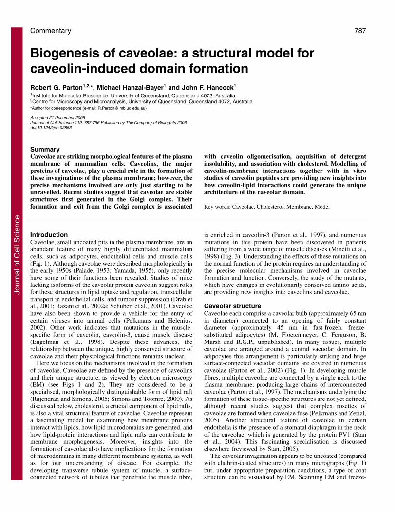
Caveolae are striking morphological features of the plasmamembrane of mammalian cells. Caveolins, the majorproteins of caveolae, play a crucial role in the formation ofthese invaginations of the plasma membrane; however, theprecise mechanisms involved are only just starting to beunravelled. Recent studies suggest that caveolae are stablestructures first generated in the Golgi complex. Theirformation and exit from the Golgi complex is associated
with caveolin oligomerisation, acquisition of detergentinsolubility, and association with cholesterol. Modelling ofcaveolin-membrane interactions together with in vitrostudies of caveolin peptides are providing new insights intohow caveolin-lipid interactions could generate the uniquearchitecture of the caveolar domain.
Key words: Caveolae, Cholesterol, Membrane, Model
Summary
Biogenesis of caveolae: a structural model forcaveolin-induced domain formationRobert G. Parton1,2,*, Michael Hanzal-Bayer1 and John F. Hancock1
1Institute for Molecular Bioscience, University of Queensland, Queensland 4072, Australia2Centre for Microscopy and Microanalysis, University of Queensland, Queensland 4072, Australia*Author for correspondence (e-mail: [email protected])
Accepted 21 December 2005Journal of Cell Science 119, 787-796 Published by The Company of Biologists 2006doi:10.1242/jcs.02853
Jour
nal o
f Cel
l Sci
ence
788
etch replica techniques have revealed striations that form aspiral around the cytoplasmic surface of the caveolarinvagination (Peters et al., 1985; Rothberg et al., 1992; Stan,2002). Densities corresponding to these structures are not seenroutinely in plastic sections but can be visualised under optimalconditions (see Fig. 2). The molecular composition of this coatremains unknown but it has been proposed that the filamentsmight comprise oligomers of caveolin (Monier et al., 1995;Peters et al., 1985; Rothberg et al., 1992). Caveolin oligomers
can be produced in vitro or purified from tissues (Monier et al.,1995). The oligomers have a distinctive ‘necklace’ appearancesimilar to that of the spiral coat. Indeed, purified fragmentscorresponding to the N-terminal cytoplasmic domain ofcaveolin-1 (residues 1-101; see Figs 1 and 3) form oligomersthat can assemble into filaments (Fernandez et al., 2002) andcould correspond to the striated structures of the caveolar coat.Such a model, in which caveolin forms the striations thatencircle the caveolar bulb is, however, at variance with thesuggestion that caveolin associates only with the neck of thecaveolae (Thorn et al., 2003), but is consistent with a recentfreeze-fracture study that localised caveolin to a belt aroundthe membrane-proximal region of the caveolar domain ofmouse fibroblasts (Westermann et al., 2005).
The role of caveolins in caveolae formationThe expression of caveolin-1 in cells normally lackingcaveolae causes the formation of caveolae (Fra et al., 1995) invarious different experimental systems (Breuza et al., 2002;Kirkham et al., 2005; Lipardi et al., 1998; Vogel et al., 1998)and studies showing loss of caveolae in mice lacking caveolin-1 or caveolin-3 confirm the importance of caveolins in thisprocess (Drab et al., 2001; Galbiati et al., 2001). With only afew notable exceptions (see below), caveolin-1 expressioncorrelates qualitatively with the formation of caveolae in non-muscle cells: tissues that exhibit high caveolin expression havea high density of caveolae, whereas those lacking caveolin-1lack caveolae (Parton, 1996). Both caveolin-1 and the closely
Journal of Cell Science 119 (5)
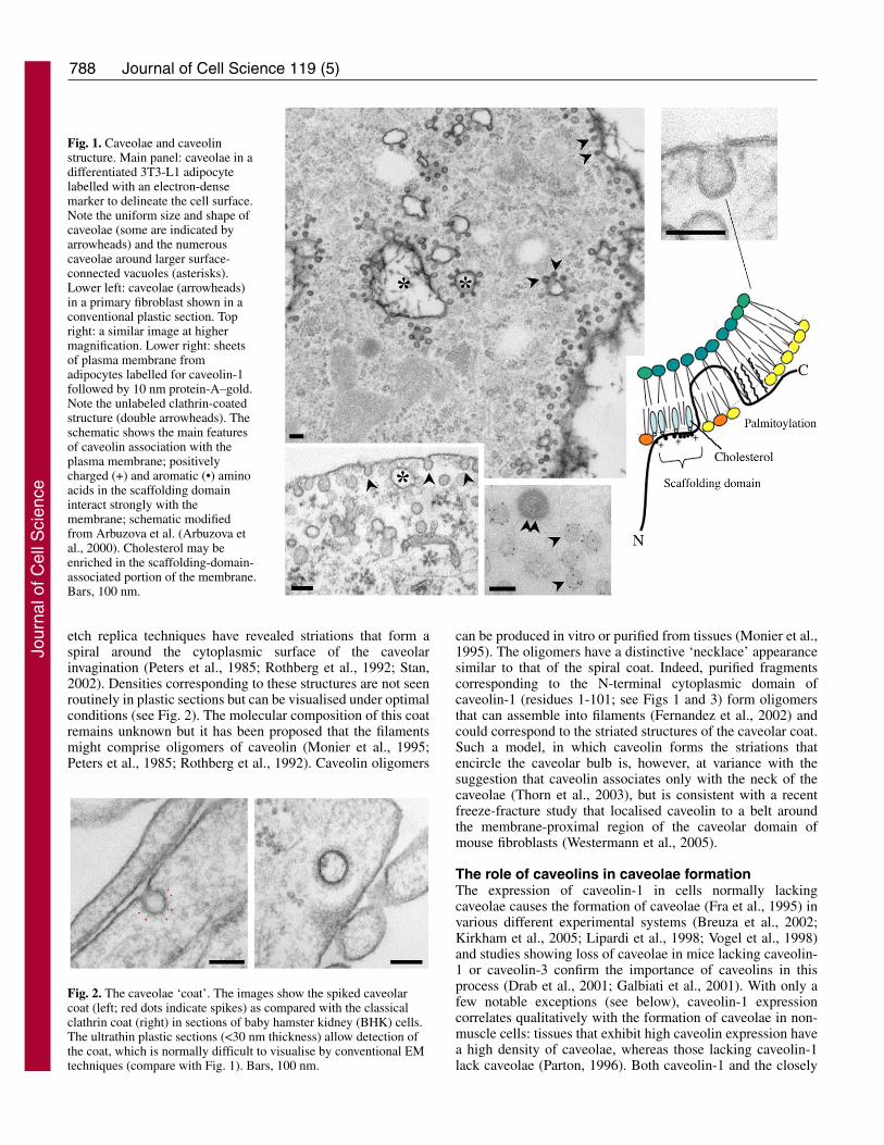
Fig. 1. Caveolae and caveolinstructure. Main panel: caveolae in adifferentiated 3T3-L1 adipocytelabelled with an electron-densemarker to delineate the cell surface.Note the uniform size and shape ofcaveolae (some are indicated byarrowheads) and the numerouscaveolae around larger surface-connected vacuoles (asterisks).Lower left: caveolae (arrowheads)in a primary fibroblast shown in aconventional plastic section. Topright: a similar image at highermagnification. Lower right: sheetsof plasma membrane fromadipocytes labelled for caveolin-1followed by 10 nm protein-A–gold.Note the unlabeled clathrin-coatedstructure (double arrowheads). Theschematic shows the main featuresof caveolin association with theplasma membrane; positivelycharged (+) and aromatic (•) aminoacids in the scaffolding domaininteract strongly with themembrane; schematic modifiedfrom Arbuzova et al. (Arbuzova etal., 2000). Cholesterol may beenriched in the scaffolding-domain-associated portion of the membrane.Bars, 100 nm.
Fig. 2. The caveolae ‘coat’. The images show the spiked caveolarcoat (left; red dots indicate spikes) as compared with the classicalclathrin coat (right) in sections of baby hamster kidney (BHK) cells.The ultrathin plastic sections (<30 nm thickness) allow detection ofthe coat, which is normally difficult to visualise by conventional EMtechniques (compare with Fig. 1). Bars, 100 nm.
Jour
nal o
f Cel
l Sci
ence
789Caveolae biogenesis
related isoform caveolin-3 can form caveolae when expressedin cells lacking caveolae (Kirkham et al., 2005). Caveolin-1 isexpressed in most non-muscle tissues and in smooth muscle,and is essential for the formation of caveolae in these tissues(Drab et al., 2001). By contrast, loss of caveolin-2, which isgenerally expressed together with caveolin-1, does not appearto affect formation of caveolae (Razani et al., 2002b) althoughcaveolin-2 might facilitate caveolae formation by caveolin-1(see below). Caveolin-3 is normally expressed in skeletalmuscle, some smooth muscle cells and in cardiac muscle (Tanget al., 1996; Way and Parton, 1995). Caveolin-3-knockout micehave no detectable caveolae in their skeletal and cardiacmuscle, which is consistent with caveolin-3 taking the place ofcaveolin-1 in those tissues (Galbiati et al., 2001). Caveolaeformed by heterologous expression of caveolin-1 or caveolin-3 are morphologically identical (M. Kirkham and R.G.P.,unpublished; T. Fujimoto, A. Carozzi and R.G.P.,unpublished).
Given the studies discussed above and the highconcentration of caveolin in caveolae (100-200 caveolinmolecules per caveola) (Dupree et al., 1993; Pelkmans andZerial, 2005), caveolin-1 and caveolin-3 might be structuralproteins involved directly in the bending of the membrane togenerate caveolae. Alternatively, the shape of the caveolardomain might be generated independently of caveolins butstabilised by caveolin or another protein (e.g. dynamin) (Nabiand Le, 2003).
Caveolin biosynthesis and traffickingAll three caveolins generally behave as integral membraneproteins, although cytosolic and secreted pools of caveolin
have also been described (Liu et al., 1999; Uittenbogaard et al.,1998). Caveolins possess a 33-residue central hydrophobicregion (the intramembrane domain), which might form ahairpin in the lipid bilayer, flanked by cytoplasmically exposedN- and C-terminal domains (Figs 1, 3, 4). They are synthesisedcotranslationally on the rough endoplasmic reticulum (ER)(Monier et al., 1995) and then, in most cells, appear to travelalong the secretory pathway to the plasma membrane (Pol etal., 2005); although, see Uittenbogaard et al. for an alternativetrafficking model (Uittenbogaard et al., 1998). The transit ofcaveolins through the Golgi complex is relatively slowcompared with that of other membrane proteins (Pol et al.,2005; Ren et al., 2004), and so a Golgi pool of newlysynthesised caveolin can be visualised in most cell types(Luetterforst et al., 1999; Nichols, 2002; Pol et al., 2005).Transport of caveolin (but not other integral membraneproteins) through the Golgi complex can be accelerated bycholesterol (Pol et al., 2005). Caveolin-1 is palmitoylated onmultiple cysteine residues (Dietzen et al., 1995) in a late-Golgi(BFA-sensitive) compartment (Parat and Fox, 2001) (Figs 1and 4). This modification is not apparently required for furthertransport of caveolin-1 or localisation to pre-existing caveolae(Dietzen et al., 1995).
At some stage in the secretory pathway, caveolin changesfrom a monomeric, detergent-soluble form to an oligomeric,detergent-insoluble form (Pol et al., 2005). The presence of thedetergent-insoluble form suggests association with lipid raftdomains, although it is important to note that the use ofdetergents to assess microdomain association has beenquestioned (Lichtenberg et al., 2005; Munro, 2003). Aminoacid substitutions and truncations that disrupt trafficking to the
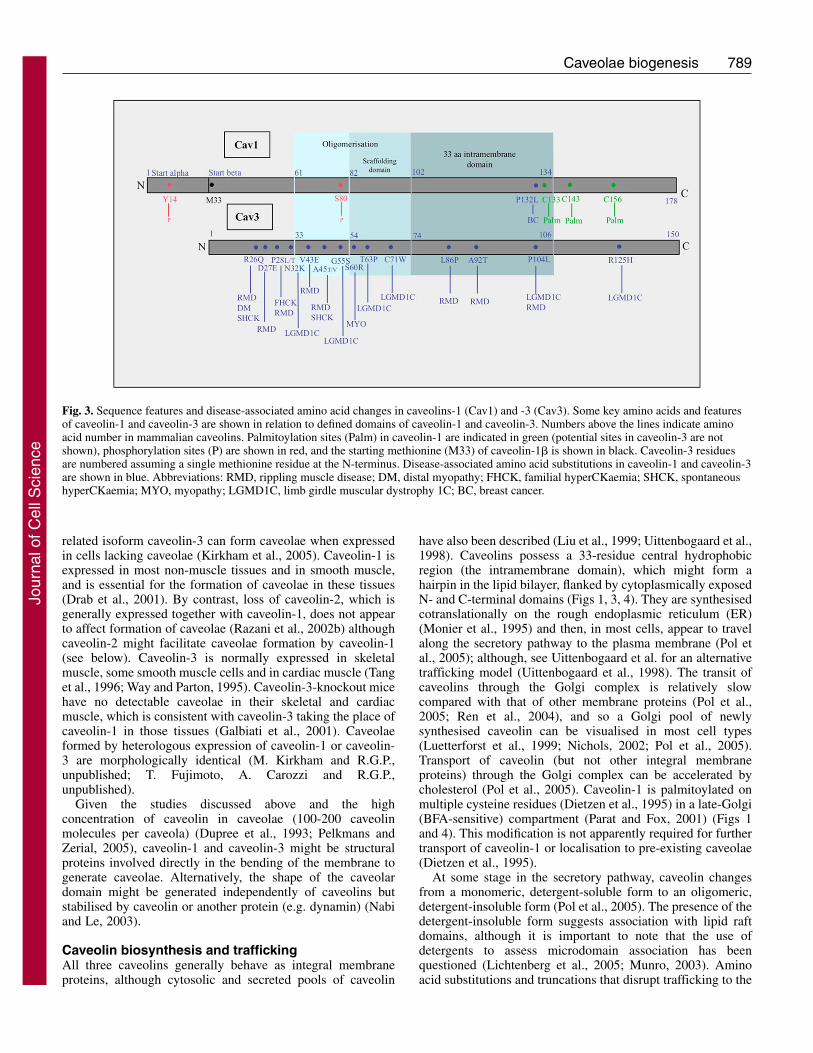
Fig. 3. Sequence features and disease-associated amino acid changes in caveolins-1 (Cav1) and -3 (Cav3). Some key amino acids and featuresof caveolin-1 and caveolin-3 are shown in relation to defined domains of caveolin-1 and caveolin-3. Numbers above the lines indicate aminoacid number in mammalian caveolins. Palmitoylation sites (Palm) in caveolin-1 are indicated in green (potential sites in caveolin-3 are notshown), phosphorylation sites (P) are shown in red, and the starting methionine (M33) of caveolin-1� is shown in black. Caveolin-3 residuesare numbered assuming a single methionine residue at the N-terminus. Disease-associated amino acid substitutions in caveolin-1 and caveolin-3are shown in blue. Abbreviations: RMD, rippling muscle disease; DM, distal myopathy; FHCK, familial hyperCKaemia; SHCK, spontaneoushyperCKaemia; MYO, myopathy; LGMD1C, limb girdle muscular dystrophy 1C; BC, breast cancer.
Jour
nal o
f Cel
l Sci
ence
790
cell surface cause accumulation of detergent-soluble caveolinin the Golgi complex (Galbiati et al., 1999; Luetterforst et al.,1999; Machleidt et al., 2000; Ren et al., 2004). Rather thanrevealing cryptic Golgi-targeting or retention sites, thesemutations might perturb the caveolin structure, leading toretention of misfolded caveolin in the Golgi complex (Ren etal., 2004). This feature of caveolin mutants is important forunderstanding certain disease states associated with bothcaveolin-1 and caveolin-3 (Galbiati et al., 1999). In a recentstudy, transport of caveolin mutants to the plasma membranecorrelated with their ability to associate with lipid raft domains– determined by an in vitro detergent-insolubility assay. Thosemutants that accumulate in the Golgi complex generally showreduced raft affinity in this assay (Ren et al., 2004). Thus,incorporation into lipid raft domains in the Golgi complexmight be required for formation of caveolae in the Golgicomplex or transport of caveolin to the surface prior to theirformation.
The site of caveola formationWhere do caveolae form? Transient expression studies andquantitative EM immunolabelling initially indicated thatcaveolae might form at the plasma membrane by suggestingthat a threshold of caveolin is required at the plasma membraneto generate caveolae (Fra et al., 1995). However, more-recentlight microscopy studies of the trafficking of newly synthesisedcaveolin-1–GFP provided evidence for the formation ofcaveolae in the Golgi complex (Tagawa et al., 2005). In the
latter experiments, the intensity of caveolin-1–GFP punctaleaving the Golgi complex is similar to that in surface caveolae,which suggests that ‘mature’ caveolae are assembled in theGolgi complex. The caveolin-1–GFP-labelled structuresapparently fuse directly with the plasma membrane. Thecaveolin-containing structures may therefore represent carriersin a Golgi-to-plasma-membrane trafficking pathway distinctfrom those carrying other exocytic markers, such as thevesicular stomatitis virus (VSV) G protein (Tagawa et al.,2005). If so, loss of caveolin-1 might cause a perturbation ofthis pathway as a result of the absence of the caveolin carriers.This is consistent with the disruption of the post-Golgitransport of specific proteins by caveolin mutants and in cellslacking caveolins (Hernández-Deviez et al., 2006; Wyse et al.,2003).
Under normal conditions, the concentration of newlysynthesised endogenous caveolin in the Golgi complex ispresumably low [the half-life of caveolin-1 is estimated to be>10 hours (Conrad et al., 1995; Dupree et al., 1993)] but aGolgi pool of newly synthesised caveolin is clearly evident(Dupree et al., 1993; Nichols, 2002; Pol et al., 2005). Ifformation of caveolar carriers in the Golgi requires a thresholdlevel of caveolin, the slow net transit of endogenous caveolinout of the Golgi complex might allow a sufficiently high levelof caveolin to be reached to allow oligomerisation, raftassociation and formation of carriers. In this model, only fullyassembled caveolae would be efficiently transported out of theGolgi complex to the plasma membrane. The characteristics of
the Golgi pool of caveolin at steady state[detergent soluble, monomeric (Pol et al.,2005)] would reflect this. Significantly, uponleaving the Golgi complex or reaching theplasma membrane, caveolin can no longer berecognised by certain caveolin antibodies (Polet al., 2005). Cholesterol depletion restores itsreactivity with these antibodies, whichsuggests a cholesterol-dependent change incaveolin structure on leaving the Golgicomplex that would be consistent with theabove model.
Once caveolae are formed, they areextremely stable. Fluorescence recovery afterphotobleaching (FRAP) experiments using
Journal of Cell Science 119 (5)
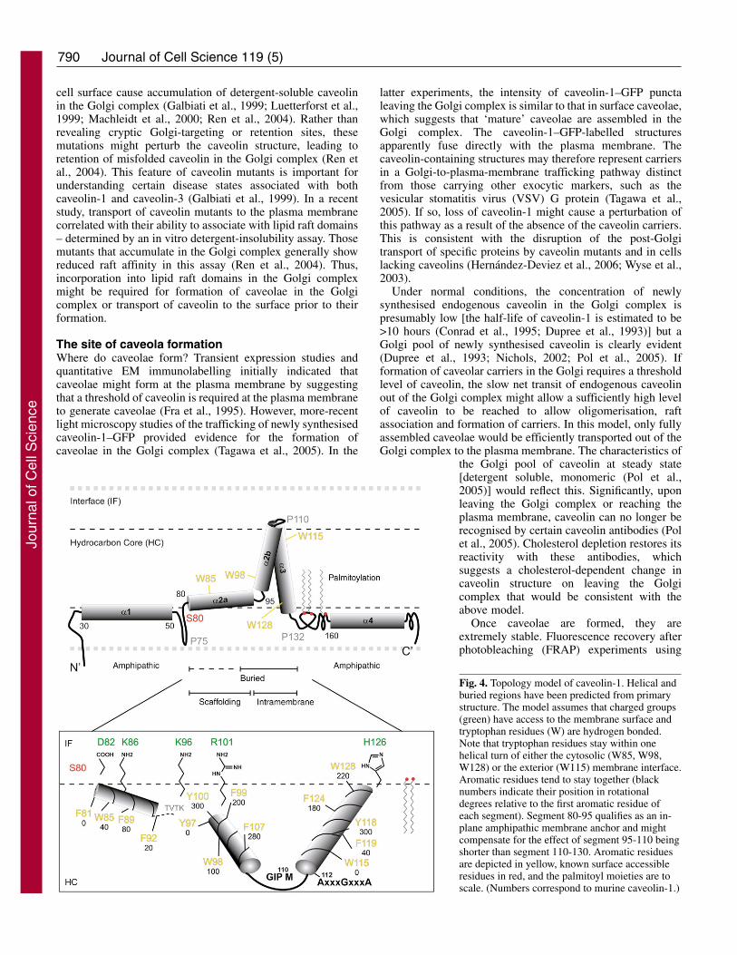
Fig. 4. Topology model of caveolin-1. Helical andburied regions have been predicted from primarystructure. The model assumes that charged groups(green) have access to the membrane surface andtryptophan residues (W) are hydrogen bonded.Note that tryptophan residues stay within onehelical turn of either the cytosolic (W85, W98,W128) or the exterior (W115) membrane interface.Aromatic residues tend to stay together (blacknumbers indicate their position in rotationaldegrees relative to the first aromatic residue ofeach segment). Segment 80-95 qualifies as an in-plane amphipathic membrane anchor and mightcompensate for the effect of segment 95-110 beingshorter than segment 110-130. Aromatic residuesare depicted in yellow, known surface accessibleresidues in red, and the palmitoyl moieties are toscale. (Numbers correspond to murine caveolin-1.)
Jour
nal o
f Cel
l Sci
ence
791Caveolae biogenesis
caveolin-GFP constructs show that caveolin does not diffuserapidly in the membrane (Pelkmans et al., 2004; Thomsen etal., 2002) and caveolin molecules within individual caveolaedo not exchange with each other (Tagawa et al., 2005).Moreover, real-time experiments have shown that caveolae arerelatively immobile. A small fraction can bud; this can beaccelerated under certain circumstances – for example,triggering by binding of the virus SV40 or introduction of aphosphatase inhibitor (Kirkham et al., 2005; Pelkmans et al.,2004; Thomsen et al., 2002). Once internalised, caveolae canfuse with a novel endosomal compartment, the caveosome(Pelkmans et al., 2001) or early endosomes (Pelkmans et al.,2004; Sharma et al., 2003; Tran et al., 1987) but remain asdiscrete units, despite the release of their cargo into theendosomal compartments (Pelkmans et al., 2004). Caveolaecan also fuse with the plasma membrane directly in a ‘kiss-and-run’ cycle (Pelkmans and Zerial, 2005). A lack of surfacecaveolae in some experiments could therefore reflect a changein the endo/exocytic cycling of caveolae rather than theinability of plasma membrane caveolins to form caveolae.
Additional proteins implicated in the formation ofcaveolaeQuantitative studies of caveolin expression suggest thatadditional factors are involved in the biogenesis of caveolae.Caveolin-2 may regulate formation of caveolae driven bycaveolin-1, particularly in epithelial cells. In these cells,caveolin-1 is detectable on both surfaces but caveolae generallyform only at the basolateral surface. Caveolin-2 is targetedspecifically to the basolateral surface (Scheiffele et al., 1998)and interacts with caveolin-1; it could thus be involved in thepolarised formation of caveolae at the basolateral surface.Evidence both for and against such a role exists.Overexpression of caveolin-2 in Madin-Darby canine kidney(MDCK) epithelial cells increases formation of caveolae at thebasolateral surface (Lahtinen et al., 2003). Furthermore, in acaveolin-negative prostate cancer cell line, caveolin-1expression is insufficient to generate caveolae but vesicles,assumed to be related to caveolae, accumulate under theplasma membrane (Sowa et al., 2003). Upon co-expressionwith wild-type caveolin-2, but not caveolin-2 lacking keyphosphorylation sites, caveolae are produced. These findingsled to a model in which caveolin-2 phosphorylation on Ser23and Ser36 by casein kinase 2 (or a related kinase) regulatescaveola formation. An alternative possibility, which might becompatible with other in vivo and in vitro studies, is thatcaveolae are very dynamic in this particular cell type. Theputative internal caveolae visualised in this study uponcaveolin-1 expression might represent budded caveolae, andthe lack of surface caveolae might reflect a change in theirendo/exocytic cycling. Caveolin-2 might negatively regulatethis process in a phosphorylation-dependent manner. With thepowerful systems now available to follow budding of caveolaein real time (Pelkmans and Zerial, 2005) and quantitativelyassess budding of individual caveolae by EM (Kirkham et al.,2005), this question can now be addressed.
A general role for caveolin-2 in driving caveola formationappears unlikely. Caveolin-1 expressed at levels similar tothose observed in naturally expressing caveolin-containing celllines causes formation of caveolae in the absence of caveolin-2 (Fra et al., 1995). Moreover, caveolin-2-null mice appear to
have normal caveolae in the tissues examined (Razani et al.,2002b). Other studies have also shown no effect of caveolin-2on the formation of caveolae when it is expressed together withcaveolin-1 (Breuza et al., 2002) or have shown that caveolin-2causes formation of more-deeply invaginated caveolae(Fujimoto et al., 2000).
Despite the striking morphology of interconnected caveolaewithin the developing T-tubule system of muscle (Ishikawa,1968; Parton et al., 1997), caveolin-3 alone causes formationof caveolae identical to those formed by caveolin-1 whenexpressed in a non-muscle system (Kirkham et al., 2005) (M.Kirkham and R.G.P., unpublished). Additional, presumablymuscle-specific, factors must therefore help form thisspecialised tubular system during muscle differentiation.Amphiphysin 2/Bin1 (M-Amph2) might provide this role.When expressed in fibroblasts, M-Amph2 generates longsurface-connected tubules to which caveolin is recruited (Leeet al., 2002). In developing muscle, it partially colocalises withcaveolin-3 and binds to phosphatidylinositol (4,5)-bisphosphate [PtdIns(4,5)P2], which is dramaticallyupregulated during muscle differentiation (Lee et al., 2002).Although caveolin-3 is not essential for T-tubule development,the cooperation between caveola-generating caveolin-3 andtubule-generating amphiphysin may be important for thedevelopment of the characteristic beaded intermediatesinvolved in the generation of the mature T-tubule (Galbiati etal., 2001; Lee et al., 2002).
The importance of cholesterolCholesterol is integral to the formation and maintenance ofcaveolae. Early studies showed enrichment of sterols at theneck of caveolae (Montesano, 1979). Depletion of cholesterolcauses flattening of caveolae (Rothberg et al., 1992;Westermann et al., 2005) and an increase in mobility ofcaveolin-1–GFP in the plasma membrane (Thomsen et al.,2002). Caveolin binds cholesterol tightly in a 1:1 ratio (Murataet al., 1995), and crosslinking studies using a photoactivatablederivative of cholesterol show that it is a major cholesterol-interacting protein (Thiele et al., 2000). Expression of caveolinin cells lacking caveolae causes enrichment of cholesterol in alow-density, detergent-insoluble floating fraction but theincrease is greater than predicted for a 1:1 caveolin-cholesterolinteraction (Pike et al., 2002).
The cholesterol-binding domain of caveolin has not yet beenpinpointed, but its scaffolding domain (residues 82-101 incaveolin-1; Fig. 3), originally considered to be a protein-interaction domain, is now thought to play a role in membraneinteractions. This highly conserved region of caveolin hasnumerous bulky aromatic residues and key positively chargedresidues (Figs 1, 4). Interestingly, expression of a dystrophy-associated point mutant of caveolin-3 (C71W) (McNally et al.,1998; de Paula et al., 2001) disrupts Ras signalling in acholesterol-dependent manner (Carozzi et al., 2002).Substitution of tryptophan, rather than loss of cysteine, isimportant for this effect (Carozzi et al., 2002), which suggeststhat an additional tryptophan residue could influenceassociation of cholesterol with caveolae. This is an interestingpossibility given a recent in vitro study showing thattryptophan-cholesterol interactions can modulate bilayercurvature (van Duyl et al., 2005). Thus, the interaction ofcaveolin with cholesterol may be fundamental to the generation
Jour
nal o
f Cel
l Sci
ence
792
of caveolae. Intriguingly, in a completely unrelated system, acysteine-to-tryptophan change (but not substitution by otherresidues) in a lipid-exposed transmembrane segment of thenicotinic acetylcholine receptor dramatically enhances theresponse of the receptor to cholesterol modulation (Santiago etal., 2001).
The N-terminal end of the scaffolding domain of caveolin-1 contains a conserved serine residue (S80; Fig. 3) that playsa role in regulating cholesterol binding. Phosphorylation ofS80 decreases caveolin-1-associated cholesterol whereas apoint mutant, S80A, shows increased sterol binding (Fieldinget al., 2004). Platelet-derived growth factor (PDGF) stimulatesphosphorylation of Y14 upon loss of sterol (Fielding et al.,2004), which suggests that there are complex interactionsbetween different domains of caveolin-1 that link signaltransduction to cholesterol binding.
In vitro studies of caveolin-cholesterol interactions havelargely focused on the scaffolding domain of caveolin-1 (seeFigs 1, 3). The scaffolding domain might at least partially insertinto the membrane (Arbuzova et al., 2000) and form an in-plane amphipathic helix with one face exposed (see below).Cholesterol promotes the deeper insertion of the peptides intothe membrane but the strongest interaction involves theinterfacial region of the membrane. Caveolins contain apotential cholesterol recognition and/or interaction amino acidconsensus (CRAC) motif (Li and Papadopoulos, 1998). CRACmotifs have the consensus sequence L/V-(X)(1-5)-Y-(X)(1-5)-R/K- (in which X represents any amino acid) and are presentin many proteins that interact with cholesterol (Li andPapadopoulos, 1998). The CRAC motif in caveolin-1 (residues94 to 101) has not been directly implicated in a simple 1:1interaction with cholesterol. A comparison of three peptidesderived from this region of caveolin-1, one corresponding tothe entire scaffolding domain, a second shorter peptidecontaining the CRAC motif (VTKYWFYR) and a third thatdoes not (KYWFYR) (Epand et al., 2005), shows that the twoCRAC-motif-containing peptides associate with liposomes,insert into the membrane and promote segregation ofcholesterol into domains (Epand et al., 2005). The sequesteredcholesterol is present in crystalline complexes, which indicatesthat the enrichment is not a result of peptide-cholesterolbinding but to an alteration of membrane properties that allowscholesterol enrichment. Indeed, concentration of cholesterol incaveolae is unlikely to reflect a simple 1:1 binding ofcholesterol to caveolin because cholesterol is in great excessof any membrane protein.
These studies show that caveolin-cholesterol interactions arecomplex but that caveolin could cause lateral segregation ofcholesterol in the membrane. How this operates in the contextof the entire caveolin molecule, which also interacts with themembrane through its intramembrane domain andpalmitoylated C-terminus, has yet to be determined.Nevertheless, we can speculate about how these and otherregions of caveolin could contribute to caveola formation bydrawing comparisons with other membrane-modellingprocesses.
A model for formation of caveolaeIf caveolins are the driving force for formation of caveolae,how does this occur? As described above, one importantproperty of the caveolin-1 and caveolin-3 proteins is their
ability to self-associate to form higher-order homo-oligomericstructures. Caveolin-1 forms a 350 kDa complex containing anestimated 14-16 monomers of caveolin-1 (Monier et al., 1996;Sargiacomo et al., 1995). The 41-residue stretch preceding theputative intramembrane domain mediates homo-oligomerisation of caveolin (Sargiacomo et al., 1995). Inaddition, the C-terminus of caveolin-1 interacts with its N- andC-terminal domains (Song et al., 1997) and this suggested anelegant model for the generation of higher-order oligomericstructures. Oligomer formation would allow formation ofdiscrete membrane domains enriched in caveolin. Whereasoligomerisation is therefore likely to facilitate formation ofcaveolae, this has not been directly demonstrated and one studyconcluded that formation of caveolae driven by differentcaveolin-1–caveolin-2 hybrids correlates with lipid raftassociation not oligomerisation (Breuza et al., 2002).
How then could caveolin (in the form of monomers oroligomers) modulate membrane curvature? To begin to tacklethis question, we have modelled the caveolin-membraneinteraction (Fig. 4). Caveolin-1 possesses a centralhydrophobic region encompassing both the scaffolding andintramembrane domains (residues 80-130). Its membraneassociation is likely to be limited on either side byphosphorylation of S80 and palmitoylation of C133respectively, two processes that probably require access to thecytosol. Using a variety of algorithms, we found that the entireregion has high helical probabilities, residues 113-127 (withinthe intramembrane domain) in particular displaying a plateau(P>0.95) and exceptional hydrophobicity. We could alsopredict the existence of an additional six-turn helix upstream(�1) between residues 30-50 (P>0.5) that has sharp boundariesand alternates every half turn between buried and solvent-exposed states, which is indicative of an interface helix. TheC-terminal residues 160-178 exhibit similar characteristics butlower helical probabilities.
The second half of the hydrophobic region is routinelyidentified as a transmembrane helix. Using whole-residuehydrophobicity scales (Wimley and White, 1996), we foundthat residues 110-130 clearly prefer the membrane’shydrocarbon interior to the interface and we assigned them tohelix �3. Helix �3 possesses a defined end, showing a sharpdrop in helical probability at position P132, a residue thathelical C-termini do not tolerate. The complete conservation ofP132 in caveolins indicates that this residue might be crucialfor a stable structure. Because prolines are present at N-terminiof known transmembrane helices, the start of helix �3 isdefined by the minimal length necessary to traverse themembrane rather than by residue P110. Our assignment isfurther supported by the evolutionary conservation of anAxxxGxxxA motif following A112, which resembles the well-known GxxxG motif of transmembrane helices (Russ andEngelman, 2000) and might be required for the closepositioning of helices in hairpin structures. Moreover, helix �3places W115 and W128 within one helical turn of themembrane interface, which is consistent with experimentalevidence that tryptophan residues resist translocation into thehydrocarbon core. W115 and W128 are separated by 12residues, which places their respective C� atoms ~1.8 nmapart. Taking into account both the length of a hydrogen bond(~0.3 nm) and the distance between N� and C� within atryptophan side chain (~0.4 nm), we can conclude that these
Journal of Cell Science 119 (5)
Jour
nal o
f Cel
l Sci
ence
793Caveolae biogenesis
12 residues are sufficient to allow both residues to contact lipidhead group carboxyl functions in opposing leaflets (~3.2 nmapart). In addition, it has been suggested that tryptophanresidues interact with cholesterol by contacting the hydroxylgroup and aligning their imidazole moieties with the steroidrings. Thus, in terms of both penetration depth and side-chainorientation, W115 and W128 would be in an ideal position.
Modelling the remainder of the hydrophobic region (thescaffolding domain and first half of the intramembrane domainupstream of �3) is less straightforward. The experimentalevidence that the scaffolding domain participates in membraneinteraction (Arbuzova et al., 2000; Epand et al., 2005) makesthe hydrophobic region much longer than the 33-residueintramembrane domain (102-134) originally proposed,essentially eliminating the need for a hairpin structure. Becausea second pair of tryptophan residues (W85 and W98) spaced12 residues apart is present, one could assign residues 81-101to a single outbound helix �2 (not shown in Fig. 4). This wouldraise the interesting possibility that caveolin-2 cannot formcaveolae because it lacks the two tryptophan residues in theouter leaflet (W98 in helix �2 and W115 in helix �3). However,we dismiss such a structure because it would connect the twohelices through a ten-residue loop (residues 99-109) containingcharged residues. This would be highly unfavoured whencaveolins localise to lipid droplets because these charges wouldbe placed inside their completely hydrophobic interior(Ostermeyer et al., 2004). Alternatively, if a helical hairpin, i.e.a tight turn connecting two �-helices, were present, it wouldrequire turn-forming residues to assume dihedral angles notavailable to all amino acids. G108 and/or P110 are the onlynearby residues that have high turn potential (Monne et al.,1999). However, helical probabilities and hydrophobicityvalues rise immediately C-terminal of residue S80, whichwould make helix �2 significantly longer than helix �3. Twohelices of 20 residues each would require a very tight turn ata position with low turn propensities, position bulky residuesbetween both helices, and/or place charged residues andtryptophan residues into the hydrocarbon core.
Modelling this 50-residue hydrophobic stretch into asymmetrical structure that does not produce hydrophobicmismatch and yet avoids energetically costly conformationstherefore constitutes a major difficulty. One particularlyinteresting solution to this problem is to orientate region 80-95(helix �2a) in the plane of the membrane, with all four aromaticresidues (F81, W85, F89, F92) pointing into the hydrocarboncore and charged residues (D82 and K86) accessing the cytosol.In this conformation, the diameter of helix �2a in combinationwith the length of the remaining section (helix �2b) correspondsto the length of helix �3. Interestingly, such in-plane helicalmembrane anchors have been identified before, and a recentalgorithm indicates that the scaffolding domain, which insertsinto the interfacial region of the membrane and recruitscholesterol (Arbuzova et al., 2000; Epand et al., 2005), iscompatible with such a structure (N. Sapay, Y. Guermeur andG. Deléage, personal communication). We propose thatinsertion of the scaffolding domain into the membrane isnecessary to trigger formation of a caveola. This might occurin the late Golgi, which would be consistent with a cholesterol-dependent conformational ‘maturation’ step that rendersspecific caveolin epitopes inaccessible (Pol et al., 2005).
In contrast to the transient recruitment and regulated
assembly of coat proteins involved in the formation of clathrin-coated pits and COP-coated buds, the association of caveolinwith the caveolar membrane is very stable and clearly quitedifferent. Yet analysis of vesicle formation in these systems andtheir accessory proteins in particular can provide interestinginsights into possible mechanisms of formation of caveolae.Epsin, for example, actually inserts into the bilayer to expandthe area of the cytoplasmic leaflet and so facilitates clathrin-coated pit formation (Ford et al., 2002). Because estimates ofthe number of caveolin molecules per caveola and the size ofcytoplasmic–lipid-raft domains suggest that caveolin–lipid-raftmicrodomains could cover the entire cytoplasmic face of thecaveolar bulb, the high local concentration of caveolin in thebilayer might likewise expand the cytoplasmic leaflet.Tryptophan residues at the membrane interface could furtherincrease cholesterol recruitment and insertion of thescaffolding domain into the membrane, achieving greaterexpansion of the cytoplasmic leaflet of the caveolar bulb.
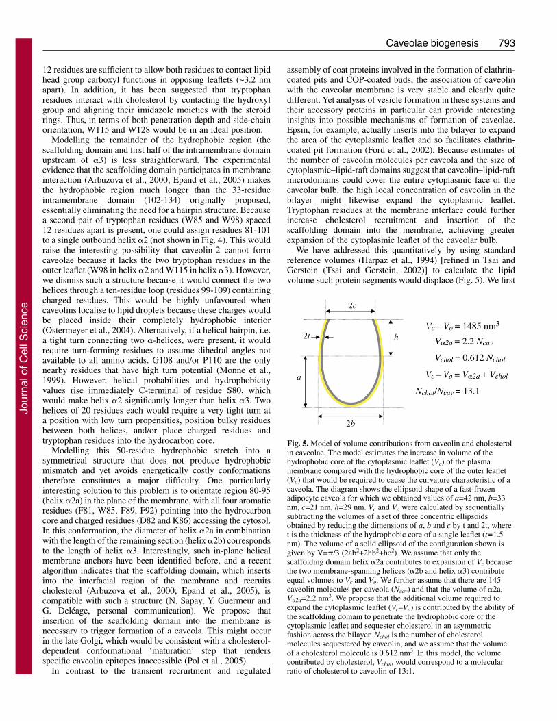
We have addressed this quantitatively by using standardreference volumes (Harpaz et al., 1994) [refined in Tsai andGerstein (Tsai and Gerstein, 2002)] to calculate the lipidvolume such protein segments would displace (Fig. 5). We first
Fig. 5. Model of volume contributions from caveolin and cholesterolin caveolae. The model estimates the increase in volume of thehydrophobic core of the cytoplasmic leaflet (Vc) of the plasmamembrane compared with the hydrophobic core of the outer leaflet(Vo) that would be required to cause the curvature characteristic of acaveola. The diagram shows the ellipsoid shape of a fast-frozenadipocyte caveola for which we obtained values of a=42 nm, b=33nm, c=21 nm, h=29 nm. Vc and Vo were calculated by sequentiallysubtracting the volumes of a set of three concentric ellipsoidsobtained by reducing the dimensions of a, b and c by t and 2t, wheret is the thickness of the hydrophobic core of a single leaflet (t=1.5nm). The volume of a solid ellipsoid of the configuration shown isgiven by V=�/3 (2ab2+2hb2+hc2). We assume that only thescaffolding domain helix �2a contributes to expansion of Vc becausethe two membrane-spanning helices (�2b and helix �3) contributeequal volumes to Vc and Vo. We further assume that there are 145caveolin molecules per caveola (Ncav) and that the volume of �2a,V�2a=2.2 nm3. We propose that the additional volume required toexpand the cytoplasmic leaflet (Vc–Vo) is contributed by the ability ofthe scaffolding domain to penetrate the hydrophobic core of thecytoplasmic leaflet and sequester cholesterol in an asymmetricfashion across the bilayer. Nchol is the number of cholesterolmolecules sequestered by caveolin, and we assume that the volumeof a cholesterol molecule is 0.612 nm3. In this model, the volumecontributed by cholesterol, Vchol, would correspond to a molecularratio of cholesterol to caveolin of 13:1.
Jour
nal o
f Cel
l Sci
ence
794
calculated the volumes of the inner and outer leaflet of a typicalcaveolar membrane and compared the relative volume increaseof the cytosolic leaflet with the volume contributed by caveolinand cholesterol. We assume that only the scaffolding domain(helix �2a) contributes to expansion of the cytosolic leaflet(Vc) because the two membrane-spanning helices (�2b andhelix �3) contribute equal volumes to the inner and outerleaflets of the membrane. These calculations indicate thatconfinement of the scaffolding domain within the cytosolicleaflet (Fig. 4), together with about 13 molecules of cholesterolper caveolin monomer, could indeed suffice to generate thecurvature observed in caveolar invaginations (Fig. 5).
Cholesterol clearly plays an important role in the formationof caveolae but its concentration in the cytoplasmic leaflet ofthe curved caveolar bulb is energetically less favourable owingto its negative spontaneous curvature (van Duyl et al., 2005).Differences in the lengths of the transmembrane segments ofcaveolin might therefore be functionally important. Forinstance, a negative hydrophobic mismatch of helix �2b and/orthe insertion of the scaffolding domain as an in-plane helix cangenerate local membrane deformations that have positivecurvature, thus stabilising cholesterol in the vicinity (van Duylet al., 2005). Alternatively, this could create a curved proteinsurface, as in the case of amphiphysin, in which membranecurvature is generated through the electrostatic interactionbetween phospholipids and the concave surface of the BAR(Bin/amphiphysin/Rvs) domain (Peter et al., 2004). Helices ofunequal lengths could also produce structural perturbations(Ren et al., 2004) that have effects elsewhere: these could beresponsible for the slow net transit out of the Golgi complex,in which caveolin cycling could facilitate caveolin-caveolinand caveolin-cholesterol interactions required for formation ofcaveolae.
Clearly, many aspects of this model require validation. Theabsence of structural data on caveolins prevents detailedmechanistic insights at present but the high density of caveolinin the caveolar membrane (Dupree et al., 1993) and theassociation of caveolin with lipid rafts are certainly consistentwith a structural model in which caveolin-cholesterolinteractions drive caveolae formation. Our model can alsoaccommodate regulatory mechanisms. For instance,phosphorylation of S80 might cause relocations as a result ofchanged electrostatics and desolvation energies. The role ofthis modification and of other specific amino acids can now betested in relation to the proposed model.
Concluding remarksCaveolae represent a stable membrane unit built up aroundcaveolin and cholesterol-rich domains in a late Golgicompartment. Caveolin-membrane interactions, andspecifically insertion of the scaffolding domain into themembrane and its interaction with cholesterol, might providethe driving force for generation of the unique caveolarstructure. This process must also be subject to regulation;recent high-throughput studies of caveolae internalisationimplicate kinases in the regulation of caveolin assembly anddisassembly (Pelkmans et al., 2005; Pelkmans and Zerial,2005). Downregulation of the kinase ARAF1, for example,causes loss of caveolin from caveolae and an increase incaveolin mobility. Detailed molecular dissection of theseeffects is now possible. High-resolution structural analysis of
caveolae and in vitro reconstitution of caveolae formation willalso provide new insights into the molecular mechanismsinvolved. This will have general implications for theunderstanding of membrane morphogenesis as well asproviding specific insights into caveola-related disease.
This work was supported by a Program Grant from the NHMRCof Australia and an RO1 grant from the NIH of USA. We would liketo thank Charles Ferguson for assistance with electron microscopy,Matthias Floetenmeyer for structural analysis of caveolae, NicolasSapay for running a prediction prior to public release, and membersof the Parton laboratory for discussions. We are also extremelygrateful to Stuart McLaughlin and Josh Zimmerberg for their expertcomments on the manuscript.
ReferencesArbuzova, A., Wang, L., Wang, J., Hangyas-Mihalyne, G., Murray, D., Honig, B.
and McLaughlin, S. (2000). Membrane binding of peptides containing both basicand aromatic residues. Experimental studies with peptides corresponding to thescaffolding region of caveolin and the effector region of MARCKS. Biochemistry 39,10330-10339.
Breuza, L., Corby, S., Arsanto, J. P., Delgrossi, M. H., Scheiffele, P. and Le Bivic, A.(2002). The scaffolding domain of caveolin 2 is responsible for its Golgi localizationin Caco-2 cells. J. Cell Sci. 115, 4457-4467.
Carozzi, A. J., Roy, S., Morrow, I. C., Pol, A., Wyse, B., Clyde-Smith, J., Prior, I. A.,Nixon, S. J., Hancock, J. F. and Parton, R. G. (2002). Inhibition of lipid raft-dependent signaling by a dystrophy-associated mutant of caveolin-3. J. Biol. Chem.277, 17944-17949.
Conrad, P. A., Smart, E. J., Ying, Y. S., Anderson, R. G. and Bloom, G. S. (1995).Caveolin cycles between plasma membrane caveolae and the Golgi complex bymicrotubule-dependent and microtubule-independent steps. J. Cell Biol. 131, 1421-1433.
de Paula, F., Vainzof, M., Bernardino, A. L., McNally, E., Kunkel, L. M. and Zatz,M. (2001). Mutations in the caveolin-3 gene: When are they pathogenic? Am. J. Med.Genet. 99, 303-307.
Dietzen, D. J., Hastings, W. R. and Lublin, D. M. (1995). Caveolin is palmitoylated onmultiple cysteine residues. Palmitoylation is not necessary for localization of caveolinto caveolae. J. Biol. Chem. 270, 6838-6842.
Drab, M., Verkade, P., Elger, M., Kasper, M., Lohn, M., Lauterbach, B., Menne, J.,Lindschau, C., Mende, F., Luft, F. C. et al. (2001). Loss of caveolae, vasculardysfunction, and pulmonary defects in caveolin-1 gene-disrupted mice. Science 9, 9.
Dupree, P., Parton, R. G., Raposo, G., Kurzchalia, T. V. and Simons, K. (1993).Caveolae and sorting in the trans-Golgi network of epithelial cells. EMBO J. 12, 1597-1605.
Engelman, J. A., Zhang, X., Galbiati, F., Volonte, D., Sotgia, F., Pestell, R. G., Minetti,C., Scherer, P. E., Okamoto, T. and Lisanti, M. P. (1998). Molecular genetics of thecaveolin gene family: implications for human cancers, diabetes, Alzheimer disease, andmuscular dystrophy. Am. J. Hum. Genet. 63, 1578-1587.
Epand, R. M., Sayer, B. G. and Epand, R. F. (2005). Caveolin scaffolding region andcholesterol-rich domains in membranes. J. Mol. Biol. 345, 339-350.
Fernandez, I., Ying, Y., Albanesi, J. and Anderson, R. G. (2002). Mechanism ofcaveolin filament assembly. Proc. Natl. Acad. Sci. USA 99, 11193-11198.
Fielding, P. E., Chau, P., Liu, D., Spencer, T. A. and Fielding, C. J. (2004). Mechanismof platelet-derived growth factor-dependent caveolin-1 phosphorylation: relationship tosterol binding and the role of serine-80. Biochemistry 43, 2578-2586.
Ford, M. G., Mills, I. G., Peter, B. J., Vallis, Y., Praefcke, G. J., Evans, P. R. andMcMahon, H. T. (2002). Curvature of clathrin-coated pits driven by epsin. Nature 419,361-366.
Fra, A. M., Williamson, E., Simons, K. and Parton, R. G. (1995). De novo formationof caveolae in lymphocytes by expression of VIP21-caveolin. Proc. Natl. Acad. Sci.USA 92, 8655-8659.
Fujimoto, T., Kogo, H., Nomura, R. and Une, T. (2000). Isoforms of caveolin-1 andcaveolar structure. J. Cell Sci. 19, 3509-3517.
Galbiati, F., Volonte, D., Minetti, C., Chu, J. B. and Lisanti, M. P. (1999). Phenotypicbehavior of caveolin-3 mutations that cause autosomal dominant limb girdle musculardystrophy (LGMD-1C). Retention of LGMD-1C caveolin-3 mutants within the Golgicomplex. J. Biol. Chem. 274, 25632-25641.
Galbiati, F., Engelman, J. A., Volonte, D., Zhang, X. L., Minetti, C., Li, M., Hou, H.,Jr, Kneitz, B., Edelmann, W. and Lisanti, M. P. (2001). Caveolin-3 null mice showa loss of caveolae, changes in the microdomain distribution of the dystrophin-glycoprotein complex, and t-tubule abnormalities. J. Biol. Chem. 276, 21425-21433.
Harpaz, Y., Gerstein, M. and Chothia, C. (1994). Volume changes on protein folding.Structure 2, 641-649.
Hernández-Deviez, D. J., Martin, S., Laval, S. H., Lo, H. P., Cooper, S. T., North, K.N. N., Bushby, K. and Parton, R. G. (2006). Aberrant dysferlin trafficking in cellslacking caveolin or expressing dystrophy mutants of caveolin-3. Hum. Mol. Genet. 15,129-142.
Ishikawa, H. (1968). Formation of elaborate networks of T-system tubules in cultured
Journal of Cell Science 119 (5)
Jour
nal o
f Cel
l Sci
ence
795Caveolae biogenesis
skeletal muscle with special reference to the T-system formation. J. Cell Biol. 38, 51-66.
Kirkham, M., Fujita, A., Chadda, R., Nixon, S. J., Kurzchalia, T. V., Sharma, D. K.,Pagano, R. E., Hancock, J. F., Mayor, S. and Parton, R. G. (2005). Ultrastructuralidentification of uncoated caveolin-independent early endocytic vehicles. J. Cell Biol.168, 465-476.
Lahtinen, U., Honsho, M., Parton, R. G., Simons, K. and Verkade, P. (2003).Involvement of caveolin-2 in caveolar biogenesis in MDCK cells. FEBS Lett. 538, 85-88.
Lee, E., Marcucci, M., Daniell, L., Pypaert, M., Weisz, O. A., Ochoa, G. C., Farsad,K., Wenk, M. R. and De Camilli, P. (2002). Amphiphysin 2 (Bin1) and T-tubulebiogenesis in muscle. Science 297, 1193-1196.
Li, H. and Papadopoulos, V. (1998). Peripheral-type benzodiazepine receptor functionin cholesterol transport. Identification of a putative cholesterol recognition/interactionamino acid sequence and consensus pattern. Endocrinology 139, 4991-4997.
Lichtenberg, D., Goni, F. M. and Heerklotz, H. (2005). Detergent-resistantmembranes should not be identified with membrane rafts. Trends Biochem. Sci. 30,430-436.
Lipardi, C., Mora, R., Colomer, V., Paladino, S., Nitsch, L., Rodriguez-Boulan, E.and Zurzolo, C. (1998). Caveolin transfection results in caveolae formation but notapical sorting of glycosylphosphatidylinositol (GPI)-anchored proteins in epithelialcells. J. Cell Biol. 140, 617-626.
Liu, P., Li, W. P., Machleidt, T. and Anderson, R. G. (1999). Identification of caveolin-1 in lipoprotein particles secreted by exocrine cells. Nat. Cell Biol. 1, 369-375.
Luetterforst, R., Stang, E., Zorzi, N., Carozzi, A., Way, M. and Parton, R. G. (1999).Molecular characterization of caveolin association with the Golgi complex:identification of a cis-Golgi targeting domain in the caveolin molecule. J. Cell Biol.145, 1443-1459.
Machleidt, T., Li, W. P., Liu, P. and Anderson, R. G. (2000). Multiple domains incaveolin-1 control its intracellular traffic. J. Cell Biol. 148, 17-28.
McNally, E. M., de Sa Moreira, E., Duggan, D. J., Bonnemann, C. G., Lisanti, M. P.,Lidov, H. G., Vainzof, M., Passos-Bueno, M. R., Hoffman, E. P., Zatz, M. et al.(1998). Caveolin-3 in muscular dystrophy. Hum. Mol. Genet. 7, 871-877.
Minetti, C., Sotgia, F., Bruno, C., Scartezzini, P., Broda, P., Bado, M., Masetti, E.,Mazzocco, M., Egeo, A., Donati, M. A. et al. (1998). Mutations in the caveolin-3gene cause autosomal dominant limb-girdle muscular dystrophy. Nat. Genet. 18, 365-368.
Monier, S., Parton, R. G., Vogel, F., Behlke, J., Henske, A. and Kurzchalia, T. V.(1995). VIP21-caveolin, a membrane protein constituent of the caveolar coat,oligomerizes in vivo and in vitro. Mol. Biol. Cell 6, 911-927.
Monier, S., Dietzen, D. J., Hastings, W. R., Lublin, D. M. and Kurzchalia, T. V. (1996).Oligomerization of VIP21-caveolin in vitro is stabilized by long chain fatty acylationor cholesterol. FEBS Lett. 388, 143-149.
Monne, M., Nilsson, I., Elofsson, A. and von Heijne, G. (1999). Turns intransmembrane helices: determination of the minimal length of a “helical hairpin” andderivation of a fine-grained turn propensity scale. J. Mol. Biol. 293, 807-814.
Montesano, R. (1979). Inhomogeneous distribution of filipin-sterol complexes in smoothmuscle cell plasma membrane. Nature 280, 328-329.
Munro, S. (2003). Lipid rafts: elusive or illusive? Cell 115, 377-388.Murata, M., Peranen, J., Schreiner, R., Wieland, F., Kurzchalia, T. V. and Simons,
K. (1995). VIP21/caveolin is a cholesterol-binding protein. Proc. Natl. Acad. Sci. USA92, 10339-10343.
Nabi, I. R. and Le, P. U. (2003). Caveolae/raft-dependent endocytosis. J. Cell Biol. 161,673-677.
Nichols, B. J. (2002). A distinct class of endosome mediates clathrin-independentendocytosis to the Golgi complex. Nat. Cell Biol. 4, 374-378.
Ostermeyer, A. G., Ramcharan, L. T., Zeng, Y., Lublin, D. M. and Brown, D. A.(2004). Role of the hydrophobic domain in targeting caveolin-1 to lipid droplets. J.Cell Biol. 164, 69-78.
Palade, G. E. (1953). Fine structure of blood capillaries. J. Appl. Phys. 24, 1424.Parat, M. O. and Fox, P. L. (2001). Palmitoylation of caveolin-1 in endothelial cells is
post-translational but irreversible. J. Biol. Chem. 276, 15776-15782.Parton, R. G. (1996). Caveolae and caveolins. Curr. Opin. Cell Biol. 8, 542-548.Parton, R. G., Way, M., Zorzi, N. and Stang, E. (1997). Caveolin-3 associates with
developing T-tubules during muscle differentiation. J. Cell Biol. 136, 137-154.Parton, R. G., Molero, J. C., Floetenmeyer, M., Green, K. M. and James, D. E. (2002).
Characterization of a distinct plasma membrane macrodomain in differentiatedadipocytes. J. Biol. Chem. 277, 46769-46778.
Pelkmans, L. and Helenius, A. (2002). Endocytosis via caveolae. Traffic 3, 311-320.Pelkmans, L. and Zerial, M. (2005). Kinase-regulated quantal assemblies and kiss-and-
run recycling of caveolae. Nature 436, 128-133.Pelkmans, L., Kartenbeck, J. and Helenius, A. (2001). Caveolar endocytosis of simian
virus 40 reveals a new two-step vesicular-transport pathway to the ER. Nat. Cell Biol.3, 473-483.
Pelkmans, L., Burli, T., Zerial, M. and Helenius, A. (2004). Caveolin-stabilizedmembrane domains as multifunctional transport and sorting devices in endocyticmembrane traffic. Cell 118, 767-780.
Pelkmans, L., Fava, E., Grabner, H., Hannus, M., Habermann, B., Krausz, E. andZerial, M. (2005). Genome-wide analysis of human kinases in clathrin- andcaveolae/raft-mediated endocytosis. Nature 436, 78-86.
Peter, B. J., Kent, H. M., Mills, I. G., Vallis, Y., Butler, P. J., Evans, P. R. andMcMahon, H. T. (2004). BAR domains as sensors of membrane curvature: theamphiphysin BAR structure. Science 303, 495-499.
Peters, K. R., Carley, W. W. and Palade, G. E. (1985). Endothelial plasmalemmal
vesicles have a characteristic striped bipolar surface structure. J. Cell Biol. 101, 2233-2238.
Pike, L. J., Han, X., Chung, K. N. and Gross, R. W. (2002). Lipid rafts are enrichedin arachidonic acid and plasmenylethanolamine and their composition is independentof caveolin-1 expression: a quantitative electrospray ionization/mass spectrometricanalysis. Biochemistry 41, 2075-2088.
Pol, A., Martin, S., Fernandez, M. A., Ingelmo-Torres, M., Ferguson, C., Enrich, C.and Parton, R. G. (2005). Cholesterol and fatty acids regulate dynamic caveolintrafficking through the Golgi complex and between the cell surface and lipid bodies.Mol. Biol. Cell 16, 2091-2105.
Rajendran, L. and Simons, K. (2005). Lipid rafts and membrane dynamics. J. Cell Sci.118, 1099-1102.
Razani, B., Combs, T. P., Wang, X. B., Frank, P. G., Park, D. S., Russell, R. G., Li,M., Tang, B., Jelicks, L. A., Scherer, P. E. et al. (2002a). Caveolin-1-deficient miceare lean, resistant to diet-induced obesity, and show hypertriglyceridemia withadipocyte abnormalities. J. Biol. Chem. 277, 8635-8647.
Razani, B., Wang, X. B., Engelman, J. A., Battista, M., Lagaud, G., Zhang, X. L.,Kneitz, B., Hou, H., Jr, Christ, G. J., Edelmann, W. et al. (2002b). Caveolin-2-deficient mice show evidence of severe pulmonary dysfunction without disruption ofcaveolae. Mol. Cell. Biol. 22, 2329-2344.
Ren, X., Ostermeyer, A. G., Ramcharan, L. T., Zeng, Y., Lublin, D. M. and Brown,D. A. (2004). Conformational defects slow Golgi exit, block oligomerization, andreduce raft affinity of caveolin-1 mutant proteins. Mol. Biol. Cell 15, 4556-4567.
Rothberg, K. G., Heuser, J. E., Donzell, W. C., Ying, Y. S., Glenney, J. R. andAnderson, R. G. (1992). Caveolin, a protein component of caveolae membrane coats.Cell 68, 673-682.
Russ, W. P. and Engelman, D. M. (2000). The GxxxG motif: a framework fortransmembrane helix-helix association. J. Mol. Biol. 296, 911-919.
Santiago, J., Guzman, G. R., Rojas, L. V., Marti, R., Asmar-Rovira, G. A., Santana,L. F., McNamee, M. and Lasalde-Dominicci, J. A. (2001). Probing the effects ofmembrane cholesterol in the Torpedo californica acetylcholine receptor and the novellipid-exposed mutation alpha C418W in Xenopus oocytes. J. Biol. Chem. 276, 46523-46532.
Sargiacomo, M., Scherer, P. E., Tang, Z., Kubler, E., Song, K. S., Sanders, M. C. andLisanti, M. P. (1995). Oligomeric structure of caveolin: implications for caveolaemembrane organization. Proc. Natl. Acad. Sci. USA 92, 9407-9411.
Scheiffele, P., Verkade, P., Fra, A. M., Virta, H., Simons, K. and Ikonen, E. (1998).Caveolin-1 and -2 in the exocytic pathway of MDCK cells. J. Cell Biol. 140, 795-806.
Schubert, W., Frank, P. G., Razani, B., Park, D. S., Chow, C. W. and Lisanti, M. P.(2001). Caveolae-deficient endothelial cells show defects in the uptake and transportof albumin in vivo. J. Biol. Chem. 276, 48619-48622.
Sharma, D. K., Choudhury, A., Singh, R. D., Wheatley, C. L., Marks, D. L. andPagano, R. E. (2003). Glycosphingolipids internalized via caveolar-relatedendocytosis rapidly merge with the clathrin pathway in early endosomes and formmicrodomains for recycling. J. Biol. Chem. 278, 7564-7572.
Simons, K. and Toomre, D. (2000). Lipid rafts and signal transduction. Nat. Rev. Mol.Cell Biol. 1, 31-39.
Song, K. S., Tang, Z., Li, S. and Lisanti, M. P. (1997). Mutational analysis of theproperties of caveolin-1. A novel role for the C-terminal domain in mediating homo-typic caveolin-caveolin interactions. J. Biol. Chem. 272, 4398-4403.
Sowa, G., Pypaert, M., Fulton, D. and Sessa, W. C. (2003). The phosphorylation ofcaveolin-2 on serines 23 and 36 modulates caveolin-1-dependent caveolae formation.Proc. Natl. Acad. Sci. USA 100, 6511-6516.
Stan, R. V. (2002). Structure and function of endothelial caveolae. Microsc. Res. Tech.57, 350-364.
Stan, R. V. (2005). Structure of caveolae. Biochim Biophys Acta. 1746, 334-348.Stan, R. V., Tkachenko, E. and Niesman, I. R. (2004). PV1 is a key structural
component for the formation of the stomatal and fenestral diaphragms. Mol. Biol. Cell15, 3615-3630.
Tagawa, A., Mezzacasa, A., Hayer, A., Longatti, A., Pelkmans, L. and Helenius, A.(2005). Assembly and trafficking of caveolar domains in the cell: caveolae as stable,cargo-triggered, vesicular transporters. J. Cell Biol. 170, 769-779.
Tang, Z., Scherer, P. E., Okamoto, T., Song, K., Chu, C., Kohtz, D. S., Nishimoto, I.,Lodish, H. F. and Lisanti, M. P. (1996). Molecular cloning of caveolin-3, a novelmember of the caveolin gene family expressed predominantly in muscle. J. Biol. Chem.271, 2255-2261.
Thiele, C., Hannah, M. J., Fahrenholz, F. and Huttner, W. B. (2000). Cholesterol bindsto synaptophysin and is required for biogenesis of synaptic vesicles. Nat. Cell Biol. 2,42-49.
Thomsen, P., Roepstorff, K., Stahlhut, M. and van Deurs, B. (2002). Caveolae arehighly immobile plasma membrane microdomains, which are not involved inconstitutive endocytic trafficking. Mol. Biol. Cell 13, 238-250.
Thorn, H., Stenkula, K. G., Karlsson, M., Ortegren, U., Nystrom, F. H.,Gustavsson, J. and Stralfors, P. (2003). Cell surface orifices of caveolae andlocalization of caveolin to the necks of caveolae in adipocytes. Mol. Biol. Cell 14,3967-3976.
Tran, D., Carpentier, J. L., Sawano, F., Gorden, P. and Orci, L. (1987). Ligandsinternalized through coated or noncoated invaginations follow a common intracellularpathway. Proc. Natl. Acad. Sci. USA 84, 7957-7961.
Tsai, J. and Gerstein, M. (2002). Calculations of protein volumes: sensitivity analysisand parameter database. Bioinformatics 18, 985-995.
Uittenbogaard, A., Ying, Y. and Smart, E. J. (1998). Characterization of a cytosolicheat-shock protein-caveolin chaperone complex. Involvement in cholesterol trafficking.J. Biol. Chem. 273, 6525-6532.
Jour
nal o
f Cel
l Sci
ence
796
van Duyl, B. Y., Meeldijk, H., Verkleij, A. J., Rijkers, D. T., Chupin, V., de Kruijff,B. and Killian, J. A. (2005). A synergistic effect between cholesterol and tryptophan-flanked transmembrane helices modulates membrane curvature. Biochemistry 44,4526-4532.
Vogel, U., Sandvig, K. and van Deurs, B. (1998). Expression of caveolin-1 and polarizedformation of invaginated caveolae in Caco-2 and MDCK II cells. J. Cell Sci. 111, 825-832.
Way, M. and Parton, R. G. (1995). M-caveolin, a muscle-specific caveolin-relatedprotein. FEBS Lett. 376, 108-112.
Westermann, M., Steiniger, F. and Richter, W. (2005). Belt-like localisation of caveolin
in deep caveolae and its re-distribution after cholesterol depletion. Histochem. CellBiol. 123, 613-620.
Wimley, W. C. and White, S. H. (1996). Experimentally determined hydrophobicityscale for proteins at membrane interfaces. Nat. Struct. Biol. 3, 842-848.
Wyse, B. D., Prior, I. A., Qian, H., Morrow, I. C., Nixon, S., Muncke, C., Kurzchalia,T. V., Thomas, W. G., Parton, R. G. and Hancock, J. F. (2003). Caveolin interactswith the angiotensin II type 1 receptor during exocytic transport but not at the plasmamembrane. J. Biol. Chem. 278, 23738-23746.
Yamada, E. (1955). The fine structures of the gall bladder epithelium of the mouse. J.Biophys. Biochem. Cytol. 1, 445-458.
Journal of Cell Science 119 (5)
Jour
nal o
f Cel
l Sci
ence