Gene knockout of the KCNJ8-encoded Kir6.1 KATP channel imparts fatal susceptibility to endotoxemia
Upload
independentCategory
view
2download
0
1
Congenital Aortic Stenosis in Adults
Update on clinical outcome, diagnostic methods and pregnancy

2
Colofon
Yap, SC
Congenital aortic stenosis in adults - Update on clinical outcome, diagnostic methods and pregnancy.
ISBN: 978-90-8559-330-0
© Copyright 2007 S.C.Yap
All rights reserved. No part of this publication may be reproduced, stored in a retrieval system, or
transmitted in any form or by any means, electronical, mechanical, photocopying, recording, or
otherwise, without prior written permission of the holder of the copyright. Several chapters of this
thesis are based on published papers, which are reproduced with permission of the co-authors and
the publishers. Copyright of these papers remains with the publishers.
Cover: Véro Crickx, Sirene ontwerpers, Rotterdam, The Netherlands
Lay-out: S.C.Yap
Printed by Optima Grafische Communicatie, Rotterdam, The Netherlands
Financial support by the Netherlands Heart Foundation for the publication of this thesis is gratefully
acknowledged.

3
Congenital Aortic Stenosis in Adults
Update on clinical outcome, diagnostic methods and pregnancy
Congenitale aortastenose in volwassenen
Nieuwe inzichten in klinische uitkomsten, diagnostische methoden en
zwangerschap
Proefschrift
ter verkrijging van de graad van doctor aan de
Erasmus Universiteit Rotterdam op gezag van de rector magnificus
Prof.dr. S.W.J. Lamberts
en volgens besluit van het College voor Promoties.
De openbare verdediging zal plaatsvinden op
woensdag 19 december 2007 om 9.45 uur
door
Sing-Chien Yap
geboren te ’s-Hertogenbosch

4
Promotiecommissie
Promotor: Prof.dr. M.L. Simoons Copromotor: Dr. J.W. Roos-Hesselink Overige leden: Prof.dr. A.J.J.C. Bogers
Prof.dr. B.M.J. Mulder Prof.dr. W.A. Helbing

5
Chapter 1. General introduction 9
Part 1. Clinical outcome of congenital aortic stenosis Chapter 2. Aortic stenosis at young adult age 18 Expert Rev Cardiovasc Ther 2005;3:1087-98
Chapter 3 Congenital aortic stenosis in adults: rate of progression and 42
predictors of clinical outcome Int J Cardiol 2007 Feb 7; [Epub ahead of print]
Chapter 4 When to intervene in aortic ectasia 56
Focused review. Braunwald’s Heart Disease: A textbook of
cardiovascular medicine
Chapter 5 Outcome of implantable cardioverter defibrillators in adults with 66
congenital heart disease: a multi-centre study Eur Heart J 2007;28:1854-61
Part 2. Diagnostic methods for congenital aortic stenosis Chapter 6 Steepened aortoseptal angle may be a risk factor for discrete 86
subaortic stenosis in adult patients Int J Cardiol 2007 April 13; [Epub ahead of print]
Chapter 7 Abnormal aortic elastic properties in adults with congenital 92
valvular aortic stenosis Int J Cardiol 2007 Aug 7; [Epub ahead of print]
Chapter 8 Rapid and accurate measurement of LV mass by RT3DE 104
in patients with concentric LV hypertrophy: comparison to CMR Eur J Echocard 2007 Jun 29; [Epub ahead of print]
Chapter 9 A simplified continuity equation approach to the quantification of 118
stenotic bicuspid aortic valves using velocity-encoded CMR J Cardiovasc Magn Reson 2007 (in press)
Contents

6
Part 3. Pregnancy and congenital aortic stenosis Chapter 10 Complications during pregnancy in women with congenital heart 136
disease: a literature review JACC 2007;49:2303-11
Chapter 11 Risk of complications during pregnancy in women with congenital 154
aortic stenosis Int J Cardiol 2007 May 3; [Epub ahead of print]
Chapter 12 Outcome of pregnancy in women after pulmonary autograft valve 168
replacement for congenital aortic valve disease J Heart Valve Dis 2007;16:398-403
Chapter 13 Percutaneous triple-valve balloon valvuloplasty in a pregnant 180
woman using intracardiac echocardiography: case report J Heart Valve Dis 2006;15:459-64
Chapter 14 General discussion, conclusions and future directions 193 Chapter 15 Samenvatting 207 Chapter 16 Dankwoord 215
List of publications 218
Curriculum vitae 221

7
1
Congenital aortic stenosis in adults
General introduction
C H A P T E R

Chapter 1
8

General introduction
9
INTRODUCTION Congenital aortic stenosis (AS) encompass a series of stenotic lesions starting in the anatomic left ventricular outflow tract (LVOT) and stretches toward the ascending aorta. Obstruction may be subvalvar, valvar, or supravalvar. All of these lesions impose an increased afterload on the left ventricle (LV), which can result in LV hypertrophy, dilatation and eventually heart failure, when left untreated. Congenital valvar AS is usually the result of a stenotic bicuspid aortic valve (BAV).1,2 Variants range from a nearly trileaflet bicommissural valve with mild cusp inequality to a unicuspid unicommissural valve. Recently, BAV has been the focus of much research as it identifies patients at risk of development of stenosis and regurgitation, and in addition predisposes patients to ascending aorta dilatation and dissection. BAV is the most common congenital malformations and has a prevalence of 1-2% in the general population.3,4 It results from fusion of one of the commissures (usually present as a raphe),5 resulting in two rather than three valve leaflets. No clear etiology has been defined for BAV, and no specific gene has been identified. Recent findings support the suggestion that all anomalies of the left ventricular outflow tract obstruction spectrum are developmentally related and multiple genes have been implicated. Experimental evidence suggests that the expression of endothelial nitric oxide synthase (eNOS) may have an influence on aortic valve anatomy and aneurysmal dilatation of the aorta.6,7 Mutations in the signaling and transcriptional regulator NOTCH1 gene result in developmental aortic valve abnormalities and severe valve calcification in affected families.8 Ubiquitin fusion degradation 1-like gene is another potential candidate, which is highly expressed in the cardiac outflow tract during embryogenesis and is downregulated in patients with BAV.9 Furthermore, familial clustering of BAV and other left ventricular outflow tract obstructions has been described.10,11 PROGNOSIS AND TREATMENT Patients with congenital AS with severe symptoms in infancy and childhood have a poor prognosis without intervention.12 Sudden cardiac death may occur, especially in the setting of physical acitivity and exertion.13 Even after surgical valvotomy the incidence of sudden death is still 0.4% per year.14 A bicuspid valve may function normally throughout a lifetime, but usually is associated with the development of either progressive stenosis or regurgitation.15,16 BAV accounts for approximately 50% of all aortic valve replacements of isolated AS in adults.1 The main indication for aortic valve replacement is the presence of symptoms (i.e., angina, syncope, congestive heart failure) as this is associated with a worse outcome (i.e., overall cardiac mortality and sudden cardiac death).17-19 Ballloon valvotomy may be an

Chapter 1
10
attractive option in children, adolescents and young adults who have pliable, noncalcified valves with fusion of the commissures, at the cost of restenosis or regurgitation.20,21 The Ross procedure (pulmonary autograft) is the treatment of choice in the pediatric population due to its growth potential, but the high reoperation rate and progressive autograft dilatation renders it unsuitable in adults.22 AORTIC DILATATION There is a high incidence of aortic disease in patients with congenital aortic valve malformations suggesting a causal relationship between these two conditions. Controversy exists regarding the etiology of aortic dilatation in BAV patients. Once thought to be the consequence of post-stenotic dilatation, evidence is accumulating that intrinsic aortic medial weakness should be regarded as the underlying cause of aortic dilatation. Several findings support this hypothesis. Approximately 50% of patients with normally functioning BAV have an aortic dilatation, thus independent of the presence of stenosis or regurgitation.23-26 Also after aortic valve replacement aortic dilatation may occur. Furthermore, the histology of the ascending aortic wall is similar to that of the Marfan syndrome.27 Medial disease is present, as are varying degrees of abnormalities of the smooth muscle, extracellular matrix, elastin, and collagen.27-29 Dilatation of the aorta may be the result of the disruption of the extracellular matrix by upregulation of matrix metalloproteinase-2 that is triggered by an inherent deficiency of fibrillin-1.28,30-32 Furthermore, premature smooth muscle cell apoptosis leads to upregulation of matrix metalloproteinase-2.33,34 PREGNANCY Cardiovascular physiology changes profoundly during pregnancy. Cardiac output increases 30-50% due to increases in both stroke volume and heart rate.35,36 During labour, cardiac output increases further due to pain and uterine contractions.37,38 The haemodynamic impact of AS is aggravated by the physiological changes during pregnancy. Pregnancy in AS patients has been the focus of some reports because of concern for development of heart failure and mortality during pregnancy. 39,40 The review of Lao et al. published in 1993 demonstrated seven deaths among 65 women, resulting in a maternal mortality rate of 11% 41. Earlier diagnosis and treatment of patients with severe stenosis (e.g. balloon aortic valvotomy) has resulted in more women with congenital AS reaching childbearing age in relatively good condition. Recent pregnancy reports in AS patients are encouraging, showing a favorable pregnancy outcome with low maternal mortality.42-44

General introduction
11
AIMS AND OUTLINE OF THE THESIS The aim of the present thesis is to present an update on clinical outcome, diagnostic methods and pregnancy in adults with congenital AS. It will not address congenital AS in the pediatric population. In part 1 (chapters 2-5) of this thesis, I will present an overview of the current management of patients with congenital aortic stenosis with regard to timing of surgical or percutaneous intervention, aortic dilatation, exercise, pregnancy, and potential medical treatment.46 Secondly, we investigated the natural history of a cohort of patients with congenital aortic stenosis, with special emphasis on the rate of stenosis progression and its determinants.47 Furthermore, a discussion is presented regarding the optimal timing of prophylactic aortic replacement in dilated aorta, which is a common finding in BAV patients. Finally, as patients with AS are at increased risk of sudden cardiac death, the outcome of ICD therapy in patients with CHD is presented.48 In part 2 (chapters 6-9), we investigated the use of diagnostic techniques and innovative approaches in patients with congenital AS. A cohort of patients with congenital valvar AS was investigated using different imaging modalities (i.e., M-mode, 2D-, 3D-echocardiography, and cardiac magnetic resonance). The aortic stiffness could be investigated using M-mode echocardiography. Furthermore, real-time 3D-echocardiography was compared with M-mode and 2D-echocardiography in determining LV mass. The role of velocity-encoded cardiac magnetic resonance in determining aortic valve area using a modified continuity equation was established. Finally, we investigated the aortoseptal angle in adults with discrete subaortic stenosis (DSS) compared to controls using 2D-echocardiography, in testing the hypothesis that altered flow patterns are a substrate for development of DSS.49
In part 3 (chapters 10-13), the risk of pregnancy in women with congenital aortic stenosis is evaluated. Chapter 10 provides a general overview of complications encountered during pregnancy in women with congenital heart disease. Chapter 11 and 12 investigate pregnancy in women with congenital aortic stenosis without previous aortic valve replacement and with pulmonary autograft valve replacement, respectively.50 These studies are part of the ZAHARA study, investigating pregnancy in congenital heart disease. For the selection of women in their fertile ages we used the Dutch CONCOR registry (CONgenital CORvitia; www.concor.net), which is a registry of all adult patients with CHD in the Netherlands.51 Chapter 13 presents a case report demonstrating the value of intracardiac echocardiography during percutaneous intervention in a pregnant woman thereby reducing radiation exposure to the foetus.52
Finally, a general discussion is presented, including conclusions and recommendations for future research and clinical practice.

Chapter 1
12
REFERENCES 1. Roberts WC, Ko JM. Frequency by decades of unicuspid, bicuspid, and tricuspid aortic valves
in adults having isolated aortic valve replacement for aortic stenosis, with or without
associated aortic regurgitation. Circulation. 2005;111:920-5.
2. Aboulhosn J, Child JS. Left ventricular outflow obstruction: subaortic stenosis, bicuspid aortic
valve, supravalvar aortic stenosis, and coarctation of the aorta. Circulation. 2006;114:2412-22.
3. Larson EW, Edwards WD. Risk factors for aortic dissection: a necropsy study of 161 cases.
Am J Cardiol. 1984;53:849-55.
4. Hoffman JI, Kaplan S. The incidence of congenital heart disease. J Am Coll Cardiol.
2002;39:1890-900.
5. Sievers HH, Schmidtke C. A classification system for the bicuspid aortic valve from 304
surgical specimens. J Thorac Cardiovasc Surg. 2007;133:1226-33.
6. Aicher D, Urbich C, Zeiher A, Dimmeler S, Schafers HJ. Endothelial nitric oxide synthase in
bicuspid aortic valve disease. Ann Thorac Surg. 2007;83:1290-4.
7. Lee TC, Zhao YD, Courtman DW, Stewart DJ. Abnormal aortic valve development in mice
lacking endothelial nitric oxide synthase. Circulation. 2000;101:2345-8.
8. Garg V, Muth AN, Ransom JF, Schluterman MK, Barnes R, King IN, Grossfeld PD, Srivastava
D. Mutations in NOTCH1 cause aortic valve disease. Nature. 2005;437:270-4.
9. Mohamed SA, Hanke T, Schlueter C, Bullerdiek J, Sievers HH. Ubiquitin fusion degradation 1-
like gene dysregulation in bicuspid aortic valve. J Thorac Cardiovasc Surg. 2005;130:1531-6.
10. Huntington K, Hunter AG, Chan KL. A prospective study to assess the frequency of familial
clustering of congenital bicuspid aortic valve. J Am Coll Cardiol. 1997;30:1809-12.
11. Wessels MW, Berger RM, Frohn-Mulder IM, Roos-Hesselink JW, Hoogeboom JJ, Mancini GS,
Bartelings MM, Krijger R, Wladimiroff JW, Niermeijer MF, Grossfeld P, Willems PJ. Autosomal
dominant inheritance of left ventricular outflow tract obstruction. Am J Med Genet A.
2005;134:171-9.
12. Keane JF, Driscoll DJ, Gersony WM, Hayes CJ, Kidd L, O'Fallon WM, Pieroni DR, Wolfe RR,
Weidman WH. Second natural history study of congenital heart defects. Results of treatment
of patients with aortic valvar stenosis. Circulation. 1993;87:I16-27.
13. Driscoll DJ, Edwards WD. Sudden unexpected death in children and adolescents. J Am Coll
Cardiol. 1985;5:118B-121B.
14. Hsieh KS, Keane JF, Nadas AS, Bernhard WF, Castaneda AR. Long-term follow-up of
valvotomy before 1968 for congenital aortic stenosis. Am J Cardiol. 1986;58:338-41.
15. Fenoglio JJ, Jr., McAllister HA, Jr., DeCastro CM, Davia JE, Cheitlin MD. Congenital bicuspid
aortic valve after age 20. Am J Cardiol. 1977;39:164-9.
16. Ward C. Clinical significance of the bicuspid aortic valve. Heart. 2000;83:81-5.
17. Otto CM. Valvular aortic stenosis: disease severity and timing of intervention. J Am Coll
Cardiol. 2006;47:2141-51.
18. Braunwald E. On the natural history of severe aortic stenosis. J Am Coll Cardiol.
1990;15:1018-20.
19. Kelly TA, Rothbart RM, Cooper CM, Kaiser DL, Smucker ML, Gibson RS. Comparison of
outcome of asymptomatic to symptomatic patients older than 20 years of age with valvular
aortic stenosis. Am J Cardiol. 1988;61:123-30.
20. Bonow RO, Carabello BA, Kanu C, de Leon AC, Jr., Faxon DP, Freed MD, Gaasch WH, Lytle
BW, Nishimura RA, O'Gara PT, O'Rourke RA, Otto CM, Shah PM, Shanewise JS, Smith SC,

General introduction
13
Jr., Jacobs AK, Adams CD, Anderson JL, Antman EM, Fuster V, Halperin JL, Hiratzka LF,
Hunt SA, Nishimura R, Page RL, Riegel B. ACC/AHA 2006 guidelines for the management of
patients with valvular heart disease: a report of the American College of Cardiology/American
Heart Association Task Force on Practice Guidelines (writing committee to revise the 1998
Guidelines for the Management of Patients With Valvular Heart Disease): developed in
collaboration with the Society of Cardiovascular Anesthesiologists: endorsed by the Society
for Cardiovascular Angiography and Interventions and the Society of Thoracic Surgeons.
Circulation. 2006;114:e84-231.
21. Reich O, Tax P, Marek J, Razek V, Gilik J, Tomek V, Chaloupecky V, Bartakova H, Skovranek
J. Long term results of percutaneous balloon valvoplasty of congenital aortic stenosis:
independent predictors of outcome. Heart. 2004;90:70-6.
22. Klieverik LM, Takkenberg JJ, Bekkers JA, Roos-Hesselink JW, Witsenburg M, Bogers AJ. The
Ross operation: a Trojan horse? Eur Heart J. 2007.
23. Nistri S, Sorbo MD, Marin M, Palisi M, Scognamiglio R, Thiene G. Aortic root dilatation in
young men with normally functioning bicuspid aortic valves. Heart. 1999;82:19-22.
24. Pachulski RT, Weinberg AL, Chan KL. Aortic aneurysm in patients with functionally normal or
minimally stenotic bicuspid aortic valve. Am J Cardiol. 1991;67:781-2.
25. Hahn RT, Roman MJ, Mogtader AH, Devereux RB. Association of aortic dilation with
regurgitant, stenotic and functionally normal bicuspid aortic valves. J Am Coll Cardiol.
1992;19:283-8.
26. Ferencik M, Pape LA. Changes in size of ascending aorta and aortic valve function with time
in patients with congenitally bicuspid aortic valves. Am J Cardiol. 2003;92:43-6.
27. Niwa K, Perloff JK, Bhuta SM, Laks H, Drinkwater DC, Child JS, Miner PD. Structural
abnormalities of great arterial walls in congenital heart disease: light and electron microscopic
analyses. Circulation. 2001;103:393-400.
28. Fedak PW, Verma S, David TE, Leask RL, Weisel RD, Butany J. Clinical and
pathophysiological implications of a bicuspid aortic valve. Circulation. 2002;106:900-4.
29. de Sa M, Moshkovitz Y, Butany J, David TE. Histologic abnormalities of the ascending aorta
and pulmonary trunk in patients with bicuspid aortic valve disease: clinical relevance to the
ross procedure. J Thorac Cardiovasc Surg. 1999;118:588-94.
30. Fedak PW, de Sa MP, Verma S, Nili N, Kazemian P, Butany J, Strauss BH, Weisel RD, David
TE. Vascular matrix remodeling in patients with bicuspid aortic valve malformations:
implications for aortic dilatation. J Thorac Cardiovasc Surg. 2003;126:797-806.
31. Boyum J, Fellinger EK, Schmoker JD, Trombley L, McPartland K, Ittleman FP, Howard AB.
Matrix metalloproteinase activity in thoracic aortic aneurysms associated with bicuspid and
tricuspid aortic valves. J Thorac Cardiovasc Surg. 2004;127:686-91.
32. Borger MA, Preston M, Ivanov J, Fedak PW, Davierwala P, Armstrong S, David TE. Should
the ascending aorta be replaced more frequently in patients with bicuspid aortic valve
disease? J Thorac Cardiovasc Surg. 2004;128:677-83.
33. Bonderman D, Gharehbaghi-Schnell E, Wollenek G, Maurer G, Baumgartner H, Lang IM.
Mechanisms underlying aortic dilatation in congenital aortic valve malformation. Circulation.
1999;99:2138-43.
34. Schmid FX, Bielenberg K, Schneider A, Haussler A, Keyser A, Birnbaum D. Ascending aortic
aneurysm associated with bicuspid and tricuspid aortic valve: involvement and clinical
relevance of smooth muscle cell apoptosis and expression of cell death-initiating proteins. Eur
J Cardiothorac Surg. 2003;23:537-43.

Chapter 1
14
35. Thornburg KL, Jacobson SL, Giraud GD, Morton MJ. Hemodynamic changes in pregnancy.
Semin Perinatol. 2000;24:11-4.
36. Mabie WC, DiSessa TG, Crocker LG, Sibai BM, Arheart KL. A longitudinal study of cardiac
output in normal human pregnancy. Am J Obstet Gynecol. 1994;170:849-56.
37. Siu SC, Colman JM. Heart disease and pregnancy. Heart. 2001;85:710-5.
38. Thilen U, Olsson SB. Pregnancy and heart disease: a review. Eur J Obstet Gynecol Reprod
Biol. 1997;75:43-50.
39. Arias F, Pineda J. Aortic stenosis and pregnancy. J Reprod Med. 1978;20:229-32.
40. Easterling TR, Chadwick HS, Otto CM, Benedetti TJ. Aortic stenosis in pregnancy. Obstet
Gynecol. 1988;72:113-8.
41. Lao TT, Sermer M, MaGee L, Farine D, Colman JM. Congenital aortic stenosis and
pregnancy--a reappraisal. Am J Obstet Gynecol. 1993;169:540-5.
42. Silversides CK, Colman JM, Sermer M, Farine D, Siu SC. Early and intermediate-term
outcomes of pregnancy with congenital aortic stenosis. Am J Cardiol. 2003;91:1386-9.
43. Hameed A, Karaalp IS, Tummala PP, Wani OR, Canetti M, Akhter MW, Goodwin I,
Zapadinsky N, Elkayam U. The effect of valvular heart disease on maternal and fetal outcome
of pregnancy. J Am Coll Cardiol. 2001;37:893-9.
44. Siu SC, Sermer M, Colman JM, Alvarez AN, Mercier LA, Morton BC, Kells CM, Bergin ML,
Kiess MC, Marcotte F, Taylor DA, Gordon EP, Spears JC, Tam JW, Amankwah KS,
Smallhorn JF, Farine D, Sorensen S. Prospective multicenter study of pregnancy outcomes in
women with heart disease. Circulation. 2001;104:515-21.
45. Deanfield J, Thaulow E, Warnes C, Webb G, Kolbel F, Hoffman A, Sorenson K, Kaemmer H,
Thilen U, Bink-Boelkens M, Iserin L, Daliento L, Silove E, Redington A, Vouhe P, Priori S,
Alonso MA, Blanc JJ, Budaj A, Cowie M, Deckers J, Fernandez Burgos E, Lekakis J, Lindahl
B, Mazzotta G, Morais J, Oto A, Smiseth O, Trappe HJ, Klein W, Blomstrom-Lundqvist C, de
Backer G, Hradec J, Parkhomenko A, Presbitero P, Torbicki A. Management of grown up
congenital heart disease. Eur Heart J. 2003;24:1035-84.
46. Yap SC, Takkenberg JJ, Witsenburg M, Meijboom FJ, Roos-Hesselink JW. Aortic stenosis at
young adult age. Expert Rev Cardiovasc Ther. 2005;3:1087-1098.
47. Yap SC, Kouwenhoven GC, Takkenberg JJ, Galema TW, Meijboom FJ, van Domburg R,
Simoons ML, Roos-Hesselink JW. Congenital aortic stenosis in adults: Rate of progression
and predictors of clinical outcome. Int J Cardiol. 2007.
48. Yap SC, Roos-Hesselink JW, Hoendermis ES, Budts W, Vliegen HW, Mulder BJ, van Dijk AP,
Schalij MJ, Drenthen W. Outcome of implantable cardioverter defibrillators in adults with
congenital heart disease: a multi-centre study. Eur Heart J. 2006.
49. Yap SC, Roos-Hesselink JW, Bogers AJ, Meijboom FJ. Steepened aortoseptal angle may be
a risk factor for discrete subaortic stenosis in adults. Int J Cardiol. 2007.
50. Yap SC, Drenthen W, Pieper PG, Moons P, Mulder BJ, Mostert B, Vliegen HW, van Dijk AP,
Meijboom FJ, Steegers EA, Roos-Hesselink JW. Risk of complications during pregnancy in
women with congenital aortic stenosis. Int J Cardiol. 2007.
51. Vander VE, Vriend JW, Mannens MM, Uiterwaal CS, Brand R, Mulder BJ. CONCOR, an
initiative towards a national registry and DNA-bank of patients with congenital heart disease in
the Netherlands: rationale, design, and first results. Eur J Epidemiol. 2005;20:549-57.
52. Yap SC, de Jaegere PP, Ligthart JM, Serruys PW, Roos-Hesselink JW. Percutaneous triple-
valve balloon valvulotomy in a pregnant woman using intracardiac echocardiography: case
report. J Heart Valve Dis. 2006;15:459-64.
15
Part 1
Clinical outcome of congenital aortic stenosis

16

17
2
Aortic stenosis at young adult age
Yap SC,a Takkenberg JJ,b Witsenburg M,c Meijboom FJ,a Roos-Hesselink JW.a
Expert Rev Cardiovasc Ther 2005;3:1087-98.
a Department of Cardiology, Erasmus Medical Center, Rotterdam, The Netherlands, b Department of Cardiothoracic Surgery, Erasmus Medical Center, Rotterdam, The Netherlands, c Department of Pediatrics, Division of Pediatric Cardiology, Sophia Children’s Hospital, Rotterdam, The Netherlands
C H A P T E R

Chapter 2
18
SUMMARY Aortic stenosis at young adult age is usually the result of a stenotic bicuspid aortic valve, which is the most common cardiac congenital anomaly. In clinical practice, exercise and pregnancy are important topics. Furthermore, the timing of intervention is under debate, as little information is available on the natural history and outcome after aortic valve replacement in these young adults. In older patients, there is a trend towards earlier intervention. With the increased knowledge of the pathophysiology of aortic stenosis, studies have focused on the dilatation of the ascending aorta with risk of dissection. Recently, it has been suggested that pharmacological treatment of aortic stenosis could be beneficial for these young adults.

Aortic stenosis at young adult age
19
Aortic stenosis (AS) at young adult age is usually the result of a stenotic bicuspid aortic valve (BAV), which is the most common cardiac congenital anomaly, occurring in approximately 1% of the general population.1,2 Aortic valve replacement is currently the only definitive treatment for AS. Despite a better understanding of the pathophysiology of the calcification of BAV, there is currently no accepted pharmacological therapy to prevent disease progression. However, recent studies have demonstrated promising results for directing pharmacologic treatment modalities. This review will summarize the current knowledge regarding the management of AS at young adult age, with special emphasis on exercise, pregnancy and timing of intervention. New pharmacological treatment modalities will also be discussed.
Figure 1. Stenotic bicuspid valve. The mar-
gins of one of the cusps have a V-shaped con-
figuration, with the apex of the V pointing to-
ward a raphe. Reprinted with permission.4
NATURAL HISTORY The most common fate of a congenital BAV is gradual development of a progressive calcific stenosis, as demonstrated by autopsy studies.1,3 Other important complications of BAV are aortic regurgitation, infective endocarditis and aortic complications, such as dilatation, dissection and rupture. AS associated with congenital aortic valve malformation is age dependent; the fewer the number of cusps, the younger stenosis develops (Figure 1 and 2).4 Sclerosis of BAV usually begins in the second decade of life, and aortic valve calcium is noted from the fourth decade.5 Several factors that are associated with coronary artery disease also seem to be related to the presence and progression of AS. A case-controlled study by Chan and colleagues demonstrated that the atherosclerotic risk factors of total cholesterol and systemic hypertension were associated with stenosis in patients with BAV.6 Progression of cusp sclerosis was faster in patients with anteroposteriorly

Chapter 2
20
located cusps than in those with right-left-located cusps, and was faster in those with eccentric than symmetric cusps.5 The incidence of AS increases progressively with age, and approximately 73% of patients with BAV over age 70 years of age have AS.3
Figure 2. Calcified tricuspid aortic valve.
Reprinted with permission.4
Little information exists on the natural history of young adults with AS; most studies describe the natural history of children or the elderly. The natural history of elderly patients with AS consists of a period with well-compensated, and thus asymptomatic, stenosis, which may be stable for a period of years, followed by a short symptomatic period that, without intervention, rapidly leads to death.7 The average survival of older patients with AS developing symptoms was 2-5 years (5 years after angina, 3 years after syncope and 2 years after congestive heart failure).8 With the onset of symptoms, the occurrence of sudden death increases by 15-20%. Sudden death in elderly asymptomatic patients with severe AS appears to be very rare, with a rate of less than 1% per year.9,10 As the development of symptoms is associated with poor survival, this is a generally accepted strong indication for aortic valve replacement.
After aortic valve replacement, life expectancy is reduced in patients with a stenotic BAV compared with the general population. A prospective study at the authors’ center showed that survival in young adults with AS (mean age at operation 44 years, range 18-54 years) 11 years after aortic valve replacement was 86% (Figure 3) [Unpublished data]. Data from this study shows that a 44-year-old male in the general population has a 95% chance of survival after 11 years.11 However, age-corrected long-term survival after aortic valve replacement is nearly normalized in older patients undergoing surgery due to calcific AS.12 It is not clear why patients with stenotic BAV in particular have a reduced life expectancy after valve replacement. This may be related to factors other than valvular problems. Severe left ventricular (LV) hypertrophy may remain present after valve surgery, with the risk of arrhythmias, heart failure and death, or these patients remain at risk for aortic dissection, as a result of the dilated ascending aorta.

Aortic stenosis at young adult age
21
0,50
0,60
0,70
0,80
0,90
1,00
0 2 4 6 8 10 12Time (years since operation)
Cu
mu
lati
ve s
urv
iva
l (%
/100
)
At risk: N=85 N=60 N=45 N=26N=90
Time (years since operation)
Cum
ulat
ive
surv
ival
(%
)
At risk: n=90 n=85 n=60 n=45 n=26
Figure 3. Cumulative survival after aortic valve replacement in 94 young adult patients with aortic
stenosis (solid lines with 95% confidence interval) compared with the survival of a 44-year old man in
the general population (stippled line). [Unplublished data].
TIMING AND CHOICE OF INTERVENTION Management depends on age at presentation, severity of obstruction, presence of symptoms, presence or absence of associated lesions and previous interventions. In general, patients with mild-to-moderate AS and normal LV dimensions and function should have medical follow-up with regular checks monitoring the onset of symptoms, changes in exercise tolerance and echocardiography. In case of moderate-to-severe calcification and peak aortic jet velocity of more than 4 m/s, patients should be re-evaluated every 6 months. Serial echocardiographic assessment should include aortic gradient and valve area, the diameter of the ascending aorta measured at several levels, LV dimensions, wall thickness and LV function. As patients with AS are at high-risk for infective endocarditis, antibiotic prophylaxis for nonsterile interventions is strongly recommended.
The current American Collge of Cardiology/American Heart Association (ACC/AHA) guidelines indicate cardiac catheterization and possible balloon valvuloplasty for children and young adults with severe gradients (Doppler peak gradients >70-80 mmHg), those who develop LV repolarization or ischemic changes on the electrocardiogram (ECG) at rest or with exercise, and those with symptoms.13 Patients with less severe gradients (Doppler peak gradient between 50 and70

Chapter 2
22
mmHg) who are interested in participating in vigorous sports or those contemplating pregnancy are also commonly referred for balloon valvuloplasty. When balloon aortic valvuloplasty is ineffective or significant aortic regurgitation is present, valve replacement is the first choice treatment.
The indications for aortic valve replacement of the calcified stenotic BAV in young adults are not described separately and thus the guidelines for degenerative senile calcified stenosis of a tricuspid valve should be used.13 Aortic valve replacement is indicated for symptomatic patients with severe AS. Valve surgery is also advised in patients who have severe AS and severe LV dysfunction or an abnormal exercise test. Furthermore, moderate-to-severe valvular calcification, a peak echo aortic velocity greater than 4.0 m/s and a rapid increase in the aortic velocity on serial studies (>0.3 m/s per year) have all been shown to predict patients at increased risk for developing symptoms or needing aortic valve replacement.10,14,15 The timing and choice of intervention in young adults is difficult. Aortic valve replacement is certainly indicated when symptoms develop. More liberal approaches can be chosen if the valve is suitable for balloon valvuloplasty. It is not clear whether aortic valve replacement should take place earlier in selected asymptomatic patients with severe AS, as detailed information on the natural history of young adult patients and predictors of outcome are lacking. It is possible that biochemical markers will become available for risk stratification. Recent studies have demonstrated that in patients with AS, plasma levels of natriuretic peptides (e.g., brain natriuretic peptide [BNP] and N-terminal pro-B-type [Nt-proBNP]) are related to severity of stenosis,16 symptomatic status17 and predict symptom-free survival and postoperative outcome.18 Thus, natriuretic peptides may improve our ability to select mildly symptomatic or asymptomatic patients who will benefit from early elective surgery.
When the decision has been made to intervene, one should consider the best treatment modality. In children, the treatment of choice has become balloon valvuloplasty, which has largely replaced surgical valvotomy. Balloon valvuloplasty is associated with good long-term outcome in children, adolescents, and young adults.19 In adults with calcified AS, first choice treatment is aortic valve replacement. In young adults with non-calcified valves, it is not clear which treatment modality is the best. One small retrospective study of patients with congenital AS aged 17-40 years (mean age 23 years) treated with balloon valvuloplasty reported no deaths. However, 50% required a reintervention at a mean follow-up of 3.1 years.20 There is too little data to provide a guideline for the age at which age balloon dilatation is less effective. Severe calcification is a likely contraindication. For patients requiring aortic valve replacement, the surgical options include replacement with a mechanical valve, an allograft, homograft or pulmonary autograft (Ross procedure). When considering the optimal type of valve for aortic valve replacement, several factors are important. These include patient age, life expectancy, coexisting medical problems, lifestyle, and cardiologist and patient preference. The main advantage of mechanical valves is

Aortic stenosis at young adult age
23
their durability. Disadvantages are thromboembolism, need for anticoagulation therapy, hemorrhage, imperfect hemodynamic performance, increased risk of endocarditis and a high sound level. Tissue valves solve some of these problems, but have a finite durability and the risk of endocarditis, although slightly less than that for mechanical valves, is still significant. Young adult patients wishing to participate in body-contact sports, and female patients contemplating pregnancy are candidates for a bioprosthesis or homograft, to prevent complications of anticoagulation therapy. Implantation of a bioprosthesis or homograft at young adult age does imply reoperation(s). Bioprosthetic heart valves have an increased rate of structural deterioration for patients younger than 60 years of age.21 Furthermore, deterioration of bioprosthetic heart valves during pregnancy has been reported in several studies, but this could reflect the deterioration of tissue valves in young individuals.22 There is still much controversy concerning the use of the Ross procedure in adult patients with BAV. These patients have more severe degenerative changes in the media of the ascending aorta and main pulmonary artery than patients with tricuspid aortic valve disease (discussed hereafter), and are therefore at a higher risk for postoperative dilatation of the aortic autograft.23 However, reported series showed an absence of high risk for autograft dilatation in patients with BAV.24 The Ross procedure is recommended in children due to growth possibilities. INFECTIVE ENDOCARDITIS All patients with BAV are at risk for infective endocarditis, which is a serious complication. It usually presents in the fourth and fifth decades of life, requiring major surgery in most cases. In the series of Lamas and colleagues, BAV accounted for 12% of the 408 cases of native valve endocarditis.25 Overall mortality was 14%, and surgical mortality was 9%. Of importance is that in many cases of endocarditis related to BAV, patients were unaware of the structural valve disease, preventing adequate antibiotic prophylaxis. With the virtual disappearance of rheumatic fever in the developed world, BAV is likely to become the most important intrinsic cardiac predisposition for infective endocarditis. Patients with BAV require antibiotic prophylaxis, and attention should be paid to fever and malaise.

Chapter 2
24
AORTIC DILATATION AND DISSECTION BAV is associated with aortic root dilatation, presumably secondary to abnormalities within the media of the aorta. Histologic examination of the dilated aorta showed cystic medial necrosis.26 This is characterized by degeneration and fragmentation of elastic fibers, loss of smooth muscle cells, an increase in collagenous fibers and replacement of the degenerated tissue with interstitial collections of basophilic-staining ground substance.23,27,28 Marfan syndrome is the prototypic disease in which cystic medial necrosis is described. In the past, the dilatation of the aorta was attributed to poststenotic turbulence. Arguments against this theory are that ascending aortic turbulence generated by discrete congenital subvalvular AS is accompanied by no more than moderate aortic dilatation,29 and that the acquired AS of trileaflet aortic valves is accompanied by normal media and no more than moderate dilatation.27 The ascending aorta in patients with BAV can be dilated whether the valve is stenotic, incompetent, or functionally normal.30-32 In fact, approximately 50% of patients with a mean age of 18 years with a normally functioning BAV have echocardiographic evidence of aortic dilatation.30 Even after aortic valve replacement, patients with BAV show progressive dilatation of the proximal ascending aorta.33 These findings suggest that, in addition to hemodynamic alterations due to a stenotic BAV, there is a structural weakness of the aortic wall secondary to a degenerative process. Several underlying mechanisms have been proposed including excessive apoptosis, deficient fibrillin-1 content and increased matrix metalloproteinases.28,34
A significant number of patients with BAV have aortic root dilatation (50-60%).30-32,35 This is presumed to be a precursor of aortic rupture and dissection, which are both potentially fatal events. BAV is found in 8% of individuals who have suffered from aortic dissection, and approximately 5% of patients with BAV will develop aortic dissection during lifetime.1-3 The risk of dissection in patients with BAV is not associated with the degree of stenosis.36 Aortic aneurysm may involve the sinuses but often extends into the sinotubular junction and the ascending aorta, with the greatest degree of dilatation in the ascending aorta.32,35 Aortic dilatation is often accompanied by aortic regurgitation. Progression of aortic dilatation may increase the degree of regurgitation and, vice versa, increased stroke volume due to the higher degree of aortic regurgitation may increase the hemodynamic stress imposed on the ascending aorta. A retrospective study in the authors’ center of 82 young adults (mean age 18 years) with congenital AS showed a mean progression of the ascending aorta diameter of 0.9 mm per year [Unpublished data]. Ferencik and colleagues observed the same high rate of progression.35 These findings warrant close follow-up of the ascending aorta in young adult patients with BAV.
At present, surgical replacement of the ascending aorta with tricuspid aortic valves is recommended when the diameter exceeds 5.5-6.0 cm. In patients with

Aortic stenosis at young adult age
25
Marfan syndrome replacement of the ascending aorta is recommended when the diameter exceeds 5.0 cm. Given the fact that the aorta of patients with BAV have similarities to the aorta of Marfan patients, with respect to an increased risk of dissection and rupture, some authors have suggested that the aorta be replaced earlier. Patients with moderate aortic dilatation (4.5 to 5.0 cm) had reduced long-term survival compared with patients with no or only mild aortic dilatation (<4.5 cm).37 As previously mentioned, aortic valve replacement does not prevent aortic dilatation in patients with BAV. These findings provide evidence to support a more aggressive approach to ascending aorta replacement in patients with BAV.
Badminton, Cross-country skiing (classic technique), Field hockey*, Orientering, Race walking, Racquetball/ Squash, Running (long distance), Soccer*, Tennis
Baseball/ Softball*, Fencing, Table tennis, Volleyball
Billiards, Bowling, Cricket,Curling, Golf, Riflery
Basketball*, Ice hockey*, Cross-country skiing (skating technique), Lacrosse*, Running (middle distance), Swimming, Team handball
American football*, Field events (jumping), Figure skating*, Rodeoing*†, Rugby*, Running (sprint), Surfing*†, Synchronisedswimming†
Archery, Auto racing*†,Diving*†, Equestrian*†,Motorcycling*†
Boxing, Canoeing/ Kayaking, Cycling*†, Decathlon, Rowing, Speed-skating*†, Triathlon*†
Body building*†, Downhill skiing*†, Skateboarding*†, Snowboarding*†, Wrestling*
Bobsledding/ Luge*†, Field events (throwing), Gymnastics*†, Martial arts*, Sailing, Sport climbing, Water skiing*†, Weight lifting*†, Windsurfing*†
A. Low
(<40% Max O2)
C. High
(>70% Max O2)
B. Moderate
(40-70% Max O2)
Increasing dynamic component
Incre
asin
g s
tati
c c
om
po
nen
t
I. L
ow
(<20
% M
CV
)
II. M
od
era
te
(20-5
0%
MC
V)
III. H
igh
(>5
0%
MC
V)
Figure 4. Classification of sports.41 This classification is based on peak static and dynamic
components achieved during competition. From Mitchell JH, Haskell W, Snell P, Van Camp SP. Task
Force 8: Classification of sports. JACC 2005;45:1362-7. MVC: Maximal voluntary contraction.
*Danger of bodily collision. †Increased risk if syncope occurs.

Chapter 2
26
EXERCISE The recommendations regarding exercise are mainly based on the occurrence of sudden death, which is the most compelling concern for patients with AS. AS is responsible for 2.6% of sudden death in young athletes.38 It has been suggested that between 20 and 80% of sudden deaths in patients with severe AS have been found to occur during exercise.39 Although peak exercise is undesirable in these patients, there is little knowledge regarding the influence of chronic exercise on ventricular function and the effects for the patients. The guidelines of the 36th Bethesda Conference, (NO, USA), recommend that asymptomatic patients with mild AS (mean gradient ≤20 mmHg) can participate in all competitive sports (Box 1).40 Patients with moderate AS should avoid competitive sports that involve high dynamic and static muscular demands (Figure 4).41 Other forms of exercise can be performed safely, but it is recommended to evaluate such patients with an exercise test to ascertain the recommended level of physical activity. Patients with moderate AS interested in participating in vigorous sports are recommended balloon valvuloplasty. The guidelines recommend that patients who have severe AS (Doppler mean aortic valve pressure gradient ≥ 40 mm Hg) should not participate in competitive sports and should limit their activity to relatively low levels.
Exercise testing provides an objective measure of exercise capacity of patients with AS. Severe AS has traditionally been regarded as a contraindication to exercise testing, and even asymptomatic moderate stenosis are regarded as a relative contraindication.42 However, exercise testing is safe in asymptomatic patients with AS when using the modified Bruce exercise test, and testing is discontinued if symptoms, hypotension or significant ST-depression develop.14,43,44 Furthermore, Das and colleagues demonstrated that in asymptomatic physically active patients younger than 70 years of age, limiting symptoms on exercise testing indicate a very high likelihood (79%) of symptom development within 12 months.44 The difference in prognosis between asymptomatic and symptomatic AS is great, and an objective measure for its distinction is clinically relevant. The authors believe that exercise testing can be of value in the risk stratification of asymptomatic patients with severe AS.

Aortic stenosis at young adult age
27
Box 1. Recommendations of the 36th
Bethesda Conference (2005).40
Aortic stenosis (AS):
- Athletes with mild AS can participate in all competitive sports, but should undergo serial
evaluations of AS severity on at least an annual basis.
- Athletes with moderate AS can engage in low-intensity competitive sports (class IA). Selected
athletes may participate in low and moderate static or low and moderate dynamic competitive
sports (classes IA, IB, and IIA) if exercise tolerance testing to at least the level of activity
achieved in competition demonstrates satisfactory exercise capacity without symptoms, ST-
segment depression or ventricular tachycardias (VT), and with a normal blood pressure
response. Those athletes with supraventricular tachyarrhythmias or multiple or complex VT at
rest or with exercise -can participate only in low-intensity competitive sports (class IA).
- Patients with severe AS or symptomatic patients with moderate AS should not engage in any
competitive sports.
Bicuspid aortic valves with aortic root dilatation:
- Patients with BAV with no aortic root dilatation (<40 mm) and no significant AS or aortic
regurgitation may participate in all competitive sports.
- Patients with BAV and dilated aortic roots between 40 and 45 mm may participate in low and
moderate static or low and moderate dynamic competitive sports (classes IA, IB, IIA, and IIB),
but should avoid any sports in these categories that involve the potential for bodily collision or
trauma.
- Patients with BAV and dilated aortic roots greater than 45 mm can participate in only low-
intensity competitive sports (class IA).
PREGNANCY Women with AS whose contemplating pregnancy should be counselled regarding the potential maternal and fetal risks of pregnancy, and expected long-term maternal morbidity and survival as well as the risk of congenital malformations in the offspring. During pregnancy, normal physiological changes, such as an increase in stroke volume and a fall in peripheral resistance are largely responsible for the increase in the gradient across the aortic valve. There is a 50% increase in circulating blood volume during pregnancy that is accompanied by an increase in cardiac output that usually peaks between the midportion of the second and third trimester. The clinical consequences of the increased aortic gradient depend on the degree of pre-existing LV hypertrophy and LV systolic function. Furthermore, as aortic dilatation is commonly associated with BAV and hormonal influences have an impact on connective tissue in the aorta wall, pregnant women with AS are also at increased risk for aortic dissection, typically in the third trimester and early postpartum period.
Older series dealing with severe AS cite a 17% rate of maternal mortality.45 According to the ACC/AHA guidelines, women with severe AS (mean pressure gradient >50 mm Hg) or symptoms should be advised against pregnancy until the

Chapter 2
28
stenosis is relieved.13 Recent studies are more encouraging.46-48 Silverside and colleagues reported no maternal death during pregnancy in women with congenital AS, of whom 59% had severe AS (AVA ≤1 cm2 or peak gradient ≥64 mm Hg). However, women with severe AS had more cardiac complications (pulmonary edema and arrhythmias) during pregnancy compared to women with mild-to-moderate AS. Fetal outcome remains compromized; 10% of pregnancies were associated with adverse fetal events.48 This may be mediated by an inadequate placental perfusion, which results in fetal growth retardation or premature labour.
In general, pregnant women with mild-to-moderate AS (Doppler peak gradient before pregnancy <70 mm Hg) without complaints usually tolerate pregnancy well. When heart failure develops or syncope occurs during pregnancy, balloon valvuloplasty or valve replacement can be considered. Balloon valvuloplasty is possible in noncalcified valves with only low complication risks from radiation exposure when transesophageal echocardiography is used for guidance during the procedure. Several studies on balloon valvuloplasty for severe AS during pregnancy suggest favourable outcomes for both mother and fetus.49,50 The use of balloon valvuloplasty in pregnancy is useful as a palliative procedure, allowing deferral of valve replacement until after birth. Valve replacement with the need for cardiopulmonary bypass carries a higher risk for the fetus, with fetal wastage of up to 20%.51 Anticoagulation
In women already treated with a mechanical prosthesis, there is a significant risk of maternal mortality during pregnancy estimated at 1-4%, with the main cause of death being related to valve thrombosis.52 Pregnancy involves a state of hypercoagulability caused by hormone-induced increased levels of various clotting factors and increased blood viscosity. Thromboembolic prophylaxis of women with mechanical heart valves during pregnancy is best achieved with oral anticoagulants, but this is associated with an increased risk of embryopathy (6.4%), particularly when used between the 6th and 9th weeks of pregnancy.53 Unfractionated heparin does not cross the placenta and substitution of the oral anticoagulants with unfractionated heparin in the first trimester (between 6 and 12 weeks’ gestation) reduces the risk of embryopathy. However, it is less effective and carries an increased risk of thromboembolic complications for the mother (9.2% vs. 3.9% for oral anticoagulants). Furthermore, long-term use of unfractionated heparin is associated with osteoporosis, alopecia and heparin-induced thrombocytopenia. Low-molecular weight heparins may be attractive because they are easier to use and have fewer side effects than unfractionated heparin. However, no randomized trials are reported documenting the benefits of low-molecular weight heparin in pregnant patients with a mechanical valve, and some case reports of trombosed prosthetic aortic valves under low-molecular-weight heparin have been reported in pregnant women.54

Aortic stenosis at young adult age
29
Patients with a mechanical valve in the aortic position should be considered at low risk for valve thrombosis as compared with patients with a mechanical valve in the mitral position.22 In the authors’ opinion, oral anticoagulation should be used from weeks 14 to 35, with the use of low-molecular-weight heparin for the first 14 weeks and after 35 weeks in anticipation of labour. In patients with a high risk of valve thrombosis a regimen of oral anticoagulation, replaced by low-molecular-weight heparin after 35 weeks should be used. Inheritance risk
When advising women with AS about pregnancy, the risk of recurrence should be discussed, as the pattern of inheritance of BAV suggests an autosomal dominant trend, with reduced penetrance.55 The occurrence rates of congenital heart disease in offspring is reported to be 3-26% in patients with AS.56,57 The risk is higher if the affected parent is the mother rather than the father (risk ratio 6.3).58 Recent reports showed that the incidence of familial clustering is high in patients with BAV; approximately 35% of patients have at least one additional family member with BAV, and the prevalence of BAV in first-degree relatives is 9%.55,59 This is much higher than the 1% rate expected in the general population.2 Recently, the increased risk of identifying BAV in the parent or sibling of the proband with any form of left-heart obstructive lesion was described.60 This suggested that BAV may represent a very mild manifestation of the genetic risk that underlies severe LV outflow tract obstruction, and that consequently, patients with BAV are at high risk for having a severely affected offspring. BAV usually occurs in isolation, but association with other cardiovascular anomalies is found in approximately 20%. Coarctation of the aorta (6-9%) and a patent ductus arteriosus occur most frequently.1 Importantly, BAV occurs in 50-60% of patients with coarctation of the aorta,61 and in 11% of patients with hypoplastic left heart syndrome or interrupted aortic arch.62 The association of BAV with abnormalities of the ascending aorta and coarctation of the aorta suggests a common underlying developmental defect involving the aortic valve and the wall of the aorta. Interestingly, both structures have a common neuroectodermal origin.28 The specific genetic locus and protein abnormality in patients with BAV is still unknown. Knowing the gene or genes responsible for the development of BAV will enable better genetic counselling.

Chapter 2
30
POTENTIAL MEDICAL TREATMENTS Statins
At present, there is no generally accepted pharmacological therapy for AS. Recent insights into the pathogenesis of calcification of the aortic valve suggest that it is an active process with features reminiscent of atherosclerosis. Immunohistochemical studies on trileaflet aortic valves with varying degrees of valve stenosis have demonstrated the presence of inflammation, lipid infiltration63 and production of proteins that mediate tissue calcification.64,65 In comparing the disease process at the tissue level in patients with severe stenosis undergoing aortic valve replacement, T-lymphocyte infiltration was present in both bicuspid and tricuspid aortic valves, with a similar pattern and extent.66 These findings provide evidence that the pathophysiology of the stenotic process in patients with BAV and tricuspid aortic valves is similar, but that different stress-sharing properties of BAV give rise to an earlier onset and more rapid progression of stenosis.
Additionally, atherosclerotic risk factors, such as total cholesterol and systemic hypertension, were associated with AS in patients with BAV.6 Other epidemiologic studies have confirmed that AS and atherosclerosis share several common risk factors, including older age, male sex, diabetes, smoking, hypertension and increased serum low-density lipoprotein (LDL) and lipoprotein (a).67 These observations have led to the hypothesis that pharmacological strategies effective in atherosclerosis might slow the progression of AS. Statins have emerged as a powerful and safe pharmacotherapy for both primary and secondary prevention of coronary heart disease. Evidence from experimental, animal and clinical studies have demonstrated that statins can also reduce the progression of AS. Statins inhibit calcification in cultured porcine aortic valve myofibroblasts by inhibiting the cholesterol biosynthetic pathway.68 Recent clinical studies demonstrate that treatment with statins in patients with calcific AS may slow the rate of progression of stenosis,69-72 decrease native aortic valve calcium accumulation,73,74 and delay the degeneration of bioprosthetic aortic valve (Tables 1 and 2).75 However, these studies were retrospective and nonrandomized. It was hypothesized that statins can exert their beneficial effect by lowering low-density lipoprotein (LDL)-cholesterol, which is an important risk factor for AS. Interestingly, Rosenhek and colleagues showed that cholesterol levels did not correlate with hemodynamic progression of AS, and suggested that the effect of statins may be caused by their pleiotropic or anti-inflammatory effects. In addition, some investigators even suggest that statins may limit aortic dilatation by reducing the production of matrix metalloproteinases in the aortic wall, thereby preventing the degradation of matrix components. Recently, Cowell and colleagues reported the results of the first randomized trial of statin therapy in patients with calcific AS.76 In contrast to the previous retrospective studies, this study did not show slower progression of AS in patients receiving statins.

Aortic stenosis at young adult age
31
Furthermore, aortic valve calcium progression was similar in the intervention and placebo groups. The authors proposed several reasons for the lack of a beneficial effect of statins. Statin treatment at an advanced stage may not be beneficial, and it is possible that statin treatment only has a favourable effect in an earlier stage of the disease. Secondly, a small effect on the progression of AS may have been missed, as the study was powered to detect a difference in progression of aortic jet velocity of 0.15 m/s/year. A small decrease in disease progression is still clinically important in young adult patients. In addition, there are differences between AS and atherosclerosis that may explain the difference in effect of statins. In contrast to atherosclerosis, AS is associated with a virtual absence of smooth muscle cell proliferation and lipid-laden macrophages, and is dominated by earlier and more extensive mineralization. Finally, the mechanism of clinical events is different. In atherosclerosis, plaque instability is the key element. In AS, progressive increase of the leaflet stiffness is responsible for the onset of events. These differences may have implications for the pharmacologic treatment and management of AS. Results of larger, randomized, controlled trials are needed before statins can be excluded as a potential treatment for AS. The authors’ group will start a smaller randomized study dedicated to AS in young adults (18-45 years of age) at the end of 2005 in the Netherlands and Belgium.
Table 1. Studies on the effect of statins on the progression of aortic stenosis
* Only patients with a initial serum low-density lipoprotein cholesterol ≥ 125 mg/dl. FU = Follow-up
Author &
Year Study design
Study
population
(n)
Mean
age
(years)
Mean
FU
(years)
Parameter
progression
aortic
stenosis Statins No Statins P Value
Aronow
(2001)69 Retrospective 180 82 2.8
Increase in
peak gradient
(mmHg/yr) 3.4 ± 1.0 6,3 ± 1.4* <0.0001
Novaro
(2001)71 Retrospective 174 68 1.8
Decrease in
valve area
(cm2/yr) 0.06 ± 0.16 0.11 ± 0.18 0.03
Bellamy
(2002)70 Retrospective 156 77 3.7
Decrease in
valve area
(cm2/yr) 0.04 ± 0.15 0.09 ± 0.17 0.04
Rosenhek
(2004)72 Retrospective 211 70 2.0
Increase in
aortic velocity
(m/s/yr) 0.10 ± 0.41 0.39 ± 0.42 0.0001
Cowell
(2005)76 Prospective 134 68 2.1
Increase in
aortic velocity
(m/s/yr) 0.20 ± 0.21 0.20 ± 0.21 0.95

Chapter 2
32
Table 2. Studies of low-density lipoprotein levels and medication use on change in aortic valve
calcium detected by electron-beam computed tomography.
Author Year Study design
Study
population
(n)
Mean
age
(years)
Mean
FU
(years) Study groups
Increase in
aortic valve
calcium P Value
Pohle73 2001 Retrospective 104 65 1.3 LDL ≤ 3.36 mmol/L 9% <0.0001
LDL > 3.36 mmol/L 43%
Shavelle74 2002 Retrospective 65 67 2.5 Statin therapy 12.1% 0.006
No statin therapy 32%
Cowell76 2005 Prospective 133 68 2.1 Statin therapy 22.3% 0.93
No statin therapy 21.7%
O’Brien80 2005 Retrospective 123 68 2.6 ACEI 6.4% <0.001
No ACEI 29.3%
Angiotensin-converting enzyme inhibitors
As previously mentioned AS and atherosclerosis share several similarities. Angiotensin-converting enzyme (ACE) inhibitors have been demonstrated to be effective in decreasing clinical events in patients with atherosclerosis.77 ACE inhibitors interfere with the renin-angiotensin system and exert various beneficial actions on vascular tissues beyond their blood pressure-lowering effect. ACE inhibitors reduce atherogenesis in experimental models both by inhibiting the conversion of inactive angiotensin I to active angiotensin II, and by decreasing bradykinin levels, which in turn releases nitric oxide. This results in improved endothelial function and a decrease in smooth muscle cell proliferation.78 Recently, the renin-angiotensin system has also been investigated in aortic valve disease pathogenesis. In an immunohistochemical study, the presence of ACE and its enzymatic product angiotensin II was demonstrated in sclerotic and stenotic aortic valves, which cannot be found in normal valve tissue.79 ACE and angiotensin II colocalize with calcium in aortic valve lesions. It remains unclear whether ACE is produced locally by lesion macrophages or whether it is carried into lesions on LDL-cholesterol particles. These findings provide the basis for further investigations examining the role of ACE inhibitors in reducing the progression of AS. Recently, the same group also found a significant association between ACE inhibitors use and a lower rate of aortic valve calcium accumulation assessed by serial electron beam computed tomography (Table 2).80 However, in the retrospective study by Rosenhek and colleagues, there was no significant difference in disease progression between patients taking ACE inhibitors compared with those who did not.72 It cannot be excluded that the initiation of ACE inhibitors at an earlier stage of disease and longer treatment intervals may lead to a positive effect on disease progression, but so far, convincing evidence is lacking. It is possible that ACE inhibitors exert their long-term clinical effect by other mechanisms, in that they may have a favorable impact on the

Aortic stenosis at young adult age
33
LV hypertrophy and remodeling as a response to pressure overload. LV hypertrophy is an important risk factor for adverse cardiac events and has also been identified as an important predictor of outcome after aortic valve replacement. Finally, there is some concern that patients with AS might be at particular risk of severe hypotension when treated with ACE inhibitors. Theoretically this effect is caused by a sudden decrease in the systemic vascular resistance in patients with fixed cardiac output, but there are few data to support this. Two recent studies suggest that the use of ACE inhibitors may be safe in AS. O’Brien and colleagues demonstrated that the use of ACE inhibitors is safe and well tolerated in patients with mild-to-moderate AS with preserved LV function.81 Chockalingam and colleagues showed that ACE inhibitors were well tolerated in symptomatic patients with severe AS who were not candidates for surgery, but patients with reduced LV function were prone to develop hypotension.82 A low start dose and individual titration seems warranted. β-blockers
In patients with Marfan syndrome, β-blocker therapy is of proven benefit for slowing the rate of aortic root dilatation, reducing the development of aortic complications and improving survival rate.83 Increased body weight or an end-diastolic aortic diameter of more than 40 mm was significantly associated with lack of desired response,83 suggesting that β-blockers must be given at an adequate dose and early in the course of the disease to optimize the potential benefit. β-blockers are believed to exert a beneficial effect by their negative chronotropic and inotropic actions, which lessen the rate and rise of the arterial pulse over time. Marfan syndrome results from mutations in the gene encoding fibrillin-1, a fundamental extracellular matrix component of the aortic media.84 The fibrillin-rich microfibrils play a prominent role in maintaining tissue elasticity by linking vascular smooth muscle cells to adjacent elastin fibrils. Fibrillin-1 content has been shown to be reduced in the aortic wall of patients with BAV, suggesting a similar pathophysiology.26 As previously mentioned, young adult patients with AS usually have BAV, which is associated with a high risk of aortic dilatation and dissection. Whether β-blockers are useful in the treatment of patients with BAV is not clear, but by extrapolating the results of Marfan patients it seems logical to advice β-blockade at least in patients who already have dilatation of the ascending aorta. A prospective study is needed, especially since theoretically, a negative influence of ß-blockade on the clinical condition of the patient may be expected in the case of moderate-to-severe stenosis.

Chapter 2
34
EXPERT COMMENTARY AS is not an innocent disease, and although balloon valvuloplasty and valve replacement can be performed with low peri-procedural morbidity and mortality, even after successful intervention, these patients definitely have a lower survival rate. Symptomatic patients are at high risk for sudden death. In addition to symptoms and severity of stenosis, severe LV hypertrophy may become an independent indicator for intervention. The implication of severe LV hypertrophy in these patients is unknown, but may be the cause of ventricular arrhythmias. Experimental models are elucidating the role of LV hypertrophy as a compensatory or maladaptive response to AS. The other possible cause for early death is aortic dissection, and careful follow-up of the dimensions of the ascending aorta with echo and magnetic resonance imaging is necessary, possibly with active intervenance.
Pregnancy is often well tolerated in patients with AS, and in an asymptomatic patient with normal exercise testing, the risks and disadvantages of an intervention performed only on the basis that the patient is contemplating pregnancy outweigh the risks of a well-guided pregnancy. Pregnancy should be discouraged in patients with severe AS. Iatrogenic damage (e.g., cesarean delivery or labor induction) should be avoided in the practical care during pregnancy and delivery. A team including an obstetrician, anaesthetist and cardiologist should make a individual delivery plan for each patient that is written down and also available during night-shifts.
Experimental studies provided insight into the pathogenesis of calcification of the aortic valve, laying the foundation for clinical studies. Progress has been made toward medical therapy for AS. More evidence is mounting from retrospective clinical studies that statins could be of special interest in preventing AS progression. However, the first randomized trial in patients with calcific AS failed to show a benefit of statins. Larger trials are ongoing and may provide a definitive answer. Special considerations should be given to the timing of therapy (early valve lesion versus end-stage calcific AS), the sample size needed to demonstrate any clinical effect, and the dose of the medication. FIVE-YEAR VIEW AS in young adults is usually the result of a stenotic BAV, and more insight in the pathophysiology of the valvular and vascular manifestations of the BAV will provide better management of these patients. The pathogenesis of calcification of the aortic valve has been elucidated by immunohistochemical and clinical studies. Despite the lack of a beneficial effect of statins in the first randomized trial in older patients with AS, the results of two larger randomized trials will become available in 5 years time. Currently, most patients with AS will eventually require an intervention. Technical

Aortic stenosis at young adult age
35
advances in the field of surgery or percutaneous aortic valve replacement will be of great benefit for the patient. As the ideal prosthesis to replace the diseased human aortic valve is not yet available, research on tissue engineering focusing on the in vitro generation of functional, living semilunar heart valve replacements is promising, but many technical obstacles must be overcome before tissue engineered heart valves are introduced into routine surgical practice.
The natural history of young adult patients with AS is not clear and it would be of great value if a national or international registry were set up. In The Netherlands, great efforts are made on the registry of adult patients with a congenital heart disease (CONCOR®). Long-term follow-up of patients with AS will provide valuable data regarding outcome and risk stratification. Finally, the authors expect that the gene or genes responsible for the development of BAV will be found within 5 years, enabling better genetic counselling.
Key issues
- Aortic stenosis (AS) at young adult age is usually the result of a stenotic bicuspid aortic valve (BAV),
eventually requiring an intervention in nearly all patients.
- BAV, which is the most common cardiac congenital anomaly, is not only associated with valvular
complications, but aortic dilatation/dissection are also important complications.
- Pregnancy is feasible in patients with mild and moderate AS with low maternal mortality, but
multidisciplinary guidance and individual information on inheritance risks is essential. Patients with severe
AS have more cardiac complications during pregnancy and should be advised against it until relief of
stenosis.
- Calcification of BAV likely results from a combination of mechanical stress on the abnormal valve and an
active disease process reminiscent of atherosclerosis. Inflammation, lipid infiltration, and proteins that
mediate tissue calcification play a pivotal role.
- Pharmacological treatment, such as statins, angiotensin-converting enzyme inhibitors and β-blockers, may
be of use in patients with AS. Further research investigating their potential beneficial role is necessary.

Chapter 2
36
REFERENCES 1. Roberts WC. The congenitally bicuspid aortic valve. A study of 85 autopsy cases. Am J
Cardiol. 1970;26:72-83.
2. Larson EW, Edwards WD. Risk factors for aortic dissection: a necropsy study of 161 cases.
Am J Cardiol. 1984;53:849-55.
3. Fenoglio JJ, Jr., McAllister HA, Jr., DeCastro CM, Davia JE, Cheitlin MD. Congenital bicuspid
aortic valve after age 20. Am J Cardiol. 1977;39:164-9.
4. Roberts WC, Ko JM. Frequency by decades of unicuspid, bicuspid, and tricuspid aortic valves
in adults having isolated aortic valve replacement for aortic stenosis, with or without
associated aortic regurgitation. Circulation. 2005;111:920-5.
5. Beppu S, Suzuki S, Matsuda H, Ohmori F, Nagata S, Miyatake K. Rapidity of progression of
aortic stenosis in patients with congenital bicuspid aortic valves. Am J Cardiol. 1993;71:322-7.
6. Chan KL, Ghani M, Woodend K, Burwash IG. Case-controlled study to assess risk factors for
aortic stenosis in congenitally bicuspid aortic valve. Am J Cardiol. 2001;88:690-3.
7. Horstkotte D, Loogen F. The natural history of aortic valve stenosis. Eur Heart J. 1988;9 Suppl
E:57-64.
8. Ross J, Jr., Braunwald E. Aortic stenosis. Circulation. 1968;38:61-7.
9. Pellikka PA, Nishimura RA, Bailey KR, Tajik AJ. The natural history of adults with
asymptomatic, hemodynamically significant aortic stenosis. J Am Coll Cardiol. 1990;15:1012-
7.
10. Rosenhek R, Binder T, Porenta G, Lang I, Christ G, Schemper M, Maurer G, Baumgartner H.
Predictors of outcome in severe, asymptomatic aortic stenosis. N Engl J Med. 2000;343:611-
7.
11. Takkenberg JJ, Puvimanasinghe JP, Grunkemeier GL. Simulation models to predict outcome
after aortic valve replacement. Ann Thorac Surg. 2003;75:1372-6.
12. Lindblom D, Lindblom U, Qvist J, Lundstrom H. Long-term relative survival rates after heart
valve replacement. J Am Coll Cardiol. 1990;15:566-73.
13. Bonow RO, Carabello B, de Leon AC, Jr., Edmunds LH, Jr., Fedderly BJ, Freed MD, Gaasch
WH, McKay CR, Nishimura RA, O'Gara PT. ACC/AHA guidelines for the management of
patients with valvular heart disease. A report of the American College of Cardiology/American
Heart Association. Task Force on Practice Guidelines (Committee on Management of Patients
with Valvular Heart Disease). J Am Coll Cardiol. 1998;32:1486-588.
14. Otto CM, Burwash IG, Legget ME, Munt BI, Fujioka M, Healy NL, Kraft CD, Miyake-Hull CY,
Schwaegler RG. Prospective study of asymptomatic valvular aortic stenosis. Clinical,
echocardiographic, and exercise predictors of outcome. Circulation. 1997;95:2262-70.
15. Rosenhek R, Klaar U, Schemper M, Scholten C, Heger M, Gabriel H, Binder T, Maurer G,
Baumgartner H. Mild and moderate aortic stenosis. Natural history and risk stratification by
echocardiography. Eur Heart J. 2004;25:199-205.
16. Weber M, Arnold R, Rau M, Brandt R, Berkovitsch A, Mitrovic V, Hamm C. Relation of N-
terminal pro-B-type natriuretic peptide to severity of valvular aortic stenosis. Am J Cardiol.
2004;94:740-5.
17. Gerber IL, Stewart RA, Legget ME, West TM, French RL, Sutton TM, Yandle TG, French JK,
Richards AM, White HD. Increased plasma natriuretic peptide levels reflect symptom onset in
aortic stenosis. Circulation. 2003;107:1884-90.

Aortic stenosis at young adult age
37
18. Bergler-Klein J, Klaar U, Heger M, Rosenhek R, Mundigler G, Gabriel H, Binder T, Pacher R,
Maurer G, Baumgartner H. Natriuretic peptides predict symptom-free survival and
postoperative outcome in severe aortic stenosis. Circulation. 2004;109:2302-8.
19. Reich O, Tax P, Marek J, Razek V, Gilik J, Tomek V, Chaloupecky V, Bartakova H, Skovranek
J. Long term results of percutaneous balloon valvoplasty of congenital aortic stenosis:
independent predictors of outcome. Heart. 2004;90:70-6.
20. Rosenfeld HM, Landzberg MJ, Perry SB, Colan SD, Keane JF, Lock JE. Balloon aortic
valvuloplasty in the young adult with congenital aortic stenosis. Am J Cardiol. 1994;73:1112-7.
21. Rahimtoola SH. Choice of prosthetic heart valve for adult patients. J Am Coll Cardiol.
2003;41:893-904.
22. Elkayam U, Bitar F. Valvular heart disease and pregnancy: part II: prosthetic valves. J Am Coll
Cardiol. 2005;46:403-10.
23. de Sa M, Moshkovitz Y, Butany J, David TE. Histologic abnormalities of the ascending aorta
and pulmonary trunk in patients with bicuspid aortic valve disease: clinical relevance to the
ross procedure. J Thorac Cardiovasc Surg. 1999;118:588-94.
24. Schmidtke C, Bechtel M, Hueppe M, Sievers HH. Time course of aortic valve function and root
dimensions after subcoronary ross procedure for bicuspid versus tricuspid aortic valve
disease. Circulation. 2001;104:I21-4.
25. Lamas CC, Eykyn SJ. Bicuspid aortic valve--A silent danger: analysis of 50 cases of infective
endocarditis. Clin Infect Dis. 2000;30:336-41.
26. Fedak PW, David TE, Borger M, Verma S, Butany J, Weisel RD. Bicuspid aortic valve
disease: recent insights in pathophysiology and treatment. Expert Rev Cardiovasc Ther.
2005;3:295-308.
27. Niwa K, Perloff JK, Bhuta SM, Laks H, Drinkwater DC, Child JS, Miner PD. Structural
abnormalities of great arterial walls in congenital heart disease: light and electron microscopic
analyses. Circulation. 2001;103:393-400.
28. Bonderman D, Gharehbaghi-Schnell E, Wollenek G, Maurer G, Baumgartner H, Lang IM.
Mechanisms underlying aortic dilatation in congenital aortic valve malformation. Circulation.
1999;99:2138-43.
29. Katz NM, Buckley MJ, Liberthson RR. Discrete membranous subaortic stenosis. Report of 31
patients, review of the literature, and delineation of management. Circulation. 1977;56:1034-8.
30. Nistri S, Sorbo MD, Marin M, Palisi M, Scognamiglio R, Thiene G. Aortic root dilatation in
young men with normally functioning bicuspid aortic valves. Heart. 1999;82:19-22.
31. Pachulski RT, Weinberg AL, Chan KL. Aortic aneurysm in patients with functionally normal or
minimally stenotic bicuspid aortic valve. Am J Cardiol. 1991;67:781-2.
32. Hahn RT, Roman MJ, Mogtader AH, Devereux RB. Association of aortic dilation with
regurgitant, stenotic and functionally normal bicuspid aortic valves. J Am Coll Cardiol.
1992;19:283-8.
33. Yasuda H, Nakatani S, Stugaard M, Tsujita-Kuroda Y, Bando K, Kobayashi J, Yamagishi M,
Kitakaze M, Kitamura S, Miyatake K. Failure to prevent progressive dilation of ascending aorta
by aortic valve replacement in patients with bicuspid aortic valve: comparison with tricuspid
aortic valve. Circulation. 2003;108 Suppl 1:II291-4.
34. Nataatmadja M, West M, West J, Summers K, Walker P, Nagata M, Watanabe T. Abnormal
extracellular matrix protein transport associated with increased apoptosis of vascular smooth
muscle cells in marfan syndrome and bicuspid aortic valve thoracic aortic aneurysm.
Circulation. 2003;108 Suppl 1:II329-34.

Chapter 2
38
35. Ferencik M, Pape LA. Changes in size of ascending aorta and aortic valve function with time
in patients with congenitally bicuspid aortic valves. Am J Cardiol. 2003;92:43-6.
36. Roberts CS, Roberts WC. Dissection of the aorta associated with congenital malformation of
the aortic valve. J Am Coll Cardiol. 1991;17:712-6.
37. Borger MA, Preston M, Ivanov J, Fedak PW, Davierwala P, Armstrong S, David TE. Should
the ascending aorta be replaced more frequently in patients with bicuspid aortic valve
disease? J Thorac Cardiovasc Surg. 2004;128:677-83.
38. Maron BJ. Sudden death in young athletes. N Engl J Med. 2003;349:1064-75.
39. Driscoll DJ, Edwards WD. Sudden unexpected death in children and adolescents. J Am Coll
Cardiol. 1985;5:118B-121B.
40. Bonow RO, Cheitlin MD, Crawford MH, Douglas PS. Task Force 3: valvular heart disease. J
Am Coll Cardiol. 2005;45:1334-40.
41. Mitchell JH, Haskell W, Snell P, Van Camp SP. Task Force 8: classification of sports. J Am
Coll Cardiol. 2005;45:1364-7.
42. Gibbons RJ, Balady GJ, Bricker JT, Chaitman BR, Fletcher GF, Froelicher VF, Mark DB,
McCallister BD, Mooss AN, O'Reilly MG, Winters WL, Antman EM, Alpert JS, Faxon DP,
Fuster V, Gregoratos G, Hiratzka LF, Jacobs AK, Russell RO, Smith SC. ACC/AHA 2002
guideline update for exercise testing: summary article. A report of the American College of
Cardiology/American Heart Association Task Force on Practice Guidelines (Committee to
Update the 1997 Exercise Testing Guidelines). J Am Coll Cardiol. 2002;40:1531-40.
43. Amato MC, Moffa PJ, Werner KE, Ramires JA. Treatment decision in asymptomatic aortic
valve stenosis: role of exercise testing. Heart. 2001;86:381-6.
44. Das P, Rimington H, Chambers J. Exercise testing to stratify risk in aortic stenosis. Eur Heart
J. 2005.
45. Arias F, Pineda J. Aortic stenosis and pregnancy. J Reprod Med. 1978;20:229-32.
46. Hameed A, Karaalp IS, Tummala PP, Wani OR, Canetti M, Akhter MW, Goodwin I,
Zapadinsky N, Elkayam U. The effect of valvular heart disease on maternal and fetal outcome
of pregnancy. J Am Coll Cardiol. 2001;37:893-9.
47. Lao TT, Sermer M, MaGee L, Farine D, Colman JM. Congenital aortic stenosis and
pregnancy--a reappraisal. Am J Obstet Gynecol. 1993;169:540-5.
48. Silversides CK, Colman JM, Sermer M, Farine D, Siu SC. Early and intermediate-term
outcomes of pregnancy with congenital aortic stenosis. Am J Cardiol. 2003;91:1386-9.
49. Lao TT, Adelman AG, Sermer M, Colman JM. Balloon valvuloplasty for congenital aortic
stenosis in pregnancy. Br J Obstet Gynaecol. 1993;100:1141-2.
50. Banning AP, Pearson JF, Hall RJ. Role of balloon dilatation of the aortic valve in pregnant
patients with severe aortic stenosis. Br Heart J. 1993;70:544-5.
51. Bernal JM, Miralles PJ. Cardiac surgery with cardiopulmonary bypass during pregnancy.
Obstet Gynecol Surv. 1986;41:1-6.
52. Reimold SC, Rutherford JD. Clinical practice. Valvular heart disease in pregnancy. N Engl J
Med. 2003;349:52-9.
53. Chan WS, Anand S, Ginsberg JS. Anticoagulation of pregnant women with mechanical heart
valves: a systematic review of the literature. Arch Intern Med. 2000;160:191-6.
54. Obergfell A, Langenfeld H, Hopp H, Steigerwald U, Grossmann R. Thrombosis of a prosthetic
aortic valve during pregnancy despite therapeutic dosage of Nadroparin. Thromb Haemost.
2004;91:839-40.

Aortic stenosis at young adult age
39
55. Huntington K, Hunter AG, Chan KL. A prospective study to assess the frequency of familial
clustering of congenital bicuspid aortic valve. J Am Coll Cardiol. 1997;30:1809-12.
56. Driscoll DJ, Michels VV, Gersony WM, Hayes CJ, Keane JF, Kidd L, Pieroni DR, Rings LJ,
Wolfe RR, Weidman WH. Occurrence risk for congenital heart defects in relatives of patients
with aortic stenosis, pulmonary stenosis, or ventricular septal defect. Circulation.
1993;87:I114-20.
57. Whittemore R, Hobbins JC, Engle MA. Pregnancy and its outcome in women with and without
surgical treatment of congenital heart disease. Am J Cardiol. 1982;50:641-51.
58. Nora JJ, Nora AH. Maternal transmission of congenital heart diseases: new recurrence risk
figures and the questions of cytoplasmic inheritance and vulnerability to teratogens. Am J
Cardiol. 1987;59:459-63.
59. Cripe L, Andelfinger G, Martin LJ, Shooner K, Benson DW. Bicuspid aortic valve is heritable. J
Am Coll Cardiol. 2004;44:138-43.
60. Lewin MB, McBride KL, Pignatelli R, Fernbach S, Combes A, Menesses A, Lam W, Bezold LI,
Kaplan N, Towbin JA, Belmont JW. Echocardiographic evaluation of asymptomatic parental
and sibling cardiovascular anomalies associated with congenital left ventricular outflow tract
lesions. Pediatrics. 2004;114:691-6.
61. Roos-Hesselink JW, Meijboom FJ, Spitaels SE, van Domburg R, van Rijen EH, Utens EM,
Bogers AJ, Simoons ML. Excellent survival and low incidence of arrhythmias, stroke and heart
failure long-term after surgical ASD closure at young age. A prospective follow-up study of 21-
33 years. Eur Heart J. 2003;24:190-7.
62. Fernandes SM, Sanders SP, Khairy P, Jenkins KJ, Gauvreau K, Lang P, Simonds H, Colan
SD. Morphology of bicuspid aortic valve in children and adolescents. J Am Coll Cardiol.
2004;44:1648-51.
63. O'Brien KD, Reichenbach DD, Marcovina SM, Kuusisto J, Alpers CE, Otto CM.
Apolipoproteins B, (a), and E accumulate in the morphologically early lesion of 'degenerative'
valvular aortic stenosis. Arterioscler Thromb Vasc Biol. 1996;16:523-32.
64. O'Brien KD, Kuusisto J, Reichenbach DD, Ferguson M, Giachelli C, Alpers CE, Otto CM.
Osteopontin is expressed in human aortic valvular lesions. Circulation. 1995;92:2163-8.
65. Mohler ER, 3rd, Gannon F, Reynolds C, Zimmerman R, Keane MG, Kaplan FS. Bone
formation and inflammation in cardiac valves. Circulation. 2001;103:1522-8.
66. Wallby L, Janerot-Sjoberg B, Steffensen T, Broqvist M. T lymphocyte infiltration in non-
rheumatic aortic stenosis: a comparative descriptive study between tricuspid and bicuspid
aortic valves. Heart. 2002;88:348-51.
67. Stewart BF, Siscovick D, Lind BK, Gardin JM, Gottdiener JS, Smith VE, Kitzman DW, Otto
CM. Clinical factors associated with calcific aortic valve disease. Cardiovascular Health Study.
J Am Coll Cardiol. 1997;29:630-4.
68. Wu B, Elmariah S, Kaplan FS, Cheng G, Mohler ER, 3rd. Paradoxical effects of statins on
aortic valve myofibroblasts and osteoblasts: implications for end-stage valvular heart disease.
Arterioscler Thromb Vasc Biol. 2005;25:592-7.
69. Aronow WS, Ahn C, Kronzon I, Goldman ME. Association of coronary risk factors and use of
statins with progression of mild valvular aortic stenosis in older persons. Am J Cardiol.
2001;88:693-5.
70. Bellamy MF, Pellikka PA, Klarich KW, Tajik AJ, Enriquez-Sarano M. Association of cholesterol
levels, hydroxymethylglutaryl coenzyme-A reductase inhibitor treatment, and progression of
aortic stenosis in the community. J Am Coll Cardiol. 2002;40:1723-30.

Chapter 2
40
71. Novaro GM, Tiong IY, Pearce GL, Lauer MS, Sprecher DL, Griffin BP. Effect of
hydroxymethylglutaryl coenzyme a reductase inhibitors on the progression of calcific aortic
stenosis. Circulation. 2001;104:2205-9.
72. Rosenhek R, Rader F, Loho N, Gabriel H, Heger M, Klaar U, Schemper M, Binder T, Maurer
G, Baumgartner H. Statins but not angiotensin-converting enzyme inhibitors delay progression
of aortic stenosis. Circulation. 2004;110:1291-5.
73. Pohle K, Maffert R, Ropers D, Moshage W, Stilianakis N, Daniel WG, Achenbach S.
Progression of aortic valve calcification: association with coronary atherosclerosis and
cardiovascular risk factors. Circulation. 2001;104:1927-32.
74. Shavelle DM, Takasu J, Budoff MJ, Mao S, Zhao XQ, O'Brien KD. HMG CoA reductase
inhibitor (statin) and aortic valve calcium. Lancet. 2002;359:1125-6.
75. Antonini-Canterin F, Zuppiroli A, Popescu BA, Granata G, Cervesato E, Piazza R, Pavan D,
Nicolosi GL. Effect of statins on the progression of bioprosthetic aortic valve degeneration. Am
J Cardiol. 2003;92:1479-82.
76. Cowell SJ, Newby DE, Prescott RJ, Bloomfield P, Reid J, Northridge DB, Boon NA. A
randomized trial of intensive lipid-lowering therapy in calcific aortic stenosis. N Engl J Med.
2005;352:2389-97.
77. Yusuf S, Sleight P, Pogue J, Bosch J, Davies R, Dagenais G. Effects of an angiotensin-
converting-enzyme inhibitor, ramipril, on cardiovascular events in high-risk patients. The Heart
Outcomes Prevention Evaluation Study Investigators. N Engl J Med. 2000;342:145-53.
78. Lonn E. Angiotensin-converting enzyme inhibitors and angiotensin receptor blockers in
atherosclerosis. Curr Atheroscler Rep. 2002;4:363-72.
79. O'Brien KD, Shavelle DM, Caulfield MT, McDonald TO, Olin-Lewis K, Otto CM, Probstfield JL.
Association of angiotensin-converting enzyme with low-density lipoprotein in aortic valvular
lesions and in human plasma. Circulation. 2002;106:2224-30.
80. O'Brien KD, Probstfield JL, Caulfield MT, Nasir K, Takasu J, Shavelle DM, Wu AH, Zhao XQ,
Budoff MJ. Angiotensin-converting enzyme inhibitors and change in aortic valve calcium. Arch
Intern Med. 2005;165:858-62.
81. O'Brien KD, Zhao XQ, Shavelle DM, Caulfield MT, Letterer RA, Kapadia SR, Probstfield JL,
Otto CM. Hemodynamic effects of the angiotensin-converting enzyme inhibitor, ramipril, in
patients with mild to moderate aortic stenosis and preserved left ventricular function. J Investig
Med. 2004;52:185-91.
82. Chockalingam A, Venkatesan S, Subramaniam T, Jagannathan V, Elangovan S, Alagesan R,
Gnanavelu G, Dorairajan S, Krishna BP, Chockalingam V. Safety and efficacy of angiotensin-
converting enzyme inhibitors in symptomatic severe aortic stenosis: Symptomatic Cardiac
Obstruction-Pilot Study of Enalapril in Aortic Stenosis (SCOPE-AS). Am Heart J.
2004;147:E19.
83. Shores J, Berger KR, Murphy EA, Pyeritz RE. Progression of aortic dilatation and the benefit
of long-term beta-adrenergic blockade in Marfan's syndrome. N Engl J Med. 1994;330:1335-
41.
84. Milewicz DM, Dietz HC, Miller DC. Treatment of aortic disease in patients with Marfan
syndrome. Circulation. 2005;111:e150-7.

41
3
Congenital aortic stenosis in adults:
Rate of progression and predictors of clinical outcome
Yap SC,a Kouwenhoven GC,a Takkenberg JJM,b Galema TW,a Meijboom FJ,a Van Domburg RT,a Simoons ML,a Roos-Hesselink JW.a
Int J Cardiol 2007 Feb 7; [Epub ahead of print].
a Department of Cardiology, Erasmus Medical Center, Rotterdam, The Netherlands, b Department of Cardiothoracic Surgery, Erasmus Medical Center, Rotterdam, The Netherlands
C H A P T E R

Chapter 3
42
ABSTRACT Background
Little data are available on the natural history of young adults with congenital valvular aortic stenosis (AS). The aim of the present study was to determine the progression rate of AS in young adults, and to identify predictors of stenosis progression and outcome. Methods
Retrospective study of all patients seen at a single centre diagnosed with congenital AS (≥2.5 m/s) between 1992 and 2005, excluding patients with severe aortic regurgitation. The slope of the regression of the aortic jet velocity on the time elapsed since the baseline study was used to define the rate of progression of stenosis. Results
A total of 84 adults (mean age, 23.5±7.9 years) were studied who had at least two echocardiograms >1 year (5.6±2.6 years) apart. The annual progression of aortic jet velocity was 0.09±0.15 m/s per year. Multivariable linear regression analysis identified older age (p<0.001) as an independent predictor of faster haemodynamic progression. During the follow-up period of 7.7±2.7 years, no patient died and 35 patients (42%) underwent aortic valve intervention. By multivariable Cox regression analysis, severe AS (≥4.0 m/s) and rapid progression of aortic jet velocity (≥0.2 m/s/year) were independent predictors of intervention. Cumulative intervention-free survival for patients with severe AS was 78±8% at 3 years and 48±10% at 5 years versus respectively 98±2% and 96±3% for patients with mild-to-moderate AS (log-rank: p<0.001). Conclusions
Progression of congenital AS was relatively low in young adults compared to elderly with degenerative AS. Older age was associated with more rapid progression.

Rate of progression and predictors of clinical outcome
43
INTRODUCTION Congenital valvulvar aortic stenosis (AS) is the most common left heart obstructive lesion in young adults. Scarce data, however, are available on the natural history of congenital AS in this population. Most natural history studies were performed in children, where the disease is considered progressive with a high-risk of sudden cardiac death.1-5 Based on this high risk, the ACC/AHA guidelines recommend intervention in children and young adults when the peak aortic gradient exceeds 70-80 mmHg even when there are no symptoms.6 As interventions (e.g. balloon valvuloplasty, Ross procedure) for AS become more frequent, information on the natural history in young adults becomes more difficult to acquire. This information is important as a reference for the management of these patients and is necessary for developing guidelines for this special patient group.
So far studies on rate of progression of AS were mainly performed in older patients with calcified aortic valves or mixed groups.7-9 Only limited serial echo-Doppler data are available in young adult patients with congenital AS.10 No predictors of progression and outcome have been established in this patient group.
The aim of the present study was to determine the rate of haemodynamic progression of stenosis, and identify predictors of AS progression and clinical outcome in young adult patients with congenital valvular AS. METHODS Patient selection
All consecutive patients, who attended the outpatient clinic for adult congenital heart disease at the Thoraxcentre between 1992 and 2005, with isolated congenital valvular AS were identified. Inclusion criteria were: (1) aortic jet velocity ≥ 2.5 m/s, (2) no severe aortic regurgitation (grade 4), (3) at least two complete echocardiographic studies ≥ 1 year apart, (4) age 16-50 years, and (5) no history of aortic valve replacement. Eligible patients were not excluded for previous balloon or surgical valvulotomy or (repaired) aortic coarctation. Two hundred and twelve adult patients with congenital valvular AS were identified in the database of the adult congenital heart clinic at the Thoraxcentre. Eighty-four patients were included. One hundred and twenty-eight patients were excluded for the following reasons: aortic valve replacement (either during childhood or before 1992) in 68 patients; no or very mild AS with aortic jet velocity < 2.5 m/s in 38 patients; severe AR in 5 patients, and absence of a follow-up echocardiography in 17 patients.

Chapter 3
44
Clinical data
Follow-up status was obtained by review of hospital records and by use of civil registries. At baseline the following variables were recorded: age, gender, cardiac symptoms, cardiac rhythm, prior surgical procedures, comorbidity and medical history using the European Paediatric Cardiac Coding. From each clinical visit, clinical events were recorded including onset of symptoms (e.g. angina, congestive heart failure, (near)syncope, or decreased exercise tolerance), and cardiac interventions (e.g. balloon aortic valvuloplasty, surgical valvulotomy, and aortic valve replacement). The primary indication for aortic valve intervention was also recorded. End points were defined as death or aortic valve intervention during follow-up.
Information on survival was obtained in 82 of the 84 patients (98%). The survival status of two patients who had moved abroad could not be retrieved, and the last available follow-up data were used. Echocardiographic data
All echocardiographic studies were performed in a standard manner and included parasternal long- and short-axis, as well as four-chamber, two-chamber and apical long-axis views. The aortic jet velocity was recorded using continuous-wave Doppler from that window yielding the highest velocity signal. Aortic and mitral regurgitant severity was graded none to severe (grade 0 to 4) on the basis of colour flow imaging.11 Left ventricular (LV) end-systolic diameter, LV end-diastolic diameter (LVEDD), interventricular septum (IVS) and posterior left ventricular wall thickness (LVPW) were derived from the M-mode analysis at the level of the mitral valve chordae. LV mass was calculated using the Devereux-modified formula 12: LV mass = 0.8{1.04 ((LVEDD+LVPW+IVS)3 – LVEDD3)}+0.6. LV hypertrophy was defined by a body surface area-indexed threshold of >134 g/m2 and >110 g/m2 for men and women, respectively.13 The aortic valve was defined as calcified if there was thickening and increased echogenicity of the leaflets in the parasternal long-axis views.
In order to obtain robust data on stenosis progression, all available echocardiographic studies (n = 411) of the study group between 1992 and 2005 were used to determine the rate of haemodynamic progression of stenosis. The first available study with an aortic jet velocity ≥2.5 m/s was used as the baseline study and the most recent study or the last study before an end point (i.e. balloon aortic valvulotomy, surgical valvulotomy, aortic valve replacement or death) was used as the final study. The mean number of studies used per patient was 5±2.
There are different echocardiographic measures of stenosis severity, e.g. aortic jet velocity, mean transvalvular gradient, and continuity equation aortic valve area. As our aim was to follow disease severity over time, we used the most reproducible measure which is the aortic jet velocity.14

Rate of progression and predictors of clinical outcome
45
Statistical analysis
To estimate the annual change of aortic stenosis severity, we used the slope of the regression of the aortic jet velocity on the time elapsed since the baseline study. Exploratory analysis of a series of echocardiographic studies with an interval of >10 years, showed that the aortic jet velocity tended to change in a linear manner over time. The patients’ slopes were given equal weight in subsequent analyses, even when based on different numbers of observations. Continuous data are presented as mean ± standard deviation and categorical variables as counts and percentages. Differences between groups were analyzed by the unpaired Student’s t tests for continuous variables and χ2 analysis or Fisher’s exact tests for categorical variables, where appropriate. Correlations were sought with Pearson’s correlation coefficient. Stepwise multiple linear regression analysis was performed for determining independent predictors of stenosis progression. Probabilities of intervention-free survival were obtained by Kaplan-Meier estimates (including standard errors) for the levels of various prognostic factors. Cox regression analysis was used to evaluate the prognostic significance of these variables. Variables with a p-value <0.10 in the univariable Cox regression analysis were included in the multivariable Cox regression model. The proportional hazard assumption was checked for each categorical variable through visual inspection of log-log survival curves. For continuous variables, the linearity assumption was checked graphically using the Martingale residuals. A p-value of <0.05 was considered statistically significant. All statistical analyses were performed using SPSS 12.0 for Windows (SPSS, Chicago, III, USA). RESULTS Characteristics of the Patients
Eighty-four patients with serial echocardiographic examinations at least 1 year apart were included in the study (Table 1). Mean age at baseline visit was 23.5±7.9 years (range, 16-50) and 59 patients (70%) were male. Associated congenital heart disease was encountered in 15 patients (18%): aortic coarctation (n=13, repaired in 11), closed VSD (n=3), and repaired patent arterial duct (n=5) (not mutually exclusive). Twenty-four patients (29%) had a prior history of aortic valve intervention (13 open aortic valvotomies, 15 balloon valvuloplasties). All patients were asymptomatic and were in sinus rhythm at baseline. Progression rate of aortic stenosis severity and its predictors
The mean interval between the first and final echocardiographic study was 5.6±2.6 years (range, 1.0-11.8). Overall, aortic jet velocity increased with a rate of 0.09±0.15 m/s per year (Figure 1).

Chapter 3
46
Table 1. Baseline patient characteristics of all patients and of patients with or without intervention
during follow-up
All patients
N=84
Intervention
N=35
No intervention
N=49
Age (years), range 23.5±7.9 25.8±9.7 22.0±5.9
Age ≥25 years 28 (33%) 15 (43%) 13 (27%)
Gender (male) 59 (70%) 25 (71%) 34 (69%)
Bicuspid aortic valve 50 (60%) 26 (74%) 24 (49%)
Prior intervention aortic valve* 24 (29%) 11 (31%) 13 (27%)
Open aortic valvulotomy 13 (16%) 6 (17%) 7 (14%)
Balloon aortic valvuloplasty 15 (18%) 6 (17%) 9 (18%)
Aortic jet velocity (m/s) 3.63±0.65 4.09±0.57 3.29±0.49
Aortic jet velocity ≥4.0 m/s 27 (32%) 23 (66%) 4 (8%)
Peak aortic gradient (mmHg) 54.3±19.6 68.2±17.9 44.3±13.8
Progression aortic jet velocity ≥
0.2 m/s/year
16 (19%) 13 (37%) 3 (6%)
Aortic valve calcification 8 (10%) 8 (23%) 0 (0%)
Aortic regurgitation
None/ Grade 1 32 (38%) 12 (14%) 20 (24%)
Grade 2 27 (32%) 12 (34%) 15 (31%)
Grade 3 25 (30%) 11 (31%) 14 (29%)
LV end-diastolic dimension (mm) 53.3±7.1 53.7±6.6 53.0±7.5
LV end-systolic dimension (mm) 30.8±6.3 29.2±6.3 31.9±6.1
Fractional shortening (%)† 42.4±7.4 45.9±7.1 39.9±6.1
LV mass index (g/m2) 128.4±40.4 141.9±37.4 118.8±40.0
LV hypertrophy 42 (50%) 23 (66%) 19 (39%)
Duration of follow-up (years) 7.7±2.7 9.4±2.0 6.5±2.5
* 2 patients had an open aortic valvulotomy and balloon aortic valvuloplasty
† All patients had a normal systolic left ventricular function, determined by a fractional shortening >
25%
According to univariable analysis the annual rate of progression of aortic jet velocity was significantly related to age and aortic valve calcification (Table 2). The rate of progression of aortic jet velocity in those with calcification of the aortic valve compared with those without calcification was 0.20±0.18 m/s per year versus 0.08±0.14 m/s per year (p=0.03), respectively. The mean age was higher in patients with aortic valve calcification than without calcification (34.0±12.5 versus 22.4±6.4, p<0.001). The youngest patient with calcification was 17 years. Rate of progression was not statistically different in those with or those without bicuspid aortic valve, male sex, left ventricular hypertrophy, grade 3 aortic regurgitation, or prior intervention of the aortic valve. Stepwise multiple regression analysis was performed to identify

Rate of progression and predictors of clinical outcome
47
independent predictors of the annual rate of progression of aortic jet velocity. All variables with a p-value <0.10 in the univariable analysis were entered in the model. Annual rate of progression was independently influenced by age (p<0.001, Beta 0.36).
2,0
3,0
4,0
5,0
6,0
7,0
-2 0 2 4 6 8 10 12
Duration of follow-up (yrs)
Aor
tic je
t vel
ocity
(m
/s)
2,0
3,0
4,0
5,0
6,0
7,0
-2 0 2 4 6 8 10 12
Duration of follow-up (yrs)
Aor
tic je
t vel
ocity
(m
/s)
Intervention group (n=35)
No intervention group (n=49)
Duration of follow-up (yrs)
Aor
tic je
t vel
ocity
(m
/s)
Aor
tic je
t vel
ocity
(m
/s)
A.
B.
Figure 1. Changes in aortic jet velocity during follow up. (A) Patients without aortic valve intervention
during follow-up. (B) Patients with aortic valve intervention during follow-up.
Table 2 . Predictors of progression of aortic jet velocity
Univariable analysis Multivariable
analysis
Variable t r p-value β p-value
Age (years) 0.36 0.001 0.36 <0.001
Aortic valve calcification 2.3 0.03 -
Aortic jet velocity (m/s) 0.18 0.09 -
Fractional shortening (%) 0.17 0.12
Bicuspid aortic valve 1.3 0.18
Gender (male) -1.1 0.28
Prior aortic valve intervention 0.8 0.40
Moderate to-severe AR -0.5 0.60
Left ventricular hypertrophy 0.4 0.66

Chapter 3
48
Clinical outcome
During a mean follow-up duration of 7.7±2.7 years (range, 1.0-13.1) no mortality occurred. Of the total study group, 35 of 84 patients (42%) underwent an intervention of the aortic valve. Twenty-nine patients (83%) had an aortic valve replacement (17 mechanical valves, 7 homografts, 5 autografts), five patients (14%) had a balloon valvuloplasty, and one patient (3%) a surgical valvulotomy. Two patients had concomitant coronary artery bypass grafting during aortic valve replacement. Overall, mean age at intervention was 30.8±9.9 years. Mean age at intervention was higher in patients who received a mechanical aortic valve compared to patients who received a tissue valve or valvotomy (34.6±11.7 versus 25.7±5.1, p=0.006). Severe symptomatic AS was the reason for intervention in 16 patients (46%), including 3 patients with concomitant severe AR. Symptoms included decreased exercise tolerance confirmed by exercise testing (n=5), angina (n=4), dizziness (n=5), dyspnoea at exercise (n=3), and syncope (n=2) (not mutually exclusive). Severe asymptomatic AS was the reason for intervention in 17 patients (51%), including 4 patients with a pregnancy wish and 3 patients with concomitant severe AR. Severe asymptomatic AR was the primary indication for intervention in two patients, and one of them had an impaired left ventricular function. All other patients had a normal left ventricular systolic function. Overall, excluding one patient with severe AR and mild AS, the mean aortic jet velocity at the final study before intervention was 4.84±0.60 m/s. No difference in stenosis severity was noted just before intervention between asymptomatic and symptomatic patients. Kaplan-Meier intervention-free survival for the total group was 92±3% at 3 years, 81±4% at 5 years, and 49±7% at 8 years (Figure 2A). In order to study the natural history, a sub analysis was performed for patients without prior aortic intervention (Figure 2B). In this group, estimated intervention-free survival was 97±2% at 3 years, 88±4% at 5 years, and 52±8% at 8 years. Predictors of clinical outcome
The severity of baseline aortic stenosis was the most powerful predictor of intervention (Table 3). Estimated intervention-free survival for patients with severe (≥4.0 m/s) aortic stenosis was 78±8% at 3 years, and 48±10% at 5 years as compared to 98±2% at 3 years, and 96±3% at 5 years for patients with mild to moderate stenosis (log rank: 42.7, p<0.001) (Figure 3A).
Patients with a faster progression of aortic jet velocity (≥0.2 m/s/year) also had a significantly higher intervention-rate (Figure 1 and 3B). Estimated intervention-free survival for patients with fast progression aortic stenosis was 73±11% at 3 years, and 52±13% at 5 years as compared to 96±2% at 3 years, and 87±4% at 5 year for patients with slower progression (log rank: 21.3, p<0.001). Gender, age, prior aortic valve intervention, aortic valve calcification, bicuspid aortic valve, and left ventricular hypertrophy were not found to be significant predictors of outcome.

Rate of progression and predictors of clinical outcome
49
0,0 2,0 4,0 6,0 8,0 10,0
Duration of follow-up (yrs)
0,0
0,2
0,4
0,6
0,8
1,0
Inte
rven
tion-
free
surv
ival
0,0 2,0 4,0 6,0 8,0 10,0
Duration of follow-up (yrs)
0,0
0,2
0,4
0,6
0,8
1,0
Inte
rven
tion-
free
surv
ival
A. B.
No prior aorticvalve intervention
Prior aortic valveIntervention
Numbers at risk:
84 82 62 43 16 8
P=0.10
Figure 2. (A) Cumulative Kaplan-Meier intervention-free survival for the total group (interventions:
aortic valve replacement n=18, balloon valvuloplasty n=5, surgical valvotomy n=1). (B) Cumulative
Kaplan-Meier intervention-free survival for patients with prior aortic valve intervention compared to
patients without intervention (log-rank: 2.6, p=0.10). Table 3. Predictors of aortic valve intervention Univariable analysis Multivariable analysis
Variable Hazard ratio
(95% CI) p-value
Hazard ratio
(95% CI) p-value
Aortic jet velocity (m/s) ≥4.0 m/s 7.8 (3.8-15.9) <0.001 6.0 (2.8-13.1) <0.001
Annual change in aortic jet velocity ≥0.2
m/s/year 4.6 (2.3-9.3) <0.001 4.6 (2.1-9.9) <0.001
Aortic valve calcification 4.9 (2.1-11.4) <0.001 2.3 (0.9-5.7) 0.07
Left ventricular hypertrophy 2.3 (1.2-4.7) 0.02 1.7 (0.8-3.7) 0.19
Bicuspid aortic valve 2.3 (1.1-4.8) 0.04 1.8 (0.8-4.1) 0.14
Prior aortic valve intervention 1.8 (0.9-3.7) 0.11
Age ≥25 years 1.6 (0.8-3.1) 0.17
Moderate-to-severe aortic regurgitation 0.9 (0.4-1.9) 0.80
Gender (male) 1.1 (0.5-2.2) 0.87
CI, confidence interval

Chapter 3
50
DISCUSSION This study of young adult patients with asymptomatic congenital valvular AS showed a low rate of progression, with a wide variability. Older age was associated with more rapid progression of stenosis. As expected, severe AS and rapid progression of aortic jet velocity were independent predictors of aortic valve intervention.
The rate of progression of aortic jet velocity was relatively low, being only 0.09±0.15 m/s per year. The observed rate is much lower than the rate of progression in older patient groups (mean age 55-75) with degenerative AS, which varies from 0.23 to 0.40 m/s per year.7-9 We found that older age was an independent predictor of faster progression. Aging leads to calcification of the aortic valve and this process is likely to be the cause of faster stenosis progression. In our study, patients with aortic valve calcification were older than patients without calcification. Remarkably, aortic valve calcification was not found to be an independent predictor of haemodynamic progression by multivariable analysis. As the study population was young (upper limit of inclusion was 50 years), there was only a small number of patients with aortic valve calcification (10% of the total population). This could be an explanation for this negative finding.
0,0 2,0 4,0 6,0 8,0 10,0
Duration of follow-up (yrs)
0,0
0,2
0,4
0,6
0,8
1,0
Inte
rven
tion-
free
sur
viva
l
0,0 2,0 4,0 6,0 8,0 10,0
Duration of follow-up (yrs)
0,0
0,2
0,4
0,6
0,8
1,0
Inte
rven
tion-
free
sur
viva
l
A. B.
Jet velocity< 4.0 m/s
Jet velocity> 4.0 m/s
Progression< 0.2 m/s/year
Progression> 0.2 m/s/year
P<0.001 P<0.001
Figure 3. (A) Cumulative Kaplan-Meier intervention-free survival for patients with severe AS (≥4.0
m/s) compared to patients with mild or moderate AS (<4.0 m/s) (log-rank: 42.7, p<0.001). (B)
Cumulative Kaplan-Meier intervention-free survival for patients with fast progression (≥0.2 m/s per
year) compared to patients having slow progression (<0.2 m/s per year) (log-rank: 21.3, p<0.001).
Recent insights into the pathogenesis of calcification of the aortic valve suggest that this is an active process with features reminiscent of atherosclerosis.15 Inflammation, lipid infiltration, dystrophic calcification, ossification, and endothelial dysfunction have

Rate of progression and predictors of clinical outcome
51
been observed in both diseases.16-19 Furthermore, hypercholesterolaemia, raised lipoprotein Lp(a), smoking, hypertension, and diabetes have been reported to be common risk factors for both.15 Although, the pathogenesis of calcification of tricuspid and bicuspid aortic valves are similar, calcification has been described to occur earlier in patients with bicuspid aortic valve.19,20 In our study one patient with a bicuspid aortic valve showed calcification at age 17. This is probably related to altered mechanical stress on the valve due to malformation of the aortic valve, leading to endothelial disruption.
A high rate of aortic valve interventions was observed: 8% had an intervention at 3 years of follow-up, increasing to 19% at 5 years, and 51% at 8 years. Severe AS and rapid progression of aortic jet velocity were independent predictors of aortic valve intervention. These results are not surprising, and reflect common practice to intervene in patients with severe AS with rapid increase in severity of stenosis. Interestingly, 54% of the patients who underwent an intervention were asymptomatic, stressing the fact that a more aggressive approach (i.e. early intervention) is applied in this young patient group as compared to elderly with AS, where usually a wait-for-symptoms strategy is employed. In elderly, valve replacement in asymptomatic patients is not recommended, although recently a trend towards earlier intervention is seen.6,21 In young adults with AS, there is controversy about the optimal timing of aortic valve replacement, especially in asymptomatic patients. The risk of sudden death must be weighed against operative risks and long-term outcome after operation. The Second Study on the Natural History of Congenital Heart Defects showed that sudden cardiac death occurred in 6.7% of children and adolescents with congenital AS.1 However, scarce information is available on the risk of sudden death in the adult population with congenital AS and thus a high-risk group cannot be identified.22,23 Potential risk factors that can identify young adult patients with AS who will benefit more from early intervention are abnormal exercise testing (e.g. symptoms, abnormal blood pressure response), marked LV hypertrophy, LV systolic dysfunction and high plasma levels of natriuretic peptides.24,25 In our study, left ventricular hypertrophy was only a predictor of valve intervention in the univariable analysis, suggesting that hypertrophy was not an independent predictor but rather an adaptive response to pressure overload. In patients undergoing aortic valve intervention, left ventricular systolic function at the timing of intervention was normal in almost all patients (97%). This implies that in our study population left ventricular function was not an issue in selecting patients for valve intervention.
The treatment modalities in young adults differ from the elderly population. In children and young adults, balloon valvuloplasty is recommended for stenotic non-calcified aortic valves with no significant regurgitation.6 However, only five patients underwent balloon valvuloplasty during the study period, while 29 underwent valve replacement. The long-term outcome of balloon valvuloplasty is good, but restenosis, significant aortic regurgitation, and ultimate need for valve replacement are important

Chapter 3
52
sequeala.26 In our study, there was a trend towards more interventions in patients who had a prior intervention (e.g. balloon or surgical valvotomy) compared with patients without a prior intervention (true natural history group) (Figure 2B). When valve replacement was performed, most patients received a mechanical valve (59%), followed by homografts (24%) and autografts (17%). The benefits of the complex autograft procedure in young adults, however, seem less clear than in children (i.e. growth potential). Clinical implications
The present study provides data on the clinical course of adult patients with congenital AS. The rate of progression in the total population is low, however increases when the patient gets older. Based on this study, half of the patients with an aortic jet velocity ≥ 4.0 m/s will have an intervention of the aortic valve within the next 5 years. As patients with severe AS have a high risk of aortic valve intervention in the near future, they require careful follow-up every 6 months. In contrast, patients with mild to moderate AS are unlikely to have an intervention and may need check-ups at larger intervals.
The similarities between aortic stenosis and atherosclerosis have been a great stimulus for research on drugs that can delay stenosis progression, the most promising agents being statins and ACE-inhibitors.27 Surprisingly, a recent randomised trial failed to show an effect of statins in older patients with calcified aortic valves.28 Maybe, statin therapy should be started earlier, when no calcification is present. Study limitations
Several potential limitations must be noted. By excluding patients who underwent valve replacement before the study period only the natural history of less severe patients were selected. The retrospective design necessitated a review of patients’ medical records, and therefore the exact reason for intervention in asymptomatic patients was sometimes not exactly clear. Furthermore, our study population consisted of patients receiving care in a tertiary centre and may not be completely representative owing to referral bias. Together with the given sample size and the possible effects of multitesting, e.g. inflation of type I error, all conclusions of the present study must be drawn with caution. Conclusion
Young adults with congenital valvular AS had a lower rate of progression than elderly with degenerative AS. Older age was an independent predictor of fast progression. Despite the low progression rate, there was a high rate of aortic valve interventions in this tertiary referral centre.

Rate of progression and predictors of clinical outcome
53
REFERENCES 1. Keane JF, Driscoll DJ, Gersony WM, Hayes CJ, Kidd L, O'Fallon WM, Pieroni DR, Wolfe RR,
Weidman WH. Second natural history study of congenital heart defects. Results of treatment
of patients with aortic valvar stenosis. Circulation. 1993;87:I16-27.
2. Kitchiner DJ, Jackson M, Walsh K, Peart I, Arnold R. Incidence and prognosis of congenital
aortic valve stenosis in Liverpool (1960-1990). Br Heart J. 1993;69:71-9.
3. Campbell M. The natural history of congenital aortic stenosis. Br Heart J. 1968;30:514-26.
4. Hossack KF, Neutze JM, Lowe JB, Barratt-Boyes BG. Congenital valvar aortic stenosis.
Natural history and assessment for operation. Br Heart J. 1980;43:561-73.
5. Tuuteri L, Landtman B. Natural history of congenital aortic stenosis. Acta Paediatr Scand
Suppl. 1970;206:Suppl 206:51+.
6. Bonow RO, Carabello B, de Leon AC, Jr., Edmunds LH, Jr., Fedderly BJ, Freed MD, Gaasch
WH, McKay CR, Nishimura RA, O'Gara PT. ACC/AHA guidelines for the management of
patients with valvular heart disease. A report of the American College of Cardiology/American
Heart Association. Task Force on Practice Guidelines (Committee on Management of Patients
with Valvular Heart Disease). J Am Coll Cardiol. 1998;32:1486-588.
7. Rosenhek R, Rader F, Loho N, Gabriel H, Heger M, Klaar U, Schemper M, Binder T, Maurer
G, Baumgartner H. Statins but not angiotensin-converting enzyme inhibitors delay progression
of aortic stenosis. Circulation. 2004;110:1291-5.
8. Otto CM, Burwash IG, Legget ME, Munt BI, Fujioka M, Healy NL, Kraft CD, Miyake-Hull CY,
Schwaegler RG. Prospective study of asymptomatic valvular aortic stenosis. Clinical,
echocardiographic, and exercise predictors of outcome. Circulation. 1997;95:2262-70.
9. Faggiano P, Ghizzoni G, Sorgato A, Sabatini T, Simoncelli U, Gardini A, Rusconi C. Rate of
progression of valvular aortic stenosis in adults. Am J Cardiol. 1992;70:229-33.
10. Pachulski RT, Chan KL. Progression of aortic valve dysfunction in 51 adult patients with
congenital bicuspid aortic valve: assessment and follow up by Doppler echocardiography. Br
Heart J. 1993;69:237-40.
11. Quinones MA, Otto CM, Stoddard M, Waggoner A, Zoghbi WA. Recommendations for
quantification of Doppler echocardiography: a report from the Doppler Quantification Task
Force of the Nomenclature and Standards Committee of the American Society of
Echocardiography. J Am Soc Echocardiogr. 2002;15:167-84.
12. Devereux RB, Alonso DR, Lutas EM, Gottlieb GJ, Campo E, Sachs I, Reichek N.
Echocardiographic assessment of left ventricular hypertrophy: comparison to necropsy
findings. Am J Cardiol. 1986;57:450-8.
13. Abergel E, Tase M, Bohlender J, Menard J, Chatellier G. Which definition for
echocardiographic left ventricular hypertrophy? Am J Cardiol. 1995;75:498-502.
14. Otto CM. Valvular aortic stenosis: disease severity and timing of intervention. J Am Coll
Cardiol. 2006;47:2141-51.
15. Mohler ER, 3rd. Mechanisms of aortic valve calcification. Am J Cardiol. 2004;94:1396-402,
A6.
16. O'Brien KD, Reichenbach DD, Marcovina SM, Kuusisto J, Alpers CE, Otto CM.
Apolipoproteins B, (a), and E accumulate in the morphologically early lesion of 'degenerative'
valvular aortic stenosis. Arterioscler Thromb Vasc Biol. 1996;16:523-32.
17. O'Brien KD, Kuusisto J, Reichenbach DD, Ferguson M, Giachelli C, Alpers CE, Otto CM.
Osteopontin is expressed in human aortic valvular lesions. Circulation. 1995;92:2163-8.

Chapter 3
54
18. Mohler ER, 3rd, Adam LP, McClelland P, Graham L, Hathaway DR. Detection of osteopontin
in calcified human aortic valves. Arterioscler Thromb Vasc Biol. 1997;17:547-52.
19. Wallby L, Janerot-Sjoberg B, Steffensen T, Broqvist M. T lymphocyte infiltration in non-
rheumatic aortic stenosis: a comparative descriptive study between tricuspid and bicuspid
aortic valves. Heart. 2002;88:348-51.
20. Beppu S, Suzuki S, Matsuda H, Ohmori F, Nagata S, Miyatake K. Rapidity of progression of
aortic stenosis in patients with congenital bicuspid aortic valves. Am J Cardiol. 1993;71:322-7.
21. Otto CM. Timing of aortic valve surgery. Heart. 2000;84:211-8.
22. Oechslin EN, Harrison DA, Connelly MS, Webb GD, Siu SC. Mode of death in adults with
congenital heart disease. Am J Cardiol. 2000;86:1111-6.
23. Silka MJ, Hardy BG, Menashe VD, Morris CD. A population-based prospective evaluation of
risk of sudden cardiac death after operation for common congenital heart defects. J Am Coll
Cardiol. 1998;32:245-51.
24. Amato MC, Moffa PJ, Werner KE, Ramires JA. Treatment decision in asymptomatic aortic
valve stenosis: role of exercise testing. Heart. 2001;86:381-6.
25. Bergler-Klein J, Klaar U, Heger M, Rosenhek R, Mundigler G, Gabriel H, Binder T, Pacher R,
Maurer G, Baumgartner H. Natriuretic peptides predict symptom-free survival and
postoperative outcome in severe aortic stenosis. Circulation. 2004;109:2302-8.
26. Reich O, Tax P, Marek J, Razek V, Gilik J, Tomek V, Chaloupecky V, Bartakova H, Skovranek
J. Long term results of percutaneous balloon valvoplasty of congenital aortic stenosis:
independent predictors of outcome. Heart. 2004;90:70-6.
27. Baumgartner H. Aortic stenosis: medical and surgical management. Heart. 2005;91:1483-8.
28. Cowell SJ, Newby DE, Prescott RJ, Bloomfield P, Reid J, Northridge DB, Boon NA. A
randomized trial of intensive lipid-lowering therapy in calcific aortic stenosis. N Engl J Med.
2005;352:2389-97.

55
4
When to intervene in aortic ectasia
Roos-Hesselink JW a and Yap SC.a
Focused Review, Braunwald’s Heart Disease: A textbook of cardiovascular medicine,
7th edition.
a Department of Cardiology, Erasmus Medical Center, Rotterdam, The Netherlands
C H A P T E R

Chapter 4
56
SUMMARY The optimal timing of surgical repair of aortic ectasia is determined by the natural course of the underlying disease, risk of surgery, and post-surgical outcome. In general, surgery for ascending thoracic aortic aneurysms is indicated at a diameter of 5.5 cm or greater. Earlier surgery (4.5-5.0 cm) is recommended for patients who are at increased risk of aortic dissection and rupture, including patients with Marfan’s syndrome, familial thoracic aortic aneurysm, bicuspid aortic valve, family history of Marfan’s syndrome plus aortic dissection, and patients with a rapid progressive aortic dilatation (≥0.5 cm/year). For most descending thoracic aortic aneurysms, surgery is recommended at an aortic diameter of ≥6.0 cm. Symptomatic aneurysms require surgery regardless of size.

When to intervene in aortic ectasia
57
INTRODUCTION The incidence of thoracic aortic aneurysm is estimated to be around six cases per 100,000 patient years, with males being affected two to four times more often than females. The location of the aneurysms is most often the ascending aorta (60%), followed by aneurysms of the descending aorta (40%), whereas arch aneurysms (10%) and thoracoabdominal aneurysms (10%) occur less frequently.1 The normal diameter of the ascending aorta is <2,1 cm/m2 and <1.6 cm/m2 for the descending aorta (corrected for body surface area).2 Dilatation is considered a diameter above the norm for age and body surface area, and an aneurysm has been defined as a 50% increase above the normal diameter. Several causes of thoracic aortic aneurysm have been identified, including Marfan’s syndrome, familial thoracic aneurysms, and bicuspid aortic valves (Table 1).1 The feared consequences of thoracic aortic aneurysms are dissection and rupture, both of which are lethal conditions. Mortality is especially high in case of rupture, with fewer than half of the patients reaching the hospital alive, mortality at 6 hours is 54% and at 24 hours reaches 76%. Detailed information on the natural history and operative risk of thoracic aortic aneurysms is necessary to determine the optimal timing of prophylactic thoracic aortic surgery. PATHOGENESIS AND ETIOLOGY Aneurysms of the ascending aorta most often result from cystic medial degeneration, which has the appearance of smooth muscle cell necrosis and elastic fiber degeneration with cystic spaces in the media filled with mucoid material.1 Medial degeneration leads to weakening of the aortic wall, which in turn results in aortic dilatation and aneurysm formation. Cystic medial degeneration occurs to some extent with aging and is accelerated by hypertension. When such aneurysms involve the aortic root, the anatomy is often referred to as annuloaortic ectasia. Marfan’s syndrome
Marfan’s syndrome, with a prevalence of 1 per 5,000, is the classical disorder associated with cystic medial degeneration of the ascending aorta. More than 100 mutations have been described in one of the genes for fibrillin-1 (FB1), which is a major component of microfibrils of elastin. These mutations result in both a decrease in the amount of elastin in the aortic wall and a loss of elastin’s normally highly organized structure. As a consequence, the aorta exhibits markedly abnormal elastic properties leading to progressive increases in stiffness and dilatation.1

Chapter 4
58
Table 1. Causes of thoracic aortic aneurysms1
Cause Remarks
Hereditary fibrillinopathies:
- Marfan syndrome Classical disorder with cystic medial degeneration due to
mutations in the fibrillin-1 gene. Aortic aneurysms typically occur
at the level of the sinuses but can occur in the entire aorta.
Accounts for 6-9% of dissections.
- Ehlers-Danlos syndrome Defects in type III collagen cause hyperelasticity, aortic root
disease is uncommon.
- Familial TAA syndrome Autosomal-dominant mode of inheritance, but marked variability
in expression and penetrance. Males present at a younger age.
Hereditary vascular diseases:
- Bicuspid aortic valve Approximately 50% have a dilatation of the ascending aorta,
cystic medial degeneration is regarded as the main cause.
Vascular inflammation:
- Takayasu arteritis Results in aortic dilatation in 15% of cases, mostly in women.
- Syphilis Spirochetal infection of the aortic media cause obliterative
endarteritis of the vasa vasorum. Currently rare due to antibiotic
treatment.
- Giant-cell arteritis Typically affects the temporal or cranial arteries, but can also
produce thoracic aortic aneurysms (11%).
- Ankylosing spondylitis Associated with inflammation of fibrocartilage, potentially directed
at tissues rich in fibrillin-1.
- Behçet disease Leads more to local aneurysm formation and perforation than
dissection
- Kawasaki syndrome Coronary aneurysms are the main sign, but also other arterial
segments can be involved. More circumscript aneurysm
formation.
Deceleration trauma:
- High speed accidents Often involves the aortic isthmus, however, most often result in
transection of the descending aorta at the level adjacent to the
left subclavian artery
TAA, thoracic ascending aneurysm Familial thoracic aortic aneurysm
It has been estimated that as many as 19% of patients with a thoracic aortic aneurysm or dissection have a family history of this disorder independent of those with a connective tissue disorder (e.g., Marfan or Ehlers-Danlos syndrome).1 An autosomal-dominant mode of inheritance has been suggested, but there is marked variability in the expression and penetrance of the disorder. Some candidate genes have been proposed but the genetic background of this syndrome is incompletely understood and therefore genetic screening is not always feasible.

When to intervene in aortic ectasia
59
Bicuspid aortic valve
There is a strong association between bicuspid aortic valves and ascending aortic aneurysms. Approximately 50% of young men with normally functioning bicuspid aortic valves have an aortic dilatation (20% at the level of the sinuses, 44% at the proximal ascending aorta). Once thought to be the consequence of post-stenotic dilatation, currently cystic medial degeneration has been found to be the underlying cause of aortic dilatation. Fedak et al showed that ascending aortic specimens from patients with bicuspid aortic valves have significantly less fibrillin-1 compared to patients with tricuspid aortic valves, independent of patient age or aortic valve function.3 Interestingly, samples of the pulmonary arteries of the same subjects showed a similar reduction in fibrillin-1 content among those with bicuspid aortic valves, explaining the high degree of neo-aortic dilatation in patients with a pulmonary autograft procedure. In a recent study, Schmid et al found that compared with tricuspid aortic valve controls, the aortic aneurysm of those with a bicuspid aortic valve demonstrated more lymphocyte infiltration and smooth muscle cell apoptosis.4 These results suggest that walls of aneurysms associated with bicuspid aortic valves are weaker compared to other types of aneurysms and are more similar to findings in Marfan patients.
Although patients with Marfan’s syndrome are at a higher risk of aortic dissection compared to patients with a bicuspid aortic valve (44% versus 6%), bicuspid aortic valve disease are responsible for more cases of aortic dissection than Marfan’s syndrome (14% versus 6-9%) due to the higher incidence of bicuspid aortic valves in the general population (1-2%). NATURAL HISTORY During life the size of the aorta increases, with a normal expansion rate of about 0.0 to 0.2 cm/year.2 In a large longitudinal study (n=332) by Davies et al the mean rate of growth for all thoracic aneurysms was found to be 0.1 cm/year.5 The rate of growth was greater for aneurysms of the descending or thoracoabdominal aorta (0.19 cm/year), chronic dissected thoracic aneurysms (0.14 cm/year), and aneurysms of patients with Marfan’s syndrome.5 Furthermore, larger aneurysms expand more rapidly than smaller ones, probably due to La Place’s law (circumferential wall stress is proportional to the product of pressure and radius). However, the rate of expansion within a population is extremely variable, thus rendering such mean growth rate of little value in predicting aneurysm growth for the individual patient.
Several studies have shown that the most important determinant of dissection or rupture is the size of the aneurysm (Figure 1).5,6 It should be noted that the annual risk of adverse events is extremely high for patients whose aneurysms have exceeded 6.0 cm in diameter (odds ratio for rupture: 27 compared to those with an

Chapter 4
60
aneurysm <4.9 cm). Cut-off values of aneurysm size for the cumulative lifetime risk of rupture or dissection were previously found by the same group to occur at 6.0 cm for the ascending aorta and 7.0 cm for the descending (Figure 2).6 More recently, the relative aortic size (aortic diameter/ body surface area) has been identified as a more important predictor of complications than the absolute aortic size.7
Aneurysms appear to rupture at somewhat smaller sizes in patients with Marfan syndrome. Many patients with Marfan syndrome and aortic dissection have a family history of dissection, and these patients are at increased risk of rupture or dissection. Pregnant women with Marfan syndrome are at particular risk for aortic dissection, particularly those who already have aortic root dilatation, with most cases of dissection occurring during the third trimester or peripartum period. However, this risk appears to be low in women with an aortic root diameter <40 mm.
7,2
5,3
6,5
14,1
0
5
10
15
Yearl
y r
isk (
%)
3,5 to 3,9 4,0 to 4,9 5,0 to 5,9 =6,0 cm
Rupture Dissection Death Rupture, Dissection, and Death
Figure 1. Yearly risk of adverse events (rupture, dissection, and death) according to aortic size.6
RISK OF THORACIC AORTIC SURGERY For optimal timing of intervention, it is also important to determine the surgical risk. The operative risk of morbidity and mortality has decreased significantly, perhaps attributable to improving surgical techniques and increased use of end-organ protective adjuncts (e.g., selective antegrade cerebral perfusion, hypothermia). Current data on the risk of elective thoracic aortic surgery are presented in Table 2.6 It is important to realize that the presented complication rates only hold for experienced surgeons in large centers. Surgery of the aortic arch carries a high risk for stroke from embolization of atherosclerotic debris or from global ischemic injury during circulatory arrest.1 The most feared nonfatal complication of resection of thoracic descending aortic aneurysm is spinal cord injury leading to paraplegia.1

When to intervene in aortic ectasia
61
-10
0
10
20
30
40
3 4 5 6 7 8
0
10
20
30
40
50
3 4 5 6 7 8
Descending Aortic Size (cm)
Inc
reas
ed
Ris
k o
f C
om
pli
ca
tio
ns
(%
)In
cre
as
ed
Ris
k o
f C
om
pli
ca
tio
ns
(%
)
Ascending Aortic Size (cm)
Figure 2. Influence of aortic size on cumulative, lifetime incidence of natural complications of aortic
aneurysm. The incidence of natural complications (rupture or dissection) is plotted along the y-axis,
and the aortic size along the x-axis. The plot for the ascending aorta is shown in the upper panel and
for the descending aorta in the lower panel. Note the hinge point at 6 and 7 cm for the ascending and
descending aorta, respectively. Reprinted from Elefteriades,6 with permission from the Society of
Thoracic Surgeons.
Elective repair of aortic root disease is associated with lower operative risk compared to emergency repair for marked dilatation or dissection. In a study of 675 patients with Marfan’s syndrome the 30 day mortality for elective repair, urgent repair, or emergency repair was 1.5, 2.6, and 11.7%, respectively.8 Elective surgical repair has been shown to improve survival significantly, confirming that surgical repair protects life long-term.5 Table 2. Current risk of elective thoracic aortic surgery6
Mortality Stroke Paraplegia
Ascending aorta/ aortic arch 2.5% 8.3% 0.0%
Descending aorta/ thoracoabdominal aorta 8.2% 4.1% 2.0%

Chapter 4
62
MANAGEMENT Serial imaging of aneurysms
The large individual variation in growth rate, requires regular follow-up of patients with an aneurysm. The optimal imaging modality depends on the location of the aneurysm. Transthoracic echocardiography is effective for imaging the aortic root. For aneurysms at other sites, contrast-enhanced computed tomography (CT) scanning and magnetic resonance angiography (MRA) are the preferred methods, as they can visualize the entire aorta. This is important as multiple aneurysms occur in 13% of patients diagnosed with a thoracic aneurysm. Every patient with an aortic aneurysm detected by echocardiography or suspected otherwise (i.e. chest x-ray) should receive an initial CT or MRA of the entire aorta. Patients with Marfan’s syndrome and bicuspid aortic valve can then be followed by serial evaluation with transthoracic echocardiography on a yearly basis, with a repeat CT or MRA every 3-5 years. These studies are indicated more frequently when there is a significant increase in aneurysm size. For patients with an aneurysm in the aortic arch or descending thoracic aorta, serial follow-up with a CT or MRA is required on a yearly basis and even every 3-6 months when there is a significant increase in aneurysm size (according to the aortic diameter).1 Ideally, the serial studies should be performed with the same technique in the same center, so that direct comparisons can be made between comparable images. Finally, in first-degree relatives of patients with a thoracic aortic aneurysm (with a familiar etiology) screening with CT or MR is indicated. Surgical management
The decision to operate is determined by the natural history of the aneurysm and the anticipated morbidity and mortality of the proposed surgical procedure (see above). As the size of the aneurysm is the most important predictor of dissection or rupture, elective surgery must be performed before the aneurysm reaches its critical size. In general, surgery for thoracic aortic aneurysms is indicated at a diameter ≥5.5 cm for ascending aortic aneurysms and ≥6.0 cm for descending aortic aneurysms.1 Earlier surgery for ascending aortic aneurysms, at a diameter of 5.0 cm or greater, is recommended for patients who are at increased risk of aortic dissection or rupture (e.g., Marfan’s syndrome, familial thoracic aortic aneurysm, or bicuspid aortic valve).1 When patients with a bicuspid valve develop severe stenosis or regurgitation necessitating aortic valve surgery, prophylactic replacement of the ascending aorta can be considered even earlier (≥4.5 cm), as these patients would otherwise remain at high risk for subsequent aortic dissection and would need another operation. Moreover, in Marfan patients a cut-off value of 4.5 cm is considered for those with rapid and progressive aortic dilatation (≥0.5 cm/year), those with a family history of Marfan’s syndrome plus aortic dissection, and women contemplating pregnancy. In

When to intervene in aortic ectasia
63
contrast to abdominal aortic aneurysms, however, no randomized trial has evaluated the benefit of early surgery versus careful routine follow-up for small thoracic aortic aneurysms. It is important to realize that these data are for asymptomatic aneurysms and that symptomatic aneurysms require surgery regardless of size. Medical management
Currently, the choice of medical treatment to prevent aortic dilatation and reduce the risk of dissection or rupture is limited. In a small randomized study of patients with Marfan’s syndrome, treatment with propranolol has shown to slow the progression rate of aortic dilatation, and was associated with fewer aortic events and lower mortality.9 Whether this treatment is of value for other patients with thoracic aortic aneurysms is not known, but seems plausible. The beneficial effects of β-adrenergic blockers are probably secondary to blunting of the hemodynamic stress (reduction of dP/dt) that is imposed on a structurally deficient aortic wall, however, lowering blood pressure alone could also explain the beneficial effect. Recently, losartan, an angiotensin II type 1 receptor (AT1) blocker, has shown to prevent aortic aneurysm in a mouse model of Marfan’s syndrome by antagonizing transforming growth factor β (TGF-β) signaling.10 In contrast to propranolol, AT1 blockade by losartan appears to achieve full correction of the phenotypic abnormalities (e.g., elastic fiber fragmentation) in the aortic wall. This suggests that AT1 antagonism might achieve superior protection over β-adrenergic blocking agents. Furthermore, HMG-CoA reductase inhibitors (statins) and angiotensin-converting enzyme inhibitors have been associated with a reduced abdominal aortic aneurysm growth. Further research is necessary to elucidate their role in patients with thoracic aortic aneurysms. Conclusions
Although data on the natural history of thoracic aortic aneurysms are limited, it is well established that the risk of rupture or dissection increases with aneurysm size. Prophylactic surgery for ascending thoracic aneurysms is indicated at a diameter of 5.5 cm or greater for those with low operative risk. Earlier surgery, at a diameter of 5.0 cm or greater, is indicated for patients at high risk for dissection or rupture, including patients with Marfan’s syndrome, familial thoracic aortic aneurysm, and bicuspid aortic valves. Surgery can be considered even sooner (≥4.5 cm) in special cases, including Marfan syndrome patients with a family history of Marfan’s syndrome plus aortic dissection, Marfan women contemplating pregnancy, and patients with bicuspid aortic valve undergoing aortic valve replacement. For most descending thoracic aortic aneurysms, surgery is recommended at an aortic diameter of ≥6.0 cm. Symptomatic aneurysms require surgery regardless of size.

Chapter 4
64
REFERENCES 1. Isselbacher EM. Thoracic and abdominal aortic aneurysms. Circulation. 2005;111:816-28.
2. Erbel R, Eggebrecht H. Aortic dimensions and the risk of dissection. Heart. 2006;92:137-42.
3. Fedak PW, de Sa MP, Verma S, Nili N, Kazemian P, Butany J, Strauss BH, Weisel RD, David
TE. Vascular matrix remodeling in patients with bicuspid aortic valve malformations:
implications for aortic dilatation. J Thorac Cardiovasc Surg. 2003;126:797-806.
4. Schmid FX, Bielenberg K, Schneider A, Haussler A, Keyser A, Birnbaum D. Ascending aortic
aneurysm associated with bicuspid and tricuspid aortic valve: involvement and clinical
relevance of smooth muscle cell apoptosis and expression of cell death-initiating proteins. Eur
J Cardiothorac Surg. 2003;23:537-43.
5. Davies RR, Goldstein LJ, Coady MA, Tittle SL, Rizzo JA, Kopf GS, Elefteriades JA. Yearly
rupture or dissection rates for thoracic aortic aneurysms: simple prediction based on size. Ann
Thorac Surg. 2002;73:17-27; discussion 27-8.
6. Elefteriades JA. Natural history of thoracic aortic aneurysms: indications for surgery, and
surgical versus nonsurgical risks. Ann Thorac Surg. 2002;74:S1877-80; discussion S1892-8.
7. Davies RR, Gallo A, Coady MA, Tellides G, Botta DM, Burke B, Coe MP, Kopf GS,
Elefteriades JA. Novel measurement of relative aortic size predicts rupture of thoracic aortic
aneurysms. Ann Thorac Surg. 2006;81:169-77.
8. Gott VL, Greene PS, Alejo DE, Cameron DE, Naftel DC, Miller DC, Gillinov AM, Laschinger
JC, Pyeritz RE. Replacement of the aortic root in patients with Marfan's syndrome. N Engl J
Med. 1999;340:1307-13.
9. Shores J, Berger KR, Murphy EA, Pyeritz RE. Progression of aortic dilatation and the benefit
of long-term beta-adrenergic blockade in Marfan's syndrome. N Engl J Med. 1994;330:1335-
41.
10. Habashi JP, Judge DP, Holm TM, Cohn RD, Loeys BL, Cooper TK, Myers L, Klein EC, Liu G,
Calvi C, Podowski M, Neptune ER, Halushka MK, Bedja D, Gabrielson K, Rifkin DB, Carta L,
Ramirez F, Huso DL, Dietz HC. Losartan, an AT1 antagonist, prevents aortic aneurysm in a
mouse model of Marfan syndrome. Science. 2006;312:117-21.

65
5
Outcome of implantable cardioverter
defibrillators in adults with congenital
heart disease: A multi-centre study
Yap SC,a Roos-Hesselink JW,a Hoendermis ES,b Budts W,c Vliegen HW,d Mulder BJM,e Van Dijk AP,f Schalij MJ,d Drenthen W.b
Eur Heart J 2007;28:1854-61.
a Department of Cardiology, Erasmus Medical Center, Rotterdam, The Netherlands, b Department of Cardiology, University Medical Center Groningen, Groningen, The Netherlands, c Department of Cardiology, University Hospitals Leuven, Leuven, Belgium, d Department of Cardiology, Leiden University Medical Center, Leiden, The Netherlands, e Department of Cardiology, Academic Medical Center, Amsterdam, The Netherlands, f Department of Cardiology, Radboud University Medical Center, Nijmegen, The Netherlands.
C H A P T E R

Chapter 5
66
ABSTRACT Aims
To investigate outcome and complications of implantable cardioverter defibrillators (ICD) in adults with congenital heart disease (CHD) and to identify predictors of (in-) appropriate shocks. Methods and results
Sixty-four CHD patients ≥18 years at first ICD-implantation (63% tetralogy of Fallot, age at implantation 37±13 years) were identified using the Dutch adult CHD registry and a Belgian tertiary care centre database. Median follow-up duration was 3.7 years. Early complications included pocket haematoma (n=3), lead failure (n=2) and pneumothorax (n=2). Late complications occurred in 11 (17%) patients, including lead failure (n=6), and electrical storm (n=3). Overall, 30 device-related re-interventions were performed in 20 patients (31%), including 4 premature generator changes and 7 lead replacements. Half of the patients received ≥1 shock, and 46 shocks in 15 patients (23%) were classified as appropriate. One-hundred-and-sixty shocks in 26 patients (41%) were classified inappropriate. No predictors of (in-) appropriate shocks were identified, except tetralogy of Fallot being associated with less appropriate shocks than patients with other CHD (HR 0.29, p=0.02). Conclusion
The ICD provided effective therapy in a quarter of adults with CHD with low complication rates. The incidence of inappropriate shocks, however, appeared to be excessive and warrants further attention.

Outcome of ICDs in adults with CHD
67
INTRODUCTION Sudden cardiac death (SCD) is the leading cause of mortality in adults with congenital heart disease (CHD), particularly in patients with repaired cyanotic defects and left heart obstructive lesions.1,2 The incidence of SCD has been estimated to be ~1 per 1000 patient-years, which is 25 to 100 times greater than in the general population.2 Several predictors of late SCD have been proposed, including the occurrence of (non-) sustained ventricular tachyarrhythmias, prolonged QRS duration, increase in QRS duration, older age at repair and atrial arrhythmias.3,4
Implantable cardioverter defibrillators (ICD) have emerged as the primary therapeutic option for survivors of SCD and high-risk, mainly ischemic heart disease, patients.5 Information on the outcome of ICD therapy in adults with CHD is limited.6-9 In reports that focus on paediatric congenital heart disease ICD recipients, the prior surgical interventions and the complex cardiac anatomy have been shown to complicate the procedure. Furthermore, growth has been related to long-term complications, like lead failure.6 In adults, additional long-term sequelae (i.e. arrhythmias) and residual lesions may decrease the benefit of ICD therapy. Adults with CHD, especially those with tetralogy of Fallot (TOF), are prone to develop supraventricular tachyarrhythmias, which are a well-known cause of inappropriate shocks.10 Inappropriate shocks and the need for re-interventions with their own challenges are important issues in this relatively young and active population. The impact of ICD therapy on the quality of life in this relatively young patient population may warrant specific considerations.
The objective of the present multi-centre study was to investigate complications and outcome of ICD therapy in adults with CHD, including the possible identification of predictors of (in-) appropriate shocks. METHODS For the present study, all adults with CHD after ICD implantation were identified using the nation-wide CONgenital CORvitia (CONCOR) registry in the Netherlands and a Belgian tertiary care centre’s adult CHD database. Crosscheck with the local ICD registries of the 6 participating tertiary centres implanting ICDs revealed a total of 83 ICD recipients. After exclusion of patients < 18 years at first implantation and those with additional primary electrical disease or cardiomyopathy, 64 patients with structural CHD were identified. The central medical ethics committee in the Netherlands and the local Belgian ethics board approved the protocol and informed consent was obtained from all patients. Baseline data prior to ICD implantation were registered from the patient files including age, gender, height, weight, blood pressure, cardiac diagnosis, surgical

Chapter 5
68
procedures, New York Heart Association (NYHA) functional class at time of implantation, and use of anti-arrhythmic drugs. The last echocardiogram prior to ICD implantation was used to determine pulmonary and systemic ventricular end diastolic diameter, qualitative ventricular function, degree of right and left ventricular outflow tract obstruction, and severity of subpulmonary and systemic valve regurgitation. The QRS width and QTc interval were measured on the last ECG prior to ICD implantation. The presence of ventricular ectopy and (non-) sustained VT were identified on the last Holter monitoring. Detailed information concerning the ICD implantation were recorded, including index event for ICD, type of ICD, type of leads, defibrillation threshold, duration of intervention, and duration of postoperative stay. Early (intervention-related) and late complications (e.g. pocket haematoma, pleural effusion, lead failure, thrombo-embolic events, pneumothorax, haemothorax, T wave oversensing, pocket and other infections) were documented. Follow-up data were obtained from review of medical records and stored intracardiac electrocardiograms. Appropriate shock was defined as an ICD shock, delivered in response to a ventricular arrhythmia. Inappropriate shock was an ICD shock delivered for reasons other than ventricular arrhythmia. Outcome-data included appropriate shocks, inappropriate shocks, ICD re-interventions, and cardiac and non-cardiac death. The scheduled follow-up differed between centres, but close follow-up (every 3 to 6 months) is standard after ICD implantation. Median follow-up time was 3.7 years (range: 0.1-13.6). Statistical analysis
Descriptive statistics for nominal data were expressed in absolute numbers and percentages. After checking for normality, mean values and standard deviations were calculated for normally distributed continuous variables. Medians and ranges were computed for continuous variables with non-normal distribution. Comparison of continuous variables between groups were made by unpaired Student’s t-tests. In case of a skewed distribution, the Mann-Whitney U test was used. When comparing frequencies, the χ2 test or Fisher Exact test was used, where applicable. Cumulative Kaplan-Meier survival curves were constructed for each outcome variable. Patients without an inappropriate or appropriate shock, respectively, were censored at the date of most recent follow-up. Univariable Cox-regression analysis was used to evaluate the prognostic significance of the following variables concerning the occurrence of appropriate or inappropriate shocks: age at ICD implantation, gender, body mass index, QRS width>180 ms, history of atrial arrhythmias, tetratology of Fallot, single chamber ICD device, secondary prophylaxis, positive PES, impaired subpulmonary ventricular function, and impaired systemic ventricular function. A separate analysis was performed for the subgroup of TOF patients including additional variables as age at repair, moderate/severe pulmonary regurgitation, and right ventricular dilatation. Hazard ratios (95% CI) are presented. The proportional
Outcome of ICDs in adults with CHD
69
hazard assumption was checked for each categorical variable through visual inspection of log-log survival curves. For continuous variables, the linearity assumption was checked graphically for all variables using the Martingale residuals. There was no sign of violation of the assumptions. Multivariable analysis was not attempted because of the low number of events. All tests were two-tailed and a p-value <0.05 was considered statistically significant. As there was a high amount of testing, we present exact p-values to show the significance of the findings. All statistics were performed using SPSS version 12.0. RESULTS Patient characteristics
The baseline characteristics of the 64 included adults with CHD are summarized in Table 1. The majority of patients had repaired TOF (n=40, 63%). In the remaining 24 patients the following structural defects were present: complete transposition of great arteries after atrial switch operation (d-TGA) (n=7), corrected double-outlet right ventricle (n=5), repaired ventricular septal defects (n=3), congenital aortic regurgitation with aortic valve replacement (n=2), repaired aortic coarctation (n=2), atrial septal defect (n=2, 1 uncorrected), unrepaired double chambered right ventricle (n=1), congenital pulmonary stenosis with previous balloon dilatation (n=1) and uncorrected Ebstein malformation (n=1).
The index event before ICD implantation was cardiac arrest in 13 (20%), spontaneous sustained VT in 26 (41%), (pre)syncope in 14 (22%), palpitations in 4 (6%), and other in 7 (11%). Detailed data on the sustained monomorphic VT was available in 20 of 26 patients (77%) , showing a mean cycle length of 302±75 ms. The sustained monomorphic VT was hemodynamically tolerated in 8 of 26 patients (31%) . In the subgroup of TOF patients, the index event was cardiac arrest in 6 (15%), spontaneous sustained VT in 18 (45%), (pre)syncope in 11 (28%), palpitations in 1 (3%), and other in 4 patients (10%). Nine TOF patients (23%) had a relieve of residual hemodynamic lesions just before (n=7) or after (n=2) ICD implantation. The following interventions were performed: pulmonary valve replacement (n=7), balloon dilatation of pulmonary valve (n=1), and infundibulectomy (n=1).
Programmed Electrical Stimulation (PES) performed in 44 patients prior to ICD implantation, mainly resulted in sustained monomorphic VTs. In 20 of the 25 patients (80%), who did not present with cardiac arrest or spontaneous sustained ventricular tachyarrhythmia, PES was performed. In 18 (90%) of them sustained ventricular tachyarrhythmias were induced. In the two patients without a positive PES, one had experienced syncope and a spontaneous nonsustained VT documented on Holter

Chapter 5
70
Table 1. Baseline characteristics of 64 adults with CHD ≥18 years at first ICD implantation.
Variable Tetralogy of
Fallot (n=40)
Other CHD
(n=24)
Total
(n=64)
Age at first implantation 36±10 38±17 37±13 Male 24 (60) 19 (79) 43 (67) BMI (kg/m2) 23.7±5.5 23.6±3.5 23.6±4.8 NYHA functional class
I 27 (68) 18 (75) 45 (70) II 10 (25) 5 (21) 15 (23) III 3 (8) 1 (4) 4 (6)
History of atrial tachyarrhythmias 12 (30) 8 (33) 20 (31) Anti-arrhythmic drug therapy
Class I 1 (3) 2 (8) 3 (5) Class II 11 (28) 4 (17) 15 (23) Class III 21 (53) 9 (38) 30 (47) Class IV 3 (8) 1 (4) 4 (6)
ECG Available 39 (98) 22 (92) 61 (95) Sinus rhythm 34 (87) 18 (82) 52 (85) Paced 4 (10) 3 (14) 7 (12) Atrial fibrillation 0 (0) 1 (3) 1 (2) Atrial rhythm 1 (3) 0 (0) 1 (2) QRS duration (ms)* 172±23 131±34 158±33 QRS duration >180 ms* 10 (29) 1 (5) 11 (20) QTc interval (ms)* 479±50 453±34 470±46 24-h Holter Performed 20 (50) 18 (75) 38 (59) Mean heart rate (beats/min) 71±15 72±11 72±13 Minimum heart rate (beats/min) 51±14 45±10 48±13 Maximum heart rate (beats/min) 120±24 121±26 121±24 Documented episodes of NSVT 6 (30) 8 (44) 14 (37) Programmed electrical stimulation Performed 31 (78) 13 (54) 44 (69) Negative 4 (13) 2 (15) 6 (14) NSVT 1 (3) 1 (7) 2 (5) SMVT 21 (68) 6 (46) 27 (61) SPVT 4 (13) 0 (0) 4 (9) VF 1 (3) 4 (31) 5 (11) Echocardiogram
Performed 40 (100) 23 (96) 63 (98) Impaired pulmonary ventricular function 13 (33) 7 (30) 20 (32) Impaired systemic ventricular function 4 (10) 7 (30) 11 (17) Pulmonary regurgitation (≥moderate) 8 (20) 1 ( 4) 9 (14)
All data are presented as n (%), unless stated otherwise.
BMI, body mass index; NYHA, New York Heart Association; NSVT, nonsustained ventricular
tachycardia; SMVT, sustained monomorphic ventricular tachycardia; SPVT, sustained polymorphic
ventricular tachycardia; VF, ventricular fibrillation
* Excluding patients with pacemaker.

Outcome of ICDs in adults with CHD
71
registration, and the other had a history of presyncope without documented VT. In the 5 patients who did not have PES, 4 patients had had spontaneous nonsustained VT (Holter monitoring (n=3), telemetry(n=1)) and the remaining fifth patient had experienced two episodes of unexplained syncope.
In 7 patients at least 1 ablation procedure to address the monomorphic VT was performed. All were unsuccessful and the attending cardiologists subsequently decided to implant an ICD.
Details on the ICD implantation, complications and outcome are summarized in Table 2. The devices were implanted between March 1990 and October 2005 (Figure 1). ICDs were manufactured by Medtronic (n=37), Guidant (n=19), St Jude Medical (n=6), ELA Medical and Biotronik (both n=1). Almost all ICDs (97%) were implanted in the subclavicular position using standard trans-venous techniques. One ICD was implanted in the abdominal position and another in the subclavicular position using epicardial patches. ICD programming was intended to avoid inappropriate therapy and tailored according to the clinical presentation. In 36 patients (56%) one tachycardia detection zone (VF zone: 314±20 ms) was programmed. In 28 patients (44%) the tachycardia detection zones were programmed to recognize fibrillation and either one (n=24) or two tachycardia zones (n=4). The programmed fibrillation and tachycardia zones were 288±24 ms and 346±48 ms respectively, and antitachycardia pacing (ATP) was activated in 22 of these 28 patients (79%). Complications and re-interventions
Eight early complications occurred in 8 different patients (13%) leading to extended hospital stay or re-hospitalisation. The most common early complication was pocket haematoma (n=3, two patients were on anticoagulation). Other early complications were: micro-dislodgment requiring lead repositioning (n=2), pneumothorax (n=2), and fever without known cause (n=1). There was no peri-procedural mortality.
Late complications, excluding inappropriate shocks, occurred in 11 patients (17%). The cumulative rate of late complications was 3%, 10%, and 28% at 1, 3, and 5 years, respectively. Lead failure, the most prominent complication, was observed in 6 patients (9%) with a median time to failure of 3.9 years (range 2.1-6.5). No inappropriate therapies were caused by these lead dysfunctions. Thrombo-embolic events occurred in four TOF patients, including 2 cerebrovascular accidents, 1 transient ischemic attack and 1 venous occlusion (see below). No new-onset atrial fibrillation was documented in these patients. One patient had a persistent left superior venous caval system connecting to the left atrium, who developed a stroke four months after ICD implantation shortly after an attempted closure with an Amplatzer device. Another patient with a history of Wallenberg’s syndrome developed a stroke 2 years after implantation. The third patient developed a transient ischemic attack 4 years after implantation. In these last two patients no right-left

Chapter 5
72
Table 2. Implantation related data, complications and outcome of ICD therapy in 64 adults with CHD.
Variable Tetralogy of
Fallot (n=40)
Other CHD
(n=24)
Total
(n=64)
Median duration of follow-up (years) 4.0 (0.1-13.6) 2.8 (0.3-7.3) 3.7 (0.1-13.6)
Type of ICD
Single chamber 19 (48) 9 (38) 28 (44)
Dual chamber 21 (52) 15 (62) 36 (56)
Defibrillation threshold testing
Available 13 (33) 21 (88) 34 (53)
Defibrillation threshold (J) 15±7 18±6 16±6
Median duration of postoperative stay (days) 3 (1-25) 3 (1-50) 3 (1-50)
Early complications
Pocket haematoma 3 (8) 0 (0) 3 (5)
Lead failure 1 (3) 1 (4) 2 (3)
Pneumothorax 2 (5) 0 (0) 2 (3)
Fever without known cause 1 (3) 0 (0) 1 (2)
Late complications
Lead failure 4 (10) 2 (8) 6 (9)
Thrombo-embolic event 4 (10) 0 (0) 4 (6)
Electrical storm 1 (3) 2 (8) 3 (5)
Infection 0 (0) 1 (4) 1 (2)
T-wave oversensing 1 (3) 0 (0) 1 (2)
Luxation ICD 1 (3) 0 (0) 1 (2)
Appropriate therapy
Number of patients 7 (18) 8 (33) 15 (23)
Median time to first shock (years) 4.8 (0.4-6.6) 1.5 (0.0-3.4) 2.3 (0.0-6.6)
Median number of shocks 1 (1-16) 2 (1-9) 1 (1-16)
Inappropriate therapy
Number of patients 16 (40) 10 (42) 26 (41)
Median time to first shock (years) 1.7 (0.0-4.4) 0.3 (0.0-6.2) 0.6 (0.0-6.2)
Median number of shocks 4 (1-22) 4 (1-11) 4 (1-22)
Re-intervention of ICD 17 (43) 3 (13) 20 (31)
Anti-arrhythmic drug therapy at last follow-up
Class I 1 (3) 1 (4) 2 (3)
Class II 17 (43) 7 (29) 24 (38)
Class III 16 (40) 8 (33) 24 (38)
Class IV 1 (3) 1 (4) 2 (3)
All data are presented as n (%), unless stated otherwise.

Outcome of ICDs in adults with CHD
73
0
5
10
15
20
25
1990-
1995
1996/
1997
1998/
1999
2000/
2001
2002/
2003
2004/
2005
Dual Single
Figure 1. Bar graph of number of adults with congenital heart disease undergoing first implantation of
ICD per calendar year. Note the steady increase in the number of ICD implantations.
shunting was identified and the cerebrovascular accidents were considered not to be ICD-related.
Thirty device-related re-interventions were performed in 20 patients (31%). The cumulative rate of re-interventions was 0%, 14%, and 51% at 1, 3, and 5 years, respectively. Four patients had 2 re-interventions and 2 patients had 4 re-interventions. Twenty-five of the 30 re-interventions were ICD replacements performed in 19 patients, including 19 uncomplicated ICD replacements, 3 upgrades to dual-chamber devices, 1 upgrade of abdominal device to transvenous single-chamber device, 1 downgrade to a single-chamber device due to venous occlusion, and 1 downgrade to a pacemaker (on patient’s request). Reasons for ICD replacement were: early end-of-life (<3 years after implantation) in 4, late end-of-life in 16, and device recall in 5. In addition, 7 lead replacements were necessary in 6 patients and reposition of the ICD in 1 patient. Three patients had an ICD replacement and lead replacement during the same procedure. During follow-up, the following complications of re-interventions were observed: pocket haematoma (n=2) and a high defibrillation threshold needing a high-energy device (n=1). Appropriate and inappropriate ICD shocks
During the study interval, 32 of the 64 patients (50%) had at least one shock (88 episodes, 206 shocks). Of the 206 shocks, 46 (22%) in 15 patients (23%) were appropriate, with a median time to first appropriate shock of 2.3 years (range 0.0-6.6) (Table 2). The number of appropriate shocks for the 15 patients ranged from 1 to 16 shocks (median 1). Forty-three shocks were given for spontaneous monomorphic VT (cycle length 291±56 ms), 2 for VF, and 1 for spontaneous polymorphic VT. The index events of the subgroup of patients with an appropriate shock during follow-up

Chapter 5
74
were: cardiac arrest in 3, spontaneous sustained VT in 5 (4 hemodynamically not well tolerated), (pre)syncope in 4, palpitations in 2, and spontaneous nonsustained VT in 1. Eleven of these 15 patients (73%) had a PES, which were all positive. Furthermore, 11 of 36 patients (31%) with a positive pre-implant PES experienced an appropriate shock for ventricular arrhythmia.
The majority of shocks, 160 shocks (78%) in 26 patients (41%), were classified as inappropriate with a median time to first inappropriate shock of 0.6 years (range 0.0-6.2). Patients who had inappropriate shocks experienced a median of 4 inappropriate shocks, with a range of 1 to 22 shocks. All inappropriate shocks were triggered by supraventricular (including sinus) tachycardias. In response to inappropriate shocks, anti-arrhythmic drugs (mainly beta-blockers) were instituted or increased in 19 of 26 patients (73%) . VT/VF detection zones were reset in 10 patients, algorithms for SVT discrimination and ATP were changed in 3 and 2 patients, respectively. In 7 of the 64 patients (11%) radiofrequency isthmus ablation after ICD implantation was performed, of which 5 patients had experienced inappropriate shocks. Additional His-bundle ablation during the same hospital stay and redo-isthmus ablation were necessary in two of the 7 patients. After ablations, no inappropriate shocks were recorded.
Predictors for appropriate and inappropriate ICD shocks
None of the recorded baseline characteristics, besides the diagnosis of tetralogy of Fallot, including well-known risk factors of SCD appeared to predict (in-)appropriate therapies in our patient population (Table 3). The presence of TOF was associated with less appropriate shocks compared to patients with other congenital heart defects (HR 0.29, 0.10-0.84, p=0.02). A separate analysis was performed for the subgroup of TOF patients including additional variables as age at repair, moderate/severe pulmonary regurgitation, and right ventricular dilatation. No predictors of inappropriate or appropriate shocks were detected. Freedom of (in)appropriate shocks and overall survival
In an attempt to estimate the risk of recurrent SCD, and thus potential impact of ICD on survival, freedom from appropriate shock therapy was estimated (Figure 2). Freedom was 94%, 85%, 72% at 1, 3, and 5 years, respectively. Freedom from inappropriate shocks, however, was 77%, 67%, 47% at 1, 3, and 5 years, respectively.
Overall survival was 98% at 5 years. One patient with d-TGA and Mustard correction died six months after ICD implantation due to septic shock, associated with endocarditis. He received the ICD as a bridge to heart transplantation after experiencing spontaneous nonsustained VT and severe dysfunction of the systemic ventricle.

Outcome of ICDs in adults with CHD
75
Table 3. Predictors of appropriate and inappropriate therapy in adults with CHD (n=64)
Variable Appropriate shock therapy Inappropriate shock therapy
HR (95%CI) p-value HR (95%CI) p-value
Age at ICD implantation 0.96 (0.92-1.01) 0.12 0.98 (0.95-1.02) 0.29
Male gender 1.09 (0.37-3.20) 0.88 1.49 (0.62-3.57) 0.38
BMI>25 0.43 (0.14-1.34) 0.14 1.23 (0.55-2.75) 0.61
QRS width >180 ms 2.00 (0.60-6.70) 0.26 0.77 (0.26-2.34) 0.65
History of atrial arrhythmias 0.97 (0.33-2.84) 0.95 1.29 (0.57-2.88) 0.54
Tetralogy of Fallot 0.29 (0.10-0.84) 0.02 0.77 (0.34-1.76) 0.53
Single chamber ICD device 1.68 (0.55-5.14) 0.36 1.90 (0.85-4.25) 0.12
Secondary prophylaxis* 0.47 (0.16-1.38) 0.17 1.75 (0.73-4.21) 0.21
Positive PES† 1.96 (0.25-15.4) 0.52 0.69 (0.19-2.48) 0.57
Impaired subpulmonary
ventricular function
1.28 (0.42-3.84) 0.66 1.19 (0.50-2.84) 0.70
Impaired systemic ventricular
function
2.34 (0.62-8.87) 0.21 1.22 (0.36-4.15) 0.76
* Index event: cardiac arrest or spontaneous sustained VT.
† Compared to patients with a negative PES.
0,0 1,0 2,0 3,0 4,0 5,0 6,0
Time since ICD implantation (years)
0,0
0,2
0,4
0,6
0,8
1,0
Fre
ed
om
fro
m a
pp
rop
riate
sh
ock t
hera
py
Number at risk:
64 52 38 32 26 14 8
0,0 1,0 2,0 3,0 4,0 5,0 6,0
Time since ICD implantation (years)
0,0
0,2
0,4
0,6
0,8
1,0
Fre
ed
om
fro
m in
ap
pro
pri
ate
sh
ock t
hera
py
Numbers at risk:
64 43 31 25 16 8 4
A. B.
Figure 2. Cumulative Kaplan-Meier survival depicting freedom from appropriate (A) and inappropriate
shocks (B) after first ICD implantation. Freedom from appropriate shocks was 94% at 1 year, 87% at 2
years, and 72% at 5 years after ICD implantation. Freedom from inappropriate shocks was 77% at 1
year, 71% at 2 years, and 47% at 5 years after ICD implantation.
Chapter 5
76
DISCUSSION The present, to our knowledge, largest and first multi-centre study that analysed the outcome of ICD therapy in adults with CHD suggests that this therapy is effective in a quarter of the patients (23%) with low early and late complication rates during a median follow-up of 3.7 years. The number of inappropriate shocks (41%), however, is relatively high caused by supraventricular tachycardias. In the face of the increased risk of SCD in CHD patients, the search for predictors of SCD and risk reducing therapy is rational. Silka et al. showed in a large CHD cohort (n=3,589) that primarily patients with repaired cyanotic (e.g. TOF and TGA) and left heart obstructive lesions (e.g. corrected aortic stenosis and aortic coarctation) are at risk.2 The majority of our study patients (72%) had TOF or TGA, however, only 3% had left heart obstructive lesions. No clear explanation for this discrepancy was found. Perhaps, these patients receive ICD therapy at a younger age. This is supported by the survey of the Paediatric Electrophysiology Society reporting left heart obstructive lesions in 5 of the 22 paediatric patients (23%) with CHD receiving an ICD.11
Most adult SCD research has also focussed on TOF and TGA patients. Gatzoulis et al. stressed the importance of QRS duration and the annual progression of QRS duration as independent risk factors for the development of sustained VT and sudden death.3 The early lengthening of the QRS duration after TOF repair results from surgical injury to the right bundle branch and myocardium, whereas later widening reflects right ventricular dilation often due to chronic pulmonary regurgitation.12,13 Other important risk factors are older age at repair, transventricular approach of repair, RV dilatation, and pulmonary regurgitation. 3,13-15 Recently, an impaired left ventricular function was identified as an important risk factor.16
After identification of the patients at risk, interventions to reduce the risks need to be searched. Catheter ablation and surgical repair of underlying hemodynamic abnormalities are sound treatment options. Therrien et al. have shown that pulmonary valve replacement for pulmonary regurgitation, a well-known long-term sequel in patients with previous TOF repair leads to stabilization of QRS duration. In conjunction with intraoperative cryo-ablation, it decreased the incidence of pre-existing atrial and ventricular tachyarrhythmia.17 Van Huysduynen et al. showed that pulmonary valve replacement even reduces QRS duration and that the amount of QRS reduction is related to the success of the operation, as expressed by the reduction in RV end-diastolic volume.18
ICD therapy has proven a welcome addition in the therapeutic arsenal against SCD. Many randomised controlled trials investigating the use of ICDs for primary or secondary prevention have shown to improve survival in a spectrum of patients with ischemic heart disease. Even without the evidence of randomised controlled trials, there is consensus that SCD survivors with CHD are also candidates for ICD

Outcome of ICDs in adults with CHD
77
implantation.5 However, no consensus exists regarding prophylactic therapy for adults with CHD who are at lower risk for SCD, such as nonsustained VT, induced VT, or unexplained syncope. In spite of the diagnostic tools and abundance of risk factors, no single predictor of SCD in adults with CHD has been found. In the present study, we tried to identify potential predictors of appropriate shocks. However, the known risk factors of SCD, including a positive PES, did not predict the occurrence of appropriate shocks. PES was performed in 69% of the patients, with induced sustained ventricular tachyarrhythmias in 82%. In contrast, Khairy et al., demonstrated that a positive PES in patients after TOF repair did predict future clinically VT and SCD.19 It is important to realize that our study population was a selected population at high-risk of sudden cardiac death. Known risk factors for sudden cardiac death are already weighted in the clinical decision process. This could potentially influence our ability to identify predictors of appropriate shocks.
Our finding that the diagnosis of TOF was associated with less appropriate shocks might imply that the abundance of risk factors described for this subgroup has decreased the threshold to consider ICD therapy in these patients. An decreased threshold could mean that more TOF patients had an ICD as primary prevention. However, this was not the case as the percentage of primary prevention was identical in both groups (TOF vs. other CHD, 40% vs. 38%, p=1.0). Another explanation could be that the other CHD group might consist of patients at higher risk for SCD. To identify adults with CHD who are at high risk of SCD and profit from ICD therapy, more large scale, international, cohort studies with long-term follow-up are required for sufficient statistical power to sort out the independent risk factors.4
Despite the complexity of the cardiac anatomy, the associated extra-cardiac malformations and underdeveloped or obstructed vascular access that may exist in this population, only few early complications (13%) occurred after ICD implantation. Alexander et al. reported a similar incidence of early complications.6 Pocket haematoma was the most frequent (5%) implantation related complication. In most reports on ICD placement in CHD patients, mainly paediatric patients, early lead dysfunction was a more prominent complication.6,20,21
Lead dysfunction and electrical storms were the most important late complications. A new finding was the relatively frequent occurrence of cerebral vascular events during follow-up. The presence of a right to left shunt could have played a role in 1 of the 3 events. To what extent the cerebral vascular events could be attributed to the ICD implantation remains to be elucidated.
Although most re-interventions could be attributed to expected end of life, two of the 4 patients needing re-intervention for early replacement had received multiple inappropriate shocks (6 and 8 shocks, respectively). Prevention of inappropriate shocks and improvement of technical capability of the battery may reduce the need for re-interventions, which subsequently may decrease the accompanying complications.

Chapter 5
78
The percentage of patients receiving inappropriate therapy in this study is high (41%) in comparison with other types of cardiovascular disease and even compared to other reports on CHD patients; the majority of articles report a rate below 25%.6,7,21-23 Most inappropriate shocks were due to atrial arrhythmias, which are known long-term complications in TOF and TGA patients.10,24 This implies that ICD therapy should be combined with therapy directing atrial arrhythmias. Radiofrequency ablation has been successfully employed for supraventricular tachycardia and may be of value in selected patients.25 For patients with atrial arrhythmias requiring re-operation, a modified maze procedure should be considered. Furthermore, patients need to be educated that compliance with rate control anti-arrhythmic drugs is essential when they need an ICD. The management of inappropriate shocks in CHD patients poses a unique challenge,9 and every effort should be taken to avoid inappropriate shocks. Moreover, the PainFREE trial has clearly shown that activating ATP for fast VT is effective in 81% of episodes, thereby significantly reducing the number of appropriate shocks.26 The majority of appropriate shocks in our study cohort were given for VT. Several appropriate shocks could have been prevented by using two tachycardia detection zones with activation of ATP.
ICD therapy was effective, delivering ≥ 1 potential life saving shock, in 23% of the patients during an average of 3.8 years follow-up. Interestingly, no difference in the percentage of appropriate shocks was found between patients receiving ICD implantation for primary or secondary prevention (28% vs. 21%, p=0.49). The percentage of patients receiving appropriate shocks equalled to an average of ~6% per year of follow-up. This rate is approximately the same as found in patients with hypertrophic obstructive cardiomyopathy (7% per year), where the indication for ICD implantation is more clear.27 Other CHD studies report higher appropriate shock rates ranging from 7.5% up to 22.4% per year of follow-up, in a more secondary preventive setting. Appropriate shocks, however, cannot be used as a surrogate mortality end point because VT is not invariably fatal and mortality due to bradyarrhythmias are also prevented by an ICD, and therefore care must be taken not to extrapolate the results to a survival benefit. Study Limitations
This study has limitations inherent to any retrospective study. Moreover, this report has the limitations of a limited cohort size and lack of a control group. The small numbers limited our ability to identify predictors of (in-)appropriate therapy. Together with the possible effects of multitesting, e.g. inflation of type I error, all conclusions of the present study must be drawn with caution.

Outcome of ICDs in adults with CHD
79
Conclusions
In summary, ICD therapy proved to be effective in 23% of adults with CHD and was associated with low complication rates. The high incidence of atrial arrhythmias in CHD patients, especially in TOF and d-TGA patients, caused an excessive incidence of inappropriate shocks, with probably high impact on the mental state of this vulnerable patient population, warranting a careful decision process for ICD implantation. A prospective controlled study with long-term follow-up will be required to definitively establish the benefit of ICD therapy in adults with CHD. The present study does not support a more liberal use of ICDs in CHD patients without covering the problem of inappropriate shocks.

Chapter 5
80
REFERENCES 1. Oechslin EN, Harrison DA, Connelly MS, Webb GD, Siu SC. Mode of death in adults with
congenital heart disease. Am J Cardiol. 2000;86:1111-6.
2. Silka MJ, Hardy BG, Menashe VD, Morris CD. A population-based prospective evaluation of
risk of sudden cardiac death after operation for common congenital heart defects. J Am Coll
Cardiol. 1998;32:245-51.
3. Gatzoulis MA, Balaji S, Webber SA, Siu SC, Hokanson JS, Poile C, Rosenthal M, Nakazawa
M, Moller JH, Gillette PC, Webb GD, Redington AN. Risk factors for arrhythmia and sudden
cardiac death late after repair of tetralogy of Fallot: a multicentre study. Lancet. 2000;356:975-
81.
4. Bricker JT. Sudden death and tetralogy of Fallot. Risks, markers, and causes. Circulation.
1995;92:158-9.
5. Gregoratos G, Cheitlin MD, Conill A, Epstein AE, Fellows C, Ferguson TB, Jr., Freedman RA,
Hlatky MA, Naccarelli GV, Saksena S, Schlant RC, Silka MJ, Ritchie JL, Gibbons RJ, Eagle
KA, Gardner TJ, Lewis RP, O'Rourke RA, Ryan TJ, Garson A, Jr. ACC/AHA guidelines for
implantation of cardiac pacemakers and antiarrhythmia devices: a report of the American
College of Cardiology/American Heart Association Task Force on Practice Guidelines
(Committee on Pacemaker Implantation). J Am Coll Cardiol. 1998;31:1175-209.
6. Alexander ME, Cecchin F, Walsh EP, Triedman JK, Bevilacqua LM, Berul CI. Implications of
implantable cardioverter defibrillator therapy in congenital heart disease and pediatrics. J
Cardiovasc Electrophysiol. 2004;15:72-6.
7. Dore A, Santagata P, Dubuc M, Mercier LA. Implantable cardioverter defibrillators in adults
with congenital heart disease: a single center experience. Pacing Clin Electrophysiol.
2004;27:47-51.
8. Sueblinvong V, Ovadia M. Defibrillator for primary prevention in congenital heart disease.
Pacing Clin Electrophysiol. 2004;27:1167-9.
9. Cohen MI, Jedeikin R, Pfeiffer S, Pedersen S. Unipolar ventricular pacing reduces
inappropriate shocks from a separate cardioverter defibrillator in a post-Fontan patient.
Europace. 2006;8:293-6.
10. Roos-Hesselink J, Perlroth MG, McGhie J, Spitaels S. Atrial arrhythmias in adults after repair
of tetralogy of Fallot. Correlations with clinical, exercise, and echocardiographic findings.
Circulation. 1995;91:2214-9.
11. Silka MJ, Kron J, Dunnigan A, Dick M, 2nd. Sudden cardiac death and the use of implantable
cardioverter-defibrillators in pediatric patients. The Pediatric Electrophysiology Society.
Circulation. 1993;87:800-7.
12. Norgard G, Gatzoulis MA, Moraes F, Lincoln C, Shore DF, Shinebourne EA, Redington AN.
Relationship between type of outflow tract repair and postoperative right ventricular diastolic
physiology in tetralogy of Fallot. Implications for long-term outcome. Circulation.
1996;94:3276-80.
13. Gatzoulis MA, Till JA, Somerville J, Redington AN. Mechanoelectrical interaction in tetralogy
of Fallot. QRS prolongation relates to right ventricular size and predicts malignant ventricular
arrhythmias and sudden death. Circulation. 1995;92:231-7.
14. Murphy JG, Gersh BJ, Mair DD, Fuster V, McGoon MD, Ilstrup DM, McGoon DC, Kirklin JW,
Danielson GK. Long-term outcome in patients undergoing surgical repair of tetralogy of Fallot.
N Engl J Med. 1993;329:593-9.

Outcome of ICDs in adults with CHD
81
15. Dietl CA, Cazzaniga ME, Dubner SJ, Perez-Balino NA, Torres AR, Favaloro RG. Life-
threatening arrhythmias and RV dysfunction after surgical repair of tetralogy of Fallot.
Comparison between transventricular and transatrial approaches. Circulation. 1994;90:II7-12.
16. Ghai A, Silversides C, Harris L, Webb GD, Siu SC, Therrien J. Left ventricular dysfunction is a
risk factor for sudden cardiac death in adults late after repair of tetralogy of Fallot. J Am Coll
Cardiol. 2002;40:1675-80.
17. Therrien J, Siu SC, Harris L, Dore A, Niwa K, Janousek J, Williams WG, Webb G, Gatzoulis
MA. Impact of pulmonary valve replacement on arrhythmia propensity late after repair of
tetralogy of Fallot. Circulation. 2001;103:2489-94.
18. van Huysduynen BH, van Straten A, Swenne CA, Maan AC, van Eck HJ, Schalij MJ, van der
Wall EE, de Roos A, Hazekamp MG, Vliegen HW. Reduction of QRS duration after pulmonary
valve replacement in adult Fallot patients is related to reduction of right ventricular volume.
Eur Heart J. 2005;26:928-32.
19. Khairy P, Landzberg MJ, Gatzoulis MA, Lucron H, Lambert J, Marcon F, Alexander ME, Walsh
EP. Value of programmed ventricular stimulation after tetralogy of fallot repair: a multicenter
study. Circulation. 2004;109:1994-2000.
20. Korte T, Koditz H, Niehaus M, Paul T, Tebbenjohanns J. High incidence of appropriate and
inappropriate ICD therapies in children and adolescents with implantable cardioverter
defibrillator. Pacing Clin Electrophysiol. 2004;27:924-32.
21. Stefanelli CB, Bradley DJ, Leroy S, Dick M, 2nd, Serwer GA, Fischbach PS. Implantable
cardioverter defibrillator therapy for life-threatening arrhythmias in young patients. J Interv
Card Electrophysiol. 2002;6:235-44.
22. Stephenson EA, Batra AS, Knilans TK, Gow RM, Gradaus R, Balaji S, Dubin AM, Rhee EK,
Ro PS, Thogersen AM, Cecchin F, Triedman JK, Walsh EP, Berul CI. A multicenter
experience with novel implantable cardioverter defibrillator configurations in the pediatric and
congenital heart disease population. J Cardiovasc Electrophysiol. 2006;17:41-6.
23. Eicken A, Kolb C, Lange S, Brodherr-Heberlein S, Zrenner B, Schreiber C, Hess J.
Implantable cardioverter defibrillator (ICD) in children. Int J Cardiol. 2006;107:30-5.
24. Puley G, Siu S, Connelly M, Harrison D, Webb G, Williams WG, Harris L. Arrhythmia and
survival in patients >18 years of age after the mustard procedure for complete transposition of
the great arteries. Am J Cardiol. 1999;83:1080-4.
25. Kanter RJ, Papagiannis J, Carboni MP, Ungerleider RM, Sanders WE, Wharton JM.
Radiofrequency catheter ablation of supraventricular tachycardia substrates after mustard and
senning operations for d-transposition of the great arteries. J Am Coll Cardiol. 2000;35:428-
41.
26. Wathen MS, DeGroot PJ, Sweeney MO, Stark AJ, Otterness MF, Adkisson WO, Canby RC,
Khalighi K, Machado C, Rubenstein DS, Volosin KJ. Prospective randomized multicenter trial
of empirical antitachycardia pacing versus shocks for spontaneous rapid ventricular
tachycardia in patients with implantable cardioverter-defibrillators: Pacing Fast Ventricular
Tachycardia Reduces Shock Therapies (PainFREE Rx II) trial results. Circulation.
2004;110:2591-6.
27. Maron BJ, Shen WK, Link MS, Epstein AE, Almquist AK, Daubert JP, Bardy GH, Favale S,
Rea RF, Boriani G, Estes NA, 3rd, Spirito P. Efficacy of implantable cardioverter-defibrillators
for the prevention of sudden death in patients with hypertrophic cardiomyopathy. N Engl J
Med. 2000;342:365-73.

82

83
Part 2
Diagnostic methods for congenital aortic stenosis

84

85
6
Steepened aortoseptal angle may be a risk
factor for discrete subaortic stenosis in
adult patients
Yap SC,a Roos-Hesselink JW,a Bogers AJJC,b Meijboom FJ.a
Int J Cardiol 2007 April 13; [Epub ahead of print].
a Department of Cardiology, Erasmus Medical Center, Rotterdam, The Netherlands, b Department of Cardiothoracic surgery, Erasmus Medical Center, Rotterdam, The Netherlands.
C H A P T E R

Chapter 6
86
ABSTRACT Discussion exists whether discrete subaortic stenosis (DSS) is a congenital or acquired cardiac defect. Currently, it is regarded an “acquired” cardiac defect presumably secondary to altered flow patterns due to morphological abnormalities in the left ventricular outflow tract, as have been shown by some studies in the pediatric population. In this report, we demonstrated a steepened aortoseptal angle in adults with DSS without previous cardiac surgery in comparison to controls. Our results strengthen the hypothesis that altered flow patterns due to a steepened aortoseptal angle are a substrate for development of DSS in adults.

Steepened aortoseptal angle may be a risk factor for DSS
87
INTRODUCTION Controversy exists regarding the origin (congenital vs. acquired) of discrete subaortic stenosis (DSS).1,2 Currently, DSS is considered an “acquired” cardiac defect, presumed to be secondary to altered flow patterns due to morphological abnormalities in the left ventricular outflow tract3,4 These morphological abnormalities may include subtle abnormalities such as a small aortic annulus, an increased mitral-aortic valve separation and a steep aortoseptal angle.4-6 DSS usually is diagnosed after the first year of life and causes left ventricular outflow tract obstruction with sometimes rapid progression of stenosis during childhood.7,8 The relationship between morphological abnormalities and the occurrence of DSS has been studied in the pediatric population, however, no data exist for the patients with DSS at adult age. The main purpose of the present study was to assess the aortoseptal angle in adults with DSS in comparison to age-matched controls. METHODS Patients with isolated DSS without previous cardiac surgery and ≥18 years old were identified from our local database. DSS was diagnosed, by means of two-dimensional Doppler echocardiography, when a fixed subvalvular obstruction causing subaortic flow acceleration detectable by color Doppler imaging was present. Only patients with short segment subaortic obstruction according to the morphologic classification of Choi and Sullivan, were included in the study. Exclusion criteria were: 1) dynamic subaortic obstruction, associated or not, with hypertrophic cardiomyopathy, 2) LVOT obstruction caused by either accessory mitral valve tissue or the support system of mitral valve prosthesis, 3) complex left ventricular outflow tract obstruction, such as tunnel subaortic narrowing or subaortic stenosis secondary to malalignment of the ventricular septum. An age- and gender-matched control group (healthy volunteers) with a normal echocardiographic examination was chosen for comparison.
The maximal flow velocity in the LVOT was recorded using continuous-wave Doppler from that window yielding the highest velocity signal. Aortic regurgitant severity was graded none to severe on the basis of colour flow imaging.9 The aortoseptal angle was determined from the standard parasternal long-axis view, defined as the angle formed by the long axis of the ascending aorta and the plane of the ventricular septum (Figure 1A). This measurement was performed in the manner of Sigfusson et al. at end-diastole, just before aortic valve opening.6 To measure the aortoseptal angle, the midline axis of the aortic root was constructed by bisecting the root at the level of the aortic annulus and above the sinotubular junction. The midline of the septum was constructed by bisecting the septum at the level of the mitral

Chapter 6
88
leaflet tips and 2 cm apically from that point. These measurements were performed by two independent observers (SCY and JWRH) unaware of the results of the other observer.
Continuous data are presented as mean ± standard deviation and dichotomous variables as counts and percentages. Differences between groups were analyzed by the unpaired Student’s t tests for continuous variables and χ2 analysis or Fisher’s exact tests for categorical data, where appropriate. The reliability of the aortoseptal angle measurements was assessed by evaluating the interobserver variability for all subjects, which was measured as the absolute difference between observers as percentage of the mean. A two-tailed p-value of <0.05 was considered statistically significant.
no yes100
110
120
130
AoSA
A. B.
Controls Patients
P < 0.01
Aor
tose
ptal
ang
le (
°)
Figure 1. (A) Measurement of the aortoseptal angle (AoSA) from the parasternal long-axis view in
early systole. (B) Results of aortoseptal angle measurements in both patients and controls. Error bars
denoted 95% confidence interval of the mean.
RESULTS A total of 23 patients (14 males) with DSS and 15 controls (11 males) were included in the present study. Age at study was similar in patients and controls (33 ± 13 versus 33 ± 6 years, respectively, p = NS). The mean maximal LVOT velocity was 3.4 ± 0.9 m/s. Aortic regurgitation was present in 19 patients (87%), and was graded mild in 8 patients (35%), moderate in 10 (43%), and severe in 1 (4%). The aortoseptal angle was significantly steeper in patients with DSS than in controls (115 ± 9° vs. 123 ± 7°, respectively, p <0.01, Figure 1B). The interobserver variability for measurement of the aortoseptal angle was 2.7 ± 1.8%.

Steepened aortoseptal angle may be a risk factor for DSS
89
DISCUSSION To the best of our knowledge, the present study shows for the first time that the aortoseptal angle is steeper in adults with isolated DSS compared to normal control subjects. The mechanisms of DSS development and progression have been studied in the pediatric population.3,4,6 Chronic flow disturbances at the LVOT level have been postulated as the cause of altered septal shear stress. Different morphologic abnormalities can cause nonlaminar flow that increase septal shear stress, including an increased mitral-aortic fibrous distance, a smaller LVOT and a steeper aortoseptal angle.3,4,6 Kleinert et al. have clearly demonstrated that these morphologic features precede the development of DSS in children.4 Experimental flow studies have demonstrated that an increased septal shear stress triggers a basic genetic predisposition to developing cellular growth factors. The morphologic changes act both in the onset and in the progression of the subaortic obstruction. As a steep aortoseptal angle is associated with a higher septal shear stress,3, this report suggests that increased shear stress is a cause of DSS development in adults. In conclusion, in accordance to children a steepened aortoseptal angle was found in adults with DSS, strengthening the hypothesis that this morphologic feature may be responsible for DSS development in adults.

Chapter 6
90
REFERENCES 1. Gewillig M, Daenen W, Dumoulin M, Van der Hauwaert L. Rheologic genesis of discrete
subvalvular aortic stenosis: a Doppler echocardiographic study. J Am Coll Cardiol.
1992;19:818-24.
2. Borow KM, Glagov S. Discrete subvalvular aortic stenosis: is the presence of upstream
complex blood flow disturbances an important pathogenic factor? J Am Coll Cardiol.
1992;19:825-7.
3. Cape EG, Vanauker MD, Sigfusson G, Tacy TA, del Nido PJ. Potential role of mechanical
stress in the etiology of pediatric heart disease: septal shear stress in subaortic stenosis. J Am
Coll Cardiol. 1997;30:247-54.
4. Kleinert S, Geva T. Echocardiographic morphometry and geometry of the left ventricular
outflow tract in fixed subaortic stenosis. J Am Coll Cardiol. 1993;22:1501-8.
5. Rosenquist GC, Clark EB, McAllister HA, Bharati S, Edwards JE. Increased mitral-aortic
separation in discrete subaortic stenosis. Circulation. 1979;60:70-4.
6. Sigfusson G, Tacy TA, Vanauker MD, Cape EG. Abnormalities of the left ventricular outflow
tract associated with discrete subaortic stenosis in children: an echocardiographic study. J Am
Coll Cardiol. 1997;30:255-9.
7. Freedom RM, Pelech A, Brand A, Vogel M, Olley PM, Smallhorn J, Rowe RD. The
progressive nature of subaortic stenosis in congenital heart disease. Int J Cardiol. 1985;8:137-
48.
8. Leichter DA, Sullivan I, Gersony WM. "Acquired" discrete subvalvular aortic stenosis: natural
history and hemodynamics. J Am Coll Cardiol. 1989;14:1539-44.
9. Quinones MA, Otto CM, Stoddard M, Waggoner A, Zoghbi WA. Recommendations for
quantification of Doppler echocardiography: a report from the Doppler Quantification Task
Force of the Nomenclature and Standards Committee of the American Society of
Echocardiography. J Am Soc Echocardiogr. 2002;15:167-84.

91
7
Abnormal aortic elastic properties in
adults with congenital valvular aortic
stenosis
Yap SC,a Nemes A,a Meijboom FJ,a Galema TW,a Geleijnse ML,a Ten Cate FJ,a
Simoons ML,a Roos-Hesselink JW.a
Int J Cardiol 2007 Aug 7; [Epub ahead of print].
a Department of Cardiology, Erasmus Medical Center, Rotterdam, The Netherlands.
C H A P T E R

Chapter 7
92
ABSTRACT Background
Abnormalities of the aortic root are common in patients with a bicuspid aortic valve. Our aim was to investigate the elastic properties of the aortic root in patients with congenital aortic valvular stenosis (AS) in comparison with age- and gender-matched controls, and to investigate the influence of stenosis severity and aortic size on aortic root elasticity. Methods
Thirty-two adults (mean age 30.4 ± 7.5 years, 22 men) with congenital AS without previous cardiovascular surgery were prospectively studied. Aortic root elasticity indices such as aortic stiffness index (ASI), aortic root distensibility (ARD), and aortic strain were calculated with the use of M-mode echocardiography.
Results
ASI was significantly higher in patients compared to controls, 8.5 ± 8.4 versus 4.0 ± 1.4, respectively (P < 0.01). Other indices of aortic root elasticity were similar between patients and controls: ARD was 4.2 ± 3.6 versus 4.3 ± 1.9 x 10⁻⁶ cm2/dynes, respectively, and aortic strain was 12.4 ± 9.6 versus 13.5 ± 5.0%, respectively (P = NS for all). Correlations were found between aortic size and indices of aortic elasticity (i.e., aortic strain and ARD), denoting that an increased aortic dimension is associated with a stiffer aorta. Interestingly, no correlations were found between indices of severity of AS and aortic elasticity, suggesting that an abnormal aortic elasticity is independent of stenosis severity. Conclusions
Congenital AS results in abnormal aortic elastic properties, independent of stenosis severity. Furthermore, there seems to be a relationship between aortic dimensions and aortic stiffness.

Abnormal aortic elastic properties in adults with congenital AS
93
INTRODUCTION Abnormalities of the thoracic aorta are common in patients with a bicuspid aortic valve (BAV). Approximately 50% of patients with BAV have a dilated aortic root regardless of the presence or absence of hemodynamically significant valve dysfunction,1-3 which is probably due to aortic medial disease (i.e., fragmentation of elastin, abnormal collagen and smooth muscle cell, increased ground substance).4-8 These abnormalities within the aortic media results in BAV being responsible for 6-10% of all aortic dissections.9 It has been estimated that approximately 5% of BAV patients will develop aortic dissection during their lifetime.9 Besides aortic dilatation and dissections, a clear correlation with the presence of aortic coarctation has been found, BAV occurs in 25-85% of patients with coarctation of the aorta.10
Once thought to be the consequence of post-stenotic dilatation, currently aortic medial disease (“cystic medial degeneration”) has been found to be the underlying cause of aortic dilatation and possibly abnormal aortic elastic properties.4-8 Patients with normally functioning BAV have been shown to have abnormal elastic properties of the aortic root compared to controls, expressed by an increased aortic stiffness and a decreased aortic distensibility assessed by M-mode echocardiography.11 Moreover, a recent MRI study has shown that the aortic stiffness in BAV patients is comparable to those of Marfan patients.12 However, the effect of a stenotic BAV on the elastic properties of a structurally deficient aortic wall has not been investigated. Local flow perturbations above the stenotic valve could have an influence on the aortic elasticity. The aim of the present study was to compare the elastic properties of the aortic root in a homogenous population of patients with congenital valvular aortic stenosis (AS) to age- and gender-matched controls. In addition, we investigated whether stenosis severity or aortic dimensions influenced aortic elasticity. METHODS Study population
Thirty-two young adults with congenital valvular AS (aortic jet velocity ≥2.5 m/s, all BAV) were prospectively examined from May 2005 till November 2006. Exclusion criteria were: 1) previous cardiovascular surgery; 2) the presence of moderate-to-severe aortic regurgitation; 3) aortic coarctation. All results were compared with those of 32 age- and gender-matched controls. All patients underwent transthoracic echocardiography. The study was approved by the local ethics committee, and informed consent was obtained from each patient.

Chapter 7
94
Blood pressure measurement
With the patient in supine position, systolic (SBP) and diastolic blood pressures (DBP) were measured with an automated mercury cuff sphygmomanometer from the left arm after 10 minutes of rest. Blood pressure values were averaged from 3 consecutive measurements. Transthoracic echocardiography.
While in the left lateral decubitus position, all patients underwent a complete two-dimensional echocardiography and Doppler study using a Philips Sonos 7500 system (Philips, Best, The Netherlands). Systolic and diastolic aortic root diameters (SD and DD, respectively) were recorded in M-mode at a level of 2 cm from the annulus from a parasternal long-axis view (Figure 1). Gain, depth, and sector angles were individualized for the best measurement. SD and DD were measured at the time of maximum anterior motion of the aorta and at the start of the QRS complex, respectively.
Figure 1. Measurements of systolic (SD) and
diastolic (DD) diameters of the aortic root are shown
on the M-mode tracing obtained at a level of 2 cm
above the aortic valve.
Aortic elastic properties were calculated according to previously proposed and evaluated formulas:13,14 aortic stiffness index (ASI) = ln(SBP/ DBP) × DD/ (SD − DD), aortic root distensibility (ARD) (cm2/dyne × 10− 6) = 2 × (SD − DD)/ (DD × PP), and aortic strain (%)= ((SD − DD) / DD) x 100%, where ln = natural logarithm, and PP = pulse pressure. M-mode tracings of the left ventricular dimension in end-diastole and end-systole, interventricular septal thickness and left ventricular posterior wall thickness were measured according to the criteria of the American Society of Echo-cardiography.15 Aortic jet velocity (Vmax) was assessed by continuous-wave Doppler recordings from multiple windows. The peak aortic gradient was calculated using the simplified Bernoulli equation (4 x Vmax
2), and the aortic valve area was calculated using the continuity equation.
DDSD

Abnormal aortic elastic properties in adults with congenital AS
95
Statistical analysis
All continuous data are expressed as mean ± SD. For comparison between patients and controls, an unpaired Student’s t test was used. Correlations between aortic elasticity (i.e., ASI, ARD, aortic strain), aortic dimensions (i.e., aortic systolic and diastolic dimension), and stenosis severity (i.e., aortic jet velocity, peak aortic gradient, and aortic valve area) in patients were investigated with Pearson’s correlation analysis. SPSS (version 13.0, SPSS Inc., Chicago, Illinois) was used for statistical calculations. A two-tailed P <0.05 was considered statistically significant. The reproducibility of the aortic diameter measurements was tested in all patients at both systole and diastole by 2 independent observers (SCY and AN). The interobserver variability was expressed as the absolute difference between 2 measurements in percent of their mean. In addition, the Bland-Altman method was used to determine the agreement between 2 observers. RESULTS Clinical, echocardiographic, hemodynamic, and aortic elasticity data are presented in Table 1. Patients and controls were similar with regard to age, body surface area, systolic blood pressure, and pulse pressure. As expected, patients with congenital AS had higher LV mass and larger aortic roots compared to controls. Furthermore, there was a small but significant difference in the DBP between groups (73.5 ± 7.8 in patients versus 78.7 ± 11.7 mm Hg in controls, P = 0.04). When comparing indices of aortic elasticity between patients and controls, ASI was significantly higher in patients compared to controls (8.5 ± 8.5 versus 4.0 ± 1.4, respectively, P < 0.01). However, aortic strain and ARD were similar in both groups (Figure 2).
Correlation analysis showed an association between end-diastolic aortic dimensions and several indices of aortic elasticity (Figure 3). No correlations were found with systolic aortic dimensions. There was a significant correlation between end-diastolic aortic size and aortic strain (r = –0.40, P = 0.02), and between end-diastolic aortic size and ARD (r = -0.47, P = 0.006). No correlations were found between three indices of stenosis severity (i.e., aortic jet velocity, peak aortic gradient, and aortic valve area) and indices of aortic elasticity or aortic dimensions. The interobserver variability as expressed as the mean ± SD difference in values obtained by 2 observers for the measurements of aortic diameter at systole was –0.07 ± 0.15 cm, with an absolute difference (as percent of the mean) of 3.9 ± 2.9%. At diastole, the difference between these observations was –0.01 ± 0.14 cm, with an absolute difference of 3.4 ± 3.2%.

Chapter 7
96
Table 1. Clinical and echocardiographic characteristics of the study population
Characteristic Controls (n=32) AS patients (n=32) P-value
Clinical data
• Age (yrs) 30.5 ± 8.2 30.4 ± 7.5 NS
• Body surface area (m2) 1.96 ± 0.16 1.96 ± 0.24 NS
• Gender (male/ female) 22 / 10 22 / 10 NS
Echocardiographic data
• End-diastolic LV diameter (mm) 46.9 ± 3.9 54.5 ± 7.4 <0.001
• End-systolic LV diameter (mm) 29.2 ± 3.7 33.9 ± 7.1 <0.01
• LV mass (g) 147.4 ± 31.7 209.3 ± 69.2 <0.001
• LV mass index (g/m2) 75.4 ± 15.7 106.0 ± 30.4 <0.001
• Peak aortic gradient (mm Hg) - 57.6 ± 22.3 -
• Aortic valve area (cm2) - 1.34 ± 0.59 -
Hemodynamic data
• Systolic BP (mm Hg) 127.7 ± 16.2 125.6 ± 13.9 NS
• Diastolic BP (mm Hg) 78.7 ± 11.7 73.5 ± 7.8 0.04
• Pulse pressure (mm Hg) 49.0 ± 10.4 52.1 ± 15.5 NS
Aortic root data
• Aortic systolic diameter (cm) 2.65 ± 0.29 3.41 ± 0.56 <0.001
• Aortic diastolic diameter (cm) 2.34 ± 0.28 3.05 ± 0.53 <0.001
• Aortic strain (%) 13.5 ± 5.0 12.4 ± 9.6 NS
• Aortic distensibility (cm2/dynes x 10-6) 4.3 ± 1.9 4.2 ± 3.6 NS
• Aortic stiffness index (β) 4.0 ± 1.4 8.5 ± 8.4 <0.01
Variables are expressed as means ± SD. BP = Blood pressure; LV = left ventricle.
DISCUSSION Although numerous studies have demonstrated abnormalities of the aortic wall in BAV patients,1-8 the number of studies evaluating the effect of BAV on the vascular function of the aorta is limited.11,12,16,17 In the present study, we demonstrated that these patients have an abnormal aortic elasticity, which seems to be related to the dimensions of the aorta, but is independent of stenosis severity.
BAV is associated with aortic root dilatation.1,18 Histological examination of the dilated aorta in BAV patients have shown aortic medial disease, which is characterized by degeneration and fragmentation of elastic fibers, loss of smooth muscle cells, increase in collagenous fibers, and replacement of degenerated tissue with interstitial collections of mucoid material.4-8 Marfan syndrome is the classical disorder with aortic medial disease (“cystic medial necrosis”),19 but there is increasing evidence that the presence of a BAV is also associated with aortic medial disease.4-8
Nistri et al.11 have previously shown that patients with a non-stenotic BAV have abnormal elastic properties of the aortic root compared to controls. BAV patients showed an increased ASI compared to controls (10.2 ± 5.3 versus 5.0 ± 2.0, respectively, P <0.001).11 Similar results were found in our patient population with
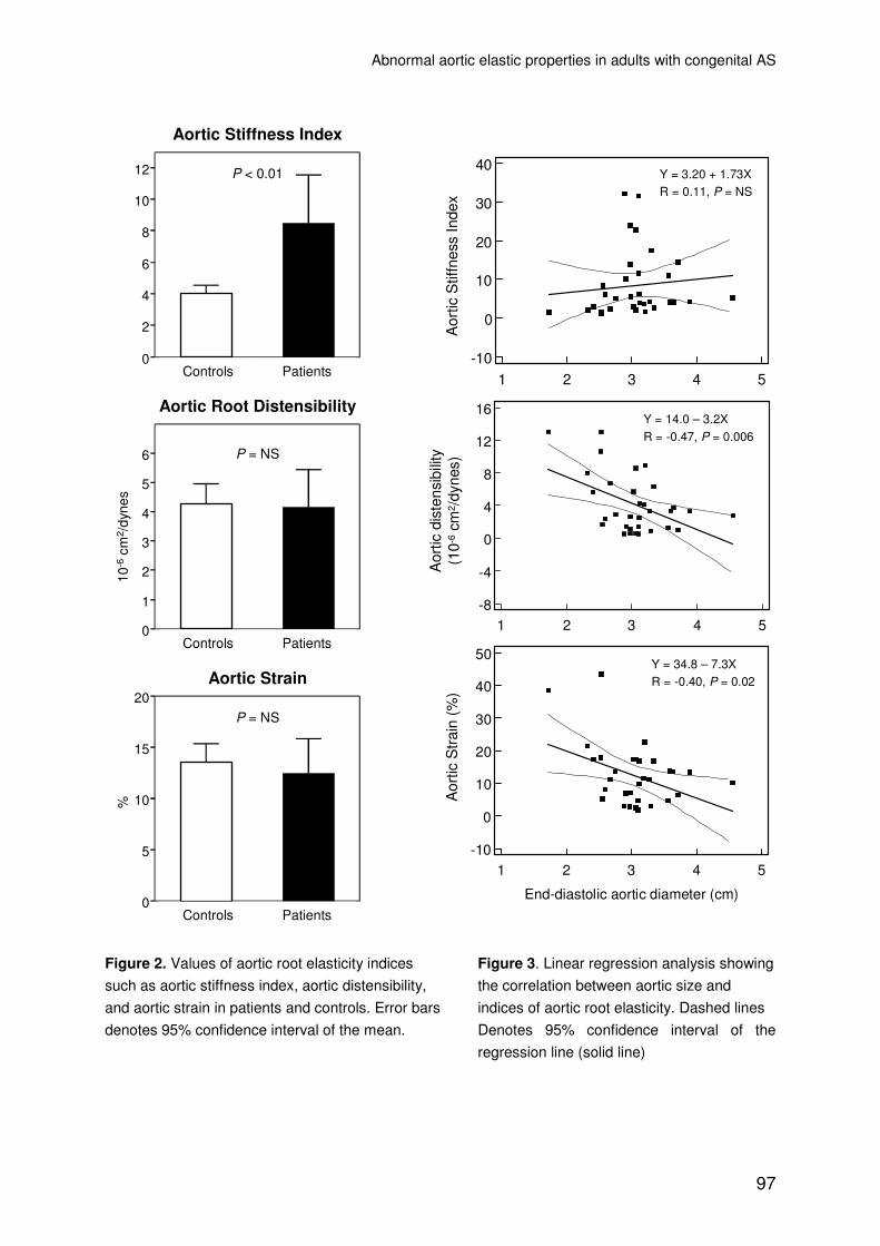
Abnormal aortic elastic properties in adults with congenital AS
97
Control patient AS patient0
5
10
15
20
Control patient AS patient0
1
2
3
4
5
6
Control patient AS patient0
2
4
6
8
10
12
Controls Patients
Patients
Patients
Controls
Controls
P = NS
P = NS
P < 0.01
Aortic Stiffness Index
Aortic Root Distensibility
10-6
cm2 /
dyne
s
Aortic Strain
%
1 2 3 4 5
40
30
20
10
0
-10
End-diastolic aortic diameter (cm)
Aor
tic S
tiffn
ess
Inde
x
1 2 3 4 5
16
12
8
4
0
-4
-8
End-diastolic aortic diameter
Aor
tic d
iste
nsib
ility
1 2 3 4 5
50
40
30
20
10
0
-10
End-diastolic aortic diameter (cm)
Aor
tic s
trai
n (%
)A
ortic
dis
tens
ibili
ty(1
0-6
cm2 /
dyne
s)A
ortic
Stif
fnes
s In
dex
Aor
tic S
trai
n (%
)
Y = 3.20 + 1.73XR = 0.11, P = NS
Y = 14.0 – 3.2XR = -0.47, P = 0.006
Y = 34.8 – 7.3XR = -0.40, P = 0.02
Figure 2. Values of aortic root elasticity indices Figure 3. Linear regression analysis showing
such as aortic stiffness index, aortic distensibility, the correlation between aortic size and
and aortic strain in patients and controls. Error bars indices of aortic root elasticity. Dashed lines
denotes 95% confidence interval of the mean. Denotes 95% confidence interval of the
regression line (solid line)

Chapter 7
98
stenotic bicuspid valves (ASI: 8.5 ± 8.5 versus 4.0 ± 1.4, respectively, P < 0.01). In our study, we observed no differences regarding ARD and aortic strain between patients and controls. These elastic parameters, however, are more pressure dependent than ASI.20,21 Thus, ASI is a more robust marker of elasticity, than ARD and aortic strain.
Remarkably, no correlation was found between indices of stenosis severity and indices of aortic elasticity or aortic dimensions. These results strengthen the hypothesis that aortic root abnormalities (i.e., dilatation and stiffness) are independent of stenosis severity, and this study confirms more strongly that this is mainly a consequence of the presence of a congenital malformed aorta instead of previously thought post-stenotic dilatation. Furthermore, the higher aortic stiffness in patients with congenital valvular AS could contribute to an increased afterload, which potentially could have a negative influence on LV function and on the development of LV hypertrophy, as demonstrated previously for patients with degenerative AS.22
Another interesting finding of our study is that there seems to be a correlation between aortic dimensions and aortic elasticity (Figure 3). A larger aortic size was associated with reduced ARD and aortic strain, however, no correlation was found between aortic size and ASI. The exact mechanism is not clear. A possible relationship between aortic size and stiffness could be explained by the presence of “cystic medial necrosis”. Elastin fibers normally bear aortic stresses, providing the aorta its biological Windkessel function.23 When the elastin fibers fragment and degenerate, the aortic wall stretches and the vessel dilates. Wall stress is than transferred to the less extensible collagenous elements in the aortic wall, leading to increased stiffness. As the aorta becomes stiffer, it opposes a higher afterload to left ventricular ejection, it augments systolic and pulse pressure, and it reduces coronary blood flow.24 Furthermore, Bonderman et al.8 showed that in BAV patients arterial medial remodelling precedes aortic dilatation. It would be interesting to know whether an increased aortic stiffness could be used as a risk factor for identifying patients at risk of progressive aortic dilatation, and subsequently dissection. We recently published a prospective study in older patients (mean age 65 ± 11 years) with degenerative AS who were planned for aortic valve replacement.25 One year after aortic valve replacement, ASI changed from 19.6 ± 10.8 at baseline to 8.4 ± 3.5 (P <0.05), and was comparable with those of age-, sex-, and risk-factor-matched controls. These results suggest improvement of aortic elastic properties after relieve of the obstruction. Whether this also occurs in patients with congenital AS seems unlikely, as we were unable to find a correlation between stenosis severity and aortic root elasticity. In congenital AS patients requiring aortic valve replacement, ASI may be used in addition to the aortic dimension in the decision process whether or not to include the aortic root in the operation.

Abnormal aortic elastic properties in adults with congenital AS
99
Study limitations
Several potential limitations must be noted. First, the method we used to evaluate aortic root elasticity is non-invasive and used pulse pressure in the brachial artery instead of the aortic pulse pressure in order to calculate ASI. These two pressures are not identical because of pulse pressure amplification from center to periphery.23 Furthermore, M-mode measurements may be problematic in the aortic root because the aorta is displaced to the apex of the heart during systole. An alternative would be 2-D measurements to evaluate aortic size. However, the method we used is well established, has good reproducibility, and its results are closely related to those obtained by direct invasive measurements.11,26,27 Second, the observation that an increased aortic dimension is associated with more stiffening would be strengthened by a longitudinal follow-up study with repeated echocardiograms at regular intervals. In particular, it would be interesting to see whether progression of aortic dilatation is associated with more stiffening, or vice versa. Third, a recent study demonstrated the influence of BAV phenotype (anterior-posterior vs. left-right orientation) on aortic elastic properties.16 In our study population the exact BAV phenotype could not be reliably determined with transthoracic echocardiography. This merits further attention. Finally, given the small sample size, all conclusions of the present study must be drawn with caution. Conclusions
Adults with congenital valvular AS have abnormal aortic elastic properties in comparison to age- and gender-matched controls. Moreover, the increased aortic stiffness is independent of stenosis severity, suggesting that the decreased aortic elasticity is mainly the consequence of the abnormal aortic wall associated with bicuspid aortic valves. Finally, an increased aortic dimension is associated with a decreased aortic elasticity, suggesting stiffening of the aorta when dilatation occurs.

Chapter 7
100
REFERENCES 1. Nistri S, Sorbo MD, Marin M, Palisi M, Scognamiglio R, Thiene G. Aortic root dilatation in
young men with normally functioning bicuspid aortic valves. Heart. 1999;82:19-22.
2. Keane MG, Wiegers SE, Plappert T, Pochettino A, Bavaria JE, Sutton MG. Bicuspid aortic
valves are associated with aortic dilatation out of proportion to coexistent valvular lesions.
Circulation. 2000;102:III35-9.
3. Hahn RT, Roman MJ, Mogtader AH, Devereux RB. Association of aortic dilation with
regurgitant, stenotic and functionally normal bicuspid aortic valves. J Am Coll Cardiol.
1992;19:283-8.
4. de Sa M, Moshkovitz Y, Butany J, David TE. Histologic abnormalities of the ascending aorta
and pulmonary trunk in patients with bicuspid aortic valve disease: clinical relevance to the
ross procedure. J Thorac Cardiovasc Surg. 1999;118:588-94.
5. Fedak PW, de Sa MP, Verma S, Nili N, Kazemian P, Butany J, Strauss BH, Weisel RD, David
TE. Vascular matrix remodeling in patients with bicuspid aortic valve malformations:
implications for aortic dilatation. J Thorac Cardiovasc Surg. 2003;126:797-806.
6. Niwa K, Perloff JK, Bhuta SM, Laks H, Drinkwater DC, Child JS, Miner PD. Structural
abnormalities of great arterial walls in congenital heart disease: light and electron microscopic
analyses. Circulation. 2001;103:393-400.
7. Nataatmadja M, West M, West J, Summers K, Walker P, Nagata M, Watanabe T. Abnormal
extracellular matrix protein transport associated with increased apoptosis of vascular smooth
muscle cells in marfan syndrome and bicuspid aortic valve thoracic aortic aneurysm.
Circulation. 2003;108 Suppl 1:II329-34.
8. Bonderman D, Gharehbaghi-Schnell E, Wollenek G, Maurer G, Baumgartner H, Lang IM.
Mechanisms underlying aortic dilatation in congenital aortic valve malformation. Circulation.
1999;99:2138-43.
9. Larson EW, Edwards WD. Risk factors for aortic dissection: a necropsy study of 161 cases.
Am J Cardiol. 1984;53:849-55.
10. Roos-Hesselink JW, Scholzel BE, Heijdra RJ, Spitaels SE, Meijboom FJ, Boersma E, Bogers
AJ, Simoons ML. Aortic valve and aortic arch pathology after coarctation repair. Heart.
2003;89:1074-7.
11. Nistri S, Sorbo MD, Basso C, Thiene G. Bicuspid aortic valve: abnormal aortic elastic
properties. J Heart Valve Dis. 2002;11:369-73; discussion 373-4.
12. Oosterhof T, Nollen GJ, van der Wall EE, Spijkerboer AM, Hrudova J, Bouma BJ, Dijkgraaf
MG, Mulder BJ. Comparison of aortic stiffness in patients with juvenile forms of ascending
aortic dilatation with versus without Marfan's syndrome. Am J Cardiol. 2005;95:996-8.
13. Hirata K, Triposkiadis F, Sparks E, Bowen J, Wooley CF, Boudoulas H. The Marfan
syndrome: abnormal aortic elastic properties. J Am Coll Cardiol. 1991;18:57-63.
14. Karpanou EA, Vyssoulis GP, Papakyriakou SA, Toutouza MG, Toutouzas PK. Effects of
menopause on aortic root function in hypertensive women. J Am Coll Cardiol. 1996;28:1562-6.
15. Lang RM, Bierig M, Devereux RB, Flachskampf FA, Foster E, Pellikka PA, Picard MH, Roman
MJ, Seward J, Shanewise JS, Solomon SD, Spencer KT, Sutton MS, Stewart WJ.
Recommendations for chamber quantification: a report from the American Society of
Echocardiography's Guidelines and Standards Committee and the Chamber Quantification
Writing Group, developed in conjunction with the European Association of Echocardiography,
a branch of the European Society of Cardiology. J Am Soc Echocardiogr. 2005;18:1440-63.

Abnormal aortic elastic properties in adults with congenital AS
101
16. Schaefer BM, Lewin MB, Stout KK, Byers PH, Otto CM. Usefulness of bicuspid aortic valve
phenotype to predict elastic properties of the ascending aorta. Am J Cardiol. 2007;99:686-90.
17. Grotenhuis HB, Ottenkamp J, Westenberg JJ, Bax JJ, Kroft LJ, de Roos A. Reduced aortic
elasticity and dilatation are associated with aortic regurgitation and left ventricular hypertrophy
in nonstenotic bicuspid aortic valve patients. J Am Coll Cardiol. 2007;49:1660-5.
18. Ferencik M, Pape LA. Changes in size of ascending aorta and aortic valve function with time
in patients with congenitally bicuspid aortic valves. Am J Cardiol. 2003;92:43-6.
19. Isselbacher EM. Thoracic and abdominal aortic aneurysms. Circulation. 2005;111:816-28.
20. Kawasaki T, Sasayama S, Yagi S, Asakawa T, Hirai T. Non-invasive assessment of the age
related changes in stiffness of major branches of the human arteries. Cardiovasc Res.
1987;21:678-87.
21. Hayashi K, Handa H, Nagasawa S, Okumura A, Moritake K. Stiffness and elastic behavior of
human intracranial and extracranial arteries. J Biomech. 1980;13:175-84.
22. Briand M, Dumesnil JG, Kadem L, Tongue AG, Rieu R, Garcia D, Pibarot P. Reduced
systemic arterial compliance impacts significantly on left ventricular afterload and function in
aortic stenosis: implications for diagnosis and treatment. J Am Coll Cardiol. 2005;46:291-8.
23. Laurent S, Cockcroft J, Van Bortel L, Boutouyrie P, Giannattasio C, Hayoz D, Pannier B,
Vlachopoulos C, Wilkinson I, Struijker-Boudier H. Expert consensus document on arterial
stiffness: methodological issues and clinical applications. Eur Heart J. 2006;27:2588-605.
24. Nemes A, Forster T, Csanady M. Decreased aortic distensibility and coronary flow velocity
reserve in patients with significant aortic valve stenosis with normal epicardial coronary
arteries. J Heart Valve Dis. 2004;13:567-73.
25. Nemes A, Galema TW, Geleijnse ML, Soliman OI, Yap SC, Anwar AM, Ten Cate FJ. Aortic
valve replacement for aortic stenosis is associated with improved aortic distensibility at long-
term follow-up. Am Heart J. 2007;153:147-51.
26. Stefanadis C, Stratos C, Boudoulas H, Kourouklis C, Toutouzas P. Distensibility of the
ascending aorta: comparison of invasive and non-invasive techniques in healthy men and in
men with coronary artery disease. Eur Heart J. 1990;11:990-6.
27. Vlachopoulos C, Aznaouridis K, Stefanadis C. Clinical appraisal of arterial stiffness: the
Argonauts in front of the Golden Fleece. Heart. 2006;92:1544-50.

102

103
8
Rapid and accurate measurement of LV
mass by biplane RT3DE in patients with
concentric LV hypertrophy:
Comparison to CMR
Yap SC,a Van Geuns RJ,a,b Nemes A,a Meijboom FJ,a McGhie JS,a Geleijnse ML,a Simoons ML,a Roos-Hesselink JW.a
Eur J Echocard 2007 Jun 29; Epub ahead of print.
a Department of Cardiology, Erasmus Medical Center, Rotterdam, The Netherlands, b Department of
Radiology, Erasmus Medical Center, Rotterdam, The Netherlands.
C H A P T E R
Chapter 8
104
ABSTRACT Aims
To evaluate the accuracy of real-time three-dimensional echocardiography (RT3DE) using a biplane and multiplane method in determining left ventricular (LV) mass compared to cardiac magnetic resonance imaging (CMR).
Methods and Results
LV mass was measured in 18 adult patients with congenital aortic stenosis using CMR and echocardiography (M-mode, two-dimensional echocardiography (2DE), and RT3DE). RT3DE data were analysed using a biplane and multiplane method. No geometric assumptions were necessary using the multiplane RT3DE method.
With regard to biplane or multiplane RT3DE, no tendency of over- or underestimation of LV mass was observed. Pearson’s correlation coefficients for RT3DE versus CMR were 0.84 and 0.90 for the biplane and multiplane method, respectively. In addition, the accuracy of both RT3DE methods were comparable (Fisher’s R-to-Z transformation: Z=0.69, P=NS). Finally, off-line analysis using biplane RT3DE was significantly faster than multiplane RT3DE (3.8±1.2 vs. 7.8±1.7 minutes, P<0.001).
Conclusions
Biplane RT3DE provided an accurate estimate of LV mass in patients with concentric left ventricular hypertrophy, which was not improved by multiplane RT3DE. The accuracy and speed of analysis renders biplane RT3DE an attractive tool in daily clinical practice for assessing the degree of LV hypertrophy.

Rapid and accurate measurement of LV mass by biplane RT3DE
105
INTRODUCTION Left ventricular (LV) hypertrophy is associated with increased morbidity and mortality in patients with congenital aortic stenosis and may be an important risk factor for long term outcome.1 To use the severity of LV hypertrophy as an indicator for surgical intervention, a reliable, reproducible, and widely available diagnostic method is warranted. Echocardiography is the imaging modality most commonly used to evaluate LV mass because of its practical and economical advantages over cardiac magnetic resonance (CMR). However, the accuracy of linear (M-mode) or 2-dimensional echocardiography (2DE) images is limited, as they both rely on geometric assumptions of uniform chamber size and shape. Furthermore, the unintended use of oblique planes leads to overestimation of LV mass measurements by M-mode, and foreshortening of apical views leads to underestimation of LV mass measurements by 2DE.2-7 Considering these limitations, real-time three-dimensional echocardiography (RT3DE) appears to be an attractive alternative for accurate evaluation of LV mass, especially since it can be measured directly from the three-dimensional data sets. Several methods for LV mass analysis from RT3DE data sets are available. A limited approach of RT3DE data is to select the nonforeshortened 2- and 4-chamber long-axis view and compute LV mass derived from model based biplane volumes (biplane RT3DE analysis).6 A more comprehensive approach is to trace the endocardial and epicardial surfaces in multiple long-axis planes and correct the tracings in the short-axis views (multiplane RT3DE analysis). We hypothesize that a more accurate estimate of LV mass will be acquired with multiplane RT3DE analysis, as no geometrical assumptions are required.8-10
The aim of the present study was to evaluate the accuracy of biplane and multiplane RT3DE compared to the current “gold standard” for LV mass values obtained from CMR images. In addition, we evaluated if RT3DE was more accurate than the conventional M-mode and 2DE techniques. METHODS We prospectively recruited 18 consecutive adult patients (13 men, age 30±8 years, weight 80±12 kg) with congenital aortic stenosis who visited the outpatient clinic for Adult Congenital Heart Disease of the Thoraxcentre. The M-mode, 2DE and RT3DE data acquisition were performed on the same day, and the CMR study was performed 26±14 days after the echocardiographic data acquisition. There were no changes in the patient’s clinical status between the echocardiographic examination and the CMR study. The mean peak aortic jet velocity was 3.7 ± 0.8 m/s and all patients were in sinus rhythm. The institutional review board approved the study, and all patients gave written informed consent.

Chapter 8
106
CMR
A clinical 1.5-Tesla MRI scanner with a dedicated cardiac four-element phased-array receiver coil was used for imaging (Signa CV/I, GE Medical Systems, Milwaukee, Wisconsin USA). Repeated breath-holds and gating to the electrocardiogram were applied to minimize the influence of cardiac and respiratory motion on data collection. Cine-MRI was performed with a steady-state free-precession technique (FIESTA, GE) with the following imaging parameters: repetition time, 3.0 to 3.4 ms; echo time, 1.5 ms; flip angle, 45°; temporal resolution, 47 ms; section thickness, 8mm; section gap, 2 mm; field of view, 300 x 340 mm; and matrix size 224 x 256. To cover the entire LV, 10 to 12 consecutive slices were acquired in short axis orientation and a single 4-chamber and 2-chamber orientation.
CMR cine loops were analyzed offline with dedicated commercial software (CAAS- MRV, Pie Medical Imaging, Maastricht, The Netherlands) by one experienced CMR cardiologist (RJMvG) blinded to the results of the echocardiographic analysis. The endocardial and epicardial border of the LV were traced manually on the long-axis views. The calculated intersection points with the short-axis images were the basis of the automatic contour detection.11 In every short-axis slice, manually corrections were performed when necessary, including the papillary muscles and trabeculations in the LV cavity. Myocardial mass was calculated by taking the difference of the end-diastolic endocardial and epicardial volume multiplied by 1.05 g/mL, which is the density of myocardium.12,13 RT3DE acquisition
Data acquisition was performed by one experienced sonographer (JSM) using second harmonic imaging with a matrix-array transducer (X4, 2 to 4 MHz) connected to a commercial ultrasound system (SONOS 7500, Philips Medical Systems, Best, The Netherlands). Care was taken to include the entire LV within the pyramidal scan volume. RT3DE datasets were then acquired with wide-angled acquisition (93° x 84°) mode in which 4 wedge-shaped sub volumes (93° x 21° each) were obtained from 4 different cardiac cycles during held respiration without moving the transducer. Acquisition was triggered to the R wave of every other cardiac cycle to allow time for storage of each sub volume, resulting in a total acquisition time of 7 heartbeats. The RT3DE datasets were analyzed using 2 different commercial software packages: 3DQ-Qlab (Philips Medical Systems, Best, The Netherlands) and 4D Echo-View (TomTec Imaging Systems GmbH, Munich, Germany) for the biplane and multiplane analysis, respectively. Two independent observers (SCY and AN) blinded to the results of the CMR analyzed the RT3DE datasets by using first the biplane method, followed by the multiplane method. LV mass was calculated with the use of a specific mass of myocardial tissue of 1.05 g/mL.

Rapid and accurate measurement of LV mass by biplane RT3DE
107
Figure 1. (A) Biplane analysis: an example of anatomically correct LV apical views from the RT3DE
data set by 3DQ-Qlab. The red line in the short-axis view corresponds to the apical 2-chamber view
and the green line in the short-axis view corresponds to the apical 4-chamber view. (B) Multiplane
analysis: an example of endocardial and epicardial tracings in LV apical views by 4D Echo-View. Note
the short-axis view (bottom left) showing the tracings corresponding to the long-axis views. A2C =
apical 2-chamber; A4C = apical 4-chamber; SA = short-axis.
Figure 2. An example of corresponding short-axis views from apex to base. (A) CMR images. (B)
Short-axis views in the same patient showing the manually traced endocardial and epicardial surfaces
by 4D Echo-View. This view allows manually correction of tracings made in the long-axis images.

Chapter 8
108
Biplane RT3DE data analysis
The pyramidal volume data were displayed in 3 different cross sections (Figure 1A). The anatomically correct 2- and 4-chamber views with the largest long-axis dimensions were selected as previously described by Mor-Avi et al.6 In these 2 planes, endocardial and epicardial contours were traced manually at end-diastole. The papillary muscles and trabeculations were included in the LV cavity to be consistent with the CMR measurements. The traced contours were then used to calculate LV volumes by use of the biplane Simpson formula incorporated in the analysis software. In addition, the LV long-axis dimension in the 2- and 4-chamber view was measured as the distance between the level of the mitral annulus and the most distal point at the apical endocardium. Multiplane analysis RT3DE data
Multiplane analysis was performed as previously described by our group8. The anatomically correct 2- and 4-chamber views with the largest long-axis dimension were selected in the same manner as described above (Figure 1B). Around this user-defined LV long axis, the software generated 8 uniformly spaced apical long-axis images 22.5° apart. In each view, epicardial and endocardial contours were manually traced at end-diastole with the papillary muscles and trabeculations included in the LV cavity. Once the points were manually traced on the eight long-axis planes, the position of the manually traced contours was verified in multiple short axis views from base to apex, and corrected when necessary (Figure 2). The traced contours were then used to calculate LV myocardial volume and mass. 2D Echocardiographic measurements
The 2DE imaging was performed by one experienced sonographer (JSM) with a S3 transducer with second harmonic imaging from the apical window with the patient in the left lateral decubitus position. The 4- and 2-chamber views were acquired during held respiration, while care was taken to avoid foreshortening. Images were stored digitally and analyzed offline (EnConcert, Philips Medical Systems) by 2 independent observers (SCY and AN). For both apical views, end-diastolic frames were selected at the start of the R wave. In each view, endocardial and epicardial contours, including the papillary muscles and trabeculations in the LV cavity, were traced manually. The traced contours were used to calculate endocardial and epicardial LV volumes by the biplane Simpson’s formula.14 The difference between the epicardial and endocardial volumes was computed for each view and multiplied by the specific mass of myocardial tissue to represent a biplane estimate of LV mass. In addition, the LV long-axis dimension in the 2- and 4-chamber view was measured as described for biplane RT3DE.

Rapid and accurate measurement of LV mass by biplane RT3DE
109
M-mode measurements
M-mode imaging was performed by one experienced sonographer (JSM) from a parasternal long-axis position using a standard transthoracic transducer. Measurements of the interventricular wall thickness (IVS), posterior wall thickness (PW), and LV internal diameter (LVID) were performed at end-diastole according to the recommendations of the American Society of Echocardiography.15 LV mass was calculated according to the cube formula using the correction described by Devereux et al.: LV mass (g) = 0.8 {1.04((IVS+LVID+PW)3 – LVEDD3)} + 0.6.16 Inter- and intraobserver variability
To determine the interobserver variability for 2DE and RT3DE evaluations of LV mass, all measurements were repeated by a second observer (AN) blinded to the values obtained by the first observer (SCY). To assess intraobserver variability, all measurements were repeated 1 month later by an observer (SCY) blinded to the results of the previous measurements. Inter- and intraobserver variability was calculated as the absolute difference between the 2 measurements in percent of their mean. The inter- and intraobserver variability could not be measured for the M-mode technique as the measurements were already performed during acquisition, making blinding impossible. Statistical analysis
All values were expressed as mean±SD. Difference between each echocardiographic technique and CMR was evaluated by a paired t test. Agreement between techniques was evaluated by linear regression analysis with Pearson’s correlation coefficient and the standard error of the estimate (SEE). The difference between the correlation coefficients of the biplane and multiplane RT3DE was tested using the Fisher R-to-Z transformation test. In addition, Bland-Altman analysis was used to determine the bias and 95% limits of agreement (1.96 SD) between echocardiographic measurements and CMR.17 These analyses were performed for the first observer (SCY). Inter- and intraobserver variability values were averaged for all patients and tested by use of a paired t test for significance of differences between techniques. Values of P <0.05 were considered significant. RESULTS Acquisition of RT3DE data sets was feasible in all patients (acquisition time <2 minutes). The CMR value of LV mass was 177±48 g. Off-line image processing and tracing required 2.0±0.4 minutes for 2DE, 3.8±1.2 minutes for biplane RT3DE and 7.8±1.7 minutes for multiplane RT3DE (P <0.001 between techniques). Linear

Chapter 8
110
regression analysis and Bland-Altman analysis are shown in Figures 3 and 4, respectively.
Mean LV mass with M-mode echocardiography was 209±68 g, which was significantly higher than the CMR values (P= 0.005). A Pearson’s correlation of r = 0.79 (SEE 43 g, P<0.001) with CMR values was observed. Bland-Altman analysis confirmed the overestimation by M-mode echocardiography by demonstrating a bias of 32 g with 95% limits of agreement at ±82 g.
Mean LV mass with 2DE was 155±31 g, which was significantly lower than the CMR values (P=0.02). A correlation of r=0.69 (SEE 23 g, P<0.001) with CMR values was observed. Bland-Altman analysis confirmed the underestimation by 2DE by demonstrating a bias of -22 g with 95% limits of agreement at ±68 g. The LV long-axis dimension was 8.8±0.8 cm in the 4-chamber view and 8.8±0.6 cm in the 2-chamber view.
Biplane RT3DE yielded a mean LV mass of 183±46 g, which was similar to the CMR values (P=NS). A Pearson’s correlation of r=0.84 (SEE 26 g, P<0.001) with CMR values was observed. Bland-Altman analysis showed no significant over- or underestimation by the RT3DE-technique as reflected by a bias of 6 g with 95% limits of agreement at ±53 g. The LV long-axis dimension was 9.2±0.9 cm in the 4-chamber view and 8.8±0.7 cm in the 2-chamber view. The LV long-axis value in the 4-chamber view was significantly larger using biplane RT3DE compared to 2DE (P<0.05).
50 150 250 350
350
250
150
50
CMR (g)
M-m
ode
(g)
50 150 250 350
350
250
150
50
CMR (g)
2D
E (
g)
50 150 250 350
350
250
150
50
CMR (g)
Bip
lane
RT
3DE
(g)
50 150 250 350
350
250
150
50
CMR (g)
Mul
tipla
ne R
T3D
E (
g)
Y = 0.81x + 39R2 = 0.70
Y = 0.82x + 36R2 = 0.81
Y = 1.14x + 8R2 = 0.63
Y = 0.44x + 77R2 = 0.47
Figure 3. Regression analyses of LV mass measurements by M-mode (upper left), 2DE (upper right),
biplane RT3DE (bottom left), and multiplane RT3DE (bottom right) against CMR values. Solid line:
regression line; dashed lines: 95% confidence intervals.

Rapid and accurate measurement of LV mass by biplane RT3DE
111
50 150 250 350
150
0
-150
(CMR + M-mode) / 2 (g)
M-m
ode
- C
MR
(g)
Mean
31,9
-1.96 SD
-50,2
+1.96 SD
114,0
50 150 250 350
150
0
-150
(CMR + biplane RT3DE) / 2 (g)
Bip
lane
RT
3DE
- C
MR
(g
)
Mean
5,9-1.96 SD
-46,9
+1.96 SD
58,8
50 150 250 350
150
0
-150
(CMR + multiplane RT3DE) / 2 (g)
Mul
tipla
ne R
T3D
E -
CM
R (
g)
Mean
4,3-1.96 SD
-36,7
+1.96 SD
45,3
50 150 250 350
150
0
-150
(CMR + 2DE) / 2 (g)
2DE
- C
MR
(g)
Mean
-21,9
-1.96 SD
-89,9
+1.96 SD
46,0
4
-37
45
-47
6
59
46
-22
-90
32
114
-50
Figure 4. Bland-Altman analyses of LV mass measurements by M-mode (upper left), 2DE (upper
right), biplane RT3DE (bottom left), and multiplane RT3DE (bottom right) against the mean value of
CMR and echocardiographic measurements. Horizontal solid lines represent mean difference between
each echocardiographic technique and CMR; dashed lines, 95% limits of agreement (±1.96 SD
around the mean).
Finally, multiplane RT3DE yielded an LV mass of 181±41 g, which was similar to the CMR values (P=NS). A correlation of r = 0.90 (SEE 20 g, P<0.001) with CMR values was observed. Bland-Altman analysis showed no significant over- or underestimation by the RT3DE-technique as reflected by a bias of 4 g with 95% limits of agreement at ±41 g. The accuracy of LV mass measurement was similar between the biplane and multiplane RT3DE method (Fisher’s R-to-Z transformation: Z=0.69, P=NS). The interobserver variability was 17±13% for the 2DE technique, 9±7% for the biplane RT3DE technique, and 11±6% for the multiplane RT3DE technique (P<0.05 between 2DE and biplane RT3DE, P=0.11 between 2DE and multiplane RT3DE). No difference in interobserver variability was observed between the biplane and multiplane RT3DE technique. The intraobserver variability was 8±6% for the 2DE technique, 9±9% for the biplane RT3DE technique, and 11±7% for the multiplane RT3DE technique (P=NS between techniques).

Chapter 8
112
DISCUSSION As expected, LV mass measurements from RT3DE data could be achieved with higher accuracy and lower interobserver variability than conventional echocardiographic techniques (M-mode, 2DE). Interestingly, LV mass measurements by biplane RT3DE was as accurate as multiplane RT3DE in patients with concentric LV hypertrophy.
Quantification of LV mass has traditionally been based on M-mode measurements of myocardial thickness, coupled with geometrical modelling of the LV, or model-based calculations from manually traced endocardial and epicardial contours obtained from 2DE images. Our results, however, showed that M-mode significantly overestimates LV mass probably due to oblique cuts,and that 2DE underestimates LV mass due to foreshortening.2-7 An improved accuracy of LV mass measurement was found for RT3DE, either by biplane or multiplane analysis, compared to conventional echo techniques. Measurements of the LV long-axis dimensions showed that offline cross sectioning of the RT3DE data provided significantly less foreshortened and thus anatomically more correct apical views than conventional 2DE measurements, thus explaining the improved accuracy of biplane RT3DE analysis. Furthermore, we hypothesized that the accuracy of RT3DE could be improved further by increasing the number of long-axis planes used to trace the endocardial and epicardial boundaries and to correct the tracings on short-axis images. We found, however, that the accuracy of multiplane RT3DE analysis was not better than biplane RT3DE analysis using 2 orthogonal planes. A similar outcome has been shown by Teupe et al., reinforcing the fact that using multiple planes does not increase accuracy in normally shaped ventricles.18 Multiplane RT3DE analysis may be of extra value in patients with nonconcentric LV hypertrophy, like hypertrophic obstructive cardiomyopathy, but not convincingly in a population with concentric LV hypertrophy.9 We recently demonstrated that LV mass measurement can be achieved with high accuracy by multiplane RT3DE in patients with abnormally shaped left ventricles due to congenital heart disease.8
Another important finding of our study is that RT3DE measurements varied less between observers compared to 2DE measurements (significant for the biplane analysis, and showing a trend for multiplane analysis), reflecting reduced operator dependency. Both the interoberver and intraobserver variability of RT3DE are ±10%, indicating that this technique is acceptable for clinical use and will improve the reliability of follow-up data. CMR, however, remains superior with variability values 2-4% reported by multiple investigators.13,19 This difference may be explained by the relatively limited ability of ultrasound imaging compared with CMR to visualize the endocardial and epicardial borders.
The time needed for the acquisition and analysis of RT3DE data are acceptable. The acquisition time for one RT3DE data set is relatively fast and is

Rapid and accurate measurement of LV mass by biplane RT3DE
113
limited to 7 heartbeats, with an acquisition time of less than 2 minutes. Off-line image processing and tracing required 3.8±1.2 minutes for biplane RT3DE analysis and 7.8±1.7 minutes for multiplane RT3DE analysis. Interestingly, it is possible to directly measure LV mass using the biplane RT3DE method on the ultrasound system (not in this study) during acquisition thus avoiding off-line analysis. Recently, custom software has been developed, incorporating semi-automated detection of the LV endocardial and epicardial surface, which will allow even more rapid and accurate measurement of LV mass.20
Several limitations are inherent to RT3DE. The size of the heart may be to big to fit into the scan volume thereby reducing accurate measurements of LV mass.9 Fortunately, this was not the case in our population, but this can be encountered in patients with LV dilatation or ventricular aneurysms. Another limitation of RT3DE is that factors that reduce 2DE image quality, including a small intercostal space and abundant body fat, also apply for RT3DE. In addition, the relatively large footprint of the X4 matrix transducer probably influences image quality in a negative way. Technical improvements in the near future will probably include an expansion of the scan volume without loss of spatial resolution and a smaller footprint of the matrix transducer.
Study limitations
Several potential limitations must be noted. Test-retest variability (summation of variability in acquisition and analysis), which is more representative for the clinical situation, was not tested in the current study. Despite this shortcoming, one would expect that the variability in acquisition is lower when using RT3DE as it encompasses the whole left ventricle in one dataset in contrast to 2DE. This should however be confirmed in another study. Together with the given sample size, all conclusions of the present study must be drawn with caution. Conclusions
Despite several limitations of RT3DE, our study showed that RT3DE data obtained with commercially available equipment provided images with sufficient detail to allow easy and fast offline analysis of LV mass. The biplane RT3DE method is a rapid and accurate method for clinical assessment of LV hypertrophy, avoiding overestimation of LV mass using M-mode which is currently the most applied clinical method for determining LV hypertrophy. Multiplane RT3DE might be the preferred method for eccentric LV hypertrophy and abnormal shaped ventricles. Biplane RT3DE is as accurate as multiplane RT3DE in patients with concentric LV hypertrophy. Although CMR remains the gold standard for assessement of LV hypertrophy, in daily clinical practice biplane RT3DE is an attractive tool due to the low costs, easy accessibility, and speed of acquisition and analysis.

Chapter 8
114
REFERENCES 1. Keane JF, Driscoll DJ, Gersony WM, Hayes CJ, Kidd L, O'Fallon WM, et al. Second natural
history study of congenital heart defects. Results of treatment of patients with aortic valvar
stenosis. Circulation. 1993;87:I16-27.
2. Kuhl HP, Bucker A, Franke A, Maul S, Nolte-Ernsting C, Reineke T, et al. Transesophageal 3-
dimensional echocardiography: in vivo determination of left ventricular mass in comparison
with magnetic resonance imaging. J Am Soc Echocardiogr. 2000;13:205-15.
3. Kuhl HP, Hanrath P, Franke A. M-mode echocardiography overestimates left ventricular mass
in patients with normal left ventricular shape: a comparative study using three-dimensional
echocardiography. Eur J Echocardiogr. 2003;4:312-9.
4. Missouris CG, Forbat SM, Singer DR, Markandu ND, Underwood R, MacGregor GA.
Echocardiography overestimates left ventricular mass: a comparative study with magnetic
resonance imaging in patients with hypertension. J Hypertens. 1996;14:1005-10.
5. Myerson SG, Montgomery HE, World MJ, Pennell DJ. Left ventricular mass: reliability of M-
mode and 2-dimensional echocardiographic formulas. Hypertension. 2002;40:673-8.
6. Mor-Avi V, Sugeng L, Weinert L, MacEneaney P, Caiani EG, Koch R, et al. Fast measurement
of left ventricular mass with real-time three-dimensional echocardiography: comparison with
magnetic resonance imaging. Circulation. 2004;110:1814-8.
7. Chuang ML, Beaudin RA, Riley MF, Mooney MG, Mannin WJ, Douglas PS, et al. Three-
dimensional echocardiographic measurement of left ventricular mass: comparison with
magnetic resonance imaging and two-dimensional echocardiographic determinations in man.
Int J Card Imaging. 2000;16:347-57.
8. van den Bosch AE, Robbers-Visser D, Krenning BJ, McGhie JS, Helbing WA, Meijboom FJ, et
al. Comparison of real-time three-dimensional echocardiography to magnetic resonance
imaging for assessment of left ventricular mass. Am J Cardiol. 2006;97:113-7.
9. Oe H, Hozumi T, Arai K, Matsumura Y, Negishi K, Sugioka K, et al. Comparison of accurate
measurement of left ventricular mass in patients with hypertrophied hearts by real-time three-
dimensional echocardiography versus magnetic resonance imaging. Am J Cardiol.
2005;95:1263-7.
10. Jenkins C, Bricknell K, Hanekom L, Marwick TH. Reproducibility and accuracy of
echocardiographic measurements of left ventricular parameters using real-time three-
dimensional echocardiography. J Am Coll Cardiol. 2004;44:878-86.
11. van Geuns RJ, Baks T, Gronenschild EH, Aben JP, Wielopolski PA, Cademartiri F, et al.
Automatic quantitative left ventricular analysis of cine MR images by using three-dimensional
information for contour detection. Radiology. 2006;240:215-21.
12. Sakuma H, Fujita N, Foo TK, Caputo GR, Nelson SJ, Hartiala J, et al. Evaluation of left
ventricular volume and mass with breath-hold cine MR imaging. Radiology. 1993;188:377-80.
13. Myerson SG, Bellenger NG, Pennell DJ. Assessment of left ventricular mass by
cardiovascular magnetic resonance. Hypertension. 2002;39:750-5.
14. Helak JW, Reichek N. Quantitation of human left ventricular mass and volume by two-
dimensional echocardiography: in vitro anatomic validation. Circulation. 1981;63:1398-407.
15. Sahn DJ, DeMaria A, Kisslo J, Weyman A. Recommendations regarding quantitation in M-
mode echocardiography: results of a survey of echocardiographic measurements. Circulation.
1978;58:1072-83.

Rapid and accurate measurement of LV mass by biplane RT3DE
115
16. Devereux RB, Alonso DR, Lutas EM, Gottlieb GJ, Campo E, Sachs I, et al. Echocardiographic
assessment of left ventricular hypertrophy: comparison to necropsy findings. Am J Cardiol.
1986;57:450-8.
17. Bland JM, Altman DG. Statistical methods for assessing agreement between two methods of
clinical measurement. Lancet. 1986;1:307-10.
18. Teupe C, Takeuchi M, Yao J, Pandian N. Determination of left ventricular mass by three-
dimensional echocardiography: in vitro validation of a novel quantification method using
multiple equi-angular rotational planes for rapid measurements. Int J Cardiovasc Imaging.
2002;18:161-7.
19. Vignaux O, Duboc D, Coste J, Argaud C, Carlier P, Le Roux P, et al. Reproducibility of left
ventricular mass measurement using a half-Fourier black-blood single-shot fast spin-echo
sequence within a single breath hold: comparison with a conventional multiple breath-hold
segmented gradient echo technique in patients. J Magn Reson Imaging. 2002;15:654-60.
20. Caiani EG, Corsi C, Sugeng L, MacEneaney P, Weinert L, Mor-Avi V, et al. Improved
quantification of left ventricular mass based on endocardial and epicardial surface detection
with real time three dimensional echocardiography. Heart. 2006;92:213-9.

116

117
9
A simplified continuity equation approach
to the quantification of stenotic bicuspid
aortic valves using velocity-encoded CMR
Yap SC,a Van Geuns RJ,a,b Meijboom FJ,a Kirschbaum SW,a,b McGhie JS,a Simoons ML,a Kilner PJ,c Roos-Hesselink JW.a
J Cardiovasc Magn Reson 2007 (in press).
a Department of Cardiology, Erasmus Medical Center, Rotterdam, The Netherlands, b Department of Rardiology, Erasmus Medical Center, Rotterdam, The Netherlands, c Cardiovascular Magnetic Resonance Unit, Royal Brompton Hospital, London, United Kingdom.
C H A P T E R

Chapter 9
118
ABSTRACT Aims
We aimed to compare velocity-encoded cine cardiovascular magnetic resonance (CMR) with an established echocardiographic method for noninvasive measurement of aortic valve area (AVA) using the continuity equation.
Methods and Results
Twenty consecutive young adults with stenotic bicuspid aortic valves were examined with CMR and transthoracic echocardiography (TTE). CMR AVA was calculated by the continuity equation, dividing stroke volume by the maximum jet velocity-time integral (VTIAorta) in the aorta, the stroke volume measured both by ventricular volume analysis and by phase contrast velocity mapping at 4 levels (1 subvalvar and 3 supravalvar). Stroke volumes measured at all levels correlated well with those from volumetric analysis. The CMR AVAs calculated using volumetric analysis and VTIAorta from jet velocity mapping correlated and agreed well with TTE AVA measurements (R2 = 0.83). When CMR AVA was calculated more rapidly using volume flow and VTIAorta both measured from the same trans-jet velocity acquisition, R2 was 0.74, with a bias and limits of agreement of 0.02 (-0.44, 0.47) cm2.
Conclusions
Continuity equation calculation of the AVA using CMR velocity mapping, with or without ventricular volumetric measurement, correlated and agreed well with the comparable and widely accepted TTE approach.

Quantification of AS using velocity-encoded CMR
119
INTRODUCTION Timing of intervention in adult patients with aortic valve stenosis is largely based on the severity of stenosis and the presence of symptoms.1 Currently, transthoracic echocardiography (TTE) is the clinical standard for the evaluation of aortic stenosis. Standard TTE parameters of aortic stenosis severity include measurement of peak aortic velocity (Vmax), mean transaortic pressure gradient, and continuity equation aortic valve area (AVA).2 Continuity equation AVA (actually area of the vena contracta) is calculated from the principle that volume flow proximal to the valve equals volume flow through the narrowed orifice. Measurements of AVA are achieved by recording two velocity-time integrals (VTIs) from Doppler velocity waveforms acquired proximal and distal to the valve, along with a measurement of the cross sectional area of the left ventricular outflow tract (LVOT).3 Several studies have shown good concordance of echocardiographic and invasive catheter-based estimates of aortic valve area.3-5
To date, most cardiac magnetic resonance (CMR) studies for the quantification of stenotic aortic valves have focused on direct planimetry of the stenotic aortic valve,6-12 although the dimensions of CMR voxels relative to the size and shape of the stenotic orifices make this approach questionable, especially in severe or irregularly shaped stenoses. Only one previous study has investigated the use of velocity-encoded cine magnetic resonance (VEC-MR) for calculation of AVA by the continuity equation.13 In the standard continuity equation, the stroke volume is estimated by multiplying VTILVOT by the cross-sectional LVOT area. But this is not the only possible approach by CMR since phase contrast velocity mapping can also determine the stroke volume more directly by measuring the volume of flow through planes transecting the LVOT, the stenotic jet or the proximal ascending aorta. Furthermore, the stroke volume calculated from standard left ventricular analysis can be used, as long as there is no significant mitral regurgitation. In theory, it is thus possible to determine AVA using the continuity equation [AVA = stroke volume/ VTIAorta] with direct input of stroke volumes instead of LVOT areas and VTILVOT.
The purpose of the present study was to compare CMR measurements of AVA using the continuity equation with those calculated by echocardiography using the standard continuity equation, the LV stroke volumes being measured by CMR either by volumetric analysis or by direct flow measurement at one or more levels.

Chapter 9
120
METHODS Patient Population
We prospectively recruited 20 consecutive young adult patients (13 men, age 35 ± 4 years) with aortic stenosis (all bicuspid aortic valves) who visited the outpatient clinic for Adult Congenital Heart Disease of the Thoraxcenter from December 1, 2005 until September 25, 2006. The only exclusion criteria were those for general CMR suitability14 and the presence of a significant mitral regurgitation. The institutional review board approved the study protocol, and all patients gave written informed consent. Transthoracic echocardiography
Patients were imaged with TTE by a single experienced sonographer (JMG), with the use of a commercially ultrasound system (Sonos 7500, Philips Medical Systems, Best, The Netherlands) with a 2 to 4-MHz, 128-element, phased-array transducer. Images were acquired by using standard imaging windows with short breath-holds used as needed. Doppler flow data were acquired from the LVOT region in pulsed wave mode and from the aortic valve in continuous wave mode. The LVOT diameter was measured in the parasternal long-axis view in midsystole at the same level as the LVOT pulsed wave Doppler velocity measurement, and then converted to LVOT area [LVOT area = π*(0.5*LVOT diameter)2].
Peak velocities and VTIs were used to calculate AVA according to the continuity equation [AVA = stroke volume/ VTIAorta], where the Doppler-derived stroke volume is calculated by [SV = LVOT area * VTILVOT]. Calculations were based on the single best representative heartbeat as selected independently by the expert sonographer (JMG), blinded to the CMR data.
Cardiovascular magnetic resonance imaging
A clinical 1.5-Tesla MRI scanner with a dedicated cardiac eight-element phased-array receiver coil was used for imaging (Signa CV/I, GE Medical Systems, Milwaukee, Wisconsin). Electrocardiographic gating was used, with cine images acquired during expiratory breath-holds. A standard ventricular function examination was performed by initial acquisition of steady-state free-precession (SSFP) cine images in standard long axis planes (2- and 4-chamber view, and LVOT view) by one experienced CMR cardiologist (RJMvG). The following imaging parameters were used: 6-10 s per breath-hold per slice (depending on heart rate); 24 phases per slice location; field of view (FOV), 300 x 340 mm2; repetition time, 3.0 to 3.4 ms; echo time, 1.5 ms; flip angle (α), 45°; matrix, 224 x 256. To cover the entire left ventricle, 9-12 short axis slices, 8 mm slice thickness with 2 mm gap, were acquired perpendicular to the 4-chamber long-axis view of the left ventricle.

Quantification of AS using velocity-encoded CMR
121
Figure 1. (A) Cine CMR image in the
LVOT plane showing the location of the
four velocity mapping planes at Levels 0,
+, ++ and –. From these, both magnitude
(B and D) and corresponding phase
images (C and E) were reconstructed.
Note the stenotic bicuspid aortic valve
(arrow in B) and the oval LVOT tract
(arrow in D).
For the quantitative flow measurements, a retrospectively gated phase contrast sequence was used during expiratory breath-holds (<15 seconds), velocity encoded through-plane in the slice select gradient direction. The following imaging parameters were used: 30 frames/ heartbeat; FOV, 400 x 200 mm2; repetition time, 6.5 ms; echo time, 3.1 ms; flip angle (α), 30°; matrix, 256 x 128. The LVOT cine views were used to locate four velocity-mapping planes parallel to the aortic valve plane at its greatest excursion toward the apex (typically, end-systole). One plane was located at the tips of the aortic valve, and the other three were positioned parallel to this, but offset +12, +6, and -18 mm (Figure 1). The naming convention adopted for these slices is as follows, moving from the aorta towards the LVOT: Level ++, Level +, Level 0, and
A
C
D
B
+++
-
0
E
Level -
Level +

Chapter 9
122
Level -. Typically, the velocity encoding range (VENC) was 2.0 m/s for the LVOT (Level -) and 4.0 m/s in the aorta (Levels 0, + and ++). For the aorta, VENC was initially 4.0 m/s but was increased a priori if the peak velocity could be predicted to be greater, based on the patient’s functional images, for example when they showed limited valve opening or a narrow systolic jet. Flow images were re-acquired with a higher VENC if velocity aliasing occurred. For the quantitative flow measurements, the data were transmitted to an offline image processing station (Cine version 3.4, GE Medical Systems, Milwaukee, Wisconsin), and the quantitative flow images were analyzed by an independent observer (SCY) unaware of the echocardiographic data. For each of the 4 CMR imaging levels, regions of interest (ROIs) were drawn on each of the 30 frames of the cine to include the lumen of the LVOT, the aortic valve, or the aorta, depending on slice position. Peak velocities were determined by extracting the greatest velocity recorded in any pixel within the ROI across the valve flow field. Flow and peak velocity data (Figure 2) were exported to a spreadsheet and stroke volumes and VTIs determined. VTIs and stroke volumes were calculated by using Simpson’s rule to integrate the peak velocity (cm/s) and aortic flow (ml/s), respectively, versus time (ms) during systole. Only the VTI from the level with the highest Vmax (VTIAorta) was used for further calculation of AVA and statistical analysis.
-100
0
100
200
300
400
500
0 200 400 600 800
Time (ms)
Aor
tic fl
ow r
ate
(ml/s
)P
eak
aort
ic v
eloc
ity (
cm/s
)
Figure 2. Aortic flow rate (solid line) and peak aortic velocities (dashed line) measured at level +. The
area under the curve during systole was used for calculation of stroke volume and VTIAorta,
respectively. Note the presence of aortic regurgitation characterized by a negative flow.

Quantification of AS using velocity-encoded CMR
123
CMR AVA was calculated using the continuity equation [AVA = SV / VTIAorta], the stroke volumes being determined by three different methods (Table 1). To investigate if stroke volumes measured from the flow images were accurate, these stroke volumes were compared with those calculated by the standard left ventricular analysis using commercially available software (CAAS-MRV, Pie Medical Imaging, Maastricht, The Netherlands) by one independent observer (RJvG) unaware of stroke volumes measured from the flow images.
When using method C (comparable to the method used in ultrasound), there are several approaches for the measurement of the LVOT area by CMR. In ultrasound the practices norm is to measure the diameter of the LVOT and assume the cross-section to be circular. The LVOT diameter was therefore measured from the LVOT view at mid systole at Level -, and assumed to be circular for consistency with ultrasound.
According to the American College of Cardiology/ American Heart Association (ACC/ AHA) guidelines,1 aortic stenosis with a valve area >1.5 cm2 was considered as mild, 1.0 to 1.5 cm2 as moderate, and < 1.0 cm2 as severe. Table 1. Three different methods of AVA calculation by CMR
Method Formula AVA calculation
(SV/ VTIAorta)
Description SV determination
A (LVEDV – LVESV) / VTIAorta SV by LV volumes
B Flow-time integral aorta / VTIAorta SV by VEC-MR (flow data) at aortic level
C (LVOT area * VTILVOT) / VTIAorta SV by VEC-MR (peak velocity data) at LVOT level
AVA = Aortic valve area, LV = Left ventricle, LVEDV = Left ventricular end-diastolic volume, LVESV =
Left ventricular end-systolic volume, LVOT = Left ventricular outflow tract, SV = Stroke volume, VEC-
MR = Velocity-encoded cine magnetic resonance, VTI = Velocity-time integral
Statistical analysis
All continuous data are expressed as mean ± SD. The correlation between CMR and TTE methods was determined by linear regression analysis, including standard errors of the estimate (SEE). Agreement between techniques was evaluated by the standard paired t-test. Furthermore, Bland-Altman analysis was used to determine the mean of the difference with 95% limits of agreement (±1.96 standard deviation).15 A two-tailed probability value <0.05 was considered statistically significant. All statistical analyses were performed using SPSS software (version 12.0, SPSS Inc., Chicago, Illinois).

Chapter 9
124
RESULTS All patients (n=20) completed the imaging protocols without difficulty. The CMR study was performed 24 ± 14 days after the echocardiographic data acquisition. There was no change in clinical status or medication use between the echocardiographic exam and the CMR study. The patient population showed a wide range in severity of aortic stenosis, with valve areas measured by TTE from 0.80 to 2.28 cm2 (mean 1.34 ± 0.45 cm2). Based on TTE, patients were classified as having mild (n=6), moderate (n=9), or severe aortic stenosis (n=5). All patients were in sinus rhythm, and 12 patients (60%) had at least mild aortic regurgitation (maximal 2+, qualitative assessment). The left ventricular ejection fraction measured by CMR averaged 59 ± 9%, ranging from 42% to 74%. Flow measurements
Measurements by CMR of peak velocities in the aorta correlated well with those by TTE (R2 = 0.88, SEE 0.30 m/s). The correlation was less good between CMR and TTE measurement at LVOT level (R2 = 0.60, SEE 0.15 m/s). Bland-Altman analysis gave the following mean differences and limits of agreement between CMR and TTE peak velocities: –0.11 (-0.68 to 0.46) m/s at aortic level, and 0.12 (-0.17 to 0.41) m/s at LVOT level. The highest peak aortic velocity was found at level 0 (at the tips of the valve) in 9 patients (45%), and at level + in the remainder (55%). VTIs incorporate more data than peak velocities and are thus more robust. CMR data correlated well with TTE data for VTIAorta, and correlated moderately with TTE for VTILVOT (Figure 3). Bland-Altman analysis demonstrated a small but significant difference between CMR and TTE measurements for VTIAorta and VTILVOT with relatively large limits of agreement for VTILVOT. Stroke volumes measured directly from flow acquisitions at all four levels showed good correspondence with stroke volume measurements by volumetric analysis, with no statistically significant differences (Table 2). The best correlation with volumetric measurements (R2 = 0.73, SEE 12.3 ml) was found when flow volume was measured at level +, with a bias of 0.3 ml and limits of agreement of –23.3 to 23.8 ml. Doppler-derived stroke volumes showed only moderate correlation (R2 = 0.42, SEE 17.2 ml) with CMR volumetric data, with a nonsignificant bias of 6.2 ml and limits of agreement of -28.1 to 40.4 ml.
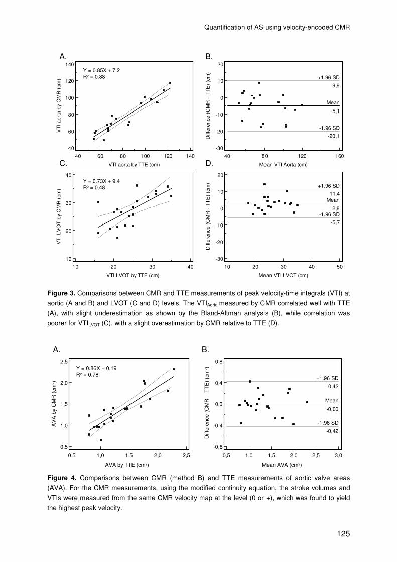
Quantification of AS using velocity-encoded CMR
125
40 60 80 100 120 140
140
120
100
80
60
40
VTI aorta by TTE (cm)
VT
I aor
ta b
y C
MR
(cm
)
40 80 120 160
20
10
0
-10
-20
-30
Mean VTI Aorta (cm)
Diff
eren
ce (C
MR
- T
TE
) (c
m)
Mean
-5,1
-1.96 SD
-20,1
+1.96 SD
9,9
10 20 30 40
40
30
20
10
VTI LVOT by TTE (cm)
VT
I LV
OT
by
CM
R (
cm)
10 20 30 40 50
20
10
0
-10
-20
-30
Mean VTI LVOT (cm)
Diff
eren
ce (
CM
R -
TT
E)
(cm
)Mean
2,8-1.96 SD
-5,7
+1.96 SD
11,4
Y = 0.85X + 7.2R2 = 0.88
Y = 0.73X + 9.4R2 = 0.48
A. B.
C. D.
Figure 3. Comparisons between CMR and TTE measurements of peak velocity-time integrals (VTI) at
aortic (A and B) and LVOT (C and D) levels. The VTIAorta measured by CMR correlated well with TTE
(A), with slight underestimation as shown by the Bland-Altman analysis (B), while correlation was
poorer for VTILVOT (C), with a slight overestimation by CMR relative to TTE (D).
0,5 1,0 1,5 2,0 2,5 3,0
0,8
0,4
0,0
-0,4
-0,8
Mean AVA (cm )
Diff
eren
ce (
CM
R -
TT
E)
(cm
)
Mean
-0,00
-1.96 SD
-0,42
+1.96 SD
0,42
0,5 1,0 1,5 2,0 2,5
2,5
2,0
1,5
1,0
0,5
AVA by TTE (cm )
AV
A b
y C
MR
(cm
)
Mean AVA (cm2)AVA by TTE (cm2)
AV
A b
y C
MR
(cm
2 )
Diff
eren
ce (
CM
R –
TT
E)
(cm
2 )
B.A.
Y = 0.86X + 0.19R2 = 0.78
Figure 4. Comparisons between CMR (method B) and TTE measurements of aortic valve areas
(AVA). For the CMR measurements, using the modified continuity equation, the stroke volumes and
VTIs were measured from the same CMR velocity map at the level (0 or +), which was found to yield
the highest peak velocity.

Chapter 9
126
Table 2. Comparison of stroke volume measurements by VEC-MR at each of the four levels to CMR
ventricular volumetric analysis
Stroke volume (ml) Linear regression analysis Bland-Altman analysis
R2 SEE Slope Interce
pt
Bias Limits of agreement
Level - 0.64 11.03 0.75 24.6 0.1 -23.0, 23.2
Level 0 0.65 12.91 0.89 11.1 0.8 -24.2, 25.7
Level + 0.73 12.31 1.03 -2.7 0.3 -23.3, 23.8
Level ++ 0.64 16.35 1.10 -8.4 1.5 -29.9, 32.9
Table 3. Comparison of AVA calculation by CMR to TTE
Linear regression analysis Bland-Altman analysis
R2 SEE Slope Interce
pt
Bias Limits of agreement
Method A 0.83 0.17 0.81 0.25 -0.01 -0.38, 0.36
Method B 0.74 0.23 0.83 0.25 0.02 -0.44, 0.47
Method C 0.69 0.36 1.19 0.11 0.37* -0.34, 1.08
* p<0.001 by paired Student’s t test
AVA measurements
Table 3 shows linear regression and Bland-Altman comparisons of the four types of CMR measurement of AVA that we tried with those by TTE. Figure 4A shows the AVAs measured by CMR, with stroke volume and jet velocity measurements derived from the same velocity-encoded acquisition (method B), plotted against the TTE AVA measurements for all 20 patients. The Bland-Altman plot shows a mean bias, CMR-TTE, of only 0.02 cm2 with relatively small limits of agreements (Figure 4B).
Measurements of AVA by the standard continuity equation using CMR (method C), assuming a circular LVOT, were larger than by TTE, despite a good correlation (bias = 0.37 cm2, p = 0.04, R2 = 0.69, SEE 0.36 cm2). DISCUSSION In routine clinical practice, TTE has become the accepted standard for evaluation of aortic valve stenosis, as it is non-invasive, readily available, fast, and can be performed at reasonable cost. The echocardiographic assessment includes measurement of peak aortic velocity and calculation of continuity equation AVA. Peak velocities, however, are dependent on the volume flow rate, overestimating the severity of stenosis when flow is elevated, as in the presence of aortic regurgitation, or underestimating the severity of stenosis if flow is reduced, i.e., when there is left ventricular systolic dysfunction or mitral regurgitation. Calculation of AVA by the

Quantification of AS using velocity-encoded CMR
127
continuity equation has the advantage of taking the flow rate into account. Echocardiographic measurements have prognostic value as most studies of outcomes have used echocardiographic indices.16-18 In most patients the echocardiographic study is adequate for the assessment of stenosis, although in certain situations, TTE may not be reliable due to poor acoustic windows, calcification of the aortic valve limiting accurate measurements of LVOT diameter, eccentric jets (especially in patients with congenital aortic stenosis), and inaccurate jet localization. Where echocardiographic results are inconclusive, a comparable investigation by CMR for congenital or acquired valvular heart disease would be valuable. There may also be cases where CMR is chosen anyway for the investigation of additional congenital or acquired cardiovascular pathology.
Most CMR studies for the quantification of stenotic aortic valves have focused on direct planimetry of the stenotic aortic valve or jet area, and they have shown varied although generally acceptable results.6-12 However, our clinical experience regarding the accuracy of CMR planimetry is not consistent, there being cases where the borders of the orifice, or rather the jet, remain unclear. This is not surprising given the typical CMR slice thicknesses of 5mm or more, relative large pixel dimensions compared to jet area, and the splayed or fragmented nature of some post stenotic jets. There is also potential for error due to misplacement of the slice due to motion of the valve or inconsistency in the position of breath holds. It is also unclear how signal loss due to parajet shear and turbulence effects edge discrimination.
The use of velocity encoding to determine the peak VTI within a jet may be less subject to these limitations because only the voxel or voxels located fully within the core of the jet are required for determination of the peak velocity and peak VTI, and the jet core will usually extend 10 mm or more beyond the orifice rendering exact plane position less crucial. Furthermore, voxels that span the boundaries of the jet or vessel lumen do contribute to volume flow measurements, but, theoretically at least, partial volume averaging in these voxels should matter less when flow (spatial mean of velocity x area for each phase of the cycle) rather than jet area is the subject of measurement. This appears to have been the case as the cardiac outputs derived from our CMR flow measurements correlated and agreed well with those by CMR ventricular volumetric analysis in our study population, where no patient had significant mitral regurgitation. Furthermore, although much reluctance exists to measure stroke volume at the level of the highest recorded velocity, the correlation and agreement were as good if not better when stroke volume was measured at this level, presumably at or close to the vena contracta. This may be explained by the relatively laminar flow within the jet at this level, rather than the convergent flow upstream or the divergent and turbulent flow downstream. However, it is important to realize that our patient cohort did not include patients with peak velocities >5.0 m/s. At high values phase contrast CMR underestimate velocity (and therefore flow),13 which is suggested to be due to intra-voxel dephasing characterized by signal loss in

Chapter 9
128
the magnitude image. Echo time minimization is essential to reduce error. Other problems associated with CMR velocity data are ghosting artifacts, and movement of the aortic valve during contraction.
In our comparative study, CMR results correlated and agreed well with TTE with respect to aortic jet velocity data (i.e. peak velocities and VTIs) and continuity equation AVAs based on volumetric (method A) or aortic flow measurement (method B) of stroke volumes. Previously, Caruthers et al.13 have shown that CMR correlated well with TTE when the standard continuity equation (method C) was used (R2 = 0.69), by means of the identified best approach), which was confirmed by our study. The present study is, to our knowledge, the first to demonstrate the usefulness and relative advantage of aortic flow measurement of stroke volumes (method B) in determining the continuity equation AVA by CMR. This method is easier to use than the method previously proposed by Caruthers et al., and both are equally accurate.13 The continuity equation AVA using volumetric analysis has been previously proposed for ultrasound (not for CMR) by Dumont et al.,19 demonstrating an additive value of the method, especially in patients with LVOT flow acceleration. Furthermore, Haghi et al.20,21 have shown that a hybrid approach, combining stroke volume measured by volumetric CMR data and continuous-wave Doppler data by TTE, for determining continuity equation AVA using the modified continuity equation correlates well with TTE. However, both studies determined stroke volume by the difference between end-diastolic and end-systolic volumes. This method could be incorrect when associated mitral regurgitation is present, whereas the measurements of aortic flow avoid this limitation. Another advantage of aortic flow measurement is that it is easier and faster to analyze than ventricular volumes measured from multiple short axis cine acquisitions. Our findings showed that estimates of stroke volumes were accurate at all 4 levels, and that the highest peak aortic velocities were acquired at 2 levels (level 0 or +). Therefore, using the modified continuity equation, only 2 flow velocity maps will be necessary to estimate AVA.
There are several reasons why the modified continuity equation (method A or B) is theoretically preferable to the standard continuity equation (method C) when using CMR. First, reliable measurements of LVOT peak velocity and area are needed for method C. This represents a potential source of error, in part because of the converging boundaries and ovoid cross section of the outflow tract. Waters et al.22 have shown that VTI measurements at different levels in the post stenotic jet were comparable, indicating relative insensitivity to the position of the imaging plane in the aorta. In the LVOT, however, measurements were position-dependent, observing higher values when the image plane was close to the valve, which would explain the lower correlation between CMR and ultrasound measurements of LVOT velocities and VTIs in our study. Secondly, the assumption that the LVOT area is circular is incorrect (see Figure 1D). Baumgartner et al.23 have shown that by using the standard continuity equation and assuming a circular LVOT, AVA is significantly

Quantification of AS using velocity-encoded CMR
129
underestimated by TTE compared to cardiac catheterization using the Gorlin formula. By using direct planimetry of the LVOT by TTE, results were more consistent with invasive data. By using method A or B, no assumptions regarding stroke volumes are required and the problems associated with determination of LVOT velocity and area are avoided. In spite of the limitations of the standard continuity equation used by TTE, however, we found relatively good correlation and agreement between AVAs determined by this approach and by CMR using the modified continuity equation. No clear explanation exists for this finding.
Study limitations
First, our study lacks a reliable gold standard because the accuracy of the results obtained by TTE remains uncertain. More data on the correlation between the described methods and invasive catheterization data (using the Gorlin formula) and other imaging modalities (for example direct planimetry by CT or transesophageal echocardiography) would be valuable.
Second, we studied a relatively selected patient population of young adults with congenital aortic stenosis. However, the approach used should also be applicable to older patients with degenerative aortic stenosis in whom calcification is unlikely to compromise stroke volume and VTI measurements as much as it does planimetric approaches. Third, our study protocol did not include repeated measurements of one patient, therefore no reproducibility data are available. Finally, due to the small sample size, all conclusions of the present study must be drawn with caution.
Conclusions
We conclude that the use of the modified continuity equation for determining AVA by velocity-encoded CMR is feasible, simple and compares well with the established echocardiographic approach in patients with stenotic bicuspid aortic valves, providing an attractive alternative non-invasive approach to the quantification of aortic stenosis severity.

Chapter 9
130
REFERENCES 1. Bonow RO, Carabello BA, Chatterjee K, de Leon AC, Jr., Faxon DP, Freed MD, Gaasch WH,
Lytle BW, Nishimura RA, O'Gara PT, O'Rourke RA, Otto CM, Shah PM, Shanewise JS, Smith
SC, Jr., Jacobs AK, Adams CD, Anderson JL, Antman EM, Fuster V, Halperin JL, Hiratzka LF,
Hunt SA, Nishimura R, Page RL, Riegel B. ACC/AHA 2006 guidelines for the management of
patients with valvular heart disease: a report of the American College of Cardiology/American
Heart Association Task Force on Practice Guidelines (writing Committee to Revise the 1998
guidelines for the management of patients with valvular heart disease) developed in
collaboration with the Society of Cardiovascular Anesthesiologists endorsed by the Society for
Cardiovascular Angiography and Interventions and the Society of Thoracic Surgeons. J Am
Coll Cardiol. 2006;48:e1-148.
2. Otto CM. Valvular aortic stenosis: disease severity and timing of intervention. J Am Coll
Cardiol. 2006;47:2141-51.
3. Otto CM, Pearlman AS, Comess KA, Reamer RP, Janko CL, Huntsman LL. Determination of
the stenotic aortic valve area in adults using Doppler echocardiography. J Am Coll Cardiol.
1986;7:509-17.
4. Skjaerpe T, Hegrenaes L, Hatle L. Noninvasive estimation of valve area in patients with aortic
stenosis by Doppler ultrasound and two-dimensional echocardiography. Circulation.
1985;72:810-8.
5. Zoghbi WA, Farmer KL, Soto JG, Nelson JG, Quinones MA. Accurate noninvasive
quantification of stenotic aortic valve area by Doppler echocardiography. Circulation.
1986;73:452-9.
6. Sondergaard L, Hildebrandt P, Lindvig K, Thomsen C, Stahlberg F, Kassis E, Henriksen O.
Valve area and cardiac output in aortic stenosis: quantification by magnetic resonance velocity
mapping. Am Heart J. 1993;126:1156-64.
7. Friedrich MG, Schulz-Menger J, Poetsch T, Pilz B, Uhlich F, Dietz R. Quantification of valvular
aortic stenosis by magnetic resonance imaging. Am Heart J. 2002;144:329-34.
8. John AS, Dill T, Brandt RR, Rau M, Ricken W, Bachmann G, Hamm CW. Magnetic resonance
to assess the aortic valve area in aortic stenosis: how does it compare to current diagnostic
standards? J Am Coll Cardiol. 2003;42:519-26.
9. Kupfahl C, Honold M, Meinhardt G, Vogelsberg H, Wagner A, Mahrholdt H, Sechtem U.
Evaluation of aortic stenosis by cardiovascular magnetic resonance imaging: comparison with
established routine clinical techniques. Heart. 2004;90:893-901.
10. Debl K, Djavidani B, Seitz J, Nitz W, Schmid FX, Muders F, Buchner S, Feuerbach S, Riegger
G, Luchner A. Planimetry of aortic valve area in aortic stenosis by magnetic resonance
imaging. Invest Radiol. 2005;40:631-6.
11. Thomas B, Freitas A, Ferreira R, Tavares NJ. The complementary role of cardiac magnetic
resonance imaging in the evaluation of patients with aortic stenosis. Rev Port Cardiol.
2005;24:1117-21.
12. Strohm O, Schulz-Menger J, Hanlein D, Dietz R, Friedrich MG. Magnetic resonance
planimetry of the vena contracta as a new approach to assessment of stenotic heart valves:
an in vitro study. J Magn Reson Imaging. 2001;14:31-4.
13. Caruthers SD, Lin SJ, Brown P, Watkins MP, Williams TA, Lehr KA, Wickline SA. Practical
value of cardiac magnetic resonance imaging for clinical quantification of aortic valve stenosis:
comparison with echocardiography. Circulation. 2003;108:2236-43.

Quantification of AS using velocity-encoded CMR
131
14. Ahmed S, Shellock FG. Magnetic resonance imaging safety: implications for cardiovascular
patients. J Cardiovasc Magn Reson. 2001;3:171-82.
15. Bland JM, Altman DG. Statistical methods for assessing agreement between two methods of
clinical measurement. Lancet. 1986;1:307-10.
16. Otto CM, Burwash IG, Legget ME, Munt BI, Fujioka M, Healy NL, Kraft CD, Miyake-Hull CY,
Schwaegler RG. Prospective study of asymptomatic valvular aortic stenosis. Clinical,
echocardiographic, and exercise predictors of outcome. Circulation. 1997;95:2262-70.
17. Rosenhek R, Klaar U, Schemper M, Scholten C, Heger M, Gabriel H, Binder T, Maurer G,
Baumgartner H. Mild and moderate aortic stenosis. Natural history and risk stratification by
echocardiography. Eur Heart J. 2004;25:199-205.
18. Pellikka PA, Nishimura RA, Bailey KR, Tajik AJ. The natural history of adults with
asymptomatic, hemodynamically significant aortic stenosis. J Am Coll Cardiol. 1990;15:1012-
7.
19. Dumont Y, Arsenault M. An alternative to standard continuity equation for the calculation of
aortic valve area by echocardiography. J Am Soc Echocardiogr. 2003;16:1309-15.
20. Haghi D, Papavassiliu T, Kalmar G, Schroder M, Neff W, Kaden JJ, Muller U, Haase KK,
Borggrefe M, Suselbeck T. A hybrid approach for quantification of aortic valve stenosis using
cardiac magnetic resonance imaging and echocardiography. J Cardiovasc Magn Reson.
2005;7:581-6.
21. Haghi D, Suselbeck T, Fluechter S, Kalmar G, Schroder M, Kaden JJ, Poerner T, Borggrefe
M, Papavassiliu T. A hybrid approach for quantification of aortic valve stenosis using cardiac
magnetic resonance imaging and echocardiography: comparison to right heart catheterization
and standard echocardiography. Clin Res Cardiol. 2006;95:162-7.
22. Waters EA, Caruthers SD, Wickline SA. Correlation analysis of stenotic aortic valve flow
patterns using phase contrast MRI. Ann Biomed Eng. 2005;33:878-87.
23. Baumgartner H, Kratzer H, Helmreich G, Kuehn P. Determination of aortic valve area by
Doppler echocardiography using the continuity equation: a critical evaluation. Cardiology.
1990;77:101-11.

132

133
Part 3
Pregnancy and congenital aortic stenosis

134

135
10
Complications during pregnancy in women
with congenital heart disease:
A literature review
Drenthen W,a Pieper PG,a Roos-Hesselink JW,b Van Lottum WA,a Voors AA,a Mulder BJM,c Van Dijk APJ,d Vliegen HW,e Yap SC,b Moons P,f Ebels T,g Van Veldhuisen DJ,a on behalf of the ZAHARA investigators. JACC 2007;49:2303-11. a Department of Cardiology, University Medical Center Groningen, Groningen, The Netherlands, b Department of Cardiology, Erasmus Medical Center, Rotterdam, The Netherlands, c Department of Cardiology, Academic Medical Center, Amsterdam, The Netherlands, d Department of Cardiology, Radboud University Medical Center, Nijmegen, The Netherlands, e Department of Cardiology, Leiden University Medical Center, Leiden, The Netherlands, f Department of Cardiology, University Hospitals Leuven, Leuven, Belgium, g Department of Thoracic Surgery, University Medical Center Groningen, Groningen, The Netherlands.
C H A P T E R

Chapter 10
136
ABSTRACT A search of peer-reviewed literature was conducted to identify reports that provide data on complications associated with pregnancy in women with structural congenital heart disease. This review describes the outcome of 2491 pregnancies, including 377 miscarriages (15%) and 114 elective abortions (5%). Important cardiac complications were seen in 11% of the pregnancies. Obstetric complications do not appear to be more prevalent. In complex CHD, premature delivery rates are high and more children are small for gestational age. The offspring mortality was high throughout the spectrum and related to the relatively high rate of premature delivery and recurrence of congenital heart disease.

Pregnancy in women with CHD
137
INTRODUCTION Progress in the fields of diagnostic techniques and surgical intervention has dramatically improved long-term outcome in congenital heart disease. As a consequence, most patients with congenital cardiac malformations reach childbearing age, and many of these women wish to become pregnant. Pregnancy itself is a circulatory burden, primarily due to volume loading, which has a significant impact even on a healthy women’s life. In the face of residual lesions or sequelae after correction or an uncorrected maternal congenital heart defect this burden may have deleterious effects on the health of both the mother and her offspring. To provide an accurate and contemporary overview of the cardiac, obstetric and neonatal complications during pregnancies in women with congenital heart disease, a literature review was conducted. METHODS A systematic search of peer-reviewed literature using PubMed, MEDLINE and the Cochrane Library databases was performed. Predefined limits were: a) date of publication after January 1st 1985 for reasons of contemporaneous applicability, b) the main body of text of the manuscript needed to be in English, German or French to reduce misinterpretation, c) the reported complications (reported in methods or in the results section) needed to be traceable to the patient’s primary congenital heart disease, e) the number of completed (not aborted or miscarried) pregnancies for the primary CHD category needed to be available, f) reviews and case reports describing ≤1 completed pregnancy were excluded. The search was performed separately by two independent researchers (W.D. and W.L). For the present review the baseline primary congenital heart disease, the number of completed pregnancies, miscarriages and therapeutic abortions were recorded. In addition, the following complications were recorded for each completed pregnancy. Cardiac complications: reported clinically significant (“requiring treatment”) episodes of arrhythmias (including origin and type of arrhythmia) or heart failure; cardiovascular events (myocardial infarction, cardiovascular mortality and/or cerebrovascular accidents), endocarditis (including first 6 months postpartum). Obstetric complications: pregnancy induced hypertension; preeclampsia; eclampsia; Haemolysis Elevated Liver enzymes Low Platelets (HELLP) syndrome; thrombo-embolic events (confirmed deep venous thrombosis or pulmonary embolus); premature rupture of membranes (PROM); premature labor (< 37 weeks gestation); postpartum hemorrhage.

Chapter 10
138
Offspring complications: premature birth (delivery <37 weeks); small-for-gestational-age birth weight (<10th percentile); fetal demise (intra-uterine death ≥ 20 weeks of gestation); and / or perinatal mortality (death within the first year of life). The used query consisted of: congenital heart disease (congenital heart disease (CHD) , atrial septal defect (ASD), atrioventricular septal defect (AVSD), ventricular septal defect (VSD), aortic stenosis (AOS), aortic coarctation, pulmonary stenosis (PS), pulmonary atresia (PA), tetralogy of Fallot (TOF), transposition of the great arteries / vessels (TGA), Ebstein’s, cyanotic heart disease, Eisenmenger and Fontan) followed by: AND (pregnancy OR delivery). Abstracts of the identified articles were read, based on this information the eligible articles were identified. Subsequently, full-text papers were ordered or downloaded. If a report could not be ordered through our library, attempts were made to contact the authors. Additional reports were found through cross-reading of the reference lists of the eligible articles. The available reports were independently read by two researchers (W.D. and W.L.) and classified according to the above-mentioned criteria. When discrepancies were discovered between the two researchers, a third independent adult congenital cardiologist was asked to interpret the data. This third party then decided how the data should be interpreted. Efforts were made to filter out duplicates; publications from the same institution were checked for the period of data collection. The format of the review was structured and analyses were performed according to the Cochrane guidelines. RESULTS The literature search was performed between November 1st 2005 and October 1st 2006. Searching the Cochrane Library database no reviews were found. Systematic literature search of PubMed and MEDLINE retrieved 48 different mainly retrospective publications, describing 2491 pregnancies, including 377 miscarriages (15%) and 114 (5%) elective abortions.1-48 It proved impossible to subdivide complications based on surgical status prior to pregnancy for every CHD category separately. Patients with TGA had undergone atrial correction prior to pregnancy, except for 3 arterial switch patients. Patients with TOF all had undergone definitive repair. All PAVSD patients had received bi-ventricular repair. The remaining uncorrected or palliated TGA, TOF and PAVSD patients were incorporated in the category (palliated or uncorrected) cyanotic heart disease. The other CHD cohorts contain a mix of (un-) corrected patients. Figure 1 provides an overview of the distribution of gestations (completed, miscarriage and elective abortions) per congenital heart disease. Figure 2 shows the occurrence of the most important cardiac, obstetric and offspring complications recorded in the reviewed literature during completed (not aborted or miscarried)

Pregnancy in women with CHD
139
pregnancies organised per CHD. Table 1-3 provides detailed information on each complications. The most frequently encountered cardiac complication (Table 1) was clinically significant heart failure (4.8%). Especially patients with complex CHD: Eisenmenger, other cyanotic heart disease and PAVSD appeared to be at risk. Arrhythmias complicated 70 (4.5%) of the completed pregnancies. Most of the encountered arrhythmias were supraventricular tachyarrhythmias (n = 48). Ventricular tachyarrhythmias were reported in 9, brady-arrhythmias in 3, WPW syndrome in one patient, while in 9 pregnancies the exact nature of the arrhythmia was not reported. Cardiovascular events (myocardial infarction, stroke and cardiovascular mortality) were primarily documented in Eisenmenger patients and those with palliated or uncorrected cyanotic heart disease.
0%
10%
20%
30%
40%
50%
60%
70%
80%
90%
100%
AS
D
VS
D
AV
SD
PS
Ebs
tein
AO
S
Coa
rcta
tio
CC
-TG
A
TG
A
PA
VS
D
Fon
tan
Cya
notic
Eis
enm
enge
r
Ove
rall
Exp
ecte
delective abortions
completed pregnancies
miscarriages
Figure 1. Distribution of miscarriages, completed pregnancies and elective abortions for each
congenital heart disease separately and the overall rates. On the right hand sided also the expected
rates in healthy women are depicted.
Abbreviations: ASD = Atrial septal defect, AVSD = atrioventricular septal defects, AOS = aortic
stenosis, CC-TGA = congenital corrected transposition of the great arteries, CHD = congenital heart
disease, CoA = aortic coarctation, Ebstein = Ebstein’s anomaly, Eisenmenger = Eisenmenger
syndrome, Fontan = patients after Fontan repair, PAVSD = pulmonary atresia with ventricular septal
defects, PS = pulmonary valve stenosis, TGA = complete transposition of the great arteries, TOF =
tetralogy of Fallot, VSD = ventricular septal defect.

Chapter 10
140
0
5
10
15
20
25
30
35
ASD VSDAVSD PS
Ebste
inAOS
CoA
CC-TGA
TGA
TOF
PAVSD
Font
an
Cyano
tic C
HD
Eisenm
enge
r
Overa
ll
Expecte
d
Arrhythmias
Heart failure
Cardiovascular event
0
5
10
15
20
25
30
35
ASD VSD
AVSD
PS
Ebstein
AOS
CoA
CC-T
GA
TGA
TOF
PAVSD
Fontan
Cyano
tic C
HD
Eisenm
enge
r
Ove
rall
Expec
ted
TEC
Hypert.disorders
Prem. labour
0
10
20
30
40
50
60
70
80
ASD VSDAVS
D PS
Ebste
inAO
SCoA
CC-TGA
TGA
TOF
PAVSD
Font
an
Cyano
tic C
HD
Eisenm
enge
r
Ove
rall
Expec
ted
Prem. Delivery
SGA
Offspring mortality

Pregnancy in women with CHD
141
Figure 2. Overview of the most important complications encountered during pregnancy in women with
structural congenital heart disease for each congenital heart disease separately and the overall rates.
On the right hand sided also the expected rates in healthy women are depicted.
Abbreviations as Figure 1.
Obstetric complications (Table 2) were less well documented. In general, hypertensive disorders related to pregnancy do not appear to be more prevalent for the whole CHD cohort. Eclampsia and HELLP syndrome were reported in respectively 3 (PS n=2 and PAVSD n=1) and 2 (PAVSD and TGA) of the 904 completed pregnancies (not depicted in Table 2). Especially the incidence of preeclampsia and eclampsia in patients with TGA, PS, aortic coarctation and PAVSD exceeded the expected rate of 2-3%. Thrombo-embolic events, predominantly pulmonary embolisms, were observed in approximately 2% of the pregnancies. Foetal complications (Table 3) were incorporated more frequently in the methodology of the reviewed articles. Importantly overall offspring mortality was 4%, which could at least in part be attributed to the relatively high overall premature birth rate (16%) and the recurrence of CHD. The recurrence rate depended on the type of CHD and ranged between 0.6% (TGA) and 8% (AVSD). DISCUSSION The present systematic review described the outcome of 2491 pregnancies in women with different types of congenital heart disease. Most pregnancies were successful, though complications were observed. Arrhythmias, heart failure, cardiovascular events, rarely seen in the healthy general population, were documented during 11% of completed pregnancies. Obstetric complications, on the other hand, do not appear to be more prevalent, except for hypertensive disorders and thrombo-embolic events. Offspring mortality was high (4% versus < 1% in the general population), and particularly occurred in CHD cohorts with high rates of premature delivery and/ or recurrence of congenital heart disease. Supraventricular and ventricular arrhythmias requiring treatment are rarely seen during pregnancy in healthy women.51,52 Potential factors that promote the development of arrhythmias are the additional circulatory burden of pregnancy and local electrophysiological effects, more specifically the extra volume load and the enhanced adrenergic receptor excitability mediated by estrogens and progesterone.51,52 Structural cardiac defects or residual defects after repair may contribute to the occurrence of clinically relevant arrhythmias. Most recorded arrhythmias were supraventricular in origin. In particular patients with TGA, Fontan repair and atrioventricular septal defects appeared at risk. Surgical scar tissue

Chapter 10
142

Pregnancy in women with CHD
143

Chapter 10
144

Pregnancy in women with CHD
145
formation may play a role in the pathophysiological mechanism. In the TGA population most patients had undergone atrial repair (Mustard or Senning) with baffle formation. Patients after Fontan repair, especially after atrio-pulmonary anastomosis and atrio-ventricular connection, were at risk for atrial tachyarrhythmias due to the exposure of right atrial tissue to higher than normal pressures. In patients with AVSD, macro-re-entrant tachycardias are well known complications after repair and also (residual) left atrioventricular regurgitation may play a role in the development of arrhythmias. In the healthy general population heart failure needing medical intervention is uncommon and mainly related to the development of peripartum cardiomyopathy.53 In our review, patients with complex CHD (in particular patients with cyanotic heart disease, Eisenmenger syndrome and PAVSD) appeared prone to develop heart failure. Overall, clinically significant (“needing medical intervention”) heart failure was observed in almost 5% of the completed pregnancies. Most episodes resolved without sequelae using medication. It needs to be addressed, however, that the given heart failure rate may be an underestimation considering that early heart failure was an important reason for elective abortion. Cardiovascular events (myocardial infarction, cerebrovascular accidents and mortality) are observed during 1:50 pregnancies. Mortality was particularly high in patients with Eisenmenger syndrome. Therefore, we still advocate that pregnancy should be discouraged in these patients. It needs to be taken into account, however that mortality is likely to be underestimated as most research is performed in retrospective or so-called “survivor” cohorts. Therefore, we have not described mortality as a separate complication. For accurate estimations of serious and relatively rare complications a large-scale (multi-centre and international) prospective registration remains necessary. With regard to the occurrence of endocarditis, it is striking that patients with simple secundum ASD appear to be at greater risk. Details on the site of infection, the causative organism and circumstances including the presence of concomitant minor (anatomical or physiologic) abnormalities which may increase the risk of endocarditis are not described. This needs to be further investigated. Pregnancy related hypertensive disorders were documented in 8.7% of the pregnancies which is comparable to the 8% found in the general population.54 In four CHD categories, however, the incidence of hypertensive disorders appears substantially higher. In patients with AOS, PS, Aortic coarctation, and TGA, hypertensive disorders occurred in respectively 12.8, 14.3, 16.0 and 16.3 percent of completed pregnancies. Preeclampsia is a relatively rare condition with a reported incidence in the developed countries between 2-3%; eclampsia is even rarer with estimated incidences of 4-5 cases per 10.000 live births. Both entities are generally associated with substantial maternal and neonatal morbidity.55,56 Preeclampsia

Chapter 10
146
appeared to cluster in patients with aortic coarctation, PS, PAVSD and TGA. Several mechanisms, either solitary or combined, may be hypothesized. First, activation of neurohormonal pathways in patients with congenital heart disease may alter vascular remodeling associated with pregnancy induced hypertension and (pre-) eclampsia. Second, endothelial dysfunction is present in patients with congenital heart disease, and last oxidative stress may interact with the pathophysiological mechanisms behind pregnancy induced hypertension and (pre-) eclampsia. During pregnancy and postpartum period, patients are at risk for thrombo-embolic
complications due to the presence of all three components of Virchow's triad: venous stasis, endothelial injury, and a hypercoagulable state. Nevertheless, thrombo embolic events are normally seen only once per 1000 to 2000 pregnancies.57,58 Fifteen thrombo-embolic complications occurred, which suggests that the incidence in women with CHD (1:50) is substantially increased compared with the general population. Importantly, associated disorders, e.g. the presence of inherited thrombophilia, malignancy, systemic disease, recent surgery or trauma, disease needing hormonal replacement therapy, or bone marrow diseases, was insufficiently documented. This finding merits further investigation. Premature birth rate (16%) also appeared higher than that generally reported in the industrialised world (10-12%). In patients with more complex CHD, including Ebstein, TGA, PAVSD, Fontan, Cyanotic CHD and Eisenmenger, the preterm delivering rates range between 22 and 65%. Premature labor seems to play an important role in the higher incidence of premature birth in these CHD categories. Several premature deliveries were iatrogenic in nature, which may imply a greater tendency of the attending gynaecologist to intervene in women with congenital heart disease. A high incidence of premature rupture of membranes may have played a role in patients with TGA and Fontan. Spontaneous premature deliveries occurred frequently prior to 34 weeks of gestation, which has important clinical repercussions. Small for gestational age children were documented in the same types of CHD as premature delivery, except for patients with Fontan repair. In Fontan, premature deliveries could mainly be attributed to the excessive occurrence of premature rupture of membranes. In the other types of CHD, a similar pathophysiological mechanism may explain the higher incidence of premature delivery as well as the small for gestational age children. In both complications, placental insufficiency plays a pivotal role. Investigation of the foetal-placental circulation in these women is necessary. An important finding of the present systematic review is that children of women with congenital heart disease are at higher risk for foetal and perinatal mortality. In the industrialised world foetal and perinatal mortality is below 1%. Therefore, the chance of offspring mortality is on average increased by 4 times. Importantly in the types of CHD with high rates of premature delivery and/ or CHD recurrence this risk

Pregnancy in women with CHD
147
was more profound, ranging up to 27.7% in women with Eisenmenger syndrome. The recurrence risks are in agreement with the larger genetic cohort studies. Several limitations of the present systematic review need to be discussed. The quality of the review depends on the design of the articles included; the study designs ranged from prospectively included cohort studies to case series reporting 2 completed pregnancies, therefore the results need to be judged with caution. Moreover, publication bias is an important factor, especially in case series. Selection bias is introduced by excluding articles based on earlier mentioned criteria. Under-reporting of complications may also be an important problem.

Chapter 10
148
REFERENCES 1. Actis Dato GM, Rinaudo P, Revelli A, Actis DA, Punta G, Centofanti P, Cavaglia M,
Barbato L, Massobrio M. Atrial septal defect and pregnancy: a retrospective analysis of
obstetrical outcome before and after surgical correction Minerva Cardioangiol 1998;46:63-8.
2. Avila WS, Grinberg M, Snitcowsky R, Faccioli R, Da Luz PL, Bellotti G, Pileggi F. Maternal
and fetal outcome in pregnant women with Eisenmenger's syndrome. Eur Heart J
1995;16:460-4.
3. Beauchesne LM, Connolly HM, Ammash NM, Warnes CA. Coarctation of the aorta: outcome
of pregnancy. J Am Coll Cardiol 2001 ;38:1728-33.
4. Canobbio MM, Mair DD, van d, V, Koos BJ. Pregnancy outcomes after the Fontan repair. J
Am Coll Cardiol 1996;28:763-7.
5. Carp H, Jayaram A, Vadhera R, Nichols M, Morton M. Epidural anesthesia for cesarean
delivery and vaginal birth after maternal Fontan repair: report of two cases. Anesth Analg
1994;78:1190-2.
6. Clarkson PM, Wilson NJ, Neutze JM, North RA, Calder AL, Barratt-Boyes BG. Outcome of
pregnancy after the Mustard operation for transposition of the great arteries with intact
ventricular septum. J Am Coll Cardiol 1994;24:190-3.
7. Connolly HM, Warnes CA. Outcome of pregnancy in patients with complex pulmonic valve
atresia. Am J Cardiol 1997;79:519-21.
8. Connolly HM, Grogan M, Warnes CA. Pregnancy among women with congenitally corrected
transposition of great arteries J Am Coll Cardiol 1999;33:1692-5.
9. Connolly HM, Warnes CA. Ebstein's anomaly: outcome of pregnancy. J Am Coll Cardiol
1994;23:1194-8.
10. Donnelly JE, Brown JM, Radford DJ. Pregnancy outcome and Ebstein's anomaly. Br Heart J
1991;66:368-71.
11. Drenthen W, Pieper PG, Roos-Hesselink JW, van Lottum WA, Voors AA, Mulder BJ, van Dijk
AP, Vliegen HW, Sollie KM, Moons P, Ebels T, van Veldhuisen DJ. Pregnancy and delivery in
women after Fontan palliation. Heart 2006;92:1290-4.
12. Drenthen W, Pieper PG, Ploeg M, Voors AA, Roos-Hesselink JW, Mulder BJ, Vliegen HW,
Sollie KM, Ebels T, van Veldhuisen DJ. Risk of complications during pregnancy after Senning
or Mustard (atrial) repair of complete transposition of the great arteries. Eur Heart J
2005;26:2588-95.
13. Drenthen W, Pieper PG, van der Tuuk K, Roos-Hesselink JW, Voors AA, Mostert B, Mulder
BJ, Moons P, Ebels T, van Veldhuisen DJ. Cardiac complications relating to pregnancy and
recurrence of disease in the offspring of women with atrioventricular septal defects. Eur Heart
J 2005;26:2581-7.
14. Easterling TR, Chadwick HS, Otto CM, Benedetti TJ. Aortic stenosis in pregnancy. Obstet
Gynecol 1988;72:113-8.
15. Genoni M, Jenni R, Hoerstrup SP, Vogt P, Turina M. Pregnancy after atrial repair for
transposition of the great arteries. Heart 1999;81:276-7.
16. Guedes A, Mercier LA, Leduc L, Berube L, Marcotte F, Dore A. Impact of pregnancy on the
systemic right ventricle after a Mustard operation for transposition of the great arteries. J Am
Coll Cardiol 2004 ;44:433-7.

Pregnancy in women with CHD
149
17. Hameed A, Karaalp IS, Tummala PP, Wani OR, Canetti M, Akhter MW, Goodwin I,
Zapadinsky N, Elkayam U. The effect of valvular heart disease on maternal and fetal
outcome of pregnancy J Am Coll Cardiol 2001;37:893-9.
18. Hoare JV, Radford D. Pregnancy after fontan repair of complex congenital heart disease. Aust
N Z J Obstet Gynaecol 2001;41:464-8.
19. Jackson GM, Dildy GA, Varner MW, Clark SL. Severe pulmonary hypertension in pregnancy
following successful repair of ventricular septal defect in childhood. Obstet Gynecol
1993;82:680-2.
20. Kaemmerer H, Bauer U, Stein JI, Lemp S, Bartmus D, Hoffmann A, Niesert S, Osmers R,
Fratz S, Rossa S, Lange PE, Beitzke A, Schneider KT, Hess J. Pregnancy in congenital
cardiac disease: an increasing challenge for cardiologists and obstetricians -- a prospective
multicenter study. Z Kardiol 2003 ;92:16-23.
21. Lao TT, Sermer M, MaGee L, Farine D, Colman JM. Congenital aortic stenosis and
pregnancy--a reappraisal. Am J Obstet Gynecol 1993;169:540-5.
22. Lao TT, Sermer M, Colman JM. Pregnancy following surgical correction for transposition of the
great arteries. Obstet Gynecol 1994;83:665-8.
23. Lynch-Salamon DI, Maze SS, Combs CA. Pregnancy after mustard repair for transposition of
the great arteries. Obstet Gynecol 1993;82:676-9.
24. Megerian G, Bell JG, Huhta JC, Bottalico JN, Weiner S. Pregnancy outcome following Mustard
procedure for transposition of the great arteries: a report of five cases and review of the
literature. Obstet Gynecol 1994;83:512-6.
25. Meijer JM, Pieper PG, Drenthen W, Voors AA, Roos-Hesselink JW, van Dijk AP, Mulder BJ,
Ebels T, van Veldhuisen DJ. Pregnancy, fertility, and recurrence risk in corrected tetralogy of
Fallot. Heart 2005; 91:801-5.
26. Neumayer U, Somerville J. Outcome of pregnancies in patients with complex pulmonary
atresia. Heart 1997;78:16-21.
27. Oliveira TA, Avila WS, Grinberg M. Obstetric and perinatal aspects in patients with congenital
heart diseases. Sao Paulo Med J 1996;114:1248-54.
28. Orme RM, Grange CS, Ainsworth QP, Grebenik CR. General anaesthesia using remifentanil
for caesarean section in parturients with critical aortic stenosis: a series of four cases. Int J
Obstet Anesth 2004;13:183-7.
29. Paternoster DM, De FD, Santarossa C, Laureti E. Congenital heart disease in pregnancy.
Minerva Ginecol 1999;51:299-302.
30. Patton DE, Lee W, Cotton DB, Miller J, Carpenter RJ, Jr., Huhta J, Hankins G. Cyanotic
maternal heart disease in pregnancy. Obstet Gynecol Surv 1990 ;45:594-600.
31. Presbitero P, Somerville J, Stone S, Aruta E, Spiegelhalter D, Rabajoli F. Pregnancy in
cyanotic congenital heart disease. Outcome of mother and fetus. Circulation 1994; 89:2673-6.
32. Saidi AS, Bezold LI, Altman CA, Ayres NA, Bricker JT. Outcome of pregnancy following
intervention for coarctation of the aorta. Am J Cardiol 1998 ;82:786-8.
33. Shime J, Mocarski EJ, Hastings D, Webb GD, McLaughlin PR. Congenital heart disease in
pregnancy: short- and long-term implications. Am J Obstet Gynecol 1987; 156:313-22.
34. Silversides CK, Colman JM, Sermer M, Farine D, Siu SC. Early and intermediate-term
outcomes of pregnancy with congenital aortic stenosis. Am J Cardiol 2003; 91:1386-9.
35. Siu SC, Sermer M, Harrison DA, Grigoriadis E, Liu G, Sorensen S, Smallhorn JF, Farine D,
Amankwah KS, Spears JC, Colman JM. Risk and predictors for pregnancy-related
complications in women with heart disease. Circulation 1997; 96:2789-94.

Chapter 10
150
36. Siu SC, Sermer M, Colman JM, Alvarez AN, Mercier LA, Morton BC, Kells CM, Bergin ML,
Kiess MC, Marcotte F, Taylor DA, Gordon EP, Spears JC, Tam JW, Amankwah KS, Smallhorn
JF, Farine D, Sorensen S. Prospective multicenter study of pregnancy outcomes in women
with heart disease. Circulation 2001;104:515-21.
37. Smedstad KG, Cramb R, Morison DH. Pulmonary hypertension and pregnancy: a series of
eight cases. Can J Anaesth 1994;41:502-12.
38. Su NY, Lin SM, Hseu SS, Chu YC, Tsou MY, Lee TY, Tsai SK. Anesthetic management of
parturients with Eisenmenger's syndrome--report of two cases. Acta Anaesthesiol Sin
2001;39:139-44.
39. Therrien J, Barnes I, Somerville J. Outcome of pregnancy in patients with congenitally
corrected transposition of the great arteries. Am J Cardiol 1999 ;84:820- 4.
40. Veldtman GR, Connolly HM, Grogan M, Ammash NM, Warnes CA. Outcomes of pregnancy in
women with tetralogy of Fallot. J Am Coll Cardiol 2004;44:174-80.
41. Venning S, Freeman LJ, Stanley K. Two cases of pregnancy with coarctation of the aorta. J R
Soc Med 2003;96:234-6.
42. Vriend JW, Drenthen W, Pieper PG, Roos-Hesselink JW, Zwinderman AH, van Veldhuisen
DJ, Mulder BJ. Outcome of pregnancy in patients after repair of aortic coarctation. Eur Heart J
2005;26:2173-8.
43. Walker E, Malins AF. Anaesthetic management of aortic coarctation in pregnancy. Int J Obstet
Anesth 2004;13:266-70.
44. Yentis SM, Dob DP. Caesarean section in the presence of aortic stenosis. Anaesthesia
1998;53:606-7.
45. Zuber M, Gautschi N, Oechslin E, Widmer V, Kiowski W, Jenni R. Outcome of pregnancy in
women with congenital shunt lesions. Heart 1999;81:271-5.
46. Drenthen W, Pieper PG, Zoon N, Roos-Hesselink JW, Voors AA, Mulder BJ, van Dijk AP,
Sollie KM, Vliegen HW, Ebels T, van Veldhuisen DJ. Pregnancy After Bi-ventricular Repair For
Pulmonary Atresia With Ventricular Septal Defect. Am J Cardiol 2006;98:262-6.
47. Canobbio MM, Morris CD, Graham TP, Landzberg MJ. Pregnancy outcomes after atrial repair
for transposition of the great arteries. Am. J. Cardiol 2006;98:668-672
48. Drenthen W, Pieper PG, Roos-Hesselink JW, Schmidt ACM, Mulder BJM, van Dijk APJ,
Vliegen HW, Sollie KM, Voors AA, Ebels T, van Veldhuisen DJ. Non-cardiac complications
during pregnancy in women with isolated congenital pulmonary valve stenosis. Heart 2006,
Epub ahead of print July 2006
49. Ferrero S, Colombo BM, Ragni N. Maternal arrhythmias during pregnancy. Arch Gynecol
Obstet 2004;269:244-53.
50. Romem A, Romem Y, Katz M, Battler A. Incidence and characteristics of maternal cardiac
arrhythmias during labor. Am J Cardiol 2004 ;93:931-3.
51. Clark SL, Cotton DB, Lee W, Bishop C, Hill T, Southwick J, Pivarnik J, Spillman T, DeVore
GR, Phelan J, . Central hemodynamic assessment of normal term pregnancy. Am J Obstet
Gynecol 1989;161:1439-42.
52. Hunter S, Robson SC. Adaptation of the maternal heart in pregnancy. Br Heart J 1992;68:540-
3.
53. Sliwa K, Fett J, Elkayam U. Peripartum cardiomyopathy. Lancet 2006;368:687-93.
54. Roberts JM, Pearson GD, Cutler JA, Lindheimer MD. Summary of the NHLBI Working Group
on Research on Hypertension During Pregnancy. Hypertens Pregnancy 2003;22:109-27.
55. Douglas KA, Redman CW. Eclampsia in the United Kingdom. BMJ 1994 26;309:1395-400.

Pregnancy in women with CHD
151
56. Sibai BM, Anderson GD, Abdella TN, McCubbin JH, Dilts PV, Jr. Eclampsia. III. Neonatal
outcome, growth, and development. Am J Obstet Gynecol 1983;146:307-16.
57. Greer IA. Thrombosis in pregnancy: maternal and fetal issues. Lancet 1999;353:1258-65.
58. Toglia MR, Weg JG. Venous thromboembolism during pregnancy. N Engl J Med
1996;335:108-14.

152

153
11
Risk of complications during pregnancy in
women with congenital aortic stenosis
Yap SC,a Drenthen W,b Pieper PG,b Moons P,c Mulder BJM,d Vliegen HW,e Van Dijk AP,f Meijboom FJ,a Steegers EAP,g Roos-Hesselink JW,a
on behalf of the ZAHARA investigators. Int J Cardiol 2007 May 3 [Epub ahead of print].
a Department of Cardiology, Erasmus Medical Center, Rotterdam, The Netherlands, b Department of Cardiology, University Medical Center Groningen, Groningen, The Netherlands, c Department of Cardiology, University Hospitals Leuven, Leuven, Belgium, d Department of Cardiology, Academic Medical Center, Amsterdam, The Netherlands, e Department of Cardiology, Leiden University Medical Center, Leiden, The Netherlands, f Department of Cardiology, Radboud University Medical Center, Nijmegen, The Netherlands, g Department of Obstetrics and Gynaecology, Division of Obstetrics and Perinatal Medicine, Erasmus Medical Center, Rotterdam, The Netherlands.
C H A P T E R

Chapter 11
154
ABSTRACT Background
Pregnancy in women with congenital aortic stenosis (AS) is associated with increased cardiac complications. Data on non-cardiac complications are limited, and this information is crucial for prenatal counseling and perinatal care. The aim of this study was to present the maternal and perinatal outcome of pregnancy in women with congenital AS. Methods
By review of the Dutch CONCOR national registry and a local Belgian tertiary care centre database, 35 women with congenital AS with a history of completed pregnancy before aortic valve replacement were enrolled in this study. Medical history and maternal and perinatal outcome were determined.
Results
Thirty-five women had 58 pregnancies resulting in 53 successful pregnancies, three miscarriages, and two abortions. The most serious cardiac complications were heart failure (n=2, 3.8%) and atrial arrhythmia (n=3, 5.7%). Although cardiac complications were present (9.4%), obstetric (22.6%) and perinatal (24.5%) complications were observed more often. A total of six pregnancies (11.3%) were complicated by hypertension-related disorders, including one case of eclampsia. Furthermore, 7 premature births (13.2%) and 7 small-for-gestational-age births (13.2%) were encountered. Pregnancy in women with severe AS was characterized by an increased incidence of heart failure and premature labour, and shorter pregnancy duration. Older women (>30 years) were at increased risk of perinatal events (odds ratio 4.38, 95% confidence interval 1.02 to 18.81).
Conclusions
Pregnancy is generally well tolerated in women with congenital AS. Importantly, an excess of obstetric and perinatal complications was found, requiring more meticulous attention.

Pregnancy in women with congenital AS
155
INTRODUCTION Congenital AS has an incidence of 5% in patients with a congenital heart disease, and is more frequently encountered in males. The stenotic lesion can occur at valvular, subvalvular, or supravalvular level.1 In older series, pregnancy in women with congenital AS is associated with a high maternal mortality rate (15-20%).2-4 Earlier diagnosis and treatment of stenosis (e.g. balloon aortic valvotomy) has resulted in more women with congenital AS reaching childbearing age in relatively good condition. Recent pregnancy reports in AS patients (less severe patients compared to the older studies) are encouraging, showing a favorable pregnancy outcome with low maternal mortality.5-7 The current guidelines consider women with severe AS high-risk patients for pregnancy.8,9
Previous studies focused mainly on cardiac complications,2-7 and only limited information exists on obstetric and perinatal complications in women with congenital AS. In these women, reduced placental perfusion may occur secondary to the fixed stenosis, resulting in an increased incidence of gestational disorders and other signs of inadequate placental perfusion (e.g. hypertensive disorders), as has been found in women with other left heart obstructive lesions, like unrepaired aortic coarctation.10
The objective of the present study was to identify the magnitude of pregnancy risks in women with congenital AS without previous aortic valve replacement, with emphasis on obstetric and perinatal complications. METHODS Study population
In 2005, the Dutch CONCOR registry (CONgenital CORvitia; www.concor.net) and a local Belgian tertiary care centre database were reviewed for all women with a main diagnosis of congenital AS (age 18-45 years), excluding women with concomitant aortic coarctation (Figure 1). Congenital AS was defined as stenosis secondary to bicuspid aortic valves, subvalvular, or supravalvular lesions. All women were followed in one of the seven participating tertiary academic centres. The institutional review board or ethics committee at each participating centres approved the protocol. Overall, 117 of the 154 women provided written informed consent and were enrolled in this study (participation rate 76.0%). The final study population consisted of 35 women with ≥ 1 completed pregnancy (> 20 weeks gestation) without previous aortic valve replacement. Study protocol
A detailed structured questionnaire was obtained from each patient by telephone. The results of the questionnaire were compared with and completed by obstetrical

Chapter 11
156
Women with congenital AS of childbearing age(n=154)
Women who provided written informed consent(n=117)
Women without completed pregnancy
(n=73)
Women with acompleted pregnancy
after AVR(n=9)
Final study population:Women with
a completed pregnancy without previous AVR
(n=35)
Figure 1. Flow chart of enrollment of the final study population. AVR = aortic valve replacement
data from medical records. Data were collected on congenital cardiovascular anomalies, previous surgical/ interventional procedures, medical history, age at menarche, medication, miscarriages (spontaneous foetal loss before 20 weeks gestation), and elective abortions.
Detailed information concerning each completed pregnancy (> 20 weeks gestation) was recorded: parity status, New York Heart Association (NYHA) functional class, mode of delivery, use of cigarettes, use of alcohol, use of drugs, use of medication, and physical examination. Transthoracic echocardiographic assessment of left ventricular systolic function during the antepartum (baseline) and postpartum (within two years after delivery) period was recorded, including Doppler quantitation of severity of AS and aortic regurgitation. Aortic stenosis severity was graded mild (<3.0 m/s), moderate (3.0-4.0 m/s), or severe (>4.0 m/s).8 Aortic regurgitation severity was graded based on the jet diameter seen with colour flow Doppler.11 Cardiac, obstetric, and perinatal events were classified according to predefined criteria. Cardiac complications: symptomatic documented arrhythmia or symptomatic heart failure requiring treatment (according to the attending cardiologist); persistent NYHA functional class deterioration (>1 year postpartum); syncope; thrombo-embolic complications; aortic dissection; and/or endocarditis.
Obstetric complications: pregnancy-induced hypertension (PIH, new onset hypertension after ≥20 weeks of gestation: blood pressure >140 mmHg systolic or >90 mmHg diastolic without significant proteinuria); preeclampsia (PIH criteria and >0.3 gram proteinuria in 24-hour urine sample); eclampsia (preeclampsia with grand

Pregnancy in women with congenital AS
157
mal seizures); Haemolysis Elevated Liver Enzymes Low Platelets (HELLP) syndrome. Premature rupture of membranes (membrane rupture before the onset of uterine contractions); premature labour (spontaneous onset of labour <37 weeks gestation); postpartum haemorrhage (vaginal delivery >500ml, cesarean delivery >1000 ml, documented by gynecologist and requiring transfusion); and placental abruption. Perinatal complications: premature birth (delivery <37 weeks of gestation); small-for-gestational-age birth weight (<10th percentile); foetal demise (intra-uterine death ≥20 weeks of gestation); perinatal death (within the first month after birth); and/or recurrence of CHD. Statistical analysis
A Clintrial data-entry program was used to record information and was converted to SPSS (version 13.0, SPSS Inc., Chicago, Illinois) for statistical analysis. Descriptive statistics for nominal data were expressed in absolute numbers and percentages. Mean values and standard deviations were presented for normally distributed continuous variables. For non-normal distributed continuous variables median and ranges were computed. Comparison of continuous variables between groups (repaired versus unrepaired) was made by unpaired Student’s t-tests or the Mann-Whitney U test. When comparing frequencies, we applied the χ2 test or Fisher exact test, where applicable.
Univariable logistic regression analysis was performed to identify predictors of pregnancy outcome, defined by a composite outcome of cardiac events (i.e. heart failure, transient ischemic attack, arrhythmias), obstetric events (i.e. PIH, preeclampsia, eclampsia, premature labour, postpartum haemorrhage; placental abruption; premature rupture of membranes), or perinatal events (i.e. premature delivery and small-for-gestational-age births). The following six baseline variables were assessed: severe AS, prior AS repair, history of previous completed pregnancy, smoking before pregnancy, smoking during pregnancy, and maternal age at pregnancy ≥ 30 years. Odds ratios (95% confidence intervals) are presented. Multi-variable analysis was not attempted because of the low number of events. All tests were two-tailed and a P-value <0.05 was considered statistically significant. RESULTS Characteristics at inclusion of the enrolled 35 women with congenital AS are summarized in Table 1. A total of 58 pregnancies were documented, including 53 completed pregnancies, 3 spontaneous miscarriages, and 2 elective abortions. Two miscarriages occurred early (<12 weeks), and all 3 miscarriages remained unexplained. Both elective abortions were performed due to AS-related maternal risk,

Chapter 11
158
Table 1. Characteristics at inclusion of 35 women with congenital AS and a history of
completed pregnancy
Characteristic
Number of women 35
Age at menarche (yrs), mean ± SD 12.5 ± 1.4
Age at first completed pregnancy (yrs), mean ± SD 25.7 ± 4.6
Age at inclusion (yrs), mean ± SD 35.6 ± 6.8
Women with repair 27 (77.1)
Aortic valvotomy 8 (22.9)
Subaortic fibromuscular shelf resection 7 (20.0)
Surgical correction supravalvular AS 2 (5.7)
Aortic valve replacement* 14 (40.0)
AS classification
Valvular 24 (68.6)
Subvalvular 9 (25.7)
Supravalvular 2 (5.7)
Pregnancies
Completed pregnancies 53 (91.4)
Spontaneous miscarriages 3 (5.2)
Elective abortions 2 (3.4)
Data are presented as n (%), unless indicated otherwise
*All aortic valve replacements were performed after pregnancy
however, both women delivered children later in life. All women became pregnant spontaneously. Eight (22.2%) women had been advised against pregnancy because of perceived maternal and foetal risk. In the present study, we focus on the 53 completed pregnancies (between December 1975 and July 2005) in the 35 women. Nineteen women had one child, 14 had two, and three had three children. There were no twin pregnancies. The mean aortic jet velocity was 3.3 ± 0.9 m/s (range 1.7 to 5.3 m/s) before pregnancy, and all women had good systolic left ventricular function. Baseline characteristics for all completed pregnancies are outlined in Table 2, and Table 3 describes cardiac, obstetric, and perinatal complications per pregnancy. Cardiac complications
Important cardiovascular events (i.e. heart failure, transient ischemic attack, and arrhythmias) complicated 5 of 53 pregnancies (9.4%). Heart failure requiring treatment occurred during two pregnancies, both in women with severe AS. One woman with severe valvular AS required aortic valve replacement with a bioprosthesis at the end of her first trimester. Balloon aortic valvuloplasty was not possible due to severe calcification of her aortic valve. Her further pregnancy and delivery was uncomplicated and a healthy daughter was born at 38 weeks of gestation weighing 2965 grams. The second woman known with severe subvalvular

Pregnancy in women with congenital AS
159
AS had an episode of heart failure which responded well to medical treatment (i.e. diuretics and digoxin). In three pregnancies in women with moderate AS, the observed worsening of NYHA class persisted more than 1 year postpartum. Deterioration of aortic valve function could be an explanation in two cases. There was an increase in aortic jet velocity from 3.7 to 4.7 m/s in one woman, and an increase in regurgitation from moderate to severe in the other woman. In the third woman, no specific cause could be identified. All three women underwent valve replacement (Ross-procedure) at 6 months, 1.5, and 2 years after pregnancy, with improvement of NYHA class thereafter.
Clinically significant arrhythmias were documented in three pregnancies. A woman with subvalvular AS required electrical cardioversion and oral anticoagulation for drug-refractory atrial fibrillation. This same woman experienced a transient ischemic attack at 34 weeks’ gestation, with complaints of dysartria and diplopia, shortly after switching for subcutaneous heparin in contemplation of cesarean delivery. The episodes of atrial arrhythmias in two other women were successfully treated with digoxin and beta-blockers. Syncope occurred in one other woman during the delivery, however, according to the attending physician the event was probably related to pain (i.e. vasovagal reaction) rather than her cardiac condition. No episodes of aortic dissection or endocarditis were recorded. Obstetric complications/ interventions
Six pregnancies (11.3%) were complicated by hypertension-related disorders, which included PIH (n=4), pre-eclampsia (n=1), and eclampsia (n=1). The woman with eclampsia was comatose for three days. Two women with placental abruptions had no hypertensive disorders during pregnancy, and it must be noted that one of them was known with protein S deficiency. Other important obstetric complications including premature labour (7.5%), post-partum haemorrhage (1,9%), and premature rupture of membranes (1,9%) were not more prevalent compared to the general population (7%, 2% and 8%, respectively). Reasons to induce labour were: perceived maternal risk (n=9), hypertensive-related disorder (n=3), post-maturity (n=3), foetal indication (n=3), and premature rupture of membranes (n=1). No episodes of HELPP syndrome were documented.
Seven elective cesarean deliveries were performed for: hypertensive related disorders (n=3), breech presentation (n=2), placenta previa (n=1), and perceived maternal cardiovascular risk (n=1). All seven emergency caesarean deliveries were performed for obstetric indications: foetal distress (n=4, decelerations on cardiotocography), vacuum-assisted delivery failure (n=2), and failure to progress during second stage of labour (n=1). During 43 deliveries (81.1%), antibiotic prophylaxis against bacterial endocarditis was administered.

Chapter 11
160
Table 2. Baseline characteristics, hospitalizations, and mode of delivery during 53 completed
pregnancies in 35 women with congenital AS
Congenital AS
Mild/moderate AS Severe AS Total
Number of completed pregnancies 41 12 53
Maternal age at pregnancy (yrs), mean±SD 26.4 ± 4.6 26.5 ± 5.4 26.4 ± 4.7
Aortic jet velocity (m/s), mean ± SD 3.0 ± 0.7 4.5 ± 0.4 3.3 ± 0.9
Medical history
• Valvular AS 29 (70.7) 7 (58.3) 36 (67.9)
• Subvalvular AS 9 (22.0) 5 (41.7) 14 (26.4)
• Supravalvular AS 3 (7.3) 0 3 (5.7)
• Repair before pregnancy 20 (48.8) 4 (33.3) 24 (45.3)
• Aortic valvotomy 10 (24.4) 2 (16.7) 12 (22.6)
• Subaortic shelf resection 9 (22.0) 2 (16.7) 11 (20.8)
• Surgical correction supravalvular AS 3 (7.3) 0 3 (5.6)
• Smoking before pregnancy 9 (22.0) 3 (25.0) 12 (22.6)
• Smoking during pregnancy 4 (15.3) 3 (25.0) 7 (18.4)
Parity status
• Primiparous 29 (70.7) 6 (50.0) 35 (66.0)
• Multiparous 12 (29.3) 6 (50.0) 18 (34.0)
NYHA class before pregnancy*
• Class I 38 (92.7) 6 (50.0) 44 (83.0)
• Class II 3 (7.3) 6 (50.0) 9 (17.0)
All-cause hospitalization 9 (22.0) 2 (16.7) 11 (20.8)
• Hypertensive disorders 4 (44.4) 0 4 (36.4)
• Dyspnea 1 (11.1) 2 (100.0) 2 (18.2)
• Other 4 (44.4) 0 4 (36.4)
Mode of delivery
• Elective cesarean delivery 6 (14.6) 1 (8.3) 7 (13.2)
• Vaginal planned 35 (85.4) 11 (91.7) 46 (86.8)
• Induction 15 (36.6) 4 (33.3) 19 (35.8)
• Artificial rupture of membranes 18 (43.9) 2 (16.7) 20 (37.7)
• Epidural analgesia 11 (31.4) 3 (27.3) 14 (30.4)
• Episiotomy 14 (34.1) 4 (33.3) 18 (34.0)
• Instrumental-assisted deliveries 10 (24.4) 3 (25.0) 13 (24.5)
• Emergency cesarean delivery 7 (17.1) 0 7 (13.2)
Data are presented as n (%), unless indicated otherwise.
* P <0.05 Perinatal complications
There were 53 live births (100%) after a mean pregnancy duration of 38.6 ± 2.5 weeks (Table 3). A perinatal event occurred in 13 of the 53 pregnancies (24.5%). Seven children (13.2%) were born premature (4 due to preterm labour), and there were 7 small-for-gestational-age births (13.2%). Of the 53 births, two children (3%)

Pregnancy in women with congenital AS
161
Table 3. Maternal and perinatal outcome during 53 completed pregnancies in 35 women with AS
Congenital AS
Mild/moderate
AS
Severe AS Total
Number of completed pregnancies 41 12 53
Cardiac complications
• Arrhythmias 3 (7.3) 0 3 (5.7)
• Postpartum persistent NYHA ↓ 3 (7.3) 0 3 (5.7)
• Heart failure 0 2 (16.7)* 2 (3.8)
• Transient ischemic attack 1 (2.4) 0 1 (1.9)
• Syncope 1 (2.4) 0 1 (1.9)
Obstetric complications
• Hypertension-related disorders 5 (12.2) 1 (8.3) 6 (11.3)
• Pregnancy-induced hypertension 3 (7.3) 1 (8.3) 4 (7.5)
• Pre-eclampsia 1 (2.4) 0 1 (1.9)
• Eclampsia 1 (2.4) 0 1 (1.9)
• Premature labour 1 (2.4) 3 (25.0)* 4 (7.5)
• Placental abruption 1 (2.4) 1 (8.3) 2 (3.8)
• Post-partum haemorrhage 1 (2.4) 0 1 (1.9)
• Premature rupture of membranes 1 (2.4) 0 1 (1.9)
Perinatal outcome
• Offspring 41 12 53
• Pregnancy duration (weeks), mean ±
SD
39.1 ± 2.1 37.1 ± 3.3* 38.6 ± 2.5
• Premature delivery (<37 weeks
gestation)
4 (9.8) 3 (25.0) 7 (13.2)
• Infant weight (kg), mean ± SD 3.08 ± 0.58 2.77 ± 0.73 3.01 ± 0.63
• Small for gestational age (<10th
percentile)
6 (14.6) 1 (8.3) 7 (13.2)
• Recurrence of congenital heart disease 2 (4.9) 0 2 (3.8)
Data are presented as n (%), unless indicated otherwise. NYHA ↓ = NYHA functional class
deterioration.
* P <0.05
from two mothers with supravalvular AS (not related) had recurrence of congenital heart disease. Both children had a familial supravalvular AS including peripheral pulmonary stenosis, without dysmorphic features reminiscent of Williams-Beuren syndrome. No perinatal mortality was documented. Predictors of pregnancy outcome
Predictors of pregnancy outcome are depicted in Table 4. Women older than 30 years were at increased risk of having perinatal events with an odds ratio of 4.38 (95% CI 1.02 to 18.81). No other baseline variable, including severe aortic stenosis, could predict pregnancy outcome when complications were grouped as cardiac,

Chapter 11
162
obstetric, or perinatal complications. However, when individual complications were analyzed, women with severe AS had more episodes of heart failure (16.7% versus 0.0%, P <0.05), premature labours (25.0% versus 2.4%, P <0.05), and shorter pregnancy durations (37.1 ± 3.3 versus 39.1 ± 2.1 weeks, P <0.05) (Table 3). Table 4. Predictors of cardiac, obstetric, or perinatal events in women with congenital AS
Odds ratio (95% confidence interval Baseline characteristic
Cardiac events
(n=5, 9.4%)
Obstetric events
(n=12, 22.6%)
Perinatal events
(n=13, 24.5%)
Severe AS 2.53 (0.37 – 17.3) 2.06 (0.50 – 8.60) 1.78 (0.43 – 7.28)
Prior repair of AS 0.27 (0.03 – 2.61) 0.27 (0.03 – 2.61) 2.40 (0.67 – 8.67)
Previous completed
pregnancy
1.33 (0.20 – 8.80) 0.96 (0.25 – 3.77) 2.00 (0.55 – 7.22)
Smoking before pregnancy 0.84 (0.09 – 8.32) 0.62 (0.12 – 3.32) 0.55 (0.10 – 2.89)
Smoking during pregnancy 1.13 (0.11 – 11.95) 1.37 (0.22 – 8.66) 1.15 (0.19 – 7.14)
Maternal age > 30 years 1.08 (0.11 – 10.9) 0.83 (0.15 – 4.53) 4.38 (1.02 – 18.81)*
Cardiac events (i.e. heart failure, TIA, arrhythmias), obstetric events (i.e. PIH, preeclampsia,
eclampsia, premature labour, postpartum haemorrhage; placental abruption, premature rupture of
membranes), perinatal events (i.e. premature delivery, small for gestational age).
* P <0.05
DISCUSSION This study reports on 53 completed pregnancies (>20 weeks gestation) in 35 women with congenital AS without previous aortic valve replacement, which renders it one of the largest series thus far. In general, pregnancy was well tolerated, however, major cardiac complications (e.g. heart failure) were recorded in women with severe AS. Furthermore, older women experienced more perinatal events.
Cardiovascular physiology changes profoundly during pregnancy. Cardiac output increases 30-50% due to increases in both stroke volume and heart rate.12,13 During labour, cardiac output increases further due to pain and uterine contractions.14,15
The haemodynamic impact of AS is aggravated by the physiological changes during pregnancy. Pregnancy in AS patients has been the focus of some reports because of concern for development of heart failure and mortality during pregnancy.2,4 The review of Lao et al. published in 1993 demonstrated seven deaths among 65 women, resulting in a maternal mortality of 11%.3 Based on these data, women with significant AS were considered high-risk patients,8,9 and 22.2% of the women in the current study have been counseled and advised against pregnancy. In patients with severe AS, the stenosis may result in abnormal elevations of left ventricular systolic and filling pressures. This, in turn, could precipitate or exacerbate

Pregnancy in women with congenital AS
163
heart failure or ischemia, requiring prompt treatment. The recent study of Hameed et
al.6 found new onset of congestive heart failure in 5 of the 12 women with AS (41.7%). However, in our study, there was a low incidence of heart failure (3.8%), which is in agreement with most other available literature.5,7 These differences in the occurrence of heart failure can be explained by reporting bias as the first study included more women with worse NYHA functional class compared to the present study.6
Clinically documented atrial arrhythmias occurred in 5.7% of pregnancies, warranting close attention as one woman with atrial fibrillation suffered a transient ischemic attack despite optimal treatment. Our findings regarding cardiac complications are in agreement with the CARPREG study,7 where left heart obstructive lesion was identified as an important independent risk factor for maternal cardiac complications (e.g. pulmonary oedema, arrhythmia, stroke, or cardiac death).
Although present, severe cardiac complications were relatively infrequent in our study. However, obstetric (22.6%) and perinatal (24.5%) complications were observed more often. The rate of premature deliveries and small-for-gestational-age births (both 13.2%) in our study was higher than reported by the previous study of Silverside et al. (8% and 2%, respectively).5 Preterm delivery is of major importance, because it is the leading cause of infant morbidity and mortality.
Hypertension-related disorders were observed slightly more often than reported in the general population (11.3% vs. 8%).16 There was one case of eclampsia, which is normally a rare condition with a reported incidence in developed countries of 4-5 cases per 10.000 live births, and is associated with substantial maternal and neonatal morbidity.17 None of the women with hypertensive disorders had chronic hypertension, renal disease, antiphospholipid antibody syndrome, connective tissue disease or thrombophilia at the time of pregnancy or inclusion. Shime et al. reported pregnancy-induced hypertension in 25% of pregnancies in women with congenital AS.18
The higher incidence of hypertension-related disorders and perinatal complications may be related to reduced placental perfusion secondary to left heart obstruction. Several mechanisms for a reduced placental perfusion may be hypothesized. First, an inadequate increase in cardiac output during pregnancy due to left heart obstruction may be responsible for abnormal placental bed vascular remodeling and reduced placental perfusion.19 A low cardiac output results in reduced placental perfusion which is a known cause for premature deliveries, intrauterine growth restriction, and hypertension-related disorders.19,20 Second, endothelial dysfunction may be present, which is associated with increased pressor sensitivity, activation of the coagulation cascade, and loss of vascular integrity, all are responsible for decreased perfusion of the microvasculature.19 Furthermore, our data imply an increased risk of perinatal complications in older women. The aetiology is

Chapter 11
164
not clear, and it could be merely a consequence of the general relationship between maternal age and uterine dysfunction.21
A high rate of cesarean deliveries (26.4%) was observed in the present study. Current guidelines prefer vaginal delivery which, compared to cesarean delivery, causes less haemorrhage, smaller shifts in blood volume, fewer clotting complications, and fewer infections.9,22 However, prolonged and difficult labour should be avoided. Although most cesarean deliveries in our study were for obstetric indications, it is remarkable that this number is much higher than that found in the general Dutch population (14%), suggesting a low ‘doctors’ threshold for cesarean deliveries. To avoid this, labour and delivery must be planned carefully and well in advance. Decisions about timing and mode of delivery should be made by a multidisciplinary team, including cardiologists, obstetricians, and anaesthesiologists, in agreement with the patient.22 Study limitations
Several potential limitations must be noted. First, the retrospective study design necessitated a review of patient’s medical records consequently leading to missing values. Nevertheless, all mentioned complications had to be documented in the records according to the pre-set definitions before data-entry. Second, the patient sample may not be completely representative due to selection bias as only women receiving care in the tertiary centres were enrolled in this study. Third, one can argue that it would be better to use aortic valve areas instead of aortic jet velocities, to correct for low stroke volumes and aortic regurgitation. However, this was not available in all cases and for consistency we used jet velocities. Fourth, a major limitation is the lack of a control group preventing solid comparisons with a normal population. Finally, this study was performed in women who had survived pregnancy and were alive at the time of this study, thus no conclusions regarding mortality risk can be made. Conclusions
Overall, pregnancy is tolerated well in most women with congenital AS. Major cardiovascular complications were infrequent, and were only encountered in women with severe AS. However, non-cardiac complications are common, including hypertension-related disorders, preterm deliveries and small-for-gestational-age births. Not only cardiac complications, but also non-cardiac complications merit close attention from the cardiologist taking care of these patients, and antenatal counselling should be tailored accordingly. A prospective study investigating the relationship between cardiac output, placental perfusion, hypertensive disorders, neurohormones, and perinatal outcome in pregnant women with AS is mandatory.

Pregnancy in women with congenital AS
165
REFERENCES 1. Aboulhosn J, Child JS. Left ventricular outflow obstruction: subaortic stenosis, bicuspid aortic
valve, supravalvar aortic stenosis, and coarctation of the aorta. Circulation. 2006;114:2412-22.
2. Arias F, Pineda J. Aortic stenosis and pregnancy. J Reprod Med. 1978;20:229-32.
3. Lao TT, Sermer M, MaGee L, Farine D, Colman JM. Congenital aortic stenosis and
pregnancy--a reappraisal. Am J Obstet Gynecol. 1993;169:540-5.
4. Easterling TR, Chadwick HS, Otto CM, Benedetti TJ. Aortic stenosis in pregnancy. Obstet
Gynecol. 1988;72:113-8.
5. Silversides CK, Colman JM, Sermer M, Farine D, Siu SC. Early and intermediate-term
outcomes of pregnancy with congenital aortic stenosis. Am J Cardiol. 2003;91:1386-9.
6. Hameed A, Karaalp IS, Tummala PP, Wani OR, Canetti M, Akhter MW, Goodwin I,
Zapadinsky N, Elkayam U. The effect of valvular heart disease on maternal and fetal outcome
of pregnancy. J Am Coll Cardiol. 2001;37:893-9.
7. Siu SC, Sermer M, Colman JM, Alvarez AN, Mercier LA, Morton BC, Kells CM, Bergin ML,
Kiess MC, Marcotte F, Taylor DA, Gordon EP, Spears JC, Tam JW, Amankwah KS,
Smallhorn JF, Farine D, Sorensen S. Prospective multicenter study of pregnancy outcomes in
women with heart disease. Circulation. 2001;104:515-21.
8. Bonow RO, Carabello BA, Chatterjee K, de Leon AC, Jr., Faxon DP, Freed MD, Gaasch WH,
Lytle BW, Nishimura RA, O'Gara PT, O'Rourke RA, Otto CM, Shah PM, Shanewise JS, Smith
SC, Jr., Jacobs AK, Adams CD, Anderson JL, Antman EM, Fuster V, Halperin JL, Hiratzka LF,
Hunt SA, Nishimura R, Page RL, Riegel B. ACC/AHA 2006 guidelines for the management of
patients with valvular heart disease: a report of the American College of Cardiology/American
Heart Association Task Force on Practice Guidelines (writing Committee to Revise the 1998
guidelines for the management of patients with valvular heart disease) developed in
collaboration with the Society of Cardiovascular Anesthesiologists endorsed by the Society for
Cardiovascular Angiography and Interventions and the Society of Thoracic Surgeons. J Am
Coll Cardiol. 2006;48:e1-148.
9. Deanfield J, Thaulow E, Warnes C, Webb G, Kolbel F, Hoffman A, Sorenson K, Kaemmer H,
Thilen U, Bink-Boelkens M, Iserin L, Daliento L, Silove E, Redington A, Vouhe P, Priori S,
Alonso MA, Blanc JJ, Budaj A, Cowie M, Deckers J, Fernandez Burgos E, Lekakis J, Lindahl
B, Mazzotta G, Morais J, Oto A, Smiseth O, Trappe HJ, Klein W, Blomstrom-Lundqvist C, de
Backer G, Hradec J, Parkhomenko A, Presbitero P, Torbicki A. Management of grown up
congenital heart disease. Eur Heart J. 2003;24:1035-84.
10. Beauchesne LM, Connolly HM, Ammash NM, Warnes CA. Coarctation of the aorta: outcome
of pregnancy. J Am Coll Cardiol. 2001;38:1728-33.
11. Willems TP, Steyerberg EW, van Herwerden LA, Kleyburg-Linker V, Schiks-Berghout M,
Taams MA, Roelandt JR, Bos E. Reproducibility of color Doppler flow quantification of aortic
regurgitation. J Am Soc Echocardiogr. 1997;10:899-903.
12. Thornburg KL, Jacobson SL, Giraud GD, Morton MJ. Hemodynamic changes in pregnancy.
Semin Perinatol. 2000;24:11-4.
13. Mabie WC, DiSessa TG, Crocker LG, Sibai BM, Arheart KL. A longitudinal study of cardiac
output in normal human pregnancy. Am J Obstet Gynecol. 1994;170:849-56.
14. Siu SC, Colman JM. Heart disease and pregnancy. Heart. 2001;85:710-5.
15. Thilen U, Olsson SB. Pregnancy and heart disease: a review. Eur J Obstet Gynecol Reprod
Biol. 1997;75:43-50.

Chapter 11
166
16. Roberts JM, Pearson GD, Cutler JA, Lindheimer MD. Summary of the NHLBI Working Group
on Research on Hypertension During Pregnancy. Hypertens Pregnancy. 2003;22:109-27.
17. Douglas KA, Redman CW. Eclampsia in the United Kingdom. Bmj. 1994;309:1395-400.
18. Shime J, Mocarski EJ, Hastings D, Webb GD, McLaughlin PR. Congenital heart disease in
pregnancy: short- and long-term implications. Am J Obstet Gynecol. 1987;156:313-22.
19. Roberts JM, Gammill HS. Preeclampsia: recent insights. Hypertension. 2005;46:1243-9.
20. Francis ST, Duncan KR, Moore RJ, Baker PN, Johnson IR, Gowland PA. Non-invasive
mapping of placental perfusion. Lancet. 1998;351:1397-9.
21. Main DM, Main EK, Moore DH, 2nd. The relationship between maternal age and uterine
dysfunction: a continuous effect throughout reproductive life. Am J Obstet Gynecol.
2000;182:1312-20.
22. Uebing A, Steer PJ, Yentis SM, Gatzoulis MA. Pregnancy and congenital heart disease. Bmj.
2006;332:401-6.
167
12
Outcome of pregnancy in women after
pulmonary autograft valve replacement for
congenital aortic valve disease
Yap SC,a Drenthen W,b Pieper PG,b Moons P,c Mulder BJM,d Klieverik LM,e Vliegen HW,f Van Dijk AP,g Meijboom FJ,a Steegers EAP,h Roos-Hesselink JW,a
on behalf of the ZAHARA investigators.
J Heart Valve Dis 2007;16:398-403.
a Department of Cardiology, Erasmus Medical Center, Rotterdam, The Netherlands, b Department of Cardiology, University Medical Center Groningen, Groningen, The Netherlands, c Department of Cardiology, University Hospitals Leuven, Leuven, Belgium, d Department of Cardiology, Academic Medical Center, Amsterdam, The Netherlands, e Department of Cardiothoracic Surgery, Medical Center, Rotterdam, The Netherlands, f Department of Cardiology, Leiden University Medical Center, Leiden, The Netherlands, g Department of Cardiology, Radboud University Medical Center, Nijmegen, The Netherlands, h Department of Obstetrics and Gynaecology, Division of Obstetrics and Perinatal Medicine, Erasmus Medical Center, Rotterdam, The Netherlands.
C H A P T E R

Chapter 12
168
ABSTRACT Background and aim of the study
The pulmonary autograft has been recommended as the valve of choice for aortic valve replacement (AVR) in young women contemplating pregnancy. However, current information on maternal and perinatal outcome of pregnancy in women with pulmonary autograft valve replacement is limited. Methods
Using a nationwide Dutch registry (CONCOR) and a local Belgian tertiary care centre database, 17 women (age range: 18-45 years) with pulmonary autograft valve replacement were enrolled into the study. Twelve pregnancies were observed among five different women, including one miscarriage and one elective abortion. Results
Clinically significant (non-)cardiac complications were documented in two of 10 completed pregnancies. Complications included: (I) placental abruption necessitating Cesarean delivery at 29 weeks’ gestation, further complicated by postpartum hemorrhage; and (II) preterm premature rupture of the membranes resulting in premature delivery at 29 weeks’ gestation with postpartum demise of the immature born child. Two women reported primary female infertility, but both became pregnant after hormonal substitution therapy. Four women reported irregularities of their natural menstrual cycle (menorrhagia, dysmenorrhea, polymenorrhea, oligomenorrhea, or amenorrhea). Conclusions
Successful pregnancy in women with pulmonary autograft valve replacement is possible, although serious and clinically significant events occurred during gestation. Infertility and menstrual cycle disorders appear to be more prevalent.

Pregnancy after pulmonary autograft valve replacement
169
INTRODUCTION The decision to perform aortic valve replacement (AVR) in women of childbearing age is difficult, because no ideal valve is available. Mechanical valves are associated not only with the disadvantage of increased thrombogenicity but also with the requisite anticoagulation that promotest the risks associated with pregnancy for both mother and child, namely prematurity, birth defects, and neonatal mortality.1,2 On the other hand, bioprostheses have a high rate of early structural valve deterioration in young adults, earlier studies have suggested an acceleration of valve deterioration during pregnancy,3,4 although this is not reflected by current opinion.5-7 A pulmonary autograft (Ross) procedure consists of autotransplantation of the pulmonary valve to the aortic position. Subsequently, an aortic or pulmonary homograft or a heterograft is placed in the pulmonary position.8 The pulmonary autograft is not thrombogenic and provides excellent valve hemodynamics;9 hence, several groups have suggested the pulmonary autograft procedure as the preferred method for AVR in young women contemplating pregnancy.10,11 However, information on pregnancy in women after the pulmonary autograft procedure is limited.11,12 The primary objective of the study was to identify the magnitude of (non-) cardiac pregnancy risks in a contemporary cohort of women who had undergone pulmonary autograft valve replacement for congenital aortic valve disease. The secondary objectives were to assess the occurrence of infertility and menstrual cycle disorders in these patients. CLINICAL MATERIAL AND METHODS Patients
In 2005, the Dutch CONCOR registry (CONgenital CORvitia; www.concor.net) and a local Belgian tertiary centre database were reviewed for all women (age range: 18-45 years) who had received a pulmonary autograft valve replacement for congenital aortic valve disease. All women were regular visitors to the outpatient clinic of one of the six participating tertiary academic centers. The study protocol was approved by the institutional review board or ethics committee at each of the participating centers. Overall, 17 of the 20 identified women (85%) provided their written informed consent to participate in the study. The final study population included five women with one or more completed pregnancy (> 20 weeks’ gestation) after the pulmonary autograft procedure.

Chapter 12
170
Data acquisition
A detailed structured questionnaire was obtained from each patient by telephone. The results of the questionnaire were compared with obstetric data from medical records, when available. Data were collected on associated congenital cardiovascular anomalies, previous surgical/interventional procedures, medical history (as recorded by European Pediatric Cardiac Coding), age at inclusion, age at menarche, menstruation cycle (duration, regularity without hormonal substitution); primary amenorrhea (menarche not established at 16th birthday, in the presence of normal growth and secondary sexual development); secondary amenorrhea (absence of menstruation for >180 days after menarche in the absence of pregnancy, lactation or menopause); oligomenorrhea (menstrual bleeding at intervals >35 days), polymenorrhea (menstrual bleeding at interval <24 days); menorrhagia (excessive or prolonged (>7 days) menstrual bleeding occurring at regular intervals characterized by loss of blood clots or development of anemia); infertility (>2 years of pregnancy attempts, investigated and documented by a gynecologist), miscarriages (spontaneous fetal loss before 20 weeks’ gestation), and elective abortions. Detailed information regarding each completed pregnancy was also recorded according to the protocol, as described previously.13,14 Data analysis
A Clintrial data-entry program (Phase Forward, Waltham, Massachusetts, USA) was used to record data, which were then converted to SPSS (version 13.0, SPSS, Inc., Chicago, Illinois, USA). Descriptive statistics for nominal data were expressed in absolute numbers and percentages. After checking for normality, mean values and standard deviations were calculated for normally distributed continuous variables. Medians and ranges were computed for continuous variables with non-normal distribution. RESULTS The characteristics of the 17 women with pulmonary autograft valve replacement for congenital aortic valve disease enrolled in the study are summarized in Table I. Five women had 12 pregnancies after the Ross procedure, including one spontaneous (<12 weeks) miscarriage and one elective abortion. The elective abortion was performed at 8 weeks’ gestation and was due to the perceived maternal risk associated with the pulmonary autograft.
The complications encountered during the remaining 10 completed pregnancies are detailed in Table II. The mean maternal age at pregnancy was 29.6 ± 4.4 years. The mean interval between the Ross procedure and pregnancy was 5.0 ± 2.8 years (range: 1 to 9 years). Moderate aortic regurgitation was present in three

Pregnancy after pulmonary autograft valve replacement
171
women before pregnancy. Left ventricular systolic function was normal in all women. One woman was in New York Heart Association (NYHA) functional class II before pregnancy, but all others were in class I. None of the women received anticoagulants or other cardiac medication.
Two women experienced a temporary deterioration of NYHA class (from class I to II) during pregnancy, however, no drug therapy was started and both women recovered after pregnancy. No episodes of syncope, arrhythmia, angina, thromboembolism, or endocarditis occurred during pregnancy.
Serious obstetric and neonatal complications were observed during two pregnancies. Patient C, who was known to have a protein S deficiency, was admitted to the hospital at 26 weeks’ of gestation due to vaginal blood loss during her second pregnancy. Placental abruption was suspected and an emergency Cesarean delivery at 29 weeks’ gestation was performed. The abruption diagnosis was confirmed. The delivery was further complicated by postpartum hemorrhage which requirer additional blood transfusion. The baby (a male, body weight 1,400 g) was admitted to the neonatal intensive care unit for respiratory distress.
At 18 weeks into her second pregnancy, patient D’s membranes ruptured prematurely, and despite efforts to prolong the pregnancy this led to a premature delivery. At 29 weeks a male baby (body weight 700 g) was delivered, but died shortly after birth. No post-mortem examination was performed. Table I. Characteristics at inclusion of women with pulmonary autograft valve replacement (n=17)
Characteristic No. of patients
Age at menarche (years)* 13 (10-17)
Cardiovascular interventions before Ross procedure+ 15 (88%)
Open aortic valvotomy 6 (35%)
Balloon aortic valvotomy 6 (35%)
Aortic valve replacement† 2 (12%)
Subaortic fibromuscular shelf resection 4 (24%)
Closure patent arterial duct 3 (18%)
Closure atrial septum defect 1 (6%)
Age at pulmonary autograft procedure (years)** 23.6 ± 6.9
Age at inclusion (years)** 33.8 ± 6.3
Future child wish 16 (94%)
Cardiologist disapproval of pregnancy 3 (18%)
History of infertility 2 (12%)
Miscarriage 1 (6%)
Therapeutic abortion 1 (6%)
Completed pregnancies 18
Completed pregnancy after pulmonary autograft procedure 10
* Values are median (range), ** Values are mean ± SD, + Not mutually exclusive, † One mechanical
valve; one bioprosthesis

Chapter 12
172
Additional complications observed included: Cesarean delivery (n=2, for protraction of the second stage of labor), vacuum delivery (n=1), pregnancy-induced hypertension (n=1), premature labor leading to premature delivery (n=1), and small for gestational age (n=2). At a mean follow-up of 5.5 ± 2.6 years all surviving children were in general good health. No recurrence of congenital heart disease was recorded.
Eight of 17 women (47%) were childless. Their primary reasons for being childless were: age (n=3), expected health risks associated with the maternal cardiac status (n=2), socio-economic situation (n=2), and inheritance risk of congenital heart disease (n=1). Two of them were discouraged to get pregnant on cardiological grounds. Nonetheless, both women were contemplating pregnancy.
Table II. Overview of complications during 10 completed pregnancies in women after pulmonary
autograft valve replacement
Complication(s) Patient Pregnancy
number
Duration
of
gestation
(weeks)
Birth
weight
(g)
Cardiac
Obstetric Perinatal
A 1 37 3280 0 CS 0
2 39 3185 0 CS 0
B 2 38 3530 0 0 0
3 38 3340 0 0 0
4 37 3150 NYHA 0 0
C 3 39 2730 0 0 SGA
4 29 1400 0 PL, PA, CS, PPH PD
D 1 35 1700 0 PL, V PD, SGA
2 29 700 0 PPROM, PL PD, SGA, PM
E 1 40 4100 NYHA PIH 0
CS = Emergency Cesarean delivery, NYHA =NYHA class deterioration during pregnancy, PA =
Placental abruption, PD = Premature delivery, PIH = Pregnancy induced hypertension, PL =
Premature labor, PM = Perinatal mortality, PPH = Postpartum hemorrhage, PPROM = Preterm
premature rupture of membranes, SGA = Small for gestational age, V = Vacuum assisted delivery
The reported median age at menarche was 13 years (range: 10 to 17 years), with primary amenorrhea occurring in one woman. Eight women began taking oral anticonception at an early age; hence, information on patient’s menstrual cycle was gathered for the remaining nine women. The median duration of the natural menstruation cycle was 30 days (range: 28-45 days), indicating the presence of oligomenorrhea (n=1). Additional reported menstrual cycle disorders were dysmenorrhea (n=2), menorrhagia (n=1), and secondary amenorrhea (n=1, for nine months) (not mutually exclusive). Two other women reported primary female infertility, but both became pregnant after hormonal substitution therapy. Note: It must be realized that the above-mentioned menstrual cycle disorders are more

Pregnancy after pulmonary autograft valve replacement
173
related to the underlying congenital aortic valve disease than to the Ross procedure, which was performed at an older age. During a mean follow-up of 10.2 ± 3.9 years after the Ross procedure (all 17 women), two women required a reintervention for structural valve deterioration. One woman (no previous pregnancy) developed a severe stenosis of the pulmonary autograft and underwent balloon valvotomy 12 years after the Ross procedure. The valvotomy was unsuccessful and subsequently a homograft was implanted. Another woman developed complications of both the autograft in the aortic position and the allograft in the pulmonic position, consisting of moderate aortic regurgitation in combination with a severe pulmonary regurgitation. Right atrial and ventricular enlargements were also documented. At nine years after the Ross procedure (five years after her last pregnancy) the pulmonary autograft was replaced by a composite aortic repair (Bentall procedure) and a new allograft was placed in the pulmonic position. DISCUSSION The key finding of the present case series was that women with pulmonary autograft valve replacement for aortic valve disease can successfully carry pregnancy to term. Nevertheless, serious non-cardiac complications must be taken into account. Furthermore, whilst age at menarche and menstrual cycle were relatively normal, a few women reported menstrual cycle disorders (possibly related to the underlying congenital aortic valve disease, rather than to the Ross procedure). Finally, almost all women had or wished to have children in the future, illustrating the importance of pregnancy research. Pregnancy in woman with severe congenital aortic stenosis is associated with an increased risk of cardiac complications.15,16 Prophylactic valve replacement is recommended in young women with severe aortic stenosis contemplating pregnancy, in order to avoid future problems.10 The implantation of a mechanical heart valve leads to the requirement of oral anticoagulants, which in turn increases the risk of embryopathy and stillbirth.2 In contrast, bioprosthetic valves have a significantly higher incidence of valve failure than mechanical valves. A natural history study of 232 young women with prosthetic valves showed that the 10-year rates of valve loss with bioprosthetic, mechanical, and homograft valves were 82%, 29%, and 28%, respectively.6 Furthermore, earlier studies have suggested an accelerated deterioration of bioprosthetic valves during or shortly after pregnancy,3,4 although other more recent studies failed to confirm this finding.5-7 The pulmonary autograft procedure has been recommended by some groups as an attractive alternative for AVR in young women contemplating pregnancy, as it is associated with low thrombogenicity and longer valve durability.10,11 A long-term follow-up study

Chapter 12
174
investigating the outcome of the Ross procedure showed survival rates of 85% and 60% at 10 and 20 years, respectively, and freedom from any reoperation of 76% and 62%, respectively.17 A recent study conducted by Yacoub et al., investigating 264 Ross patients operated on at Harefield, UK, or at Erasmus MC, The Netherlands, demonstrated an even better outcome, with a 10-year survival rate of 95.4% and freedom from autograft reoperation of 94.9% at 10-years.18 Unfortunately, only limited data are available on the durability of pulmonary autografts in pregnant women. No evidence of valve deterioration was indicated in one report of 14 pregnancies in eight women, amongst whom right-sided obstruction occurred in two cases at nine and 15 years after the pulmonary autograft replacement (at 4 and 7 years after a second pregnancy.11 The remaining patients were free of valve deterioration, with a mean follow-up of 16.5 years. In the present study with a mean follow-up of 10.2 years after the Ross procedure, two women required reoperation for structural valve deterioration at nine and 12 years, respectively, after the Ross procedure. A summary of the available literature on pregnancy and pulmonary autograft procedure,11 together with data from the present series, is presented in Table III. These data were compared to the largest series of pregnancies in patients with congential aortic stenosis without pulmonary autograft.16 The five pregnancies in women with a pulmonary autograft in the prospective study by Siu et al were not included in the summary, as no specific pregnancy data were presented.15 Overall, serious cardiac complications were rare, and only one case of heart failure was described by Dore and Somerville.11 This woman suffered heart failure, probably due to a dilated cardiomyopathy that was unrelated to aortic or pulmonary valve dysfunction. These authors conclude that this might have been related to myocardial damage after several cardiac operations.11 Most women (88%) in the present series had undergone previous cardiovascular surgery, though none had developed cardiomyopathy during pregnancy.
Remarkably, when combining data from both series, perinatal mortality was extremely high (two of 24 pregnancies). No perinatal mortality was encountered in the series of Silverside et al, who investigated pregnancies in patients with congenital aortic stenosis.16 Perinatal mortality in developed countries is an extremely rare complication, occurring in <0.8% of the pregnancies in the general population of The Netherlands.19 The infant described by Dore and Somerville11 died on the ninth day due to spina bifida and hydrocephalus. In the present series, preterm premature rupture of the membranes leading to preterm delivery was the main cause of perinatal death.
Premature delivery is a well-known cause of infant morbidity and mortality, which was illustrated by this case. In addition, there was an overall increased rate of premature deliveries and children born as small-for-gestational age. This may be

Pregnancy after pulmonary autograft valve replacement
175
Table III. Maternal and fetal outcome of pregnancies in women with congenital aortic valve disease
with (Dore and Somerville and current series) and without (Silverside) pulmonary autograft valve
replacement
Complication(s) Series Completed
pregnancies
(n)
Birth
weight
(gram)
Cardiac Obstetric Perinatal
Dore and
Somerville11
14 3200 HF (1) CS (4) PM (1)
Current series 10 2712 NYHA (2) CS (3), PL (3), PA (1),
PPROM (1), PIH (1), V
(1)
PD (3), SGA
(3),
PM (1),
Total 24 2997 NYHA (2),
HF (1)
CS (7), PL (3), PA (1),
PPROM (1), PIH (1), V
(1)
PD (3), SGA
(3),
PM (2)
Silverside et
al.16*
49 - HF (2),
AA (1)
PPH (1) PD (5), SGA
(1)
AA = Atrial arrhythmias, HF = Heart failure. Other abbreviations as Table II.
* This study did not focuss on obstetric complications; thus, the number of obstetric complications may
be underestimated.
related to a reduced placental perfusion secondary to endothelial dysfunction in women known to have congenital aortic valve disease. Endothelial dysfunction is associated with increased pressor sensitivity, activation of the coagulation cascade, and a loss of vascular integrity, all of which are responsible for a decreased perfusion of the microvasculature.20 Among the present patients, menarche occurred at approximately 13 years, which was comparable to the period of 12.8 years reported in the general population. Delayed menarche (>16 years) with normal secondary sexual development found in one woman was suggestive of primary amenorrhea. In the guidelines of the Dutch College of General Practitioners (NHG), it is reported that <5% of menarche occurs in patients after the age of 16 years. Furthermore, two women who reported infertility became pregnant after hormonal substitution therapy. Study limitations
Within the present study, it was important to note several potential limitations. First, the retrospective study design necessitated a review of patients’ medical records, and, consequently missing values were inevitable. Second, a selection bias may have been introduced because only survivors were included. Third, the lack of a control group limited the possibility to perform solid comparisons. Fourth, only those women were included who underwent the Ross procedure for congenital aortic valve disease; hence, these data should not be extrapolated to women undergoing such surgery for other reasons. Finally, given the small sample size, all conclusions of the present study must be regarded with caution.

Chapter 12
176
REFERENCES 1. Sadler L, McCowan L, White H, Stewart A, Bracken M, North R. Pregnancy outcomes and
cardiac complications in women with mechanical, bioprosthetic and homograft valves. Bjog.
2000;107:245-53.
2. Chan WS, Anand S, Ginsberg JS. Anticoagulation of pregnant women with mechanical heart
valves: a systematic review of the literature. Arch Intern Med. 2000;160:191-6.
3. Sbarouni E, Oakley CM. Outcome of pregnancy in women with valve prostheses. Br Heart J.
1994;71:196-201.
4. Hanania G, Thomas D, Michel PL, Garbarz E, Age C, Millaire A, Acar J. Pregnancy and
prosthetic heart valves: a French cooperative retrospective study of 155 cases. Eur Heart J.
1994;15:1651-8.
5. Jamieson WR, Miller DC, Akins CW, Munro AI, Glower DD, Moore KA, Henderson C.
Pregnancy and bioprostheses: influence on structural valve deterioration. Ann Thorac Surg.
1995;60:S282-6; discussion S287.
6. North RA, Sadler L, Stewart AW, McCowan LM, Kerr AR, White HD. Long-term survival and
valve-related complications in young women with cardiac valve replacements. Circulation.
1999;99:2669-76.
7. Salazar E, Espinola N, Roman L, Casanova JM. Effect of pregnancy on the duration of bovine
pericardial bioprostheses. Am Heart J. 1999;137:714-20.
8. Gulbins H, Kreuzer E, Reichart B. Homografts: a review. Expert Rev Cardiovasc Ther.
2003;1:533-9.
9. Al-Halees Z, Pieters F, Qadoura F, Shahid M, Al-Amri M, Al-Fadley F. The Ross procedure is
the procedure of choice for congenital aortic valve disease. J Thorac Cardiovasc Surg.
2002;123:437-41; discussion 441-2.
10. Bonow RO, Carabello BA, Chatterjee K, de Leon AC, Jr., Faxon DP, Freed MD, Gaasch WH,
Lytle BW, Nishimura RA, O'Gara PT, O'Rourke RA, Otto CM, Shah PM, Shanewise JS, Smith
SC, Jr., Jacobs AK, Adams CD, Anderson JL, Antman EM, Fuster V, Halperin JL, Hiratzka LF,
Hunt SA, Nishimura R, Page RL, Riegel B. ACC/AHA 2006 guidelines for the management of
patients with valvular heart disease: a report of the American College of Cardiology/American
Heart Association Task Force on Practice Guidelines (writing Committee to Revise the 1998
guidelines for the management of patients with valvular heart disease) developed in
collaboration with the Society of Cardiovascular Anesthesiologists endorsed by the Society for
Cardiovascular Angiography and Interventions and the Society of Thoracic Surgeons. J Am
Coll Cardiol. 2006;48:e1-148.
11. Dore A, Somerville J. Pregnancy in patients with pulmonary autograft valve replacement. Eur
Heart J. 1997;18:1659-62.
12. Martin TC, Idahosa V, Ogunbiyi A, Fevrier-Roberts G, Winter A. Successful pregnancy and
delivery after pulmonary autograft operation (Ross procedure) for rheumatic aortic valve
insufficiency. West Indian Med J. 2003;52:62-4.
13. Drenthen W, Pieper PG, Ploeg M, Voors AA, Roos-Hesselink JW, Mulder BJ, Vliegen HW,
Sollie KM, Ebels T, van Veldhuisen DJ. Risk of complications during pregnancy after Senning
or Mustard (atrial) repair of complete transposition of the great arteries. Eur Heart J.
2005;26:2588-95.
14. Drenthen W, Pieper PG, van der Tuuk K, Roos-Hesselink JW, Voors AA, Mostert B, Mulder
BJ, Moons P, Ebels T, van Veldhuisen DJ. Cardiac complications relating to pregnancy and

Pregnancy after pulmonary autograft valve replacement
177
recurrence of disease in the offspring of women with atrioventricular septal defects. Eur Heart
J. 2005;26:2581-7.
15. Siu SC, Sermer M, Colman JM, Alvarez AN, Mercier LA, Morton BC, Kells CM, Bergin ML,
Kiess MC, Marcotte F, Taylor DA, Gordon EP, Spears JC, Tam JW, Amankwah KS,
Smallhorn JF, Farine D, Sorensen S. Prospective multicenter study of pregnancy outcomes in
women with heart disease. Circulation. 2001;104:515-21.
16. Silversides CK, Colman JM, Sermer M, Farine D, Siu SC. Early and intermediate-term
outcomes of pregnancy with congenital aortic stenosis. Am J Cardiol. 2003;91:1386-9.
17. Chambers JC, Somerville J, Stone S, Ross DN. Pulmonary autograft procedure for aortic
valve disease: long-term results of the pioneer series. Circulation. 1997;96:2206-14.
18. Yacoub MH, Klieverik LM, Melina G, Edwards SE, Sarathchandra P, Bogers AJ, Squarcia U,
Sani G, van Herwerden LA, Takkenberg JJ. An evaluation of the Ross operation in adults. J
Heart Valve Dis. 2006;15:531-9.
19. Buitendijk S, Zeitlin J, Cuttini M, Langhoff-Roos J, Bottu J. Indicators of fetal and infant health
outcomes. Eur J Obstet Gynecol Reprod Biol. 2003;111 Suppl 1:S66-77.
20. Roberts JM, Gammill HS. Preeclampsia: recent insights. Hypertension. 2005;46:1243-9.

178
179
13
Percutaneous triple-valve balloon
valvuloplasty in a pregnant woman using
intracardiac echocardiography:
Case report
Yap SC,a De Jaegere PPT,a Ligthart JM,a Serruys PW,a Roos-Hesselink JW.a J Heart Valve Dis 2006;15:459-64.
a Department of Cardiology, Erasmus Medical Center, Rotterdam, The Netherlands.
C H A P T E R

Chapter 13
180
ABSTRACT The management of multivalvular heart disease during pregnancy is difficult, as no guidelines currently exist. Herein is reported the first case of percutaneous triple-valve balloon valvulotomy guided by intracardiac echocardiography (ICE) in a pregnant woman with multivalvular rheumatic heart disease. ICE provided excellent imaging and guidance, which resulted in a low radiation exposure to the mother and fetus.

Percutaneous triple-valve balloon valvuloplasty in a pregnant woman
181
INTRODUCTION Valvular heart disease in young women is most commonly due to rheumatic heart disease, congenital abnormalities, or previous endocarditis. Although mitral stenosis is the most common rheumatic valvular lesion in pregnancy,1 the rheumatic disease process also can affect the aortic valve and, less commonly, the tricuspid valve. Conventional options for the management of symptomatic patients with valvular disease during pregnancy have included therapeutic abortion or valvular surgery. Valve surgery with the need for cardiopulmonary bypass (CPB), is associated with a fetal mortality of approximately 20%.2 An alternative to valve surgery is percutaneous balloon valvulotomy, which has been reported in increasing numbers of pregnant patients with mitral stenosis,3-6 and in a few with aortic stenosis.7-13 The long-term outcome of percutaneous balloon valvulotomy has been favorable for both mother and fetus, though special considerations for conducting the procedure in gravide patients include radiation exposure, the risk of urgent surgery and pregnancy outcome. Transesophageal echocardiography (TEE) can be used to reduce irradiation, and the technique and use of TEE during percutaneous balloon mitral valvulotomy have been well described.14,15
Intracardiac echocardiography (ICE) offers an alternative means for guidance during valvulotomy, especially in multi-valve pathology. The technique combines the superior imaging resolution of TEE without exposing the patient to risks of sedation or esophageal intubation. The AcuNav (Acuson, Mountain View, CA, USA) ICE catheter is a 10-Fr ultrasound-tipped catheter capable of 5.5-10-MHz imaging of intracardiac structures via the right atrium or ventricle of the heart. Using this catheter, visualization of both left- and right-sided intracardiac structures is possible. Herein is described the use of ICE during percutaneous triple-valve balloon valvulotomy in a pregnant woman. CASE REPORT A 25-year-old Iraqi primigravida was admitted to the authors’ hospital with a diagnosis of congestive heart failure (NYHA functional class III) at 16 weeks’ gestation. Her medical history reported rheumatic fever in 1996 that was treated with penicillin in Iraq. A physical examination revealed a blood pressure of 100/65 mmHg and a regular heart rate of 75 beats per minute. The patient’s height was 165 cm, and body weight 65 kg. The jugular venous pressure was slightly elevated, and the lungs were clear. On cardiac examination, a parasternal thrill radiating to the carotid arteries was felt. There was a loud S1 and a soft S2, a grade 4/6 systolic ejection murmur was best heard at the right parasternal border, and a high frequent holosystolic murmur and a diastolic rumble was noted at the apex. The liver was

Chapter 13
182
palpable 1 cm below the costodiafragmatic border. Electrocardiography revealed a normal sinus rhythm, a normal axis, and signs of biatrial enlargement. Chest X-radiography was deferred.
Transthoracic echocardiography (TTE) disclosed rheumatic deformity of the mitral, tricuspid and aortic valve without calcification. There was doming of the anterior mitral leaflet, and the posterior mitral leaflet showed no forward movement in diastole. Both mitral leaflets were thickened, and there was biatrial enlargement. An echo-Doppler examination showed severe aortic stenosis despite good mobility of the slightly thickened tricuspid aortic valve, moderate to severe aortic regurgitation, moderate to severe mitral stenosis, mild mitral regurgitation, moderate tricuspid stenosis, and severe tricuspid regurgitation (Table 1). There was left ventricular (LV) hypertrophy, with the interventricular septum measuring 11 mm and the posterior wall 12 mm. The LV end-diastolic diameter was 43 mm, and the LV end-systolic diameter 23 mm (fractional shortening 47%). The aortic annular diameter measured 19 mm in the parasternal long-axis view. The patient underwent TEE to further evaluate the abnormalities seen by TTE, confirming the results seen at TTE and showing no left atrial thrombus.
During her hospital stay, the patient complained of shortness of breath and also experienced chest pain while walking. Diuretic treatment was started. The obstetricians determined that fetal development was within normal limits. The presence of cardiac symptoms and the expected increasing hemodynamic burden of pregnancy aggravating her symptoms were a clear indication for intervention. As the patient wished not to terminate the pregnancy, the risks and benefits of cardiothoracic surgery versus percutaneous balloon valvulotomy were discussed with the patient and her family.The high risk of fetal loss, durability of tissue valves and anticoagulation with mechanical valves were adressed with regard to surgery. With respect to percutaneous balloon valvulotomy, the potential need for emergency operation and temporary solution were addressed. Consequently, the patient agreed to undergo a percutaneous triple-valve balloon valvulotomy of the aortic, mitral and tricuspid valves. Cardiac catheterization
The procedure was performed at 18 weeks’ gestation, without the use of sedatives. The total radiation exposure to the fetus was reduced by using ICE and setting the frame rate of fluoroscopy at 6.25 frames per second, without using cineruns. Fluoroscopic images were stored digitally. A lead apron for the mother was not used as this would not reduce the internal radiation scatter. A 12-F sheath was inserted into the patient’s left femoral vein to allow for insertion of the 10-F AcuNav ICE catheter. The mitral valve was dilated first, to enhance the forward flow of blood with venous loading and to prevent hypotension during aortic balloon valvulotomy. The interatrial septum was visualized, and transseptal punction performed with a

Percutaneous triple-valve balloon valvuloplasty in a pregnant woman
183
Table 1. Echocardiographic parameters before and after valvulotomy
Before
valvulotomy
After
valvulotomy
Aortic valve:
- Peak velocity (m/s) 5.5 3.5
- Peak gradient (mmHg) 121 49
- Mean gradient (mmHg) 65 28
- Valve area (cm2) 0.5* 1.3*
- Regurgitation 3+ 3+
Mitral valve:
- Peak velocity (m/s) 2.4 1.4
- Peak gradient (mmHg) 23 9
- Mean gradient (mmHg) 9 4
- Valve area (cm2) 1.0† 1.9†
- Regurgitation 1+ 1+
Tricuspid valve:
- Peak velocity (m/s) 1.4 1.1
- Peak gradient (mmHg) 8 6
- Mean gradient (mmHg) 7 3
- Pressure half time (ms) 262 135
- Regurgitation 4+ 4+
* Calculated using the continuity equation.
† Based on the pressure half-time.
transseptal dilator through an 8-Fr Mullins sheath, and a standard Brockenbrough needle. After entry into the left atrium, 7000 U heparin was administered. An Inoue balloon (30 mm) (Toray Industries, Tokyo, Japan) was passed through the interatrial septum, mitral orifice, and into the left ventricle under continuous ICE guidance, with the ICE catheter in the right atrium. The balloon catheter was positioned across the mitral valve (Figure 1). Two sequential balloon inflations were successful in decreasing the mean gradient across the mitral valve from 10 to 5 mmHg (Table 2). After completion of mitral valvulotomy, the aortic valve was crossed retrogradely, facilitated by ICE guidance. After crossing with a back-up wire (Boston Scientific Corporation, Miami, FL, USA), a balloon catheter (20 mm by 4.0 cm) was positioned across the aortic valve and inflated (Figure 2). Two attempts without pacing were unsuccessful because the balloon moved into the aorta during balloon inflation. The third inflation was made with rapid pacing using a bipolar pacing catheter (Arrow, Reading, PA, USA) in the right ventricle, and successfully reduced the peak-to-peak gradient from 75 to 35 mmHg.
Tricuspid valvulotomy was performed last. The Inoue balloon (30 mm) was positioned across the tricuspid valve and inflated. Although fluoroscopy showed good position of the balloon (Figure 3A), ICE showed the balloon to be positioned under

Chapter 13
184
Table 2. Catheterization-derived hemodynamic data at rest and after balloon valvulotomy
Resting Post- MV
valvulotomy
Post-AoV
valvulotomy
Post-TV
valvulotomy
Mean LAP (mmHg) 18 9 N/A N/A
LV (mmHg) 162 / 9 175 / 13 156 / 16 N/A
Aorta pressure(mmHg) 99 / 51 100 / 66 121 / 78 N/A
Mean RAP (mmHg) N/A N/A 15 12
RV pressure (mmHg) N/A N/A 34 / 5 35 / 9
Mean gradient MV (mmHg) 10 5 N/A N/A
Mean gradient AoV (mmHg) N/A 55 23 N/A
Mean gradient TV (mmHg) N/A N/A 5 1
Heart rate (beats/min) 82 87 79 78
AoV, aortic valve; LAP, left atrial pressure; LV, left ventricle; MV, mitral valve; N/A, no data available;
RAP, right atrial pressure; RV, right ventricle; TV, tricuspid valve
Figure 1. Inflation of the Inoue balloon across the mitral valve imaged with intracardiac
echocardiography. The asterisk indicates the Inoue balloon, and the arrowhead the mitral annulus. LA:
Left atrium; LV = Left ventricle
the tricuspid valve in the right ventricle (Figure 3B). The following inflation (Figure 3C and D) with correct balloon positioning was successful, and the mean gradient decreased from 5 to 1 mmHg. There were no complications. Using ICE for guidance, and fluoroscopy for fine-tuning, the total dose-area product (DAP) was 16.29 Gy·cm2.
Radiation exposure to the fetus was calculated as 93 µSv, which was much lower than the maximal allowable dosis of 1000 µSv or 1 mSv. The total duration of the procedure was 200 min.

Percutaneous triple-valve balloon valvuloplasty in a pregnant woman
185
Figure 2. Inflation of the balloon across the aortic valve imaged with intracardiac echocardiography.
The asterisk indicates the Inoue balloon. Ao: Aorta; LA; Left atrium.
The mother’s postoperative course was unremarkable while in the cardiac care unit and on the hospital ward. TTE performed two days after the intervention confirmed the positive results (Table 1). Color Doppler showed the same regurgitant jets across the valves as before the procedure. The patient was discharged in good clinical condition at one week after the intervention, and remained symptom-free during the subsequent pregnancy. A healthy son (weight 2710 g) was delivered vaginally after spontaneous labor at 38 weeks of gestation, without maternal and neonatal complications. DISCUSSION Pregnancy and rheumatic heart disease
Pregnancy is associated with a 30-50% increase in circulating blood volume , and an approximate 5% rise in cardiac output. The latter begins to rise around the fifth week and increases rapidly until the 24th week, when it levels off or continues to rise slightly.16 These hemodynamic changes are usually well tolerated, but valvular heart disease may complicate the course of pregnancy and increase the maternal and fetal risks associated with pregnancy.

Chapter 13
186
Figure 3. Inflation of the Inoue balloon in the right ventricle just below the tricuspid valve (upper)
imaged with (A) fluoroscopy and (B) intracardiac echocardiography (ICE). Iinflation of the Inoue
balloon across the tricuspid valve imaged with (C) fluoroscopy and (D) ICE. The asterisk indicates the
Inoue balloon, and the arrowhead the tricuspid valve. RA: Right atrium; RV: Right ventricle. Mitral stenosis is the most common valvular lesion during pregnancy, and is caused in most cases by rheumatic heart disease.17 Although commonly associated with mitral regurgitation, hemodynamic and symptomatic problems are predominantly caused by the stenosis. The increased volume load and increased cardiac output associated with pregnancy leads to an increase in pressure gradient, which causes an increase in left atrial pressure and pulmonary venous filling pressures, and worsening of symptoms such as dyspnea, decreased exercise capacity, orthopnea, and pulmonary edema. Rheumatic aortic stenosis occurs in approximately 5% of pregnant women with rheumatic valvular disease, almost always in combination with mitral valve disease. The combination of mitral and aortic stenosis is associated with marked increase in maternal morbidity and an unfavourable effect on fetal outcome.18 Management of valvular heart disease during pregnancy
Pregnant patients with mild rheumatic heart disease can be managed conservatively. The cautious use of diuretics, avoidance of excessive salt, and restriction of physical
Percutaneous triple-valve balloon valvuloplasty in a pregnant woman
187
activity reduce cardiovascular load. When pregnant patients do not respond to medical treatment, then either therapeutic abortion or an intervention (e.g., surgery or percutaneous balloon valvulotomy) should be considered. Repair19 or replacement of a dysfunctional valve during pregnancy is possible, the operative risk of cardiac surgery being comparable in pregnant and non-pregnant women.20 However, the present patient suffered from multiple valvular lesions and required at least double valve replacement, the operative risk for which is 70% higher than for single valve replacement.19 The use of CPB is associated with a high risk of fetal loss (ca. 20%).21
Pregnant patients with severe mitral stenosis who develop NYHA class III-IV symptoms are recommended to undergo percutaneous balloon valvulotomy.22 Although reported results with mitral balloon valvulotomy during pregnancy have been excellent, with few maternal and/or fetal complications,3-6 very few reports have been made with balloon aortic valvulotomy during pregnancy.7-13 The combination of multiple valvular lesions adds to the already complex hemodynamic changes accompanying pregnancy. As few data exist to guide the management of multiple valve disease during pregnancy, each case must be considered individually and management based on the potential derangements in hemodynamics and the probable risk/ benefit of intervention. The present patient, who had rheumatic multivalvular disease, exhibited symptoms of congestive heart failure and chest discomfort with mild exercise, caused mainly by the severity of aortic and mitral stenoses. In consideration of the valvular lesions, the duration of pregnancy (16 weeks) and the expected further hemodynamic changes during pregnancy, it was considered that the continuation of pregnancy would aggravate any symptoms and be associated with high risks for both mother and fetus. Thus, a percutaneous balloon valvulotomy of three valves was performed, having taken into account that the procedure might aggravate the regurgitation of the treated valves. However, mitral and aortic regurgitation is generally tolerated well during pregnancy, due to a fall in systemic vascular resistance. Imaging technique during percutaneous balloon valvulotomy
When considering percutaneous balloon valvulotomy during pregnancy the radiation exposure to the fetus must be kept to a minimum. TEE can be used to guide transseptal puncture and positioning of the balloon catheter across the valves, but it is uncomfortable for the patient and conscious sedation or even general anesthesia may be needed, especially if prolonged intraprocedure imaging is required. In patients with severe aortic stenosis, general anesthesia carries an increased risk and requires scrupulous anesthetic handling. These problems were avoided using ICE, the imaging technique having been shown to be both feasible and safe during percutaneous mitral valvulotomy.23 Consequently, ICE has been adopted as the standard adjunct for imaging during patent foramen ovale and atrial septal defect closure as it minimizes patient risk, reduces radiation exposure and provides

Chapter 13
188
excellent imaging guidance during percutaneous procedures. These advantages were clearly demonstrated in the present patient at inflation of the balloon catheter across the tricuspid valve, when fluoroscopy showed the Inoue balloon catheter to be in the correct position, but ICE proved otherwise (Figure 3). Notably, imaging of the tip and length of the Brockenbrough needle with ICE is comparable to that with TEE; this is important, as transseptal puncture is a high-risk procedure that could result in atrial or aortic perforation.
Some potential disadvantages with the use of ICE must be mentioned, however. First, the cost of the ICE catheter is high for a single use. Second, there is an important learning curve associated with the new technology; in contrast to standard TEE, the orientations of ICE images are different and require experience for their interpretation and the visual identification of important intracardiac landmarks. There is also a potential for venous access complications.
To date, only one report has been made of successful (for both mother and fetus) percutaneous triple-valve balloon valvulotomy during pregnancy,9 although other less successful cases may not have been reported. Hemodynamic measurements in the present patient post-valvulotomy showed significant reductions of the gradients across all valves, without worsening of regurgitation. The long-term outcome and need for surgery is not yet known.
In conclusion, the present case report highlights the technical feasibility, safety and effectiveness of performing triple-valve balloon valvulotomy by using ICE. In addition, percutaneous balloon valvulotomy guided by ICE represents an important alternative for women with valvular heart disease during pregnancy.

Percutaneous triple-valve balloon valvuloplasty in a pregnant woman
189
REFERENCES 1. Reimold SC, Rutherford JD. Clinical practice. Valvular heart disease in pregnancy. N Engl J
Med. 2003;349:52-9.
2. Weiss BM, von Segesser LK, Alon E, Seifert B, Turina MI. Outcome of cardiovascular surgery
and pregnancy: a systematic review of the period 1984-1996. Am J Obstet Gynecol.
1998;179:1643-53.
3. Birincioglu CL, Kucuker SA, Yapar EG, Yildiz U, Ulus AT, Yamak B, Katircioglu SF, Tasdemir
O. Perinatal mitral valve interventions: a report of 10 cases. Ann Thorac Surg. 1999;67:1312-
4.
4. de Souza JA, Martinez EE, Jr., Ambrose JA, Alves CM, Born D, Buffolo E, Carvalho AC.
Percutaneous balloon mitral valvuloplasty in comparison with open mitral valve
commissurotomy for mitral stenosis during pregnancy. J Am Coll Cardiol. 2001;37:900-3.
5. Mangione JA, Lourenco RM, dos Santos ES, Shigueyuki A, Mauro MF, Cristovao SA, Del
Castillo JM, Siqueira EJ, Bayerl DM, Lins Neto OB, Salman AA. Long-term follow-up of
pregnant women after percutaneous mitral valvuloplasty. Catheter Cardiovasc Interv.
2000;50:413-7.
6. Kinsara AJ, Ismail O, Fawzi ME. Effect of balloon mitral valvoplasty during pregnancy on
childhood development. Cardiology. 2002;97:155-8.
7. Angel JL, Chapman C, Knuppel RA, Morales WJ, Sims CJ. Percutaneous balloon aortic
valvuloplasty in pregnancy. Obstet Gynecol. 1988;72:438-40.
8. McIvor RA. Percutaneous balloon aortic valvuloplasty during pregnancy. Int J Cardiol.
1991;32:1-3.
9. Savas V, Grines CL, O'Neill WW. Percutaneous triple-valve balloon valvuloplasty in a
pregnant woman. Cathet Cardiovasc Diagn. 1991;24:288-94.
10. Banning AP, Pearson JF, Hall RJ. Role of balloon dilatation of the aortic valve in pregnant
patients with severe aortic stenosis. Br Heart J. 1993;70:544-5.
11. Lao TT, Sermer M, MaGee L, Farine D, Colman JM. Congenital aortic stenosis and
pregnancy--a reappraisal. Am J Obstet Gynecol. 1993;169:540-5.
12. Bhargava B, Agarwal R, Yadav R, Bahl VK, Manchanda SC. Percutaneous balloon aortic
valvuloplasty during pregnancy: use of the Inoue balloon and the physiologic antegrade
approach. Cathet Cardiovasc Diagn. 1998;45:422-5.
13. Radford DJ, Walters DL. Balloon aortic valvotomy in pregnancy. Aust N Z J Obstet Gynaecol.
2004;44:577-9.
14. Goldstein SA, Campbell AN. Mitral stenosis. Evaluation and guidance of valvuloplasty by
transesophageal echocardiography. Cardiol Clin. 1993;11:409-25.
15. Das P, Prendergast B. Imaging in mitral stenosis: assessment before, during and after
percutaneous balloon mitral valvuloplasty. Expert Rev Cardiovasc Ther. 2003;1:549-57.
16. Elkayam U, Gleicher N. Hemodynamic and cardiac function during normal pregnancy and the
puerperium. In: Elkayam U, Gleicher N, eds. Cardiac Problems in Pregnancy. 3rd ed. New
York: Wiley-Liss; 1998:3-20.
17. Elkayam U, Bitar F. Valvular heart disease and pregnancy part I: native valves. J Am Coll
Cardiol. 2005;46:223-30.
18. Hameed A, Karaalp IS, Tummala PP, Wani OR, Canetti M, Akhter MW, Goodwin I,
Zapadinsky N, Elkayam U. The effect of valvular heart disease on maternal and fetal outcome
of pregnancy. J Am Coll Cardiol. 2001;37:893-9.

Chapter 13
190
19. Liuzzo JP, Shin YT, Lucariello R, Klapholz M, Lang SJ, Braff R, Guan H, Coppola JT,
Ambrose JA. Triple valve repair for rheumatic heart disease. J Card Surg. 2005;20:358-63.
20. Parry AJ, Westaby S. Cardiopulmonary bypass during pregnancy. Ann Thorac Surg.
1996;61:1865-9.
21. Becker RM. Intracardiac surgery in pregnant women. Ann Thorac Surg. 1983;36:453-8.
22. Bonow RO, Carabello B, de Leon AC, Jr., Edmunds LH, Jr., Fedderly BJ, Freed MD, Gaasch
WH, McKay CR, Nishimura RA, O'Gara PT. ACC/AHA guidelines for the management of
patients with valvular heart disease. A report of the American College of Cardiology/American
Heart Association. Task Force on Practice Guidelines (Committee on Management of Patients
with Valvular Heart Disease). J Am Coll Cardiol. 1998;32:1486-588.
23. Green NE, Hansgen AR, Carroll JD. Initial clinical experience with intracardiac
echocardiography in guiding balloon mitral valvuloplasty: technique, safety, utility, and
limitations. Catheter Cardiovasc Interv. 2004;63:385-94.

191
14
General discussion
C H A P T E R

Chapter 14
192

General discussion
193
INTRODUCTION – BENIGN DISEASE OR NOT? Obstruction to left ventricular outflow is localized most commonly at the level of the aortic valve. Congenital malformations of the aortic valve may be unicuspid, bicuspid, or tricuspid. In this thesis we focused on congenital AS in adults, where usually a stenotic bicuspid aortic valve is responsible for the obstruction (chapter 2). For a long time, BAV has been regarded a benign disease. However, the abnormal architecture induces turbulent flow, which traumatize the leaflets and leads to fibrosis, increased rigidity, calcification of the leaflets, resulting in narrowing of the aortic orifice in adulthood. AS is responsible for 2.6% of sudden cardiac deaths (SCD) in young athletes.1 Even after surgical valvotomy the incidence of SCD is still 0.4% per year.2 BAV also poses patients at risk of infective endocarditis, sometimes with fatal outcome, with an overall mortality of 14% and surgical mortality of 9%.3 Furthermore, aortic media abnormalities has been described in BAV patients, resulting in aotic dilatation, dissection or rupture. Approximately 5% of patients with BAV will develop aortic dissection during their lifetime.4 As patients with BAV may be asymptomatic and go undetected throughout life despite the presence of severe obstruction, it should be seen as a silent danger. The high inheritance risk warrants familial screening to detect asymptomatic BAV. NATURAL HISTORY AND PROGNOSIS Scarce data are available on the natural history of congenital AS in adults. Most natural history studies were performed in children, where the disease is considered progressive with a high-risk of SCD.5-9 Based on this high risk, the ACC/AHA guidelines recommend intervention in children and young adults when the peak aortic gradient exceeds 70-80 mmHg even when there are no symptoms.10 In young adults the rate of progression of aortic jet velocity, however, is relatively low, being only 0.09±0.15 m/s per year (chapter 3).11 The observed rate is much lower than the rate of progression in older patient groups (mean age 55-75) with degenerative AS, which varies from 0.23 to 0.40 m/s per year.12-14 We found that older age was an independent predictor of faster progression. Aging leads to calcification of the aortic valve and this process is likely to be the cause of faster stenosis progression.
There is a high rate of aortic valve interventions in young adults with congenital AS: 19% had an intervention at 5 years, and 51% at 8 years (chapter 3). Severe AS and rapid progression of aortic jet velocity are independent predictors of aortic valve intervention, which is not surprising, and reflect common practice to intervene in patients with severe AS with rapid increase in severity of stenosis. Interestingly, 54% of the patients who underwent an intervention were asymptomatic, stressing the fact that a more aggressive approach (i.e. early intervention) is applied
Chapter 14
194
in this young patient group as compared to elderly with AS, where usually a wait-for-symptoms strategy is employed.15 In young adults with AS, there is controversy about the optimal timing of aortic valve replacement, especially in asymptomatic patients. Potential risk factors that can identify young adult patients with AS who will benefit more from early intervention are abnormal exercise testing (e.g. symptoms, abnormal blood pressure response), marked LV hypertrophy (chapter 8), LV systolic dysfunction and high plasma levels of natriuretic peptides.16,17
SCD is the leading cause of mortality in adults with CHD, particularly in patients with repaired cyanotic defects and left heart obstructive lesions (i.e., corrected AS and aortic coarctation).18,19 Implantable cardioverter defibrillators (ICD) have emerged as the primary therapeutic option for survivors of SCD and high-risk, mainly ischemic heart disease, patients.20 Information on the outcome of ICD therapy in adults with CHD is limited.21-24 We present the largest and first multi-centre study that analysed the outcome of ICD therapy in adults with CHD describing that this therapy is effective in a quarter of the patients (23%) with low early and late complication rates during a median follow-up of 3.7 years (chapter 5). The number of inappropriate shocks (41%) is relatively high, mainly caused by supraventricular tachycardias.
The majority of our study patients (72%) had tetralogy of Fallot or transposition of the great arteries, only 3% had left heart obstructive lesions. As patients with left heart obstructive lesions are known to be at high risk of SCD, it is remarkable that only a small percentage of them receives an ICD in contrast the patients with tetralogy of Fallot. No clear explanation for this discrepancy was found. Perhaps patients with left heart obstructive lesions receive ICD therapy at a younger age. This is supported by the survey of the Paediatric Electrophysiology Society reporting left heart obstructive lesions in 23% of paediatric patients with CHD receiving an ICD.25 AORTIC DILATATION There is a strong association between bicuspid aortic valves and ascending aortic aneurysms (chapter 2 and 4). Approximately 50% of young men with normally functioning bicuspid aortic valves have an aortic dilatation.26 Once thought to be the consequence of post-stenotic dilatation, currently cystic medial degeneration (i.e., fragmentation of elastin, abnormal collagen and smooth muscle cell, increased ground substance) has been found to be the underlying cause of aortic dilatation.27-29 Dilatation of the aorta may be the result of the disruption of the extracellular matrix by upregulation of matrix metalloproteinase-2 that is triggered by an inherent deficiency of fibrillin-1.27,30-32 Interestingly, samples of the pulmonary arteries of BAV patients have been shown to have a similar reduction in fibrillin-1 content,30 explaining the high degree of neo-aortic dilatation in patients with a pulmonary autograft

General discussion
195
procedure.33 Furthermore, premature smooth muscle cell apoptosis leads to upregulation of matrix metalloproteinase-2.34,35
Abnormalities in the aortic media not only cause aortic dilatation, but also abnormal elastic properties.36-38 BAV patients demonstrate a stiffer aorta, which seems to be related to the dimensions of the aorta, but is independent of stenosis severity (chapter 7). A possible relationship between aortic size and stiffness could be explained by the presence of “cystic medial necrosis”. Elastin fibers normally bear aortic stresses, providing the aorta its biological Windkessel function.39 When the elastin fibers fragment and degenerate, the aortic wall stretches and the vessel dilates. Wall stress is than transferred to the less extensible collagenous elements in the aortic wall, leading to increased stiffness. As the aorta becomes stiffer, it opposes a higher afterload to left ventricular ejection, it augments systolic and pulse pressure, and it reduces coronary blood flow.40 Furthermore, Bonderman et al.34 showed that in BAV patients arterial medial remodelling precedes aortic dilatation. It would be interesting to know whether an increased aortic stiffness could be used as a risk factor for identifying patients at risk of progressive aortic dilatation, and subsequently dissection
The optimal timing of surgical repair of aortic dilatation is determined by the natural course of the underlying disease, risk of surgery, and post-surgical outcome (chapter 4). Although data on the natural history of thoracic aortic aneurysms are limited, it is well established that the risk of rupture or dissection increases with aneurysm size.41,42 In general, surgery for ascending thoracic aortic aneurysms is indicated at a diameter of 5.5 cm or greater. Earlier surgery (4.5-5.0 cm) is recommended for patients who are at increased risk of aortic dissection and rupture, including patients with Marfan’s syndrome, familial thoracic aortic aneurysm, BAV, family history of Marfan’s syndrome plus aortic dissection, and patients with a rapid progressive aortic dilatation (≥0.5 cm/year). As can be appreciated, serial imaging of the aorta is mandatory to estimate optimal timing of surgery. PREGNANCY Pregnancy in AS patients has been the focus of some reports because of concern for development of heart failure and mortality during pregnancy.43,44 The review of Lao et
al. published in 1993 demonstrated seven deaths among 65 women, resulting in a maternal mortality of 11%.45 Based on these data, women with significant AS are considered high-risk patients,46,47 and 22.2% of the women in our study have been counseled and advised against pregnancy (chapter 11). In patients with severe AS, the stenosis may result in abnormal elevations of left ventricular systolic and filling pressures. This, in turn, could precipitate or exacerbate heart failure or ischemia, requiring prompt treatment.

Chapter 14
196
Recent pregnancy reports in AS patients (less severe patients compared to the older studies) are more encouraging, showing a favorable pregnancy outcome with low maternal mortality.48-51 Previous studies focused mainly on cardiac complications,43-45,48-50 and only limited information exists on obstetric and perinatal complications in women with congenital AS.
In our study, severe cardiac complications were relatively infrequent (chapter
11). However, obstetric (22.6%) and perinatal (24.5%) complications were observed often. Even in the whole spectrum of pregnancy in patients with CHD, patients with congenital AS do experience relatively more events of hypertension related-disorders (chapter 10). The higher incidence of hypertension-related disorders and perinatal complications may be related to reduced placental perfusion secondary to left heart obstruction. Several mechanisms for a reduced placental perfusion may be hypothesized. First, an inadequate increase in cardiac output during pregnancy due to left heart obstruction may be responsible for abnormal placental bed vascular remodeling and reduced placental perfusion.52 A low cardiac output results in reduced placental perfusion which is a known cause for premature deliveries, intrauterine growth restriction, and hypertension-related disorders.52,53 Second, endothelial dysfunction may be present, which is associated with increased pressor sensitivity, activation of the coagulation cascade, and loss of vascular integrity, all are responsible for decreased perfusion of the microvasculature.52
According to the ACC/AHA guidelines women with severe AS should be counselled against pregnancy until relief of their stenosis.15 The pulmonary autograft has been recommended as the valve of choice for aortic valve replacement in young women contemplating pregnancy.15,54 However, information on maternal and perinatal outcome of pregnancy in women with pulmonary autograft valve replacement is limited.54 We report on 12 pregnancies in 5 different women, including 1 miscarriage and 1 elective abortion (chapter 12). Clinically significant (non-)cardiac complications were documented in 2 of 10 completed pregnancies, including placental abruption and preterm premature rupture of the membranes resulting in postpartum demise of the immature born child. We conclude that successful pregnancy in women with pulmonary autograft valve replacement is possible, although serious and clinically significant events can occur.

General discussion
197
FUTURE DIRECTIONS Medical treatment
Recent insights into the pathogenesis of calcification of the aortic valve suggest that this is an active, rather than a passive process, with features reminiscent of atherosclerosis (chapter 2).55 Inflammation, lipid infiltration, dystrophic calcification, ossification, and endothelial dysfunction have been observed in both diseases.56-59 Furthermore, hypercholesterolaemia, raised lipoprotein Lp(a), smoking, hypertension, and diabetes have been reported to be common risk factors for both.55
The similarities between AS and atherosclerosis have been a great stimulus for research on drugs that can delay stenosis progression, the most promising agents being statins.60 First, statins by virtue of lowering LDL-cholesterol reduce an important risk factor for BAV disease progression. Second, statins, through their powerful anti-inflammatory actions, may limit the extent of aortic valve calcification, critical to the development of BAV stenosis. Third, statins may limit aortic dilatation by reducing the production of matrix metalloproteinases, which are critical to the aberrant aortic remodelling seen in BAV. Currently, there are four retrospective studies that have shown that statins can reduce stenosis progression.12,61-63 Recently, two landmark trials showed conflicting results.64,65 The SALTIRE study was a randomized, double-blind trial (n=134) which showed no effect of 80 mg atorvastatin on the progression of calcific AS.64 In a subsequent prospective open-label study, RAAVE study, treatment with rosuvastatin according to the NCEP-ATPIII guidelines did slow the hemodynamic progression of AS.65 As the last trial was not-randomized it should be regarded as hypothesis-generating. The RAAVE study comprised less severe AS patients, and thus suggests that earlier treatment with statins is more efficacious than late treatment. Results of larger randomized trials like ASTRONOMER (Canada), SEAS (Europe) and STOP-AS (USA) should be awaited.
Currently, the choice of medical treatment to prevent aortic dilatation and reduce the risk of dissection or rupture is limited. In a small randomized study of patients with Marfan’s syndrome, treatment with propranolol has shown to slow the progression rate of aortic dilatation, and was associated with fewer aortic events and lower mortality.66 Whether this treatment is of value for other patients with thoracic aortic aneurysms is not known, but seems plausible. The beneficial effects of β-adrenergic blockers are probably secondary to blunting of the hemodynamic stress (reduction of dP/dt) that is imposed on a structurally deficient aortic wall, however, lowering blood pressure alone could also explain the beneficial effect. Recently, losartan, an angiotensin II type 1 receptor (AT1) blocker, has shown to prevent aortic aneurysm in a mouse model of Marfan’s syndrome by antagonizing transforming growth factor β (TGF-β) signaling.67 In contrast to propranolol, AT1 blockade by losartan appears to achieve full correction of the phenotypic abnormalities (e.g., elastic fiber fragmentation) in the aortic wall. This suggests that AT1 antagonism

Chapter 14
198
might achieve superior protection over β-adrenergic blocking agents. Prospective, randomized, placebo-controlled studies are required to definitely address these issues.
Percutaneous aortic valve replacement
Despite the promising results of medical treatment, most patients with AS will eventually require valve replacement. Percutaneous aortic valve replacement (PAVR) is a technique where the artificial valve is loaded inside a catheter and self-expands to fix in position. The first human report of PAVR by Cribier et al. dates back to 2002 and describes a single patient who died 17 weeks after the implant of complications related to peripheral artery disease.68 In 2006, the same group reported their overall clinical experience: in 36 patients with end-stage AS PAVR could be performed successfully in 27 patients. Thirty-day major adverse events were 26% (pericardial tamponade, stroke, arrhythmia, urosepsis, and one unexplained death). Eleven patients were alive with a follow-up ranging from 9 to 26 months. All patients experienced amelioration of symptoms (>90% NYHA functional class I to II). Percutaneous heart valve function remained unchanged during follow-up, and no deaths were device-related.69 More recently, Grube et al. reported their experience with the self-expanding CoreValve aortic valve prosthesis in 25 elderly patients with multiple high-risk comorbid conditions.70 Device success and procedural success were achieved in 88% and 84% of patients, respectively. Major in-hospital cardiovascular and cerebral events occurred in 32%, including mortality in 5 patients (20%).
Despite these disappointing results, the technique has the potential to become the treatment option of choice as many patients in the future will prefer a percutaneous intervention rather than an open-heart surgery. However, several issues remain to be solved before widespread implementation of the technique will take place. Undoubtly, the technical challenges, reliability, and ability to achieve consistent results (not to mention appropriate patient selection) will be an area of active research and development in the near future. Optimal positioning of the stent to avoid coronary ostial impairment and paravalvular leakage is still a challenge. The durability of the tissue valve within a metal stent is unknown. Future will tell whether these challenges may be overcome. Genetic inheritance
Recent findings support the suggestion that all anomalies of the left ventricular outflow tract obstruction spectrum are developmentally related and multiple genes have been implicated. Endothelium-derived nitric oxide synthase (eNOS) has been implicated because mice deficient in this gene frequently develop BAV.71,72 Mutations in the signaling and transcriptional regulator NOTCH1 gene result in developmental aortic valve abnormalities and severe valve calcification in affected families.73

General discussion
199
Ubiquitin fusion degradation 1-like gene is another potential candidate, which is highly expressed in the cardiac outflow tract during embryogenesis and is downregulated in patients with BAV.74
BAVs appear to be genetically inherited in an autosomal dominant fashion with reduced penetrance at least in some families.75,76 With the use of echocardiography to screen the family members of patients with a BAV, 36.7% of the screened families had more than one first-degree relative with a BAV,76 suggesting that echocardiographic screening of first-degree relatives may be valuable especially in families with more than one affected family members. Furthermore, familial clustering of BAV and other left ventricular outflow tract obstructions has been described.77 Identification of the genes responsible for the development of BAV may enable better genetic counseling.

Chapter 14
200
REFERENCES 1. Maron BJ. Sudden death in young athletes. N Engl J Med. 2003;349:1064-75.
2. Hsieh KS, Keane JF, Nadas AS, Bernhard WF, Castaneda AR. Long-term follow-up of
valvotomy before 1968 for congenital aortic stenosis. Am J Cardiol. 1986;58:338-41.
3. Lamas CC, Eykyn SJ. Bicuspid aortic valve--A silent danger: analysis of 50 cases of infective
endocarditis. Clin Infect Dis. 2000;30:336-41.
4. Larson EW, Edwards WD. Risk factors for aortic dissection: a necropsy study of 161 cases.
Am J Cardiol. 1984;53:849-55.
5. Keane JF, Driscoll DJ, Gersony WM, Hayes CJ, Kidd L, O'Fallon WM, Pieroni DR, Wolfe RR,
Weidman WH. Second natural history study of congenital heart defects. Results of treatment
of patients with aortic valvar stenosis. Circulation. 1993;87:I16-27.
6. Kitchiner DJ, Jackson M, Walsh K, Peart I, Arnold R. Incidence and prognosis of congenital
aortic valve stenosis in Liverpool (1960-1990). Br Heart J. 1993;69:71-9.
7. Campbell M. The natural history of congenital aortic stenosis. Br Heart J. 1968;30:514-26.
8. Hossack KF, Neutze JM, Lowe JB, Barratt-Boyes BG. Congenital valvar aortic stenosis.
Natural history and assessment for operation. Br Heart J. 1980;43:561-73.
9. Tuuteri L, Landtman B. Natural history of congenital aortic stenosis. Acta Paediatr Scand
Suppl. 1970;206:Suppl 206:51+.
10. Bonow RO, Carabello B, de Leon AC, Jr., Edmunds LH, Jr., Fedderly BJ, Freed MD, Gaasch
WH, McKay CR, Nishimura RA, O'Gara PT. ACC/AHA guidelines for the management of
patients with valvular heart disease. A report of the American College of Cardiology/American
Heart Association. Task Force on Practice Guidelines (Committee on Management of Patients
with Valvular Heart Disease). J Am Coll Cardiol. 1998;32:1486-588.
11. Yap SC, Kouwenhoven GC, Takkenberg JJ, Galema TW, Meijboom FJ, van Domburg R,
Simoons ML, Roos-Hesselink JW. Congenital aortic stenosis in adults: Rate of progression
and predictors of clinical outcome. Int J Cardiol. 2007.
12. Rosenhek R, Rader F, Loho N, Gabriel H, Heger M, Klaar U, Schemper M, Binder T, Maurer
G, Baumgartner H. Statins but not angiotensin-converting enzyme inhibitors delay progression
of aortic stenosis. Circulation. 2004;110:1291-5.
13. Otto CM, Burwash IG, Legget ME, Munt BI, Fujioka M, Healy NL, Kraft CD, Miyake-Hull CY,
Schwaegler RG. Prospective study of asymptomatic valvular aortic stenosis. Clinical,
echocardiographic, and exercise predictors of outcome. Circulation. 1997;95:2262-70.
14. Faggiano P, Ghizzoni G, Sorgato A, Sabatini T, Simoncelli U, Gardini A, Rusconi C. Rate of
progression of valvular aortic stenosis in adults. Am J Cardiol. 1992;70:229-33.
15. Bonow RO, Carabello BA, Kanu C, de Leon AC, Jr., Faxon DP, Freed MD, Gaasch WH, Lytle
BW, Nishimura RA, O'Gara PT, O'Rourke RA, Otto CM, Shah PM, Shanewise JS, Smith SC,
Jr., Jacobs AK, Adams CD, Anderson JL, Antman EM, Fuster V, Halperin JL, Hiratzka LF,
Hunt SA, Nishimura R, Page RL, Riegel B. ACC/AHA 2006 guidelines for the management of
patients with valvular heart disease: a report of the American College of Cardiology/American
Heart Association Task Force on Practice Guidelines (writing committee to revise the 1998
Guidelines for the Management of Patients With Valvular Heart Disease): developed in
collaboration with the Society of Cardiovascular Anesthesiologists: endorsed by the Society
for Cardiovascular Angiography and Interventions and the Society of Thoracic Surgeons.
Circulation. 2006;114:e84-231.

General discussion
201
16. Amato MC, Moffa PJ, Werner KE, Ramires JA. Treatment decision in asymptomatic aortic
valve stenosis: role of exercise testing. Heart. 2001;86:381-6.
17. Bergler-Klein J, Klaar U, Heger M, Rosenhek R, Mundigler G, Gabriel H, Binder T, Pacher R,
Maurer G, Baumgartner H. Natriuretic peptides predict symptom-free survival and
postoperative outcome in severe aortic stenosis. Circulation. 2004;109:2302-8.
18. Oechslin EN, Harrison DA, Connelly MS, Webb GD, Siu SC. Mode of death in adults with
congenital heart disease. Am J Cardiol. 2000;86:1111-6.
19. Silka MJ, Hardy BG, Menashe VD, Morris CD. A population-based prospective evaluation of
risk of sudden cardiac death after operation for common congenital heart defects. J Am Coll
Cardiol. 1998;32:245-51.
20. Gregoratos G, Cheitlin MD, Conill A, Epstein AE, Fellows C, Ferguson TB, Jr., Freedman RA,
Hlatky MA, Naccarelli GV, Saksena S, Schlant RC, Silka MJ, Ritchie JL, Gibbons RJ, Eagle
KA, Gardner TJ, Lewis RP, O'Rourke RA, Ryan TJ, Garson A, Jr. ACC/AHA guidelines for
implantation of cardiac pacemakers and antiarrhythmia devices: a report of the American
College of Cardiology/American Heart Association Task Force on Practice Guidelines
(Committee on Pacemaker Implantation). J Am Coll Cardiol. 1998;31:1175-209.
21. Alexander ME, Cecchin F, Walsh EP, Triedman JK, Bevilacqua LM, Berul CI. Implications of
implantable cardioverter defibrillator therapy in congenital heart disease and pediatrics. J
Cardiovasc Electrophysiol. 2004;15:72-6.
22. Dore A, Santagata P, Dubuc M, Mercier LA. Implantable cardioverter defibrillators in adults
with congenital heart disease: a single center experience. Pacing Clin Electrophysiol.
2004;27:47-51.
23. Sueblinvong V, Ovadia M. Defibrillator for primary prevention in congenital heart disease.
Pacing Clin Electrophysiol. 2004;27:1167-9.
24. Cohen MI, Jedeikin R, Pfeiffer S, Pedersen S. Unipolar ventricular pacing reduces
inappropriate shocks from a separate cardioverter defibrillator in a post-Fontan patient.
Europace. 2006;8:293-6.
25. Silka MJ, Kron J, Dunnigan A, Dick M, 2nd. Sudden cardiac death and the use of implantable
cardioverter-defibrillators in pediatric patients. The Pediatric Electrophysiology Society.
Circulation. 1993;87:800-7.
26. Nistri S, Sorbo MD, Marin M, Palisi M, Scognamiglio R, Thiene G. Aortic root dilatation in
young men with normally functioning bicuspid aortic valves. Heart. 1999;82:19-22.
27. Fedak PW, Verma S, David TE, Leask RL, Weisel RD, Butany J. Clinical and
pathophysiological implications of a bicuspid aortic valve. Circulation. 2002;106:900-4.
28. Niwa K, Perloff JK, Bhuta SM, Laks H, Drinkwater DC, Child JS, Miner PD. Structural
abnormalities of great arterial walls in congenital heart disease: light and electron microscopic
analyses. Circulation. 2001;103:393-400.
29. de Sa M, Moshkovitz Y, Butany J, David TE. Histologic abnormalities of the ascending aorta
and pulmonary trunk in patients with bicuspid aortic valve disease: clinical relevance to the
ross procedure. J Thorac Cardiovasc Surg. 1999;118:588-94.
30. Fedak PW, de Sa MP, Verma S, Nili N, Kazemian P, Butany J, Strauss BH, Weisel RD, David
TE. Vascular matrix remodeling in patients with bicuspid aortic valve malformations:
implications for aortic dilatation. J Thorac Cardiovasc Surg. 2003;126:797-806.
31. Boyum J, Fellinger EK, Schmoker JD, Trombley L, McPartland K, Ittleman FP, Howard AB.
Matrix metalloproteinase activity in thoracic aortic aneurysms associated with bicuspid and
tricuspid aortic valves. J Thorac Cardiovasc Surg. 2004;127:686-91.

Chapter 14
202
32. Borger MA, Preston M, Ivanov J, Fedak PW, Davierwala P, Armstrong S, David TE. Should
the ascending aorta be replaced more frequently in patients with bicuspid aortic valve
disease? J Thorac Cardiovasc Surg. 2004;128:677-83.
33. Takkenberg JJ, van Herwerden LA, Galema TW, Bekkers JA, Kleyburg-Linkers VE, Eijkemans
MJ, Bogers AJ. Serial echocardiographic assessment of neo-aortic regurgitation and root
dimensions after the modified Ross procedure. J Heart Valve Dis. 2006;15:100-6; discussion
106-7.
34. Bonderman D, Gharehbaghi-Schnell E, Wollenek G, Maurer G, Baumgartner H, Lang IM.
Mechanisms underlying aortic dilatation in congenital aortic valve malformation. Circulation.
1999;99:2138-43.
35. Schmid FX, Bielenberg K, Schneider A, Haussler A, Keyser A, Birnbaum D. Ascending aortic
aneurysm associated with bicuspid and tricuspid aortic valve: involvement and clinical
relevance of smooth muscle cell apoptosis and expression of cell death-initiating proteins. Eur
J Cardiothorac Surg. 2003;23:537-43.
36. Nistri S, Sorbo MD, Basso C, Thiene G. Bicuspid aortic valve: abnormal aortic elastic
properties. J Heart Valve Dis. 2002;11:369-73; discussion 373-4.
37. Oosterhof T, Nollen GJ, van der Wall EE, Spijkerboer AM, Hrudova J, Bouma BJ, Dijkgraaf
MG, Mulder BJ. Comparison of aortic stiffness in patients with juvenile forms of ascending
aortic dilatation with versus without Marfan's syndrome. Am J Cardiol. 2005;95:996-8.
38. Schaefer BM, Lewin MB, Stout KK, Byers PH, Otto CM. Usefulness of bicuspid aortic valve
phenotype to predict elastic properties of the ascending aorta. Am J Cardiol. 2007;99:686-90.
39. Laurent S, Cockcroft J, Van Bortel L, Boutouyrie P, Giannattasio C, Hayoz D, Pannier B,
Vlachopoulos C, Wilkinson I, Struijker-Boudier H. Expert consensus document on arterial
stiffness: methodological issues and clinical applications. Eur Heart J. 2006;27:2588-605.
40. Nemes A, Forster T, Csanady M. Decreased aortic distensibility and coronary flow velocity
reserve in patients with significant aortic valve stenosis with normal epicardial coronary
arteries. J Heart Valve Dis. 2004;13:567-73.
41. Davies RR, Goldstein LJ, Coady MA, Tittle SL, Rizzo JA, Kopf GS, Elefteriades JA. Yearly
rupture or dissection rates for thoracic aortic aneurysms: simple prediction based on size. Ann
Thorac Surg. 2002;73:17-27; discussion 27-8.
42. Elefteriades JA. Natural history of thoracic aortic aneurysms: indications for surgery, and
surgical versus nonsurgical risks. Ann Thorac Surg. 2002;74:S1877-80; discussion S1892-8.
43. Arias F, Pineda J. Aortic stenosis and pregnancy. J Reprod Med. 1978;20:229-32.
44. Easterling TR, Chadwick HS, Otto CM, Benedetti TJ. Aortic stenosis in pregnancy. Obstet
Gynecol. 1988;72:113-8.
45. Lao TT, Sermer M, MaGee L, Farine D, Colman JM. Congenital aortic stenosis and
pregnancy--a reappraisal. Am J Obstet Gynecol. 1993;169:540-5.
46. Bonow RO, Carabello BA, Chatterjee K, de Leon AC, Jr., Faxon DP, Freed MD, Gaasch WH,
Lytle BW, Nishimura RA, O'Gara PT, O'Rourke RA, Otto CM, Shah PM, Shanewise JS, Smith
SC, Jr., Jacobs AK, Adams CD, Anderson JL, Antman EM, Fuster V, Halperin JL, Hiratzka LF,
Hunt SA, Nishimura R, Page RL, Riegel B. ACC/AHA 2006 guidelines for the management of
patients with valvular heart disease: a report of the American College of Cardiology/American
Heart Association Task Force on Practice Guidelines (writing Committee to Revise the 1998
guidelines for the management of patients with valvular heart disease) developed in
collaboration with the Society of Cardiovascular Anesthesiologists endorsed by the Society for

General discussion
203
Cardiovascular Angiography and Interventions and the Society of Thoracic Surgeons. J Am
Coll Cardiol. 2006;48:e1-148.
47. Deanfield J, Thaulow E, Warnes C, Webb G, Kolbel F, Hoffman A, Sorenson K, Kaemmer H,
Thilen U, Bink-Boelkens M, Iserin L, Daliento L, Silove E, Redington A, Vouhe P, Priori S,
Alonso MA, Blanc JJ, Budaj A, Cowie M, Deckers J, Fernandez Burgos E, Lekakis J, Lindahl
B, Mazzotta G, Morais J, Oto A, Smiseth O, Trappe HJ, Klein W, Blomstrom-Lundqvist C, de
Backer G, Hradec J, Parkhomenko A, Presbitero P, Torbicki A. Management of grown up
congenital heart disease. Eur Heart J. 2003;24:1035-84.
48. Silversides CK, Colman JM, Sermer M, Farine D, Siu SC. Early and intermediate-term
outcomes of pregnancy with congenital aortic stenosis. Am J Cardiol. 2003;91:1386-9.
49. Hameed A, Karaalp IS, Tummala PP, Wani OR, Canetti M, Akhter MW, Goodwin I,
Zapadinsky N, Elkayam U. The effect of valvular heart disease on maternal and fetal outcome
of pregnancy. J Am Coll Cardiol. 2001;37:893-9.
50. Siu SC, Sermer M, Colman JM, Alvarez AN, Mercier LA, Morton BC, Kells CM, Bergin ML,
Kiess MC, Marcotte F, Taylor DA, Gordon EP, Spears JC, Tam JW, Amankwah KS,
Smallhorn JF, Farine D, Sorensen S. Prospective multicenter study of pregnancy outcomes in
women with heart disease. Circulation. 2001;104:515-21.
51. Yap SC, Drenthen W, Pieper PG, Moons P, Mulder BJ, Mostert B, Vliegen HW, van Dijk AP,
Meijboom FJ, Steegers EA, Roos-Hesselink JW. Risk of complications during pregnancy in
women with congenital aortic stenosis. Int J Cardiol. 2007.
52. Roberts JM, Gammill HS. Preeclampsia: recent insights. Hypertension. 2005;46:1243-9.
53. Francis ST, Duncan KR, Moore RJ, Baker PN, Johnson IR, Gowland PA. Non-invasive
mapping of placental perfusion. Lancet. 1998;351:1397-9.
54. Dore A, Somerville J. Pregnancy in patients with pulmonary autograft valve replacement. Eur
Heart J. 1997;18:1659-62.
55. Mohler ER, 3rd. Mechanisms of aortic valve calcification. Am J Cardiol. 2004;94:1396-402,
A6.
56. O'Brien KD, Reichenbach DD, Marcovina SM, Kuusisto J, Alpers CE, Otto CM.
Apolipoproteins B, (a), and E accumulate in the morphologically early lesion of 'degenerative'
valvular aortic stenosis. Arterioscler Thromb Vasc Biol. 1996;16:523-32.
57. O'Brien KD, Kuusisto J, Reichenbach DD, Ferguson M, Giachelli C, Alpers CE, Otto CM.
Osteopontin is expressed in human aortic valvular lesions. Circulation. 1995;92:2163-8.
58. Mohler ER, 3rd, Adam LP, McClelland P, Graham L, Hathaway DR. Detection of osteopontin
in calcified human aortic valves. Arterioscler Thromb Vasc Biol. 1997;17:547-52.
59. Wallby L, Janerot-Sjoberg B, Steffensen T, Broqvist M. T lymphocyte infiltration in non-
rheumatic aortic stenosis: a comparative descriptive study between tricuspid and bicuspid
aortic valves. Heart. 2002;88:348-51.
60. Baumgartner H. Aortic stenosis: medical and surgical management. Heart. 2005;91:1483-8.
61. Bellamy MF, Pellikka PA, Klarich KW, Tajik AJ, Enriquez-Sarano M. Association of cholesterol
levels, hydroxymethylglutaryl coenzyme-A reductase inhibitor treatment, and progression of
aortic stenosis in the community. J Am Coll Cardiol. 2002;40:1723-30.
62. Novaro GM, Tiong IY, Pearce GL, Lauer MS, Sprecher DL, Griffin BP. Effect of
hydroxymethylglutaryl coenzyme a reductase inhibitors on the progression of calcific aortic
stenosis. Circulation. 2001;104:2205-9.

Chapter 14
204
63. Aronow WS, Ahn C, Kronzon I, Goldman ME. Association of coronary risk factors and use of
statins with progression of mild valvular aortic stenosis in older persons. Am J Cardiol.
2001;88:693-5.
64. Cowell SJ, Newby DE, Prescott RJ, Bloomfield P, Reid J, Northridge DB, Boon NA. A
randomized trial of intensive lipid-lowering therapy in calcific aortic stenosis. N Engl J Med.
2005;352:2389-97.
65. Moura LM, Ramos SF, Zamorano JL, Barros IM, Azevedo LF, Rocha-Goncalves F,
Rajamannan NM. Rosuvastatin affecting aortic valve endothelium to slow the progression of
aortic stenosis. J Am Coll Cardiol. 2007;49:554-61.
66. Shores J, Berger KR, Murphy EA, Pyeritz RE. Progression of aortic dilatation and the benefit
of long-term beta-adrenergic blockade in Marfan's syndrome. N Engl J Med. 1994;330:1335-
41.
67. Habashi JP, Judge DP, Holm TM, Cohn RD, Loeys BL, Cooper TK, Myers L, Klein EC, Liu G,
Calvi C, Podowski M, Neptune ER, Halushka MK, Bedja D, Gabrielson K, Rifkin DB, Carta L,
Ramirez F, Huso DL, Dietz HC. Losartan, an AT1 antagonist, prevents aortic aneurysm in a
mouse model of Marfan syndrome. Science. 2006;312:117-21.
68. Cribier A, Eltchaninoff H, Bash A, Borenstein N, Tron C, Bauer F, Derumeaux G, Anselme F,
Laborde F, Leon MB. Percutaneous transcatheter implantation of an aortic valve prosthesis for
calcific aortic stenosis: first human case description. Circulation. 2002;106:3006-8.
69. Cribier A, Eltchaninoff H, Tron C, Bauer F, Agatiello C, Nercolini D, Tapiero S, Litzler PY,
Bessou JP, Babaliaros V. Treatment of calcific aortic stenosis with the percutaneous heart
valve: mid-term follow-up from the initial feasibility studies: the French experience. J Am Coll
Cardiol. 2006;47:1214-23.
70. Grube E, Laborde JC, Gerckens U, Felderhoff T, Sauren B, Buellesfeld L, Mueller R,
Menichelli M, Schmidt T, Zickmann B, Iversen S, Stone GW. Percutaneous implantation of the
CoreValve self-expanding valve prosthesis in high-risk patients with aortic valve disease: the
Siegburg first-in-man study. Circulation. 2006;114:1616-24.
71. Aicher D, Urbich C, Zeiher A, Dimmeler S, Schafers HJ. Endothelial nitric oxide synthase in
bicuspid aortic valve disease. Ann Thorac Surg. 2007;83:1290-4.
72. Lee TC, Zhao YD, Courtman DW, Stewart DJ. Abnormal aortic valve development in mice
lacking endothelial nitric oxide synthase. Circulation. 2000;101:2345-8.
73. Garg V, Muth AN, Ransom JF, Schluterman MK, Barnes R, King IN, Grossfeld PD, Srivastava
D. Mutations in NOTCH1 cause aortic valve disease. Nature. 2005;437:270-4.
74. Mohamed SA, Hanke T, Schlueter C, Bullerdiek J, Sievers HH. Ubiquitin fusion degradation 1-
like gene dysregulation in bicuspid aortic valve. J Thorac Cardiovasc Surg. 2005;130:1531-6.
75. Cripe L, Andelfinger G, Martin LJ, Shooner K, Benson DW. Bicuspid aortic valve is heritable. J
Am Coll Cardiol. 2004;44:138-43.
76. Huntington K, Hunter AG, Chan KL. A prospective study to assess the frequency of familial
clustering of congenital bicuspid aortic valve. J Am Coll Cardiol. 1997;30:1809-12.
77. Wessels MW, Berger RM, Frohn-Mulder IM, Roos-Hesselink JW, Hoogeboom JJ, Mancini GS,
Bartelings MM, Krijger R, Wladimiroff JW, Niermeijer MF, Grossfeld P, Willems PJ. Autosomal
dominant inheritance of left ventricular outflow tract obstruction. Am J Med Genet A.
2005;134:171-9.
205
15
Samenvatting
C H A P T E R

Chapter 15
206

Samenvatting
207
Een aangeboren aortastenose is een vernauwing ter hoogte van de aortaklep. De vernauwing kan zich ook onder of boven de klep bevinden, ook wel de subvalvulaire en supravalvulaire vorm genoemd. De normale aortaklep bestaat uit 3 even grote klepbladen. Een aangeboren aortastenose op klepniveau is het resultaat van een abnormale aanleg van de aortaklep, waardoor er 1 tot 4 klepbladen ontstaan. De meest voorkomende klepafwijking is een tweekleppige of bicuspide aortaklep (figuur 1), welke verantwoordelijk is voor >95% van alle gevallen van aangeboren aortastenose. Een bicuspide aortaklep komt bij 1% van de bevolking voor, driemaal vaker bij mannen dan bij vrouwen. Door zijn afwijkende morfologie kan een bicuspide aortaklep leiden tot een klepvernauwing of –lekkage. Bovendien is de klep extra vatbaar voor infecties. Verder gaat een bicuspide aortaklep samen met een verwijding van het opstijgende deel van de grote lichaamsslagader (aorta ascendens), wat vermoedelijk secundair is aan afwijkingen in de aortawand. Een verwijde aorta ascendens kan leiden tot levensbedreigende complicaties zoals een beschadiging van de binnenwand van de aorta waardoor er bloed tussen de binnen- en buitenwand van de aorta ophoopt (dissectie) of een totale scheur van de aorta (ruptuur). De meeste patiënten met aangeboren aortastenose hebben geen klachten, maar op het moment dat er klachten optreden is er sprake van een ernstige situatie. Dit proefschrift beschrijft verschillende klinische aspecten van een aangeboren aortastenose bij volwassenen. Het eerste deel van dit proefschrift beschrijft de huidige inzichten in de ontstaansmechanismen en het natuurlijk beloop van de ziekte. Het tweede deel richt zich op nieuwe diagnostische technieken welke van additionele waarde kunnen zijn voor de follow-up van patiënten met een aangeboren aortastenose. Het laatste deel beschrijft de zwangerschapsuitkomsten bij vrouwen met een aangeboren aortastenose.
Figuur 1. Een bicuspide aortaklep

Chapter 15
208
KLINISCHE INZICHTEN Hoofdstuk 1 geeft een algemene inleiding over aangeboren aortastenose en geeft een overzicht van de onderwerpen die in dit proefschrift worden besproken. Hoofdstuk 2 geeft een overzicht van de meest recente studies met betrekking tot aangeboren aortastenose bij volwassenen. De huidige richtlijnen worden besproken welke betrekking hebben op het tijdstip van percutane (ingreep via de lies) en chirurgische interventie, zwangerschap en sport. De meeste patiënten met een aangeboren aortastenose zijn klachtenvrij. Zodra zich klachten voordoen zoals pijn op de borst, benauwdheid of flauwvallen, dan heeft dit een negatief effect op de gemiddelde overlevingsduur. Het optreden van klachten is een harde indicatie voor een behandeling zoals een aortaklepvervanging. Een andere mogelijkheid is een percutane interventie waarbij de aortaklep wordt verwijd middels een ballon die is ingebracht via de lies, maar dit is een tijdelijke oplossing (het effect blijft gemiddeld 4 jaar) doordat de klep vaak op termijn weer vernauwd. Plotse hartdood, vooral bij inspanning, is een beruchte complicatie van een aangeboren aortastenose. Er wordt geadviseerd piekinspanningen te vermijden. Dit heeft consequenties ten aanzien van sportbeoefening bij deze jongvolwassenen. Tot slot worden enkele veelbelovende medicijnen (statines en ACE-remmers) besproken die wellicht een positief effect zouden kunnen hebben op het natuurlijk beloop van de ziekte. Statines hebben naast hun cholesterolverlagende, tevens ontstekingsremmende effecten en deze werking kan verkalking van de klep afremmen. Deze gunstige resultaten moeten nog aangetoond worden in gerandomiseerde studies.
In hoofdstuk 3 wordt het natuurlijk beloop van een aangeboren aortastenose beschreven. De gegevens zijn gebaseerd op een populatie van 84 patiënten die tussen 1992 en 2005 in het Erasmus MC gedurende gemiddeld 8 jaar werden gevolgd. De jaarlijkse toename van de ernst van de vernauwing was relatief laag. Uit deze studie bleek verder dat een oudere leeftijd geassocieerd was met een snellere toename van de vernauwing. Dit is waarschijnlijk het gevolg van verkalking van de aortaklep. Tijdens deze studie had 42% van de patiënten een aortaklepvervanging nodig. Patiënten met een ernstige aortastenose en degenen waarbij sprake was van een snellere toename van de vernauwing hadden de grootste kans om een aortaklepvervanging te moeten ondergaan.
In hoofdstuk 4 wordt besproken wat het optimale tijdstip is voor chirurgische vervanging van een verwijde aorta. Zoals eerder aangegeven is een verwijde aorta een veel voorkomende afwijking bij patiënten met een aangeboren aortastenose welke kan leiden tot levensbedreigende complicaties zoals een dissectie of ruptuur. Het optimale tijdstip van vervanging wordt bepaald door het natuurlijk beloop van de ziekte, operatierisico’s en lange-termijn uitkomsten na operatie. Vervanging van de aorta is geïndiceerd als de aorta ascendens een diameter groter dan 5.5 cm bereikt. Voor patiënten die een verhoogd risico hebben op aorta dissectie en ruptuur wordt

Samenvatting
209
eerder vervangen van de aorta (bij een diameter van 4.5-5.0 cm) aanbevolen. Dit betreft onder andere patiënten met de ziekte van Marfan, familiaire thoracale aorta aneurysma, een bicuspide aortaklep, een snelle toename van de aorta dimensies en patiënten met een positieve familie anamnese voor de ziekte van Marfan plus aortadissectie.
In hoofdstuk 5 worden de uitkomsten van een implanteerbare cardioverter-defibrillator (ICD) bij volwassenen met een aangeboren hartafwijking besproken. Patiënten met een aangeboren aortastenose hebben een verhoogd risico op plotse hartdood. Een ICD is een soort pacemaker dat levensbedreigende hartritmestoornissen kan herkennen en vervolgens beëindigen door middel van een elektroschok. Een ICD kan een plotse hartdood voorkomen. Meerdere gerandomiseerde onderzoeken bij ouderen met ischemische hartziekten hebben aangetoond dat behandeling met een ICD leidt tot een lagere sterfte in vergelijking met medicamenteuze therapie. Deze resultaten zijn nog niet aangetoond voor jongvolwassenen met een aangeboren hartafwijking. Het in dit hoofdstuk beschreven multi-center onderzoek is gebaseerd op 64 patiënten met een aangeboren hartafwijking en een ICD. De meeste patiënten hadden tetralogie van Fallot en slechts 3% van de patiënten bleek een linkszijdige obstructieve (bijvoorbeeld aortastenose) hartaandoening te hebben. Tijdens een follow-up van bijna 4 jaar leidde een ICD bij een kwart van de patiënten tot een terechte schok na een levensbedreigende ventrikelritmestoornis. Alarmerend is het hoge aantal “onterechte” schokken dat plaats vond bij niet levensbedreigende ritmestoornissen of een normale maar snelle hartslag (41%). DIAGNOSTIEK In het tweede deel van dit proefschrift worden nieuwe diagnostische methoden besproken die van belang kunnen zijn voor de follow-up van patiënten met een aangeboren aortastenose. In hoofdstuk 6 onderzochten wij de aortaseptale hoek bij patiënten met een discrete subvalvulaire aortastenose door middel van echocardiografie. Bij een discrete subvalvulaire aortastenose is er sprake van een vernauwing onder de aortaklep bestaande uit een membraan. Het wordt tegenwoordig beschouwd als een verworven hartaandoening, secundair aan abnormale bloedstromen als gevolg van morfologische afwijkingen in de linkerventrikeluitstroombaan. Een morfologische afwijking is bijvoorbeeld een scherpere aortaseptale hoek, dit is de hoek tussen het linker ventrikel en de aorta ascendens. Uit ons onderzoek bleek dat volwassenen met een subvalvulaire aortastenose een scherpere aortaseptale hoek hadden in vergelijking tot gezonde vrijwilligers. Deze bevinding versterkt de hypothese dat veranderde ‘shear stress’ ten

Chapter 15
210
gevolge van een scherpere aortaseptale hoek een lokale prikkel geeft tot celproliferatie, leidend tot de ontwikkeling van een membraan.
In hoofdstuk 7 wordt een studie beschreven waarin de elastische eigenschappen van de aortawortel worden onderzocht in patiënten met een aangeboren aortastenose ten gevolge van een bicuspide aortaklep. Het is al lange tijd bekend dat een bicuspide aortaklep samengaat met een verwijde aorta. Deze huidige studie toont aan dat patiënten met een aangeboren aortastenose een minder elastische aorta (lees: stijvere aorta) hebben ten opzichte van gezonde vrijwilligers. Er was een negatieve correlatie tussen de diameter van de aortawortel en parameters van aorta elasticiteit. Een interessante bevinding was tevens dat er geen correlatie werd gevonden tussen de ernst van aortastenose en aorta elasticiteit. De abnormale elasticiteit van de aorta is dus onafhankelijk van de ernst van aortastenose en waarschijnlijk het gevolg van intrinsieke aortawand afwijkingen die ook leiden tot een verwijding van de aorta.
In hoofdstuk 8 wordt de rol van 3-dimensionale (3D) echocardiografie ter bepaling van de mate van verdikking van de hartspier (linkerventrikelhypertrofie) beschreven bij patiënten met aangeboren aortastenose. Diverse studies hebben aangetoond dat linkerventrikelhypertrofie gepaard gaat met een slechtere prognose. Echocardiografie is de meest gebruikte diagnostische modaliteit ter bepaling van de hypertrofie vanwege de praktische en economische voordelen ten opzichte van een MRI-scan (Magnetic Resonance Imaging). Uit onze studie blijkt dat 3D-echocardiografie waarin slechts 2 langeasdoorsneden worden gebruikt, een nauwkeurige schatting geeft van de linkerventrikelhypertrofie. De nauwkeurigheid wordt niet verbeterd door 3D-echocardiografie waarin 8 langeasdoorsneden worden gebruikt. Voor de dagelijkse praktijk is 3D-echocardiografie een simpele en aantrekkelijke methode om de mate van linkerventrikelhypertrofie te bepalen.
In hoofdstuk 9 wordt een studie beschreven waarin de rol van MRI ter bepaling van de aortaklepoppervlakte werd onderzocht bij patiënten met een aangeboren aortastenose. Eerdere MRI studies bepaalden de aortaklepoppervlakte direct. De oppervlakte kan echter ook worden berekend met behulp van de wet van massabehoud, de zogenaamde continuïteitsvergelijking. Hierbij wordt het gemeten slagvolume gedeeld door de maximale tijdsnelheidsintegraal in de aorta. Er was een goede correlatie (R2 = 0.83) tussen MRI en echocardiografie (standaard techniek) voor de bepaling van de aortaklepoppervlakte. MRI is een aantrekkelijk niet-invasieve diagnostische methode om de ernst van aortastenose te bepalen. ZWANGERSCHAP In het laatste deel van dit proefschrift worden de uitkomsten besproken van zwangerschap voor vrouwen met een aangeboren aortastenose. In hoofdstuk 10

Samenvatting
211
wordt een literatuurstudie gepresenteerd betreffende zwangerschapscomplicaties bij vrouwen met een structurele aangeboren hartafwijking (n=2491). Cardiovasculaire complicaties in het bijzonder hartritmestoornissen en NYHA klasse achteruitgang, traden vaker op tijdens een zwangerschap bij vrouwen met een aangeboren hartafwijking. Vrouwen met een complexe aangeboren hartafwijking hadden een hogere kans op vroegtijdige weëenactiviteit, vroeggeboorten en kinderen met een abnormaal laag geboortegewicht. Deze bevindingen suggereren dat het te kort schieten van het utero-placentaire (moederkoek) vaatbed een belangrijke etiologische factor is. Alarmerend is de hoge perinatale sterfte, welke gerelateerd was aan een relatief hoog aantal vroeggeboorten en aangeboren hartafwijkingen bij de kinderen.
In hoofdstuk 11 worden de specifieke zwangerschapsuitkomsten in vrouwen met een aangeboren aortastenose besproken. De huidige richtlijnen beschouwen vrouwen met een ernstige aortastenose als hoogrisico patiënten voor een zwangerschap. Tijdens de zwangerschap neemt het hartminuutvolume met 50% toe waarbij een gefixeerde vernauwing kan leiden tot een forse belasting voor het hart. Vijfendertig vrouwen met aortastenose hadden 58 zwangerschappen. Drie eindigden in een miskraam en 2 in een electieve abortus. De meest voorkomende cardiale complicaties waren hartfalen (3.8%) en boezemritmestoornissen (5.7%). Ook obstetrische (22.6%) en perinatale (24.5%) complicaties werden frequent geobserveerd. Een hypertensieve stoornis werd in 6 zwangerschappen (11.3%) vastgesteld. Bij vrouwen met een ernstige aortastenose werd een hoger aantal episoden van hartfalen, vroegtijdige weëenactiviteit en vroeggeboortes gevonden. Oudere vrouwen (>30 jaar) hadden een viermaal verhoogd risico op perinatale complicaties. Over het algemeen werd een zwangerschap goed verdragen in vrouwen met een aangeboren aortastenose.
In hoofdstuk 12 worden de zwangerschapsuitkomsten bij vrouwen met een Ross procedure besproken. Jonge vrouwen met matig tot ernstige aortastenose, die ooit zwanger willen worden, wordt aanbevolen een aortaklepvervanging te ondergaan vóór de zwangerschap. Één van de mogelijkheden is het ondergaan van een pulmonale autograft procedure (Ross procedure), waarbij de eigen longslagaderklep op de plaats van de aortaklep wordt geplaatst, in de opening van de longslagaderklep wordt dan een bioprothese geplaatst. Dit voorkomt de noodzaak voor coumarinederivaten (bloedverdunners noodzakelijk bij mechanische kleppen) die teratogeen zijn. Klinisch significante complicaties kwamen voor in 2 van de 10 complete zwangerschappen. Dit onderzoek toont dat een succesvolle zwangerschap bij vrouwen met een Ross procedure mogelijk was, hoewel ernstige en klinisch significante complicaties optraden zoals placentaloslating, voortijdig breken van de vliezen en neonatale sterfte. Tevens lijken vruchtbaarheidproblemen en cyclusstoornissen vaker voor te komen.

Chapter 15
212
In hoofdstuk 13 beschrijven we een zwangere 25-jarige vrouw (16 weken) met hartfalen bij vernauwingen van meerdere hartkleppen door acuut reuma. De behandeling van meervoudige klepproblematiek gedurende de zwangerschap is moeilijk. Dit is de eerst beschreven casus van een percutane balloondilatatie van drie kleppen gedurende een zwangerschap, waarbij gebruik wordt gemaakt van intracardiale echocardiografie. Bij intracardiale echocardiografie wordt er een catheter naar het hart gebracht via de lies en middels geluidsgolven wordt een beeld gecreëerd. Deze methode reduceert de blootstelling aan röntgenstraling voor zowel moeder als foetus. Er was sprake van een succesvolle procedure met verlaging van de drukgradiënten over de kleppen en verbetering van de klachten van hartfalen. Bij een zwangerschapsduur van 38 weken werd een gezonde zoon geboren.
Tot slot wordt in hoofdstuk 14 een algemene discussie gegeven over aangeboren aortastenose bij volwassenen. Daarnaast worden aanbevelingen gedaan voor toekomstige studies.

213
16
Dankwoord
List of publications
Curriculum Vitae
C H A P T E R

Chapter 16
214

Dankwoord
215
DANKWOORD “Genius is one per cent inspiration and ninety-nine per cent perspiration.” Thomas Alfa Edison (1847-1931) Dat promoveren hard werken is, zal niemand ontkennen. Als promovendus voel je je soms als een solo-zeiler die de overkant van de Atlantische Oceaan moet bereiken. Bij vlagen ben je geniaal, en soms is het akelig windstil. Gelukkig zijn er veel personen die je op weg helpen en ervoor zorgen dat je de overkant haalt. De volgende paar bladzijden vormen dan ook een essentieel onderdeel van het promotietraject: een gepaste dankwoord aan al diegenen die de afgelopen periode veel voor me hebben betekend en hun eigen bijdrage hebben geleverd aan de voltooiing van dit proefschrift.
Allereerst gaat mijn dank uit naar mijn co-promotor, dr. Jolien W. Roos-Hesselink. Beste Jolien, als jouw eerste promovendus heb ik zeer veel van je geleerd. Je enthousiasme is aanstekelijk en je sociale vaardigheden en ambities zijn ongeëvenaard. Onder jouw hoede heb ik met plezier en gedrevenheid gewerkt aan mijn promotie. Ik zou zonder je hulp als een molen zonder wieken zijn en ik hoop dan in de toekomst ook nog lang samen met je te kunnen werken.
Prof. dr. Simoons, bedankt dat u mij de gelegenheid gaf om in 2005 als AGIKO te beginnen in het Thoraxcentrum. Onder uw leiding is het Thoraxcentrum groots geworden. Zonder uw vertrouwen zou ik dit proefschrift niet hebben kunnen voltooien.
Graag wil ik de leden van de leescommissie, prof. dr. Bogers, prof. dr. Mulder en prof. dr. Helbing, hartelijk danken voor hun kritische beoordeling van dit proefschrift. Prof. dr. Bogers, ik heb zeer veel bewondering voor uw genuanceerde aanpak van complexe congenitale hartpatiënten tijdens de vele donderdagochtend-besprekingen. Beste Barbara, een betere vertegenwoordiger van de congenitale cardiologie in Nederland kan men zich niet wensen. Je gedrevenheid werkt aanstekelijk.
Prof. dr. Sreeram, ik was zeer vereerd dat u in mijn grote commissie wilde. Mijn wetenschappelijke activiteiten en interesse op het gebied van aangeboren hartafwijkingen zijn begonnen op de afdeling kindercardiologie in het Wilhelmina Kinderziekenhuis.
Verder wil ik iedereen van de congenitale cardiologie bedanken. Mijn dank is groot aan dr. Folkert J. Meijboom. Beste Folkert, jouw kennis van aangeboren hartafwijkingen en echocardiografie is indrukwekkend. Met interesse heb ik naar je verhalen geluisterd. Daarnaast ben je een grote steun voor me geweest in de afgelopen periode. Ik vind het erg jammer dat je naar Nijmegen gaat. Willeke, Celeste en Miranda bedankt voor jullie hulp. Beste René, bedankt voor het creëeren van alle TIF-files. De nieuwe promovendi/onderzoekers van de congenitale cardiologie, Yusuf en Els, wil ik veel succes wensen met hun komende periode. De

Chapter 16
216
“Congenitale” heeft de afgelopen jaren een bloeiende periode doorgemaakt, laten we dit samen volhouden.
Graag wil ik iedereen van de afdeling Echocardiografie onder leiding van dr. Folkert J. ten Cate bedanken voor de uitstekende samenwerking. Speciale dank gaat uit naar dr. Marcel L. Geleijnse en drs. Tjebbe W. Galema. Beste Marcel en Tjebbe, ik heb genoten van onze meetings waar ik als jullie zogenaamde statistische expert kon helpen. Uiteindelijk heeft dit zelfs enkele publicaties opgeleverd. Tjebbe, succes met je promotie.
Jackie McGhie en Wim Vletter, hartelijk bedankt voor al jullie hulp en geduld. Samen zijn jullie goed voor meer dan 40 jaar echocardiografische kennis, welke van onschatbare waarde is. Ik heb met veel plezier met jullie samengewerkt.
Dear Attila and Osama, many thanks for the interesting discussions during the many coffee breaks. Attila, good luck in Hungary, and please no more stiffness articles. Osama, no doubt that you will be a great professor in the future with your desired Circulation and NEJM papers.
Robert-Jan, Sharon en Timo, bedankt voor de vele vrijdagochtenden wanneer jullie de PROCAS patiënten scanden. Robert-Jan, vele discussies hebben we gevoerd over de continuity equation, uiteindelijk blijkt het toch te werken. Sharon, ik heb genoten van de gezellige farma-etentjes en congressen, succes met je eigen proefschrift.
Wim Drenthen, vele weekenden hebben we doorgebracht in de archieven van de Gasthuisberg te Leuven. Ik zal nooit vergeten hoe het ICD-artikel tot stand is gekomen: een mooi voorbeeld van samenwerking. Wellicht dat we dat ook in de toekomst kunnen continueren. Bedankt voor je kritische opmerkingen en natuurlijk voor je gezelligheid.
Alle leden van de Werkgroep Congenitale Cardiologie wil ik bedanken voor hun hulp. Speciale dank gaat uit naar Barbara Mulder, Els Pieper, Hubert Vliegen en Arie van Dijk voor het kritisch beoordelen van alle manuscripten. Daarnaast wil ik dr. Werner Budts en dr. Philip Moons uit Leuven bedanken voor de prettige samenwerking in de vele projecten.
Dear professor Michael Gatzoulis, I won’t forget the time I spend in the Royal Brompton Hospital (London) in 2000. You are the foundation for my enthousiasm for GUCH. It was nice to see you again in Rotterdam.
Dr. Peter De Jaegere wil ik bedanken voor het feit dat hij me heeft overtuigd en gesteund om naar Rotterdam te komen. Het is goed om in een centrum te werken met zoveel gedreven en enthousiaste mensen.
Eric Boersma, Ron van Domburg en Hanneke Takkenberg, jullie hebben me erg geholpen met de statistiek waarvoor dank. Het blijft lastige materie. Loes Klieverik, jouw promotie was zeker geen Trojaans paard, het ging als een speer.
Mijn collega’s uit het Erasmus MC wil ik bedanken voor de gezellige etentjes en koffiepauzes. Michelle, Ramon en Marco, bedankt voor de geweldige tijd in Chicago.

Dankwoord
217
De spare-ribs in Carson’s waren verrukkelijk. We zullen elkaar ongetwijfeld weer tegenkomen op de vele congressen. Jonathan Lipton, samen zijn we begonnen aan ons AGIKO-traject. Ons eerste jaar verliep zeker niet zonder slag of stoot. Je verfrissende blik werkte altijd als een goede motivator om door te gaan. Slechte momenten werden altijd gevolgd door goede momenten.
Mijn collega’s uit het Sint Franciscus Gasthuis (helaas teveel om op te noemen) wil ik bedanken voor de prettige samenwerking en sfeer. De combinatie van klinische werkzaamheden en onderzoek doen is niet altijd ideaal. Ik dank mijn opleider Arie Rietveld voor zijn begrip met betrekking tot mijn inbreng tijdens de FEAR-vergaderingen, welke minimaal was door mijn promotie. Dit gaat zeker veranderen.
Mijn vrienden wil ik bedanken voor hun gezelligheid, morele ondersteuning en voor het besef dat er meer is dan wetenschap en werk. JC Perestrojka (Rolf, Menno, Philip, Bart, Berends, Bockweg, Dirk, Steven, Koen, Menachem, Reijer) bedankt voor … ja, wat eigenlijk niet. Geen studentenkamers en brakke avonden meer, maar gewoon een huis, trouwen en kinderen. Waar zijn we aan begonnen?
Rolf en Floris, bedankt dat jullie mijn paranimfen willen zijn. Rolf je ongekende enthousiasme voor quantumdots blijft me intrigeren. Je respect voor serendipiteit zorgt altijd voor verrassende ideëen, niet alleen op wetenschappelijk maar ook op sociaal gebied. Floris, onze vriendschap is begonnen tijdens het eerste jaar van geneeskunde. Onze gezamelijke interesse in aangeboren hartafwijkingen is mooi meegenomen. Wellicht dat we in de toekomst nog veel samen zullen werken, jij als kindercardioloog en ik als congenitaal cardioloog. Veel succes met Marianne en Gijs in Keulen, je zult een geweldige tijd krijgen bij prof. Sreeram.
Mijn ouders, zus en broertje ben ik dankbaar voor het feit dat ze me hebben gestimuleerd om het beste uit mezelf naar boven te halen. Ma en Pa, ik ben er trots op jullie zoon te zijn. Ben, Joke, Ilja, Tijmen, Phileine, Eline en Harm, een betere schoonfamilie kun je je niet wensen. Bedankt voor jullie interesse in mijn academische vorderingen.
Myra, je stond altijd voor me klaar. Bedankt voor je liefde en steun, jij vormt mijn grootste inspiratiebron.
Sing-Chien Yap

Chapter 16
218
LIST OF PUBLICATIONS Peer-reviewed papers
1. Yap SC, Hoorntje T, Sreeram N. Polymorphic ventricular tachycardia after use of intravenous
amiodarone for postoperative junctional ectopic tachycardia. Int J Cardiol 2000;76:245-7.
2. Davos CH, Francis DP, Leenarts MF, Yap SC, Li W, Davlouros PA, Wensel R, Coats AJ, Piepoli
M, Sreeram N, Gatzoulis MA. Global impairment of cardiac autonomic nervous activity late after
the Fontan operation. Circulation 2003;108 Suppl 1:II180-5.
3. Yap SC, Takkenberg JJM, Witsenburg M, Meijboom FJ, Roos-Hesselnk JW. Aortic stenosis at
young adult age. Expert Rev Cardiovasc Ther 2005;3:1087-98.
4. Yap SC, De Jaegere PPT, Ligthart JM, Serruys PW, Roos-Hesselink JW. Percutaneous triple-
valve balloon valvuloplasty in a pregnant woman using intracardiac echocardiography: case report.
J Heart Valv Dis 2006;15:459-64.
5. Nemes A, Galema TW, Geleijnse ML, Soliman OII, Yap SC, Anwar AM, Ten Cate FJ. Aortic valve
replacement for aortic stenosis is associated with improved aortic distensibility at long-term follow-
up. Am Heart J 2007;153:147-51.
6. Yap SC, Roos-Hesselink JW, Hoendermis ES, Budts W, Vliegen HW, Mulder BJM, Van Dijk APJ,
Schalij MJ, Drenthen W. Outcome of implantable cardioverter defibrillators in adults with
congenital heart disease: a multi-centre study. Eur Heart J 2007;28:1854-61.
7. Yap SC, Kouwenhoven GC, Takkenberg JJM, Galema TW, Meijboom FJ, Van Domburg RT,
Simoons ML, Roos-Hesselink JW. Congenital aortic stenosis in adults: rate of progression and
predictors of clinical outcome. Int J Cardiol 2007 Feb 7, Epub ahead of print.
8. Yap SC, Roos-Hesselink JW, Bogers AJJC, Meijboom FJ. Steepened aortoseptal angle may be a
risk factor for discrete subaortic stenosis in adult patients. Int J Cardiol 2007 Apr 13; Epub ahead
of print.
9. Galema TW, Geleijnse ML, Yap SC, Van Domburg RT, Biagini E, Vletter WB, Ten Cate FJ.
Assessment of left ventricular ejection fraction after myocardial infarction using contrast
echocardiography. Eur J Echocard 2007 Apr 30, Epub ahead of print.
10. Yap SC, Drenthen W, Pieper PG, Moons P, Mulder BJM, Mostert B, Vliegen HW, Van Dijk APJ,
Meijboom FJ, Steegers EAP, Roos-Hesselink JW. Risk of complications during pregnancy in
women with congenital aortic stenosis. Int J Cardiol 2007 May 3; Epub ahead of print.
11. Bol-Raap G, Meijboom FJ, Kappetein AP, Galema TW, Yap SC, Bogers AJJC. Long-term follow-
up and quality of life after closure of ventricular septal defect in adults. Eur J Cardiothorac Surg
2007;32:215-9.
12. Drenthen W, Pieper PG, Roos-Hesselink JW, Van Lottum WA, Voors AA, Mulder BJM, Van Dijk
APJ, Vliegen HW, Yap SC, Moons P, Ebels T, Van Veldhuizen DJ. Complications during
pregnancy in women with congenital heart disease: a literature review. JACC 2007;49:2303-11.
13. Yap SC, Van Geuns RJ, Nemes A, Meijboom FJ, McGhie JS, Geleijnse ML, Simoons ML, Roos-
Hesselink JW. Rapid and accurate measurement of LV mass by real-time three-dimensional
echocardiography in patients with concentric LV hypertrophy: Comparison to CMR. Eur J
Echocard 2007 Jun 29, Epub ahead of print.

List of publications
219
14. Galema TW, Yap SC, Geleijnse ML, Van Thiel RJ, Lindemans J, Ten Cate FJ, Michels M,
Brandenburg HJ, Roos-Hesselink JW, Bogers AJJC, Simoons ML. Early detection of left
ventricular dysfunction by tissue Doppler imaging and NT-proBNP in patients with symptomatic
severe aortic stenosis. JASE 2007 Jul 10, Epub ahead of print.
15. Yap SC, Nemes A, Meijboom FJ, Galema TW, Geleijnse ML, Ten Cate FJ, Simoons ML, Roos-
Hesselink JW. Abnormal aortic elastic properties in adults with congenital valvular aortic stenosis.
Int J Cardiol 2007 Aug 7; [Epub ahead of print].
16. Engelfriet P, Drenthen W, Pieper PG, Tijssen JGP, Yap SC, Boersma E, Mulder BJM. Smoking
and its effects on mortality in adults with congenital heart disease. Int J Cardiol 2007 Aug 9; [Epub
ahead of print].
17. Drenthen W, Pieper PG, Van der Tuuk K, Roos-Hesselink JW, Hoendermis E, Voors AA, Mulder
BJM, Van Dijk APJ, Yap SC, Vliegen HW, Moons P, Ebels T, Van Veldhuisen DJ. Fertility,
pregnancy and delivery in women after bi-ventricular repair for double-outlet right ventricle.
Cardiology 2007;109:105-9.
18. Yap SC, Drenthen W, Pieper PG, Moons P, Mulder BJ, Klieverik LM, Vliegen HW, Van Dijk AP,
Meijboom FJ, Roos-Hesselink JW. Outcome of pregnancy in women after pulmonary autograft
valve replacement for congenital aortic valve disease. J Heart Valve Dis 2007,16:398-403.
19. Yap SC, Van Geuns RJ, Meijboom FJ, Kirschbaum SW, McGhie JS, Simoons ML, Kilner PJ,
Roos-Hesselink JW. A simplified continuity equation approach to the quantification of stenotic
bicuspid aortic valves using velocity-encoded CMR. J Cardiovasc Magn Reson 2007, in press.
20. Klieverik LMA, Yap SC, Roos-Hesselink JW, Bogers AJJC, Takkenberg JJM. Outcome after aortic
valve replacement with human tissue valves in women of childbearing age: does pregnancy
affects valve durability? Submitted.
21. Yap SC, Drenthen W, Roos-Hesselink JW, Moons P, Mulder BJM, Vliegen HW, Van Dijk AP,
Meijboom FJ, Pieper PG. Risk of complications during pregnancy in women with atrial septal
defects. Submitted.
22. Yap SC, Drenthen W, Pieper PG, Moons P, Mulder BJM, Vliegen HW, Van Dijk AP, Meijboom FJ,
Roos-Hesselink JW. Risk of complications during pregnancy in women with ventricular septal
defects. Submitted.
Book chapters
1. Sreeram N and Yap SC. Detachable coil occlusion of the PDA. In: Rao PS, Kern MJ. Catheter-
based devices for the treatment of non-coronary cardiovascular disease in adults and children.
Houston, Lippincott Williams & Wilkins Publishers, 2003. ISBN: 0781742307.
2. Roos-Hesselink JW and Yap SC. When to intervene in aortic ectasia. Focused Review in: Zipes
DP et al. Braunwald’s Heart Disease: A textbook of cardiovascular medicine, 7th edition. Saunders,
2005. ISBN: 1416000143.

Chapter 16
220
Oral presentations
1. Davos CH, Leenarts MF, Yap SC, Davlouros PA, Wensel R, Piepoli M, Sreeram N, Gatzoulis MA.
Global autonomic nervous system impairment late after the Fontan operation. Eur Heart J
2002;23:P1673
2. Yap SC, Drenthen W, Budts W, Mulder BJM, Pieper PG, Vliegen HW, Van Dijk APJ, Roos-
Hesselink JW. Risk of complications during pregnancy in women with congenital aortic stenosis.
Eur Heart J 2006;27:P173 (Barcelona).
3. Yap SC, Roos-Hesselink JW, Hoendermis ES, Budts W, Vliegen HW, Mulder BJM, Van Dijk APJ,
Schalij MJ, Drenthen W. Outcome of implantable cardioverter defibrillators in adults with
congenital heart disease: a multicentre study. NVVC Congress 2006 (Ermelo).
4. Yap SC, Roos-Hesselink JW, Hoendermis ES, Budts W, Vliegen HW, Mulder BJM, Van Dijk APJ,
Schalij MJ, Drenthen W. Outcome of implantable cardioverter defibrillators in adults with
congenital heart disease: a multicentre study. Circulation 2006;114:S3223 (Chicago).
5. Yap SC. Cardioverter-defibrillator implantation in adults with congenital heart disease. ESC
Congress 2007 (Wenen), invited speaker.
6. Drenthen W, Roos-Hesselink JW, Van der Tuuk K, Mulder BJM, Van Dijk APJ, Vliegen HW, Yap
SC, Moons P, Pieper PG, Van Veldhuisen DJ. Predictors of pregnancy-related cardiac, obstetric
and neonatal complications in women with congenital heart disease. Eur Heart J 2007;28:S150-1
(1092) (Wenen)
Poster presentations
1. Yap SC, Van Geuns RJ, Meijboom FJ, Kirschbaum SW, McGhie JS, Simoons ML, Roos-
Hesselink JW. Simplified approach of quantification of aortic valve area by velocity-encoded
cardiac magnetic resonance imaging. Tenth Annual SCMR Scientific Sessions/Sixth Meeting of
the Euro CMR Working Group, 2007 (Rome). J Cardiovasc Magn Reson 2007;9:210-211 (P346).
2. Yap SC, Nemes A, Meijboom FJ, Galema TW, Geleijnse ML, Ten Cate FJ, Simoons ML, Roos-
Hesselink JW. Abnormal aortic elastic properties in adults with congenital valvular aortic stenosis.
Eur Heart J 2007;28:S99 (P678).
3. Yap SC, Drenthen W, Roos-Hesselink, Moons P, Mulder BJM, Vliegen HW, Van Dijk AP,
Meijboom FJ, Pieper PG. Risk of complications during pregnancy in women with atrial septal
defects. Eur Heart J 2007;28:S100 (P682).

Curriculum vitae
221
CURRICULUM VITAE De auteur van dit proefschrift werd op 4 september 1977 geboren in ‘s-Hertogenbosch. Na het eindexamen cum laude te hebben afgelegd in 1995 aan het Sint-Jans Lyceum te ‘s-Hertogenbosch studeerde hij farmacie aan de Universiteit Utrecht. Na de loting van 1996 kon begonnen worden met de studie geneeskunde in Utrecht. Tijdens zijn studie verrichte hij onder andere onderzoek naar Fontan patiënten in het Wilhelmina Kinderziekenhuis in Utrecht (prof. dr. N. Sreeram) en in het Royal Brompton Hospital te Londen, UK (prof. dr. M.A. Gatzoulis). Hij kreeg hiervoor een reisbeurs van de Nederlandse Stichting voor Wetenschappelijk Onderwijs (NWO). De auteur is gedurende zijn studententijd actief geweest in verschillende commissies en besturen. Van 2000 tot 2001 was hij voorzitter van een studentengezelligheidsvereniging in Utrecht (Unitas S.R.). Het artsexamen werd behaald in december 2004. Hierna startte hij als assistent-geneeskundige in opleiding tot klinisch onderzoeker (AGIKO) op de afdeling Cardiologie van het Erasmus MC met zijn promotieonderzoek (co-promotor: dr. J.W. Roos-Hesselink, promotor: prof. dr. M.L. Simoons). Hij ontving hiervoor een Agiko-stipendium van ZonMW. In januari 2007 werd begonnen met de vooropleiding Interne Geneeskunde in het Sint-Franciscus Gasthuis te Rotterdam (opleider drs. A.P. Rietveld). De auteur van dit proefschrift woont samen met Myra van Leeuwen.

222

223
The studies in this thesis were supported by:
Netherlands Organization for Health Research and Development (Zon-MW Agiko stipendium 920-03-405) Additional financial support for the publication of this thesis was generously
provided by:
Erasmus University Rotterdam Netherlands Heart Foundation And by the following companies:
Astrazeneca Bristol-Myers Squibb Merck Sharp & Dohme B.V. Novartis Pharma B.V. Sanofi-Aventis Servier Nederland Farma B.V.
Copyright © 2022 FDOKUMEN