A Review of Human Circulatory System Simulation - MDPI
24
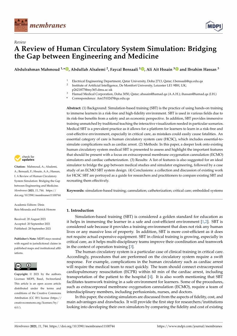
membranes Review A Review of Human Circulatory System Simulation: Bridging the Gap between Engineering and Medicine Abdulrahman Mahmoud 1, * , Abdullah Alsalemi 2 , Faycal Bensaali 1 , Ali Ait Hssain 3 and Ibrahim Hassan 3 Citation: Mahmoud, A.; Alsalemi, A.; Bensaali, F.; Hssain, A.A.; Hassan, I. A Review of Human Circulatory System Simulation: Bridging the Gap between Engineering and Medicine. Membranes 2021, 11, 744. https:// doi.org/10.3390/membranes11100744 Academic Editors: Dinis Reis Miranda and Patrick Honore Received: 20 August 2021 Accepted: 20 September 2021 Published: 28 September 2021 Publisher’s Note: MDPI stays neutral with regard to jurisdictional claims in published maps and institutional affil- iations. Copyright: © 2021 by the authors. Licensee MDPI, Basel, Switzerland. This article is an open access article distributed under the terms and conditions of the Creative Commons Attribution (CC BY) license (https:// creativecommons.org/licenses/by/ 4.0/). 1 Electrical Engineering Department, Qatar University, Doha 2713, Qatar; [email protected] 2 Institute of Artificial Intelligence, De Montfort University, Leicester LE1 9BH, UK; [email protected] 3 Hamad Medical Corporation, Doha 3050, Qatar; [email protected] (A.A.H.); [email protected] (I.H.) * Correspondence: [email protected] Abstract: (1) Background: Simulation-based training (SBT) is the practice of using hands-on training to immerse learners in a risk-free and high-fidelity environment. SBT is used in various fields due to its risk-free benefits from a safety and an economic perspective. In addition, SBT provides immersive training unmatched by traditional teaching the interactive visualization needed in particular scenarios. Medical SBT is a prevalent practice as it allows for a platform for learners to learn in a risk-free and cost-effective environment, especially in critical care, as mistakes could easily cause fatalities. An essential category of care is human circulatory system care (HCSC), which includes essential-to- simulate complications such as cardiac arrest. (2) Methods: In this paper, a deeper look onto existing human circulatory system medical SBT is presented to assess and highlight the important features that should be present with a focus on extracorporeal membrane oxygenation cannulation (ECMO) simulators and cardiac catheterization. (3) Results: A list of features is also suggested for an ideal simulator to bridge the gap between medical studies and simulator engineering, followed by a case study of an ECMO SBT system design. (4) Conclusions: a collection and discussion of existing work for HCSC SBT are portrayed as a guide for researchers and practitioners to compare existing SBT and recreating them effectively. Keywords: simulation-based training; cannulation; catheterization; critical care; embedded systems 1. Introduction Simulation-based training (SBT) is considered a golden standard for education as it helps in immersing the learner in a safe and cost-efficient environment [1,2]. SBT is considered safe because it provides a training environment that does not risk any human lives or any massive loss of property. In addition, SBT is more cost-efficient as it does not require actual expensive equipment. SBT in clinical training is growing, especially in critical care, as it helps multi-disciplinary teams improve their coordination and teamwork in the context of operation training [3]. The human circulatory system is a particular case of clinical training in critical care. Accordingly, procedures that are performed on the circulatory system require a swift response. For example, complications in the human circulatory such as cardiac arrest will require the medical team to react quickly. The team should connect extracorporeal cardiopulmonary resuscitation (ECPR) within 60 min of the cardiac arrest, including transportation of the patient to the hospital [4]. It is also worth mentioning that SBT facilitates teamwork training in a safe environment for learners. Some of the procedures, such as extracorporeal membrane oxygenation cannulation (ECMO), require a team of interdisciplinary members, including perfusionists, nurses, and doctors. In this paper, the existing simulators are discussed from the aspects of fidelity, cost, and main advantages and drawbacks. It will provide the first step for researchers/institutions looking into developing their own simulators by comparing the fidelity and cost of existing Membranes 2021, 11, 744. https://doi.org/10.3390/membranes11100744 https://www.mdpi.com/journal/membranes
-
Upload
khangminh22 -
Category
Documents
-
view
2 -
download
0
Transcript of A Review of Human Circulatory System Simulation - MDPI
membranes
Review
A Review of Human Circulatory System Simulation: Bridgingthe Gap between Engineering and Medicine
Abdulrahman Mahmoud 1,* , Abdullah Alsalemi 2, Faycal Bensaali 1 , Ali Ait Hssain 3 and Ibrahim Hassan 3
�����������������
Citation: Mahmoud, A.; Alsalemi,
A.; Bensaali, F.; Hssain, A.A.; Hassan,
I. A Review of Human Circulatory
System Simulation: Bridging the Gap
between Engineering and Medicine.
Membranes 2021, 11, 744. https://
doi.org/10.3390/membranes11100744
Academic Editors: Dinis
Reis Miranda and Patrick Honore
Received: 20 August 2021
Accepted: 20 September 2021
Published: 28 September 2021
Publisher’s Note: MDPI stays neutral
with regard to jurisdictional claims in
published maps and institutional affil-
iations.
Copyright: © 2021 by the authors.
Licensee MDPI, Basel, Switzerland.
This article is an open access article
distributed under the terms and
conditions of the Creative Commons
Attribution (CC BY) license (https://
creativecommons.org/licenses/by/
4.0/).
1 Electrical Engineering Department, Qatar University, Doha 2713, Qatar; [email protected] Institute of Artificial Intelligence, De Montfort University, Leicester LE1 9BH, UK;
[email protected] Hamad Medical Corporation, Doha 3050, Qatar; [email protected] (A.A.H.); [email protected] (I.H.)* Correspondence: [email protected]
Abstract: (1) Background: Simulation-based training (SBT) is the practice of using hands-on trainingto immerse learners in a risk-free and high-fidelity environment. SBT is used in various fields due toits risk-free benefits from a safety and an economic perspective. In addition, SBT provides immersivetraining unmatched by traditional teaching the interactive visualization needed in particular scenarios.Medical SBT is a prevalent practice as it allows for a platform for learners to learn in a risk-free andcost-effective environment, especially in critical care, as mistakes could easily cause fatalities. Anessential category of care is human circulatory system care (HCSC), which includes essential-to-simulate complications such as cardiac arrest. (2) Methods: In this paper, a deeper look onto existinghuman circulatory system medical SBT is presented to assess and highlight the important featuresthat should be present with a focus on extracorporeal membrane oxygenation cannulation (ECMO)simulators and cardiac catheterization. (3) Results: A list of features is also suggested for an idealsimulator to bridge the gap between medical studies and simulator engineering, followed by a casestudy of an ECMO SBT system design. (4) Conclusions: a collection and discussion of existing workfor HCSC SBT are portrayed as a guide for researchers and practitioners to compare existing SBT andrecreating them effectively.
Keywords: simulation-based training; cannulation; catheterization; critical care; embedded systems
1. Introduction
Simulation-based training (SBT) is considered a golden standard for education asit helps in immersing the learner in a safe and cost-efficient environment [1,2]. SBT isconsidered safe because it provides a training environment that does not risk any humanlives or any massive loss of property. In addition, SBT is more cost-efficient as it doesnot require actual expensive equipment. SBT in clinical training is growing, especially incritical care, as it helps multi-disciplinary teams improve their coordination and teamworkin the context of operation training [3].
The human circulatory system is a particular case of clinical training in critical care.Accordingly, procedures that are performed on the circulatory system require a swiftresponse. For example, complications in the human circulatory such as cardiac arrestwill require the medical team to react quickly. The team should connect extracorporealcardiopulmonary resuscitation (ECPR) within 60 min of the cardiac arrest, includingtransportation of the patient to the hospital [4]. It is also worth mentioning that SBTfacilitates teamwork training in a safe environment for learners. Some of the procedures,such as extracorporeal membrane oxygenation cannulation (ECMO), require a team ofinterdisciplinary members, including perfusionists, nurses, and doctors.
In this paper, the existing simulators are discussed from the aspects of fidelity, cost, andmain advantages and drawbacks. It will provide the first step for researchers/institutionslooking into developing their own simulators by comparing the fidelity and cost of existing
Membranes 2021, 11, 744. https://doi.org/10.3390/membranes11100744 https://www.mdpi.com/journal/membranes

Membranes 2021, 11, 744 2 of 24
simulators. A set of recommendations is then provided on the features to be includedin a human circulatory system simulator and the means to implement them using multi-disciplinary engineering skills. In addition, a case study to clarify the recommendations ofan ideal simulator is presented.
A sparse number of papers reviewed HCSC simulators, and the ones that did are onlyspecific to a particular medical operation. The main aim of this paper is to look into existingwork in a more rudimental way to allow for sharing of human circulatory simulationexperience between different clinical training of the same invasiveness. The paper willprimarily focus on two of the most intensive procedures concerning the human circulatorysystems; ECMO and cardiac catheterization. The procedures selected are chosen becausethey require high-fidelity training as both require the passing of a foreign object throughthe blood vessels. This paper aims to bridge the knowledge gap between engineering andmedicine by providing medical and engineering professionals with insight into anatomicalreview and technical engineering applications generating a conclusive narrative review.
2. Anatomical Review
When reviewing the human circulatory system from an SBT perspective, three mainaspects are considered the focal points of the review: (i) heart; (ii) blood vessels; and (iii)access points.
2.1. Heart
The heart is considered the central aspect of the human circulatory system as it pumpsblood to the whole body. Since it is a cornerstone of the circulatory system, many criticalcare procedures cater to resolving issues in the heart. Therefore, having an accuraterepresentation of the heart is quintessential to the success of the learning process. Acomputerized tomography (CT) scan of the heart could be seen in Figure 1, and in orderto emulate it in a dual loop system, the heart can be divided into two regions. The mainfeatures are the big bulge, which includes the right ventricle, left ventricle, and right atriumseen in region B. In addition, region A consists of the aorta, which is the artery that feedsthe whole body with oxygenated blood [5].
Figure 1. Human heart computerized tomography scan [5].
2.2. Blood Vessels
Blood vessels are essential for SBT. Usually, when an operation is performed on theheart, the clinicians cannot simply do it directly. That might risk damaging the heart, so one

Membranes 2021, 11, 744 3 of 24
usually inserts one’s tool into the blood vessels and passes it to the heart to decrease theprocedures’ invasiveness. The outer wall of the arteries is thicker, and its inner diameter issmaller than the veins [6].
2.3. Access Points
The access points are essential to the training process because the entry point of theprocedure tool is the first invasive step of surgeries and carries the risk of bleeding. Inmost invasive procedures, the access point is where the tool enters the circulatory systemand is usually pricked by a needle to make an entry point. After creating an entry point,the tool is inserted, such as a cannula in the case of cannulation. Common access pointsare femoral and jugular blood vessels as they can provide enough space for the large borecannula size required for ECMO and cardiac catheterization. Figure 2 shows the ultrasoundof the femoral access point ultrasound is used as it is the expected standard for guidingprocedures that involve the blood vessels as it can go through tissue and show the bloodvessels. In Figure 3, the ultrasound of the femoral region is seen. The veins are shown to beabout twice the size of both arteries combined, and as shown, arteries are situated on topof the veins and very close to them. The targeted artery is the common femoral artery to fitthe desired cannula. Common femoral arteries split into two branches deep femoral artery(DFA) and the superficial femoral artery (SFA). The SFA is the artery that feeds the leg andshould typically be avoided to keep blood flow in the leg [7].
Figure 2. Femoral region ultrasound scan [7].
Figure 3. Paper taxonomy.
3. Existing Simulators
Two types of critical care procedure simulators for the human circulatory system arereviewed in this section: extracorporeal membrane oxygenation (ECMO) cannulation andcardiac catheterization. ECMO is used as a relatively long-term ECPR and/or respiratorysystem [8]. In addition, for the ECMO machine to be connected to the patient’s body,

Membranes 2021, 11, 744 4 of 24
one must undergo an intensely invasive procedure called cannulation. Cannulation isthe act of passing the cannula from the cannulation access point (i.e., femoral or jugularregion) through the blood vessels to the desired location. The desired location for thearteries is aortal bifurcation. The desired location for the veins is the inferior vena cavaright below the heart near the hepatic vein [9]. Cardiac catheterization is the act of passinga catheter from the femoral or radial region to the heart to unblock the honorary arteryor any blood vessel, inject contrast dye to observe the heart clearly under X-ray, or fixcongenital disabilities in the heart [10]. The fact that the catheter and the cannula travel inthe blood vessels risk contact with organs, potentially leading to fatality. Auxiliary medicalequipment is an integral part of medical procedures. They are used to monitor vitals andassist in the body’s healing process by providing organ functionality while the organs heal.Therefore, incorporating auxiliary medical equipment in the emulation is quintessential.Figure 3 shows the taxonomy of the review paper where the simulators are categorizedinto simulators with a focus on ECMO, cardiac catheterization, and simulators work that isbased on the auxiliary device simulation such as designing an ECMO machine simulatoror an ultrasound machine simulator.
3.1. Methods
In this subsection, the criteria of evaluating the existing work is discussed and ana-lyzed. All the surveyed simulators are those from academic literature and the commer-cialized ones that are available in the market. The main aspects of evaluation are fidelityand cost, as they are usually tradeoffs of one another. When discussing fidelity, multi-ple aspects of fidelity are observed, including conceptual fidelity, physical fidelity, andpsychological fidelity. Conceptual fidelity is concerned with how much the sequence ofevents that the simulation-based training is providing conceptual context to the learner.This could be assessed by experts in the field and through surveys before and after thetraining [11]. Physical fidelity is a measure of how the simulator physically resembles ahuman circulatory system from the feeling to the guiding methods through the system,including fluoroscopy and ultrasound [12]. Psychological fidelity is concerned with howimmersive the SBT experience is and the capability of the system to duplicate the pressurein such engaging medical procedures. Cost as a criterion encompasses instantaneous costand maintenance cost as they both equally contribute to the overall cost of some of thesuggested solutions. The point of highlighting fidelity and cost is to showcase the aspect ofcost effectiveness as most of the commercialized simulators have mediocre fidelity and ahigh price. This paper acts as a guide to developing a sense to the cost of some features inorder to guide the readers in designing the highest cost-effective simulators.
3.2. ECMO Cannulation Systems
In this subsection, existing HCSC ECMO cannulation simulators are analyzed andreviewed based on cost and fidelity. The selected simulators are from both academicresearch and commercialized simulators.
3.2.1. Cadaveric ECMO Cannulation Simulator
According to the authors, although medical cadavers have been previously employedfor endovascular procedural training, the authors present the first clear novel modelcapable of functional venous and arterial ECMO cancellation. A novel perfusion-capablemannequin simulation has been used to supply high-fidelity training in ECPR-related crisisresource management areas. Yet, the technical aspects of cannulation are challenging torecreate. This simulator has high physical fidelity as it uses real cadavers and averageconceptual and psychological fidelity. Due to that, the cost of this simulation-based trainingis high. The cost is high because cadavers are not easy to acquire and are costly. Inaddition, when training is performed on such systems, disinfection of the whole system isrequired after the end of the training to avoid infection to learners and to uphold hygienicstandards [13].

Membranes 2021, 11, 744 5 of 24
3.2.2. A High-Fidelity Surgical Model and Perfusion Simulator
An ECMO cannulation simulator for a neonatal model is presented in the manuscript [14].The overall system seems cost-efficient, simplistic, and has mediocre overall fidelity. Thereis almost no heart emulation as it is a simple change of tubes. In addition, the bloodvessels are not sufficiently realistic as there is no distinction between arteries and veinsanatomy-wise, which makes the physical fidelity lower. The simulated blood pressure isinaccurate due to the lack of pumps in the system, and there are no false simulated pathsthat decrease the conceptual fidelity as the learners would lack the ability to differentiatebetween the arteries and veins. Lastly, the jugular cannulation access point is relativelyrealistic as it includes multiple layers that resemble flesh, blood, and skin. In addition,the tubes are of the eight Frenches size as the cannula are, which boosts the conceptualfidelity [14]. Figure 4 shows the proposed neonatal cannulation simulator.
Figure 4. Neonatal ECMO cannulation simulator [14].
3.2.3. ECMO Professional Simulator
Erler Zimmer commercializes an adult cannulation simulator that is highly realisticand could simulate multiple scenarios but comes with a hefty price tag of around USD$19.4 k, seen in Figure 5. Although the heart is vital as a stopping point, it is not evidentthat the simulator includes an emulation of the heart, which lowers the physical andconceptual fidelity. The blood vessels are realistic flow-wise and customizable enough tosimulate different scenarios and have dual loops, including arteries and veins, as seen inFigure 5 boosts the physical and conceptual fidelity greatly and allows for various scenarios.However, the blood vessel’s emulation is not anatomically accurate from the sense of wrong

Membranes 2021, 11, 744 6 of 24
tracks the guidewire can go through, which lowers the clinical fidelity. Lastly, the accesspoints are ultrasound-able, boosting the conceptual fidelity and cost-efficiency as the sizesare optimized, but the mold is not provided to fabricate the cannulation access point. Dueto the unrivaled versatility of the simulator, the overall fidelity is high [15].
Figure 5. Erler Zimmer adult cannulation simulator [15].
3.2.4. An Extracorporeal Membrane Oxygenation Cannulation Curriculum
The research team is developing a neonatal cannulation simulator with high physicalfidelity at the cannulation access points. Focusing on cannulation access point, the simulatorlacks heart emulation. The blood vessels have simplistic flow and only simulates the veinsloop, which decreases the conceptual fidelity as fewer scenarios could be simulated. Thecannulation access point is highly sophisticated as it has many scenarios and includesblood oozing and other scenarios that make the simulator have high physical fidelity andincreasing the conceptual fidelity [16]. Figure 6 shows the simulator and oozing feature.
Figure 6. Neonatal ECMO cannulation simulator featuring oozing (A) Overall system (B,C) cannula-tion with standard flow with oozing (D) bleeding from operative site [16].

Membranes 2021, 11, 744 7 of 24
3.2.5. Neonatal Cannulation Simulator
A neonatal cannulation simulator is presented by the research team that is cost-efficient.There is no emulation of the heart in the simulator despite its necessity in identifying thestopping point of the guidewire and the cannula, which lowers the physical fidelity. Theblood vessels are very simplistic and contain only one loop with constant flow lowering theconceptual fidelity. The cannulation access point is not ultrasound-able, which decreasesthe conceptual fidelity of the simulator seen in Figure 7, which deems the overall fidelity ofthe simulator low [17].
Figure 7. Neonatal ECMO cannulation simulator [17].
3.2.6. Adult Cannulation Simulator
The research team developed an adult cannulation simulator that is cost-efficientand only costs around USD $100 per simulator. The simulator does not include a heartemulation, which lowers the physical and conceptual fidelity. However, the blood vesselsare of descent physical fidelity. Despite having one closed loop, the tube circles back tomake the other loop, which gives a dual loop with a single one. The cannulation accesspoint is not of suitable physical fidelity, which lowers the emotional fidelity, a crucialaspects of the overall process, but it is very cost-efficient and emulates dilation [18]. TheEndo circuit diagram can be seen in Figure 8.

Membranes 2021, 11, 744 8 of 24
Figure 8. Endo circuit adult cannulation simulator [18].
3.2.7. ECPR Simulation Training Mannequin
Pang showcases a simulator where 3D printing was used to create a robust ECPRsimulation training mannequin. A commonly available regular CPR mannequin with anairway feature was used as a base case for adjustment. A low-cost 3D printer (Laerdal Med-ical, Stavanger, Norway) was used to 3D-print a modular plastic pelvis, which improvesits physical fidelity. A medical silicone gel to simulate the femoral silicone vasculature wasproduced in conjunction with a gravitational vascular system. The outcome is a modifiedmannequin, a part of the modular in-house ECMO cannulation and vascular structurescombined with commercially available airway and CPR components, making the overallfidelity average. The total cost of developing the simulator was valued at USD $1,394, seenin Figure 9 [19].
Figure 9. 3D-printed ECMO cannulation simulator [19].
3.2.8. ECMO Surgical Cannulation Simulators
McMullan et al. proposed a novel surgical neonatal cannulation simulator. The simula-tor uses affordable, silicone-based ECMO cannulation systems and commercially availablesilicone pads combined to reproduce layers of skin, subcutaneous tissue, blood vessels,and bones, improving the physical fidelity. The authors are working on a percutaneouscannulation simulator designed to reproduce the cervical cannulation with dual-lumen VVECMO cannulas; the focus boosts the conceptual fidelity of the system. Its cost-effectivenessmakes it desirable and easy to produce, but its limited physical and conceptual fidelity setsit back as the pumping is primitive, and the system has limited scenarios to enact [20].

Membranes 2021, 11, 744 9 of 24
3.2.9. Training the Component Steps of an ECMO Cannulation
In this research project, the authors developed a cost-efficient ECMO cannulationsimulator that contains both arterial and veins cannulation simulation at an access point.However, the system does not contain emulation of the heart or the blood loop, loweringconceptual fidelity. The team employed cheap and disposable components along with3D printing to construct an anatomically accurate cannulation access point simulator,but the lack of having a mannequin and other parts lowers the emotional fidelity seenFigure 10 [21].
Figure 10. Cannulation access point simulator [21].
3.2.10. Next-Generation Cannulation Simulator
The next-generation cannulation simulator is a joint collaboration between QatarUniversity (QU) and Hamad medical corporation (HMC), the governmental healthcareprovider in Qatar, aiming to develop a high-fidelity, low-cost ECMO cannulation simu-lator to train ECMO professionals locally. The research team has developed an ECMOcannulation simulator composed of three integrated parts: the heart, blood vessels, andaccess points. The heart emulation is made of a silicon rubber pad made with a 3D-printedmold. The pad has cavities that mimic the hepatic vein and the right atrium, which areessential for ECMO cannulation. The hepatic vein is the stopping point of the veinouscannula, which increases the conceptual fidelity. In addition, the heart pad does not includean arterial side because the arterial cannula stops at the femoral artery. Therefore, thedouble-walled blood vessels have realistic blood flow, which is turbulent for arteries andlaminar for veins increasing physical fidelity. The access point emulation is made in asimilar way to the heart pad. It features alternative tracks for the cannula to emulate therealistic difficulty of guiding the cannula and the guidewire through the human circulatorysystem, which boosts emotional fidelity. To complement the physical fidelity, the team alsodesigned a procedural emergency system by measuring the force exerted on the renal veinto predict the chance of internal hemorrhage and incorporated a flow meter to measure theamount of bleeding. On the other hand, one drawback of the system is the drift in sensorreading and the need for recalibration [22]. The simulator has an overall high fidelity andcan be seen in Figure 11.

Membranes 2021, 11, 744 10 of 24
Figure 11. Next-generation cannulation simulator [22].
3.2.11. S2225 Pediatric HAL
Gaumard developed multiple patient simulators for SBT, including the cutting-edgepediatric HAL. HAL was designed to create a life-like pediatric patient to train healthcareprofessionals in dealing with multiple clinical emergencies and cases using high-endeye and facial expressions, realistic lung simulation, patient monitor support, airway,and circulatory system emulation, and wireless connection. The combination of thesetechnologies makes this simulator of high physical and conceptual fidelity. The simulatorincludes a wide range of interactive features, including the ability to make conversationsand the device describing pain levels mimicking a human being and having an array ofsensors to immerse the learner in the experience, fully improving its emotional fidelity. TheHal simulator can be seen in Figure 12 [23].
Figure 12. Hal pediatric nursing simulator [23].
3.3. Cardiac Catherization Systems
This subsection focuses on the medical procedure of cardiac catheterization andincludes research projects and commercialized products.
3.3.1. Catheterization and Cardiovascular Interventions
An adult trans-catheter cardiovascular simulator is developed by the authors withhigh fidelity seen in Figure 13. The heart emulation in the simulator is anatomicallyaccurate as it is made using a mold that was designed and implemented using additivemanufacturing and liquid to emulate blood, improving physical fidelity. The blood vesselsare high-fidelity as it includes the renal vein as and the mold is to be filled with siliconerubber that is ultrasound-able. Although the blood vessels do not have a false path, the

Membranes 2021, 11, 744 11 of 24
catheter could go through decreasing the conceptual fidelity. The access point is made fromultrasound-able material and only includes the veins [24].
Figure 13. Multi-system cardiac catheterization simulator (A) space for silicone rubber for fluoroscopyemulation; (B) space for stereo camera for transesophageal echocardiography; (C) Catherization pad;(D) screen displaying fluoroscopy imaging; (E) electronics compartment; and (F) touch pad for thegraphical user interface of the system [24].
3.3.2. Beating Heart Porcine High-Fidelity Simulator
This research project showcases a trans-catheter cardiovascular simulator, which is ofhigh fidelity seen in Figure 14. The heart emulation of the simulator is of high fidelity as areal porcine heart is used for simulation. The blood vessels used are cannula connected toa pulsatile piston pump, which increases physical fidelity. On the other hand, there is noaccess point for the catheter lowering the conceptual fidelity [25].
Figure 14. Porcine heart cardiac catheterization simulator [25].
3.3.3. Gen II Femoral Vascular Access and Regional Anastasia Ultrasound Training Model
This product is a femoral vascular access point that could potentially be used forcannulation and catheterization. The simulator consists of a femoral access point that isanatomically accurate and resembles the ultrasound of an adult femoral region in a verydetailed way seen in Figure 15, which provides limited physical fidelity [26].

Membranes 2021, 11, 744 12 of 24
Figure 15. Femoral region cardiac catheterization simulator [26].
3.4. Auxiliary Devices
This subsection focuses on simulators that are auxiliary device simulators. All thework discussed is research work.
3.4.1. ECMO Therapy Simulator for Extracorporeal Life Support
The authors constructed an innovative ECMO simulator system demo integrated intoany currently usable full-body patient simulator and used it for medical research. Theoverall expense of the first simulator prototype is roughly USD $450 and $50 for disposablecomponents. The simulator enables many possibilities concerning the development ofscenarios, particularly: deployment, perfusion, and transport of patients using ECMO,providing conceptual fidelity [27]. A practical demonstration can be illustrated in Figure 16.
Figure 16. Adult ECMO simulator demo [27].

Membranes 2021, 11, 744 13 of 24
3.4.2. Design and Development of a Mechatronic Training Simulator for Adult ECMO
Mehta has developed an ECMO mechatronic training simulator, which can help medi-cal professionals gain the skills needed, gain familiarity and reduce errors by practicingbefore the procedure in actual patients. The simulator is built as an ultrasound-compatiblebalanced simulator with functional components such as synthetic blood vessels, cannu-lation pads, and a color-varying blood simulator to mimic both oxygenation and deoxy-genation, boosting physical fidelity. The simulator is combined with a statistical modelof human physiology to replicate real-time patient vital signs and monitor the operator’sequipment improving conceptual fidelity. Results include successful cannulation underultrasound scanning and a simple patient scenario of hypovolemia [28].
3.4.3. Hardware-in-the-Loop Test Bench for Artificial Lungs
The authors propose constructing a hardware-in-the-loop test bench to address typicalweaknesses such as sophistication, tediousness, and lack of repeatability. It was testedat a live laboratory while maintaining precise monitoring of the different control andsimulation variables, resulting in relatively higher conceptual fidelity seen in Figure 17.Current prototype results show that the proposed system allows high-fidelity testing ofinteractions with physiological ECMO conditions [29].
Membranes 2021, 11, x 14 of 25
Figure 16. Adult ECMO simulator demo [27].
3.4.2. Design and Development of a Mechatronic Training Simulator for Adult ECMO Mehta has developed an ECMO mechatronic training simulator, which can help
medical professionals gain the skills needed, gain familiarity and reduce errors by prac-ticing before the procedure in actual patients. The simulator is built as an ultrasound-compatible balanced simulator with functional components such as synthetic blood ves-sels, cannulation pads, and a color-varying blood simulator to mimic both oxygenation and deoxygenation, boosting physical fidelity. The simulator is combined with a statistical model of human physiology to replicate real-time patient vital signs and monitor the op-erator’s equipment improving conceptual fidelity. Results include successful cannulation under ultrasound scanning and a simple patient scenario of hypovolemia [28].
3.4.3. Hardware-in-the-Loop Test Bench for Artificial Lungs The authors propose constructing a hardware-in-the-loop test bench to address typ-
ical weaknesses such as sophistication, tediousness, and lack of repeatability. It was tested at a live laboratory while maintaining precise monitoring of the different control and sim-ulation variables, resulting in relatively higher conceptual fidelity seen in Figure 17. Cur-rent prototype results show that the proposed system allows high-fidelity testing of inter-actions with physiological ECMO conditions [29].
Figure 17. Artificial lungs simulator [29].
Figure 17. Artificial lungs simulator [29].
3.4.4. A Hybrid Cardiopulmonary Simulation Platform
In this project, a hybrid cardiopulmonary simulation platform was adapted for ECMOsimulation with a specially designed ECMO hydraulic model. Standard ECMO configura-tions of VA and VV were the simulation modes. Preliminary tests indicate an improvementin left ventricular afterload for VA configuration and a rise in blood recirculation for VVone. Considering the location of cannulas, the geometrical architectures of the systemicvessels and actual oxygenation offer a more practical and forward-looking simulationstrategy showcasing conceptual fidelity [30].
3.4.5. Simulation Training for Extracorporeal Membrane Oxygenation
This work describes a 1-day ECMO course that was given within the scope of ECMOsimulation. An ICU simulated room was used, bundled with a high-fidelity mannequin.The simulator was used for training ECMO staff on various scenarios and instilling thetheoretical knowledge given in lectures focusing on conceptual fidelity. Results reportthat all participants signify the awareness raised by the simulation alongside nurturingpractical team-working skills [31].
3.4.6. Dynamic Extracorporeal Membrane Oxygenation Simulation
The authors showcase a patent on a system with a clamp for an ECMO circuit, anarticulator connected to the clamp, and a simulator module connected to the articulator

Membranes 2021, 11, 744 14 of 24
to send control signals. The system also includes a pump and a display module. Thecircuit comprises a conduit, and a flow regulating device is positioned to scenario a specificECMO scenario [32].
3.4.7. Optical Skill-Assist Device for Ultrasound-Guided Vascular Access
A central vein catheterization (CVC) was implemented in this research project. Thesimulator is in a preliminary phase and only includes an access point and an ultrasoundemulator that improves physical and conceptual fidelity. The system successfully helps thelearner visualize and train using the ultrasound machine and CVC. The medical imaginingof the simulator is very accurate and human-like but lacks emotional fidelity, as seen inFigure 18 [33]. CVC is an important type of catheterization that is applied as a secondarymedical procedure with the highlight of the simulator being the auxiliary devices usedin training.
Figure 18. Central vein catheterization access point simulator (A) ultrasound emulator (B) CVC pad(C) Ultrasound image of simulator (D) diagram showing how the device is placed [33].
3.4.8. ECMO Simulation with Affordable Yet High-Fidelity Technology
This research project was developed as an initiative between QU and HMC to designan ECMO machine simulator and is the predecessor of Mahmoud et al. [22]. To eliminatemultiple of the main flaws in the existing literature. The team has successfully patented theuse of thermochromic ink to replace animal blood in training, effectively replacing animalblood that is commonly used for such training [34]. Replacing the blood with anothermaterial will provide a more straightforward method of simulating oxygenation -heating-and will end the need of disinfecting the whole loop after training, improving physicalfidelity and cost efficiency. The system is a modular training system with multiple modulesto train the learners on different scenarios, including air in the ECMO machine loop andbleeding and more the ECMO machine simulator seen in Figure 19 [35]. The projectpresents a modular, high-fidelity cost-efficient simulator with a primary drawback, whichis hard to simulate the scenario of connecting a heater in line with the ECMO machine asthat would interfere with the simulation, which highly improves the physical fidelity [36].
Figure 19. ECMO machine simulator [36].
Membranes 2021, 11, 744 15 of 24
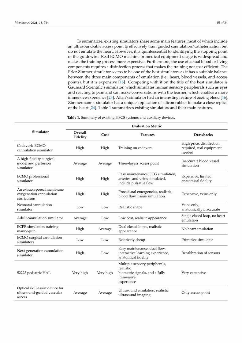
To summarize, existing simulators share some main features, most of which includean ultrasound-able access point to effectively train guided cannulation/catheterization butdo not emulate the heart. However, it is quintessential to identifying the stopping pointof the guidewire. Real ECMO machine or medical equipment usage is widespread andmakes the training process more expensive. Furthermore, the use of actual blood or livingcomponents requires a disinfection process that makes the training not cost-efficient. TheErler Zimmer simulator seems to be one of the best simulators as it has a suitable balancebetween the three main components of emulation (i.e., heart, blood vessels, and accesspoints), but it is expensive [15]. Competing with it on the title of the best simulator isGaumard Scientific’s simulator, which simulates human sensory peripherals such as eyesand reacting to pain and can make conversations with the learner, which enables a moreimmersive experience [23]. Allan’s simulator had an interesting feature of oozing blood [16].Zimmermann’s simulator has a unique application of silicon rubber to make a close replicaof the heart [24]. Table 1 summarizes existing simulators and their main features.
Table 1. Summary of existing HSCS systems and auxiliary devices.
Simulator
Evaluation Metric
OverallFidelity Cost Features Drawbacks
Cadaveric ECMOcannulation simulator High High Training on cadavers
High price, disinfectionrequired, real equipmentneeded
A high-fidelity surgicalmodel and perfusionsimulator
Average Average Three-layers access point Inaccurate blood vesselsimulation
ECMO professionalsimulator High High
Easy maintenance, ECG simulation,arteries, and veins simulated,include pulsatile flow
Expensive, limitedanatomical fidelity
An extracorporeal membraneoxygenation cannulationcurriculum
High High Procedural emergencies, realistic,blood flow, tissue simulation Expensive, veins only
Neonatal cannulationsimulator Low Low Realistic shape Veins only,
anatomically inaccurate
Adult cannulation simulator Average Low Low cost, realistic appearance Single closed loop, no heartemulation
ECPR simulation trainingmannequin High Average Dual closed loops, realistic
appearance No heart emulation
ECMO surgical cannulationsimulators Low Low Relatively cheap Primitive simulator
Next-generation cannulationsimulator High Low
Easy maintenance, dual flow,interactive learning experience,anatomical fidelity
Recalibration of sensors
S2225 pediatric HAL Very high Very high
Multiple sensory peripherals,realisticbiometric signals, and a fullyimmersiveexperience
Very expensive
Optical skill-assist device forultrasound-guided vascularaccess
Average Average Ultrasound emulation, realisticultrasound imaging Only access point

Membranes 2021, 11, 744 16 of 24
Table 1. Cont.
Simulator
Evaluation Metric
OverallFidelity Cost Features Drawbacks
Catheterization andcardiovascular interventions High Average Fully ultrasound-able system,
anatomically accurate system Does not look like a patient
Beating heart porcinehigh-fidelity simulator High High Anatomically accurate heart Requires disinfection of
equipment
Gen II femoral vascularaccess training model High High Anatomically accurate access point Only access point
ECMO therapy simulator forextracorporeal life support Low Low Cheap auxiliary device emulation,
easy to maintain
Only demo for controllingthe ECMOmachine, no surgicaltraining
Design and development of amechatronic trainingsimulator for adult ECMO
NA NA Synthetic blood emulation, vitalsignals generation Insufficient details
Hardware-in-the-loop testbench for artificial lungs High High Blood oxygenation, full ECMO
machine control
Requires disinfection,expensiveoxygenation devices
A hybrid cardiopulmonarysimulation platform Average High Blood oxygenation, full ECMO
machine control
Requires disinfection,expensiveoxygenation devices
Simulation training forextracorporeal membraneoxygenation
High Very high Blood oxygenationRequires disinfection,expensive off-the-shelfsystems
Dynamic extracorporealmembrane oxygenationsimulation
High High Blood oxygenation, full ECMOmachine control
Requires disinfection,expensiveoxygenation devices
ECMO simulation withaffordable yet high-fidelitytechnology
High Low No disinfection required, full ECMOmachine control, modular design
Heater scenariointerference
An important aspect to look at when reviewing the human circulatory system SBTis cost, as the learners need much training before they are ready to operate on patients.Multiple simulators are cost-efficient, but one of the most cost-efficient simulators is theEndo simulator, as it costs USD $100 and simulates a dual loop simulation that evensimulates dilation [18]. In addition, simulators such as Allan, Thompson, and Palmersimulators provide unique features with low costs, such as blood oozing, high fidelity, andlayered cannulation access points, respectively [14,16,17]. It is worth mentioning that amain problem with existing simulators is the use of real blood to emulate human blood,which causes the need for disinfection of the whole system, which is very costly. The onlyreviewed simulator that solved this problem with high fidelity is [36], as the research teamfound a simulation alternative to oxygenation in heating.
4. Recommendations for an Ideal Simulator
As discussed in the previous section, the existing work is conclusive. However,there is still a gap in knowledge as there much cutting-edge technology used in medicalsimulators. Therefore, in this section, features of an ideal simulator are discussed, andrecommendations for its implementation. It is worth mentioning that a common practice isto have a formal education into the anatomy of the circulatory system and non-invasiveimaging of the human circulatory system before engaging in using the recommendedideal simulator.

Membranes 2021, 11, 744 17 of 24
4.1. Heart Emulation
The best practice for heart emulation would be done using a mold to improve anatomicalaccuracy. This was done in Zimmermann’s for the learner to correctly visualize how the heartlooks under ultrasound [24]. Accurate heart emulation could be achieved by first 3D modelingan accurate shape of the heart then creating an inner and outer mold that could be later filledwith EcoFLEX silicon rubber(Smooth-On, Macungie, PA, USA). Alternatively, a block-likemold could be done as seen in the cannulation access point, as the block could have a cavityinside it that resembles a heart. To track the tool in it using ultrasound. The block could beproduced by 3D printing the mold and designing the cavity in the shape of a heart.
4.2. Blood Vessels
The following should be realistic to produce an accurate blood vessel system: the flowof simulated blood, the tubing system inside the body, should resemble the human bodyanatomically. The flow of simulated blood could be controlled using two pumps, one foreach loop. The arterial loop should have a pulsatile pump, and the veins should havea laminar pump. It is not advisable to purchase an off-the-shelf pulsatile pump for thesystem as it would be costly. Alternatively, one can buy a variable pump and use pulsewidth modulation in a microcontroller to control it. The laminar flow could be as simple asa constant flow pump. To improve functionality, one could connect it to the microcontrollerto control its pumping action as performed by the Erler Zimmer simulator (Erler-Zimmer,Lauf, Germany) [15]. To obtain an anatomically accurate blood vessel, one can use differentsize silicon tubes for each loop, with veins having a thinner inner diameter. The connectorscould also be 3D modeled and created by additive manufacturing.
4.3. Access Points
Thirdly, the access point could be made accurate by making a 3D model of the mold.The ultrasound of the femoral region should be used as a guideline to the mold design asultimately ultrasound is used for guidance when doing cannulation catheterization. It isalso worth noting that at least three access points should present two femoral access pointsand one jugular to simulate different cannulation methods and catheterization. To improvethe access point’s fidelity, one can use a mechanism fitted below the pad to make the padseem as though it is pulsating under the ultrasound. To generate the desired pulsationunder arteries, the mechanism should output linear motion. A simple application to such amechanism would be a cam follower mechanism where the cam rotates driven by a simpleDC motor. The follower is fit perpendicular to the cam to change the rotary motion tolinear therefore squeezing the pad and making it appear as if pulsating.
4.4. Embedded System
To develop a more realistic simulator, one must design an embedded system to controlthe pumps and to have sensors continuously reading key features such as the flow ofsimulated blood and the force a guidewire exerts on the organs in its path. In addition,one should have a sensor to measure the level of liquid in the tanks. Such sensor readingsmay aid in developing a better understanding of the learner’s mistakes. That is not all,as having an embedded system can enable the instructor to have simulation scenariosthat resemble procedural emergencies to assess the learner’s reaction to it, as performedby Gaumard Scientific’s simulator (Gaumard, Miami, Florida) [23]. Possible proceduralemergencies to emulate are bleeding, internal bleeding, seizures, and heart failure. Thebleeding emergency could be detected by the flow meter and the tank level and notifythe instructor. Internal bleeding could be detected by having force-sensing resistors atthe renal vein to detect if the learner might cause internal bleeding by puncturing thekidney. Seizures could be emulated by having motors vibrate the mannequin to preparethe learners for such a case. Heart failure is a procedural emergency where the instructorcan change the simulator pumping and simulated blood oxygenation to test the learner’sunderstanding of which cannulation method to do.

Membranes 2021, 11, 744 18 of 24
4.5. Instructor Application
Finally, an instructor application (app) should be developed as performed by AlDisi et al. [36] that enables the instructor to control the procedural emergencies from theconvenience of their fingertips to trigger different clinical scenarios in real-time and at thesame time observe and monitor the learner’s reaction to the procedural emergency. The appshould also control the dummy to control the simulated blood pumping and oxygenation inthe simulated blood. The app should also display the sensors on the mannequin’s readingsto evaluate the learner’s performance [37].
5. Case Study
According to the discussed recommendations for an ideal simulator, the team hasdevised a design for a modular and cost-efficient ECMO cannulation simulator with highfidelity. The system comprises three subsystems (i) the cannulation simulator, (ii) the ECMOmachine simulator, and (iii) the instructor app, as represented in Figure 20. The cannulationsimulator is the part of the simulator that emulates the body of the patient. The body isemulated using a mannequin. The cannulation system is composed of three parts, whichare the closed loop, cannulation access points, and embedded system. The ECMO machinesimulator is an instrumental part of the simulator system as it replaces the expensiveECMO machine in training. The ECMO machine uses the concept of thermochromism toemulate blood oxygenation to avoid having to disinfect the system with high fidelity. Thesystem uses a heater-cooler system that controls the amount of emulated oxygenation tosimulate different clinical cases. Lastly, the instructor application is used to control thesystem from the remote access of the instructor convenience. In this case study, the focusis on the cannulation simulator and its implementation. More details about the ECMOmachine simulator can be found in [38,39].
Figure 20. Block diagram of the overall case study.
5.1. Closed Loop
The closed-loop system of the cannulation simulator is composed of three essentialparts, presented in Figure 21, which are the tubing system, the pumping system, andthe sensors. The tubing system comprises two plastic and silicone tubes with differentwall thicknesses to simulate both veins and arteries and represented in blue and red,respectively. The pumping system is composed of two pumps that emulate the heart

Membranes 2021, 11, 744 19 of 24
pumping simulating both veins and arterial flow. A microcontroller is used to control thepumps to have pulsatile flow at the arteries and laminar flow in the veins. The sensorsin the system are force-sensing resistors (FSR) and flowmeters. The sensors are part ofthe procedural passive procedural emergencies. The force-sensing meter is located in thealternative path of the cannula at the renal vein to measure the potential force exerted onit upon failure of navigating the guidewire through the circulatory system. The FSR isessential for the simulator as exerting so much force on the kidney in real life would causefatal internal bleeding. The FSR is to be placed into the connector in Figure 22, right belowthe heart pad. The flowmeter sensor is used to calculate the deferential flow and, as abyproduct, calculate the leakage of simulated blood to measure the bleeding in the patient.
Figure 21. Overall design of the case study.
Figure 22. FSR connector design and FSR.

Membranes 2021, 11, 744 20 of 24
5.2. Cannulation Access Point
The main parts of the cannulation access point are femoral pads, heart pad, andthe pulsating mechanism. The femoral pads are located at the bottom of the mannequinand contain the blood vessels responsible for feeding the lower body. To ensure highphysical fidelity, the pads are made from a custom-made mold made possible by additivemanufacturing. The custom-made design is based on an ultrasound image of the femoralregion seen in Figure 2. The design is unpacked in Table 2. The heart pad is considered anessential part of the simulator as it represents the stopping point of the veins cannula. Theheart pad is created in a similar method to the femoral pad through a mold. In the case ofthe heart pad, a computerized tomography scan of the heart, shown in Figure 1, was usedto design the mold to guarantee high anatomical realism. The details of the heart pad molddesign are presented in Table 3. Lastly, the pulsating mechanism is a mechanism used tomove the cannulation access points from the arteries’ side to emulate pulsating arteriesunder ultrasound, which is how the practitioners identify the arteries vessels from theveins. The mechanism type is cam follower, which is a two-part mechanism that convertsfrom rotary motion to linear motion. Table 4 showcases the design of the mechanism andits main features.
Table 2. Design of the femoral pad mold.
Part Name 3D Model Description
Body • Exterior body of the femoral pad• Based on an ultrasound of the femoral region
Arterial Rod • Emulates splitting of femoral artery to the deep femoralartery and superficial artery
Veins Rod • Emulates natural curvature in blood vessels
Overview Just write for example fully designed mold for femoral pad
Table 3. Design of the heart pad mold.
Part Name 3D Model Description
Body• The exterior part of the body of the mold• Based on a CT scan of the heart
Veins Rod • Features right atrium and hepatic vein
Arterial Rod • A simple rod is used as the cannula does not reach this point.
Plug • Includes inner threading to center the veins rod
Overview • An overview of a fully connected mold
Membranes 2021, 11, 744 21 of 24
Table 4. Design of the cam and follower mechanism.
Part Name 3D Model Description
Body
• Top space reserved for the femoral pad• Lower space is for the rotation to a linear mechanism• The stand-in lower space is reserved for a DC servo motor
Cam • Steep elliptical shape to have instantaneous change in motion from the linear side
Follower • Emulates natural curvature in blood vessels
Overview• The DC motor rotates the cam, which results in translation in follower• Future improvement might include multiple mechanisms
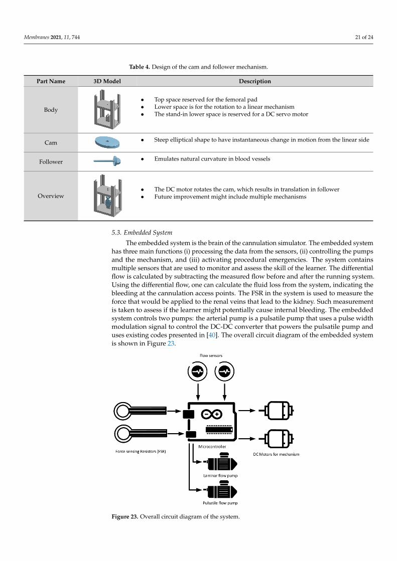
5.3. Embedded System
The embedded system is the brain of the cannulation simulator. The embedded systemhas three main functions (i) processing the data from the sensors, (ii) controlling the pumpsand the mechanism, and (iii) activating procedural emergencies. The system containsmultiple sensors that are used to monitor and assess the skill of the learner. The differentialflow is calculated by subtracting the measured flow before and after the running system.Using the differential flow, one can calculate the fluid loss from the system, indicating thebleeding at the cannulation access points. The FSR in the system is used to measure theforce that would be applied to the renal veins that lead to the kidney. Such measurementis taken to assess if the learner might potentially cause internal bleeding. The embeddedsystem controls two pumps: the arterial pump is a pulsatile pump that uses a pulse widthmodulation signal to control the DC-DC converter that powers the pulsatile pump anduses existing codes presented in [40]. The overall circuit diagram of the embedded systemis shown in Figure 23.
Figure 23. Overall circuit diagram of the system.

Membranes 2021, 11, 744 22 of 24
Similarly, the veins pump is a laminar flow. The laminar flow is controlled using thesame technique, but the signal is constant. The activation of the procedural emergencyoccurs through the embedded system as it controls the actuators. The seizure and pulsatingsystems can both use the same cam and follower mechanism in different sizes and operatingspeeds. The design of the mechanism is shown in Table 4. In both applications, rotationaction performed by DC motors is required to cause linear motion in the required objects.
6. Conclusions
In conclusion, it is evident of the knowledge gap between the advancement in engineer-ing and the application in medical simulation-based training for critical care. Accordingly,the paper has provided an anatomical review of the features of the human circulatorysystem. A review on the existing human circulatory system emulators in academia and themarket with emphasis on cost and fidelity providing a summary table (Table 1). In additionto discussing an insight into the technical aspect of creating an affordable simulator withstate-of-the-art features from an engineering perspective. The core features suggestedare better practices for developing the heart and access point emulating using additivemanufacturing and pourable silicon rubber. To improve the blood vessels emulation, thecase study introduced variable pumping speed, high-fidelity false paths, and pulsatingmechanism to help trainees differentiate between arteries and veins. Furthermore, intro-ducing an embedded system to control the system and implement sensors to provide tothe instructor in real-time. The embedded system facilitates the introduction of proceduralemergencies to immerse the learner in the learning experience. Lastly, the instructor appwas recommended to help in effectively assessing the learner and trigger procedural emer-gencies. The case study highlighted the recommendations and provided an example ofan overall high-fidelity simulator. This paper is considered one-of-a-kind and an essentialread for every institution developing a human circulatory system, emulation providing aconclusive narrative review of HSCS simulators. To summarize, an ideal HSCS simulatorshould include a high physical and conceptual fidelity heart and access point emulation.Improving the emulation of the blood vessels improve conceptual and physical fidelity.The inclusion of the embedded system and instructor app improves the psychologicalfidelity of the simulator.
Author Contributions: A.M. Collected the reviewed simulators with the help of A.A. and F.B.,A.M. drafted the manuscript. F.B. and A.A. helped draft the manuscript by refining the paper andproviding comments for all the drafts. A.A.H. and I.H. provided medical context. All authors haveread and agreed to the published version of the manuscript.
Funding: Qatar National Research Fund, Award Number: GSRA6-2-0418-19015, Recipient: A.M.;Qatar University, Award Number: M-CTP-CENG-2020-1, Recipient: F.B.
Institutional Review Board Statement: Not applicable.
Informed Consent Statement: Not applicable.
Data Availability Statement: Not applicable.
Acknowledgments: This publication was made possible by an Award (GSRA6-2-0418-19015) fromthe Qatar National Research Fund (a member of the Qatar Foundation). The contents herein aresolely the responsibility of the authors. This publication was also supported by Qatar UniversityInternal Grant No. M-CTP-CENG-2020-1. The findings achieved herein are solely the responsibilityof the authors.
Conflicts of Interest: The authors declare no conflict of interest.
References1. Kunkler, K. The role of medical simulation: An overview. Int. J. Med. Robot. Comput. Assist. Surg. 2006, 2, 203–210. [CrossRef]2. McGaghie, W.C.; Issenberg, S.B.; Cohen, E.R.; Barsuk, J.H.; Wayne, D.B. Does simulation-based medical education with deliberate
practice yield better results than traditional clinical education? A meta-analytic comparative review of the evidence. Acad. Med.2011, 86, 706–711. [CrossRef] [PubMed]

Membranes 2021, 11, 744 23 of 24
3. Alinier, G.; Platt, A. International overview of high-level simulation education initiatives in relation to critical care. Nurs. Crit.Care 2014, 19, 42–49. [CrossRef] [PubMed]
4. Hutin, A.; Abu-Habsa, M.; Burns, B.; Bernard, S.; Bellezzo, J.; Shinar, Z.; Torres, E.C.; Gueugniaud, P.Y.; Carli, P.; Lamhaut, L.Early ECPR for out-of-hospital cardiac arrest: Best practice in 2018. Resuscitation 2018, 130, 44–48. [CrossRef] [PubMed]
5. Herring, W. Learning Radiology: Recognizing the Basics; Elsevier: Philadelphia, PA, USA, 2019; ISBN 978-0323567299.6. Moore, K.L.; Dalley, A.F.; Agur, A.M. Clinically Oriented Anatomy, 7th ed.; Lippincott: Baltimore, MD, USA, 2014; ISBN
9781451119459. [CrossRef]7. Burns, K.; Fox, J.C. Venous ultrasound. In Atlas of Emergency Ultrasound; Cambridge University Press: Irvine, CA, USA, 2011.
[CrossRef]8. Biolo, M.; Salton, F.; Ruaro, B.; Busca, A.; Santagiuliana, M.; Fontanesi, L.; Gabrielli, M.; Baratella, E.; Confalonieri, M. Emergency
laser treatment of a tracheobronchial carcinoid during ECMO. Med. Res. Arch. 2020, 8. [CrossRef]9. Pavlushkov, E.; Berman, M.; Valchanov, K. Cannulation techniques for extracorporeal life support. Ann. Transl. Med. 2017, 5, 70.
[CrossRef]10. Ruaro, B.; Confalonieri, M.; Salton, F.; Wade, B.; Baratella, E.; Geri, P.; Confalonieri, P.; Kodric, M.; Biolo, M.; Bruni, C. The
relationship between pulmonary damage and peripheral vascular manifestations in systemic sclerosis patients. Pharmaceuticals2021, 14, 403. [CrossRef]
11. Rudolph, J.W.; Simon, R.; Raemer, D.B. Which reality matters? Questions on the path to high engagement in healthcare simulation.Simul. Healthc. 2007, 2, 161–163. [CrossRef]
12. Alexander, A.; Brunyé, T.T.; Sidman, J.; Weil, S.A. From Gaming to Training: A Review of Studies on Fidelity, Immersion, Presence,and Buy-in and Their Effects on Transfer in PC-Based Simulations and Games. DARWARS Train. Impact Group 2005, 5, 1–14.
13. Fagan, A.; Gould, J.; Horne, D.; Morrison, S.; Kovacs, G.; Sandeski, R. Cadaveric Ecmo Cannulation Simulator. Can. J. Cardiol.2019, 35, S93. [CrossRef]
14. Palmer, D.; Aspenleiter, M.; da Silva, J.; Castro-Medina, M.; Morell, V.; Sharma, M.; Viegas, M. A High-Fidelity Surgical Modeland Perfusion Simulator Used to Demonstrate ECMO Cannulation, Initiation, and Stabilization. J. Extra Corpor. Technol. 2019,51, 94.
15. Erler Zimmer ECMO Professional Simulator the Most Immersive ECMO Simulator on the Market. Available online: https://www.mentone-educational.com.au/simulation/surgical-simulation/ecmo-professional-simulator (accessed on 10 May 2019).
16. Allan, C.K.; Pigula, F.; Bacha, E.A.; Emani, S.; Fynn-Thompson, F.; Thiagarajan, R.R.; Imprescia, A.; Hayes, G.; Weinstock, P. Anextracorporeal membrane oxygenation cannulation curriculum featuring a novel integrated skills trainer leads to improvedperformance among pediatric cardiac surgery trainees. Simul. Healthc. 2013, 8, 221–228. [CrossRef]
17. Thompson, J.L.; Grisham, L.M.; Scott, J.; Mogan, C.; Prescher, H.; Biffar, D.; Jarred, J.; Meyer, R.J.; Hamilton, A.J. Construction of areusable, high-fidelity model to enhance extracorporeal membrane oxygenation training through simulation. Adv. Neonatal Care2014, 14, 103–109. [CrossRef]
18. Endo, T.; Kagaya, Y.; Arata, Y.; Imai, H. Long-term efficacy of an extracorporeal membrane oxygenation simulation with a novel,low-cost vascular model “Endo-Circuit”. Acute Med. Surg. 2016, 4, 79–88. [CrossRef] [PubMed]
19. Pang, G.; Futter, C.; Pincus, J.; Dhanani, J.; Laupland, K.B. Development and testing of a low cost simulation manikin forextracorporeal cardiopulmonary resuscitation (ECPR) using 3-dimensional printing. Resuscitation 2020, 149, 24–29. [CrossRef][PubMed]
20. McMullan, D.M. Novel ECMO surgical cannulation simulators. Qatar Med. J. 2017, 2017, 61. [CrossRef]21. Botden, S.M.; Bökkerink, G.M.; Leijte, E.; Antonius, T.; de Blaauw, I. Training the component steps of an extra-corporeal membrane
oxygenation (ECMO) cannulation outside the clinical setting. J. Artif. Organs 2020, 23, 328–334. [CrossRef] [PubMed]22. Mahmoud, A.; Hssain, A.A.; Alinier, G.; Hassan, I.; Khurshid, U.; Abducarim, A.; Mahmud, S.; Abdallah, O.; Mohamed, E.;
Alsalemi, A.; et al. Preliminary Implementation of the Next Generation Cannulation Simulator. In Proceedings of the 2019IEEE Student Conference on Research and Development, SCOReD 2019, Bandar Seri Iskandar, Malaysia, 15–17 October 2019.[CrossRef]
23. Gaumard Scientific S2225 Pediatric HAL|Gaumard Scientific. Available online: https://www.gaumard.com/s2225 (accessed on3 September 2020).
24. Zimmermann, J.M.; Steffen, O.J.; Vicentini, L.; Schmid Daners, M.; Taramasso, M.; Maisano, F.; Meboldt, M. Novel augmentedphysical simulator for the training of transcatheter cardiovascular interventions. Catheter. Cardiovasc. Interv. 2019, 95, 1202–1209.[CrossRef]
25. Gollmann-Tepeköylü, C.; Holfeld, J.; Pölzl, G.; Metzler, B.; Hintringer, F.; Adukauskaite, A.; Stijnen, M.; van Tuijl, S.; Müller, L.;Grimm, M.; et al. Beating heart porcine high-fidelity simulator for the training of edge-to-edge mitral valve repair. Multimed.Man. Cardiothorac. Surg. MMCTS 2018. [CrossRef]
26. Adam Rouilly Gen II Femoral Vascular Access and Regional Anesthesia Ultrasound Training Model. Available online:https://www.adam-rouilly.co.uk/products/clinical-skills-simulators/blue-phantom-ultrasound-training-models/abp136-gen-ii-femoral-vascular-access-and-regional-anesthesia-ultrasound-training-model (accessed on 3 September 2020).
27. Puslecki, M.; Ligowski, M.; Kiel, M.; Dabrowski, M.; Stefaniak, S.; Maciejewski, A.; Kiel-Puslecka, I.; Telec, W.; Czekajlo, M.;Jemielity, M. ECMO therapy simulator for extracorporeal life support. Am. J. Emerg. Med. 2018, 36, 506–508. [CrossRef]

Membranes 2021, 11, 744 24 of 24
28. Mehta, I. Design and Development of a Mechatronic Training Simulator for Adult ECMO, 2019. Master’s Thesis, University ofIllinois at Urbana-Champaign, Champaign, IL, USA, 2019.
29. Walter, M.; Eisenbrand, S.; Kopp, R.; Leonhardt, S. Hardware-in-the-loop test bench for artificial lungs. AIP Conf. Proc. 2019,2140, 020078. [CrossRef]
30. Zielinski, K.; Okrzeja, P.; Stecka, A.; Kozarski, M.; Darowski, M. A hybrid cardio-pulmonary simulation platform—An applicationfor extracorporeal assist devices. In IFMBE Proceedings, Proceedings of the World Congress on Medical Physics and BiomedicalEngineering, Coimbra, Portugal, 26–28 September 2019; Springer: Berlin/Heidelberg, Germany, 2019. [CrossRef]
31. Brum, R.; Rajani, R.; Gelandt, E.; Morgan, L.; Raguseelan, N.; Butt, S.; Nelmes, D.; Auzinger, G.; Broughton, S. Simulation trainingfor extracorporeal membrane oxygenation. Ann. Card. Anaesth. 2015, 18, 185. [CrossRef] [PubMed]
32. Curtis, P.S. Devices and Methods for Dynamic Extracorporeal Membrane Oxygenation Simulation. U.S. Patent 10,984,678, 13April 2016.
33. Asao, T.; Kikuchi, M.; Tokumine, J.; Matsushima, H.; Andoh, H.; Tanaka, K.; Kanamoto, M.; Ideno, Y. Optical skill-assist devicefor ultrasound-guided vascular access. Medicine 2019, 98, e16126. [CrossRef] [PubMed]
34. Al Disi, M.; Alsalemi, A.; Alhomsi, Y.; Bensaali, F.; Amira, A.; Alinier, G. Using thermochromism to simulate blood oxygenationin extracorporeal membrane oxygenation. Perfusion 2018, 34, 106–115. [CrossRef] [PubMed]
35. Alsalemi, A.; Al Disi, M.; Ahmed, I.; Alhomsi, Y.; Bensaali, F.; Amira, A.; Alinier, G. Developing cost-effective simulatorsfor patient management: A modular approach. In Proceedings of the International Conference on Advances in BiomedicalEngineering, ICABME, Beirut, Lebanon, 19–21 October 2017. [CrossRef]
36. Al Disi, M.; Alsalemi, A.; Alhomsi, Y.; Bensaali, F.; Amira, A.; Alinier, G. Revolutionizing ECMO simulation with affordable yethigh-Fidelity technology. Am. J. Emerg. Med. 2018, 36, 1310–1312. [CrossRef] [PubMed]
37. Mahmoud, A.; Alsalemi, A.; Bensaali, F.; Amira, A.; Hssain, A.A.; Alinier, G.; Hassan, I.F. A Leap Towards the Next GenerationCannulation Simulator. In Proceedings of the 30th Annual Elso Conference Abstracts, Austin, TX, USA, 12–15 September2019; p. 10.
38. Alhomsi, Y.; Alsalemi, A.; Noorizadeh, M.; Bensaali, F.; Meskin, N.; Hssain, A.A. A Modular Approach for a Patient Unit forExtracorporeal Membrane Oxygenation Simulator. Membranes 2021, 11, 424. [CrossRef]
39. Alsalemi, A.; Alhomsi, Y.; Bensaali, F.; Hssain, A.A. A High-Realism and Cost-Effective Training Simulator for ExtracorporealMembrane Oxygenation. IEEE Access 2021, 9, 20893–20901. [CrossRef]
40. Najjari, M.R.; Plesniak, M.W. PID controller design to generate pulsatile flow rate for in vitro experimental studies of physiologicalflows. Biomed. Eng. Lett. 2017, 7, 339–344. [CrossRef]