A respiratory syncytial virus (RSV) anti-G protein F(ab')2 monoclonal antibody suppresses mucous...
13
A Respiratory Syncytial Virus (RSV) Anti-G Protein F(ab=) 2 Monoclonal Antibody Suppresses Mucous Production and Breathing Effort in RSV rA2-line19F-Infected BALB/c Mice Seyhan Boyoglu-Barnum, a Kelsey A. Gaston, a Sean O. Todd, a Cemil Boyoglu, a Tatiana Chirkova, a Thomas R. Barnum, b Patricia Jorquera, c Lia M. Haynes, d Ralph A. Tripp, c Martin L. Moore, a Larry J. Anderson a Emory University Department of Pediatrics and Children’s Healthcare of Atlanta, Atlanta, Georgia, USA a ; Division of Viral Diseases, NCIRD, CDC, Atlanta, Georgia, USA d ; University of Georgia Department of Infectious Diseases, Animal Health Research Center, Athens, Georgia, USA c ; University of Georgia Odum School of Ecology, Athens, Georgia, USA b Respiratory syncytial virus (RSV) belongs to the family Paramyxoviridae and is the single most important cause of serious lower respiratory tract infections in young children, yet no highly effective treatment or vaccine is available. Increased airway resis- tance and increased airway mucin production are two manifestations of RSV infection in children. RSV rA2-line19F infection induces pulmonary mucous production and increased breathing effort in BALB/c mice and provides a way to assess these mani- festations of RSV disease in an animal model. In the present study, we investigated the effect of prophylactic treatment with the F(ab=) 2 form of the anti-G protein monoclonal antibody (MAb) 131-2G on disease in RSV rA2-line19F-challenged mice. F(ab=) 2 131-2G does not affect virus replication. It and the intact form that does decrease virus replication prevented increased breathing effort and airway mucin production, as well as weight loss, pulmonary inflammatory-cell infiltration, and the pulmonary sub- stance P and pulmonary Th2 cytokine levels that occur in mice challenged with this virus. These data suggest that the RSV G pro- tein contributes to prominent manifestations of RSV disease and that MAb 131-2G can prevent these manifestations of RSV dis- ease without inhibiting virus infection. R espiratory syncytial virus (RSV), a pneumovirus of the family Paramyxoviridae, is a leading cause of lower respiratory tract disease in infants and young children worldwide (1). Despite 50 years of research, there is still no highly effective treatment for or licensed vaccine to prevent RSV disease. Immune prophylaxis, initially with immune globulin with a high titer of antibody against RSV (2) and later with an RSV anti-F protein-neutralizing monoclonal antibody (MAb) (3), was shown to be effective and is used to prevent complications of infection in high-risk infants and young children (4). Good infection control practices, the other available preventive measures, are effective in preventing nosoco- mial transmission in health care settings (5). RSV encodes 11 proteins, and among them the F and G surface glycoproteins are important for inducing protective immunity (6, 7). The F protein induces higher titers of neutralizing antibodies and better cross-protection against different RSV strains in animal models (8–10). The G protein plays a substantial role in inducing and modulating the host immune response to infection (11), and some of the induced responses appear to contribute to disease. For example, during RSV infection, the presence of the RSV G protein is associated with a lower frequency of gamma interferon (IFN- )- and a higher frequency of interleukin-5 (IL-5)- and IL-6-ex- pressing T cells (12) and alterations in the production of cytokines in mice (13). The G protein has also been associated with in- creased pulmonary eosinophilia in formalin-inactivated RSV vac- cine-enhanced disease (12, 14), increased pulmonary levels of substance P (15), and decreased respiratory rates when given in- travenously to mice (16). The CX3C chemokine motif in the cen- tral conserved region of the G protein has been shown to contrib- ute to some of these activities (14, 16, 17). The G protein CX3C motif binds to the CX3C chemokine receptor CX3CR1 and mim- ics some activities of the only known CX3C chemokine, frac- talkine (18). This motif, located between amino acids (aa) 182 and 186 of the G protein, includes two of the four evolutionarily con- served cysteines at aa 173, 176, 182, and 186, which form a cysteine noose structure. Monoclonal antibody 131-2G (19, 20) binds to the central con- served region of the G protein, blocks binding to CX3CR1, and decreases several manifestations of disease in RSV-challenged mice, including increased pulmonary inflammation, G protein- induced depression of respiratory rates, and enhanced inflamma- tion in RSV-challenged formalin-inactivated RSV (FI-RSV)-vac- cinated mice (16, 21–23). Treatment with this MAb also neutralizes virus in vivo in an Fc-dependent fashion, but not in vitro (20). Importantly, 131-2G F(ab=) 2 decreases pulmonary in- flammation after both primary RSV challenge and challenge in FI-RSV-vaccinated mice without decreasing viral load (22, 23). A recently described strain of RSV, line 19, induces airway reactivity and increased mucous production in mice. This anti- genic subgroup A strain of RSV was isolated from a sick infant at the University of Michigan (24). The airway reactivity and in- creased mucous production induced by line 19 infection are asso- ciated with the F gene, and a virus derived from the RSV A2 strain that has the line 19 F gene, rA2-line19F, induces both disease manifestations in mice (25). rA2-line19F also induces the Th 2 cy- tokine IL-13, a mediator of pulmonary mucous secretion (26–28) Received 30 April 2013 Accepted 12 July 2013 Published ahead of print 24 July 2013 Address correspondence to Larry J. Anderson, [email protected]. Copyright © 2013, American Society for Microbiology. All Rights Reserved. doi:10.1128/JVI.01164-13 October 2013 Volume 87 Number 20 Journal of Virology p. 10955–10967 jvi.asm.org 10955 on September 28, 2016 by guest http://jvi.asm.org/ Downloaded from
Transcript of A respiratory syncytial virus (RSV) anti-G protein F(ab')2 monoclonal antibody suppresses mucous...
A Respiratory Syncytial Virus (RSV) Anti-G Protein F(ab=)2
Monoclonal Antibody Suppresses Mucous Production and BreathingEffort in RSV rA2-line19F-Infected BALB/c Mice
Seyhan Boyoglu-Barnum,a Kelsey A. Gaston,a Sean O. Todd,a Cemil Boyoglu,a Tatiana Chirkova,a Thomas R. Barnum,b
Patricia Jorquera,c Lia M. Haynes,d Ralph A. Tripp,c Martin L. Moore,a Larry J. Andersona
Emory University Department of Pediatrics and Children’s Healthcare of Atlanta, Atlanta, Georgia, USAa; Division of Viral Diseases, NCIRD, CDC, Atlanta, Georgia, USAd;University of Georgia Department of Infectious Diseases, Animal Health Research Center, Athens, Georgia, USAc; University of Georgia Odum School of Ecology, Athens,Georgia, USAb
Respiratory syncytial virus (RSV) belongs to the family Paramyxoviridae and is the single most important cause of serious lowerrespiratory tract infections in young children, yet no highly effective treatment or vaccine is available. Increased airway resis-tance and increased airway mucin production are two manifestations of RSV infection in children. RSV rA2-line19F infectioninduces pulmonary mucous production and increased breathing effort in BALB/c mice and provides a way to assess these mani-festations of RSV disease in an animal model. In the present study, we investigated the effect of prophylactic treatment with theF(ab=)2 form of the anti-G protein monoclonal antibody (MAb) 131-2G on disease in RSV rA2-line19F-challenged mice. F(ab=)2
131-2G does not affect virus replication. It and the intact form that does decrease virus replication prevented increased breathingeffort and airway mucin production, as well as weight loss, pulmonary inflammatory-cell infiltration, and the pulmonary sub-stance P and pulmonary Th2 cytokine levels that occur in mice challenged with this virus. These data suggest that the RSV G pro-tein contributes to prominent manifestations of RSV disease and that MAb 131-2G can prevent these manifestations of RSV dis-ease without inhibiting virus infection.
Respiratory syncytial virus (RSV), a pneumovirus of the familyParamyxoviridae, is a leading cause of lower respiratory tract
disease in infants and young children worldwide (1). Despite �50years of research, there is still no highly effective treatment for orlicensed vaccine to prevent RSV disease. Immune prophylaxis,initially with immune globulin with a high titer of antibodyagainst RSV (2) and later with an RSV anti-F protein-neutralizingmonoclonal antibody (MAb) (3), was shown to be effective and isused to prevent complications of infection in high-risk infants andyoung children (4). Good infection control practices, the otheravailable preventive measures, are effective in preventing nosoco-mial transmission in health care settings (5).
RSV encodes 11 proteins, and among them the F and G surfaceglycoproteins are important for inducing protective immunity (6,7). The F protein induces higher titers of neutralizing antibodiesand better cross-protection against different RSV strains in animalmodels (8–10). The G protein plays a substantial role in inducingand modulating the host immune response to infection (11), andsome of the induced responses appear to contribute to disease. Forexample, during RSV infection, the presence of the RSV G proteinis associated with a lower frequency of gamma interferon (IFN-�)- and a higher frequency of interleukin-5 (IL-5)- and IL-6-ex-pressing T cells (12) and alterations in the production of cytokinesin mice (13). The G protein has also been associated with in-creased pulmonary eosinophilia in formalin-inactivated RSV vac-cine-enhanced disease (12, 14), increased pulmonary levels ofsubstance P (15), and decreased respiratory rates when given in-travenously to mice (16). The CX3C chemokine motif in the cen-tral conserved region of the G protein has been shown to contrib-ute to some of these activities (14, 16, 17). The G protein CX3Cmotif binds to the CX3C chemokine receptor CX3CR1 and mim-ics some activities of the only known CX3C chemokine, frac-
talkine (18). This motif, located between amino acids (aa) 182 and186 of the G protein, includes two of the four evolutionarily con-served cysteines at aa 173, 176, 182, and 186, which form a cysteinenoose structure.
Monoclonal antibody 131-2G (19, 20) binds to the central con-served region of the G protein, blocks binding to CX3CR1, anddecreases several manifestations of disease in RSV-challengedmice, including increased pulmonary inflammation, G protein-induced depression of respiratory rates, and enhanced inflamma-tion in RSV-challenged formalin-inactivated RSV (FI-RSV)-vac-cinated mice (16, 21–23). Treatment with this MAb alsoneutralizes virus in vivo in an Fc-dependent fashion, but not invitro (20). Importantly, 131-2G F(ab=)2 decreases pulmonary in-flammation after both primary RSV challenge and challenge inFI-RSV-vaccinated mice without decreasing viral load (22, 23).
A recently described strain of RSV, line 19, induces airwayreactivity and increased mucous production in mice. This anti-genic subgroup A strain of RSV was isolated from a sick infant atthe University of Michigan (24). The airway reactivity and in-creased mucous production induced by line 19 infection are asso-ciated with the F gene, and a virus derived from the RSV A2 strainthat has the line 19 F gene, rA2-line19F, induces both diseasemanifestations in mice (25). rA2-line19F also induces the Th2 cy-tokine IL-13, a mediator of pulmonary mucous secretion (26–28)
Received 30 April 2013 Accepted 12 July 2013
Published ahead of print 24 July 2013
Address correspondence to Larry J. Anderson, [email protected].
Copyright © 2013, American Society for Microbiology. All Rights Reserved.
doi:10.1128/JVI.01164-13
October 2013 Volume 87 Number 20 Journal of Virology p. 10955–10967 jvi.asm.org 10955
on Septem
ber 28, 2016 by guesthttp://jvi.asm
.org/D
ownloaded from
which has been reported to be present in RSV-specific T cells frominfants with RSV bronchiolitis (29).
Since increased airway resistance and mucous production areassociated with RSV infection in humans (30–32), their inductionby RSV rA2-line19F provides additional clinically relevant out-comes to study in mice (25, 33). In this study, we chose to deter-mine if the F(ab=)2 form of MAb 131-2G can block these featuresof disease with rA2-line19F infection and, consequently, deter-mine if the G protein likely contributes to these manifestations ofRSV disease. We used the F(ab=)2 form of 131-2G to eliminatedecreased virus replication as a reason for the observed effects.Our results show that prophylaxis with the F(ab=)2 form of MAb131-2G, as well as the intact form, decreased airway mucin pro-duction, pulmonary cellular infiltration, pulmonary substance Plevels, and breathing effort. These data suggest that prevention ofincreased mucous production and airway dysfunction can beadded to the potential benefits of binding G protein with antibodyagainst the central conserved region of G protein.
MATERIALS AND METHODSAnimals. Animal studies were performed according to a protocol ap-proved by the Emory University (Atlanta, GA) Institutional Animal Careand Use Committee. Four- to 6-week-old, specific-pathogen-free femaleBALB/c mice (Charles River Laboratory, Wilmington, MA) were used inall experiments. Mice were housed in microisolator cages and fed steril-ized water and food ad libitum. In study 1, mice were anesthetized byintraperitoneal administration of tribromoethanol (Avertin; 2% 2,2,2-tribromoethanol, 180 to 250 mg/kg in tertiary amyl alcohol) and chal-lenged intranasally with 1 � 106 PFU of RSV A2 or rA2-line19F in serum-free minimal essential medium (MEM) (volume, 50 �l) with notreatment. In study 2, mice were intraperitoneally treated with 300 �geach of the intact and F(ab=)2 forms of anti-RSV G MAb 131-2G and theintact and F(ab=)2 forms of normal mouse IgG (nIgG; Pierce ProteinResearch Products, Rockford, IL). Two days later, they were challengedintranasally with 1 � 106 50% tissue culture infective dose (TCID50) ofRSV rA2-line19F in serum-free MEM (volume, 50 �l). At each time point,3 to 5 mice per group were examined (Fig. 1).
Virus preparation. RSV A2 or rA2-line19F was propagated in HEp-2cells. RSV A2 was included in some experiments to enhance comparisonsof the results of this study to those of earlier studies with rA2-line19F.Briefly, 5 days after inoculation of HEp-2 cells with RSV A2 or rA2-line19F at a multiplicity of infection (MOI) of 0.01, loosely capped flasks
containing the cells were transferred to a �80°C freezer overnight. Afterthawing at 4°C, cells were scraped down and the cell lysates were trans-ferred to 50-ml conical tubes. The cell debris was removed by centrifuga-tion at 3,000 rpm for 7 min at 4°C. The supernatants were pooled andpurified by centrifugation on a sucrose cushion (20% sucrose) at 16,000 �g for 4 h. After centrifugation, the supernatant was removed and the pelletwas dissolved in serum-free MEM. The viral pools were divided into ali-quots, quick-frozen in liquid nitrogen, and stored at �80°C until theywere used.
Virus infectivity titers were determined by microinfectivity assays aspreviously described (34). Briefly, serial dilutions of the virus preparationin minimal essential medium supplemented with fetal bovine serum(Fisher, Pittsburgh, PA), 2 mM L-glutamine (Gibco, Grand Island, NY),and 5,000 units/ml penicillin-streptomycin (Gibco, Grand Island, NY)were inoculated onto subconfluent HEp-2 cells in 96-well microtiterplates. After 2 h of adsorption at 37°C, 180 �l of tissue culture media wasadded, and 5 days later, the cells were fixed with 80% acetone in 1�phosphate-buffered saline (PBS). Replication of virus was determined byRSV enzyme-linked immunosorbent assay (ELISA) using plates with theacetone-fixed RSV-infected HEp-2 cells. The plates were blocked with 5%bovine serum albumin (BSA) in 1� PBS for 1 h at 37°C, and then goatanti-RSV antibody (Millipore, Billerica, MA) was added and the plateswere incubated for 1 h at 37°C. The plates were washed with 1� PBScontaining 0.05% Tween and then incubated for 1 h at 37°C with perox-idase-conjugated donkey anti-goat antibody (Jackson ImmunoResearch,West Grove, PA). After a second washing, color was developed with o-phenylenediamine (OPD) substrate (Sigma-Aldrich, St. Louis, MO) asindicated by the manufacturer. The enzyme-substrate reaction was termi-nated by the addition of a sulfuric acid solution, and absorbance wasmeasured at a wavelength of 450 nm. The infectivity titer was determinedusing the Reed and Muench method (35).
F(ab=)2 fragment preparation and treatment. Monoclonal antibody131-2G F(ab=)2 fragments were generated by pepsin digestion (Sigma-Aldrich). Briefly, purified MAb 131-2G was digested with porcine pepsinovernight at 37°C, and digested MAb was passed through a protein GSepharose column (GE Healthcare, Alpharetta, GA) to eliminate Fc frag-ments and undigested antibodies. Purified F(ab=)2 fragments were dia-lyzed and concentrated using a Centricon spin column (Millipore,Temecula, CA) with a 30-kDa cutoff. The purity of the F(ab=)2 fragmentswas determined by SDS-PAGE (Bio-Rad, Hercules, CA) under nonreduc-ing conditions. The protein concentration was determined by bicin-choninic acid (BCA) protein assay (Pierce Protein Research Products,Rockford, IL). Endotoxin concentrations were determined using a Limu-
FIG 1 Experimental schedule for animal studies. Days are numbered relative to the day of RSV challenge.
Boyoglu-Barnum et al.
10956 jvi.asm.org Journal of Virology
on Septem
ber 28, 2016 by guesthttp://jvi.asm
.org/D
ownloaded from
lus amebocyte lysate chromogenic endpoint assay in accordance with themanufacturer’s instructions (Lonza, Atlanta, GA). Intact 131-2G was in-cluded in some experiments to determine if it had effects on rA2-line19Fdisease similar to those of F(ab=)2 131-2G and if it would also decreasereplication of rA2-line19F in mice.
Quantification of lung viral load. Pulmonary viral load replicationwas assessed by measuring infectious virus in homogenized lung tissue.To homogenize the lungs, we used a BeadBeater (Biospec Products,Bartlesville, OK). Two-milliliter-deep 96-well plates (Axygen Scientific,Union City, CA) were loaded with 1 ml of zirconium beads (BiospecProducts, Bartlesville, OK), sealed with a rubber lid (Axiomat; AxygenScientific, Union City, CA), and autoclaved. Sterile basal MEM (500 �l)was added to each well, and the plates were briefly centrifuged and thenkept on ice. For each mouse, the left lung lobe was harvested, weighed, cutinto four to five pieces, and placed into a well of a 2-ml-deep 96-well plateon ice. An additional 400 �l of basal MEM was added to each well, and theplates were sealed with the Axiomat lids. The plates were shaken in theBeadBeater for 1 min, followed by a 1-min plunge in an ice and waterslurry. This was repeated 12 times for a total of 12 1-min homogeniza-tions. The plates were centrifuged at 2,000 rpm for 5 min at 4°C. Lungsupernatants were divided into aliquots; one aliquot was used for virusinfectivity titration and the remaining aliquots were stored at �80°C forfuture studies. Virus infectivity titers were determined as described abovein the section “Virus Preparation.”
Viral RNA levels were determined by RSV real-time PCR. Total RNAwas extracted from bronchoalveolar lavage (BAL) fluid using a Qiagentotal-RNA extraction kit (Qiagen, Valencia, CA) according to the manu-facturer’s instructions and stored at �80°C. Quantitative real-time PCRwas performed by using an AgPath-ID one-step reverse transcription(RT)-PCR kit (Applied Biosystems, Foster City, CA) and an ABI 7700sequence detection system (PE Applied Biosystems, Foster City, CA).Thermal cycling conditions included 45°C for 2 min and 95°C for 10 min,followed by 40 cycles of amplification at 95°C for 15 s and 55°C for 2 minfor denaturing and annealing, respectively. The primers and probes forthe RSV matrix (M) gene (forward primer, 5=-GGC AAA TAT GGA AACATA GCT GAA-3=; reverse primer, 5=-TCT TTT TCT AGG ACA TTGTAY TGA ACA G-3=; probe, 5=-6-carboxyfluorescein (FAM)-TGT CCGTCT TCT ACG CCC TCG TC- black hole quencher 1 (BHQ-1)-3=) (36)were obtained from Integrated DNA Technologies (IDT) (Coralville, IA).Threshold cycles (CT) for each sample were calculated. Assays were per-formed in duplicate for each experiment, and they were repeated for threedifferent sets of mice.
BAL leukocyte specimens. Mice were anesthetized and euthanizedby exsanguination after severing of the left axillary artery. Leuckocyteswere harvested by lavaging the lungs 3 times using 1 ml sterile 1� phos-phate-buffered saline (PBS) for each wash. The BAL cells were stained forextracellular markers using microculture staining according to the proto-col described by Tripp et al. (12). Briefly, BAL cells were blocked in Fcblocker (anti-CD16/32) in 1� PBS with 1% bovine serum albumin for 15min at 4°C and then stained for 30 min at 4°C in the dark with the appro-priate combinations of anti-CD3 (17A2) (T cells), anti-CD4 (GK1.5) (Tcells), anti-CD8 (53-6.7) (T cells), anti-CD45R/B220 (RA3-6B2) (B cells),anti-CD11b (M1/70) (macrophages, dendritic cells, and monocytes), an-ti-mouse Ly-6G/Gr-1 (RB6-8C5) (polymorphonuclear cells [PMNs]),anti-mouse CD49b/integrin alpha 2 (DX5) (NK and NK T cells), andmouse isotype antibody controls (all from eBiosciences, San Diego, CA)diluted in staining buffer. The distributions and patterns of cell surfacemarkers were determined for 10,000 lymphocyte-gated events analyzedon a BD LSRII flow cytometer (BD Biosciences, Mountain View, CA), anddata were analyzed using FlowJo software (TreeStar, Ashland, OR).
Quantitation of substance P. Substance P (SP) levels in cell-free BALsupernatant were analyzed by using a competitive ELISA kit (CaymanChemical, Ann Arbor, MI) in accordance with the manufacturer’s in-structions and as previously described (37). The assay is based on thecompetition between free SP and an SP tracer for a limited number of
SP-specific binding sites. The percent sample bound/maximum boundwas calculated, and the SP concentration in each sample was determinedbased on the percent standard bound/maximum bound versus the stan-dard SP concentration. The intra- and interassay coefficients of variationare 10%.
Pulse oximetry. Pulsus paradoxus is an exaggeration of normal vari-ation in the pulse volume with respiration that can be caused by laboredbreathing and the associated increased negative and positive intrathoracicpressures with inspiration and expiration, respectively. The breath-asso-ciated difference in pulse volume (difference between systole and diastole)was assessed by measuring the absorbance of light passing through tissuewith a rodent pulse oximeter (MouseOx; Starr Life Sciences Corp., Oak-mont, PA). The difference in light absorbance between systole and di-astole was used to calculate changes in the effective path length of lightpassing through pulsating vessels or arterioles as an indicator of pulsevolume, and differences in pulse volume between inspiration and expira-tion were estimated and reported as breath distention in microns (38). Forthese measurements, mice were anesthetized using an isoflurane (2.5%)-oxygen (2 liters/min) mixture provided by an anesthesia machine(VetEquip, Pleasanton, CA). Mice were anesthetized five at a time in aPlexiglas box connected to the anesthesia machine. One mouse at a timewas removed from the Plexiglas box and placed on its back on a pad withits nose in an isoflurane-oxygen nosecone connected to the anesthesiamachine. The anesthetized mouse’s feet were restrained to a pad with tape,and the MouseOx sensor was applied to the mouse’s thigh. The mouse wascovered with a cloth to reduce ambient light, and its arterial O2 saturation,heart rate, pulse rate, pulse distension, and breath distention were re-corded approximately every 0.1 s (MouseOx software, version 4.0) for 1 to5 min. Only time points in which all of these physiological parameterswere successfully measured were included in analyses, giving 30 s to 2 minof measurements per mouse. The average recorded breath-associatedchange in vessel wall distension (in microns) was used as the value for eachmouse. Data were analyzed using Microsoft Excel and presented in mi-crons.
Determination of Muc5AC protein expression. Mucin-5AC protein(Muc5AC) concentrations in lung tissues were assayed with a mouse mu-cin-5 subtype AC ELISA kit (USCN Life Science, Inc., Wuhan, China)used according to the manufacturer’s protocol. Briefly, an aliquot of ho-mogenized lung tissue was thawed, and the total protein concentrationwas determined by a BCA protein assay kit (Thermo Scientific, Rockford,IL). Muc5AC standards and lung homogenates were added to designatedmicrotiter plate wells precoated with anti-Muc5AC antibody. After 2 h ofincubation at 37°C, biotin-conjugated polyclonal antibody was added andthe wells were incubated for 1 h at 37°C. Next, avidin conjugated to horse-radish peroxidase was added and the wells were incubated for 1 h at 37°C.After the wells were washed, color was developed with TMB substratesolution (3,3=,5,5=-tetramethylbenzidine). The enzyme-substrate reac-tion was terminated by the addition of a sulfuric acid solution, and absor-bance was measured at a wavelength of 450 nm. The concentration ofMuc5AC in the samples was determined by comparing the optical density(OD) of the samples to the standard curve generated and expressed as afold value.
Histopathology. Lungs were fixed in 10% formalin for 1 h, followedby washing with 70% ethanol and embedding in paraffin blocks. Five-micrometer sections of lung tissue were deparaffinized in xylene, rehy-drated through a graded series of ethanol washes, and stained with a pe-riodic acid-schiff (PAS) kit (Sigma-Aldrich, St. Louis, MO) to assessintracellular pulmonary mucin levels. PAS-stained slides were digitallyscanned using a Hamamatsu NanoZoomer 2.0-HT slide scanner (MeyerInstruments, Houston, TX) with a 20� objective and analyzed using Im-ageJ software. Fifteen to 20 fields (20� magnification) per tissue sectionwere examined.
For immunohistochemistry staining, deparaffinized sections were se-quentially incubated at room temperature with blocking serum for 30min; primary antibody, i.e., anti-Muc5AC (clone 45MI) (Santa Cruz, Dal-
Effects of Anti-G Protein F(ab=)2 MAb
October 2013 Volume 87 Number 20 jvi.asm.org 10957
on Septem
ber 28, 2016 by guesthttp://jvi.asm
.org/D
ownloaded from
las, TX), at 4°C overnight; biotinylated secondary antibody (Santa Cruz,South Carolina) for 30 min; 3% H2O2 for 5 min to eliminate endogenoushydrogen peroxidase activity; and horseradish peroxidase-streptavidinfor 30 min. Final visualization of antigen was achieved by incubatingsections with a diaminobenzidine-H2O2 solution for 5 min until the de-sired stain intensity developed. The sections were digitally scanned using aHamamatsu NanoZoomer 2.0-HT slide scanner with a 20� objective andanalyzed using ImageJ software. Fifteen to 25 fields in the tissue sectionswere analyzed.
For immunofluorescence staining, deparaffinized sections were se-quentially incubated at room temperature with avidin-biotin blocking(Vector Laboratories, Burlingame, CA) for 30 min; primary antibody, i.e.,goat polyclonal anti-RSV (Millipore, Burlingame, CA), for 1 h at roomtemperature; biotinylated rabbit anti-goat secondary antibody (VectorLaboratories) for 30 min; and fluorescein avidin DCS (Vector Laborato-ries) for 10 min in the dark. The slides were mounted with ProLong Goldantifade reagent with DAPI (4=,6-diamidino-2-phenylindole; Invitrogen,Grand Island, NY) at room temperature. The sections were visualizedwith an Amg Evos* fl digital inverted fluorescence microscope (FisherScientific, Pittsburgh, PA) with a 20� objective and analyzed usingImageJ software. Fifteen to 20 fields (20� magnification) per tissue sec-tion were examined.
Multiplex cytokine analysis. Cytokine levels in lung homogenateswere normalized to the protein present (in milligrams) in cell-free prep-arations of lung supernatants as measured by BCA assay used according tothe manufacturer’s protocol (Thermo Scientific, Rockford, IL). The lunghomogenates were centrifuged, and the cell-free supernatants were testedby multiplex Luminex assays for IL-13, IL-4, IL-5, monocyte chemotactic
protein 1 (MCP-1), interferon-inducible protein 10 (IP-10), and IL-12(Invitrogen, Valencia, CA) according to the manufacturer’s instructions(mouse cytokine 20-plex panel; Invitrogen, Valencia, CA) using an xMap200 system (Invitrogen). The concentration of each cytokine was deter-mined by comparison to the standard curve according to the manufactur-er’s instructions. The threshold of detection was 5 pg/ml.
Statistical analyses. Unless otherwise indicated, groups were com-pared by one-way analysis of variance (ANOVA) and post hoc Tukey’shonestly significant difference (HSD) test (P � 0.05). A P value of �0.05was considered statistically significant. All statistical analyses were per-formed using the statistical package R (R Developmental Core Team2012). Data are shown as means � standard errors of the means (SEMs).
RESULTSKinetics of rA2-line19F infection. The effects of rA2-line19F andA2 challenges (1 � 106 TCID50) on weight loss, viral load, pulmo-nary inflammation, and pulsus paradoxus were determined ondays 1, 3, 5, and 8. Both A2 and rA2-line19F infections causednoticeable weight loss compared to mock-challenged mice, withthe peak difference on day 5 postinfection (p.i.) (P � 0.05) (Fig.2A). rA2-line19F caused greater weight loss, but the differenceswere not significant. Both viruses showed the peak virus titer inthe lungs at day 5 p.i., with rA2-line19F giving a 0.42 � 0.12 logTCID50 higher titer than A2 (Fig. 2B) (P � 0.001). The kinetics ofpulmonary inflammation paralleled the titers of virus in the lungs,with peak values on day 5 p.i. rA2-line19F-infected mice showed a
FIG 2 Comparisons of challenges with two RSV strains, rA2-line19F and A2, on weight loss (n � 10 mice/group) (A), viral load (n � 3 mice/group) (B),pulmonary inflammation (n � 5 mice/group) (C), and pulsus paradaxus (n � 5 mice/group) (D). BALB/c mice were challenged with 1 � 106 TCID50 of eitherrA2-line19F or A2. Data points represent means � SEMs. �, values for rA2-line19F are significantly different (P � 0.001; ANOVA) from those for A2; #, valuesfor rA2-line19F are significantly different (P � 0.001; ANOVA) from those for mock infections; *, values forA2 are significantly different (P � 0.001; ANOVA)from those for mock infections.
Boyoglu-Barnum et al.
10958 jvi.asm.org Journal of Virology
on Septem
ber 28, 2016 by guesthttp://jvi.asm
.org/D
ownloaded from
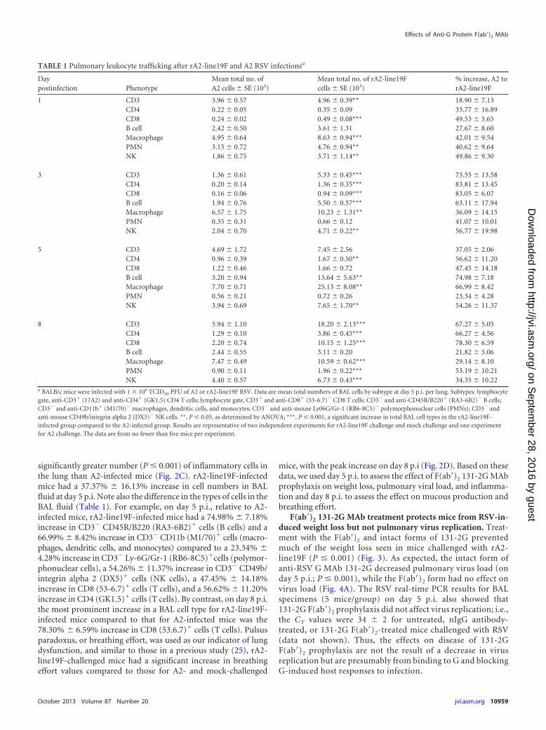
significantly greater number (P � 0.001) of inflammatory cells inthe lung than A2-infected mice (Fig. 2C). rA2-line19F-infectedmice had a 37.37% � 16.13% increase in cell numbers in BALfluid at day 5 p.i. Note also the difference in the types of cells in theBAL fluid (Table 1). For example, on day 5 p.i., relative to A2-infected mice, rA2-line19F-infected mice had a 74.98% � 7.18%increase in CD3� CD45R/B220 (RA3-6B2)� cells (B cells) and a66.99% � 8.42% increase in CD3� CD11b (M1/70)� cells (macro-phages, dendritic cells, and monocytes) compared to a 23.34% �4.28% increase in CD3� Ly-6G/Gr-1 (RB6-8C5)�cells (polymor-phonuclear cells), a 54.26% � 11.37% increase in CD3� CD49b/integrin alpha 2 (DX5)� cells (NK cells), a 47.45% � 14.18%increase in CD8 (53-6.7)� cells (T cells), and a 56.62% � 11.20%increase in CD4 (GK1.5)� cells (T cells). By contrast, on day 8 p.i.the most prominent increase in a BAL cell type for rA2-line19F-infected mice compared to that for A2-infected mice was the78.30% � 6.59% increase in CD8 (53.6.7)� cells (T cells). Pulsusparadoxus, or breathing effort, was used as our indicator of lungdysfunction, and similar to those in a previous study (25), rA2-line19F-challenged mice had a significant increase in breathingeffort values compared to those for A2- and mock-challenged
mice, with the peak increase on day 8 p.i (Fig. 2D). Based on thesedata, we used day 5 p.i. to assess the effect of F(ab=)2 131-2G MAbprophylaxis on weight loss, pulmonary viral load, and inflamma-tion and day 8 p.i. to assess the effect on mucous production andbreathing effort.
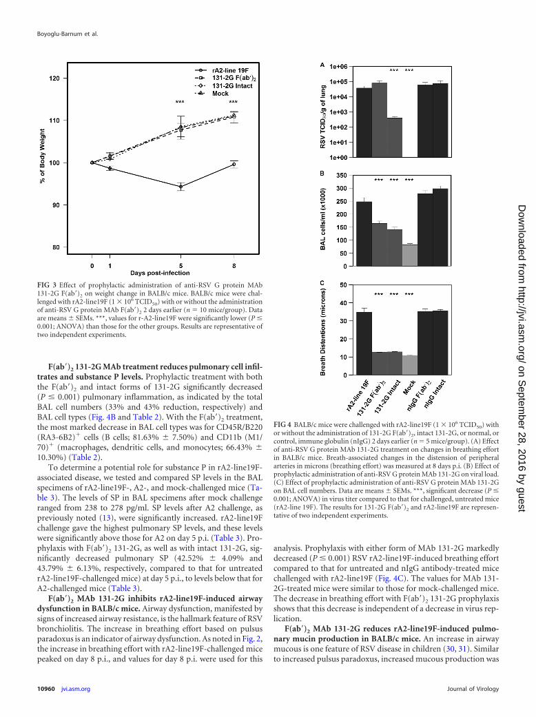
F(ab=)2 131-2G MAb treatment protects mice from RSV-in-duced weight loss but not pulmonary virus replication. Treat-ment with the F(ab=)2 and intact forms of 131-2G preventedmuch of the weight loss seen in mice challenged with rA2-line19F (P � 0.001) (Fig. 3). As expected, the intact form ofanti-RSV G MAb 131-2G decreased pulmonary virus load (onday 5 p.i.; P � 0.001), while the F(ab=)2 form had no effect onvirus load (Fig. 4A). The RSV real-time PCR results for BALspecimens (5 mice/group) on day 5 p.i. also showed that131-2G F(ab=)2 prophylaxis did not affect virus replication; i.e.,the CT values were 34 � 2 for untreated, nIgG antibody-treated, or 131-2G F(ab=)2-treated mice challenged with RSV(data not shown). Thus, the effects on disease of 131-2GF(ab=)2 prophylaxis are not the result of a decrease in virusreplication but are presumably from binding to G and blockingG-induced host responses to infection.
TABLE 1 Pulmonary leukocyte trafficking after rA2-line19F and A2 RSV infectionsa
Daypostinfection Phenotype
Mean total no. ofA2 cells � SE (103)
Mean total no. of rA2-line19Fcells � SE (103)
% increase, A2 torA2-line19F
1 CD3 3.96 � 0.57 4.96 � 0.39** 18.90 � 7.13CD4 0.22 � 0.05 0.35 � 0.09 33.77 � 16.89CD8 0.24 � 0.02 0.49 � 0.08*** 49.53 � 5.65B cell 2.42 � 0.50 3.61 � 1.31 27.67 � 8.60Macrophage 4.95 � 0.64 8.63 � 0.94*** 42.01 � 9.54PMN 3.15 � 0.72 4.76 � 0.94** 40.62 � 9.64NK 1.86 � 0.75 3.71 � 1.14** 49.86 � 9.30
3 CD3 1.36 � 0.61 5.33 � 0.45*** 73.55 � 13.58CD4 0.20 � 0.14 1.36 � 0.35*** 83.81 � 13.45CD8 0.16 � 0.06 0.94 � 0.09*** 83.05 � 6.07B cell 1.94 � 0.76 5.50 � 0.57*** 63.11 � 17.94Macrophage 6.57 � 1.75 10.23 � 1.31** 36.09 � 14.15PMN 0.35 � 0.31 0.66 � 0.12 41.07 � 10.01NK 2.04 � 0.70 4.71 � 0.22** 56.77 � 19.98
5 CD3 4.69 � 1.72 7.45 � 2.56 37.05 � 2.06CD4 0.96 � 0.39 1.67 � 0.50** 56.62 � 11.20CD8 1.22 � 0.46 1.66 � 0.72 47.45 � 14.18B cell 3.20 � 0.94 13.64 � 5.63** 74.98 � 7.18Macrophage 7.70 � 0.71 25.13 � 8.08** 66.99 � 8.42PMN 0.56 � 0.21 0.72 � 0.26 23.34 � 4.28NK 3.94 � 0.69 7.65 � 1.70** 54.26 � 11.37
8 CD3 5.94 � 1.10 18.20 � 2.13*** 67.27 � 5.05CD4 1.29 � 0.10 3.86 � 0.45*** 66.27 � 4.56CD8 2.20 � 0.74 10.15 � 1.25*** 78.30 � 6.59B cell 2.44 � 0.55 3.11 � 0.20 21.82 � 5.06Macrophage 7.47 � 0.49 10.59 � 0.62*** 29.14 � 8.10PMN 0.90 � 0.11 1.96 � 0.22*** 53.19 � 10.21NK 4.40 � 0.57 6.73 � 0.43*** 34.35 � 10.22
a BALB/c mice were infected with 1 � 106 TCID50 PFU of A2 or rA2-line19F RSV. Data are mean total numbers of BAL cells by subtype at day 5 p.i. per lung. Subtypes: lymphocytegate, anti-CD3� (17A2) and anti-CD4� (GK1.5) CD4 T cells; lymphocyte gate, CD3� and anti-CD8� (53-6.7)� CD8 T cells; CD3� and anti-CD45R/B220� (RA3-6B2)� B cells;CD3� and anti-CD11b� (M1/70)� macrophages, dendritic cells, and monocytes; CD3� and anti-mouse Ly06G/Gr-1 (RB6-8C5)� polymorphonuclear cells (PMNs); CD3� andanti-mouse CD49b/integrin alpha 2 (DX5)� NK cells. **, P � 0.05, as determined by ANOVA; ***, P � 0.001, a significant increase in total BAL cell types in the rA2-line19F-infected group compared to the A2-infected group. Results are representative of two independent experiments for rA2-line19F challenge and mock challenge and one experimentfor A2 challenge. The data are from no fewer than five mice per experiment.
Effects of Anti-G Protein F(ab=)2 MAb
October 2013 Volume 87 Number 20 jvi.asm.org 10959
on Septem
ber 28, 2016 by guesthttp://jvi.asm
.org/D
ownloaded from
F(ab=)2 131-2G MAb treatment reduces pulmonary cell infil-trates and substance P levels. Prophylactic treatment with boththe F(ab=)2 and intact forms of 131-2G significantly decreased(P � 0.001) pulmonary inflammation, as indicated by the totalBAL cell numbers (33% and 43% reduction, respectively) andBAL cell types (Fig. 4B and Table 2). With the F(ab=)2 treatment,the most marked decrease in BAL cell types was for CD45R/B220(RA3-6B2)� cells (B cells; 81.63% � 7.50%) and CD11b (M1/70)� (macrophages, dendritic cells, and monocytes; 66.43% �10.30%) (Table 2).
To determine a potential role for substance P in rA2-line19F-associated disease, we tested and compared SP levels in the BALspecimens of rA2-line19F-, A2-, and mock-challenged mice (Ta-ble 3). The levels of SP in BAL specimens after mock challengeranged from 238 to 278 pg/ml. SP levels after A2 challenge, aspreviously noted (13), were significantly increased. rA2-line19Fchallenge gave the highest pulmonary SP levels, and these levelswere significantly above those for A2 on day 5 p.i. (Table 3). Pro-phylaxis with F(ab=)2 131-2G, as well as with intact 131-2G, sig-nificantly decreased pulmonary SP (42.52% � 4.09% and43.79% � 6.13%, respectively, compared to that for untreatedrA2-line19F-challenged mice) at day 5 p.i., to levels below that forA2-challenged mice (Table 3).
F(ab=)2 MAb 131-2G inhibits rA2-line19F-induced airwaydysfunction in BALB/c mice. Airway dysfunction, manifested bysigns of increased airway resistance, is the hallmark feature of RSVbronchiolitis. The increase in breathing effort based on pulsusparadoxus is an indicator of airway dysfunction. As noted in Fig. 2,the increase in breathing effort with rA2-line19F-challenged micepeaked on day 8 p.i., and values for day 8 p.i. were used for this
analysis. Prophylaxis with either form of MAb 131-2G markedlydecreased (P � 0.001) RSV rA2-line19F-induced breathing effortcompared to that for untreated and nIgG antibody-treated micechallenged with rA2-line19F (Fig. 4C). The values for MAb 131-2G-treated mice were similar to those for mock-challenged mice.The decrease in breathing effort with F(ab=)2 131-2G prophylaxisshows that this decrease is independent of a decrease in virus rep-lication.
F(ab=)2 MAb 131-2G reduces rA2-line19F-induced pulmo-nary mucin production in BALB/c mice. An increase in airwaymucous is one feature of RSV disease in children (30, 31). Similarto increased pulsus paradoxus, increased mucous production was
FIG 3 Effect of prophylactic administration of anti-RSV G protein MAb131-2G F(ab=)2 on weight change in BALB/c mice. BALB/c mice were chal-lenged with rA2-line19F (1 � 106 TCID50) with or without the administrationof anti-RSV G protein MAb F(ab=)2 2 days earlier (n � 10 mice/group). Dataare means � SEMs. ***, values for r-A2-line19F were significantly lower (P �0.001; ANOVA) than those for the other groups. Results are representative oftwo independent experiments.
FIG 4 BALB/c mice were challenged with rA2-line19F (1 � 106 TCID50) withor without the administration of 131-2G F(ab=)2, intact 131-2G, or normal, orcontrol, immune globulin (nIgG) 2 days earlier (n � 5 mice/group). (A) Effectof anti-RSV G protein MAb 131-2G treatment on changes in breathing effortin BALB/c mice. Breath-associated changes in the distension of peripheralarteries in microns (breathing effort) was measured at 8 days p.i. (B) Effect ofprophylactic administration of anti-RSV G protein MAb 131-2G on viral load.(C) Effect of prophylactic administration of anti-RSV G protein MAb 131-2Gon BAL cell numbers. Data are means � SEMs. ***, significant decrease (P �0.001; ANOVA) in virus titer compared to that for challenged, untreated mice(rA2-line 19F). The results for 131-2G F(ab=)2 and rA2-line19F are represen-tative of two independent experiments.
Boyoglu-Barnum et al.
10960 jvi.asm.org Journal of Virology
on Septem
ber 28, 2016 by guesthttp://jvi.asm
.org/D
ownloaded from
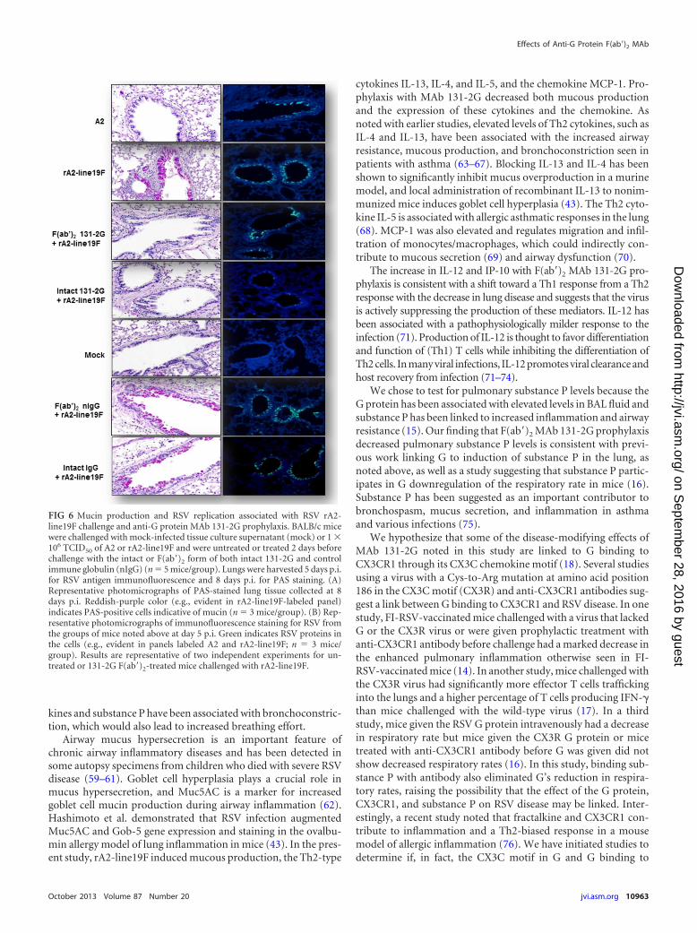
documented 8 days after challenge, and values for day 8 were usedfor these analyses. There was an increase in Muc5AC protein ex-pression in the lungs of mice challenged with rA2-line19F but notin the lungs of A2-challenged mice (Fig. 5A), which was similar tothe results of other studies (25). In a fashion similar to that forincreased pulsus paradoxus, prophylactic treatment with both theF(ab=)2 and intact forms of 131-2G significantly decreased (P �0.001) Muc5AC levels compared to those in untreated and nIgGantibody-treated mice challenged with rA2-line19F (Fig. 5B). Theability of F(ab=)2 131-2G to so effectively decrease mucin produc-tion shows that this effect is also independent of decreasing virusreplication.
Differences in pulmonary mucous production between A2 andrA2-line19F infections and between treatment with either form ofMAb 131-2G and no treatment before rA2-line19F infection wereevident in lung histopathology studies (Fig. 6A). Lung sectionsharvested 8 days p.i. showed PAS (a staining method used to de-tect intracellular pulmonary mucin levels)-stained cells for un-
treated, or control MAb-treated, and rA2-line19F-challengedmice. A2-challenged mice and MAb 131-2G-treated, rA2-line19F-challenged mice did not show PAS-stained cells. As expected, A2-challenged mice and rA2-line19F-challenged mice treated withthe F(ab=)2 form of 131-2G were positive for RSV antigens byimmunofluorescent staining (Fig. 6B).
Immunohistochemical staining (IHC) for Muc5AC productionanalyzed using ImageJ software (see Materials and Methods) wasused to quantitate differences between treatment groups. Consistentwith PAS-staining results, the percentages of Muc5AC-positivestained areas in the total epithelial area were 32.8% after rA2-line19Fchallenge, 13.2% after A2 challenge, 9.6% after mock challenge, and10.8% and 11.6% for rA2-line19F-challenged mice given prophylaxiswith intact and F(ab=)2 forms of MAb 131-2G.
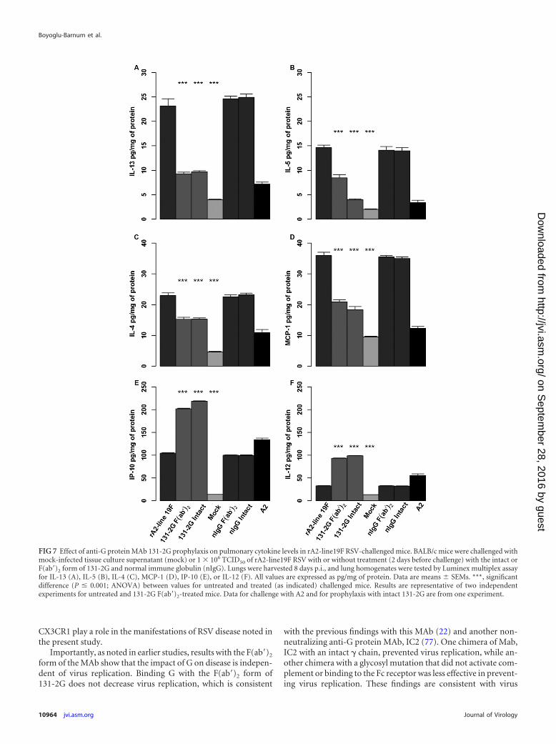
Cytokine responses. To address a potential mechanism for thedecrease in pulsus paradoxus and mucin production after prophy-laxis with the anti-RSV G protein MAb 131-2G, we determinedthe levels of pulmonary cytokines and chemokines in lung ho-mogenates. As noted with earlier studies, elevated levels of Th2cytokines, such as IL-4, IL-13, IL-5, and MCP-1, have been asso-ciated with the increased airway resistance, mucous production,and bronchoconstriction seen in patients with asthma. At 8 daysp.i., significant increases (P � 0.001) in the levels of IL-4, IL-5,IL-13, and MCP-1 were detected in untreated, nIgG antibody-treated, and untreated rA2-line19F-challenged mice (Fig. 7).These increases were significantly lower in rA2-line19F-chal-lenged mice treated with either form of MAb 131-2G and in A2-challenged mice. In addition, substantially higher levels of IP-10and IL-12 were detected in lung homogenates after prophylaxiswith either form of the 131-2G MAb than in untreated or nIgGantibody-treated, rA2-line19F-challenged mice. A2-challengedmice also had higher levels of IL-12 and IP-10 (Fig. 7).
DISCUSSION
Development of an effective vaccine or treatment for RSV infec-tion has not yet been achieved, though passive administration ofan immune globulin with high titers of RSV antibodies or a neu-tralizing MAb is effective when given prophylactically. One possi-ble explanation for success with antibody prophylaxis (4) and lim-ited success with treatment of active infection could be that thevirus-induced host inflammatory response is important to the dis-
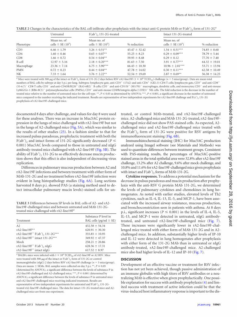
TABLE 2 Changes in the characteristics of the BAL cell infiltrate after prophylaxis with the intact anti-G protein MAb or F(ab=)2 form of 131-2Ga
Phenotype
Untreated F(ab=)2 131-2G-treated Intact 131-2G-treated
Mean no. ofcells � SE (103)
Mean no. ofcells � SE (103) % reduction
Mean no. ofcells � SE (103) % reduction
CD3 6.46 � 1.79 3.26 � 0.51** 43.47 � 32.42 1.54 � 0.51*** 74.85 � 8.60CD4 1.60 � 0.46 0.65 � 0.07** 55.25 � 8.23 0.29 � 0.09*** 78.79 � 5.44CD8 1.66 � 0.72 0.54 � 0.04** 59.95 � 8.48 0.29 � 0.12 77.70 � 7.40B cell 12.97 � 5.16 2.18 � 0.20*** 81.63 � 7.50 3.91 � 0.77*** 64.32 � 19.01Macrophage 23.36 � 7.14 6.75 � 3.90*** 66.43 � 10.30 10.96 � 2.01*** 53.71 � 13.94PMN 0.72 � 0.23 0.44 � 0.04** 47.78 � 14.02 0.30 � 0.11*** 62.58 � 10.49NK 7.33 � 1.64 3.76 � 2.22** 52.54 � 19.69 2.87 � 0.69** 56.38 � 14.25a Mice were treated with 300 �g of the intact or F(ab=)2 form of 131-2G 2 days before RSV rA2-line19F (1 � 106 TCID50) challenge (n � 5 mice/group). Data are mean totalnumbers of BAL cells by subtype at day 5 p.i. per lung. Subtypes: lymphocyte gate, anti-CD3� (17A2) and anti-CD4� (GK1.5) CD4 T cells; lymphocyte gate, CD3� and anti-CD8�
(53-6.7)� CD8 T cells; CD3� and anti-CD45R/B220� (RA3-6B2)� B cells; CD3� and anti-CD11b� (M1/70)� macrophages, dendritic cells, and monocytes; CD3� and anti-mouseLy06G/Gr-1 (RB6-8C5)� polymorphonuclear cells (PMNs); CD3� and anti-mouse CD49b/integrin alpha 2 (DX5)� NK cells. The fold reduction is the decrease in the number oftreated mice relative to the number of untreated mice for the cell type. **, P � 0.05 as determined by ANOVA; ***, P � 0.001, a significant decrease in the number of untreatedmice compared to the number receiving the indicated treatment. Results are representative of two independent experiments for rA2-line19F challenge and F(a=)2 131-2Gprophylaxis of rA2-line19F-challenged mice.
TABLE 3 Differences between SP levels in BAL cells of A2- and rA2-line19F-challenged mice and between untreated and MAb 131-2G-treated mice challenged with rA2-line19Fa
TreatmentSubstance P level inBAL cells (pg/ml � SE)
A2 432.58 � 54.34rA2-line19F** 620.91 � 30.30rA2-line19F� F(ab=)2 131-2G*** 355.85 � 10.95rA2-line19F� intact 131-2G*** 349.92 � 47.37Mock 258.21 � 20.86rA2-line19F� F(ab=)2 nIgG 628.36 � 17.31rA2-line19F� intact nIgG 617.52 � 8.97a BALB/c mice were infected with 1 � 106 TCID50 of rA2-line19F or A2 RSV. Micewere treated with 300 �g of the intact or F(ab=)2 form of 131-2G or controlimmunoglobulin (nIgG) 2 days before RSV rA2-line19F challenge (n � 5 mice/group).Data are means � SEMs. BAL samples were collected on day 5 p.i. **, P � 0.05(determined by ANOVA), a significant difference between the levels of substance P inrA2-line19F-challenged and A2-challenged mice; ***, P � 0.001 (determined byANOVA), a significant difference between the levels of substance P in untreated miceand rA2-line19F-challenged mice receiving indicated treatment. Results arerepresentative of two independent experiments for untreated and F(ab=)2 131-2G-treated rA2-line19F-challenged mice. The data for intact-131-2G-treated mice and A2-challenged mice are from one experiment.
Effects of Anti-G Protein F(ab=)2 MAb
October 2013 Volume 87 Number 20 jvi.asm.org 10961
on Septem
ber 28, 2016 by guesthttp://jvi.asm
.org/D
ownloaded from

ease process and is only partially responsive to stopping replica-tion once infection is established (39, 40). Consequently, effectivetreatment of active infection may require both anti-inflammatoryand antiviral components. RSV vaccines or prophylaxis may alsobe more effective if they have both anti-inflammatory and antivi-ral effects. There is increasing evidence for RSV G protein being animportant contributor to RSV inflammation and disease patho-genesis, and thus it is a potential target for the anti-inflammatorycomponent of vaccine and antiviral drug development strategies(22, 23, 41–44).
Since immune prophylaxis with palivizumab is about 50% ef-fective in decreasing RSV hospitalizations (4), it is possible thatbinding G and blocking G-associated disease might improve theeffectiveness of immune prophylaxis. The RSV G protein, G pep-tides, and secreted G have been associated with a number of im-mune and inflammatory responses. For example, vaccinationwith intact or secreted G and several peptides in or proximal to thecentral nonglycosylated region of G induce a Th2-biased memoryresponse resulting in increased pulmonary inflammation and eo-sinophilia after RSV challenge (45–50). In other studies, the Gprotein has been associated with suppressing some immune re-sponses, such as Toll-like receptor 3 (TLR3) or TLR4 induction ofIFN-� (48), proinflammatory responses of lung epithelial cells(51), lymphoproliferation (52), and a number of other innate re-sponses, including activation of dendritic cells (53, 54), enhancedcytotoxic T cell responses (55), and downregulation of type I IFNproduction by inducing suppression of cytokine signaling (SOCS)(56). The G protein has also been associated with depression of therespiratory rate (16), increased production of pulmonary sub-stance P (15), and antibody decoy activity (57).
Earlier studies showed that binding the G protein with MAb
131-2G prevented pulmonary inflammation after primary infec-tion, pulmonary inflammation, and eosinophilia in FI-RSV-vac-cinated mice and weight loss after primary infection or infectionin FI-RSV-vaccinated mice (21–23). The availability of the rA2-line19F virus allowed us to demonstrate that MAb 131-2G pro-phylaxis also decreases breathing effort and pulmonary mucousproduction. These findings suggest additional clinical benefits oftreatment or prophylaxis with this or a comparable anti-G proteinMAb. These findings also expand the possible contributions of theG protein to the pathogenesis of RSV disease.
The potential importance of these findings to human RSV dis-ease is highlighted by wheezing, often resulting in a diagnosis ofbronchiolitis, being the hallmark clinical feature of RSV infectionin children (32, 58). Wheezing is associated with increased airwayresistance, which can result from bronchoconstriction or pluggingof the airways with sloughed airway epithelial and inflammatorycells, other debris, and/or mucous. With rA2-line19F, we see in-creased pulsus paradoxus indicative of increased breathing effort,which indicates airway dysfunction. The data in this report anddata from Moore et al. (25) are consistent with one or severalmechanisms causing the increased breathing effort; i.e., infectionwith rA2-line19F is associated with the increased pulmonary in-flammation, Th2 cytokine levels, and mucous production thatparallel the increase in breathing effort. In addition, the data inthis report show a decrease in breathing effort with F(ab=)2 MAb131-2G treatment that correlates with decreases in BAL cell infil-trates (Fig. 5 and Table 2), pulmonary mucous production (Fig.6), pulmonary Th2 cytokines (Fig. 7), and substance P levels (Ta-ble 3). The cellular infiltration and/or mucous production canplug airways, leading to increased breathing effort, and Th2 cyto-
FIG 5 Effect of different virus strains and anti-RSV G protein MAb 131-2G treatment on mucin production in BALB/c mice. (A) BALB/c mice were challengedwith mock-infected tissue culture supernatant (mock) or 1 � 106 TCID50 of rA2-line19F or A2 RSV(n � 5mice/group). Lungs were harvested 8 days p.i., and lunghomogenates were tested for Muc5AC levels by ELISA. Data are means � SEMs. ***, significant difference (P � 0.001; ANOVA) between values for rA2-line19F-challenged mice and other indicated groups. (B) BALB/c mice were treated 2 days before rA2-line19F challenge with the intact or F(ab=)2 form of either 131-2Gor control antibody (nIgG) (n � 5mice/group). Lungs were harvested 8 days p.i., and lung homogenates were tested for Muc5AC levels. Data are means � SEMs.***, significant difference (P � 0.001; ANOVA) between values for untreated rA2-line19F-challenged mice and other indicated groups. Results are representativeof two independent experiments for untreated and F(ab=)2 131-2G-treated mice challenged with rA2-line19F. Data for challenge with A2 and for prophylaxis withintact 131-2G are from one experiment.
Boyoglu-Barnum et al.
10962 jvi.asm.org Journal of Virology
on Septem
ber 28, 2016 by guesthttp://jvi.asm
.org/D
ownloaded from
kines and substance P have been associated with bronchoconstric-tion, which would also lead to increased breathing effort.
Airway mucus hypersecretion is an important feature ofchronic airway inflammatory diseases and has been detected insome autopsy specimens from children who died with severe RSVdisease (59–61). Goblet cell hyperplasia plays a crucial role inmucus hypersecretion, and Muc5AC is a marker for increasedgoblet cell mucin production during airway inflammation (62).Hashimoto et al. demonstrated that RSV infection augmentedMuc5AC and Gob-5 gene expression and staining in the ovalbu-min allergy model of lung inflammation in mice (43). In the pres-ent study, rA2-line19F induced mucous production, the Th2-type
cytokines IL-13, IL-4, and IL-5, and the chemokine MCP-1. Pro-phylaxis with MAb 131-2G decreased both mucous productionand the expression of these cytokines and the chemokine. Asnoted with earlier studies, elevated levels of Th2 cytokines, such asIL-4 and IL-13, have been associated with the increased airwayresistance, mucous production, and bronchoconstriction seen inpatients with asthma (63–67). Blocking IL-13 and IL-4 has beenshown to significantly inhibit mucus overproduction in a murinemodel, and local administration of recombinant IL-13 to nonim-munized mice induces goblet cell hyperplasia (43). The Th2 cyto-kine IL-5 is associated with allergic asthmatic responses in the lung(68). MCP-1 was also elevated and regulates migration and infil-tration of monocytes/macrophages, which could indirectly con-tribute to mucous secretion (69) and airway dysfunction (70).
The increase in IL-12 and IP-10 with F(ab=)2 MAb 131-2G pro-phylaxis is consistent with a shift toward a Th1 response from a Th2response with the decrease in lung disease and suggests that the virusis actively suppressing the production of these mediators. IL-12 hasbeen associated with a pathophysiologically milder response to theinfection (71). Production of IL-12 is thought to favor differentiationand function of (Th1) T cells while inhibiting the differentiation ofTh2 cells. In many viral infections, IL-12 promotes viral clearance andhost recovery from infection (71–74).
We chose to test for pulmonary substance P levels because theG protein has been associated with elevated levels in BAL fluid andsubstance P has been linked to increased inflammation and airwayresistance (15). Our finding that F(ab=)2 MAb 131-2G prophylaxisdecreased pulmonary substance P levels is consistent with previ-ous work linking G to induction of substance P in the lung, asnoted above, as well as a study suggesting that substance P partic-ipates in G downregulation of the respiratory rate in mice (16).Substance P has been suggested as an important contributor tobronchospasm, mucus secretion, and inflammation in asthmaand various infections (75).
We hypothesize that some of the disease-modifying effects ofMAb 131-2G noted in this study are linked to G binding toCX3CR1 through its CX3C chemokine motif (18). Several studiesusing a virus with a Cys-to-Arg mutation at amino acid position186 in the CX3C motif (CX3R) and anti-CX3CR1 antibodies sug-gest a link between G binding to CX3CR1 and RSV disease. In onestudy, FI-RSV-vaccinated mice challenged with a virus that lackedG or the CX3R virus or were given prophylactic treatment withanti-CX3CR1 antibody before challenge had a marked decrease inthe enhanced pulmonary inflammation otherwise seen in FI-RSV-vaccinated mice (14). In another study, mice challenged withthe CX3R virus had significantly more effector T cells traffickinginto the lungs and a higher percentage of T cells producing IFN-�than mice challenged with the wild-type virus (17). In a thirdstudy, mice given the RSV G protein intravenously had a decreasein respiratory rate but mice given the CX3R G protein or micetreated with anti-CX3CR1 antibody before G was given did notshow decreased respiratory rates (16). In this study, binding sub-stance P with antibody also eliminated G’s reduction in respira-tory rates, raising the possibility that the effect of the G protein,CX3CR1, and substance P on RSV disease may be linked. Inter-estingly, a recent study noted that fractalkine and CX3CR1 con-tribute to inflammation and a Th2-biased response in a mousemodel of allergic inflammation (76). We have initiated studies todetermine if, in fact, the CX3C motif in G and G binding to
FIG 6 Mucin production and RSV replication associated with RSV rA2-line19F challenge and anti-G protein MAb 131-2G prophylaxis. BALB/c micewere challenged with mock-infected tissue culture supernatant (mock) or 1 �106 TCID50 of A2 or rA2-line19F and were untreated or treated 2 days beforechallenge with the intact or F(ab=)2 form of both intact 131-2G and controlimmune globulin (nIgG) (n � 5 mice/group). Lungs were harvested 5 days p.i.for RSV antigen immunofluorescence and 8 days p.i. for PAS staining. (A)Representative photomicrographs of PAS-stained lung tissue collected at 8days p.i. Reddish-purple color (e.g., evident in rA2-line19F-labeled panel)indicates PAS-positive cells indicative of mucin (n � 3 mice/group). (B) Rep-resentative photomicrographs of immunofluorescence staining for RSV fromthe groups of mice noted above at day 5 p.i. Green indicates RSV proteins inthe cells (e.g., evident in panels labeled A2 and rA2-line19F; n � 3 mice/group). Results are representative of two independent experiments for un-treated or 131-2G F(ab=)2-treated mice challenged with rA2-line19F.
Effects of Anti-G Protein F(ab=)2 MAb
October 2013 Volume 87 Number 20 jvi.asm.org 10963
on Septem
ber 28, 2016 by guesthttp://jvi.asm
.org/D
ownloaded from
CX3CR1 play a role in the manifestations of RSV disease noted inthe present study.
Importantly, as noted in earlier studies, results with the F(ab=)2
form of the MAb show that the impact of G on disease is indepen-dent of virus replication. Binding G with the F(ab=)2 form of131-2G does not decrease virus replication, which is consistent
with the previous findings with this MAb (22) and another non-neutralizing anti-G protein MAb, IC2 (77). One chimera of Mab,IC2 with an intact � chain, prevented virus replication, while an-other chimera with a glycosyl mutation that did not activate com-plement or binding to the Fc receptor was less effective in prevent-ing virus replication. These findings are consistent with virus
FIG 7 Effect of anti-G protein MAb 131-2G prophylaxis on pulmonary cytokine levels in rA2-line19F RSV-challenged mice. BALB/c mice were challenged withmock-infected tissue culture supernatant (mock) or 1 � 106 TCID50 of rA2-line19F RSV with or without treatment (2 days before challenge) with the intact orF(ab=)2 form of 131-2G and normal immune globulin (nIgG). Lungs were harvested 8 days p.i., and lung homogenates were tested by Luminex multiplex assayfor IL-13 (A), IL-5 (B), IL-4 (C), MCP-1 (D), IP-10 (E), or IL-12 (F). All values are expressed as pg/mg of protein. Data are means � SEMs. ***, significantdifference (P � 0.001; ANOVA) between values for untreated and treated (as indicated) challenged mice. Results are representative of two independentexperiments for untreated and 131-2G F(ab=)2-treated mice. Data for challenge with A2 and for prophylaxis with intact 131-2G are from one experiment.
Boyoglu-Barnum et al.
10964 jvi.asm.org Journal of Virology
on Septem
ber 28, 2016 by guesthttp://jvi.asm
.org/D
ownloaded from
neutralization by antibody-dependent cell-mediated cytotoxicity(ADCC)-dependent or complement-mediated mechanisms. In-tact 131-2G, as noted in the present study and previous studies(22, 23), effectively neutralizes RSV in vivo and thus in the mouseimpacts RSV disease both by downregulating G-induced inflam-mation and immune modulation and by decreasing virus replica-tion. If it impacts both in humans, it might prove more effective indecreasing disease than antiviral treatment that only decreases vi-rus replication.
The ability of an anti-G protein MAb to so effectively decreasepulmonary mucous production and breathing effort is somewhatsurprising, since the difference between RSV A2 and rA2-line19Fis five amino acid changes in the F protein and no differences inthe G protein. Consequently, both proteins must be involved inrA2-line19F infection-associated mucous production and airwayreactivity. The G and F proteins do interact during infection, andthis may explain a joint role in these manifestations of infection(78). Alternatively, the role of each may have independent effectsthat in concert result in mucous overproduction and increasedbreathing effort. We are beginning studies to better understandthe role of F and G in these manifestations of RSV disease.
Two limitations need to be considered in interpreting the datapresented in the report. First, pulsus paradoxus, or breathing ef-fort, is an indication of airway dysfunction but does not indicatethe location of this dysfunction. Previous work, however, hasshown that increases in pulsus paradoxus, or breathing effort, inthe mouse do correspond to increases in airway hyperresponsive-ness (AHR) in mice challenged with RSV rA2-line19F. AHR wasmeasured using a small animal ventilator and methacholine chal-lenge (38), and the correspondence between this measurementand increased breathing effort supports that breathing effort inthis model indicates dysfunction in the lung. We chose to usebreathing effort because it is less invasive than measuring AHRand it allows serial measures in the same mouse. Second, it isimportant to recognize that findings in mice may, or may not,apply to humans. Thus, although these data suggest a substantialrole for the G protein in the pathogenesis of human RSV disease,this suggestion needs to be confirmed by studies with humans.
Together, the data presented in this report identify additionalroles for the RSV G protein in disease pathogenesis, i.e., mucousproduction and airway dysfunction. These results and the resultsof other studies of G suggest that understanding G’s role in thepathogenesis of disease is likely essential to understanding RSVdisease and likely important in the approach to its treatment andprevention. Finally, if G’s impact on human RSV disease is similarto its impact on disease in mice, binding G with a MAb or antiviraldrug with activities similar to those of 131-2G MAb could im-prove the effectiveness of RSV prophylaxis and/or treatment. Thedata also suggest that inducing antibodies with activity similar tothat of MAb 131-2G might improve a vaccine’s ability to preventdisease with later RSV infection.
ACKNOWLEDGMENTS
This work was supported by NIH grant 1U19AI095227 awarded to M.L.M.and L.J.A., funding from Children’s Healthcare of Atlanta, support from theImmunology Core of Emory-Children’s Pediatric Research Center, and Em-ory Vaccinology Training Grant (VTP) T32 5T32AI074492-03. Martin L.Moore was supported by NIH 1R01AI087798 and NIH 1U19AI095227.
We thank Carla J. Shoffeitt for histology slide preparations and Sica L.Gabriel for histology slide analysis.
The findings and conclusions in this report are those of the authorsand do not necessarily represent the views of the Centers for DiseaseControl and Prevention.
REFERENCES1. Nair H, Nokes DJ, Gessner BD, Dherani M, Madhi SA, Singleton RJ,
O’Brien KL, Roca A, Wright PF, Bruce N, Chandran A, Theodoratou E,Sutanto A, Sedyaningsih ER, Ngama M, Munywoki PK, KartasasmitaC, Simoes EA, Rudan I, Weber MW, Campbell H. 2010. Global burdenof acute lower respiratory infections due to respiratory syncytial virus inyoung children: a systematic review and meta-analysis. Lancet 375:1545–1555.
2. Groothuis JR, Simoes E, Levin MJ, Hall CB, Long CE, Rodriguez WJ,Arrobio J, Meissner HC, Fulton DR, Welliver RC, Tristram DA, SiberGR, Prince GA, Van Raden M, Hemming VG. 1993. Prophylacticadministration of respiratory syncytial virus immune globulin to high-risk infants and young children. Respiratory Syncytial Virus ImmuneGlobulin Study Group. N. Engl. J. Med. 329:1524 –1530.
3. IMpact-RSV Study Group. 1998. Palivizumab, a humanized respiratorysyncytial virus monoclonal antibody, reduces hospitalization from respi-ratory syncytial virus infection in high-risk infants. Pediatrics 102:531–537.
4. Geevarghese B, Simões EA. 2012. Antibodies for prevention and treat-ment of respiratory syncytial virus infections in children. Antivir. Ther.17:201–211.
5. Siegel JD, Rhinehart E, Jackson M, Chiarello L. 2007. 2007 Guideline forisolation precautions: preventing transmission of infectious agents inhealth care settings. Am. J. Infect. Control 35(Suppl 2):S65–S164.
6. Collins PL, Melero JA. 2011. Progress in understanding and controllingrespiratory syncytial virus: still crazy after all these years. Virus Res. 162:80 –99.
7. Graham BS. 2011. Biological challenges and technological opportunitiesfor respiratory syncytial virus vaccine development. Immunol. Rev. 239:149 –166.
8. Connors M, Collins PL, Firestone CY, Murphy BR. 1991. Respiratorysyncytial virus (RSV) F, G, M2 (22K), and N proteins each induce resis-tance to RSV challenge, but resistance induced by M2 and N proteins isrelatively short-lived. J. Virol. 65:1634 –1637.
9. Olmsted RA, Elango N, Prince GA, Murphy BR, Johnson PR, Moss B,Chanock RM, Collins PL. 1986. Expression of the F glycoprotein ofrespiratory syncytial virus by a recombinant vaccinia virus: comparison ofthe individual contributions of the F and G glycoproteins to host immu-nity. Proc. Natl. Acad. Sci. U. S. A. 83:7462–7466.
10. Stott EJ, Taylor G, Ball LA, Anderson K, Young KK, King AM, WertzGW. 1987. Immune and histopathological responses in animals vacci-nated with recombinant vaccinia viruses that express individual genes ofhuman respiratory syncytial virus. J. Virol. 61:3855–3861.
11. Tripp RA. 2004. Pathogenesis of respiratory syncytial virus infection.Viral Immunol. 17:165–181.
12. Tripp RA, Moore D, Jones L, Sullender W, Winter J, Anderson LJ. 1999.Respiratory syncytial virus G and/or SH protein alters Th1 cytokines, nat-ural killer cells, and neutrophils responding to pulmonary infection inBALB/c mice. J. Virol. 73:7099 –7107.
13. Tripp RA, Jones L, Anderson LJ. 2000. Respiratory syncytial virus Gand/or SH glycoproteins modify CC and CXC chemokine mRNA expres-sion in the BALB/c mouse. J. Virol. 74:6227– 6229.
14. Haynes LM, Jones LP, Barskey A, Anderson LJ, Tripp RA. 2003.Enhanced disease and pulmonary eosinophilia associated with formalin-inactivated respiratory syncytial virus vaccination are linked to G glyco-protein CX3C-CX3CR1 interaction and expression of substance. P. J. Vi-rol. 77:9831–9844.
15. Tripp RA, Moore D, Winter J, Anderson LJ. 2000. Respiratory syncytialvirus infection and G and/or SH protein expression contribute to sub-stance P, which mediates inflammation and enhanced pulmonary diseasein BALB/c mice. J. Virol. 74:1614 –1622.
16. Tripp RA, Dakhama A, Jones LP, Barskey A, Gelfand EW, Anderson LJ.2003. The G glycoprotein of respiratory syncytial virus depresses respira-tory rates through the CX3C motif and substance. P. J. Virol. 77:6580 –6584.
17. Harcourt J, Alvarez R, Jones LP, Henderson C, Anderson LJ, Tripp RA.2006. Respiratory syncytial virus G protein and G protein CX3C motifadversely affect CX3CR1� T cell responses. J. Immunol. 176:1600 –1608.
Effects of Anti-G Protein F(ab=)2 MAb
October 2013 Volume 87 Number 20 jvi.asm.org 10965
on Septem
ber 28, 2016 by guesthttp://jvi.asm
.org/D
ownloaded from
18. Tripp RA, Jones LP, Haynes LM, Zheng H, Murphy PM, Anderson LJ.2001. CX3C chemokine mimicry by respiratory syncytial virus G glyco-protein. Nat. Immunol. 2:732–738.
19. Anderson LJ, Hierholzer JC, Stone YO, Tsou C, Fernie BF. 1986.Identification of epitopes on respiratory syncytial virus proteins by com-petitive binding immunoassay. J. Clin. Microbiol. 23:475– 480.
20. Anderson LJ, Bingham P, Hierholzer JC. 1988. Neutralization of respi-ratory syncytial virus by individual and mixtures of F and G proteinmonoclonal antibodies. J. Virol. 62:4232– 4238.
21. Haynes LM, Caidi H, Radu GU, Miao C, Harcourt JL, Tripp RA,Anderson LJ. 2009. Therapeutic monoclonal antibody treatment target-ing respiratory syncytial virus (RSV) G protein mediates viral clearanceand reduces the pathogenesis of RSV infection in BALB/c mice. J. Infect.Dis. 200:439 – 447.
22. Miao C, Radu GU, Caidi H, Tripp RA, Anderson LJ, Haynes LM. 2009.Treatment with respiratory syncytial virus G glycoprotein monoclonalantibody or F(ab=)2 components mediates reduced pulmonary inflamma-tion in mice. J. Gen. Virol. 90:1119 –1123.
23. Radu GU, Caidi H, Miao C, Tripp RA, Anderson LJ, Haynes LM. 2010.Prophylactic treatment with a G glycoprotein monoclonal antibody re-duces pulmonary inflammation in respiratory syncytial virus (RSV)-challenged naive and formalin-inactivated RSV-immunized BALB/cmice. J. Virol. 84:9632–9636.
24. Lukacs NW, Moore ML, Rudd BD, Berlin AA, Collins RD, Olson SJ, HoSB, Peebles RS, Jr. 2006. Differential immune responses and pulmonarypathophysiology are induced by two different strains of respiratory syn-cytial virus. Am. J. Pathol. 169:977–986.
25. Moore ML, Chi MH, Luongo C, Lukacs NW, Polosukhin VV, HuckabeeMM, Newcomb DC, Buchholz UJ, Crowe JE, Jr, Goleniewska K, Wil-liams JV, Collins PL, Peebles RS, Jr. 2009. A chimeric A2 strain ofrespiratory syncytial virus (RSV) with the fusion protein of RSV strain line19 exhibits enhanced viral load, mucus, and airway dysfunction. J. Virol.83:4185– 4194.
26. Hershey GK. 2003. IL-13 receptors and signaling pathways: an evolvingweb. J. Allergy Clin. Immunol. 111:677– 690.
27. Walter DM, McIntire JJ, Berry G, McKenzie AN, Donaldson DD,DeKruyff RH, Umetsu DT. 2001. Critical role for IL-13 in the develop-ment of allergen-induced airway hyperreactivity. J. Immunol. 167:4668 –4675.
28. Zhu Z, Homer RJ, Wang Z, Chen Q, Geba GP, Wang J, Zhang Y, EliasJA. 1999. Pulmonary expression of interleukin-13 causes inflammation,mucus hypersecretion, subepithelial fibrosis, physiologic abnormalities,and eotaxin production. J. Clin. Invest. 103:779 –788.
29. de Waal L, Koopman LP, van Benten IJ, Brandenburg AH, Mulder PG,de Swart RL, Fokkens WJ, Neijens HJ, Osterhaus AD. 2003. Moderatelocal and systemic respiratory syncytial virus-specific T-cell responsesupon mild or subclinical RSV infection. J. Med. Virol. 70:309 –318.
30. Lugo RA, Nahata MC. 1993. Pathogenesis and treatment of bronchiolitis.Clin. Pharm. 12:95–116.
31. Quinn SF, Erickson S, Oshman D, Hayden F. 1985. Lobar collapse withrespiratory syncytial virus pneumonitis. Pediatr. Radiol. 15:229 –230.
32. Hall CB, Weinberg GA, Iwane MK, Blumkin AK, Edwards KM, StaatMA, Auinger P, Griffin MR, Poehling KA, Erdman D, Grijalva CG, ZhuY, Szilagyi P. 2009. The burden of respiratory syncytial virus infection inyoung children. N. Engl. J. Med. 360:588 –598.
33. Moore ML, Peebles RS, Jr. 2006. Respiratory syncytial virus diseasemechanisms implicated by human, animal model, and in vitro data facil-itate vaccine strategies and new therapeutics. Pharmacol. Ther. 112:405–424.
34. Anderson LJ, Hierholzer JC, Bingham PG, Stone YO. 1985. Microneu-tralization test for respiratory syncytial virus based on an enzyme immu-noassay. J. Clin. Microbiol. 22:1050 –1052.
35. Reed LJ, Muench H. 1938. A simple method of estimating fifty percentendpoints. Amer. J. Hyg. 27:493– 497.
36. Kodani M, Yang G, Conklin LM, Travis TC, Whitney CG, Anderson LJ,Schrag SJ, Taylor TH, Jr, Beall BW, Breiman RF, Feikin DR, NjengaMK, Mayer LW, Oberste MS, Tondella ML, Winchell JM, LindstromSL, Erdman DD, Fields BS. 2011. Application of TaqMan low-densityarrays for simultaneous detection of multiple respiratory pathogens. J.Clin. Microbiol. 49:2175–2182.
37. Tripp RA, Barskey A, Goss L, Anderson LJ. 2002. Substance P receptorexpression on lymphocytes is associated with the immune response torespiratory syncytial virus infection. J. Neuroimmunol. 129:141–153.
38. Stokes KL, Chi MH, Sakamoto K, Newcomb DC, Currier MG, Hucka-bee MM, Lee S, Goleniewska K, Pretto C, Williams JV, Hotard A,Sherrill TP, Peebles RS, Jr, Moore ML. 2011. Differential pathogenesis ofrespiratory syncytial virus clinical isolates in BALB/c mice. J. Virol. 85:5782–5793.
39. Empey KM, Peebles RS, Kolls JK. 2010. Pharmacologic advances in thetreatment and prevention of respiratory syncytial virus. Clin. Infect. Dis.50:1258 –1267.
40. Ottolini MG, Curtis SJ, Porter DD, Mathews A, Richardson JY, Hem-ming VG, Prince GA. 2002. Comparison of corticosteroids for treatmentof respiratory syncytial virus bronchiolitis and pneumonia in cotton rats.Antimicrob. Agents Chemother. 46:2299 –2302.
41. Andersson C, Liljestrom P, Stahl S, Power UF. 2000. Protection againstrespiratory syncytial virus (RSV) elicited in mice by plasmid DNA im-munisation encoding a secreted RSV G protein-derived antigen. FEMSImmunol. Med. Microbiol. 29:247–253.
42. Collarini EJ, Lee FE, Foord O, Park M, Sperinde G, Wu H, HarrimanWD, Carroll SF, Ellsworth SL, Anderson LJ, Tripp RA, Walsh EE, KeytBA, Kauvar LM. 2009. Potent high-affinity antibodies for treatment andprophylaxis of respiratory syncytial virus derived from B cells of infectedpatients. J. Immunol. 183:6338 – 6345.
43. Hashimoto K, Graham BS, Ho SB, Adler KB, Collins RD, Olson SJ,Zhou W, Suzutani T, Jones PW, Goleniewska K, O’Neal JF, Peebles RS,Jr. 2004. Respiratory syncytial virus in allergic lung inflammation in-creases Muc5ac and gob-5. Am. J. Respir. Crit. Care Med. 170:306 –312.
44. Kauvar LM, Harcourt JL, Haynes LM, Tripp RA. 2010. Therapeutictargeting of respiratory syncytial virus G-protein. Immunotherapy 2:655–661.
45. Bembridge GP, Garcia-Beato R, Lopez JA, Melero JA, Taylor G. 1998.Subcellular site of expression and route of vaccination influence pulmo-nary eosinophilia following respiratory syncytial virus challenge inBALB/c mice sensitized to the attachment G protein. J. Immunol. 161:2473–2480.
46. Hancock GE, Speelman DJ, Heers K, Bortell E, Smith J, Cosco C. 1996.Generation of atypical pulmonary inflammatory responses in BALB/cmice after immunization with the native attachment (G) glycoprotein ofrespiratory syncytial virus. J. Virol. 70:7783–7791.
47. Johnson TR, Graham BS. 1999. Secreted respiratory syncytial virus Gglycoprotein induces interleukin-5 (IL-5), IL-13, and eosinophilia by anIL-4-independent mechanism. J. Virol. 73:8485– 8495.
48. Shingai M, Azuma M, Ebihara T, Sasai M, Funami K, Ayata M, OguraH, Tsutsumi H, Matsumoto M, Seya T. 2008. Soluble G protein ofrespiratory syncytial virus inhibits Toll-like receptor 3/4-mediated IFN-beta induction. Int. Immunol. 20:1169 –1180.
49. Sparer TE, Matthews S, Hussell T, Rae AJ, Garcia-Barreno B, MeleroJA, Openshaw PJ. 1998. Eliminating a region of respiratory syncytial virusattachment protein allows induction of protective immunity without vac-cine-enhanced lung eosinophilia. J. Exp. Med. 187:1921–1926.
50. Tebbey PW, Hagen M, Hancock GE. 1998. Atypical pulmonary eosino-philia is mediated by a specific amino acid sequence of the attachment (G)protein of respiratory syncytial virus. J. Exp. Med. 188:1967–1972.
51. Arnold R, Konig B, Werchau H, Konig W. 2004. Respiratory syncytialvirus deficient in soluble G protein induced an increased proinflammatoryresponse in human lung epithelial cells. Virology 330:384 –397.
52. Ray R, Hoft DF, Meyer K, Brown R, Lagging LM, Belshe RB. 2001.Immunoregulatory role of secreted glycoprotein G from respiratory syn-cytial virus. Virus Res. 75:147–154.
53. Johnson TR, McLellan JS, Graham BS. 2012. Respiratory syncytial virusglycoprotein G interacts with DC-SIGN and L-SIGN to activate ERK1 andERK2. J. Virol. 86:1339 –1347.
54. Polack FP, Irusta PM, Hoffman SJ, Schiatti MP, Melendi GA, DelgadoMF, Laham FR, Thumar B, Hendry RM, Melero JA, Karron RA, CollinsPL, Kleeberger SR. 2005. The cysteine-rich region of respiratory syncytialvirus attachment protein inhibits innate immunity elicited by the virusand endotoxin. Proc. Natl. Acad. Sci. U. S. A. 102:8996 –9001.
55. Bukreyev A, Serra ME, Laham FR, Melendi GA, Kleeberger SR, CollinsPL, Polack FP. 2006. The cysteine-rich region and secreted form of theattachment G glycoprotein of respiratory syncytial virus enhance the cy-totoxic T-lymphocyte response despite lacking major histocompatibilitycomplex class I-restricted epitopes. J. Virol. 80:5854 –5861.
56. Oshansky CM, Krunkosky TM, Barber J, Jones LP, Tripp RA. 2009.Respiratory syncytial virus proteins modulate suppressors of cytokine sig-
Boyoglu-Barnum et al.
10966 jvi.asm.org Journal of Virology
on Septem
ber 28, 2016 by guesthttp://jvi.asm
.org/D
ownloaded from
naling 1 and 3 and the type I interferon response to infection by a toll-likereceptor pathway. Viral Immunol. 22:147–161.
57. Bukreyev A, Yang L, Fricke J, Cheng L, Ward JM, Murphy BR, CollinsPL. 2008. The secreted form of respiratory syncytial virus G glycoproteinhelps the virus evade antibody-mediated restriction of replication by act-ing as an antigen decoy and through effects on Fc receptor-bearing leuko-cytes. J. Virol. 82:12191–12204.
58. Hall CB. 2001. Respiratory syncytial virus and parainfluenza virus. N.Engl. J. Med. 344:1917–1928.
59. Aherne W, Bird T, Court SD, Gardner PS, McQuillin J. 1970. Patho-logical changes in virus infections of the lower respiratory tract in chil-dren. J. Clin. Pathol. 23:7–18.
60. Johnson JE, Gonzales RA, Olson SJ, Wright PF, Graham BS. 2007. Thehistopathology of fatal untreated human respiratory syncytial virus infec-tion. Mod. Pathol. 20:108 –119.
61. Welliver TP, Garofalo RP, Hosakote Y, Hintz KH, Avendano L, San-chez K, Velozo L, Jafri H, Chavez-Bueno S, Ogra PL, McKinney L, ReedJL, Welliver RC, Sr. 2007. Severe human lower respiratory tract illnesscaused by respiratory syncytial virus and influenza virus is characterizedby the absence of pulmonary cytotoxic lymphocyte responses. J. Infect.Dis. 195:1126 –1136.
62. Rose MC, Voynow JA. 2006. Respiratory tract mucin genes and mucinglycoproteins in health and disease. Physiol. Rev. 86:245–278.
63. Cockcroft DW, Davis BE. 2006. Mechanisms of airway hyperresponsive-ness. J. Allergy Clin. Immunol. 118:551–559.
64. Holgate ST, Polosa R. 2008. Treatment strategies for allergy and asthma.Nat. Rev. Immunol. 8:218 –230.
65. Perkins C, Yanase N, Smulian G, Gildea L, Orekov T, Potter C,Brombacher F, Aronow B, Wills-Karp M, Finkelman FD. 2011. Selectivestimulation of IL-4 receptor on smooth muscle induces airway hyperre-sponsiveness in mice. J. Exp. Med. 208:853– 867.
66. Rosenberg HF, Phipps S, Foster PS. 2007. Eosinophil trafficking inallergy and asthma. J. Allergy Clin. Immunol. 119:1303–1310.
67. Vock C, Hauber HP, Wegmann M. 2010. The other T helper cells inasthma pathogenesis. J. Allergy 2010:519298. doi:10.1155/2010/519298.
68. Franova S, Joskova M, Sadlonova V, Pavelcikova D, Mesarosova L,Novakova E, Sutovska M. 2013. Experimental model of allergic asthma.Adv. Exp. Med. Biol. 756:49 –55.
69. Deshmane SL, Kremlev S, Amini S, Sawaya BE. 2009. Monocyte che-moattractant protein-1 (MCP-1): an overview. J. Interferon Cytokine Res.29:313–326.
70. Foster WM, Adler KB, Crews AL, Potts EN, Fischer BM, Voynow JA.2010. MARCKS-related peptide modulates in vivo the secretion of airwayMuc5ac. Am. J. Physiol. Lung Cell. Mol. Physiol. 299:L345–L352.
71. Tekkanat KK, Maassab H, Berlin AA, Lincoln PM, Evanoff HL, KaplanMH, Lukacs NW. 2001. Role of interleukin-12 and stat-4 in the regulationof airway inflammation and hyperreactivity in respiratory syncytial virusinfection. Am. J. Pathol. 159:631– 638.
72. Bortolatto J, Borducchi E, Rodriguez D, Keller AC, Faquim-Mauro E,Bortoluci KR, Mucida D, Gomes E, Christ A, Schnyder-Candrian S,Schnyder B, Ryffel B, Russo M. 2008. Toll-like receptor 4 agonistsadsorbed to aluminium hydroxide adjuvant attenuate ovalbumin-specificallergic airway disease: role of MyD88 adaptor molecule and interleukin-12/gamma interferon axis. Clin. Exp. Allergy 38:1668 –1679.
73. Romani L, Puccetti P, Bistoni F. 1997. Interleukin-12 in infectiousdiseases. Clin. Microbiol. Rev. 10:611– 636.
74. Rudd BD, Schaller MA, Smit JJ, Kunkel SL, Neupane R, Kelley L, BerlinAA, Lukacs NW. 2007. MyD88-mediated instructive signals in dendriticcells regulate pulmonary immune responses during respiratory virus in-fection. J. Immunol. 178:5820 –5827.
75. Douglas SD, Leeman SE. 2011. Neurokinin-1 receptor: functional signif-icance in the immune system in reference to selected infections and in-flammation. Ann. N. Y. Acad. Sci. 1217:83–95.
76. Mionnet C, Buatois V, Kanda A, Milcent V, Fleury S, Lair D, LangelotM, Lacoeuille Y, Hessel E, Coffman R, Magnan A, Dombrowicz D,Glaichenhaus N, Julia V. 2010. CX3CR1 is required for airway inflam-mation by promoting T helper cell survival and maintenance in inflamedlung. Nat. Med. 16:1305–1312.
77. Mekseepralard C, Toms GL, Routledge EG. 2006. Protection of miceagainst human respiratory syncytial virus by wild-type and aglycosylmouse-human chimaeric IgG antibodies to subgroup-conserved epitopeson the G glycoprotein. J. Gen. Virol. 87:1267–1273.
78. Low KW, Tan T, Ng K, Tan BH, Sugrue RJ. 2008. The RSV F and Gglycoproteins interact to form a complex on the surface of infected cells.Biochem. Biophys. Res. Commun. 366:308 –313.
Effects of Anti-G Protein F(ab=)2 MAb
October 2013 Volume 87 Number 20 jvi.asm.org 10967
on Septem
ber 28, 2016 by guesthttp://jvi.asm
.org/D
ownloaded from